Embed Size (px)
Citation preview
ISSN: 1511-3701Pertanika J. Trop. Agric. Sci. 34 (1): 163 - 166 (2011) © Universiti Putra Malaysia Press
Received: 18 August 2010Accepted: 3 October 2010*Corresponding Author
INTRODUCTIONApoptosis is generally characterized by its distinct morphological characteristics such shrinkage of cells, segmentation of nucleus, condensation, and cleavage of DNA into domain-sized fragment in most cells followed by nucleolus degradation (Wagstaff and Jans, 2009). These characteristics of cell death may be identified easily in histological sections stained with haematoxylin and eosin (H&E) reagents. The identification and counting of apoptotic cells in the H&E staining have been extensively used to study apoptosis induced by radiation or other cytotoxic agents for many years (Matsuu-Matsuyama et al., 2008). This stain is also
excellent for displaying tissue morphology, but insufficient to distinguish between the apoptotic and necrotic cells. Thus, powerful tools, such as transmission electron microscopy (TEM), are needed to solve this matter. In addition, TEM is especially used to distinguish between apoptotic and necrosis in oncolytic cell population, as these processes may share common features (Miller et al., 2002). Normally, apoptotic cells have intact membranes and organelles, as well as condensed nuclear membrane fragments. Therefore, many references found on the subject of apoptosis were appropriately observed and recorded by using the TEM equipment (Wyllie et al., 1999; Blankenberg et al., 2000).
Apoptosis and Tumour Cell Death in Response to Pro-apoptotic Gene
Nik-Mohd-Afizan, N.A.R.2, Zeenathul, N.A.1,2*, Noordin, M.M.1, Ruzila, I.2, NorHidayah, M.2 and Mohd-Azmi, M.L.1,2
1Faculty of Veterinary Medicine, Univesiti Putra Malaysia,2Institute of Bioscience, Universiti Putra Malaysia,
43400 UPM, Serdang, Selangor, Malaysia*E-mail: [email protected]
ABSTRACTThe process of cell death, or apoptosis, is commonly defined by its distinct morphological characteristic. Apoptosis is considered a vital component of various processes including normal cell turnover, proper development and functioning of the immune system, the cellular antiviral response pathway, embryonic development and chemical-induced cell death. In this study, retroviral vector was utilized as the gene delivery vehicles and the tumour-specific viral death effector VP3 as pro-apoptotic agent for malignant colon cancer cells treatment. Here, pro-apoptotic gene inducing apoptosis in CT26 colon tumour cells is reported. In addition, the activation of a typical apoptotic programme was observed in response to this therapeutic agent. An increased number of colon tumour cells suffered apoptosis upon treatment with pro-apoptotic agent. As a result, these tumour cells displayed loss of membrane asymmetry and impermeability, cell shrinkage, nuclear condensation and DNA fragmentation under H&E images. In the presence of VP3, TEM also confirmed that VP3-treated CT26 tumour cells showed apoptotic features. These results suggested that VP3 was a potential pro-apoptotic agent and the apoptosis induced by VP3 was a key anti-tumour mechanism.
Keywords: CT26, mouse models, pro-apoptotic agent, retroviral vector, tumour, VP3
Nik-Mohd-Afizan, N.A.R. et al.
164 Pertanika J. Trop. Agric. Sci. Vol. 34 (1) 2011
In the present study, a recombinant of retroviral vector containing pro-apoptotic gene (VP3 gene) was used. VP3 which is also known as apoptin is a 14-kDa basic protein derived from the chicken anemia virus (CAV) that can induce apoptosis in transformed and malignant cell lines (De Smit and Noteborn, 2009). This pro-apoptotic gene was predominantly found in the cytoplasm, whereas in transformed and tumour cells, it was located in the nucleus, suggesting that the location of apoptin is related to its activity (Los et al., 2009).
The present study focused on determining VP3 induced apoptosis in light microscopic assessment and ultrastructure of tumour cells in vivo level. This particular study also concentrated on our studies with CT26 tumour-bearing mice. Apoptosis induced by VP3 in the treated tumour was analyzed using the H&E staining and TEM analysis. This combination of H&E staining and TEM application would provide us with a powerful approach to obtain useful information about the image of tumour cells, and to explain the results at the ultrastructural level.
MATERIALS AND METHODS
Cell Culture and ReagentsCT26 cells obtained from the ATCC were grown in high-glucose RPMI6410 (Gibco, USA) supplemented with 10% fetal bovine serum (FBS; Gibco, USA), penicillin (Gibco, USA) and streptomycin (Gibco, USA). Meanwhile, retrovirus packaging cell lines, PT67 cells from ATCC, were maintained in high-glucose DMEM medium supplemented with 10% FBS.
Transfection and Virus ProductionThe recombinant retrovirus of pMSCV-VP3 was transfected into PT67 packaging cells by Calcium Phosphate Transfection System (BD Biosciences Clontech, USA). The culture media were replaced after 12 h to remove the calcium phosphate and residual DNA. Stably transfected cells were selected in a medium containing 50 mg/mL neomycin (Sigma, USA). Pooled colonies of transfected cells were allowed
to grow to confluency, and viral supernatant harvested, following overnight incubation in serum-free DMEM for infections. All the supernatants were filtered (0.45 μm) for further step.
In vivo Experiments in Mice ModelBalb/c mice at five weeks of age were kept at constant temperature in 12 h circadian cycles. They were provided with standard rodent chow and water ad libitum. For induction of tumour, 1.0 × 10⁶ CT26 mouse colon carcinoma cells in 200 μl of phosphate buffered-saline were subcutaneously injected into the right flank of each mouse. The appearance time of palpable tumour was recorded, and the longest perpendicular diameters were measured twice weekly using callipers. Tumours were allowed to grow for 7-10 days prior to administration of recombinant retrovirus particles. A total of 250 µl (3 × 10⁴) of the retroviral supernatants, produced as described above, were administrated to the tumour-bearing mice. Mice were sacrificed 48 h post-treatment, at which time small pieces of tumour tissues were then removed for histopatological processing and TEM analysis.
HistopathologySubcutaneous tumour tissues were fixed overnight in fresh buffered 4% paraformal-dehyde, and subsequently processed through a serial of alcohol dehydration, chlorate, and paraffinization. All of the tissues were embedded in paraffin and sectioned at 3 µm. All slides were stained with hematoxylin-eosin and examined microscopically.
Transmission Electron Microscopic (TEM)The tissues were also particularly observed by the TEM in following procedure. They were cut into small pieces (2-4 mm) and fixed in 2.5% glutaraldehyde in 0.1 M PBS (pH 7.2) at 4°C for 4 h. The tissues were then rinsed with cold PBS and post-fixed in 1% osmium tetroxide at room temperature for another 2 h. After dehydration with gradient ethanol and twice with
Apoptosis and Tumour Cell Death in Response to Pro-apoptotic Gene
165Pertanika J. Trop. Agric. Sci. Vol. 34 (1) 2011
acetone for 15 min each time, the samples were embedded in resin and acetone (1:1) for 30 min, followed by 100% resin for 1 h. Finally, resin was solidified at 37°C for 24 h and at 60°C for another 48 h. Ultrathin sections were obtained and subsequently stained with uranyl acetate and lead citrate for examination by TEM.
RESULTS AND DISCUSSION
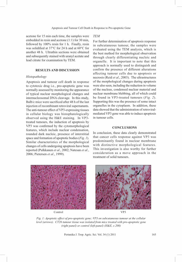
HistopathologyApoptosis and tumour cell death in response to cytotoxic drug i.e., pro-apoptotic gene was normally assessed by monitoring the appearance of typical nuclear morphological changes and internucleosomal DNA cleavage. In this study, Balb/c mice were sacrificed after 48 h of the last injection of recombinant retroviral supernatants. The anti-tumour effect of VP3-expressing tissues in cellular biology was histophatologically observed using the H&E staining. In VP3-treated tumours, the induction of apoptosis by VP3 was confirmed by the cytomorphological features, which include nuclear condensation, rounded dark nucleic, presence of interstitial space and formation of apoptotic bodies (Fig. 1). Similar characteristics of the morphological changes of cells undergoing apoptosis have been reported (Pulkkanen et al., 2002; Natesan et al., 2006; Pietersen et al., 1999).
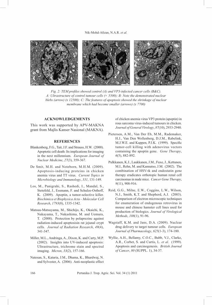
TEMFor further determination of apoptosis response in subcutaneous tumour, the samples were evaluated using the TEM analysis, which is the best method for morphological observation through clearly differentiating nucleic and organelle. It is important to note that this approach is normally used to distinguish and confirm the presence of different alterations affecting tumour cells due to apoptosis or necrosis (Reid et al., 2003). The ultrastructures of the morphological changes during apoptosis were also seen, including the reduction in volume of the nucleus, condensed nuclear material and nuclear membrane blebbing, all of which could be found in VP3-treated tumours (Fig. 2). Supporting this was the presence of some intact organelles in the cytoplasm. In addition, these data showed that the administration of retroviral-mediated VP3 gene was able to induce apoptosis in tumour cells.
CONCLUSIONSIn conclusion, these data clearly demonstrated that cancer cells response against VP3 was predominantly found in nuclear membrane with distinctive morphological features. This investigation is also worthy for further consideration as a move approach in the treatment of solid tumours.
Control VP3
Fig. 1: Apoptotic effect of pro-apoptotic gene; VP3 on subcutaneous tumour at the cellular level (arrows). CT26 tumour tissue was isolated from mice treated with pro-apoptotic gene
(right panel) or control (left-panel) (H&E, x 200)
Nik-Mohd-Afizan, N.A.R. et al.
166 Pertanika J. Trop. Agric. Sci. Vol. 34 (1) 2011
ACKNOWLEDGEMENTSThis work was supported by APV-MAKNA grant from Majlis Kanser Nasional (MAKNA).
REFERENCESBlankenberg, F.G., Tait, J.F. and Strauss, H.W. (2000).
Apoptotic cell death: Its implications for imaging in the next millennium. European Journal of Nuclear Medicine, 27(3), 359-367.
De Smit, M.H. and Noteborn, M.H.M. (2009). Apoptosis-inducing proteins in chicken anemia virus and TT virus. Current Topics in Microbiology and Immunology, 331, 131-149.
Los, M., Panigrahi, S., Rashedi, I., Mandal, S., Stetefeld, J., Essmann, F. and Schulze-Osthoff, K. (2009). Apoptin, a tumor-selective killer. Biochimica et Biophysica Acta - Molecular Cell Research, 1793(8), 1335-1342.
Matsuu-Matsuyama, M., Shichijo, K., Okaichi, K., Nakayama, T., Nakashima, M. and Uemura, T. (2008). Protection by polaprezinc against radiation-induced apoptosisin rat jejunal crypt cells. Journal of Radiation Research, 49(4), 341-347.
Miller, M.L., Andringa, A., Dixon, K. and Carty, M.P. (2002). Insights into UV-induced apoptosis: Ultrastructure, trichrome stain and spectral imaging. Micron, 33(2), 157-166.
Natesan, S., Kataria, J.M., Dhama, K., Bhardwaj, N. and Sylvester, A. (2006). Anti-neoplastic effect
of chicken anemia virus VP3 protein (apoptin) in rous sarcoma virus-induced tumours in chicken. Journal of General Virology, 87(10), 2933-2940.
Pietersen, A.M., Van Der Eb, M.M., Rademaker, H.J., Van Den Wollenberg, D.J.M., Rabelink, M.J.W.E. and Kuppen, P.J.K. (1999). Specific tumor-cell killing with adenovirus vectors containing the apoptin gene. Gene Therapy, 6(5), 882-892.
Pulkkanen, K.J., Laukkanen, J.M., Fuxe, J., Kettunen, M.I., Rehn, M. and Kannatso, J.M. (2002). The combination of HSV-tk and endostatin gene therapy eradicates orthotopic human renal cell carcinomas in nude mice. Cancer Gene Therapy, 9(11), 908-916.
Reid, G.G., Milne, E.W., Coggins, L.W., Wilson, N.J., Smith, K.T. and Shepherd, A.J. (2003). Comparison of electron microscopic techniques for enumeration of endogenous retrovirus in mouse and chinese hamster cell lines used for production of biologics. Journal of Virological Methods, 108(1), 91-96.
Wagstaff, K.M. and Jans, D.A. (2009). Nuclear drug delivery to target tumour cells. European Journal of Pharmacology, 625(1-3), 174-180.
Wyllie, A.H., Bellamy, C.O.C., Bubb, V.J., Clarke, A.R., Corbet, S. and Curtis, L. et al. (1999). Apoptosis and carcinogenesis. British Journal of Cancer, 80 (SUPPL. 1), 34-37.
Fig. 2: TEM profiles showed control (A) and VP3-infected cancer cells (B&C). A: Ultrastructure of control tumour cells (× 5500); B: Note the demonstrated nuclear blebs (arrow) (x 12500); C: The features of apoptosis showed the shrinkage of nuclear
membrane which had become smaller (arrows) (x 7700)