Embed Size (px)
Citation preview

Page 1/16
Feasibility of Endoscopic Transforaminal LumbarInterbody Fusion through the Posterior ParaspinalApproach: Technical Note and Preliminary ResultHyeun-Sung Kim ( [email protected] )
Nanoori Hospital Gangnam https://orcid.org/0000-0001-5361-5234Harshavardhan Dilip Raorane
Gangnam Nanoori HospitalPang Hung Wu
Gangnam Nanoori HospitalDong Hwa Heo
Gangnam Nanoori HospitalYeon Jin Yi
Gangnam Nanoori HospitalKyung-Hoon Yang
Gangnam Nanoori HospitalIl Tae Jang
Gangnam Nanoori Hospital
Research article
Keywords: Endoscopic TLIF, paraspinal approach, feasibility, end plate preservation
Posted Date: May 27th, 2020
DOI: https://doi.org/10.21203/rs.3.rs-28307/v1
License: This work is licensed under a Creative Commons Attribution 4.0 International License. Read Full License

Page 2/16
AbstractBackground: The implement of endoscopic spinal surgery into degenerative spinal disease hasminimized the requirement of fusion procedures. However, it is still necessary to develop endoscopicspine surgery in certain patients requiring fusion such as instability. We performed a full-endoscopictransforaminal lumbar interbody fusion(eTLIF) through a conventional paraspinal approach. Thefeasibility of procedure and early outcome were evaluated.
Materials and Methods: eighteen consecutive patients with degenerative lumbar disease underwent eTLIFthrough a conventional paraspinal approach. Their clinical outcomes were evaluated with visual analogscale(VAS) leg pain score, Oswestry Disability Index(ODI) and the MacNab's criteria; radiological outcomemeasured with segmental lordosis, global lumbar lordosis, disc height on plain radiograph andpercentage of potential fusion mass on CT scan at pre-operative, post-operative and �nal follow upperiod. intra operative and post-operative complications noted.
Results: Mean age was 63. 71 years and Mean follow-up periods was 7.78 months. According to the level,L2-3 (1 case), L3-4 (4 cases), L4-5 (13 cases) and L5-S1 (2 cases). In the X-ray result, mean segmentallordosis angle(SLA) improved in pre-operative/post-operative/follow-up period 9.87±2.74 degree/11.79±3.74 degree/ 10.56±3.69 degree (p > 0.01); mean lumbar lordosis angle(LLA) improved 37.1±7.04degree/ 39.2±7.13 degree/ 35.7±7.25 degree(p > 0.01). Mean preoperative disc height(DH) improved from8.97±1.49 mm/ 12.34±1.39 mm/ 11.44±1.98 mm (p < 0.01). In the CT result, Average percentage offusion mass was 42.61%.
VAS was improved signi�cantly, 7.67 / 3.39 / 2.5 and ODI was improved signi�cantly, 74.9 / 34.56 /27.76 by each preoperative / postoperative / �nal follow-up. In the clinical result, excellent was 5 casesand good was 13 cases.
Conclusion: According to the results of this study, eTLIF was competent enough to perform as open TLIF.and good results were obtained in the form of endplate preservation, disc height restoration, minimalblood loss and post-operative pain with early mobilization. In addition, the fusion volume including thecage and the bone graft material occupies 40% to 50% of disc space is expected to give su�cient fusionby using 3D printed cages which gives the high fusion rate. In conclusion, eTLIF is considered to be aviable surgical procedure.
1. BackgroundImplementation of endoscopic spine surgery in management of degenerative lumbar diseases hassigni�cantly reduced the need of fusion surgeries. [1,2] Actually, one of the principles of prof. A young'sphilosophy of minimally invasive spine surgery states that staged approach to the therapeuticinterventions should be utilized beginning with the least invasive treatment options; while keepingsalvage procedure like fusion as last resort in the pain management. [3,4] However there is some greyarea like gross instability, high grade of spondylolisthesis and deformity where fusion surgery still stands

Page 3/16
as a 1st line of management. interbody fusion procedures has evolved from open TLIF to minimalinvasive MIS-TLIF. However, it gives some di�culties of visualizing the disc space, contralateraldecompression and end plate preparation.
The objective of this study was to analyze the preliminary clinical and radiological outcomes ofendoscopic transforaminal lumbar interbody fusion(TLIF) through paraspinal approach in patients withthe degenerative lumbar spine disease using the endoscopic spine surgery.
2. Materials And Methods2.1. Subjects
This is retrospective case study was exempted from institutional review board of Nanoori Hospital, Seoul,Republic of Korea. The informed consent was obtained from all patients participated in study. We studied18 consecutive patients with 20 levels of lumbar degenerative disease; (6 males, 12 females; Mean age63.71 years) who underwent Endoscopic Transforaminal Lumbar Interbody Fusion(TLIF) through thePosterior Paraspinal Approach at a spine specialty hospital between august 2018 and January 2019.Inclusion criteria was degenerative grade 1 spondylolisthesis, lumbar central canal stenosis withforaminal stenosis and segmental instability con�rmed on dynamic radiograph. Spondylolisthesis grade2 and more and lumbar canal stenosis with more than 50% loss of disc height are excluded from studydue to di�culty to be managed with endoscopic fusion.
2.2. Surgical procedure
Procedure done under general anaesthesia. Patient placed in prone position on radiolucent table overWilson's frame. Entry point is located under �uoroscopy guidance. Target point is ipsilateral facet at thelevel and it is approached through conventional Wiltse's approach between multi�dus and longissimusmuscle. Usually interval located about 3cm lateral to the midline. A longitudinal skin incision ofapproximately 1cm in length was made 4cm lateral to the midline on affected side. It facilitates themedial trajectory of working cannula and cage into the disc space. Serial dilators introduced through theinterval and �nally working channel introduced along with endoscope. For initial dissection we useworking cannula with outer diameter of 13.7 mm and bevelled tip, the endoscope has 15 degree viewingangle, outer diameter of 10 mm, working channel diameter of 6 mm and working length 125 mm.ILESSYS-DeltaR (Joimax Gmbh, Germany).
The soft tissue over facet joint is dissected using radiofrequency ablator to expose inferior articularprocess(IAP) of superior vertebra. Endoscopic drill is used to hollow out the groove at IAP in inverted "L"fashion and its resected with bone cutter and forceps. IAP harvested in total to use as a autologus graftlater. Underlying superior articular process(SAP) burred with drill to expose foraminal part of deep layer ofligamentum �avum. Central and contra lateral decompression can conveniently achieved though thesame interval by simply tilting the endoscope. [�gure 1] Haemostasis and annulotomy done withradiofrequency probe (Elliquence, New York, USA). Disc material removed with forceps. End plate

Page 4/16
preparation is most important step of the procedure and advantage of endoscopic spine surgery ismeticulous end plate preparation can be done under direct vision without damaging it. End plate cartilageis removed with dissector and curettes to prepare fusion bed in order to protect traversing root bevel tip ofcannula is rotated from superior to medial direction and inserted into the disc space through theannulotomy site. Autologus graft mixed with allograft is tamped into the disc space under image�uoroscopy guidance. Single large 3D printed cage (GENQSS, GS Medical, Republic of korea) withdemineralised bone matrix(DBM) is inserted as oblique as possible and �nal position of cage is checkedunder both �uoroscope as well as endoscope. Working cannula is withdrawn keeping suction drain insitu. [picture 1]
After completion of interbody fusion procedure construct is stabilized with percutaneous pedicle screw�xation through same incision on ipsilateral side. Same procedure is repeated on contra lateral sidethrough 2 separate incisions and rods are inserted with compression in routine fashion. If required pediclescrew �xation is augmented with bone cement. [video]
2.3 Outcome evaluation
a) Clinical evaluation: Demographic data collected and all 18 patients were clinically evaluated on thebasis of the Visual Analogue Scale(VAS) score for the back, Oswestry Disability Index(ODI) and MacNab'scriteria preoperatively, postoperatively and �nal follow up (minimum 6 months). Completeness ofdecompression was documented with postoperative magnetic resonance imaging(MRI). Patients are alsoassessed for any intra or post operative complications and recurrence of symptoms.
b) Radiological evaluation:
The radiological assessment was performed by independent observer who was blinded to the patient'sclinical information. Plain radiograph used to evaluate disc height(DH), lumbar lordosis angle(LLA) andsegmental lordosis angle(SLA) pre and post operatively.
The fusion status was assessed using computed tomography(CT) at 6 months and 1 year asrecommended by CT protocol. [5] Bony fusion was de�ned when there is continuous contact of trabecularbone between upper and lower endplates of fusion segments. Osteolysis at the margins of fusion devices(pedicle screw), cystic changes within endplates adjacent to the cage, linear defect in trabecular bonebridge of fusion were considered features of delayed or failed fusion.
to evaluate fusion mass, CT scan done with axial slices parallel to the end plates of fused segment.Percentage of fusion mass surface area is calculated on CT images in axial cut using software (In�nitt,inc, Seoul, Korea) in following steps -
1. The surface area of fusion mass was measured at mid disc level from the region of interest (ROI)(surface area of cage is also included as we have used 3D-printed cage with osteo-integrationproperty; surface area of osteophytes at end plate excluded)

Page 5/16
2. the surface area of upper and lower end plates measured from the slices going through or close tothe end plates
3. average of the upper and lower end plates surface area calculated
percentage of fusion mass surface area calculated with the formula:
percentage = surface area of fusion mass/average end plate surface area X 100 [�gure 2]
2.4 Statistical analysis
Clinical data was analyzed with SPSS version 18 statistical analysis software (IBM corporation, NewYork). The continuous variables were expressed as mean and standard deviation(SD). The paired t test isused for comparison of preoperative and postoperative VAS, ODI scores; LLA, SLA and DH values. A valueof (p < 0.01) considered signi�cant.
3. Results3.1 Clinical outcomes
Out of 18 patients, there were 10 patients with grade 1 spondylolisthesis, 4 patients with segmentalinstability, 3 patients with lumbar canal stenosis and 1 patient with lumbar disc herniation. 16 patientsoperated for single level including L2-3 (1 patient), L3-4 (3 patients), L4-5 (11 patients) and L5-S1 (1patient) while 2 patients operated for 2 levels including 1 patient at L3-4, L4-5 and 1 patient at L4-5, L5-S1.
All patients are followed up for 6 months to 1year with Mean follow-up periods was 7.78 months. Themean preoperative VAS score was improved signi�cantly, 7.67 / 3.39 / 2.5 (p < 0.01) and ODI wasimproved signi�cantly, 74.9 / 34.56 / 27.76(p < 0.01) by each preoperative / postoperative / �nal follow-up. Based on MacNab's criteria, the clinical result was excellent in 5 patients and good in 13 patients.[�gure 3]
3.2 Radiological outcomes
In the X-ray result, mean preoperative segmental lordosis angle(SLA) 9.87+2.74 degree, immediatepostoperative was 11.79+3.74 degree and at the �nal follow-up was 10.56+3.69 degree (p > 0.01). Meanpreoperative lumbar lordosis angle(LLA) 37.1+7.04 degree, immediate postoperative was 39.2+7.13degree (p < 0.01) and at the �nal follow-up was 35.7+7.25 degree. (p > 0.01) Mean preoperative discheight(DH) was 8.97+1.49 mm, immediate postoperative was 12.34+1.39 mm (p < 0.01) and at the �nalfollow-up was 11.44+1.98 mm (p < 0.01). We have also compared disc height between immediate postop and �nal follow up (p < 0.01). In the CT result, vertebral body end-plate average surface area was

Page 6/16
1563.7 mm2 and average surface area of fusion mass was 663.7 mm2. Average percentage of fusionmass was 42.61%. [table 1]
3.3 Recurrence and complications
We noticed 2 intra operative complications in the form dural tear over traversing root sleeve which ismanaged conservatively using the Fibrin Sealant Patch. Both patients were asymptomatic in subsequentfollow up and clinically recovered well.
We didn't experience any post-operative complication like hematoma collection, infections, implantmigration, implant breakage or any other signs of non union. No patient required revision surgery.
4. DiscussionTraditionally, open PLIF and TLIF are already established as gold standard treatment in the managementof degenerative lumbar diseases; however, it comes with unavoidable complications like damage to theposterior ligaments complex and paraspinal muscles which ultimately leads to failed back syndrome. [6]in 2003, foley et al[7] developed MIS-TLIF technique to overcome this problem using 20-24 mm tubularretractor. However, it is di�cult to observe end plate during end plate preparation and relatively di�cultwhile decompressing the contralateral side. Lateral lumbar interbody fusion(XLIF) and oblique lumbarinterbody fusion(OLIF) developed to avoid injury to back muscles and improve visualization of endplates. [8] however it is also associated with approach related serious complications like potential injuryto major vessels, ureter and lumbar plexus. [9-10]
Successful clinical results in fusion surgery depends upon a high fusion rate along with maintainace ofdisc height and sagittal balance. Failure to achieve bony fusion may result in implant loosening,breakage and back pain in long term. Previously reported fusion rates for PLIF ranged from 56 to 100 %;[11,12] while fusion rate for open TLIF ranged from 86 to 100%.[13,14] despite of narrow operative �eld inMIS-TLIF; several articles have demonstrated good fusion rate ranging from 92-100%.[15,16]
In turn, successful fusion primarily depends upon surface area of fusion bed and correct placement ofbone graft along with interbody cage over fusion bed. Sometimes inaccurate placement of bone graftover unprepared end plate with intact cartilage may results in pseudoarthrodesis. Potter et al[13] havereported that for obtaining �rm interbody fusion, exposure of more than 30% endplate is required andclinically with transforaminal approach 56% of endplate can be prepared. At the same time, we have to becareful not to remove excess of subchondral bone. Overenthusiastic disc preparation can result intosubsidence and recurrent foraminal stenosis; particularly in osteoporotic patients. As we have used 15degree endoscope inside disc space which can be rotated 360 degree it allows access to 60-80% of theend plate. [17] we have calculated the mean percentage of surface area of fusion mass to be 42.61%which is more than su�cient to achieve interbody fusion we have used 3D-printed porous titaniuminterbody cage which provides osteo-conductive scaffold for new bone growth. It provides short termstability due to friction and long term stability due to osteoblast adhesion, proliferation and bone

Page 7/16
ingrowth. Single large cage improves the construct stability and bone ingrowth decreases subsidence dueto load distribution. [18] fusion rate can be further improved with the use of rh-BMP and variousosteoinductive bone material.
In 2012, Said Osman [19] �rst reported 60 cases of endoscopic TLIF for treatment of degenerative lumbardiseases with 59.6% solid fusion and 36.2% stable �xation, but complication rate was upto 20%. Recently,Mongesterns[17] in their 30 patients operated with percutaneous TLIF with mean follow up period of 38months reported 100% fusion rate using rigid PEEK cage and expandable titanium cage. Lee et al[20]performed percutaneous TLIF in 18 patients using titanium expandable spacer(B-Twin) under localanesthesia with conscious sedation. He achieved good clinical results with 88% fusion rate (16 out of 18)with average subsidence 2.1 mm. Subsidence of interbody disc height is expected in post operativeperiod; however progressive subsidence can cause decrease in foraminal height which results in recurrentforaminal stenosis also destruction of sagittal balance. In present study we didn't noticed any signs ofnon fusion but experienced subsidence rate of 0.9 mm. However, there was signi�cant difference betweenpre operative and �nal follow up (p < 0.01). Signi�cant subsidence is de�ned as a decrease in disc heightof more than 3mm. Though our subsidence rate is low; it needs close long term follow up. Jacquet etal[21] considered procedure controversial due to technical di�culties and high complication rate(up to36%). They also added need for technical improvements to the procedure. Certain studies also proposedfeasibility of biportal endoscopic surgery for interbody fusion with signi�cant clinical results. [22,23]
All this studies utilized classical endoscopic transforaminal approach with expansile foraminoplasty fordisc space preparation and cage insertion. Majority of this studies experienced common complication ofpost-operative dysesthesia due to irritation of DRG while performing disc preparation and insertion oflarger cage into the disc space. In our study we have utilized conventional Wiltse's approach whichminimizes the need of retraction of exiting root and DRG. However, in our study we experiencedcomplication rate of 3.6%. We noticed dural tear in 2 patients over traversing root sleeve by bevel tip dueto incomplete retraction of traversing root. This complication can be minimized by carefully rotating thebevel tip and introducing it into the disc space. We didn't notice post-operative dysesthesia orneurological de�cit in any patient. Interbody fusion via transforaminal approach has certain limitationslike inadequate foraminoplasty may impose some di�culties while inserting cage into the disc space andobstacle of iliac crest for L5-S1 disc space. It can be comfortably overcome by paraspinal approach.
Despite of its good clinical results endoscopic TLIF have certain limitations which needs further technicaldevelopment; it is not suitable for grade 2 or more spondylolisthesis due to di�culty to achieve completereduction and sagittal balance. In patient where disc space is collapsed more than 50% it is very di�cultto introduced endoscope into disc space. Said Osman [17] has advised prior pedicle screwinstrumentation and distraction of disc space for the same. In our study we didn't noticed statisticallysigni�cant difference between pre-operative and follow up segmental balance. Though technique of endoTLIF is similar to open TLIF its signi�cance for restoration of segmental balance is doubtful as perpresent study. Further technical development in the instrumentation may require for disc spacedistraction, cage design in form of expandable cage or lordotic cage. Lewandrowski et al[24] tried

Page 8/16
endoscopic TLIF with standalone lordotic cage for a single level with good clinical results(77.8%).However, he reported cage subsidence in all the cases with revision surgery rate around 8%(4 out of 48).Long term radiological studies with respect to subsidence rate, sagittal balance, effect at adjacentsegment, fusion mass distribution and fusion rate will be helpful for future development of endoscopicinstrumentation for fusion surgery.
This is retrospective study with small sample size and preliminary results. We need to compare studygroup with matched control group like open or MIS TLIF group to evaluate effectiveness of thisprocedure. Prospective study with large sample size will give more comprehensive results.
ConclusionAccording to the results of this study, endoscopic TLIF was comparable to an open TLIF with favorableresults obtained in the form of endplate preservation, disc height restoration, minimal blood loss and postoperative pain with early mobilization. In addition, the fusion volume including the cage and the bonegraft material as measured on post-operative CT scans, occupied 40% to 50% of disc space which wouldbe expected to give high fusion rates with the use of 3D printed cages. In conclusion, endoscopic TLIF isconsidered to be a viable surgical procedure.
AbbreviationsSLA – Segmental lordosis angle
LLA – Lumbar lordosis angle
DH – Disc height
PLIF – Posterior lumbar interbody fusion
TLIF – Transforaminal lumbar interbody fusion
OLIF – Oblique lumbar interbody fusion
DeclarationsEthics approval and consent to participate
The current study is approved from institutional review board, Nanoori hospital, Gangnam, Seoul,Republic of Korea.
Informed consent is obtained from all the participants in the study.
Consent for publication
Page 9/16
Not applicable.
Availability of data and materials
The datasets used and/or analyzed during the current study are available from the corresponding authoron reasonable request.
Competing interests
The authors declare that they have no competing interests
Funding
No funds are received for the current study.
Author contributions
(1) Conception and design: HK
(2) Administrative support: IJ
(3) Provision of study material or patients: HK
(4) Collection and assembly of data: HK, HR
(5) Data analysis and interpretation: HK, HR, WP
(6) Manuscript writing: All authors
(7) Final approval of manuscript: All authors
Acknowledgment:
We would like to acknowledge scienti�c team members Ms. Jae Eun Park and Mr. Kyeong Rae Kim forproviding assistance in acquiring full text articles and managing digital works.
References1. 1. Kim M, Kim HS, Oh SW, Adsul NM, Singh R, Kashlan ON, Noh JH, Jang IT, Oh SH. [2019] Evolution
of Spinal Endoscopic Surgery. 16(1):6-14. doi: 10.14245/ns.1836322.161.[PubMed: 30943702];[PubMed Central: PMC6449828].
2. Wang JC, Kim HS. Endoscopic Spinal Surgery (ESS): Prepare for a Happy 100-Year-Old! Neurospine. 2019 Mar;16(1):4-5. doi: 10.14245/ns.19int001. Epub 2019 Mar 31. PubMed PMID:30943701; PubMed Central PMCID: PMC6449830.

Page 10/16
3. Yeung A, Kotheeranurak V. Transforaminal Endoscopic Decompression of the Lumbar Spine forStable Isthmic Spondylolisthesis as the Least Invasive Surgical Treatment Using the YESS SurgeryTechnique. Int J Spine Surg. 2018 Jun;12(3):408-414. doi: 10.14444/5048. eCollection 2018Jun. PubMed PMID: 30276099; PubMed Central PMCID: PMC6159720.
4. Gore S, Yeung A: The “inside out” transforaminal technique to treat lumbar spinal pain in an awakeand aware patient under local anesthesia: results and a review of the literature. International Journalof Spine Surgery 8, 2014.
5. Williams A.L., Gornet M.F., Burkus J.K. CT evaluation of lumbar interbody fusion: Current concepts(2005) American Journal of Neuroradiology, 26 (8), pp. 2057-2066.
�. Kawaguchi Y, Matsui H, Tsuji H. Back muscle injury after posteriorlumbar spine surgery. Part 1:Histologic and histochemical analyses inrats. Spine 1994; 19:2590 –7.
7. Foley KT, Holly LT, Schwender JD. Minimally invasive lumbarfusion.Spine (Phila Pa 1976).2003;28(15 Suppl): S26-35.
�. Anand N, Rosemann R, Khalsa B, Baron EM. Mid-term and long-term clinical and functionaloutcomes of minimally invasive correction and fusion for adults with scoliosis. Neurosurg Focus2010; 28:E6
9. Walker CT, Farber SH, Cole TS, et al. Complications for minimally invasive lateral interbodyarthrodesis: a systematic review and meta-analysis comparing prepsoas and transpsoasapproaches. J Neurosurg Spine. 2019 Jan 25; 1-15. doi: 10.3171/2018.9. SPINE18800. PMID:30684932.
10. Joseph JR, Smith BW, La Marca F, Park P. Comparison of complication rates of minimally invasivetransforaminal lumbar interbody fusion and lateral lumbar interbody fusion: a systematic review ofthe literature. Neurosurg Focus. 2015Oct;39(4): E4. doi:10.3171/2015.7. FOCUS15278. PubMedPMID: 26424344
11. Brantigan JW, Steffee AD, Lewis ML, Quinn LM, Persenaire JM: Lumbar interbody fusion using theBrantigan I/F cage for posterior lumbar interbody fusion and the variable pedicle screw placementsystem: two-year results from a Food and Drug Administration investigational device exemptionclinical trial. Spine (Phila Pa 1976) 25:1437-1446, 2000
12. Kai Y, Oyama M, Morooka M: Posterior lumbar interbody fusion using local facet joint autograft andpedicle screw �xation.Spine (Phila Pa 1976) 29:41-46, 2004
13. Potter BK, Freedman BA, Verwiebe EG, Hall JM, Polly DW,Jr., Kuklo TR: Transforaminal lumbarinterbody fusion: clinical and radiographic results and complications in 100 consecutive patients. JSpinal Disord Tech 18:337-346, 2005
14. Hackenberg L, Halm H, Bullmann V, Vieth V, Schneider M,Liljenqvist U: Transforaminal lumbarinterbody fusion: a safe technique with satisfactory three to �ve year results. Eur SpineJ 14:551-558,2005
15. Schwender JD, Holly LT,Rouben DP, Foley KT: Minimally invasive transforaminal lumbar interbodyfusion (TLIF): technical feasibility and initial results. J Spinal Disord Tech 18 Suppl: S1-6, 2005
Page 11/16
1�. Kim MC, Chung HT, Kim DJ, Kim SH, Jeon SH: The clinical and radiological outcomes of minimallyinvasive transforaminal lumbar interbody single level fusion. Asian Spine J 5:111-116,
17. R. Morgenstern and C. Morgenstern, “Percutaneous Transforaminal lumbar interbody fusion (pTLIF)with a posterolateral approach for the treatment of denegerative disk disease: feasibility andpreliminary results,” International Journal of Spine Surgery, vol. 27, no. 9, article 41, 2015.
1�. McGilvray KC, Easley J, Seim HB, et al. Bony ingrowth potential of 3D-printed porous titanium alloy: adirect comparison of interbody cage materials in an in vivo ovine lumbar fusion model. Spine J.2018;18(7):1250–1260. doi:10.1016/j.spine.2018.02.018
19. S. G. Osman, “Endoscopic transforaminal decompression, interbody fusion, and percutaneouspedicle screw implantation of the lumbar spine: A case series report,” International Journal of SpineSurgery, vol. 6, no. 1, pp. 157–166, 2012.
20. S. H. Lee,H. Y. Erken, and J. Bae, “Percutaneous transforaminal endoscopic lumbar interbody fusion:clinical and radiological results of mean 46-month follow-up,” BioMed Research International, vol.2017,Article ID 3731983, 2017.
21. F. Jacquot andD. Gastambide, “Percutaneous endoscopic transforaminal lumbar interbody fusion: isit worth it?” International Orthopaedics, vol. 37, no. 8, pp. 1507–1510, 2013.
22. Hwa Eum J, Hwa Heo D, Son SK, Park CK. Percutaneous biportal endoscopic decompression forlumbar spinal stenosis: a technical note and preliminary clinical results. J Neurosurg Spine. 2016Apr;24(4):602-7. doi: 10.3171/2015.7.SPINE15304.
23. Kim JE, Choi DJ. Biportal Endoscopic Transforaminal Lumbar Interbody Fusion withArthroscopy. Clin Orthop Surg. 2018;10(2):248–252. doi:10.4055/cios.2018.10.2.248
24. Lewandrowski KU, Ransom NA, Ramírez León JF, Yeung A. The Concept for A Standalone LordoticEndoscopic Wedge Lumbar Interbody Fusion: The LEW-LIF.Neurospine. 2019 Mar;16(1):82-95. doi:10.14245/ns.1938046.023.
TableTable 1: Patient's radiological data pre operative post operative and final follow up period (values arepresented as mean+standard deviation, p value < 0.01 considered as significant)

Page 12/16
Radiological Data value P value
Lumbar lordosis angle
pre operative 37.1+7.040
post operative 39.2+7.136 (p < 0.01)
final follow up 35.7+7.252
Segmental lordosis angle
pre operative 9.87+2.743
post operative 11.79+3.742 (p < 0.01)
final follow up 10.56+3.690
Disc height
pre operative 8.97+1.490
post operative 12.34+1.399 (p < 0.01)
final follow up 11.44+1.986 (p < 0.01)
Mean percentage of
surface area of fusion mass
42.61%.
Additional File LegendPicture 1: A & B beveled obturator inserted with its tip inside disc space and end plate preparation done C& D 3-D printed cage inserted through obturator and then it is removed E & F Final position of implantchecked under endoscopic view and �uoroscopy guidance
Figures
Page 13/16
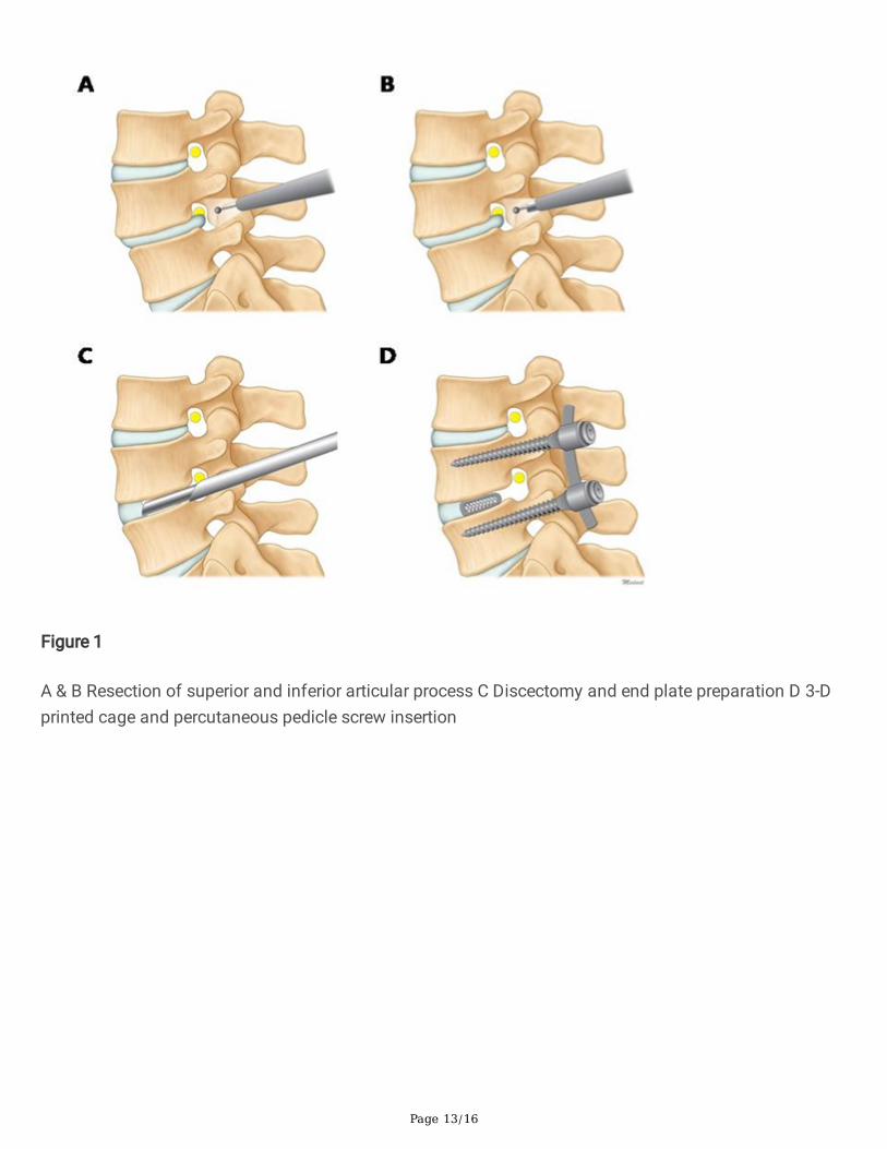
Figure 1
A & B Resection of superior and inferior articular process C Discectomy and end plate preparation D 3-Dprinted cage and percutaneous pedicle screw insertion

Page 14/16
Figure 2
Radiological assessment Plain radiograph used to evaluate A. Disc height(DH) distance between upperand lower end plate, B. Lumbar lordosis angle(LLA) angle between superior end plate of L1 andS1vertebra and C. segmental lordosis angle(SLA) angle between superior end plate of upper and inferiorend plate of lower vertebra of fused segment

Page 15/16
Figure 3
patients pain assessment during pre operative post operative and �nal follow up period
Figure 4
representative case - 62 years female patient operated for L5-S1 endoscopic TLIF through paraspinalapproach A & B T2 weighted sagittal and axial image showing L5-S1 grade 1 spondylolisthesis withforaminal stenosis C & D Dynamic radiograph showing angular instability E Post-operative radiograph

Page 16/16
Supplementary Files
This is a list of supplementary �les associated with this preprint. Click to download.
renamed53d99.wmv
Picture1.jpg