Embed Size (px)
Citation preview
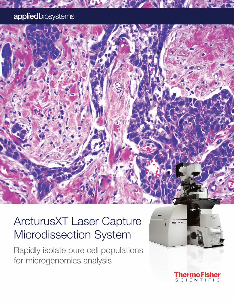
ArcturusXT Laser Capture Microdissection SystemRapidly isolate pure cell populationsfor microgenomics analysis
The Applied Biosystems™ ArcturusXT™ LCM System is a unique
microdissection system that combines the power of infrared (IR)
laser capture and ultraviolet (UV) laser cutting in one modular
platform. The solid-state IR laser delivers a gentle capture
technique that preserves the overall biomolecular integrity of cells,
and is ideal for both single cells and small numbers of cells. The
solid-state UV laser permits superior speed and precision, and
is well suited for microdissecting dense tissue structures, and
capturing large numbers of cells. This unique combination of
two lasers in one system helps ensure that sample custody is
maintained at all times, thereby enabling researchers to uncover
unique molecular signatures that would have otherwise been
obscured in a heterogeneous cell population.
ArcturusXT Laser Capture Microdissection (LCM) System—a complete solution for microgenomics
3
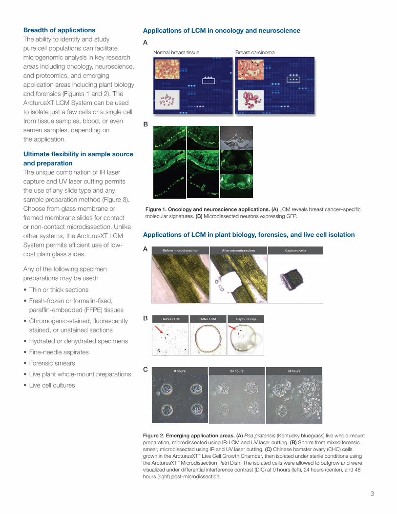
Breadth of applicationsThe ability to identify and study pure cell populations can facilitate microgenomic analysis in key research areas including oncology, neuroscience, and proteomics, and emerging application areas including plant biology and forensics (Figures 1 and 2). The ArcturusXT LCM System can be used to isolate just a few cells or a single cell from tissue samples, blood, or even semen samples, depending on the application.
Ultimate fl exibility in sample source and preparationThe unique combination of IR laser capture and UV laser cutting permits the use of any slide type and any sample preparation method (Figure 3). Choose from glass membrane or framed membrane slides for contact or non-contact microdissection. Unlike other systems, the ArcturusXT LCM System permits effi cient use of low-cost plain glass slides.
Any of the following specimen preparations may be used:
• Thin or thick sections
• Fresh-frozen or formalin-fi xed, paraffi n-embedded (FFPE) tissues
• Chromogenic-stained, fl uorescently stained, or unstained sections
• Hydrated or dehydrated specimens
• Fine-needle aspirates
• Forensic smears
• Live plant whole-mount preparations
• Live cell cultures
Applications of LCM in oncology and neuroscience
Applications of LCM in plant biology, forensics, and live cell isolation
A
B
A
B
C
Before microdissection After microdissection Captured cells
0 hours 24 hours 48 hours
Figure 1. Oncology and neuroscience applications. (A) LCM reveals breast cancer–specifi c molecular signatures. (B) Microdissected neurons expressing GFP.
Figure 2. Emerging application areas. (A) Poa pratensis (Kentucky bluegrass) live whole-mount preparation, microdissected using IR-LCM and UV laser cutting. (B) Sperm from mixed forensic smear, microdissected using IR and UV laser cutting. (C) Chinese hamster ovary (CHO) cells grown in the ArcturusXT™ Live Cell Growth Chamber, then isolated under sterile conditions using the ArcturusXT™ Microdissection Petri Dish. The isolated cells were allowed to outgrow and were visualized under differential interference contrast (DIC) at 0 hours (left), 24 hours (center), and 48 hours (right) post-microdissection.
Before LCM After LCM CapSure cap
Normal breast tissue Breast carcinoma
4
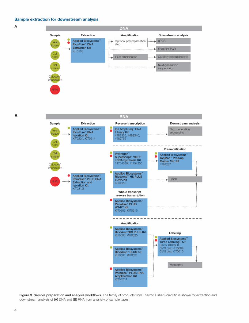
Figure 3. Sample preparation and analysis workflows. The family of products from Thermo Fisher Scientific is shown for extraction and downstream analysis of (A) DNA and (B) RNA from a variety of sample types.
B RNA
FFPE
Sample Extraction Reverse transcription
Cellsmear
Cytospin™
preparation
Freshfrozen
Cellpellet
Applied Biosystems™ PicoPure™ RNA Isolation KitKIT0204, KIT0214
Ion AmpliSeq™ RNA Library Kit4482335, 4482340, 4482752
Invitrogen™ SuperScript™ VILO™ cDNA Synthesis Kit11754050, 11754250
Applied Biosystems™ RiboAmp™ HS PLUS cDNA KitKIT0529
Downstream analysis
qPCR
Microarray
Next-generation sequencing
Amplification
Applied Biosystems™ RiboAmp™HS PLUS KitKIT0505, KIT0525
Applied Biosystems™ RiboAmp™ PLUS KitKIT0501, KIT0521
Applied Biosystems™ Paradise™ PLUS RNA Extraction and Isolation KitKIT0312I
Preamplification
Applied Biosystems™ TaqMan™ PreAmp Master Mix Kit4384267
Labeling
Applied Biosystems™ Turbo Labeling™ KitBiotin: KIT0608Cy®3 dye: KIT0609Cy®5 dye: KIT0610
Whole transcript reverse transcription
Applied Biosystems™ Paradise™ PLUS WT-RT KitKIT0305, KIT0315
Applied Biosystems™ Paradise™ PLUS RNA Amplification KitKIT0321A
DNA
FFPE
Sample Extraction Amplification
Cellsmear
Cytospin™
preparation
Freshfrozen
Cellpellet
Applied Biosystems™ PicoPure™ DNA Extraction KitKIT0103
Optional preamplification step
PCR amplification
Downstream analysis
qPCR
Endpoint PCR
Capillary electrophoresis
Next-generation sequencing
Sample extraction for downstream analysis
A
5
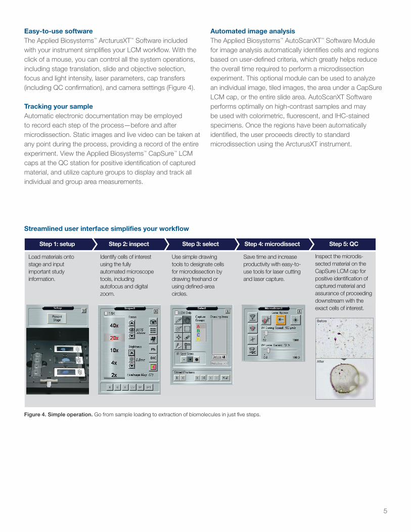
Easy-to-use softwareThe Applied Biosystems™ ArcturusXT™ Software included with your instrument simplifi es your LCM workfl ow. With the click of a mouse, you can control all the system operations, including stage translation, slide and objective selection, focus and light intensity, laser parameters, cap transfers (including QC confi rmation), and camera settings (Figure 4).
Tracking your sampleAutomatic electronic documentation may be employed to record each step of the process—before and after microdissection. Static images and live video can be taken at any point during the process, providing a record of the entire experiment. View the Applied Biosystems™ CapSure™ LCM caps at the QC station for positive identifi cation of captured material, and utilize capture groups to display and track all individual and group area measurements.
Figure 4. Simple operation. Go from sample loading to extraction of biomolecules in just fi ve steps.
Streamlined user interface simplifi es your workfl ow
Step 1: setup
Load materials onto stage and input important study information.
Step 2: inspect
Identify cells of interest using the fullyautomated microscope tools, includingautofocus and digital zoom.
Step 3: select
Use simple drawing tools to designate cells for microdissection by drawing freehand orusing defi ned-area circles.
Step 4: microdissect
Save time and increase productivity with easy-to-use tools for laser cutting and laser capture.
Step 5: QC
Inspect the microdis-sected material on the CapSure LCM cap for positive identifi cation of captured material and assurance of proceeding downstream with the exact cells of interest.
Before
After
Automated image analysisThe Applied Biosystems™ AutoScanXT™ Software Module for image analysis automatically identifi es cells and regions based on user-defi ned criteria, which greatly helps reduce the overall time required to perform a microdissection experiment. This optional module can be used to analyze an individual image, tiled images, the area under a CapSure LCM cap, or the entire slide area. AutoScanXT Software performs optimally on high-contrast samples and may be used with colorimetric, fl uorescent, and IHC-stained specimens. Once the regions have been automatically identifi ed, the user proceeds directly to standard microdissection using the ArcturusXT instrument.

6
Ensure the integrity of microdissected cellsSpecially designed for research use with the ArcturusXT LCM System, all CapSure LCM caps have a transfer fi lm bonded to the lower cap surface. Using the ArcturusXT LCM System, an infrared laser pulses through the top of the cap during LCM and interacts with the transfer fi lm, which then melts and bonds to the cells or regions of interest. The fi lm absorbs the laser radiation—instead of the tissue or cell sample—creating a gentle, nondamaging microdissection that preserves the integrity of the captured material.
HistoGene™ kits for frozen tissue and immunofl uo-rescence staining
ArcturusXT™ microdissectioninstrument for capturing pure cell populations
PicoPure™ kits for DNA and RNA extraction and isolation
Paradise™ PLUS reagents for staining, RNA isolation and amplifi cation, and whole-transcript reverse transcription from FFPE samples
RiboAmp™ PLUS kits for RNA linear amplifi cation from frozen samples
Find the products you need for every step
Identify cells Select cells Purify DNA/RNA Amplify DNA/RNA
Figure 5. Experimental workfl ow. Depending upon the sample types and the applications needed in the microgenomics workfl ow, we offer a variety of kits to choose from.
Step 1 Step 2 Step 3 Step 4
Complete solution for microgenomicsThermo Fisher Scientifi c offers everything you need for your microgenomics workfl ow (Figure 5). From tissue staining kits and LCM instrumentation, to kits for extraction, amplifi cation, and labeling, we have what you need for your microgenomics project.
We also provide superior applications and technical support with a dedicated team of highly qualifi ed and expertly trained application scientists and technical support specialists to assist researchers in any molecular or cellular biology research area.
7
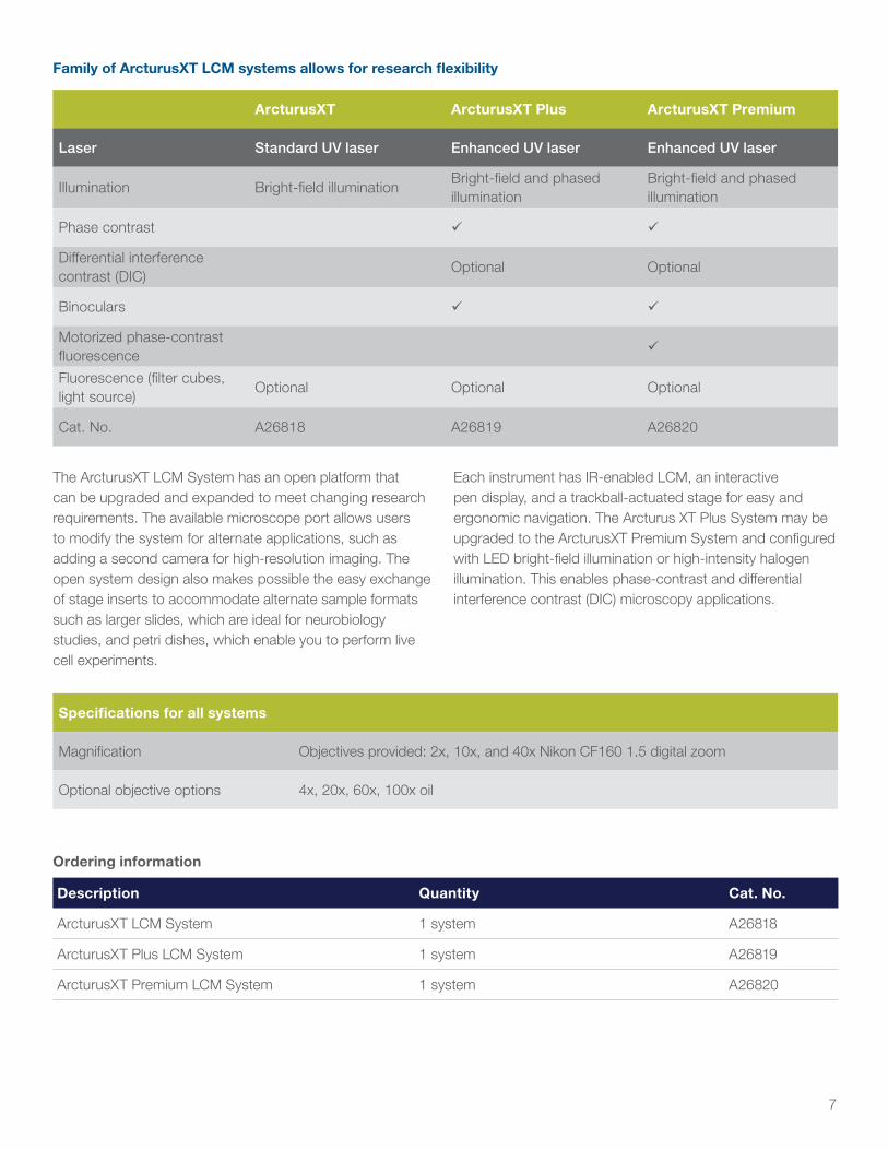
Family of ArcturusXT LCM systems allows for research flexibility
ArcturusXT ArcturusXT Plus ArcturusXT Premium
Laser Standard UV laser Enhanced UV laser Enhanced UV laser
Illumination Bright-field illuminationBright-field and phased illumination
Bright-field and phased illumination
Phase contrast
Differential interference contrast (DIC)
Optional Optional
Binoculars
Motorized phase-contrast fluorescence
Fluorescence (filter cubes, light source)
Optional Optional Optional
Cat. No. A26818 A26819 A26820
The ArcturusXT LCM System has an open platform that can be upgraded and expanded to meet changing research requirements. The available microscope port allows users to modify the system for alternate applications, such as adding a second camera for high-resolution imaging. The open system design also makes possible the easy exchange of stage inserts to accommodate alternate sample formats such as larger slides, which are ideal for neurobiology studies, and petri dishes, which enable you to perform live cell experiments.
Each instrument has IR-enabled LCM, an interactive pen display, and a trackball-actuated stage for easy and ergonomic navigation. The Arcturus XT Plus System may be upgraded to the ArcturusXT Premium System and configured with LED bright-field illumination or high-intensity halogen illumination. This enables phase-contrast and differential interference contrast (DIC) microscopy applications.
Specifications for all systems
Magnification Objectives provided: 2x, 10x, and 40x Nikon CF160 1.5 digital zoom
Optional objective options 4x, 20x, 60x, 100x oil
Ordering information
Description Quantity Cat. No.
ArcturusXT LCM System 1 system A26818
ArcturusXT Plus LCM System 1 system A26819
ArcturusXT Premium LCM System 1 system A26820
Find out more at thermofisher.com/lcm
For Research Use Only. Not for use in diagnostic procedures. © 2016 Thermo Fisher Scientific Inc. All rights reserved. All trademarks are the property of Thermo Fisher Scientific and its subsidiaries unless otherwise specified. Nikon is a trademark of the Nikon Corporation. Cy is a trademark of GE Healthcare UK Limited. COL21154 0416
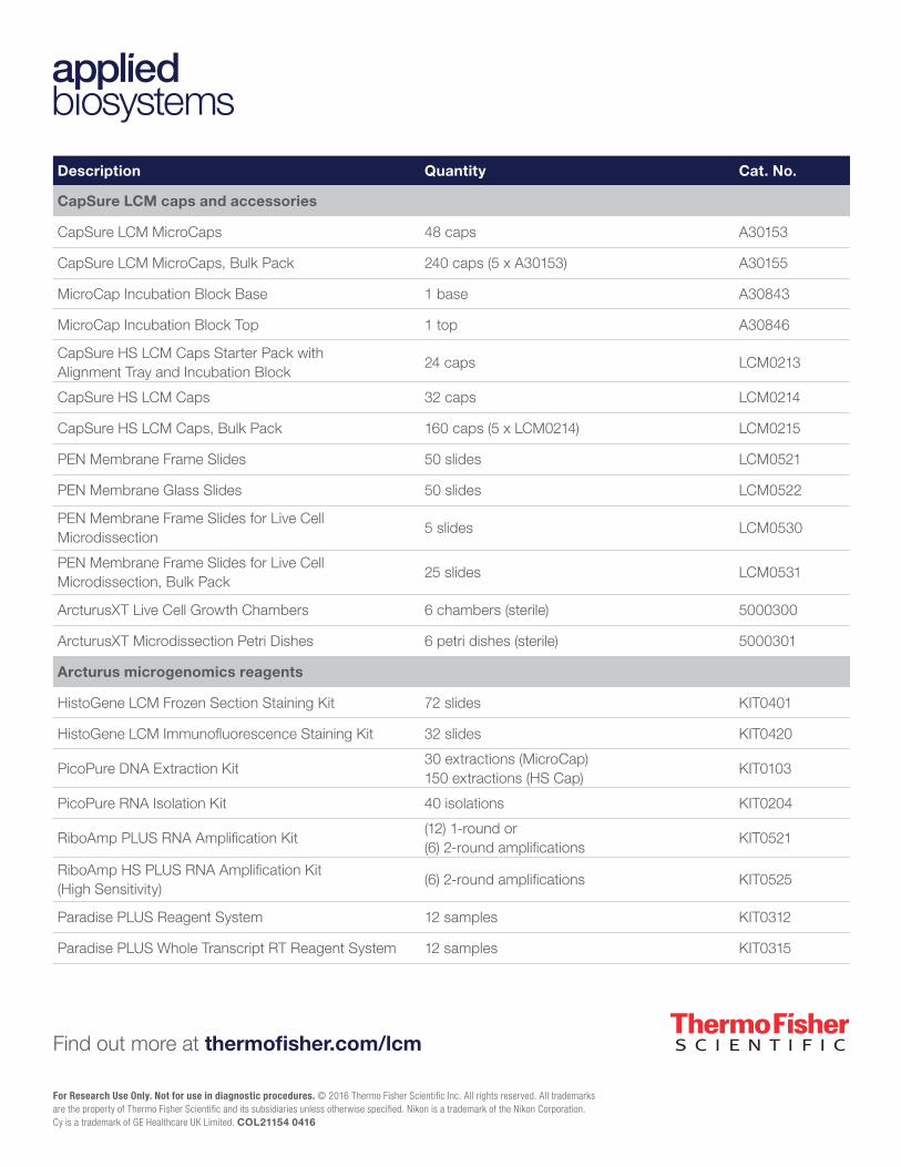
Description Quantity Cat. No.
CapSure LCM caps and accessories
CapSure LCM MicroCaps 48 caps A30153
CapSure LCM MicroCaps, Bulk Pack 240 caps (5 x A30153) A30155
MicroCap Incubation Block Base 1 base A30843
MicroCap Incubation Block Top 1 top A30846
CapSure HS LCM Caps Starter Pack withAlignment Tray and Incubation Block
24 caps LCM0213
CapSure HS LCM Caps 32 caps LCM0214
CapSure HS LCM Caps, Bulk Pack 160 caps (5 x LCM0214) LCM0215
PEN Membrane Frame Slides 50 slides LCM0521
PEN Membrane Glass Slides 50 slides LCM0522
PEN Membrane Frame Slides for Live Cell Microdissection
5 slides LCM0530
PEN Membrane Frame Slides for Live Cell Microdissection, Bulk Pack
25 slides LCM0531
ArcturusXT Live Cell Growth Chambers 6 chambers (sterile) 5000300
ArcturusXT Microdissection Petri Dishes 6 petri dishes (sterile) 5000301
Arcturus microgenomics reagents
HistoGene LCM Frozen Section Staining Kit 72 slides KIT0401
HistoGene LCM Immunofluorescence Staining Kit 32 slides KIT0420
PicoPure DNA Extraction Kit30 extractions (MicroCap) 150 extractions (HS Cap)
KIT0103
PicoPure RNA Isolation Kit 40 isolations KIT0204
RiboAmp PLUS RNA Amplification Kit(12) 1-round or (6) 2-round amplifications
KIT0521
RiboAmp HS PLUS RNA Amplification Kit (High Sensitivity)
(6) 2-round amplifications KIT0525
Paradise PLUS Reagent System 12 samples KIT0312
Paradise PLUS Whole Transcript RT Reagent System 12 samples KIT0315