Embed Size (px)
Citation preview
Naval Medical Research InstituteSetheeda, MO 20814-6056 NMRI 88-94 December 1988
ARGININE VASOPRESSIN LOWERS PULMONARY ARTERY
(0 PRESSURE IN HYPOXIC RATS BY RELEASING00
ATRIAL NATRIURETIC PEPTIDEIn
"i? DEC 1 41989Y-F. Chen
R-H. Yang
T. M. McKenna
R. M. Jackson
S. Opari Approved for public release;distribution is unlimited
Naval Medical Researchand Development CommandBethesda, Maryland 20814-5044
Department of the NavyNaval Medical CommandWashington, D.C. 20372-5210
$9 12 "-*
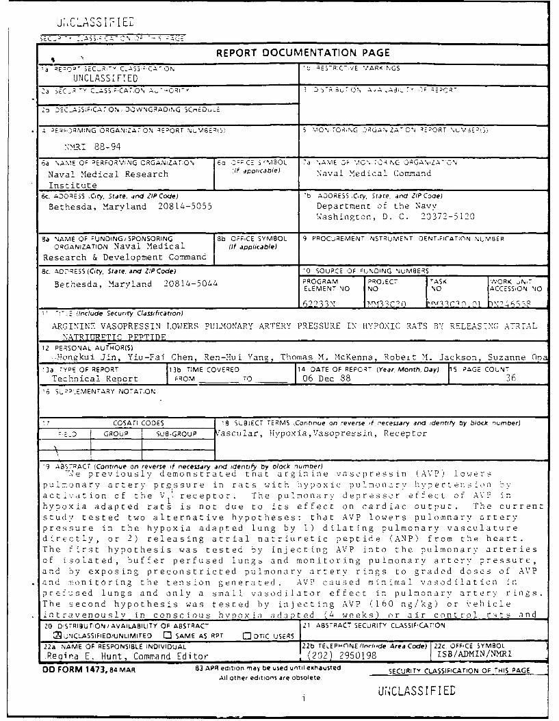
REPORT DOCUMENTATION PAGE.3R SO EC,.R-y C..ASS ;-CA' ON o qESRIC 'IE M1ARK r'IS
UNCLASS I FIED,a 3C 7CASCAO &-R 3 D -'R 3U70N AjA A3._71y 3 - CR-
2:) DECASiFCA7 ON, DOQVVNCRADiNG SCHEDu_E
4~ DERP- RMING ORGA.%!ZA7 ON REPORT %NuVBEP(S; 5 %1ONj 7ORNG P RAN ZA7 ON4 PEl-OR7 UB 2 3
88 0-946a %AME Of DERFORMING ORGANiZAT;ON 6o 5~ CE 30L 7,3 NAMOvE 3; '0OTO CN- C ORG A%,ZA 7C N
Naval Medical Research if applicabie) Naval Medical CommandInstitute_________ ____________________ ____
6c. ADDRESS ,Ciry, State, anld ZIP Code) 'b ADDRESS Crty, Stare, and ZIP Coae)
Bethnesda, Maryland 20814-5055 Department of the NavyWashington, D. C. 20372-5120
Sa~ NAME OF FUNDING; SPONSORING 8b OFFICE SYMBOL 9 PROCjREMENT NSTRUMENT .DENTFCATnN %UMBERORGANIZATION Naval Medical j(if appficable)
Research & Development CommandI8c. ADCRPESS (City, State, and ZIP Code) .0SOUPCE OF rUNDING NUMBERS
Bethesda, Maryland 20814-5044 PROGRAM PROjECT -ASK I WORK -N,7ELEMENT NO NO jNO ACCESSiON N
______________________6 1_______3_____ NT 1M3 3C3 0 1, (,,1 - P1 4 ,:;,5
1 !_ InclUde Security Classification)
ARCININE VASOPRESSIN LOW4ERS PUL-MONARY ARTERY PRESSURE IN HYPOXIC PA-'TS By, RELEASING ATRIALNATRIURFTTC PFPTTnF
12 PERSONAL AUH-OR(S)--Hongkui Jin, Yiu-Fai Chen, Ren-Hui Yang, Thomas M. McKenna, Robett M. Jackson, Suzanne On~
! 3a. TYPE OF REPORT 1i3b. TIME COVERED 114 DATE OF REPORT (Year, Month, Day) 15 PAGE COUNT
Technical Report FROM 7O ___106 Dec 88 I366 SL:.2!E:MENTARY NOTATION
7 COSATI CODES I8 SUBJECT TERMS Continue on reverse if necessary and identify by block number)
z - ROUP SUB-GROUP Vascular, Hlypoxia, Vasopressin, Receptor
9ABOTRACT (Continue on reverse if necessary and identify by block number).,q previously demonstrated tnat arginine vasopressin AVP) low,,er5,
pulmnonary artery pre ssure in rats with hypoxic pulmnonarv, hypertension '-Vactivation of the V receptor. The pulmonary depressor effect of AVP inhypoxia adapted rats is not due to its effect on cardiac output. The currentstudyi tested two alternative hypotheses: that AVP lowers pulomnary arterypressure in the hypoxia adapted lung by 1) dilating pulmonary vasculaturedirectly, or 2) releasing atrial natriuretic peptide (ANP) from the heart.The first hypothesis was tested by injecting AVP into the pulmonary arteriesof isolated, buffer perfused lung-s and monitoring pulmonary artery pressure,and by exposing preconstricted pulmonary artery rings to graded doses of AVPand monitoring the tension generated. AVP caused minimal vasodilation inprefused lungs and only a small vasodilator effect in pulmonary artery rings.The second hypothesis was tested by injecting AVP (160 ng/kg) or v'ehicleintravenouslv in conscious hv oxia ada ted (4 weeks) or air control rats and20 DISTRISUITION /AVAILABILITY OF ABSTRACT 21 ABSTRACT SECURITY CLASSIFICATIO0N
M NCLASSIFIED/UNLI MITE D (: SAME AS RPT C3 DTIC USERS
22a NAME OF RESPONSIBLE !NOIVIDUAL 22b TELEP.4CNE (Irr,de Area Code) 22c. OFFICE SYMBOLPeaina E. Hunt, Command Editor (202L2950198 ISB/ADMIN/N-MRI
DO FORM 1473, 84 MAR 83 APR edition may be used until exhausted SECURITY CLASSIFICATION OF THIS PAGEAll other editions are obsolete
UNCLASSIFIEE
UNCLASS IF I EDSECURITY CLASSIFICATION OF T IIS PAGE
measuring ANP in arterial blood and atria, and by testing pretreatment, witl
the V1 receptor antagonist d(CH 2 )5 Tyr(Me)AVP (130 vg/kg) on the AVP-induced
increase in plasma ANP. AVP produced a 7-fold increase in plasma ANP (209±33
to 1346t233 pg/ml; p(O.05) 'in hypoxia adapted rats and a 5-fold increase in
ANP (122±22 to 573±174 pg/ml; p< 0 .0 5 ) in air controls. ANP release was
abolished by pretreatment of both groups with d(CH) Tyr(Me)AV?. The AV?
induced ANP release came mainly from left atrium. T ese data strong!y suggest
that the pulmonary depressor effects of AVP in hypoxia adapted rats is due :o
augmented V receptor-induced release of ANP from left atrium.
1i
UNCLASSIFIEDSECURITY CLASSIFICATION OF TMS PAGE
ACKNOWLEDGEMENTS
A portion of this work was performed under Naval Medical Research and
Development Command Work Unit No. MM33C30.01.I00i. The opinions and
assertions contained herein are the private ones of the writers and are not to
be construed as official or reflecting the views of the Navy Department or the
Naval Service at large. The excellent technical assistance of David Reusch
and John Lueders and editorial assistance of Charlane Crouse are gratefully
acknowledged. We would also like to thank Wyeth Pharmaceuticals for providing
the anti-ANP antibody. The experiments reported herein were conducted
accordiig to Lhe pLii cpies seL rorth in the "Guide for Care and Use ot
Laboratory Animals," Institute of Laboratory Animal Resources, National
Research Council, DHHS, Pub. No. (NIH)86-23. This work has also been
supported by grants from NIH/NHLBI HI-35051 and HL-22544; and the National
Dairy Board, administered in cooperation with the National Dairy Council.
iii
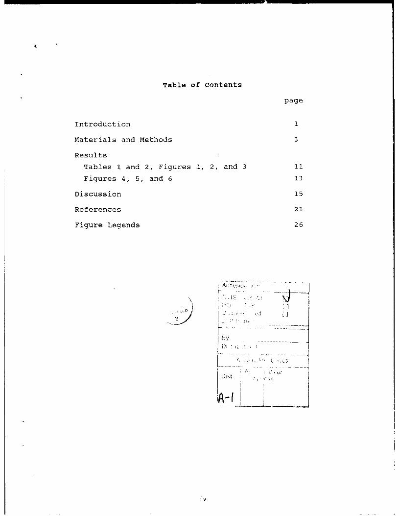
Table of Contents
page
Introduction 1
Materials and Methods 3
Results
Tables 1 and 2, Figures 1, 2, and 3 11
Figures 4, 5, and 6 13
Discussion 15
References 21
Figure Legends 26
J,
A-1iiv
iv
INTRODUCTION
Previous studies (15,17) from our laboratory have demonstrated that
arginine vasopressin (AVP) lowers pulmonary artery pressure in rats with
hypoxic pulmonary hypertension. The pulmonary depressor effect of AVP in
hypoxia-adapted rats was dose dependent and appeared to ba mediated by
activption of the V1 receptor. In contrast, AVP had little effect on
pulmonary artery pressure in air control rats. AVP in doses sufficient to
lower pulmonary artery pressure elicited a systemic pressor response in
hypoxia-adapted rats that was significantly blunted compared to air controls.
To test the hypothesis that the pulmonary depressor action of AVP is due to
the effects of the peptide on the hearr and systemic vqsculature, we .i-zured
cardiac output before and after AVP administration in hypoxia-adapted and air
control rats (17). The AVP did not cause a greater reduction in cardiac
output of hypoxia-aeapted rats compared to air controls. Calculated pulmonary
vascular resistance fell slightly followii.g AVP administration in hypoxia-
adapted rats, but. incraased in air controls. The hemodynamic data indicate
that the AVP-induced fall in cardiac output fails to account for the decrease
in pulmonary artery pressure in the hypoxic group.
In the current study, we examined the effects of AVP on the pulmonary
vasculature of the hypoxia-adapted and air control rats in two preparations:
isolated, non-blood perfused lungs and pulmonary artery rings. AVP had little
vasodilator effect in perfused lungs, and only a small vasodilator effect in
pulmonary artery rings. Further, since previous studies have shown that AVP
induces the release of atrial natriuretic peptide (ANP) from isolated atria
II
(35) and increases circulating levels of ANP in intact rats (19), we then
tested the hypothesis that AVP dilates the pulmonary vasculature in rats with
hypoxic pulmonary hypertension by releasing ANP.
2
MATERIALS AND METHODS
Male Sprague-Dawley rats (Charles River Breeding Laboratories,
Wilmington, MA) weighing approximately 2 20g were maintained in either 10% 02
(hypoxia) or room air (normo::ic controls) for periods of 28 days as previouslv
described (14). Rats were exposed to hypoxia in a 330 liter plexiglas glove
box (Manostat, Brooklyn, NY). Hypoxic exposures (range 10.0±0.5% 02), were
accomplished by adding N2 (Southern Welding, Birmingham, AL) to the chamber
intermittently from a liquid N2 reservoir, the gds outflow of which was
regulated by a solenoid valve controlled by the recorder output of an S3-A 02
analyzer (Applied Electrochemistry, Sunnyvale, CA) through a control circuit
(model 371-K, LFE, Clinton, MA). A baralym- (Allied Health Care Products, St.
Louis, MO) CO2 scrubber kept the [CO 2 ] at <0.2%. Relative humidity within the
chamber was kept a <70% with anhydrous CaSO4 . Boric acid was used to keep NH3
levels within the chamber at a minimum. Animals were permitted to have
standard laboratory chow and tap water ad libitum. Daily animal maintenance
was carried out without interruption of the exposures. Control animals were
caged qimilarly and were exposed to filtered room air for identical periods.
In the initial experiment, the direct effect of AVP on pulmonary arterial
pressure in isolated perfused lungs was determined. Eight hypoxic and A
normoxic control rats were anesthetized with pentobarbital sodium (50 mg/kg
i.p.) after 4 weeks of exposure to hypoxia or room air. A tracheostomy was
performed, -nd a 15 gauge Luer stub was secured in the trachea. The lungs
were ventilated with a Harvard rodent respirator (tidal volune, I ml/100 g
body wt; rate, 60 min-1 ; end-expiratory pressure, 2.5 cm H O) using a 5% C0 2 -
20% 02-75% N 2 gas mixture (Southern Welding, Birmingham, AL). The thoracic
and abdominal cavities were opened. The rats were then killed by severing the
3
abdominal aorta and vena cava. A 16 gauge bulb-tipped cannula was inserted
into the main pulmonary artery. The left atrium was transacted. The lunts
were perfused free of blood using a peristaltic pump (2 mlmin) from a
separate reservoir that contained perfusate at 37°C. T.hen cl ,,ired of blood.
the lungs were removed from the thorax and transferred to the perfusion
chamber, where they were perfused witn a similar periaattic pump (Harvard
Apparatus, South Natick, MA) at a flow rate of 8-12 ml/min in order to
maintain MPAP at levels similar to those observed in vivo. The perfusate
consisted of Krebs-Ringer bicarbonate buffer containing 3% fatty acid- poor
albumin (Bovuminar, Armour Pharmaceutical, Tarrytown, NY) and 5 m1M glucose (pH
7.4; 37'C) (37). Perfusate was continuously equilibrated with ventilator gas
in a aerator, and perfusate pH was monitored throughout the experiment with a
flow-through pH cuvette. Pulmonary artery and airway pressures were monitored
and recorded continuously, because they are reliable indicators of edema
formation in this preparation (9). After a 10 min equilibration period, 50 ng
(in 50 ul saline). AVP ([Arg 8 ]-vasopressin, Sigma Chemical Co.. St. Louis, MO0
(a dose previously shown to reduce MPAP by approximately 24 of the
pretreatment level in intact conscious hypoxia-adapted rats). 50 ul saline, or
12.R mg hydralazine (Sigma Chemical Co., St. Louis, MO) were injected into the
pulmonary artery cannula in random order. The interval bctv¢ecn injcctions
10-15 minutes. Maximal changes in MPAP after injection were recorded. Each
pair uf iungs ;ccived a total of 3 injections, and the total duration of each
perfusion was 40-45 minutes. Lung wet weight-to-dry weig!t ratios (by oven
drying at 800 C for 72 hr) were obtained after each perfusion period to monitor
weight gain of the preparation.
4
In a second protocol, the influence of AVP on the cotp..'tile performance
of thoracic aorta and pulmonary artery tissue isolated frnA ::-2ts adapted to
hypoxia for 4 weeks or air controls was examined. The ravr ..ere killed b.'
decapitation and the thoracic aorta and pulmonary arterv r i:rrediatelv
excised. The adventitia was removpd from both vessels and :. attached
remnant of the thoracic aorta was removed from the pulmonary: irterv with th,
aid of a binocular dissecting microscope. The pulmonary ar-'p.v was cut into 1
ring approximately 2 mm in length. The thwracic aorta was nwctioned intn i
ring 3.5 mm in length. De-endothelialized tissue was prepard from the
remaining portion of the aorta, prior to sectioning into in idditional ring.
by perfusion of the isolated aorta with 2 mg/ml sodium den:vcholate in Krebs-
Ringer Bicarbonate buffer (KRB) for 20 sec, followed hv a Tin wash wick KPB
Krmillimolar composition: NaCl 118, KCI 4.7, CaC1 2 1.3, MgSO 1.2, KH2P0 1.2.
NaHCO, 25.0, glucose 11.7) at pH 7.4 while bubbled with 90 l).-5% CO,.
Each ring was mounted between 2 stainless steel hooks in a 10 ml jacketed
nrqan bath and maintained in KRB at 37'C and continually fnccd. One hook was
stationarv and the other was attached to an isometric forc- -:ansducer (Kulite
Semiconductor BG-10). Ring tension was recorded on a Gouid V'rush 2400 chart
recorder. All rings were subjected to a conditioning protoco: that included
1) equilibration to a resting tension of 2.5 g for 30-45 min preliminary
experiments showed this to be the optimal length-tension reinnio-nhip for ring
contraction,) 2) application of norepinephrine (NE, 3 X 10 :11
(norepinephrine bitartrate, Sigma Chemical Co., St. Louis .A, to un, organ
bath with acetylcholine (Ach, 10 - 6 M) (pcetvlcholine chlorido. Calbiochom
Behringer, La Jolla, CA) added at maximal tension to confirm the functional
presence or absence of the endothelium 11) and 3) applicAtion of an
5
additic".l dose of NE (3 x 20- 7 M) for 20 min to assure tWa: *he rings coul d
sustain a stable tension for this time period. The ri:ws.........I ushed with
KRB between ea' conditioning treatment until a stable bhA.u:,e tension was
reartai ' ,'
The influence of adaptation to hypoxia on thoracic aioy: con'tractile
response to AVP was examined by subjecting conditioned endut-,elium-intact or
de-endothelialized aortic rings isolated from hypoxia-adapced and control rats
to cumulative doses of AVP from 10-1" to 10-6 M. Vascular tissue contraction
can he characterized by two distinct responses during exposnre to an agonist:
1) sensitivity to the agonist and 2) development of tensio,. Therefore, both
serisitivitv to AVP and measured tension were taken into account bv integrating
to tension developed by aortic rings in response to the cq'"iatie doses of
AVP: i.e., mg tension/ng tissue versus In [AVP] M. Integrated dose responses
for aortic ring contractile responses to AVP were -alculared over the entire
dose range of 10-10 to 10-6 M. In addition, because large doses of AVP
Sctallv di.minished vascular contraction in some preparatuio:';, integrated dose
r-s onses were also calculated over a subset of the dose rari-e (10-n to 3 >1
1 (1 1) in -wlhich all preparations displayed a maximal contr-,ction. EC. values
' 7- = concentration of AVP causing a half maximal contrac 'I oNn were
c<alclsPted bv linear regression after logit-log transform,- in of dose
responses.
The influence of adaptation to hvpoxia on pulmonarv a,:,rv relaxation to
A.' was measured in conditioned pulmonary artery rings isol red from hypoxia-
, a nd 'ont ro rats . The rings were pre-contracte-d hv X1 - M NE nd.
t,- a i:.:mal contraction was attained, relaxation of ,he :iu l,, o :Ti rv vessels to
,. was induced bv subjecting the rings to cumulative dos,; of AVP from 1i0-
to h 0 M. Each sequential dose of AVP was applied after maximal relaxatiop
had occurred inresponse to the preceding dose. All rings were removed from
the organ baths, blotted and weighed after'completion of the experiments.
In a third set of experiments, effects of AVP on plasma and atrial ANP
levels were assessed in hypoxia adapted and air control rats. Polyethylene
catheters (PE-lO fused to PE-50) filled with heparin-saline solution (50 U/ml)
were implanted in the femoral artery and vein for blood sampling and drug
administration respectively under ether anesthesia after 25 days of hypoxic or
normoxic exposure. Following catheter implantation, all rats were housed
individually, and the hypoxia adapted rats were returned to the hypoxic
chamber. Two days after implantation, tubing was connected with the femoral
arterial catheter for blood sampling. At least 1 hr was allowed to pass
before 1.0 ml of blood was collected from conscious, unrestrained, resting
animals 5 min after i.v. injection of AVP (160 ng/kg) or saline vehicle for
ANP determination. The blood withdrawn was immediately replaced with an equal
volume of 0.9% saline. Blood was placed in iced tubes containing 1.5 mg EDTA
and 1 trypsin-inhibitor unit of aprotinin. Rats were then decapitated, and
left and right atria were removed quickly. Plasma was separated by
centrifugation. Plasma and atrial samples were stored at -80'C until
radioimmunoassay (RIA) for ANP.
In a separate group of hypoxia-adapted and air control rats, the effects
of AVP on MSAP and MPAP were examined. After 25 days of exposure to hypoxia
or room air, catheters were implanted in the femoral artery and vein as
indicated above. The pulmonary artery was catheterized in situ via the right
jugular vein by a modification of the closed chest technique of Rabinovitch et
al. (29), as described in our previous reports (16,17). Two days after the
7
catheterization, MSAP and MPAP were recorded in conscious rats, through the
pulmonary and femoral arterial catheter using a Model CP-01 pressure
transducer (Century Technology Company, Inglewood, CA) coupled to a Grass
Model 7 polygraph (Grass Instrument Co., Quincy, MA). After stable HSAP and
MPAP were obtained, AVP (160 ng/kg) or saline vehicle were injected
intravenously and peak changes in MSAP and MPAP were recorded.
ANP content of plasma and atria was measured by a modification of the RIA
of Tanaka et al. (38), Eskay et al. (8) and Nakao et al. (24). Plasma for ANP
determination was extracted with Sep-Pak C-18 cartridges (Waters Associates.
Milford, MA) by the method of Eskay et al. (8). Extracts were dried under
vacuum and reconstituted in RIA buffer (see below). Tissue samples were
prepared by a modification of the methods of Tanaka et al. (38) and Nakao et
al. (24). Briefly, left and right atria were weighed and homogenized in 2 ml
of 1 M acetic acid containing 20 mM HCI. The homogenate was heated in a
boiling water bath for 10 min and centrifuged at 25,000 x g for 30 min at 4'C.
The supernatant was lyophilized overnight and reconstituted in RIA buffer (see
below).
Rat 8-33 ANP (Peninsula Labs, Belmont, CA) was used as the reference
standard. Rabbit anti-rat alpha AVP antiserum was generously donated by Wyeth
Laboratories. During the assay, 10 ul of standard (2-250 pg) or sample were
incubated for 48 hrs at 4*C with 100 ul (8,000 cpm) of 1251 labeled rat ANP
(DuP2ont/NEN Research Products, Boston, MA), 100 ul of ANP antiserum (twice
the dilution recommended by the manufacturer) and 200 ul RIA buffer (50 mM
potassium phosphate buffer, pH 7.4, containing 0.1% bovine serum albumin,
0.01% NaN 3, 0.1% Triton X-100, 50 uM phenylmethylsulfonyl fluoride, 50 mM NaCI
and 0.0005% aprotinin). Separation of bound from free tracer was done by
8
adding 750 ul of 20% polyethylene glycol-8000 and 75 ul of 1.5% bovine gamma
globulin to each assay tube and centrifuging for 1 hr at 2,200g (18).
Recovery of ANP from plasma, as assessed by addition of unlabeled 8-33 ANP to
normal rat plasma, was 91±4% Non-specific binding of the tracer was 3%. The
sensitivity of the ANP-RIA was 3.3 pg/assay tube, with 50% displacement at 33
pg/assay tube.
The final experiment tested the functional significance of AVP-induced
ANP release in mediating the pulmonary artery pressure lowering effect of AVP
in hypoxia adapted rats. We had previous shown that the V, receptor
antagonist d(CH2)5 Tyr(Me)AVP administered in a dose of 130 ug/kg, i.v.,
inhibits completely the AVP-induced pulmonary depressor and systemic pressor
effects in hypoxia adapted rats (15). We reasoned that, if the pulmonary
depressor effect of AVP is secondary to ANP release,. the V, receptor
antagonist should also prevent ANP release in this setting. Accordingly, the
effect of the V, receptor antagonist on the AVP-induced increase in plasma ANP
was determined. .The experiment was performed 2 days after catheterization of
the femoral artery and vein as described above. AVP (160 ng/kg), was injected
i.v. 5 min after d(CH2 )5 Tyr(Me)AVP (130 ug/kg), into hypoxia adapted and air
control rats in the conscious, resting state. Five min later, 1 ml of blood
was collected for ANP measurement as previously described.
9
Statistical Analysis. Results are expressed as mean ± SEM. Comparisons
of paired and independent mean treatment effects were by the appropriate
Student's t-test in the first and second experiments. Comparisons of
relaxation responses, expressed as a percentage of initial contraction, were
by Mann-Whitney U-test (36). In the third experiment, results were analyzed
by 2 way analysis of variance; significant differences were then subjected to
Newman-Keuls post-hoc analysis.
10
RESULTS
Rats kept in hypoxia for 4 weeks had reduced body weights compared to air
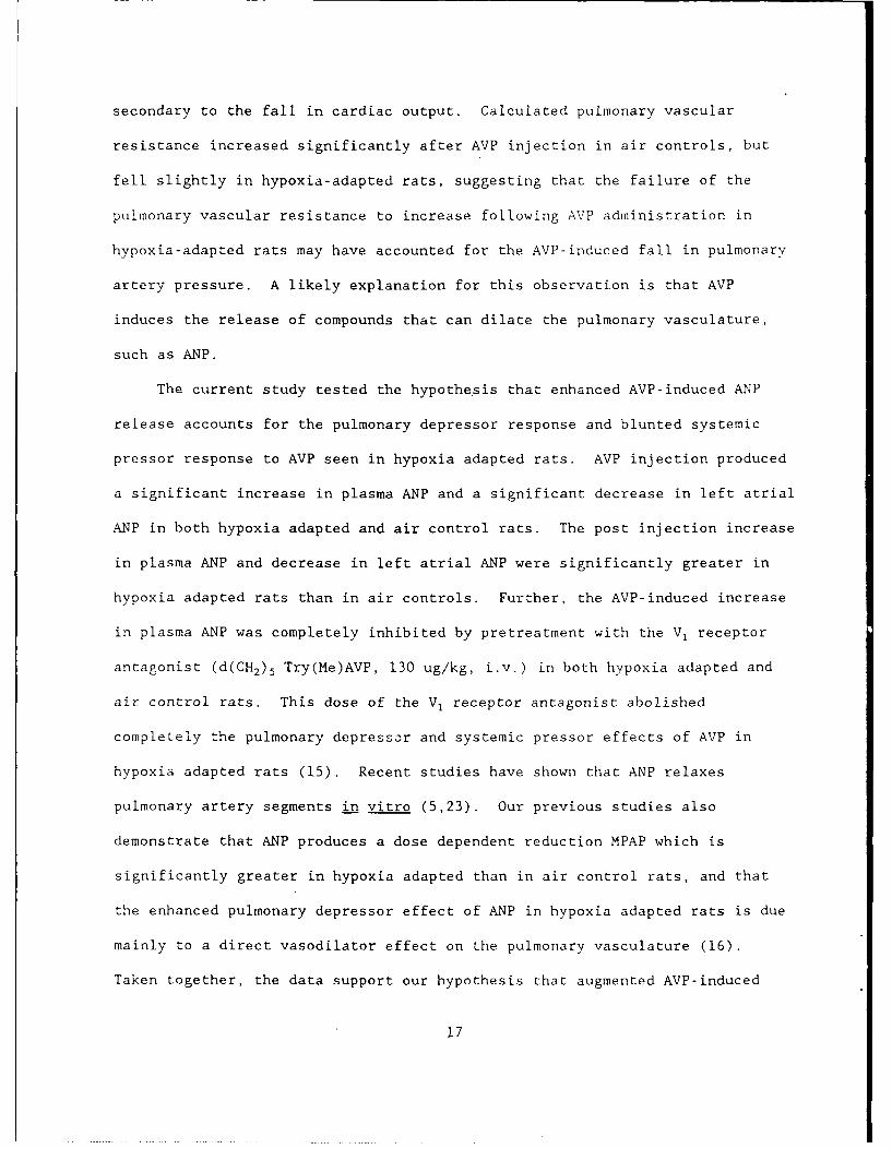
controls (Table 1). Baseline MPAP in isolated perfused lungs from hypoxia
adapted rats was significantly higher (p<0.01) than in those from air controls
(Table 1). The wet weight-to-dry weight ratios of perfused lungs were between
6.0 and 6.5 in both hypoxic and normoxic groups, indicating that significant
alveolar edema did not develop in the course of in vitro perfusion. Bolus
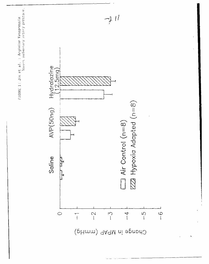
injection of 50 ng AVP into the pulmonary cannula produced only a slight
decrease in MPAP in isolated perfused lungs from both hypoxic and normoxic
rats (Figure 1). Saline vehicle did not alter MPAP in either group. In
contrast, hydralazine (12.5 mg) injection through the pulmonary cannula caused
a significant decrease in MPAP in both groups. The pulmonary depressor
response to hydralazine was not significantly different in hypoxia adapted
rats from air controls.
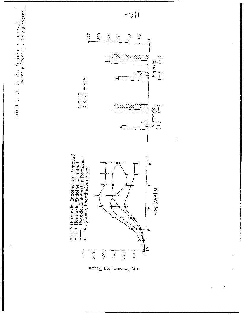
Thoracic aorta and pulmonary artery rings, pre-contracted by NE during
the conditioning.protocol, exhibited robust endothelium-dependent relaxation
in response to Ach (at least 40% by both hypoxia-adapted and control rats,
p<O.001, Figures 2 and 3). Removal of the endothelium prevented the Ach-
induced vascular relaxation (Figure 2).
Thoracic aorta rings from hypoxia-adapted rats, with and without intact
endothelium, were unable to maintain contraction to large doses of AVP;
equivalently prepared rings from control rats performed significantly better
(Figure 2, Table 2). The basis for the relaxation by rings from hypoxia-
adapted rats to large doses of AVP cannot be determined from the current
experiments. However, if the aortic ring responses to AVP were integrated
over a restricted dose range of 10-10 to 3 X 10⁻⁸ M (a dose at which all
11
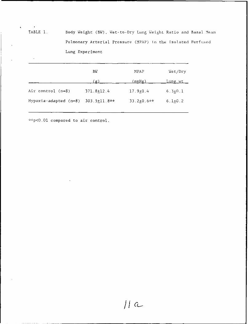
TABLE 1. Body Weight (BW), Wet-to-Dry Lung Weight Ratio and Basal Mean
Pulmonary Arterial Pressure (MPAP) in the Isolated Perfused
Lung Experiment
BW MPAP Wet/Dry
(F-) (mmHg) Lungl wt
Air control (n=8) 371.8+12.4 17.9+0.4 6.3+n.1
Hypoxia-adapted (n=8) 303.5+ll.8** 33.2+0.6** 6.1+0.2
**p<O.Ol compared to air control.
/CL,
L')n
L >>
C) X
'CL
0 ~ wu-1 CjV)j Lfl GbOq
ci
C-,
2-0 0 0 00 C. C 0 0 0
CC-F---- --- -- X
0
UL L
LiL
0>E0 0-
.E E JI -E / E0 0S
.LJ LLJ
o00 > >,
0 0 0 0-0 0o
C) 0 0
~~~l 5w 'U N~j5
5u/, lSuo
C),
-z z
o L,
00
E oCL 75
00
0~(
'0
0 C C0 o o 1U0140J-)U O ID4!Ul o IU0ja'
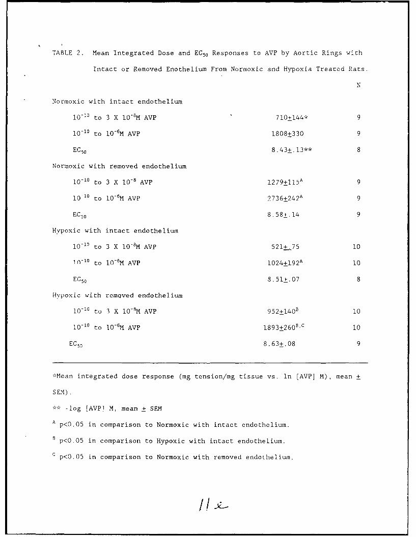
TABLE 2. Mean Integrated Dose and EC5 0 Responses to AVP by Aortic Rings with
Intact or Removed Enothelium From Normoxic and Hypoxia Treated Rats.
N
Normoxic with intact endothelium
I0- 3 to 3 X 10⁻⁸ M AVP 710+144* 9
10-10 to 10-6 M AVP 1808+330 9
EC 5 0 8.43+.13** 8
Normoxic with removed endothelium
10-1" to 3 X 10⁻⁸ AVP 1 2 7 9±1 15 A 9
10-10 to 10- 6M AVP 2 7 3 6+24 2A 9
EC 5 0 8.58*.14 9
Hypoxic with intact endothelium
10-10 to 3 X 10-M AVP 521+.75 10
in-1° to 10-6M AVP 1 0 24 ± 1 9 2A 10
EC 50 8.51±.07 8
Hypoxic with remQved endothelium
10- 0 to 3 X 10⁻⁸M AVP 952±140" 10
10 -10 to 10-6M AVP 1 89 3± 260B C 10
EC50 8.63+.08 9
*Mean integrated dose response (mg tension/mg tissue vs. in [AVP] M), mean +
SEM).
** -log [AVP] M, mean + SEM
A p<0 .0 5 in comparison to Normoxic with intact endothelium.
B p<0.05 in comparison to Hypoxic with intact endothelium.
C p<0.05 in comparison to Normoxic with removed endothelium.
treatment groups had attained maximal contraction), then no significant
differences in integrated dose responses (Table 2) between equivalently
preparE_ rings isolated from hypoxia-adapted and control rats were apparent.
Removal of the endothelium significantly enhanced contraction to AVP by aortic
rings from both hypoxia-adapted and control rats; the improvement in
performance could be attributed to an enhancement in the magnitude of
contraction, since no increase in sensitivity (i.e. decrease in EC5 0) to AVP
was present in de-endothelialized aortic rings from either hypoxia-adapted or
control rats (Figure 2, Table 2).
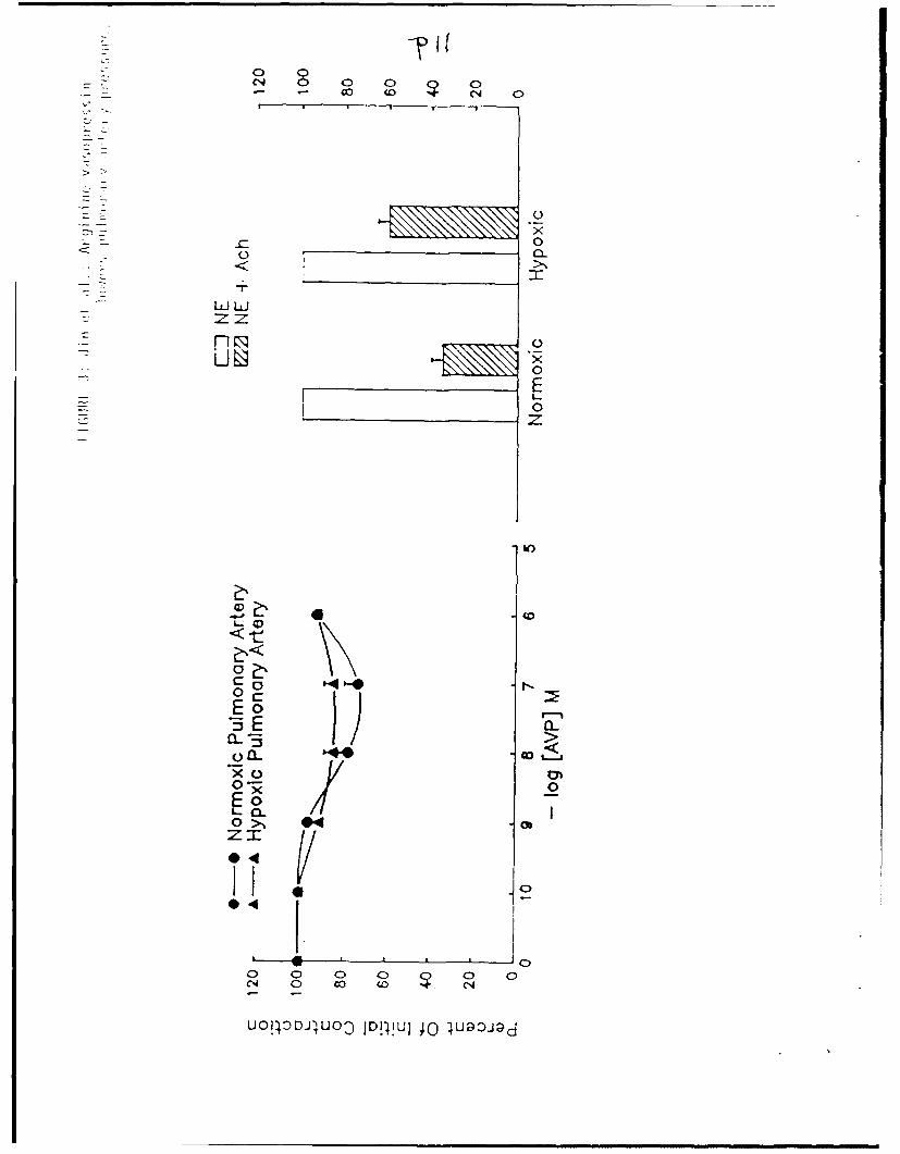
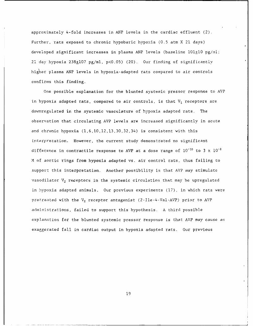
Pulmonary artery rings from hypoxia-adapted and control rats both
manifested weak, but significant, vasodilatory responses to AVP. Rings
isolated from hypoxia-adapted rats and pre-contracted by NE responded to AVP
with increasing vasodilation; a dose of l0-7 M resulted a 14.6±.2% decrease
from the initial NE-induced tension (p<O.Ol, Figure 3). Similarly treated
rings isolated from control rats exhibited a maximal decrease in tension of
28.2±0.4% (p<O.Oi, Pigure 3); the magnitude of tht vasodilatory responses of
rings from hypoxia-adapted and control rats were not significantly difterent
from each other. AVP was significantly less effective in inducing
vasodialtion in pulmonary artery rings pre-contracted by NE than was
the endothelium-dependent mechanism activated by treatment with Ach. Rings
isolated from hypoxia-adapted rats responded to Ach treatement with a
41.04-0.2% relaxation in comparison to the 14.6+0.2% relaxation measured after
exposure to AVP (p<O.O01, Figure 3). Rings isolated from control rats
exhibited a 65.1+0.2% relaxation to NE versus a 28.2+0.4% relaxation after
treatment with AVP (p<0.001, Figure 3).
12
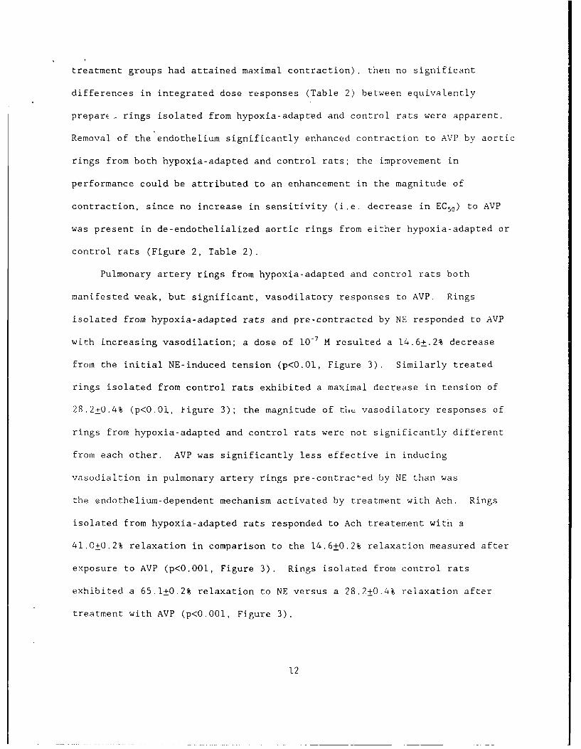
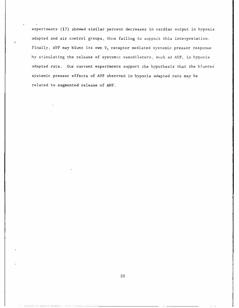
There was no difference in the basal level of MSAP between the hypoxia-
adapted and air contro. groups (Figure 4, Top panel). In contrast, the basal
MPAP in hypoxia-adapted rats was significantly higher than in air controls
(Figure 4, Bottom panel). Bolus injections of AVP (160 ng/kg) produced
significant increases in MSAP in both air control and hypoxia-adapted rats.
The systemic pressor response to AVP was blunted in the hypoxic group compared
with the air control group. The AVP injection significantly lowered MPAP in
hypoxia-adapted rats, but not in air control rats. Saline vehicle did not
alter MSAP or MPAP in either group.
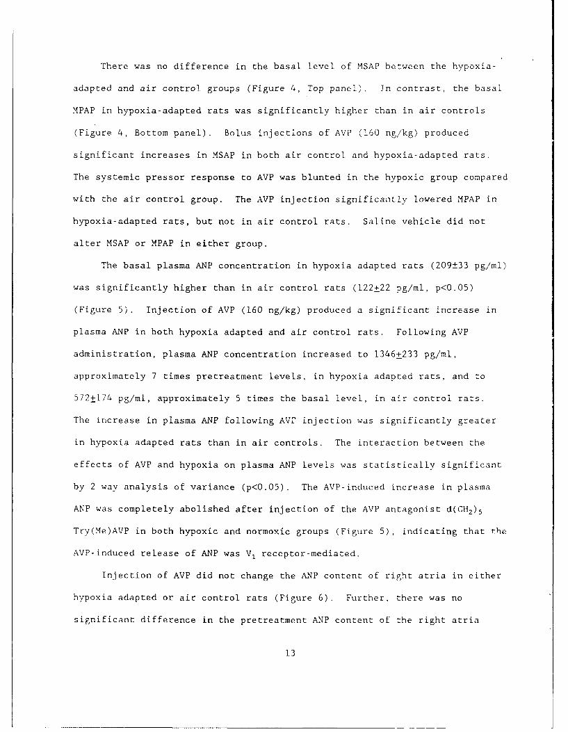
The basal plasma ANP concentration in hypoxia adapted rats (209±33 pg/ml)
was significantly higher than in air control rats (122±22 pg/ml, p<0.05)
(Figure 5). Injection of AVP (160 ng/kg) produced a significant increase in
plasma ANP in both hypoxia adapted and air control rats. Following AVP
administration, plasma ANP concentration increased to 1346+233 pg/ml,
approximately 7 times pretreatment levels, in hypoxia adapted rats, and to
572±174 pg/ml, approximately 5 times the basal level, in air control rats.
The increase in plasma ANP following AVT injection was significantly greater
in hypoxia adapted rats than in air controls. The interaction between the
effects of AVP and hypoxia on plasma ANP levels was statistically significant
by 2 way analysis of variance (p<0.05). The AVP-induced increase in plasma
ANP was completely abolished after injection of the AVP antagonist d(CH 2 )5
Try(Me)AVP in both hypoxic and normoxic groups (Figure 5), indicating that the
AVP-induced release of ANP was V, receptor-mediated.
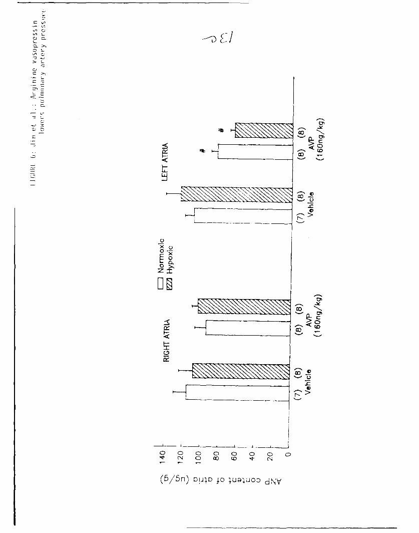
Injection of AVP did not change the ANP content of right atria in either
hypoxia adapted or air control rats (Figure 6). Further, there was no
significant difference in the pretreatment ANP content of the right atria
13
V)
C-)
0.3
x0
0>>
LDCI0 0 C I 0 U) 0 L.4 LO(oD C4 1
(6 HWw) dVSkN (HW W) d V'd 6
~~CD
U- C
C)~U 0 C
Cl)
0*
0~
z- '
00
o >Do
o0 0 0 CD 0 0D 0DN 0 cxj co I- N
(6/'5n) 0JID 40 ;ua'JOO dNV
between normoxic and hypoxic groups. In contrast, AVP injecticn produced a
significant decrease in the ANP content of left atria in both hypoxia adapted
and air control rats (Figure 6). The decrement in ANP content of left atria
was greater (approximately 50% of stores) in hypoxia adapted rats than in air
controls (approximately 25% of stores), although the difference was not
statistically significant. There was no difference in basal ANP content of
left atria between the hypoxic and air control groups.
14
DISCUSSION
The current study confirmed our previous observation that administration
of AVP produced a dose dependent pulmonary depressor effect in hypoxia-adapted
rats, but had no significant effect on pulmonary artery pressure in air
control animals (15,17). AVP had minimal vas~dilator action in isolated,
buffer perfused lungs and only a small vasodilator effect in pulmonary artery
rings in both hypoxia-adapted and air control groups. These observations
strongly suggest that the pulmonary depressor effect of AVP in intact hypoxia-
adapted rats is not due to a direct action of AVP on the pulmonary
vasculature. Published studies of the effects of AVP on the pulmonary
circulation have yielded inconsistent results. Infusion of AVP has been shown
to cause active, nonflow dependent pulmonary vasoconstriction in the intact
conscious dog (26). Other in vivo studies have not consistertly documented an
effect of AVP on the pulmonary vasculatrue in rats, cats and human subjects
(3,7,21,25,31,33). In the isolated perfused lung, AVP has been reported to
increase pulmonary vascular resistance (28), yet most in vitro studies
indicate that isolated pulmonary vascular smooth muscle is poorly responsive
to AVP (4,26). Our current observations support the latter findings.
The current study further demonstrated that AVP induced exaggerated
release of ANP from the left atrium into the circulation in hypoxia-adapted
rats compared to air controls and that the AVP-induced release of ANP was
completely abolished following pretreatment with the V1 receptor antagonist in
both hypoxic and normoxic groups. Taken together, our data suggest that the
pulmonary depressor and blunted systemic pressor effects of AVP observed in
hypoxia-adapted rats are related to augmented release of ANP from left atrium.
15
Pretreatment with a V, receptor anatgonist nearly abolished the pulmonary
depressor effect of AVP in hypoxia-adapted rats, whereas a V2 receptor
antagonist had no effect on the AVP induced response (17), indicating that the
depressor effect of AVP in the pulmonary circulation in hypoxia-adapted rats
is V1 receptor mediated. We have considered several mechanisms by which V,
receptors may mediate the pulmonary depressor effect of AVP in hypoxia-adapted
rats. One possible explanation is that V, receptors, which are absent from
the pulmonary vasculature under normal conditions, may appear de novo in the
pulmonary circulation of hypoxia-adapted rats. Alternatively, the second
messenger mechanism through which AVP operates may be altered in hypoxia-
adapted pulmonary vessels. Hypoxia causes neomuscularization of the pulmonary
vasculature (29), and the response of these new smooth muscle cells to AVP may
differ from that of normal pulmonary vascular smooth muscle. The first
explanation seems unlikely, since circulating AVP levels are increased
significantly in animals (10,30,32) and humans (1,6,12,13,34) adapted to acute
and chronic hypoxia, thus favoring down regulation of V, receptors. Further,
the observations in the current study that AVP had no significant direct
vasodilator or relaxant action in either the isolated, non-blood perfused lung
or pulmonary artery rings from either hypoxia adajpted -r air control rats,
rule against either explanation.
Exogenous AVP decreases cardiac output in conscious rats (27,39) and dogs
(22,26,28), providing another potential mechanism for the pulmonary depressor
effect of AVP in hypoxia-adapted animals. We have previously demonstrated
that the magnitude of the AVP-induced reduction in cardiac output in hypoxia
adapted rats is not different from that in air controls (17), suggesting that
the pulmonary depressor response to AVP in hypoxia adapted rats is not
16
secondary to the fall in cardiac output. Calculated pulmonary vascular
resistance increased significantly after AVP injection in air controls, but
fell slightly in hypoxia-adapted rats, suggesting that the failure of the
pulmonary vascular resistance to increase following AVP administration in
hypoxia-adapted rats may have accounted for the AVP-induced fall in pulmonary
artery pressure. A likely explanation for this observation is that AVP
induces the release of compounds that can dilate the pulmonary vasculature,
such as ANP.
The current study tested the hypothesis that enhanced AVP-induced ANP
release accounts for the pulmonary depressor response and blunted systemic
pressor response to AVP seen in hypoxia adapted rats. AVP injection produced
a significant increase in plasma ANP and a significant decrease in left atrial
ANP in both hypoxia adapted and air control rats. The post injection increase
in plasma ANP and decrease in left atrial ANP were significantly greater in
hypoxia adapted rats than in air controls. Further, the AVP-induced increase
in plasma ANP was completely inhibited by pretreatment with the V, receptor
antagonist (d(CH2) 5 Try(Me)AVP, 130 ug/kg, i.v.) in both hypoxia adapted and
air control rats. This dose of the V1 receptor antagonist abolished
completely the pulmonary depressor and systemic pressor effects of AVP in
hypoxia adapted rats (15). Recent studies have shown that ANP relaxes
pulmonary artery segments in vitro (5,23). Our previous studies also
demonstrate that ANP produces a dose dependent reduction MPAP which is
significantly greater in hypoxia adapted than in air control rats, and that
the enhanced pulmonary depressor effect of ANP in hypoxia adapted rats is due
mainly to a direct vasodilator effect on the pulmonary vasculature (16).
Taken together, the data support our hypothesis that augmented AVP-induced
17
release on ANP accounts for the pulmonary depressor effect and the blunted
systemic pressor effect of AVP observed in hypoxia adapted rats.
The mechanisms by which AVP induces release of ANP are unclear. Other
AVP analogs, such as dAVP (l-deamino-Arga-vasopressin) and oxytocin, which
have pressor activity, and several other pressor agents, such as phenylephrine
and angiotensin II, induce ANP release in the same dose-dependent manner as
AVP in intact anesthetized rats (19). In contrast, neither the nonpressor
analog dDAP nor AVP in the presence of its antipressor (V,) antagonist causes
ANP release. These observations suggest that AVP-induced ANP release is
related to the systemic pressor action of AVP. The finding from our current
study that AVP induced ANP release mainly from left atria is consistent with
the above interpretation. The AVP-induced increase in systemic pressure is
not the only factor involved in ANP AVP-induced release of ANP, however.
Manning et al. (19) have found that, although acute increases in systemic
blood pressure were maximal after the administration of 1 ug if dAVP, ANP
release continued to increase at doses up to 10 ug (doses of dAVP in excess of
10 ug were not tested). Further, there were significant differences in the
amount of ANP released in response to matched increases in MSAP within 15 min
after injection of AVP, dAVP, phenylephrine and angiotensin II. In the
current study, a blunted systemic response to AVP was associated with
augmented ANP release in hypoxia adapted rats compared to air controls,
suggesting the possibility that factors other than increased blood pressure
and atrial stretch are involved in the AVP-induced release of ANP.
Hypoxia per se stimulates release of ANP from atrial stores. Perfusion
of isolated rat and rabbit hearts with hypoxic (PO2 = 21±4 mmHg) buffer via a
Langendorff apparatus for 10 min periods has been associated with
18
approximately 4-fold increases in ANP levels in the cardiac effluent (2).
Further, rats exposed to chronic hypobaric hypoxia (0.5 atm X 21 days)
developed significant increases in plasma ANP levels (baseline 101+10 pg/ml;
21 day hypoxia 238+107 pg/ml, p<0.05) (20). Our finding of significantly
higher plasma ANP levels in hypoxia-adapted rats compared to air controls
confirms this finding.
One possible explanation for the blunted systemic pressor response to AVP
in hypoxia adapted rats, compared to air controls, is that V, receptors are
downregulated in the systemic vasculature of hypoxia adapted rats. The
observation that circulating AVP levels are increased significantly in acute
and chronic hypoxia (1,6,10,12,13,30,32,34) is consistent with this
interpretation. However, the current study demonstrated no significant
difference in contractile response to AVP at a dose range of 10-10 to 3 x 10⁻⁸
M of aortic rings from hypoxia adapted vs. air control rats, thus failing to
support this interpretation. Another possibility is that AVP may stimulate
vasodilator V2 receptors in the systemic circulation that may be upregulated
in hypoxia adapted animals. Our previous experiments (17), in which rats were
pretreated with the V2 receptor antagonist (2-Ile-4-Val-AVP) prior to AVP
administrations, failed to support this hypothesis. A third possible
explanation for the blunted systemic pressor response is that AVP may cause an
exaggerated fall in cardiac output in hypoxia adapted rats. Our previous
19
experiments (17) showed similar percent decreases in cardiac output in hypoxia
adapted and air control groups, thus failing to support this interpretation.
Finally, AVP may blunt its own V, receptor mediated systemic pressor response
by stimulating the release of systemic vasodilators, such as ANP, in hypoxia
adapted rats. Our current experiments support the hypothesis that the blunted
systemic pressor effects of AVP observed in hypoxia adapted rats may be
related to augmented release of ANP.
20
REFERENCES
1. Andrew, M., H. O'Brodovich, G. Coates, G.L. Robertson and G.W. Gray.
Hypoxia-induced vasopressin release and coagulopathy in a normal subject.
Aviat. Space Environ. Med. 56:120-123, 1985.
2. Baertschi, A.J., C. Hausmaninger, R.S. Walsh, R.M. Mentzer, D.A. Wyatt
and R.A. Pence. Hypoxia-induced release of atrial natriuretic factor
(ANF) from the isolated rat and rabbit heart. Biochem. Biophys. Res.
Commun. 140:427-433, 1986.
3. Barer, G.R. A comparison of the circulatory effects of angiotensin,
vasopressin and adrenaline in the anaesthetized cat. J. Physiol. Lond.
156:49-66, 1961.
4. Chand, N. and B.M. Altura. Reactivity and contracility of rat main
pulmonary artery to vasoactive agents. J. Appl. Physiol. 49:1016-1021,
1980.
5. Chou, J., E. Kubota, T. Sata and S.I. Said. Comparative relaxant
activities of atrial natriuretic peptide (ANPs) and vasoactive intestinal
peptide (VIP) on smooth musti- -tructutes iL lufg. Fed. Proc. 45:553,
1986 (Abstr).
6. Colice, G.L. and G. Ramirez. The effect of furosemide during normoxemia
and hypoxemia. Am. Rev. Respir. Dis. 133:279-285, 1986.
7. Emerson, T.E. Effects of angiotensin, epinephrine, norepinephrine, and
vasopressin on venous return. Am. J. Physiol. 210:933-942, 1966.
21
8. Eskay, R., Z. Zukowska-Grojec, M. Haass and N. Zamir. Characterization
of circulating atrial natriuretic peptides in conscious rats: Regulation
of release by multiple factors. Science 232:636-639, 1986.
9. Fisher, A.B., C. Dodia and J. Linask. Perfusate composition and edema
formation in isoalted rat lungs. Exp. Lung. Res. 12:241-248, 1981.
10. Forsling, M.L. and L.A. Aziz. Release of vasporessin in response to
hypoxia and the effect of aminergic and opioid antagonists. J.
Endocrinol. 99:77-86, 1983.
11. Furchgott, R.F. Role of endothelium in responses of vascular smooth
muscle. Circ. Res. 53:557-573, 1983.
12. Hackett, P.H., ML. Forsling, J.S. Milledge and D. Rennie. Release of
vasopressin in man at altitude. Horm. Metab. Res. 10:571, 1978.
13. Heyes, M.P., M.O. Farber, F. Manfredi, D. Robershaw, M. Weinberger, N.
Fineberg and G. Robertson. Acute effects of hypoxia on renal and
endocrine function on normal humans. Am. J. Physiol. 243:R265-R270,
1982.
14. Jin, H., S. Oparil, H.S. Ann, R.H. Yang and R.M. Jackson. Hypoxia-
induced inhibition of converting enzyme activity: Role in vascular
regulation. J. Appl. Physiol. 63:1012-1018, 1987.
15. Jin, H., R.H. Yang, Y.F. Chen, R. Jackson and S. Oparil. Arginine
vasopressin lowers pulmonary arterial pressure in rats adapted to chronic
hypoxia. Am. J. Med. Sci. 30:274-278, 1987.
16. Jin, H., R.H. Yang, Y.F. Chen, R.M. Thornton, R. Jackson and S. Oparil.
Atrial natriuretic peptide lowers pulmonary arterial pressure in hypoxia
adapted rats. J. Appl. Physiol. 65:1729-1735, 1988.
22
17. Jin, H., R.H. Yang, Y.F. Chen, R.M. Thornton, R.M. Jackson and S. Oparil.
Hemodynamic effects of arginine vasopressin rats adapted to chronic
hypoxia. J. Appl. Physiol., in press, 1988.
18. Larose, P., S. Meloche, P. du Souich, A. De Lean and H. Ong.
Radioimmunoassay of atrial natriuretic factor: Human plasma levels.
Biochem. Biophys. Res. Commun. 130:553-558, 1985.
19. Manning, P.T., D. Schwartz, N.C. Katsube, S.W. Holmberg and P. Needleman.
Vasopressin-stimulated release of atriopeptin: endocrine antagonist in
fluid hemostasis. Science 229:395-397, 1985.
20. McKenzie, J.C., I. Tanaka, T. Inagami, K.S. Misono and R.M. Klein.
Alterations in atrial and plasma atrial natriuretic factor (ANF) content
during development of hypoxia-induced pulmonary hypertension in the rat.
Proc. Soc. Exp. Biol. Med. 181:459-463, 1986.
21. Mols, P., R. Hallemans, M.V. Kuvk, C. Melot, P. Lejeune, H. Ham, F.
Vertongen and R. Naeije. Hemodynamic effects of vasopressin alone and in
combination .with nitroprusside, in patients with liver cirrhosis and
portal hypertpnsion. Ann. Surg. 199:176-181, 1984.
22. Montani, J.P., J.F. Liard, J. Schoun and J. Mohring. Hemodynamic effects
of exogenous and endogenous vasopressin at low plasma concentrations in
conscious dogs. Circ. Res. 47:346-355, 1980.
23. Morice, A.H., T.L. Jansen and M.J. Brown. The effect of attial
natriuretic factors on in vivo pulmonary artery pressure. Am. Rev. Resp.
Dis. 135:A300, 1987 (Abstr).
23
24. Nakao, K., A. Sugawara, N. Morii, M. Sakamoto, M. Suda, J. Soneda, T.
Ban, M. Kihara, Y. Yamori, M. Shimokura, Y. Kiso and H. Imura.
Radioimmunoassay for alpha-human and rat atrial natriuretic polypeptides.
Biochem. BioDhvs. Res. Commun. 124:815-821. 1984.
25. Nelson, R.A., L.G. May, A. Bennett, M. Kobayashi and R. Gregory.
Comparison of the effects of pressor and depressor agents and influences
on pulmonary and systemic pressures of normotensive and hypertensive
subjects. Am. Heart J. 50:172-187, 1955.
26. Nyhan, D.P., H.S. Geller, H.M. Goll and P.A. Murray. Pulmonary
vasoactive effects of exogenous and endogenous AVP in conscious dogs.
Am. J. Physiol. 251:HI009-HI016, 1986.
27. Osborn, J.W., M.M. Skelton and A.W. Cowley. Hemodynamic effects of
vasopressin compared with angiotensin II in conscious rats. Am. J.
Physiol. 252(Heart Circ. Physiol. 21):H628-H637, 1987.
28. Porcelli, R.J., A. Viau, M. Demeny, N.E. Naftchi and E.H. Bergofsky.
Relation between hypoxic pulmonary vasoconstriction, its humoral
mediators and alpha-beta adrenergic receptors. Chest 71:249-251, 1977.
29. Rabinovitch, M., W. Gamble, A.S. Nadas, O.S. Miettinen and L. Reid. Rat
pulmonary circulation after chronic hypoxia: hemodynamic and structural
features. Am. J. Physiol. 236:H818-H827, 1979.
30. Raff, H., J. Shinsako, L.C. Keil and M.F. Dallman. Vasopressin, ACTH and
corticosteriods during hypercapnia and graded hypoxia in dogs. Am. J.
Physiol. 244:E453-E458, 1983.
31. Ribot, S., H. Green, M.J. Small and S. Abramowitz. Cardiovascular
effects of v~sopressin. Am. J. Med. Sci. 242:612-619, 1061.
24
32. Rose, C.E., R.J. Anderson and R.M. Carey. Antidiuresis and vasopressin
release with hypoxemia and hypercapnia in conscious dogs. Am. J.
Physiol. 247:RI27-RI34, 1984.
33. Segel, N., T.J. Bayley, A. Paton, P.W. Dykes and J.M. Bishop. The
effects of synthetic vasopressin and angiotensin on the circulation in
cirrhosis of the liver. Clin. Sci. 25:43-55, 1963.
34. Singh, I., M.S. Malhotra, P.K. Khanna, R.B. Nanda, T. Purshottam, T.N.
Upadhyay, U. Radhakrishman and H.D. Brahma-hari. Changes in plasma
cortisol, blood antidiuretic hormone and urinary catecholamines in high
altitude pulmonary edema. Int. J. Biometeor. 18:211-221, 1974.
35. Sonnenberg, H. and A.T. Veress. Cellular mechanism of release of atrial
natriuretic factor. Biochem. Biophys. Res. Commun. 124:443-449, 1984.
36. Snedecor, G.W. and W.G. Cochran. Statistical Methods. Iowa State
University Press, Ames, Iowa, 1967, pp. 391-395.
3; Steinberg, H., R.A. Greenwald, S.A. Moak and D.K. Dos. Depression of
pulmonary 5-hydroxytryptamine uptake by metabolic inhibitors. Am. J.
Physiol. 228:1298-1303, 1975.
38. Tanaka, I., K.S. Misono and T. Inagami. Atrial natriuretic factor in rat
hypothalamus, atria and plasma: Determination by specific
radioimmunoassay. Biochem. Biophys. Res. Commun. 124:663-667, 1984.
39. Walker, B.R. Evidence tor a a vasopressin in the
conscious rat. Am. J. Physiol. 251(Heart Circ. Physiol. 20):H34-H39,
1986.
25
FIGURE LEGENDS
Figure 1: Effects of AVP and hydralazine on mean pulmonary arterial
pressure (MPAP) in isolated perfused lurgs from hypoxia adap-,d
and air control rats. Data are presented as means + SEM.
Figure 2: Contractile responses to cumulative doses of AVP (left) and
relaxation to Ach (right), by thoracic aorta rings isolated from
hypoxia-adapted and control rats. Rings possessed an intact
endothelium (+), or were de-endothelialized (-). Open histograms
(right) denote maximal contractile response to 3 X 10⁻⁷ M NE,
striated histograms denote remaining contraction subsequent to
application of 10-6 M Ach. All data are presented as mean + SEM,
N=9 to 10 per treatment group.
Figure 3: Vasodilatory responses to cumulative doses of AVP (left) and 10--
M Ach (right) by endotheliuur-intact ,iilionarv artery rings
isolated from hypoxia-adapted and control rats. Vasodilatorv
responses to both agonists are presented as the percentage
remaining of an initial contraction induced bv 3 X 10- 7 M NE. All
data are presented as mean + SEM, ',9 to 10 per treatment group.
26
Figure 4: Effects of AVP (160 ng/kg) on mean systemic arterial pressure
(MSAP) and mean pulmonary arterial pressure (MPAP) in hypoxia-
adapted and air control rats., Data are presented as mean ± SEM.
* p<0.01, compared to the respective normoxic group.
# p<O.Ol, compared to the respective basal or vehicle group.
Figure 5: Effects of AVP (160 ng/kg) on plasma ANP levels in hypoxia-
adapted and air control rats. Data are presented as mean + SEM.
* p<O.05 compared to the respective normoxic group.
# p<0.05 compared to the respective vehicle or d(CHZ)s Try(Me)AVP
+ AVP group. Numbers in parentheses - number of rats in each
group.
Figure 6: Effect of AVP (160 ng/kg) on ANP content of right and left atria
in hypoxia-adapted and air control rats. Data are presented as
mean + SEM.
# p<0.05 compared to the respective vehicle.
Numbers in parentheses - number of rats in each group.
27