Embed Size (px)
Citation preview

1
Arrhythmias and Dysrhythmias
Craig Barstow, MD, FAAFP
ACTIVITY DISCLAIMERThe material presented here is being made available by the American Academy of Family Physicians for educational purposes only. Please note that medical information is constantly changing; the information contained in this activity was accurate at the time of publication. This material is not intended to represent the only, nor necessarily best, methods or procedures appropriate for the medical situations discussed. Rather, it is intended to present an approach, view, statement, or opinion of the faculty, which may be helpful to others who face similar situations.
The AAFP disclaims any and all liability for injury or other damages resulting to any individual using this material and for all claims that might arise out of the use of the techniques demonstrated therein by such individuals, whether these claims shall be asserted by a physician or any other person. Physicians may care to check specific details such as drug doses and contraindications, etc., in standard sources prior to clinical application. This material might contain recommendations/guidelines developed by other organizations. Please note that although these guidelines might be included, this does not necessarily imply the endorsement by the AAFP.
DISCLOSUREIt is the policy of the AAFP that all individuals in a position to control content disclose any relationships with commercial interests upon nomination/invitation of participation. Disclosure documents are reviewed for potential conflict of interest (COI), and if identified, conflicts are resolved prior to confirmation of participation. Only those participants who had no conflict of interest or who agreed to an identified resolution process prior to their participation were involved in this CME activity.
All individuals in a position to control content for this session have indicated they have no relevant financial relationships to disclose.
The content of my material/presentation in this CME activity will not include discussion of unapproved or investigational uses of products or devices.
Craig Barstow, MD, FAAFPProgram Director, Hospitalist Fellowship at Womack Army Medical Center, Fort Bragg, North Carolina; Assistant Professor of Family Medicine at Uniformed Services University of Health Sciences, Bethesda, Maryland; Director of Ultrasound Education, Family Medicine Residency Program, Womack Army Medical Center; Physician, Scotland Memorial Hospital Emergency Department, Laurinburg, North Carolina.
Dr. Barstow is a graduate of the Uniformed Services University of the Health Sciences –F. Edward Herbert School of Medicine in Bethesda, Maryland. He completed undergraduate studies at the U.S. Military Academy. Dr. Barstow joined the Womack Army Medical Center Family Medicine Residency Program in 2012, and created the fellowship program, accepting the first fellow in July 2015. His areas of interest include inpatient family medicine, newborn care, and point-of-care ultrasound teaching.
Learning Objectives1. Identify the causes of ventricular arrhythmias and differentiate the types of
ventricular arrhythmias and identify the causes of atrial arrhythmias and differentiate the types of atrial arrhythmias.
2. Manage life-threatening ventricular arrhythmias, and assess, diagnose and stratify for risk patients who have, or are at risk for, ventricular arrhythmias.
3. Develop collaborative care plans with patients, emphasizing medication adherence and follow-up.
4. Establish quality improvement plans to maximize care coordination and minimize hospital readmission.
Associated Session(s)
• Arrhythmias and Dysrhythmias: PBL

2
Audience Engagement SystemStep 1 Step 2 Step 3
Arrhythmias and Dysrhythmias
Tachyarrhythmia
• Atrial fibrillation
• Supraventricular tachycardia
• Ventricular tachycardia
Bradyarrhythmia
Tachyarrhythmias
Case 1
Evelyn is a 65-year old woman who presents for a routine office visit. On physical exam, she is noted to have an irregular heart rate.
AES POLL QUESTIONWhat is the most common arrhythmia worldwide?
A. Atrioventricular blockB. Wolf Parkinson White syndromeC. Atrial fibrillationD. Atrial flutter

3
Atrial Fibrillation
• Most common cardiac arrhythmia worldwide
• Disease of aging
– 1% patients < 60
– 8-12% patients > 80
• 450,000 admission per year in the US
• Significant cause of stroke
– Increased mortality and morbidity from stroke from AFGo AS, Mozaffarian D, Roger VL, et al. Heart disease and stroke statistics‐2014 update: a report from the American Heart Association. Circulation.2014; 129(3):e28‐e292.
Atrial Fibrillation
• Paroxysmal AF
• Persistent AF
• Long-standing AF
• Permanent AF
• Nonvalvular AF
Treatment of Atrial FibrillationAcute Management
If hemodynamically unstable– Electrical cardioversion
If hemodynamically stable but symptomatic(with no pre-excitation)
– Metoprolol 2.5-5.0 mg IV bolus every 3 min; up to 3 doses– Verapamil 0.075-0.15 mg/kg IV bolus over 2 min; may give an
additional 10.0 mg after 30 min in no response, then 0.005 mg/kg/min infusion
– Diltiazem 0.25 mg/kg IV bolus over 2 min; then 5-15 mg/hrJanuary CT, Wann LS, Alpert JS, et al. 2014 AHA/ACC/HRS guideline for the management of patients with atrial fibrillation: a report of the American College of Cardiology/American Heart Association Task Force on Practice Guidelines and the Heart Rhythm Society. J AM Coll Cardiol 2014; 64: e1‐76.
Treatment of Atrial FibrillationRate vs. Rhythm Control
• AFFIRM and RACE trials• Rate control equivalent to rhythm control• Rhythm control
– Proarrhythmic– Requires monitoring– Reoccurs in 20-60% at one year– Increased hospitalization rate
Rate Control• Beta blockers
– esmolol– propranolol– metoprolol
• Nondihydropyridine calcium channel blockers– diltiazem– verapamil
• Digoxin• Amiodarone
Rhythm Control
• Cardioversion
• Antiarrhythmic drugs
• Catheter ablation
4
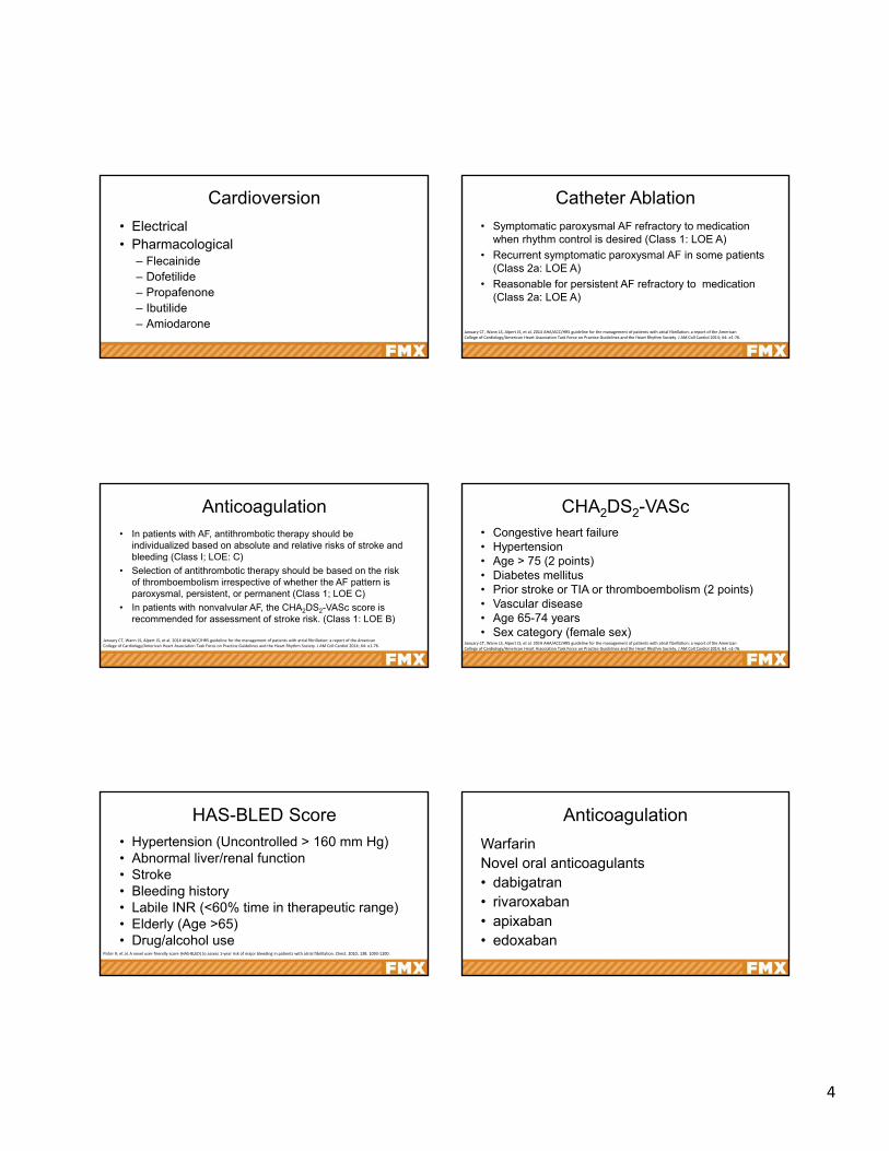
Cardioversion
• Electrical• Pharmacological
– Flecainide– Dofetilide– Propafenone– Ibutilide– Amiodarone
Catheter Ablation
• Symptomatic paroxysmal AF refractory to medication when rhythm control is desired (Class 1: LOE A)
• Recurrent symptomatic paroxysmal AF in some patients (Class 2a: LOE A)
• Reasonable for persistent AF refractory to medication (Class 2a: LOE A)
January CT, Wann LS, Alpert JS, et al. 2014 AHA/ACC/HRS guideline for the management of patients with atrial fibrillation: a report of the American College of Cardiology/American Heart Association Task Force on Practice Guidelines and the Heart Rhythm Society. J AM Coll Cardiol 2014; 64: e1‐76.
Anticoagulation• In patients with AF, antithrombotic therapy should be
individualized based on absolute and relative risks of stroke and bleeding (Class I; LOE: C)
• Selection of antithrombotic therapy should be based on the risk of thromboembolism irrespective of whether the AF pattern is paroxysmal, persistent, or permanent (Class 1; LOE C)
• In patients with nonvalvular AF, the CHA2DS2-VASc score is recommended for assessment of stroke risk. (Class 1: LOE B)
January CT, Wann LS, Alpert JS, et al. 2014 AHA/ACC/HRS guideline for the management of patients with atrial fibrillation: a report of the American College of Cardiology/American Heart Association Task Force on Practice Guidelines and the Heart Rhythm Society. J AM Coll Cardiol 2014; 64: e1‐76.
CHA2DS2-VASc• Congestive heart failure• Hypertension• Age > 75 (2 points)• Diabetes mellitus• Prior stroke or TIA or thromboembolism (2 points)• Vascular disease• Age 65-74 years• Sex category (female sex)
January CT, Wann LS, Alpert JS, et al. 2014 AHA/ACC/HRS guideline for the management of patients with atrial fibrillation: a report of the American College of Cardiology/American Heart Association Task Force on Practice Guidelines and the Heart Rhythm Society. J AM Coll Cardiol 2014; 64: e1‐76.
HAS-BLED Score• Hypertension (Uncontrolled > 160 mm Hg)• Abnormal liver/renal function• Stroke• Bleeding history• Labile INR (<60% time in therapeutic range)• Elderly (Age >65)• Drug/alcohol use
Pister R, et al. A novel user‐friendly score (HAS‐BLED) to assess 1‐year risk of major bleeding in patients with atrial fibrillation. Chest. 2010; 138: 1093‐1100.
Anticoagulation
WarfarinNovel oral anticoagulants• dabigatran• rivaroxaban• apixaban• edoxaban

5
AES POLL QUESTION
For patients with atrial fibrillation, aspirin provides an absolute risk reduction of…A. 8%B. 80%C. 4%D. 0.8%
Case 2
A 76-year old male presents to your office complaining of palpitations. He has a history of atrial fibrillation. An ECG reveals the following:
Atrial Flutter
• Reentrant atrial arrhythmia• Regular atrial rate• Constant p-wave morphology• Similar risk factors for atrial fibrillation• Atrial flutter and atrial fibrillation can
coexist in same patient
Atrial Flutter• Acute Management• Hemodynamically unstable
– Rhythm control• Synchronized cardioversion (Class 1)
– Rate control• IV amiodarone (Class 2a)
• Hemodynamically stable– Rhythm control
• Synchronized cardioversion (Calls 1)– Rate control
• IV beta blockers, diltiazem, verapamil (Class 1)• IV amiodarone (Class 2a)
Page RL, Joglar JA, Caldwell MA, et al. 2015 ACC/AHA/HRS guideline for the management of adult patients with supraventricular tachycardia: a report of the American College of Cardiology/American Heart Association Task Force on Clinical Practice Guidelines and the Heart Rhythm Society. J Am Coll Cardiol. 2016; 67: e27‐115.
Atrial Flutter
Chronic Management1. Rate control
• Beta blockers, diltiazem, verapamil (Class 1)2. Rhythm control
• Catheter ablation (Class 1)• Amiodarone, dofetiliide or sotalol (Class 2a)• Flecainide or propafenone (Class 2b)
6
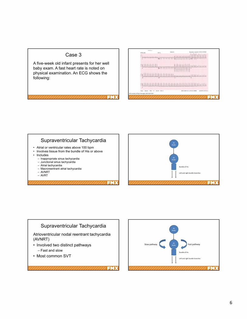
Case 3
A five-week old infant presents for her well baby exam. A fast heart rate is noted on physical examination. An ECG shows the following:
ECG courtesy of Ryan Flannigan, MD FAAP FACC
Supraventricular Tachycardia• Atrial or ventricular rates above 100 bpm• Involves tissue from the bundle of His or above• Includes
– Inappropriate sinus tachycardia– Junctional sinus tachycardia– Atrial tachycardia– Macroreentrant atrial tachycardia– AVNRT– AVRT
SA Node
AV Node
Bundle of His
Left and right bundle branches
Supraventricular Tachycardia
Atrioventricular nodal reentrant tachycardia (AVNRT)
• Involved two distinct pathways– Fast and slow
• Most common SVT
SA Node
AV Node
Bundle of His
Left and right bundle branches
Fast pathwaySlow pathway
7
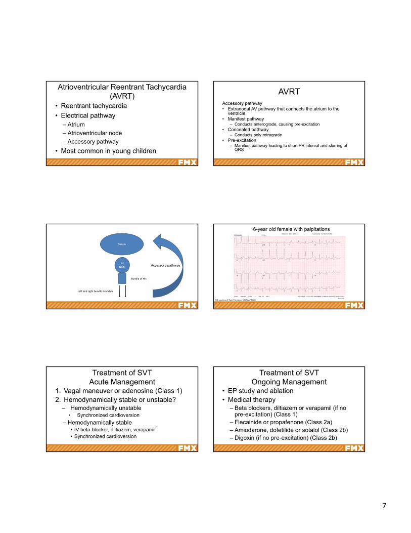
Atrioventricular Reentrant Tachycardia (AVRT)
• Reentrant tachycardia
• Electrical pathway– Atrium
– Atrioventricular node
– Accessory pathway
• Most common in young children
AVRTAccessory pathway• Extranodal AV pathway that connects the atrium to the
ventricle• Manifest pathway
– Conducts anterograde, causing pre-excitation• Concealed pathway
– Conducts only retrograde• Pre-excitation
– Manifest pathway leading to short PR interval and slurring of QRS
Atrium
AV Node
Bundle of His
Left and right bundle branches
Accessory pathway
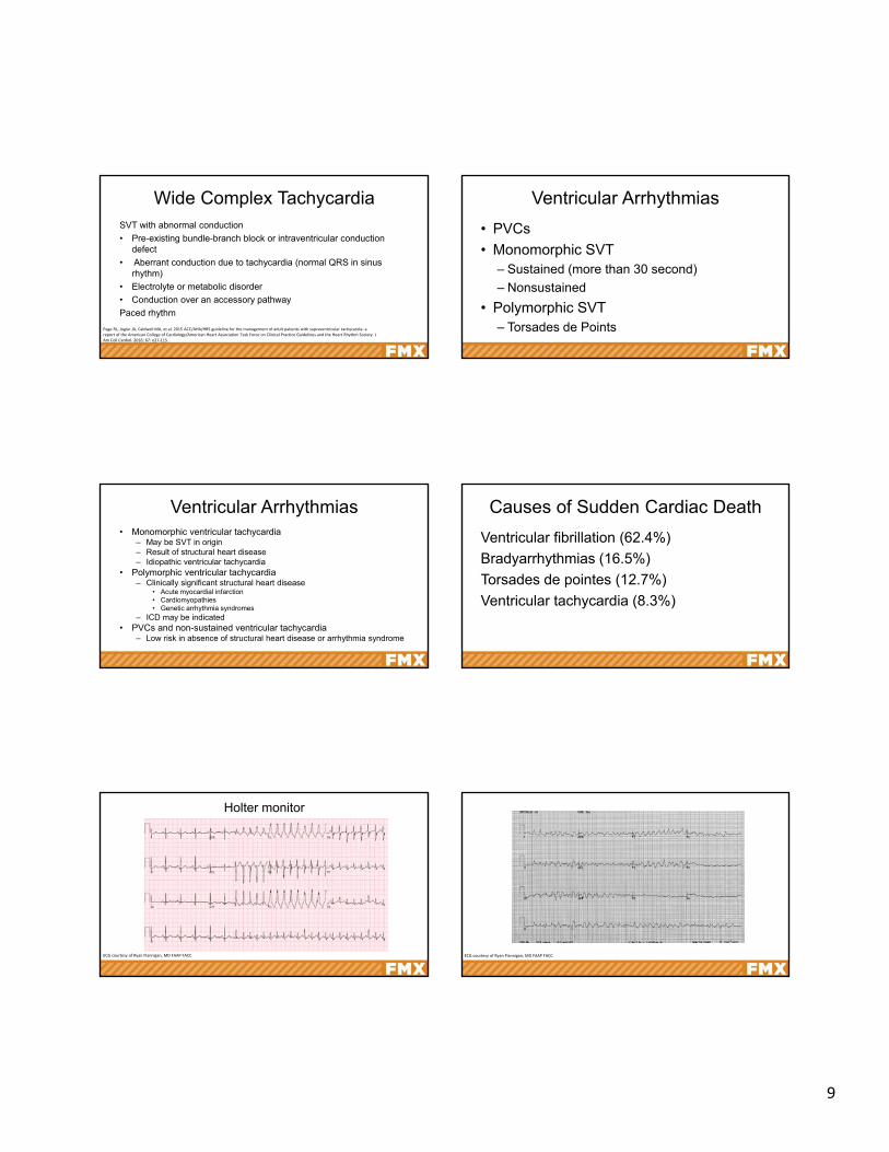
16-year old female with palpitations
ECG courtesy of Ryan Flannigan, MD FAAP FACC
Treatment of SVTAcute Management
1. Vagal maneuver or adenosine (Class 1)2. Hemodynamically stable or unstable?
– Hemodynamically unstable• Synchronized cardioversion
– Hemodynamically stable• IV beta blocker, diltiazem, verapamil• Synchronized cardioversion
Treatment of SVTOngoing Management
• EP study and ablation• Medical therapy
– Beta blockers, diltiazem or verapamil (if no pre-excitation) (Class 1)
– Flecainide or propafenone (Class 2a)– Amiodarone, dofetilide or sotalol (Class 2b)– Digoxin (if no pre-excitation) (Class 2b)

8
Tachycardia-Induced Cardiomyopathy
• Cardiomyopathy secondary to sustained tachycardia
• Dilated cardiomyopathy
• Sustained tachycardia for months to years
• Reversible with control of underlying rhythm
AES POLL QUESTION
Which of the following are relatively contraindicated in AVRT with preexcitation?
A. Adenosine
B. Beta blockers
C. Nondihydropyridine CCB
D. B and CPage RL, Joglar JA, Caldwell MA, et al. 2015 ACC/AHA/HRS guideline for the management of adult patients with supraventricular tachycardia: a report of the American College of Cardiology/American Heart Association Task Force on Clinical Practice Guidelines and the Heart Rhythm Society. J Am Coll Cardiol. 2016; 67: e27‐115.
Wide Complex Tachycardia
Case 4
A 60-year old woman presents to your clinic with palpitations and shortness of breath. She has a history of atrial fibrillation. ECG reveals a wide-complex regular tachycardia.
Wide Complex Tachycardia
• Ventricular tachycardia
• Supraventricular rhythm with abnormal conduction
9
Wide Complex TachycardiaSVT with abnormal conduction
• Pre-existing bundle-branch block or intraventricular conduction defect
• Aberrant conduction due to tachycardia (normal QRS in sinus rhythm)
• Electrolyte or metabolic disorder
• Conduction over an accessory pathway
Paced rhythm
Page RL, Joglar JA, Caldwell MA, et al. 2015 ACC/AHA/HRS guideline for the management of adult patients with supraventricular tachycardia: a report of the American College of Cardiology/American Heart Association Task Force on Clinical Practice Guidelines and the Heart Rhythm Society. J Am Coll Cardiol. 2016; 67: e27‐115.
Ventricular Arrhythmias
• PVCs
• Monomorphic SVT– Sustained (more than 30 second)
– Nonsustained
• Polymorphic SVT– Torsades de Points
Ventricular Arrhythmias• Monomorphic ventricular tachycardia
– May be SVT in origin– Result of structural heart disease– Idiopathic ventricular tachycardia
• Polymorphic ventricular tachycardia– Clinically significant structural heart disease
• Acute myocardial infarction• Cardiomyopathies• Genetic arrhythmia syndromes
– ICD may be indicated• PVCs and non-sustained ventricular tachycardia
– Low risk in absence of structural heart disease or arrhythmia syndrome
Causes of Sudden Cardiac Death
Ventricular fibrillation (62.4%)
Bradyarrhythmias (16.5%)
Torsades de pointes (12.7%)
Ventricular tachycardia (8.3%)
Holter monitor
ECG courtesy of Ryan Flannigan, MD FAAP FACC ECG courtesy of Ryan Flannigan, MD FAAP FACC
10
ECG courtesy of Ryan Flannigan, MD FAAP FACC
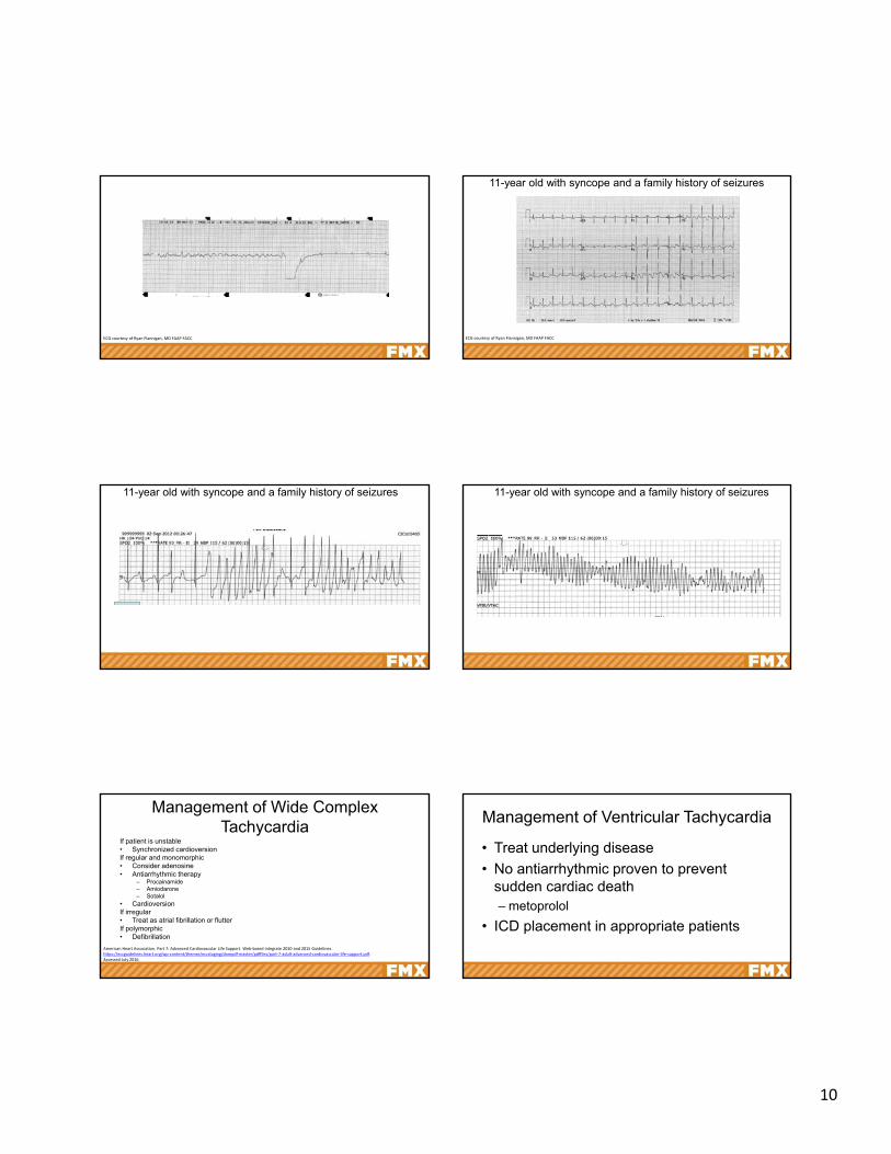
11-year old with syncope and a family history of seizures
ECG courtesy of Ryan Flannigan, MD FAAP FACC
11-year old with syncope and a family history of seizures 11-year old with syncope and a family history of seizures
Management of Wide Complex Tachycardia
If patient is unstable• Synchronized cardioversionIf regular and monomorphic• Consider adenosine• Antiarrhythmic therapy
– Procainamide– Amiodarone– Sotalol
• CardioversionIf irregular• Treat as atrial fibrillation or flutterIf polymorphic• Defibrillation
American Heart Association. Part 7: Advanced Cardiovascular Life Support. Web‐based integrate 2010 and 2015 Guidelines. https://eccguidelines.heart.org/wp‐content/themes/eccstaging/dompdf‐master/pdffiles/part‐7‐adult‐advanced‐cardiovascular‐life‐support.pdf. Accessed July 2016.
Management of Ventricular Tachycardia
• Treat underlying disease
• No antiarrhythmic proven to prevent sudden cardiac death– metoprolol
• ICD placement in appropriate patients

11
Sudden Cardiac Death (SCD)In patients with heart disease
• Older patients– Coronary artery disease– Valvular heart disease– Heart failure
• Predictors– Severity of underlying disease
• Coronary heart disease• Heart failure
– Ejection fraction strongest predictor (< 30-40%)
Sudden Cardiac Death (SCD)In patients without heart disease
• 50% have undiagnosed ischemic heart disease
• Younger patients– Chanelopathies– Cardiomyopathy– Myocarditis– Substance abuse
CardiomyopathiesPrimary Cardiomyopathies Secondary Cardiomyopathies
Genetic Mixed Acquired Infiltrative/Storage
Endocrine Other
Hypertrophic cardiomyopathy
Dilated cardiomyopathy Myocarditis Amyloidosis Diabetes mellitus Sarcoidosis
Arrhythmogenic right ventricular dysplasia
Restrictive Takotsubo Gaucher disease Hyperthyroidism Neuromuscular
LV Noncompaction Peripartum Hurler’s disease Hypothyroidism Neurological
Glycogen storage diseases Tachycardia induced Hunter’s disease Hyperparathyroidism Nutritional deficiencies
Conduction defects Hemochromatosis Pheochromocytoma Dermatomyositis
Mitochondrial myopathies
Fabry’s disease Acromegaly Scleroderma
Ion channel disorders Glycogen storage disease Electrolyte imbalance
Niemann‐Pick disease Cancer therapy
Maron BJ, Towbin JA, Thiene G, et al. Contemporary definitions and classification of the cardiomyopathies: An American Heart Association scientific statement from the council on clinical cardiology, heart failure and transplantation committee; quality of care and outcomes research and functional genomics and translational biology interdisciplinary working groups; and council on epidemiology and prevention. Circulation. 2006;113:1807‐1816.
ECG Changes in CardiomyopathyT wave inversion >1 mm 2 or more leads; V2‐V6, II and aVF or I and AVL
ST segment depression > 0.5 mm in two or more leads
Pathological Q waves > 3 mm depth or > 40ms duration two or more leads
Complete left bundle branch block QRS > 120 ms, negative QRS complex in V1 and upright monophasic R 1 and V6
Intraventricular conduction delay QRS > 140 ms
Left axis deviation ‐30o ‐ ‐90o
Left atrial enlargement
Right ventricular hypertrophy pattern
Premature ventricular contractions > 2 PVCs per 10 sec tracing
Ventricular arrhythmias Couplets, triplets and non‐sustained VT
Drezner JA, Ashley E, Baggish AL. Abnormal electrocardiographic findings in athletes: recognising changes suggestive of cardiomyopathy. Br J Sports Med. 2013;47:137‐152.
ECG courtesy of Ryan Flannigan, MD FAAP FACC
16- year old male with a history of syncope
Bradyarrhythmias
12
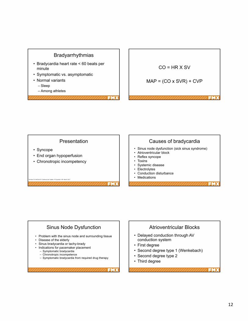
Bradyarrhythmias
• Bradycardia heart rate < 60 beats per minute
• Symptomatic vs. asymptomatic
• Normal variants– Sleep
– Among athletes
CO = HR X SV
MAP = (CO x SVR) + CVP
Presentation
• Syncope
• End organ hypoperfusion
• Chronotropic incompetency
Barstow CH, McDivitt JD. Cardiovascular Update. FP Essentials. 454: March 2017
Causes of bradycardia• Sinus node dysfunction (sick sinus syndrome)• Atrioventricular block• Reflex syncope• Toxins• Systemic disease• Electrolytes• Conduction disturbance• Medications
Sinus Node Dysfunction
• Problem with the sinus node and surrounding tissue• Disease of the elderly• Sinus bradycardia or tachy-brady• Indications for pacemaker placement
– Symptomatic bradycardia– Chronotropic incompetence– Symptomatic bradycardia from required drug therapy
Atrioventricular Blocks
• Delayed conduction through AV conduction system
• First degree• Second degree type 1 (Wenkebach)• Second degree type 2• Third degree

13
SA Node
AV Node
Bundle of His
Left and right bundle branches
ECG courtesy of Ryan Flannigan, MD FAAP FACC
ECG courtesy of Ryan Flannigan, MD FAAP FACC ECG courtesy of Ryan Flannigan, MD FAAP FACC
ECG courtesy of Ryan Flannigan, MD FAAP FACC ECG courtesy of Ryan Flannigan, MD FAAP FACC
14
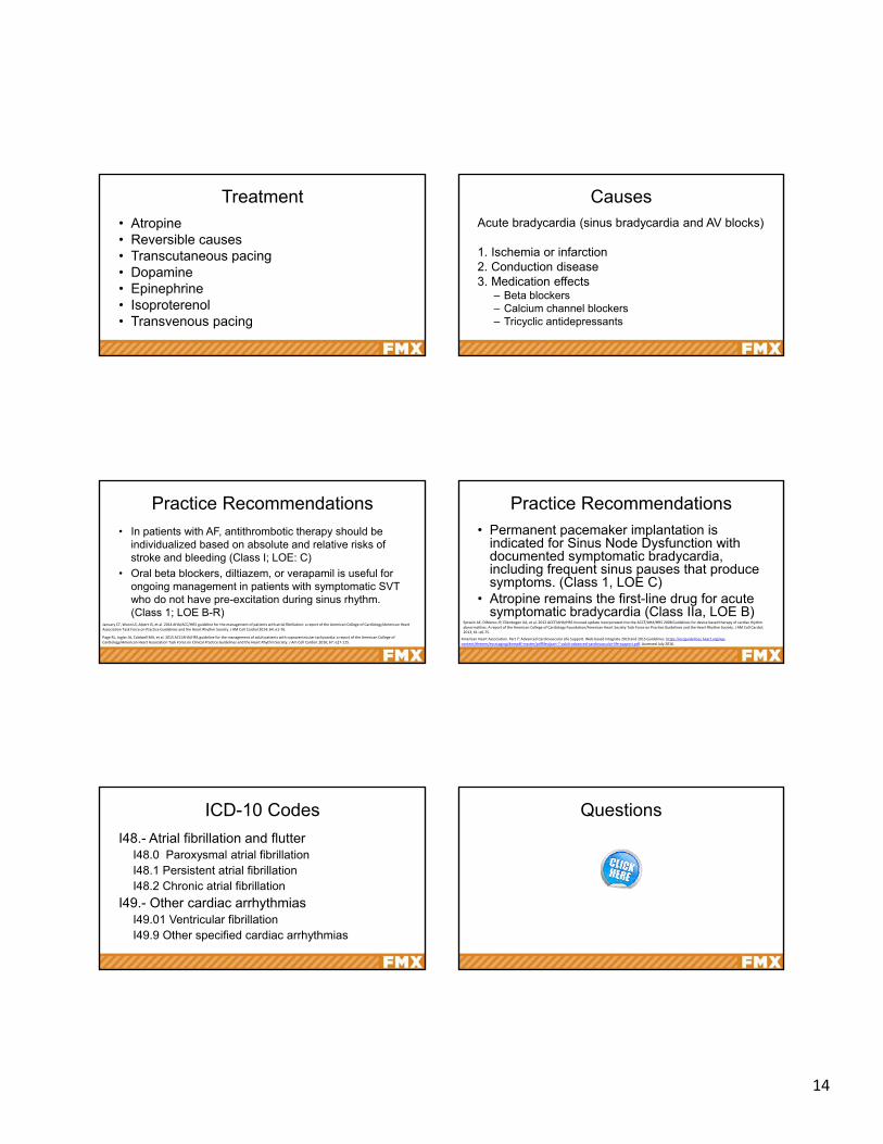
Treatment• Atropine• Reversible causes• Transcutaneous pacing• Dopamine• Epinephrine• Isoproterenol• Transvenous pacing
CausesAcute bradycardia (sinus bradycardia and AV blocks)
1. Ischemia or infarction2. Conduction disease3. Medication effects
– Beta blockers– Calcium channel blockers– Tricyclic antidepressants
Practice Recommendations
• In patients with AF, antithrombotic therapy should be individualized based on absolute and relative risks of stroke and bleeding (Class I; LOE: C)
• Oral beta blockers, diltiazem, or verapamil is useful for ongoing management in patients with symptomatic SVT who do not have pre-excitation during sinus rhythm. (Class 1; LOE B-R)
January CT, Wann LS, Alpert JS, et al. 2014 AHA/ACC/HRS guideline for the management of patients with atrial fibrillation: a report of the American College of Cardiology/American Heart Association Task Force on Practice Guidelines and the Heart Rhythm Society. J AM Coll Cardiol 2014; 64: e1‐76.
Page RL, Joglar JA, Caldwell MA, et al. 2015 ACC/AHA/HRS guideline for the management of adult patients with supraventricular tachycardia: a report of the American College of Cardiology/American Heart Association Task Force on Clinical Practice Guidelines and the Heart Rhythm Society. J Am Coll Cardiol. 2016; 67: e27‐115.
Practice Recommendations• Permanent pacemaker implantation is
indicated for Sinus Node Dysfunction with documented symptomatic bradycardia, including frequent sinus pauses that produce symptoms. (Class 1, LOE C)
• Atropine remains the first-line drug for acute symptomatic bradycardia (Class IIa, LOE B)
American Heart Association. Part 7: Advanced Cardiovascular Life Support. Web‐based integrate 2010 and 2015 Guidelines. https://eccguidelines.heart.org/wp‐content/themes/eccstaging/dompdf‐master/pdffiles/part‐7‐adult‐advanced‐cardiovascular‐life‐support.pdf. Accessed July 2016.
Eptsein AE, DiMarco JP, Ellenbogen KA, et al. 2012 ACCF/AHA/HRS focused update incorporated into the ACCF/AHA/HRS 2008 Guidelines for device based therapy of cardiac rhythm abnormalities. A report of the American College of Cardiology Foundation/American Heart Society Task Force on Practice Guidelines and the Heart Rhythm Society. J AM Coll Cardiol. 2013; 61: e6‐75.
ICD-10 Codes
I48.- Atrial fibrillation and flutterI48.0 Paroxysmal atrial fibrillationI48.1 Persistent atrial fibrillationI48.2 Chronic atrial fibrillation
I49.- Other cardiac arrhythmiasI49.01 Ventricular fibrillationI49.9 Other specified cardiac arrhythmias
Questions