Embed Size (px)
Citation preview
Negative regulation of IL6 by ARTD1 via MLL1
Page 1 of 27
ARTD1 suppresses interleukin 6 expression by repressing MLL1-1
dependent histone H3 trimethylation 2
Roberta Minotti1,†, Anneli Andersson1,2,† and Michael O. Hottiger1* 3
1Institute of Veterinary Biochemistry and Molecular Biology, University of Zurich, 4
Winterthurerstrasse 190, 8057 Zurich, Switzerland; 2Life Science Zurich Graduate School, 5
Molecular Life Science Program, University of Zurich. 6
7
Running title: Negative regulation of IL6 by ARTD1 via MLL1 8
9
†Both of these authors contributed equally to this study 10
*Address correspondence to Michael O. Hottiger, [email protected] 11
12
Key words: PARP1, gene regulation, ADP-ribosylation, inflammation, 13
14
Word count for Materials and Methods: 1,099 15
Combined word count for Introduction, Results, and Discussion: 3,457 16
17
MCB Accepted Manuscript Posted Online 6 July 2015Mol. Cell. Biol. doi:10.1128/MCB.00196-15Copyright © 2015, American Society for Microbiology. All Rights Reserved.
on April 11, 2018 by guest
http://mcb.asm
.org/D
ownloaded from

Negative regulation of IL6 by ARTD1 via MLL1
Page 2 of 27
Abstract 18
ADP-ribosyltransferase diphtheria-toxin like 1 (ARTD1/PARP1) is a chromatin-associated 19
protein in the nucleus and plays an important role in different cellular processes such as 20
regulation of gene transcription. ARTD1 has been shown to co-regulate the inflammatory 21
response by modulating the activity of the transcription factor nuclear factor kappa B (NF-22
κB), the principal regulator of interleukin 6 (IL6), an important inflammatory cytokine 23
implicated in a variety of diseases such as cancer. However, to which extent and how 24
ARTD1 regulates IL6 transcription has not been clear. 25
Here, we show that ARTD1 suppresses LPS-induced IL6 expression in macrophages, 26
without affecting the recruitment of the NF-κB subunit RelA to the IL6 promoter and 27
independent of its enzymatic activity. Interestingly, knockdown of ARTD1 did not alter H3 28
occupancy but increased LPS-induced trimethylation of histone 3 at lysine 4 (H3K4me3), a 29
hallmark of transcriptionally active genes. We found that ARTD1 mediates its effect through 30
the methyltransferase MLL1, by catalyzing H3K4me3 at the IL6 promoter and forming a 31
complex with NF-κB. These results demonstrate that ARTD1 modulates IL6 expression by 32
regulating the function of an NF-κB enhanceosome complex, which involves MLL1 and does 33
not require ADP-ribosylation. 34
35
36
on April 11, 2018 by guest
http://mcb.asm
.org/D
ownloaded from
Negative regulation of IL6 by ARTD1 via MLL1
Page 3 of 27
Introduction 37
Interleukin 6 (IL6) is an important inflammatory cytokine triggered e.g. by pathogen-38
associated molecular patterns (PAMPs) such as bacterial lipopolysaccharide (LPS) (1). 39
Increased IL6 production is linked to inflammatory diseases such as inflammatory bowel 40
disease, rheumatoid arthritis, mast cell growth proliferation, chronic inflammation and 41
obesity, and to different cancers such as breast, colon, epithelial or lung cancer (1-6). IL6 can 42
be released from tumors themselves, or from cancer-associated fibroblasts, and together with 43
other factors thereby creates a tumor-promoting microenvironment (7-9). Understanding the 44
mechanisms how IL6 transcription is regulated in different cell types is thus important for 45
diseases such as cancer and inflammatory diseases. While the expression of IL6 is principally 46
regulated by the transcription factor nuclear factor kappa B (NF-κB), epigenetic mechanisms 47
also play an important role in the regulation of IL6 gene expression (10-14). 48
NF-κB is a widely expressed, inducible transcription factor crucial for inflammation, 49
immunity, cell proliferation and apoptosis (15, 16). In mammalian cells, five members of the 50
NF-κB family exist, forming different homo- or hetero-dimers (17). The most abundant, best-51
studied and "classical" form of NF-κB is a heterodimer consisting of the two subunits p50 and 52
RelA (p65). In unstimulated cells, NF-κB is mostly sequestered in the cytoplasm as an 53
inactive transcription factor complex by its physical association with one of the several 54
inhibitors of NF-κB (IκB). PAMPs induce the classical, canonical pathway, which involves 55
the rapid activation of IKKβ, the NEMO-dependent phosphorylation and subsequent 56
degradation of IκBs, and the consequent nuclear translocation of primarily RelA-containing 57
NF-κB heterodimers. As a mechanism to control the inflammatory response, nuclear NF-κB 58
activity is selectively regulated at various levels downstream of activation, including DNA 59
methylation (which depends on the differentiation state of the cell in question), nucleosome 60
on April 11, 2018 by guest
http://mcb.asm
.org/D
ownloaded from

Negative regulation of IL6 by ARTD1 via MLL1
Page 4 of 27
positioning, histone modifications (e.g., histone methylation such as H3K4, H3K4 or H4K27 61
methylation) and via complex formation with co-regulators including p300/CBP and MLL1 62
(18-22). MLL1 is a member of the SET1/MLL family of methyltransferases and known for 63
its crucial functions for homeobox gene expression during development and embryogenesis, 64
for stem cell regulation, as well as for gene transcription in general (23, 24). Interestingly, 65
MLL1-dependent regulation of NF-κB downstream genes, including IL6, has recently been 66
reported (25). Due to these pivotal functions, mutations affecting the MLL1 gene cause 67
severe diseases and are implicated in acute leukemia in children and adults, with a particularly 68
poor prognosis (26). To prevent fatal malfunction or misregulation of MLL1, multiple 69
mechanisms control its activity in the cell (27). Together, there is a diversity of regulatory 70
mechanisms for the differential activation of NF-κB-dependent target genes within the same 71
cell or the differential activation of the same gene in different cells (20, 28). Furthermore, 72
NF-κB is subject to positive feedback regulation by cytokines such as IL6 (29, 30). 73
ADP-ribosyltransferase diphtheria-toxin like 1 (ARTD1/PARP1) is an abundant nuclear 74
protein that plays key roles in a variety of nuclear processes, including the regulation of 75
transcription (31). ARTD1 possesses an intrinsic enzymatic activity that catalyzes the 76
transfer of ADP-ribose (ADPr) units from nicotinamide adenine dinucleotide (NAD+) onto 77
target gene regulatory proteins, thereby modulating their activities, functions and interacting 78
partners (32, 33). 79
Since its discovery, most studies on ARTD1have focused on its role in DNA damage 80
detection and repair responses (34). However, over the past decade, the role of ARTD1 in 81
gene regulation has received increasing attention (31). Interestingly, ARTD1 can act as a 82
transcriptional enhancer or as an attenuator. ARTD1 can regulate transcription by binding to 83
nucleosomes, and interacts dynamically with different types of chromatin domains to 84
modulate the chromatin structure (34, 35). Nucleosome binding and auto-ADP-ribosylation 85
on April 11, 2018 by guest
http://mcb.asm
.org/D
ownloaded from

Negative regulation of IL6 by ARTD1 via MLL1
Page 5 of 27
of ARTD1 has been described as the underlying mechanism for this formation of 86
transcriptionally inactive, dense chromatin (36, 37), and have been implicated in the 87
reciprocal binding of ARTD1 and histone H1 to chromatin (38). ARTD1 has also been shown 88
to covalently modify histone and chromatin-associated non-histone proteins with poly-ADP-89
ribose (PAR) (39, 40). ARTD1 can modulate the activity of nucleosome remodelers through 90
non-covalent mechanisms, as is the case with ALC1 (amplified in liver cancer 1; also known 91
as CHD1L), a macrodomain-containing nucleosome-remodeling enzyme. PAR-dependent 92
interactions between ARTD1 and ALC1 promote nucleosome remodeling by ALC1, as well 93
as recruitment of ALC1 to sites of DNA damage in cells (41, 42). ADP-ribosylation of 94
KDM5B, a histone lysine demethylase acting on H3 lysine 4 trimethylation (H3K4me3), has 95
been shown to block the binding of KDM5B to chromatin and inhibit its demethylase activity 96
(43). This antagonism between ARTD1 and KDM5B helps to explain the high correlation 97
between ARTD1 and H3K4me3 at actively transcribed promoters. The functional interplay 98
between ARTD1 and KDM5B helps to control the chromatin state at ARTD1-regulated 99
promoters for both basal and signal-regulated transcriptional outcomes (43). Finally, ARTD1 100
may also function as a scaffold protein independent of its catalytic activities, by interacting 101
with and promoting the recruitment of other co-regulatory enzymes required for transcription. 102
We were the first to show that ARTD1 directly interacts with the NF-κB subunits and thereby 103
regulates NF-κB-dependent gene expression (44, 45). We found that ARTD1 synergistically 104
co-regulates transcription together with known NF-κB transcriptional cofactors such as p300, 105
CARM1, PRMT1 and the Mediator complex (18, 45, 46). 106
Although the enzymatic activity is not required for the transcriptional activation of 107
transiently transfected NF-κB reporter plasmids by ARTD1 or upon NLRP3 inflammasome-108
induced ARTD1 cleavage (47), we were able to link ARTD1 and ADP-ribosylation to 109
signaling during inflammation and the expression of adhesion molecules in atherogenesis as 110
on April 11, 2018 by guest
http://mcb.asm
.org/D
ownloaded from
Negative regulation of IL6 by ARTD1 via MLL1
Page 6 of 27
well as cell survival under stress conditions (48). ARTD1 and NF-κB are thus interconnected 111
in the inflammatory response. 112
At the inflammatory level, we have recently shown that non-apoptotic LPS-induced 113
caspase 7 activation via the NLRP3 inflammasome induces ARTD1 cleavage at the 114
transcriptional start site (TSS) of distinct NF-κB target genes including IL6 and thereby 115
causes elevated expression of these genes (49). The molecular mechanism responsible for the 116
repressed IL6 expression levels in the presence of ARTD1 was not elucidated, however. 117
In the present work, we have characterized and elucidated the molecular mechanism by 118
which ARTD1 regulates the transcription of IL6. Our results demonstrate that the negative 119
regulation of IL6 expression by ARTD1 is independent of its enzymatic activity and does not 120
affect RelA recruitment to the IL6 promoter. Instead, we found that ARTD1 is enriched at the 121
IL6 promoter and suppresses MLL1-dependent H3K4me3. These results uncover a new 122
mechanism of chromatin remodeling by ARTD1 and its importance for inflammation. 123
124 on April 11, 2018 by guest
http://mcb.asm
.org/D
ownloaded from
Negative regulation of IL6 by ARTD1 via MLL1
Page 7 of 27
Materials and Methods 125
Cell culture 126
Mouse leukemic monocyte macrophage cell line (Raw 264.7) was cultured in Roswell 127
Park Memorial Institute medium (RPMI) (Gibco, Invitrogen, CA, California) at 37°C. 128
NIH/3T3 and HEK 293 cells were cultivated in Dulbecco's Modified Eagle's Medium 129
(DMEM) (PAA, Pasching, Austria). Bone marrow-derived macrophages (BMDM) were 130
obtained from bones (femurs and tibia) of wildtype (WT) and ARTD1 knockout mice and 131
also cultivated in RPMI. The bones were cut from both ends, and bone marrow was flushed 132
out with complete medium using a 1 ml syringe with 23G needle until bones were completely 133
white. Cells were resuspended in RPMI supplemented with 20% of the supernatatnt of L929 134
cells and plated in bacterial dishes for 5 days. Differentiated macrophages were maintained in 135
culture for 2 weeks in RPMI supplemented with 5% L929 supernatant. All media were 136
supplemented with 1% (v/v) Penicillin/Streptavidin and 10% (v/v) fetal calf serum (Gibco, 137
Invitrogen, CA, California, USA). 138
Cells were pre-incubated with ARTD1 inhibitors Olaparib (1 μM) and ABT-888 (1 μM) 139
for 3 h before LPS (100 ng/ml, Sigma Aldrich, St. Louis, MO, USA) stimulation. Raw 264.7 140
cells stably downregulating ARTD1 were generated using viral transduction. Virus expressing 141
an shARTD1 construct were generated in HEK 293 cells. 10 µg shARTD1 plasmid, 6 µg of 142
packaging plasmid, and 3.5 µg viral envelope plasmid were transfected into HEK 293 cells 143
(4x10⁶ cells per 10 cm plate) using calcium phosphate. Medium was changed 6-8 h post 144
transfection and supernatant containing virus collected, centrifuged and filtered (0.45 µg 145
cellulose acetate filters) after 3 days. Raw 264.7 cells were seeded 1 day prior to transduction 146
on a 6-well- plate (5x10⁵ cells per well). Polybrene was added to a final concentration of 4 147
µg/ml over night. 1 ml of supernatant containing virus was added per well. The medium was 148
replaced 8 h post infection and to selective-medium after 2 day (Puromycin, 2 µg/ml) 149
on April 11, 2018 by guest
http://mcb.asm
.org/D
ownloaded from
Negative regulation of IL6 by ARTD1 via MLL1
Page 8 of 27
(Invivogen, San Diego, CA, USA). Cells were kept under constant selection. 150
151
siRNA transfection 152
Negative control allstars (siMOCK), human siPARP1 #6, mouse siPARP1 #7, mouse 153
siMLL1 #1, mouse siSet7 #1, mouse siRelA #2 were ordered from Qiagen (Hilden, 154
Germany). Cells were seeded at 50% confluency (2x10⁵ cells per well) and transfected with 155
20 nmol siRNA per well (in 6-well plate) with RNAi MAX lipofectamine (Invitrogen, 156
Carlsbad, CA, USA). Experiment was performed 3 days after transfection. 157
158
RNA extraction and qPCR analysis 159
RNA extraction was performed with the NucleoSpin® RNA II kit (Macherey-Nagel, 160
Düren, Germany). RNA was quantified with a NanoDrop (ThermoFisherScintific, Waltham, 161
MS, USA) and 2 µg RNA was reverse-transcribed according to the supplier’s protocol (High 162
Capacity cDNA Reverse Transcription Kit, Applied Biosystems, Foster City, CA, United 163
States). 164
Quantitative real-time polymerase chain reactions (qPCR) were performed with SYBR® 165
green SensiMix SYBR Hi-ROX Kit (Bioline Reagents Ltd, London, UK) and a Rotor-Gene Q 166
2plex HRM System (Qiagen, Hilden, Germany). See Supplemental Table 1 for primer 167
sequences. The relative amount of each mRNA was normalized to housekeeping genes 168
RSP12 (for mouse) and RPL28 (for human). 169
170
Cell lysis, SDS-PAGE and Western blot analysis 171
Whole cell extracts were prepared directly on plate by using a Tris lysis buffer (50 mM 172
Tris pH 8, 500 mM NaCl, 1% Triton X-100, 1 µg/ml pepstatin, 1 µg/ml bestatin, 1 µg/ml 173
leupeptin, 2 mM PMSF). Lysates were homogenized for 10 min at 4°C, followed by 10 min 174
centrifugation (maximal speed, at 4°C) to eliminate cell debris. Protein concentration was 175
on April 11, 2018 by guest
http://mcb.asm
.org/D
ownloaded from
Negative regulation of IL6 by ARTD1 via MLL1
Page 9 of 27
quantified by Bradford assay (Bio-Rad laboratories, Hercules, CA, USA) and 30 µg of protein 176
extract was separated on a 10% or 7.5% SDS-polyacrylamide gel (120 V). The gel was 177
blotted onto a PDVF membrane and analyzed using protein specific antibodies. 178
179
Co-Immunoprecipitation 180
Cells were harvested with a scraper and washed once with PBS (5 min, 900 × g). Pellet 181
was resuspended in 400 µl hypotonic buffer (0.5% NP-40, 85 mM KCl, 5 mM HEPES (pH 182
7.4)) and directly centrifuged for 10 min at 6’200 × g using a cold centrifuge. Pellet was 183
resuspended in 200 µl nuclear extraction buffer (50 mM Tris-HCl pH 7.5, 150 mM KCl, 5 184
mM MgCl2 , 0.2 mM EDTA, 20% Glycerol, 0.1% NP40) and sonicated twice for 30 s. 185
Nuclear extract was incubated for 30 min at 4°C with 1 µl DNase and sonicated again for 30 186
s, followed by 10 min centrifugation (4°C, 3’500 × g). Proteins were quantified by Bradford 187
assay (Bio-Rad laboratories, Hercules, CA, USA). Immunoprecipitation was carried out with 188
300 µg of extract at 4°C overnight with 10 µl monoclonal anti-HA-agarose beads (Sigma 189
Aldrich, St. Louis, MO, USA). After overnight incubation, beads were washed three times 190
with washing buffer (20 mM Tris-HCl pH 7.5, 0.1 M KCl, 5 mM MgCl2, 0.2 mM EDTA, 191
10% glycerol, 0.1 % Tween), resuspended in 2x Laemmli buffer (20 µl) and boiled for 5 min 192
at 95°C. SDS-PAGE and Western blot analysis were performed as described above. 193
194
Immunofluorescence microscopy 195
Cells were cultured on sterile cover slips (105 cells per well in a 24-well-plate) and grown 196
overnight. After treatment with or without H2O2 (1 mM in FCS-free medium, 10 min), cells 197
were fixed (methanol: acetic acid 3:1, 5 min on ice) and washed twice with PBS. The cells 198
were blocked for 30 min in PBS containing 5% milk powder and 0.05% Tween and incubated 199
with 10H PAR antibody (1:350) in the same buffer (1 h at room temperature). Cover slips 200
were incubated with secondary Cy3-Antibody (1 h at room temperature in the dark). After 201
on April 11, 2018 by guest
http://mcb.asm
.org/D
ownloaded from
Negative regulation of IL6 by ARTD1 via MLL1
Page 10 of 27
washing with PBS, cover slips were mounted with Vectashield containing DAPI (Vector 202
Laboratories, Burlingame, CA, USA). Conventional microscopy was carried out using a Leica 203
DMI 6000B light microscope (Leica microsystems GmbH, Wetzlar, Germany). 204
205
Chromatin Immunoprecipitation 206
ChIP analysis for H3, H3K4me3 and ARTD1 was performed as described previously 207
(Santoro et al, 2002) using magnetic Dynabeads® (Life Technologies, Carlsbad, CA, USA). 208
ChIP analysis for p65 was performed as described (50), using protein A agarose/salmon 209
sperm DNA beads (Millipore, Billerica MS, USA). 210
211
Luciferase Assay 212
Cells were transfected with specific siRNA as described above (2.104 cells per well in a 213
24-well-plate). One day after transfection, medium was changed and the different luciferase 214
constructs transfected using TransIT-3T3 Transfection Kit (Mirus, Madison, WI, USA). 215
Luciferase assay was performed after 48 h using Dual-Luciferase Reporter Assay System 216
(Promega, Madison, WI, USA) according to the supplier’s protocol. 217
218
Antibodies 219
Following antibodies were used: PARP1/ARDT1 (H-250, rabbit), PARP-1 (C2-10, mouse) 220
and p65 (C-20, rabbit) from Santa Cruz Biotechnology, Inc (Dallas, TX, USA); tubulin 221
(mouse) from Sigma Aldrich (St. Louis, MO, USA); H3K4me3 (rabbit) from Millipore 222
(Billerica MS, USA); Histone H3 (rabbit) from Abcam pls (Cambridge, UK). MLL1/HRX 223
from Millipore (Billerica MS, USA); secondary Cy3TM-conjugated AffiniPure Goat Anti-224
mouse from Jackson ImmunoResearch Laboratories (Suffolk, UK); PAR 10H (mouse) was 225
prepared in-house. 226
227
on April 11, 2018 by guest
http://mcb.asm
.org/D
ownloaded from

Negative regulation of IL6 by ARTD1 via MLL1
Page 11 of 27
Results 228
ARTD1 negatively regulates LPS-induced IL6 expression in a RelA-dependent manner 229
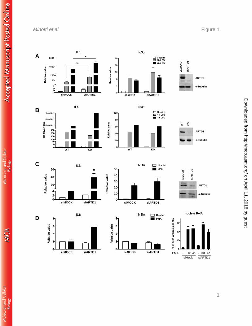
Stimulation of Raw macrophages with LPS for 1 or 4 h led to a strong induction of IL6 230
transcript levels as identified by quantitative RT-PCR (Fig. 1A). In shARTD1-treated Raw 231
cells (see Fig. 1A right panel for knockdown efficiency), IL6 expression levels were 232
comparably induced after 1 h, but significantly enhanced at 4 h after LPS stimulation, 233
suggesting that ARTD1 negatively regulates IL6 gene expression at this later time point. The 234
observed effect was specific for IL6, since the NF-κB-dependent control gene IkBα was not 235
affected by ARTD1 knockdown (Fig. 1A). Selectively enhanced IL6 expression (compared 236
to their respective controls) was also observed in LPS-stimulated primary bone marrow-237
derived macrophages (BMDMs) from ARTD1 knockout mice (Fig. 1B) and NIH/3T3 238
fibroblasts treated with siARTD1 (Fig. 1C) (see right panels for knockout/knockdown 239
efficiencies), as well as in siARTD1-treated HEK 293T cells stimulated with PMA (Fig. 1D, 240
see right panel for induction of RelA nuclear translocation), suggesting that the ARTD1-241
mediated negative regulation of IL6 expression is cell type- (and stimulus-) independent. 242
Because of their ease of transfectability, the mechanism of the negative regulatory effect of 243
ARTD1 on IL6 expression was further investigated using NIH/3T3 cells. To assess whether 244
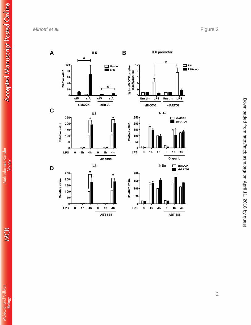
the negative regulation by ARTD1 was dependent on NF-κB, experiments with double-245
knockdown of ARTD1 and RelA were performed. Enhanced IL6 expression by ARTD1 246
knockdown in LPS-stimulated NIH/3T3 cells was completely abolished by concomitant 247
downregulation of RelA (Fig. 2A), indicating that the negative regulatory effect of ARTD1 on 248
IL6 expression was highly dependent on the induction of RelA in these cells. To corroborate 249
these results, reporter assays with a luciferase gene under the control of either the WT IL6 250
promoter or a mutated IL6 promoter lacking the NF-κB binding site were performed. In line 251
with the analysis of the transcript levels, knockdown of ARTD1 led to enhanced IL6 promoter 252
on April 11, 2018 by guest
http://mcb.asm
.org/D
ownloaded from

Negative regulation of IL6 by ARTD1 via MLL1
Page 12 of 27
transcriptional activity (Fig. 2B). This effect was not observed when the NF-κB binding site 253
was mutated, confirming that the negative regulatory effect of ARTD1 under these conditions 254
is also RelA-dependent and the chromatinization of the transfected reporter plasmids is 255
sufficient to detect the ARTD1-mediated IL6 repression. 256
257
ARTD1 negatively regulates IL6 expression independent of its enzymatic activity 258
ARTD1 can regulate gene expression by ADP-ribosylating target proteins (including itself) 259
or by its association with chromatin at the promoters of regulated genes (i.e. independently of 260
its enzymatic activity). To determine if the enzymatic activity of ARTD1 is required for the 261
observed negative regulatory effect on IL6 expression, Raw cells were stimulated with LPS in 262
the absence or presence of the ADP-ribosylation inhibitor olaparib. Again, ARTD1 263
knockdown lead to enhanced IL6 expression, while that of IkBα remained unaffected (Fig. 264
2C). Treatment of cells with olaparib, which effectively inhibited H2O2-induced PAR 265
formation at the dose used for this experiment (data not shown), neither affected IL6 266
expression in shMOCK cells nor enhanced expression in shARTD1 cells (Fig. 2C), indicating 267
that ADP-ribosylation is not involved in the negative regulatory effect of ADRT1 on IL6 268
expression. Comparable results were obtained with another ADP-ribosylation inhibitor, ABT-269
888 (Fig. 2D). 270
Together, these results demonstrate that the observed de-repression of IL6 expression upon 271
knockdown of ARTD1 is neither dependent on ARTD1-mediated ADP-ribosylation nor on 272
that by another ARTD family member. 273
274
ARTD1 does not alter the H3 occupancy at the IL6 promoter 275
To investigate whether ARTD1 represses IL6 expression indirectly by hampering RelA 276
recruitment to the IL6 promoter, LPS-induced recruitment of RelA to the IL6 and IkBα 277
on April 11, 2018 by guest
http://mcb.asm
.org/D
ownloaded from
Negative regulation of IL6 by ARTD1 via MLL1
Page 13 of 27
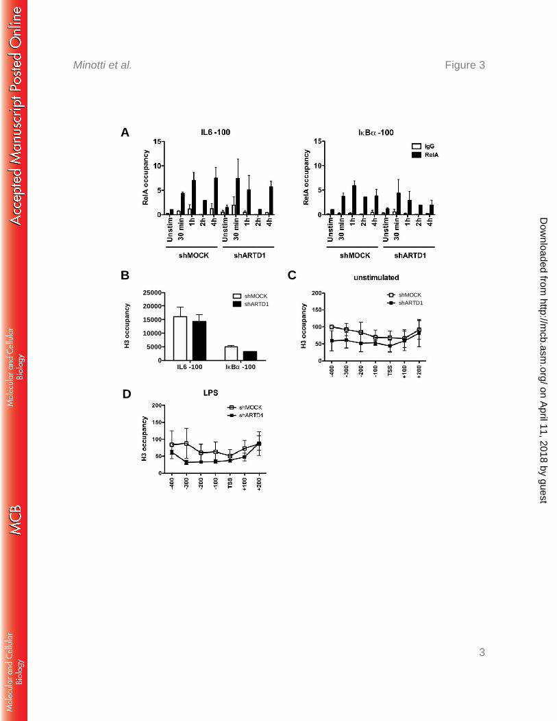
promoters (100 bp upstream of the TSS was compared in shMOCK and shARTD1-treated 278
Raw cells. The biphasic recruitment of RelA to the IL6 promoter (i.e. immediate within 1 h 279
and late at 4 h after stimulation) was not different between shMOCK and shARTD1-treated 280
cells in regard to timing and extent (Fig. 3A). The ARTD1-dependent negative regulation of 281
IL6 expression was therefore not due to an effect on RelA recruitment to the IL6 promoter. 282
Alternatively, as a chromatin-associated factor, ARTD1 may exert its regulatory function 283
through the modulation of the chromatin state at the TSS of the IL6 gene. To address the 284
function of ARTD1 as a chromatin regulator at the promoter of IL6, chromatin 285
immunoprecipitation (ChIP) experiments were performed to analyze the H3 occupancy at the 286
IL6 and IkBα promoters. These analyses revealed that the chromatin at the IL6 promoter was 287
strongly enriched (at least 3-fold) in histone H3 compared to that at the IkBα promoter in 288
shMOCK-treated cells (Fig. 3B), suggesting a more compact chromatin state at the IL6 289
promoter (51). The H3 occupancy in shARTD1-treated Raw cells under basal conditions (i.e. 290
untreated) was not significantly reduced compared to shMOCK-treated cells in the first 100 291
bp upstream of the TSS of the IL6 promoter, but also not further upstream and even 292
downstream in the gene body (Fig. 3C). Upon LPS stimulation, the measured H3 content was 293
also not significantly changed between shARTD1 and shMOCK cells (Fig. 3D), suggesting 294
that ARTD1 affects IL6 expression unlikely by altering the histone occupancy around the 295
TSS. 296
297
ARTD1 regulates IL6 expression through the H3K4me3 levels at the IL6 promoter 298
The results obtained so far indicate that ARTD1 negatively regulates IL6 expression 299
independent of H3 recruitment or its enzymatic activity. However, ARTD1 may exert its 300
regulatory function through the modulation of the histone modifications at the TSS of the IL6 301
gene. The higher occupancy of H3 at the IL6 promoter compared to the IkBα promoter (Fig. 302
on April 11, 2018 by guest
http://mcb.asm
.org/D
ownloaded from

Negative regulation of IL6 by ARTD1 via MLL1
Page 14 of 27
3B) suggests that a H3 modification may be required to induce a more permissive chromatin 303
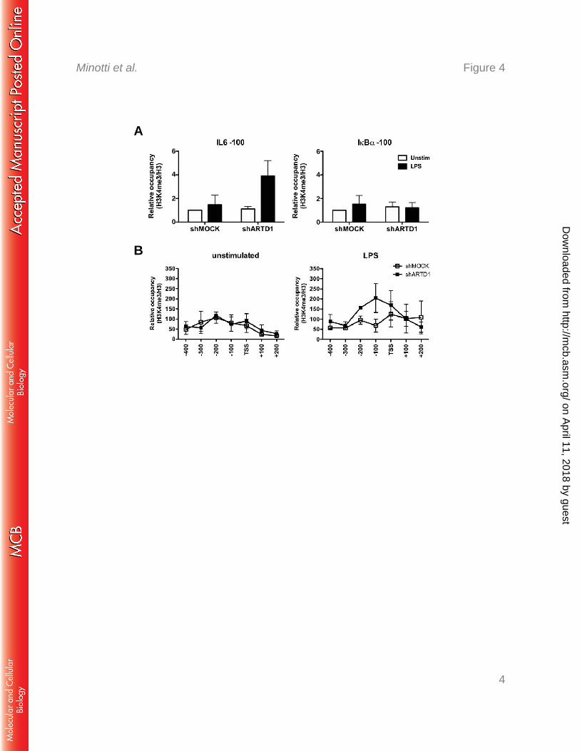
state. H3K4me3 is a mark that is associated with active gene expression (52). We therefore 304
elucidated whether ARTD1 inhibits H3K4me3 at the IL6 promoter, thereby keeping the 305
chromatin in a non-permissive state. To study this possibility, ChIP experiments for 306
H3K4me3 were performed and the IL6 and IkBα promoters analyzed in detail. Upon ARTD1 307
knockdown and LPS stimulation, the relative occupancy of H3K4me3 100 bp upstream of the 308
TSS of the IL6 promoter was strongly increased, while no significant changes were observed 309
at the IkBα promoter (Fig. 4A). More detailed analysis revealed that the strongest effects on 310
relative H3K4me3 levels upon ARTD1 down-regulation and LPS stimulation was observed 311
between the TSS and 200 bp upstream (Fig. 4B), which corresponds to the location of the NF-312
κB response element. The increased H3K4me3 was observed at the 4 h time point, supporting 313
the gene expression data and suggesting that ARTD1 represses H3K4me3 levels at the NF-κB 314
binding site of the IL6 promoter thereby repressing IL-6 gene expression. 315
316
MLL1 is responsible for the H3K4me3 at the IL6 promoter and interacts with NF-κB 317
To identify the responsible H3K4 methyltransferase at the IL6 TSS and to investigate a 318
potential antagonistic effect with ARTD1, Set7/9 and MLL1 were knocked down by siRNA 319
in NIH/3T3 cells in the presence or absence of ARTD1 (Fig. 5A, see right panels for knock-320
down efficiencies). Whereas the enhanced IL6 gene expression observed in siARTD1-treated 321
cells was unaffected by knockdown of the methyltransferase Set7/9, it was rescued by 322
knocking down MLL1, suggesting that MLL1 is the methyltransferase responsible for 323
enhanced H3K4me3 formation at the IL6 promoter upon knockdown of ARTD1 (Fig. 5A). 324
The expression of IkBα was neither significantly affected by siARTD1 nor by siMLL1 325
treatment. The H3K4me3 could be also altered by a reduced activity of the histone 326
demethylase KDM5B (43). However, knock-down of KDM5B did not lead to increased 327
on April 11, 2018 by guest
http://mcb.asm
.org/D
ownloaded from
Negative regulation of IL6 by ARTD1 via MLL1
Page 15 of 27
expression of IL6, neither in shMOCK nor in shARTD1 cells (Fig. 5B). Furthermore, 328
knockdown of MLL1, but not of Set7/9, led to a significantly reduced H3K4me3 at the IL6 329
promoter after LPS stimulation, whereas that at the control promoter IkBα was unaffected 330
(Fig. 5C). These analyses provide strong evidence that the methyltransferase MLL1 and not 331
Set7/9, or KDMB5 regulates H3K4me3 at the IL6 promoter in LPS-stimulated cells. The 332
same observation was made when MLL1 was knocked down together with ARTD1, 333
indicating that ARTD1 mediates its effect through MLL1 (Fig. 5D). 334
To test whether MLL1 forms a complex with NF-κB, complex formation was analyzed in 335
HEK 293T cells overexpressing hemagglutinin (HA)-tagged MLL1. Since HEK 293T cells 336
lack TLR4 and therefore cannot be stimulated by LPS, cells were stimulated with phorbol 337
myristate acetate (PMA). In HEK 293T cells overexpressing MLL1, complex formation of 338
MLL1 with endogenous RelA was detected in chromatin-free nuclear extracts 4 h after 339
stimulation (Fig. 5E). Interestingly, the interaction between RelA and MLL1 was observed in 340
a stimulus-dependent manner and at the same time point when the enhancement of IL6 341
expression by ARTD1 knockdown was the most prominent. Due to the lack of suitable 342
antibodies, the chromatin recruitment of MLL1 could not be analyzed, although others have 343
recently reported that MLL1 is recruited to the chromatin in an NF-κB-dependent manner to 344
regulate NF-κB-mediated gene expression (25). 345
346
ARTD1 forms as complex with MLL1, but leaves the IL6 promoter in a stimulus-347
dependent manner 348
To explain how ARTD1 interferes with MLL1-dependent IL6 expression and H3K4me3 at 349
the IL6 promoter, MLL1 expression levels were analyzed. ARTD1 knockdown did neither 350
significantly affect MLL1 expression nor MLL1 protein levels (Fig. 6A and B), suggesting 351
on April 11, 2018 by guest
http://mcb.asm
.org/D
ownloaded from

Negative regulation of IL6 by ARTD1 via MLL1
Page 16 of 27
that ARTD1 may rather be involved in the recruitment of MLL1 to the chromatin, or may 352
influence specific protein-protein interactions. 353
To test, whether ARTD1 forms a complex with MLL1, MLL1 was immunoprecipitated 354
from HEK 293T cells overexpressing MLL1 and stimulated for 4 h with PMA. An 355
interaction of MLL1 with endogenous ARTD1 was indeed detected in a stimulus-dependent 356
manner at the same time point as for RelA (i.e. 4 h after stimulation, Fig. 6C). The detected 357
signal was specific, because ARTD1 knock-down obliterated the signal. Since the negative 358
regulation of IL6 expression by ARTD1 could not be explained by an altered recruitment of 359
RelA (Fig. 3A) and the chromatin recruitment of MLL1 could not be analyzed, we 360
investigated whether the chromatin recruitment of ARTD1 is changed during LPS 361
stimulation. ChIP experiments with LPS-stimulated Raw cells revealed that ARTD1 was 362
time-dependently released from the promoters of both IL6 and IkBα upon LPS stimulation 363
(Fig. 6D), suggesting that LPS stimulation changes the occupancy of ARTD1 at different 364
chromatin loci. 365
In summary, we describe MLL1 as a new transcriptional co-activator of NF-κB and an 366
additional molecular mechanism by which ARTD1 co-regulates NF-κB-dependent 367
transcription, namely by the regulation of H3K4me3 through MLL1 and independent of 368
ARTD1's enzymatic activity. 369
370
on April 11, 2018 by guest
http://mcb.asm
.org/D
ownloaded from
Negative regulation of IL6 by ARTD1 via MLL1
Page 17 of 27
Discussion 371
ADP-ribosylation and in particular ARTD1 have been implicated in many different and 372
distinct cellular and biological processes (31). One of the most important functions of ARTD1 373
is the co-regulation of inflammatory gene expression by direct modulation of transcriptional 374
regulators or indirectly through alterations of the chromatin state (31). 375
Here we elucidated the mechanism by which ARTD1 conveys a non-permissive chromatin 376
state at the IL6 promoter, by interfering with MLL1-dependent H3K4me3 upon LPS 377
stimulation. Interestingly, expression of the gene encoding the NF-κB inhibitor IκBα and 378
other house keeping genes was not ARTD1-dependent, which may be due to different 379
chromatin architecture at the promoters of these two genes. The co-regulatory function of 380
ARTD1 was not mediated by its enzymatic activity. 381
The results presented here define the negative regulation of H3K4me3 at the IL6 promoter 382
by MLL1 as the mechanism by which ARTD1 modulates IL6 expression. Previously, 383
ARTD1 activity was shown to positively regulate transcription via the modification and 384
inhibition of the histone demethylase KDM5B, responsible for the demethylation of 385
H3K4me3 (43). In the study described here, knockdown of MLL1, but not of SET7 or 386
KDM5B, altered the H3K4me3 of the IL6 promoter. It is tempting to speculate that ARTD1 387
can dependent on the cellular context, its stimulation and the availability of its substrate 388
NAD+, either repress H3K4me3, through ADP-ribosylation-independent binding to MLL1, or 389
enhance H3K4me3 and therefore gene expression through ADP-ribosylation of KDM5B. 390
We observed that ARTD1 chromatin association is reduced upon LPS treatment after 4 h. 391
Based on our recent studies (49), it is likely that the activation of the inflammasome at this 392
time point is responsible for the proteolytic cleavage of ARTD1 at D214 and its subsequent 393
release from chromatin. Since LPS itself is not expected to activate the inflammasome, but to 394
induce the expression of its components, it remains to be elucidated by which mechanism the 395
on April 11, 2018 by guest
http://mcb.asm
.org/D
ownloaded from

Negative regulation of IL6 by ARTD1 via MLL1
Page 18 of 27
inflammasome is activated. 396
According to our study, ARTD1 is bound to the IL6 promoter even before stimulation of 397
the cells, likely regulating the compaction and basal expression levels of IL6. Similar to this 398
result, biochemical studies with reconstituted chromatin have shown that in the absence of 399
NAD+, ARTD1 promotes chromatin compaction independently of its enzymatic activity (53). 400
Our findings suggest a model in which ARTD1 represses IL6 expression by interfering 401
with the MLL1-induced H3K4me3 at the IL6 promoter. Upon stimulation of cells, ARTD1 402
leaves the IL6 promoter in a time-dependent manner. Interestingly ARTD1 forms a complex 403
with MLL1 only at the later time point (4 h, second NF-κB wave), when also complex 404
formation of MLL1 with NF-κB is observed, suggesting that upon eviction of ARTD1, 405
ARTD1 might compete with NF-κB for binding to MLL1, thus allowing only a certain 406
amount of MLL1 to bind to NF-κB and thus damping the MLL1-induced H3K4me3 levels at 407
this later time point. In contrast, in cells lacking ARTD1 or expressing reduced levels of 408
ARTD1, all MLL1 binds to NF-κB and remains associated with the chromatin, resulting in 409
increased H3K4me3 at the IL6 promoter and consequently, increased IL6 gene expression. 410
Since we were not able to investigate and quantify the recruitment of MLL1 to the IL6 411
promoter, and the co-immunoprecipitation of MLL1 with NF-κB and/or ARTD1 does not 412
allow us to distinguish whether all three proteins are found in the same complex or in distinct 413
complexes containing MLL1 only associated with NF-κB or ARTD1, additional 414
investigations are required to fully dissect the mechanism by which ARTD1 regulates MLL-415
1-driven NF-κB-dependent gene expression of IL6, including also in developmental 416
processes, homeobox gene expression or the development of acute leukemia. In this context, 417
it will be of particular interest to investigate the regulation of IL6 expression by ARTD1 in 418
cells harboring MLL1 mutations. 419
on April 11, 2018 by guest
http://mcb.asm
.org/D
ownloaded from
Negative regulation of IL6 by ARTD1 via MLL1
Page 19 of 27
In summary, our experiments have identified the dampening effect of ARTD1 on MLL1-420
dependent H3K4me3 as a new mechanism regulating the expression of the inflammatory 421
cytokine IL6. Taken together, these data strongly indicate that ARTD1 orchestrates 422
chromatin accessibility and the post-translational modification of histones indirectly through 423
the interaction with the H3K4 methyltransferase MLL1 and thereby modulates the expression 424
of IL6. This apparently occurs in an ADP-ribosylation-independent manner. These results 425
have not only important implications for our understanding and future analysis of MLL1 426
functions and IL6 regulation, but also highlight a potential crosstalk between IL6- and MLL1-427
induced pathologies. 428
on April 11, 2018 by guest
http://mcb.asm
.org/D
ownloaded from
Negative regulation of IL6 by ARTD1 via MLL1
Page 20 of 27
Conflict of Interest 429
The authors declare that they have no conflict of interest. 430
431
Acknowledgements 432
We would like to thank Dr. Robert Roeder, The Rockfeller University, New York for 433
providing the Flag-HA-tagged MLL1 pcDNA5 clone. Florian Freimoser and Stephan 434
Christen (Institute of Veterinary Biochemistry and Molecular Biology, University of Zurich, 435
Switzerland) provided editorial assistance and critical input during the writing. This work was 436
supported in part by the Swiss National Science Foundation (grant 310030B_138667 and 437
310030_157019), the Kanton of Zurich, and the Novartis Foundation (to M.O.H). 438
439
440
on April 11, 2018 by guest
http://mcb.asm
.org/D
ownloaded from
Negative regulation of IL6 by ARTD1 via MLL1
Page 21 of 27
References 441
1. Nishimoto N, Kishimoto T. 2006. Interleukin 6: from bench to bedside. Nat. Clin. 442 Pract. Rheumatol. 2:619-626. 443
2. Ganapathi MK, Weizer AK, Borsellino S, Bukowski RM, Ganapathi R, Rice T, 444 Casey G, Kawamura K. 1996. Resistance to interleukin 6 in human non-small cell 445 lung carcinoma cell lines: role of receptor components. Cell Growth Differ. 7:923-446 929. 447
3. Yamaji H, Iizasa T, Koh E, Suzuki M, Otsuji M, Chang H, Motohashi S, Yokoi S, 448 Hiroshima K, Tagawa M, Nakayama T, Fujisawa T. 2004. Correlation between 449 interleukin 6 production and tumor proliferation in non-small cell lung cancer. Cancer 450 Immunol. Immunother. 53:786-792. 451
4. Knupfer H, Preiss R. 2007. Significance of interleukin-6 (IL-6) in breast cancer. 452 Breast Cancer Res. Treat. 102:129-135. 453
5. Bromberg J, Wang T. 2009. Inflammation and cancer: IL-6 and STAT3 complete the 454 link. Cancer Cell 15:79-80. 455
6. Iliopoulos D, Hirsch HA, Wang G, Struhl K. 2011. Inducible formation of breast 456 cancer stem cells and their dynamic equilibrium with non-stem cancer cells via IL6 457 secretion. Proc Natl Acad Sci USA 108:1397-1402. 458
7. Hugo HJ, Lebret S, Tomaskovic-Crook E, Ahmed N, Blick T, Newgreen DF, 459 Thompson EW, Ackland ML. 2012. Contribution of Fibroblast and Mast Cell 460 (Afferent) and Tumor (Efferent) IL-6 Effects within the Tumor Microenvironment. 461 Cancer Microenviron. 5:83-93. 462
8. Raz Y, Erez N. 2013. An inflammatory vicious cycle: Fibroblasts and immune cell 463 recruitment in cancer. Exp. Cell Res. 319:1596-1603. 464
9. Servais C, Erez N. 2013. From sentinel cells to inflammatory culprits: cancer-465 associated fibroblasts in tumour-related inflammation. J. Pathol. 229:198-207. 466
10. Armenante F, Merola M, Furia A, Palmieri M. 1999. Repression of the IL-6 gene 467 is associated with hypermethylation. Biochem. Biophys. Res. Commun. 258:644-647. 468
11. Armenante F, Merola M, Furia A, Tovey M, Palmieri M. 1999. Interleukin-6 469 repression is associated with a distinctive chromatin structure of the gene. Nucleic 470 Acids Res. 27:4483-4490. 471
12. Dandrea M, Donadelli M, Costanzo C, Scarpa A, Palmieri M. 2009. 472 MeCP2/H3meK9 are involved in IL-6 gene silencing in pancreatic adenocarcinoma 473 cell lines. Nucleic Acids Res. 37:6681-6690. 474
13. Takeuch O, Akira S. 2011. Epigenetic control of macrophage polarization. European 475 J Immunol 41:2490-2493. 476
14. Tang B, Zhao R, Sun Y, Zhu Y, Zhong J, Zhao G, Zhu N. 2011. Interleukin-6 477 expression was regulated by epigenetic mechanisms in response to influenza virus 478 infection or dsRNA treatment. Mol. Immunol. 48:1001-1008. 479
15. Li Q, Verma IM. 2002. NF-kappaB regulation in the immune system. Nat. Rev. 480 Immunol. 2:725-734. 481
16. Karin M, Greten FR. 2005. NF-kappaB: linking inflammation and immunity to 482 cancer development and progression. Nat. Rev. Immunol. 5:749-759. 483
17. Tak PP, Firestein GS. 2001. NF-kappaB: a key role in inflammatory diseases. J. 484 Clin. Invest. 107:7-11. 485
18. Hassa PO, Haenni SS, Buerki C, Meier NI, Lane WS, Owen H, Gersbach M, 486 Imhof R, Hottiger MO. 2005. Acetylation of poly(ADP-ribose) polymerase-1 by 487
on April 11, 2018 by guest
http://mcb.asm
.org/D
ownloaded from

Negative regulation of IL6 by ARTD1 via MLL1
Page 22 of 27
p300/CREB-binding protein regulates coactivation of NF-κB-dependent transcription. 488 J. Biol. Chem. 280:40450-40464. 489
19. Mao X, Gluck N, Li D, Maine GN, Li H, Zaidi IW, Repaka A, Mayo MW, 490 Burstein E. 2009. GCN5 is a required cofactor for a ubiquitin ligase that targets NF-491 kappaB/RelA. Genes Dev. 23:849-861. 492
20. Smale ST. 2010. Selective transcription in response to an inflammatory stimulus. Cell 493 140:833-844. 494
21. Li H, Wittwer T, Weber A, Schneider H, Moreno R, Maine GN, Kracht M, 495 Schmitz ML, Burstein E. 2012. Regulation of NF-kappaB activity by competition 496 between RelA acetylation and ubiquitination. Oncogene 31:611-623. 497
22. Sen N, Paul BD, Gadalla MM, Mustafa AK, Sen T, Xu R, Kim S, Snyder SH. 498 2012. Hydrogen sulfide-linked sulfhydration of NF-kappaB mediates its antiapoptotic 499 actions. Mol. Cell 45:13-24. 500
23. Ansari KI, Mandal SS. 2010. Mixed lineage leukemia: roles in gene expression, 501 hormone signaling and mRNA processing. FEBS J. 277:1790-1804. 502
24. Hubert A, Henderson JM, Ross KG, Cowles MW, Torres J, Zayas RM. 2013. 503 Epigenetic regulation of planarian stem cells by the SET1/MLL family of histone 504 methyltransferases. Epigenetics 8:79-91. 505
25. Wang X, Zhu K, Li S, Liao Y, Du R, Zhang X, Shu HB, Guo AY, Li L, Wu M. 506 2012. MLL1, a H3K4 methyltransferase, regulates the TNFalpha-stimulated activation 507 of genes downstream of NF-kappaB. J. Cell Sci. 125:4058-4066. 508
26. Marschalek R. 2010. Mixed lineage leukemia: roles in human malignancies and 509 potential therapy. FEBS J. 277:1822-1831. 510
27. Zhang P, Bergamin E, Couture JF. 2013. The many facets of MLL1 regulation. 511 Biopolymers 99:136-145. 512
28. Ramirez-Carrozzi V, Braas D, Bhatt D, Cheng C, Hong C, Doty K, Black J, 513 Hoffmann A, Carey M, Smale S. 2009. A unifying model for the selective regulation 514 of inducible transcription by CpG islands and nucleosome remodeling. Cell 138:114-515 128. 516
29. Iliopoulos D, Hirsch H, Struhl K. 2009. An epigenetic switch involving NF-kappaB, 517 Lin28, Let-7 MicroRNA, and IL6 links inflammation to cell transformation. Cell 518 139:693-706. 519
30. Korkaya H, Liu S, Wicha MS. 2011. Regulation of cancer stem cells by cytokine 520 networks: attacking cancer's inflammatory roots. Clin. Cancer Res. 17:6125-6129. 521
31. Kraus WL, Hottiger MO. 2013. PARP-1 and gene regulation: progress and puzzles. 522 Mol. Aspects Med. 34:1109-1123. 523
32. Rosenthal F, Hottiger MO. 2014. Identification of ADP-ribosylated peptides and 524 ADP-ribose acceptor sites. Front Biosci (Landmark Ed) 19:1041-1056. 525
33. Elser M, Borsig L, Hassa PO, Erener S, Messner S, Valovka T, Keller S, 526 Gassmann M, Hottiger MO. 2008. Poly(ADP-ribose) polymerase 1 promotes tumor 527 cell survival by coactivating hypoxia-inducible factor-1-dependent gene expression. 528 Mol. Cancer Res. 6:282-290. 529
34. D'Amours D, Desnoyers S, D'Silva I, Poirier G. 1999. Poly(ADP-ribosyl)ation 530 reactions in the regulation of nuclear functions. Biochem. J. 342:249-268. 531
35. Kraus W, Lis J. 2003. PARP goes transcription. Cell 113:677-683. 532 36. de Murcia G, Huletsky A, Lamarre D, Gaudreau A, Pouyet J, Daune M, Poirier 533
G. 1986. Modulation of chromatin superstructure induced by poly(ADP-ribose) 534 synthesis and degradation. J. Biol. Chem. 261:7011-7017. 535
on April 11, 2018 by guest
http://mcb.asm
.org/D
ownloaded from
Negative regulation of IL6 by ARTD1 via MLL1
Page 23 of 27
37. Kim M, Mauro S, Gévry N, Lis J, Kraus W. 2004. NAD+-dependent modulation of 536 chromatin structure and transcription by nucleosome binding properties of PARP-1. 537 Cell 119:803-814. 538
38. Krishnakumar R, Gamble M, Frizzell K, Berrocal J, Kininis M, Kraus W. 2008. 539 Reciprocal binding of PARP-1 and histone H1 at promoters specifies transcriptional 540 outcomes. Science 319:819-821. 541
39. Altmeyer M, Messner S, Hassa PO, Fey M, Hottiger MO. 2009. Molecular 542 mechanism of poly(ADP-ribosyl)ation by PARP1 and identification of lysine residues 543 as ADP-ribose acceptor sites. Nucleic Acids Res. 37:3723-3738. 544
40. Messner S, Altmeyer M, Zhao H, Pozivil A, Roschitzki B, Gehrig P, Rutishauser 545 D, Huang D, Caflisch A, Hottiger MO. 2010. PARP1 ADP-ribosylates lysine 546 residues of the core histone tails. Nucleic Acids Res. 38:6350-6362. 547
41. Ahel D, Horejsi Z, Wiechens N, Polo SE, Garcia-Wilson E, Ahel I, Flynn H, 548 Skehel M, West SC, Jackson SP, Owen-Hughes T, Boulton SJ. 2009. Poly(ADP-549 ribose)-dependent regulation of DNA repair by the chromatin remodeling enzyme 550 ALC1. Science 325:1240-1243. 551
42. Gottschalk A, Timinszky G, Kong S, Jin J, Cai Y, Swanson S, Washburn M, 552 Florens L, Ladurner A, Conaway J, Conaway R. 2009. Poly(ADP-ribosyl)ation 553 directs recruitment and activation of an ATP-dependent chromatin remodeler. Proc 554 Natl Acad Sci USA 106:13770-13774. 555
43. Krishnakumar R, Kraus W. 2010. PARP-1 regulates chromatin structure and 556 transcription through a KDM5B-dependent pathway. Mol. Cell 39:736-749. 557
44. Hassa PO, Hottiger MO. 1999. A role of poly (ADP-ribose) polymerase in NF-κB 558 transcriptional activation. Biol. Chem. 380:953-959. 559
45. Hassa PO, Buerki C, Lombardi C, Imhof R, Hottiger MO. 2003. Transcriptional 560 coactivation of nuclear factor-κB-dependent gene expression by p300 is regulated by 561 poly(ADP)-ribose polymerase-1. J. Biol. Chem. 278:45145-45153. 562
46. Hassa PO, Covic M, Bedford MT, Hottiger MO. 2008. Protein arginine 563 methyltransferase 1 coactivates NF-κB-dependent gene expression synergistically 564 with CARM1 and PARP1. J. Mol. Biol. 377:668-678. 565
47. Hassa PO, Covic M, Hasan S, Imhof R, Hottiger MO. 2001. The enzymatic and 566 DNA binding activity of PARP-1 are not required for NF-κB coactivator function. J. 567 Biol. Chem. 276:45588-45597. 568
48. von Lukowicz T, Hassa PO, Lohmann C, Borén J, Braunersreuther V, Mach F, 569 Odermatt B, Gersbach M, Camici GG, Stähli BE, Tanner FC, Hottiger MO, 570 Lüscher TF, Matter CM. 2008. PARP1 is required for adhesion molecule expression 571 in atherogenesis. Cardiovasc. Res. 78:158-166. 572
49. Erener S, Petrilli V, Kassner I, Minotti R, Castillo R, Santoro R, Hassa PO, 573 Tschopp J, Hottiger MO. 2012. Inflammasome-activated caspase 7 cleaves PARP1 574 to enhance the expression of a subset of NF-kappaB target genes. Mol. Cell 46:200-575 211. 576
50. Saccani S, Pantano S, Natoli G. 2001. Two waves of nuclear factor kappaB 577 recruitment to target promoters. J. Exp. Med. 193:1351-1359. 578
51. Lachner M, O'Carroll D, Rea S, Mechtler K, Jenuwein T. 2001. Methylation of 579 histone H3 lysine 9 creates a binding site for HP1 proteins. Nature 410:116-120. 580
52. Santos-Rosa H, Schneider R, Bannister AJ, Sherriff J, Bernstein BE, Emre NC, 581 Schreiber SL, Mellor J, Kouzarides T. 2002. Active genes are tri-methylated at K4 582 of histone H3. Nature 419:407-411. 583
on April 11, 2018 by guest
http://mcb.asm
.org/D
ownloaded from
Negative regulation of IL6 by ARTD1 via MLL1
Page 24 of 27
53. Wacker D, Ruhl D, Balagamwala E, Hope K, Zhang T, Kraus W. 2007. The DNA 584 binding and catalytic domains of poly(ADP-ribose) polymerase 1 cooperate in the 585 regulation of chromatin structure and transcription. Mol. Cell. Biol. 27:7475-7485. 586
587
588
on April 11, 2018 by guest
http://mcb.asm
.org/D
ownloaded from

Negative regulation of IL6 by ARTD1 via MLL1
Page 25 of 27
Figure Legends 589
590
Figure 1: ARTD1 negatively regulates LPS- or PMA-induced IL6 expression. IL6 and 591
IκBα gene expression was quantified by RT-PCR in Raw cells (A), BMDMs (B), NIH/3T3 592
cells (C), and HEK cells (D). A) IL6 (left panel) and IκBα (middle panel) expression upon 593
LPS stimulation in Raw cells treated with shRNA specific against ARTD1 (n=5). Right panel 594
shows knock-down efficiency by Western blot analysis. B) IL6 (left panel) and IκBα (middle 595
panel) expression upon LPS stimulation in BMDM from wt and ARTD1 knock-out mice (1 596
representative experiment out of 3). Right panel demonstrates knock-out by Western blot 597
analysis. C) IL6 (left panel) and IκBα (middle panel) expression upon LPS stimulation in 598
NIH/3T3 cells treated with siRNA specific against ARTD1 (n=3-4). Right panel shows 599
knock-down efficiency by Western blot analysis. D) IL6 (left panel) and IκBα (middle panel) 600
expression upon PMA stimulation in HEK cells treated with siRNA specific against ARTD1 601
(n=2). Right panel demonstrates induction of p65 by PMA (quantitative representation of 602
immunofluoresence analysis). Data are presented as mean ± SD and were analyzed by 1-way 603
ANOVA followed by Bonferroni’s post-hoc test. *P < 0.05 604
605
Figure 2: ARTD1 negatively regulates IL6 expression in a p65-dependent manner and 606
independent of its enzymatic activity. A) IL6 expression upon ARTD1 and RelA 607
knockdown in NIH/3T3 cells stimulated with LPS or not (n=3). B) Expression of the 608
luciferase reporter under the control of IL6 promoter (4 h LPS or unstimulated) in NIH/3T3 609
cells (n=3). C) IL6 and IκBα expression upon Olaparib treatment (1 μM) in Raw cells (n=2). 610
D) IL6 and IκBα expression upon ABT-888 treatment (1 μM) in Raw cells (n=2). Data are 611
presented as mean ± SD and were analyzed by 1-way ANOVA followed by Bonferroni’s 612
post-hoc test. *P < 0.05 613
on April 11, 2018 by guest
http://mcb.asm
.org/D
ownloaded from
Negative regulation of IL6 by ARTD1 via MLL1
Page 26 of 27
614
Figure 3: ARTD1 helps to maintain H3 at the IL6 promoter. A) Effect of ARTD1 615
knockdown on p65 occupancy on IL6 and IκBα promoters upon LPS stimulation in Raw cells 616
(n=3). H3 occupancy at the IL6 and IκBα promoters in Raw cells under unstimulated 617
conditions (shMOCK and shARTD1) (B, C) or after 4 h of LPS treatment (D) (n=3). Data are 618
presented as mean ± SD. 619
620
Figure 4: ADRT1 knock-down is associated with increased H3K4me3 occupancy at the 621
IL6 promoter in LPS-stimulated cells. A and B) H3K4me3 occupancy at the IL6 and IκBα 622
promoters in Raw cells upon 4 h LPS stimulation (shMOCK and shARTD1) (n=3). 623
624
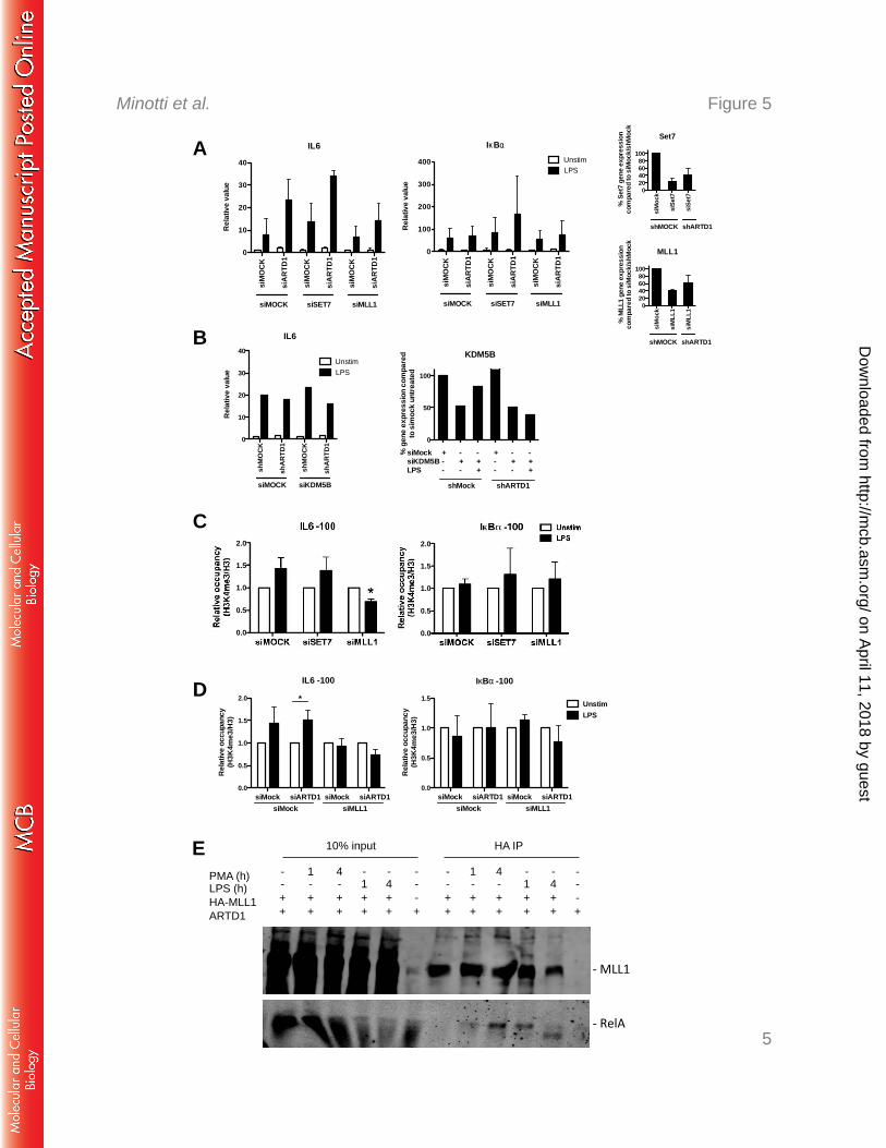
Figure 5: MLL1 regulates IL6 expression and H3K4me3 levels at the IL6 promoter 625
A) IL6 (left panel) and IκBα (middle panel) expression upon combined knock down of 626
ARTD1 and either SET7 or MLL1 in NIH/3T3 cells, 4 h LPS (n=3). Right panels show 627
knock-down efficiency of siRNA treatment against SET7 (top) and MLL1 (bottom) by qPCR 628
analysis. B) IL6 (left panel) expression upon combined knock down of ARTD1 and KDM5B 629
in NIH/3T3 cells, 4 h LPS (n=1). Right panel shows knock-down efficiency of siRNA 630
treatment against KDM5B by qPCR analysis. C) H3K4me3 occupancy at the IL6 and IkBα 631
promoters upon knock down of ARTD1 and either SET7 or MLL1 in NIH/3T3 cells, 4 h LPS 632
(n=3). Data are presented as mean ± SD and were analyzed by 1-way ANOVA followed by 633
Bonferroni’s post-hoc test. *P < 0.05. D) H3K4me3 occupancy at the IL6 and IκBα promoters 634
upon combined knock down of ARTD1 and either SET7 or MLL1 in NIH/3T3 cells, 4 h LPS 635
(n=3). E) MLL1 and p65 co-IP in HEK cells overexpressing MLL1 (lane 1 and 4: unstim, 636
lane 2: 1 h PMA, lane 3: 4 h PMA). 637
638
on April 11, 2018 by guest
http://mcb.asm
.org/D
ownloaded from
Negative regulation of IL6 by ARTD1 via MLL1
Page 27 of 27
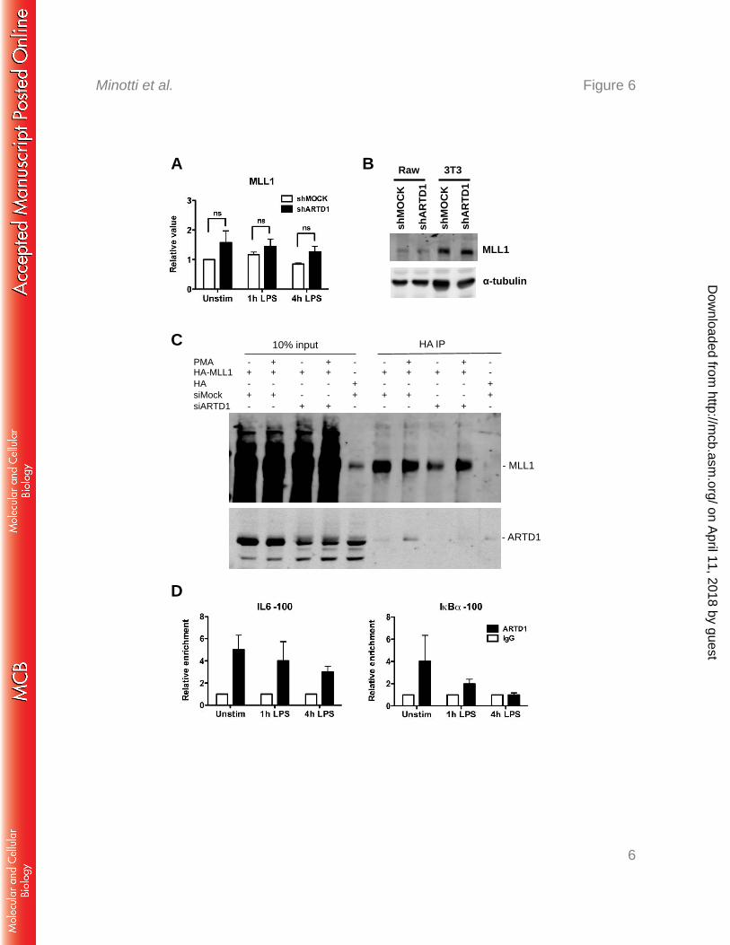
Figure 6: MLL1 interacts with ARTD1 and p65 after LPS stimulation. MLL1 gene (A) 639
and protein (B) expression in shMOCK and shARTD1-treated Raw cells (n=5 each). C) 640
MLL1 and ARTD1 co-IP in HEK cells overexpressing MLL1 and pretreated with siMOCK or 641
siARTD1 (4 h PMA). D) ChIP analysis of ARTD1 occupancy on IL6 and IκBα promoters in 642
Raw cells (n=4). 643
on April 11, 2018 by guest
http://mcb.asm
.org/D
ownloaded from
1
Figure 1 Minotti et al.
IL6
shMOCK shARTD10246
100
200
4000
8000 *ns
Rel
ativ
e va
lue
α-Tubulin
ARTD1 si
MO
CK
siA
RTD
1
shM
OC
K
shA
RTD
1
α-Tubulin
ARTD1
WT
KO
α-Tubulin
ARTD1
A
B
D
C
0
10
20
30
% o
f cel
ls w
ith n
ucle
ar p
65
nuclear RelA
siMockPMA - 30' 4h - 30' 4h
siARTD1
IL6
siMOCK siARTD1024
30
40
50
Rela
tive
valu
e
*IκBα
siMOCK siARTD10
10
20
30
40
50UnstimLPS
Rela
tive
valu
e on April 11, 2018 by guest
http://mcb.asm
.org/D
ownloaded from
2
Figure 2 Minotti et al.
A B
C
D
on April 11, 2018 by guest
http://mcb.asm
.org/D
ownloaded from
3
Figure 3 Minotti et al.
B C
D
A
IL6 -100 IκBα -1000
5000
10000
15000
20000
25000 shMOCK shARTD1
H3 o
ccup
ancy
shMOCKshARTD1
on April 11, 2018 by guest
http://mcb.asm
.org/D
ownloaded from
4
Figure 4 Minotti et al.
6
4
2
0
B
A 6
4
2
0
on April 11, 2018 by guest
http://mcb.asm
.org/D
ownloaded from
5
Figure 5 Minotti et al.
2.0
1.5
1.0
0.5
0.0
siM
ock
siM
LL1
siM
LL10
20406080
100
% M
LL1
gene
exp
ress
ion
com
pare
d to
siM
ock/
shM
ock
MLL1
shMOCK shARTD1
A IL6
siM
OC
K
siA
RTD
1
siM
OC
K
siA
RTD
1
siM
OC
K
siA
RTD
1
0
10
20
30
40
Rel
ativ
e va
lue
siSET7 siMLL1siMOCK
IκBα
siM
OC
K
siA
RTD
1
siM
OC
K
siA
RTD
1
siM
OC
K
siA
RTD
1
0
100
200
300
400 UnstimLPS
siSET7 siMLL1siMOCK
Rel
ativ
e va
lue
B
D
siM
ock
siSe
t7
siSe
t7
020406080
100
% S
et7
gene
exp
ress
ion
com
pare
d to
siM
ock/
shM
ock
Set7
shMOCK shARTD1
0
50
100
% g
ene
expr
essi
on c
ompa
red
to s
imoc
k un
treat
ed
KDM5B
siMock + - - + - -siKDM5B - + + - + +LPS - - + - - +
shMock shARTD1
0.0
0.5
1.0
1.5
2.0
Rel
ativ
e oc
cupa
ncy
(H3K
4me3
/H3)
IL6 -100
*
siMock siARTD1 siMock siARTD1siMock siMLL1
0.0
0.5
1.0
1.5
IκBα -100
Rel
ativ
e oc
cupa
ncy
(H3K
4me3
/H3)
UnstimLPS
siMock siARTD1 siMock siARTD1siMock siMLL1
E PMA (h) LPS (h) HA-MLL1 ARTD1
- - + +
1 - + +
4 - + +
- - + +
1 - + +
4 - + +
- 1 + +
- 1 + +
- 4 + +
- - - +
- 4 + +
- - - +
10% input HA IP
RelA
shM
OC
K
shA
RTD
1
shM
OC
K
shA
RTD
1
0
10
20
30
40
Rel
ativ
e va
lue
IL6
siMOCK siKDM5B
LPSUnstim
C 2.0
1.5
1.0
0.5
0.0
on April 11, 2018 by guest
http://mcb.asm
.org/D
ownloaded from
6
Figure 6 Minotti et al.
B
shM
OC
K
shA
RTD
1
shM
OC
K
shA
RTD
1
3T3 Raw
MLL1
α-tubulin
C
A
D
PMA HA-MLL1 HA siMock siARTD1
- + - + -
+ + - + -
- + - - +
+ + - - +
- - + + -
10% input HA IP
- MLL1
- ARTD1
- + - + -
+ + - + -
- + - - +
+ + - - +
- - + + -
on April 11, 2018 by guest
http://mcb.asm
.org/D
ownloaded from


![Crosstalk between SET7/9-dependent methylation and ARTD1 ... · ing on the gene, H1 can serve as either a positive or a negative regulator of transcription [5]. Similar to the core](https://img.pdfslide.net/doc/110x75/60695e88087fcc744b3dac8c/crosstalk-between-set79-dependent-methylation-and-artd1-ing-on-the-gene-h1.jpg)