Embed Size (px)
Citation preview
Asrij Maintains the Stem Cell Niche and ControlsDifferentiation during Drosophila Lymph GlandHematopoiesisVani Kulkarni., Rohan J. Khadilkar., Srivathsa M. S., Maneesha S. Inamdar*
Molecular Biology and Genetics Unit, Jawaharlal Nehru Centre for Advanced Scientific Research, Bangalore, India
Abstract
Several signaling pathways control blood cell (hemocyte) development in the Drosophila lymph gland. Mechanisms thatmodulate and integrate these signals are poorly understood. Here we report that mutation in a conserved endocytic proteinAsrij affects signal transmission and causes aberrant lymph gland hematopoiesis. Mammalian Asrij (Ociad1) is expressed instem cells of the blood vascular system and is implicated in several cancers. We found that Drosophila Asrij is a pan-hemocyte marker and localizes to a subset of endocytic vesicles. Loss of asrij causes hyperproliferation of lymph gland lobescoupled with increased hemocyte differentiation, thereby depleting the pool of quiescent hemocyte precursors. This co-relates with fewer Col+ cells in the hematopoietic stem cell niche of asrij mutants. Asrij null mutants also show excessspecification of crystal cells that express the RUNX factor Lozenge (Lz), a target of Notch signaling. Asrij mutant lymphglands show increased N in sorting endosomes suggesting aberrant trafficking. In vitro assays also show impaired traffic offluorescent probes in asrij null hemocytes. Taken together our data suggest a role for Asrij in causing increased Notchsignaling thereby affecting hemocyte differentiation. Thus, conserved endocytic functions may control blood cell progenitorquiescence and differentiation.
Citation: Kulkarni V, Khadilkar RJ, M. S. S, Inamdar MS (2011) Asrij Maintains the Stem Cell Niche and Controls Differentiation during Drosophila Lymph GlandHematopoiesis. PLoS ONE 6(11): e27667. doi:10.1371/journal.pone.0027667
Editor: Christos Samakovlis, Stockholm University, Sweden
Received February 12, 2011; Accepted October 21, 2011; Published November 14, 2011
Copyright: � 2011 Kulkarni et al. This is an open-access article distributed under the terms of the Creative Commons Attribution License, which permitsunrestricted use, distribution, and reproduction in any medium, provided the original author and source are credited.
Funding: This work was supported by the Department of Science and Technology, Government of India and the Jawaharlal Nehru Centre for Advanced ScientificResearch, Bangalore. The funders had no role in study design, data collection and analysis, decision to publish, or preparation of the manuscript.
Competing Interests: The authors have declared that no competing interests exist.
* E-mail: [email protected]
. These authors contributed equally to this work.
Introduction
The conservation of mechanisms as well as ontogeny of blood
development over the course of evolution is well established
[1,2,3]. Signaling proteins and transcription factors required for
mediating hematopoiesis are conserved between vertebrate and
Drosophila hematopoiesis [4,5]. While several signaling molecules,
receptors and transcription factors have been identified, mecha-
nisms required for transmittance of the signal are poorly
understood. Endocytic proteins form part of the cellular trafficking
machinery and are expected to play an integral role in modulating
signals and their effectors. We therefore investigated the role of an
identified hemocyte-expressed endocytic protein Asrij in Drosophila
hematopoiesis.
We previously reported asrij expression in Drosophila hemocytes
[6]. Asrij was first identified as a conserved protein expressed in
embryonic stem (ES) cells and the developing blood vasculature
[7] and is also a mouse hematopoietic stem cell marker [8].
Expression is initiated in the mouse mesoderm prior to and
overlapping with that of the hemangioblast marker Flk1/
VEGFRII, persists in the blood islands and continues in the
developing vasculature [7]. Similarly early onset of asrij expression
is also seen in Drosophila prohemocytes and is independent of the
prohemocyte marker Serpent (Srp) [6]. Asrij protein has a novel
OCIA domain with two conserved helices and named after the
human ortholog Ovarian Carcinoma Immunoreactive Antigen
domain 1 (Ociad1). Mouse Asrij localizes to endocytic vesicles [7].
A Drosophila yeast two hybrid screen [9] reported that Asrij
interacts with ADP ribosylation factor 1 (ARF1) a GTPase that
functions in endocytosis and recycling. The mutant phenotype of
asrij/ociad1 has not been reported. However, mis-regulation of
ociad1 is associated with several hematological neoplasms [10,11]
such as multiple myeloma [12] and neutrophilia [13]. To elucidate
the conserved functions of asrij in hematopoiesis, we undertook a
functional analysis of Asrij in Drosophila.
Drosophila lymph gland is the best studied site of hematopoiesis.
Lymph gland hemocytes are released only at metamorphosis
[14,15,16] or prematurely upon immune challenge [17,18,19]. At
the end of embryogenesis the lymph gland exists as a single paired
primary lobe anterior to the cardiac tube [20]. The primary lobe
of the third instar larval lymph gland is demarcated into immature
and mature hemocyte zones [4,5,21]. The outer cortical zone (CZ)
houses mature hemocytes of the myeloid lineage comprised of P1-
expressing plasmatocytes and Lz- expressing crystal cells which, in
the larva, are released into circulation only upon immune
challenge [21]. In addition a specialized hemocyte, the lamello-
cyte, is induced in response to parasitic wasp infection and is
marked by the L1 antigen [22]. The inner medullary zone (MZ) is
comprised of pro-hemocytes which express Domeless-GAL4 and
Drosophila E-Cadherin (DE-cad) [21]. A subset of Antennapedia
PLoS ONE | www.plosone.org 1 November 2011 | Volume 6 | Issue 11 | e27667
(Antp)- expressing cells in the primary lobe forms the posterior
signaling centre (PSC) which is the hematopoietic stem cell niche
[23]. The JAK-STAT, Wingless (Wg) and Hedgehog (Hh)
pathways [23,24,25] generate multiple signals that act in the
PSC and medullary zone and are integrated to control stem cell
maintenance, precursor quiescence and lineage differentiation.
By the third instar the lymph gland has additional secondary
and tertiary lobes residing between segments T3 to A3 [4]. The
origin of these additional lobes is widely debated but poorly
understood [21]. They are thought to contain undifferentiated
prohemocytes [15]. The lymph gland lobes and nephrocytic
pericardial cells (PCs) [26] flank the cardiac tube and show a
characteristic arrangement and spacing along the anterior-
posterior axis [27].
In this report, we use genetic analyses to reveal an important
role for endocytic proteins in hematopoiesis. We show that Asrij is
expressed in embryonic and lymph gland hemocytes. A null
mutation in asrij leads to a dramatic increase in the number of
lymph gland lobes. Asrij blocks hemocyte precursor differentiation
and controls hemocyte number. We present a detailed analysis of
the hematopoietic defects associated with asrij mutants. We also
show that Asrij modulates Notch signaling and discuss the
importance of endosomal trafficking in hematopoiesis. These
results provide definitive genetic evidence that loss of asrij
promotes aberrant cell proliferation and differentiation in vivo
and will help enhance our understanding of pathways affected in
hematopoietic disorders.
Materials and Methods
Fly stocks and geneticsDrosophila stocks were maintained under standard rearing
conditions at 25uC. Canton-S was used as the wild type reference
strain. Respective UAS or GAL4 parent stocks or w1118 were
used as controls where appropriate. P element stock KG08017
(Bloomington # 14935) was used to generate excision lines of asrij
by following standard procedure (see Text S1 and Figure S2). For
expression in transgenic flies, asrij cDNA (BDGP clone ID
AT12418) was cloned in pPUAST vector [28]. The construct
was injected according to standard procedures [29]. Germline
transformed, transgenic flies were selected by red eye color (w+)
and maintained as homozygotes. Multiple transgenic lines were
analyzed for each construct. For knock down experiments, UAS-
Dmasrij-RNAi transgenic flies were obtained from National
Institute of Genetics, Japan. Other stocks used in this study were
HemolectinGAL4 (Bloomington # 6395), e33cGAL4 (K.Anderson,
NY) and lzGAL4UASGFP (Bloomington # 6314), domelessGa-
l4;UAS-mCD8GFP (M. Crozatier, Toulouse, France).
Immunostaining and microscopyImmunostaining was performed on embryos as described
previously [30]. Wandering third instar larvae were used for
dissection of lymph glands. All dissections were in phosphate-
buffered saline (PBS). Dissected preparations were fixed in 4%
formaldehyde in PBS for 30 min then transferred to tubes. All
subsequent steps were with gentle agitation on a flat bed rotator,
using 1 ml of each solution at room temperature, except for the
antibody incubations, which were at 4uC. Hemolymph was
extracted into 150 ml of Schneider’s complete medium (CM;
Schneider’s insect medium supplemented with 10% FBS
(GIBCO), 1 mg/ml bovine pancreatic insulin, 150 mg/ml penicil-
lin, 250 mg/ml streptomycin, 750 mg/ml glutamine) by puncturing
the larval integument using fine forceps. Hemocytes were allowed
to attach for one hour, fixed with 2.5% paraformaldehyde,
permeabilized with 0.4% Igepal for 13 min, pre- incubated in
blocking solution (BS; medium with 2 mg/ml BSA) and followed
by incubation with primary antiserum diluted in BS. Excess
antiserum was washed off and cells were incubated with labeled
secondary antibodies diluted in BS. Images were captured with a
Zeiss LSM510-Meta confocal microscope and analyzed using
LSM510 processing software (Carl Zeiss, Inc.). Rabbit polyclonal
antibodies were raised against the full-length recombinant Asrij
protein expressed in E. coli. Antisera were checked for specificity to
the immunogen by Western blot analysis (see Text S1 and Figure
S1). Other antibodies were against: Serpent (1:800) [31], Pvr
(1:1000) [32], Rab5 (1:50) [33], Rab11 (1:1000) [34], dArl8 (1:500)
[35], GM130 (1:500) [36], Hrs (1:1000) [37], Collier (1:50) [38],
Antenapedia (1:20, Developmental Studies Hybridoma Bank, #4C3), NICD (1:50, Developmental Studies Hybridoma Bank, #C17.9C6), Odd (1:400) [39], Phospho histone H3 (Upstate # 09-
797), and mAbs H2, P1, C4 and L1 (1:50) [40]. Secondary
antibodies were Alexa-488 or Alexa-568 conjugated (Molecular
Probes, Inc.).
Molecular biologyTotal RNA was extracted from embryo, larvae, pupae and
adults using Trizol reagent (Invitrogen Bioservices). cDNA was
prepared using Superscript enzyme (Invitrogen Bioservices) and
used as a template for PCR amplification. qRT-PCR was
performed using SYBR green chemistry in a Rotor Gene 3000
(Corbett Life Science 3000) and analyzed with the accompanying
software. Primer sequences used for RT-PCR and qRT-PCR are
provided in Table S1.
Hemocyte countsCirculating hemocyte counts were obtained as described before
[41] from wandering third instar larvae. Hemocyte counts were
expressed as per animal equivalent. Appropriate control genotypes
were included to take care of variation due to genetic background.
Results
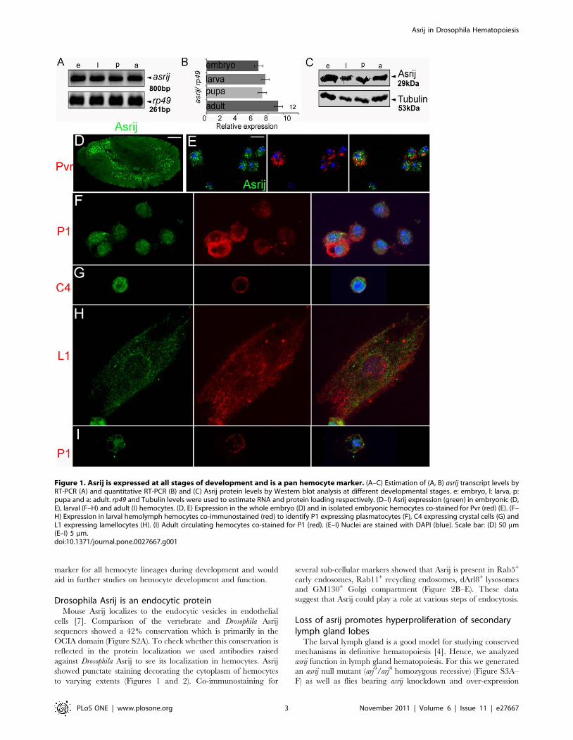
Asrij is a pan-hemocyte markerEarlier we reported asrij mRNA expression in embryonic
hemocytes [6]. Here we undertook a detailed expression analysis
of asrij RNA by Reverse Transcription-Polymerase Chain
Reaction (RT-PCR) and of protein by Western blot analysis and
immunolocalization at different developmental stages of Drosophila
melanogaster. We analyzed asrij mRNA expression at the whole
animal level and found that it is present at all developmental stages
(Figure 1A) and relative levels are comparable as seen by
quantitative RT-PCR (Figure 1B). Polyclonal antibodies against
the full- length protein (Text S1 and Figure S1) revealed an
approximately 29 kDa protein expressed throughout development
(Figure 1C). Immunolocalization showed the protein was present
in embryonic hemocytes (Figure 1D, 1E). Asrij is also expressed in
all subsets of larval (Figure 1F–H) and adult (Figure 1I)
hemolymph hemocytes such as P1+ plasmatocytes, C4+ crystal
cells and L1+ lamellocytes. In addition, we saw Asrij expression in
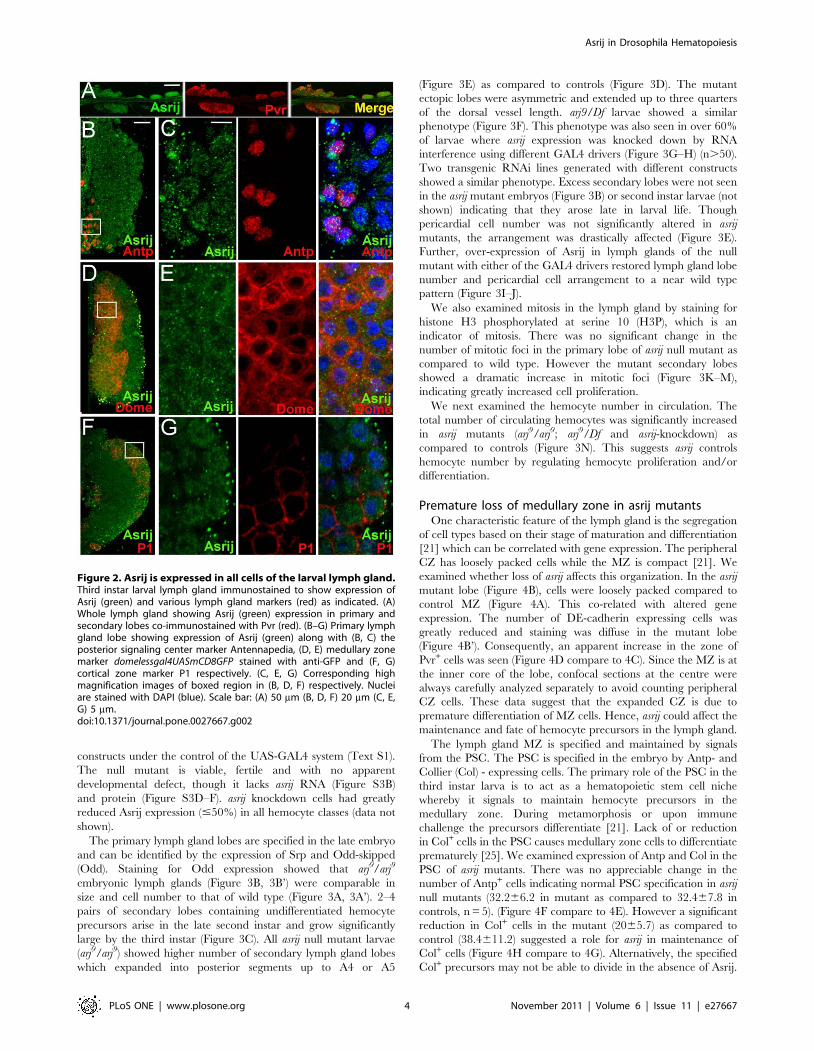
the larval lymph gland lobes (Figure 2A). To identify cell types in
the primary lobe that express Asrij we co-stained for Asrij and
PSC, MZ or CZ markers Antp, domeless (using a GFP reporter)
and P1 respectively. Asrij is expressed in all cells of the primary
lymph gland lobe (Figure 2B–G). Asrij expression could not be
detected in several other tissues examined (Figure S1). Specificity
of the Asrij antibody was confirmed by using pre-immune serum
and no primary antibody controls (not shown) as well as by
staining the null mutant hemocytes (Figure S3). Thus, Asrij is a
Asrij in Drosophila Hematopoiesis
PLoS ONE | www.plosone.org 2 November 2011 | Volume 6 | Issue 11 | e27667
marker for all hemocyte lineages during development and would
aid in further studies on hemocyte development and function.
Drosophila Asrij is an endocytic proteinMouse Asrij localizes to the endocytic vesicles in endothelial
cells [7]. Comparison of the vertebrate and Drosophila Asrij
sequences showed a 42% conservation which is primarily in the
OCIA domain (Figure S2A). To check whether this conservation is
reflected in the protein localization we used antibodies raised
against Drosophila Asrij to see its localization in hemocytes. Asrij
showed punctate staining decorating the cytoplasm of hemocytes
to varying extents (Figures 1 and 2). Co-immunostaining for
several sub-cellular markers showed that Asrij is present in Rab5+
early endosomes, Rab11+ recycling endosomes, dArl8+ lysosomes
and GM130+ Golgi compartment (Figure 2B–E). These data
suggest that Asrij could play a role at various steps of endocytosis.
Loss of asrij promotes hyperproliferation of secondarylymph gland lobes
The larval lymph gland is a good model for studying conserved
mechanisms in definitive hematopoiesis [4]. Hence, we analyzed
asrij function in lymph gland hematopoiesis. For this we generated
an asrij null mutant (arj9/arj9 homozygous recessive) (Figure S3A–
F) as well as flies bearing asrij knockdown and over-expression
Figure 1. Asrij is expressed at all stages of development and is a pan hemocyte marker. (A–C) Estimation of (A, B) asrij transcript levels byRT-PCR (A) and quantitative RT-PCR (B) and (C) Asrij protein levels by Western blot analysis at different developmental stages. e: embryo, l: larva, p:pupa and a: adult. rp49 and Tubulin levels were used to estimate RNA and protein loading respectively. (D–I) Asrij expression (green) in embryonic (D,E), larval (F–H) and adult (I) hemocytes. (D, E) Expression in the whole embryo (D) and in isolated embryonic hemocytes co-stained for Pvr (red) (E). (F–H) Expression in larval hemolymph hemocytes co-immunostained (red) to identify P1 expressing plasmatocytes (F), C4 expressing crystal cells (G) andL1 expressing lamellocytes (H). (I) Adult circulating hemocytes co-stained for P1 (red). (E–I) Nuclei are stained with DAPI (blue). Scale bar: (D) 50 mm(E–I) 5 mm.doi:10.1371/journal.pone.0027667.g001
Asrij in Drosophila Hematopoiesis
PLoS ONE | www.plosone.org 3 November 2011 | Volume 6 | Issue 11 | e27667
constructs under the control of the UAS-GAL4 system (Text S1).
The null mutant is viable, fertile and with no apparent
developmental defect, though it lacks asrij RNA (Figure S3B)
and protein (Figure S3D–F). asrij knockdown cells had greatly
reduced Asrij expression (#50%) in all hemocyte classes (data not
shown).
The primary lymph gland lobes are specified in the late embryo
and can be identified by the expression of Srp and Odd-skipped
(Odd). Staining for Odd expression showed that arj9/arj9
embryonic lymph glands (Figure 3B, 3B’) were comparable in
size and cell number to that of wild type (Figure 3A, 3A’). 2–4
pairs of secondary lobes containing undifferentiated hemocyte
precursors arise in the late second instar and grow significantly
large by the third instar (Figure 3C). All asrij null mutant larvae
(arj9/arj9) showed higher number of secondary lymph gland lobes
which expanded into posterior segments up to A4 or A5
(Figure 3E) as compared to controls (Figure 3D). The mutant
ectopic lobes were asymmetric and extended up to three quarters
of the dorsal vessel length. arj9/Df larvae showed a similar
phenotype (Figure 3F). This phenotype was also seen in over 60%
of larvae where asrij expression was knocked down by RNA
interference using different GAL4 drivers (Figure 3G–H) (n.50).
Two transgenic RNAi lines generated with different constructs
showed a similar phenotype. Excess secondary lobes were not seen
in the asrij mutant embryos (Figure 3B) or second instar larvae (not
shown) indicating that they arose late in larval life. Though
pericardial cell number was not significantly altered in asrij
mutants, the arrangement was drastically affected (Figure 3E).
Further, over-expression of Asrij in lymph glands of the null
mutant with either of the GAL4 drivers restored lymph gland lobe
number and pericardial cell arrangement to a near wild type
pattern (Figure 3I–J).
We also examined mitosis in the lymph gland by staining for
histone H3 phosphorylated at serine 10 (H3P), which is an
indicator of mitosis. There was no significant change in the
number of mitotic foci in the primary lobe of asrij null mutant as
compared to wild type. However the mutant secondary lobes
showed a dramatic increase in mitotic foci (Figure 3K–M),
indicating greatly increased cell proliferation.
We next examined the hemocyte number in circulation. The
total number of circulating hemocytes was significantly increased
in asrij mutants (arj9/arj9; arj9/Df and asrij-knockdown) as
compared to controls (Figure 3N). This suggests asrij controls
hemocyte number by regulating hemocyte proliferation and/or
differentiation.
Premature loss of medullary zone in asrij mutantsOne characteristic feature of the lymph gland is the segregation
of cell types based on their stage of maturation and differentiation
[21] which can be correlated with gene expression. The peripheral
CZ has loosely packed cells while the MZ is compact [21]. We
examined whether loss of asrij affects this organization. In the asrij
mutant lobe (Figure 4B), cells were loosely packed compared to
control MZ (Figure 4A). This co-related with altered gene
expression. The number of DE-cadherin expressing cells was
greatly reduced and staining was diffuse in the mutant lobe
(Figure 4B’). Consequently, an apparent increase in the zone of
Pvr+ cells was seen (Figure 4D compare to 4C). Since the MZ is at
the inner core of the lobe, confocal sections at the centre were
always carefully analyzed separately to avoid counting peripheral
CZ cells. These data suggest that the expanded CZ is due to
premature differentiation of MZ cells. Hence, asrij could affect the
maintenance and fate of hemocyte precursors in the lymph gland.
The lymph gland MZ is specified and maintained by signals
from the PSC. The PSC is specified in the embryo by Antp- and
Collier (Col) - expressing cells. The primary role of the PSC in the
third instar larva is to act as a hematopoietic stem cell niche
whereby it signals to maintain hemocyte precursors in the
medullary zone. During metamorphosis or upon immune
challenge the precursors differentiate [21]. Lack of or reduction
in Col+ cells in the PSC causes medullary zone cells to differentiate
prematurely [25]. We examined expression of Antp and Col in the
PSC of asrij mutants. There was no appreciable change in the
number of Antp+ cells indicating normal PSC specification in asrij
null mutants (32.266.2 in mutant as compared to 32.467.8 in
controls, n = 5). (Figure 4F compare to 4E). However a significant
reduction in Col+ cells in the mutant (2065.7) as compared to
control (38.4611.2) suggested a role for asrij in maintenance of
Col+ cells (Figure 4H compare to 4G). Alternatively, the specified
Col+ precursors may not be able to divide in the absence of Asrij.
Figure 2. Asrij is expressed in all cells of the larval lymph gland.Third instar larval lymph gland immunostained to show expression ofAsrij (green) and various lymph gland markers (red) as indicated. (A)Whole lymph gland showing Asrij (green) expression in primary andsecondary lobes co-immunostained with Pvr (red). (B–G) Primary lymphgland lobe showing expression of Asrij (green) along with (B, C) theposterior signaling center marker Antennapedia, (D, E) medullary zonemarker domelessgal4UASmCD8GFP stained with anti-GFP and (F, G)cortical zone marker P1 respectively. (C, E, G) Corresponding highmagnification images of boxed region in (B, D, F) respectively. Nucleiare stained with DAPI (blue). Scale bar: (A) 50 mm (B, D, F) 20 mm (C, E,G) 5 mm.doi:10.1371/journal.pone.0027667.g002
Asrij in Drosophila Hematopoiesis
PLoS ONE | www.plosone.org 4 November 2011 | Volume 6 | Issue 11 | e27667
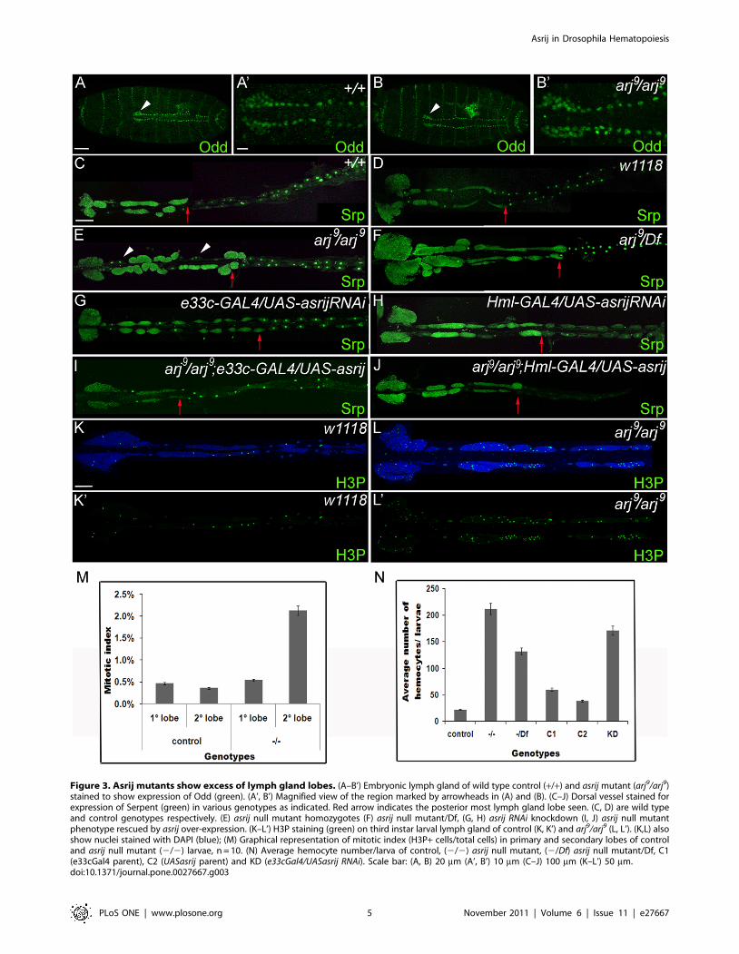
Figure 3. Asrij mutants show excess of lymph gland lobes. (A–B’) Embryonic lymph gland of wild type control (+/+) and asrij mutant (arj9/arj9)stained to show expression of Odd (green). (A’, B’) Magnified view of the region marked by arrowheads in (A) and (B). (C–J) Dorsal vessel stained forexpression of Serpent (green) in various genotypes as indicated. Red arrow indicates the posterior most lymph gland lobe seen. (C, D) are wild typeand control genotypes respectively. (E) asrij null mutant homozygotes (F) asrij null mutant/Df, (G, H) asrij RNAi knockdown (I, J) asrij null mutantphenotype rescued by asrij over-expression. (K–L’) H3P staining (green) on third instar larval lymph gland of control (K, K’) and arj9/arj9 (L, L’). (K,L) alsoshow nuclei stained with DAPI (blue); (M) Graphical representation of mitotic index (H3P+ cells/total cells) in primary and secondary lobes of controland asrij null mutant (2/2) larvae, n = 10. (N) Average hemocyte number/larva of control, (2/2) asrij null mutant, (2/Df) asrij null mutant/Df, C1(e33cGal4 parent), C2 (UASasrij parent) and KD (e33cGal4/UASasrij RNAi). Scale bar: (A, B) 20 mm (A’, B’) 10 mm (C–J) 100 mm (K–L’) 50 mm.doi:10.1371/journal.pone.0027667.g003
Asrij in Drosophila Hematopoiesis
PLoS ONE | www.plosone.org 5 November 2011 | Volume 6 | Issue 11 | e27667
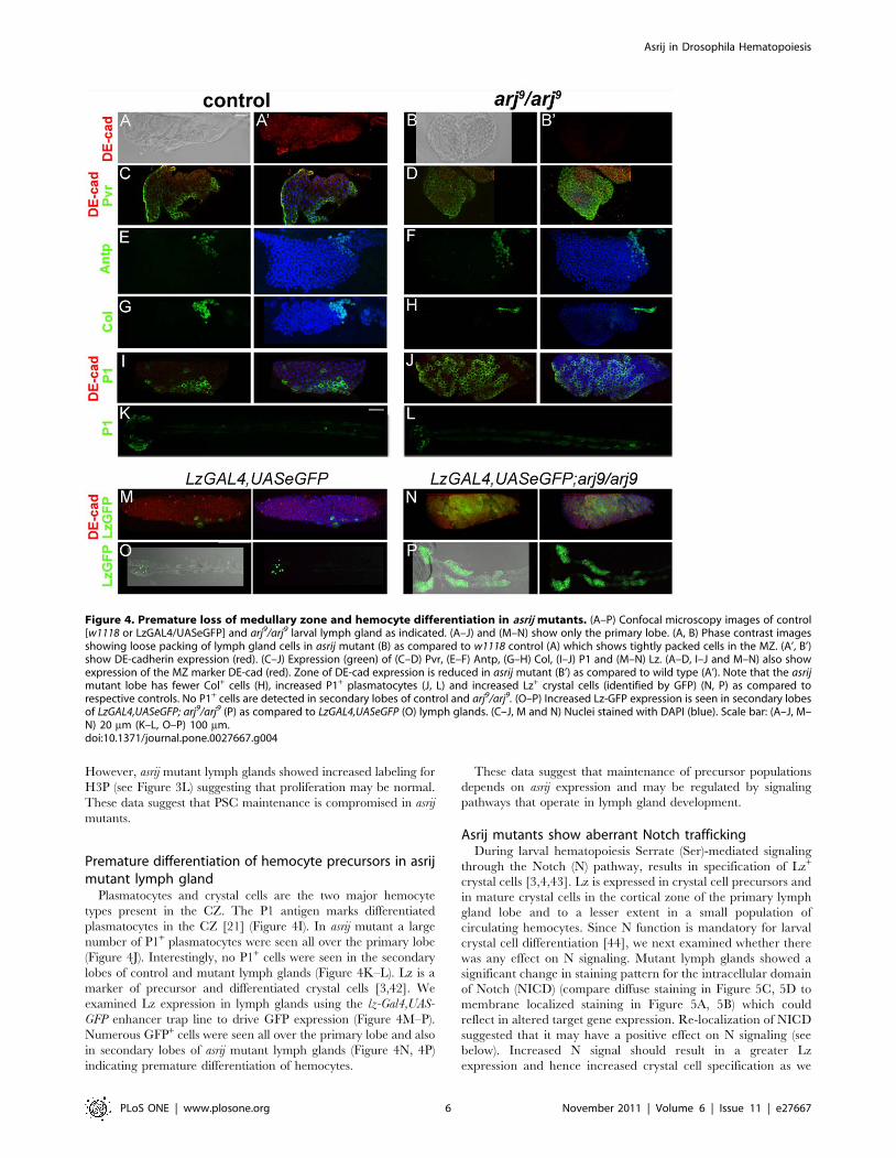
However, asrij mutant lymph glands showed increased labeling for
H3P (see Figure 3L) suggesting that proliferation may be normal.
These data suggest that PSC maintenance is compromised in asrij
mutants.
Premature differentiation of hemocyte precursors in asrijmutant lymph gland
Plasmatocytes and crystal cells are the two major hemocyte
types present in the CZ. The P1 antigen marks differentiated
plasmatocytes in the CZ [21] (Figure 4I). In asrij mutant a large
number of P1+ plasmatocytes were seen all over the primary lobe
(Figure 4J). Interestingly, no P1+ cells were seen in the secondary
lobes of control and mutant lymph glands (Figure 4K–L). Lz is a
marker of precursor and differentiated crystal cells [3,42]. We
examined Lz expression in lymph glands using the lz-Gal4,UAS-
GFP enhancer trap line to drive GFP expression (Figure 4M–P).
Numerous GFP+ cells were seen all over the primary lobe and also
in secondary lobes of asrij mutant lymph glands (Figure 4N, 4P)
indicating premature differentiation of hemocytes.
These data suggest that maintenance of precursor populations
depends on asrij expression and may be regulated by signaling
pathways that operate in lymph gland development.
Asrij mutants show aberrant Notch traffickingDuring larval hematopoiesis Serrate (Ser)-mediated signaling
through the Notch (N) pathway, results in specification of Lz+
crystal cells [3,4,43]. Lz is expressed in crystal cell precursors and
in mature crystal cells in the cortical zone of the primary lymph
gland lobe and to a lesser extent in a small population of
circulating hemocytes. Since N function is mandatory for larval
crystal cell differentiation [44], we next examined whether there
was any effect on N signaling. Mutant lymph glands showed a
significant change in staining pattern for the intracellular domain
of Notch (NICD) (compare diffuse staining in Figure 5C, 5D to
membrane localized staining in Figure 5A, 5B) which could
reflect in altered target gene expression. Re-localization of NICD
suggested that it may have a positive effect on N signaling (see
below). Increased N signal should result in a greater Lz
expression and hence increased crystal cell specification as we
Figure 4. Premature loss of medullary zone and hemocyte differentiation in asrij mutants. (A–P) Confocal microscopy images of control[w1118 or LzGAL4/UASeGFP] and arj9/arj9 larval lymph gland as indicated. (A–J) and (M–N) show only the primary lobe. (A, B) Phase contrast imagesshowing loose packing of lymph gland cells in asrij mutant (B) as compared to w1118 control (A) which shows tightly packed cells in the MZ. (A’, B’)show DE-cadherin expression (red). (C–J) Expression (green) of (C–D) Pvr, (E–F) Antp, (G–H) Col, (I–J) P1 and (M–N) Lz. (A–D, I–J and M–N) also showexpression of the MZ marker DE-cad (red). Zone of DE-cad expression is reduced in asrij mutant (B’) as compared to wild type (A’). Note that the asrijmutant lobe has fewer Col+ cells (H), increased P1+ plasmatocytes (J, L) and increased Lz+ crystal cells (identified by GFP) (N, P) as compared torespective controls. No P1+ cells are detected in secondary lobes of control and arj9/arj9. (O–P) Increased Lz-GFP expression is seen in secondary lobesof LzGAL4,UASeGFP; arj9/arj9 (P) as compared to LzGAL4,UASeGFP (O) lymph glands. (C–J, M and N) Nuclei stained with DAPI (blue). Scale bar: (A–J, M–N) 20 mm (K–L, O–P) 100 mm.doi:10.1371/journal.pone.0027667.g004
Asrij in Drosophila Hematopoiesis
PLoS ONE | www.plosone.org 6 November 2011 | Volume 6 | Issue 11 | e27667
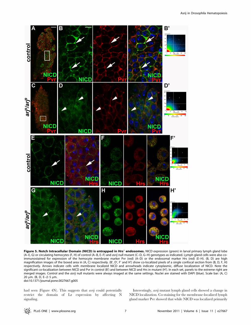
had seen (Figure 4N). This suggests that asrij could potentially
restrict the domain of Lz expression by affecting N
signaling.
Interestingly, asrij mutant lymph gland cells showed a change in
NICD localization. Co-staining for the membrane-localized lymph
gland marker Pvr showed that while NICD was localized primarily
Figure 5. Notch Intracellular Domain (NICD) is entrapped in Hrs+ endosomes. NICD expression (green) in larval primary lymph gland lobe(A–E, G) or circulating hemocytes (F, H) of control (A–B, E–F) and asrij null mutant (C–D, G–H) genotypes as indicated. Lymph gland cells were also co-immunostained for expression of the hemocyte membrane marker Pvr (red) (A–D) or the endosomal marker Hrs (red) (E–H). (B, D) are highmagnification images of the boxed area in (A, C) respectively. (B’, D’, F’ and H’) show co-localized pixels of a single confocal section from (B, D, F, H)respectively. Arrows indicate cells with membrane localized NICD and arrowheads indicate cytoplasmic, diffuse localization of NICD. Note thesignificant co-localization between NICD and Pvr in control (B’) and between NICD and Hrs in mutant (H’). In each set, panels to the extreme right aremerged images. Control and the asrij null mutants were always imaged at the same settings. Nuclei are stained with DAPI (blue). Scale bar: (A, C)20 mm. (B, D, E–J) 5 mm.doi:10.1371/journal.pone.0027667.g005
Asrij in Drosophila Hematopoiesis
PLoS ONE | www.plosone.org 7 November 2011 | Volume 6 | Issue 11 | e27667
to the membrane in control cells, asrij mutant cells showed diffuse
NICD staining (Figure 5D compare to Figure 5B). This indicates
aberrant localization of NICD in asrij mutants. During N signal
transduction NICD is cleaved by c-secretase and transported to
the nucleus through a series of endocytic compartments
[45,46,47]. Aberrant NICD localization in mutant cells suggests
that Asrij could regulate N trafficking. To analyze the effect of
Asrij on N signaling further, we examined the expression of N and
various trafficking proteins in the asrij mutant lymph gland
hemocytes compared to controls (Figure 5E, 5G). For a higher
resolution analysis of this phenotype we checked expression in
control and arj9/arj9 hemolymph hemocytes (Figure 5F, 5H).
Immunostaining showed NICD trapped in sub-cellular compart-
ments of arj9/arj9 hemocytes, which we identified as Hrs+
endosomes (Figure 5G, 5H). Hrs is required for maturation of
endosomes into multivesicular bodies (MVBs) [48,49]. Control
hemocytes showed very little co-localization of NICD with Hrs
(Figure 5F’), whereas there was increased overlap between the two
in asrij mutant hemocytes (Figure 5H’). To analyze the ability of
asrij null hemocytes to traffic generic molecules, we used
fluorescent probes. FITC-labelled dextran (F-dex) is used as an
indicator of molecular movement within the cell during endocy-
tosis [50]. Trafficking of F-dex in hemocytes has been well-
documented [50]. Asrij null hemocytes showed greatly reduced
uptake of the probe (Figure S4).
Based on these results we reasoned that in asrij null hemocytes N
is stalled in the endosomes due to lack of Asrij. Therefore Asrij is
required for NICD trafficking. Notably, we did not see any effect
on NICD localization in tissues where asrij is not normally
expressed, such as the wing disc.
Discussion
We have used Drosophila hematopoiesis as a model to study the
role of a conserved endocytic molecule in trafficking of signals
required for maintenance of stem cells and precursors. Mutants
lacking the endocytic protein have excess hemocytes in circulation,
hyperproliferation of lymph gland secondary lobes and premature
differentiation of hemocytes. In agreement with our previous
reports on mouse and Drosophila asrij we have shown that Asrij is
expressed from the earliest stages of prohemocyte specification.
While embryos homozygous for a deficiency of asrij (and therefore
deleted in multiple genes) are lethal (Inamdar 2003), asrij null
mutant is homozygous viable. Just as mutations in human asrij are
associated with cancers [51], Drosophila asrij loss-of-function
mutants also cause hyperproliferation and premature differentia-
tion of precursors, indicating that the mutant phenotype is the
result of perturbation in conserved gene function. Our results
indicate that Asrij interacts with multiple signaling pathways and
will be an important tool in the analysis of hematopoiesis.
Asrij regulates lymph gland proliferationA remarkable feature of the arj9 mutant phenotype is the
supernumerary posterior lymph gland lobes. The origin of the
secondary lymph gland lobes is not understood and no precursors
are detected in the embryo [21]. In asrij mutants we see a bona fide
primary lobe and excess secondary lobes arising in the larva. This
suggests the presence of previously undetected larval lymph gland
precursors whose proliferation was suppressed by Asrij. Alterna-
tively, asrij may suppress specification of posterior lymph gland
progenitors in larval development. In addition, asrij may control
proliferation of circulating hemocytes as we see increase in their
number. Similar phenotypes were reported for other mutants that
show overgrowth in mutant lymph glands and increase in
circulating hemocytes [41]. Asrij mutants provide an excellent tool
to elucidate events in hematopoiesis and interrogate signaling
pathways implicated in proliferation of lymph gland lobes.
The intriguing question remains of how Asrij can promote both
proliferation and differentiation in posterior lobes and differenti-
ation in the primary lobe. Multiple signals in the anterior lobe are
integrated in time and space to maintain the PSC and control
precursor differentiation. These may provide mechanisms inde-
pendent of or complementary to Asrij function in the control of
proliferation. Such details are not available for the secondary
lobes, which are believed to harbor a homogenous population of
quiescent precursors. Loss of Asrij leads to hyperplastic effects in
the secondary lobes. Hyperplasia is also a phenotype associated
with excess N signaling. We propose that asrij controls proliferation
by integrating with Notch signaling. Previous studies report the
effect of N signaling on the primary lobe or circulating hemocytes
[44], but the secondary lobes have not been analyzed in detail.
Loss of asrij leads to increased Notch signaling and hence
hyperproliferation in secondary lobes. However, additional signals
required to maintain quiescence may be absent and hence there is
increased differentiation to Lz+ crystal cells, which is a read out for
Notch signaling. On the other hand, plasmatocyte differentiation
is not seen in asrij mutant secondary lobes. This suggests that
pathways that promote plasmatocyte differentiation are not active
in the posterior or not controlled by Asrij.
Non-autonomous or secondary effects of AsrijAs none of the hemocyte-specific GAL4 drivers is reported to
function only in the lymph gland primary and secondary lobes, we
used the best available drivers e33cGAL4 (expressed in all lymph
gland cells and in other tissues) [52] and HmlGAL4 (expressed
only in the primary lobe and in hemocytes) [53] to generate asrij
knockdown or overexpression flies. Experiments using either
GAL4 driver gave similar phenotypes to those using the null
mutant (arj9/arj9) or the arj9/Df flies. These results validate that the
phenotypes seen are primarily due to the effect on asrij. Though
HmlGal4 is reported to drive expression only in the primary lobe
and hemocytes, multiple experiments that we have done clearly
show that expression using HmlGAL4 affects function in the
secondary lymph gland lobes. This could be either due to
previously unreported low level or leaky activity of the driver in
posterior lobes or due to a non-autonomous effect of manipulating
asrij activity in primary lobes and in circulating hemocytes. The
interaction between hemocytes in lymph gland posterior lobes and
in hemolymph merits further investigation.
As Asrij is involved in vesicular traffic, it may affect multiple
signaling pathways and possibly have non-autonomous or
secondary effects. Though asrij mutants show disturbed pericardial
cell arrangement, the heartbeat of arj9 homozygous larvae is
normal (data not shown) indicating no functional effect on cardiac
rhythm. This is in agreement with earlier reports that pericardial
cells are not required for normal cardiac function [26].
Asrij is required for maintenance of the stem cell nicheand precursor quiescence
Inspite of reduced Col+ cells in asrij mutant, no appreciable
reduction in Antp+ cell number was seen, suggesting that Asrij
may affect maintenance of Col+ cells, which needs to be
investigated. The reduced Col+ PSC in asrij mutants could affect
MZ quiescence. Cells in the MZ are compact, bounded by
extracellular matrix (ECM) and maintained in a slow-cycling
quiescent state by signals from the PSC [5]. Loss of MZ
accompanied by increased differentiation and release of hemocytes
into circulation is normally seen during metamorphosis or upon
Asrij in Drosophila Hematopoiesis
PLoS ONE | www.plosone.org 8 November 2011 | Volume 6 | Issue 11 | e27667
immune challenge. For this, precursor- matrix interactions have to
be modulated as cells differentiate and migrate to the periphery
where they are loosely packed [21]. The choice between
prohemocyte maintenance and its differentiation may be mediated
by changes in ECM components and in adhesive properties of a
cell. Asrij null lymph glands have loosely packed cells with greatly
reduced expression of the Wg target DE-cad (Figure 4B,B’). The
importance of deregulated adhesion in cancer is well documented.
Inactivation of E-cadherin in human and mouse is associated with
progression to metastasis and also promotes neoplasia. Increased
proliferation of precursors in the asrij mutant lymph gland and
increase in circulating hemocyte number suggest that asrij may act
on mechanisms that control DE-cad expression and indirectly
control cell adhesion. Ociad1 plays a key role in human cancer cell
adhesion [51]. Changes in Ociad1 expression levels can modulate
integrin function thereby affecting cell adhesion and the ability of
cancer cells to form secondary colonies [54]. We speculate that
Asrij/Ociad1 may play a similar role in regulating adhesion via
the Wnt pathway. Further, this change in adhesive properties
could influence the choice between stem cell maintenance and
differentiation.
Control of endocytosis is important for hematopoiesisPremature hemocyte differentiation in asrij mutant larvae
suggests a regulatory role for endocytosis during normal
development. Control levels of Asrij are required to prevent
hemocyte differentiation possibly as a secondary effect of MZ loss.
Alternatively, asrij may attenuate signals required for hemocyte
differentiation through uptake and degradation of signaling
molecules. In the absence of Asrij, control on signal amplitude
may be lost and can result in initiation of the differentiation
program. Retention of NICD in subcellular compartments
correlates well with increased Lz+ cells and supports the latter.
Further, this phenotype does correlate with a N gain of function as
seen by increase in crystal cells. Vaccari et al. [55] reported NICD
entrapped in Hrs positive endosomes in ESCRT mutants – (in
genes like tsg101, vps25, vps 20)- showed Notch gain of function
phenotypes such as overgrowth of eye imaginal discs and eye
phenotypes in adult mutant flies too. The mechanism by which
asrij affects NICD endocytosis merits further investigation.
Asrij may have context-dependent functions during hematopoi-
esis. Our observations that asrij mutants show increased N
signaling reveals a mechanism by which endocytic molecules can
regulate cell proliferation. Further, NICD is aberrantly localized in
all mutant lymph gland cells compared to only a subset of control
lymph gland cells. This is reflected in the widespread ectopic Lz+
cells in mutants compared to controls. This suggests Asrij interacts
with additional pathways that control N signaling.
Hemocytes also differentiate and are released into circulation
during systemic infection. One possibility that remains to be tested
is whether Asrij is a target of the signaling cascade triggered by
immune challenge. Reduction in Asrij levels could help rapidly
respond to immune challenge and we are testing whether this is so.
This scenario is also supported by the presence of excess
circulating hemocytes in the asrij mutant. Expression of asrij only
in the lymph glands is sufficient for complete rescue of the mutant
phenotype, indicating a function for Asrij within the lymph gland
and hemocytes. However the signaling molecules regulated by
asrij, or their effectors could be released from the lymph gland or
fat body to activate systemic targets in the larva. Further studies on
the Asrij protein and its role in cellular traffic would help address
these mechanisms.
Common endocytic pathways could mediate signalingduring hematopoiesis
In addition to interaction of Asrij with ARF1 [9], the human
ortholog Ociad1 is predicted to interact with SLC35F2 a solute
carrier family protein and also with KDR, the human Flk1/
VEGFRII homolog (http://string.embl.de/newstring_cgi/show_-
network_section.pl). Hematopoiesis in Drosophila is governed by a
transcription factor cascade initiated by the GATA factor Srp.
Hemocyte division, density and possibly viability are controlled by
the Toll/Cactus and JAK/STAT pathways which also activate
immunity genes [17,56]. Similar phenotypes of Asrij and the
conservation in expression pattern and functions suggest common
endocytic pathways that mediate hematopoiesis. Asrij being an
endocytic protein could be involved in regulation of these multiple
inputs. Human ociad1 interacts with several cellular proteins [54],
supporting our hypothesis.
Notch processing is quite complex and several tissue- specific
components are implicated in its activation [57,58]. Notch
accumulates in intracellular structures when endocytic progression
is perturbed resulting in its hyperactivation leading to hyperplasia
[37,59,60,61]. We have shown that loss of Asrij leads to retention
of Notch intracellular domain in Hrs endosomes correlating with
increased Notch activity, seen as increase in Lz+ cells. Up-
regulation of Notch has been implicated in human blood cell
disorders such as, T cell acute lymphoblastic leukemia [62].
Mutants such as lethal giant discs (lgd) that affect protein sorting in
the late endosomes or MVB result in Notch gain- of- function
phenotypes [63]. Our preliminary analysis with fluorescent probes
indicates a generic requirement for Asrij in intracellular transport
in hemocytes. Further investigation is required to understand
whether Notch activation in asrij mutant is ligand dependent or
independent. Asrij mutants provide an excellent tool to understand
the mechanism involved in precocious N signaling leading to
blood cell disorders. Further asrij mutants are viable and can be
used in studying post-embryonic Notch signaling in various
contexts.
Asrij functions at multiple levels during hematopoiesisThe importance of signaling proteins, receptors and transcrip-
tional targets of the N and Wg pathways for cancer development is
well established. In contrast, data regarding endocytic molecules
that traffic the pathway components and modulate their activity is
limited. Here we show that loss of the endocytic protein Asrij
affects Drosophila at multiple levels leading to increased hemato-
poiesis by enhancing precursor proliferation and differentiation.
Our results indicate a role for Asrij in PSC maintenance, which in
turn affects precursor quiescence. In addition, a more direct role
for Asrij is implicated in crystal cell specification via control of
NICD traffic.
Our study demonstrates the value of a comparative approach in
identifying functions of conserved mammalian genes in Drosophila.
The early onset of Asrij expression during development suggests
that it could be a key player in vertebrate hematopoiesis as well. By
virtue of its ability to control cellular traffic, Asrij may control cell
adhesion, proliferation and differentiation, which makes it difficult
to tease out the exact mechanism of its action. Understanding how
asrij controls the balance between stem cell number and
committed precursors may aid in disease correction and
regenerative medicine. Together, our findings indicate that
endocytosis is a key modulator of lymph gland hematopoiesis
and provide in vivo demonstration that genetic loss of endocytic
components can lead to accelerated hematopoietic development
and facilitate premature differentiation.
Asrij in Drosophila Hematopoiesis
PLoS ONE | www.plosone.org 9 November 2011 | Volume 6 | Issue 11 | e27667
Supporting Information
Figure S1 (A–B) Antigen-antibody competition assay tovalidate the specificity of Asrij antibody. Western blot
showing specificity of Asrij (A) antibody. Lanes: (1, 2) Blot probed
with antibody preincubated with 25 or 50 mg of corresponding
antigen. (3, 4) blot probed with antibody without preincubation
with antigen. (B–C) Asrij expression (green) by immunostaining
with anti-Asrij antibodies could not be detected in several other
tissues examined including wing disc (B), fat body (C). Nuclei
stained with DAPI (blue). Panels to the extreme right are merged
images. Scale bar: (B, C) 50 mm.
(TIF)
Figure S2 Conservation and subcellular localisation ofAsrij in Drosophila melanogaster. (A) Schematic represent-
ing conservation in OCIA domain of Asrij. The N half of the Asrij
protein including predicted helices are conserved in Drosophila,
mouse and human. (B–E) Subcellular localization of Asrij.
Immufluorescence analysis of hemocytes stained for expression
of Asrij (green, extreme left panels) and subcellular marker
proteins (red, middle panels) such as (B) Rab5, (C) Rab11, (D)
dArl8 and (E) GM130. Nuclei are stained with DAPI (blue). Panels
to the extreme right in each set are merged images. Scale bar: (B–
E): 5 mm.
(TIF)
Figure S3 Southern blot analysis confirms insertion inarj9 mutant. (A) Schematic showing the details of the asrij null
mutant. (B) Southern blot of Hind III digested genomic DNA from
asrij excision lines probed with 32P-labelled cDNA. Lanes. 1: CS, 2:
BL14935, 3: arj9/arj9 and 4: Marker. A 3.2 kbp band of expected
size is seen in wild type whereas arj9/arj9 mutant has 2 bands of
2.4 kbp and 1.3 kbp due to 550 bp remnant of P element
sequence. Analysis of asrij (C) transcript expression by RT-PCR
and (D) protein expression by immunoblot with anti-Asrij
antibody. Genotypes are as indicated above the lanes. (E–F)
Immunofluorescence analysis of Asrij (green) expression in
hemocytes of wild type (E) and arj9/arj9 mutant (F). Hemocytes
are identified by the expression of the pan hemocyte marker
Hemese (red). Nuclei marked by DAPI (blue). Scale bar: (E, F)
5 mm.
(TIF)
Figure S4 Dextran uptake is reduced in Asrij nullhemocytes. (A) Total cell associated fluorescence of internalized
FITC Dextran 5 min after starting the incubation of wild type
(CS), asrij null (arj9/arj9) and rescue (arj9/arj9; HmlGAL4/UAS
Dmasrij) hemocytes (P = 0.002). (B–C) Representative images of
wild type (B) and arj9/arj9 mutant (C) hemocytes showing the
uptake of FITC Dextran. Cell boundary is marked by a white line.
Scale bar: (B, C) 5 mm.
(TIF)
Table S1 List of primers used for RT-PCR and qRT-PCR.
(DOC)
Text S1
(DOC)
Acknowledgments
We thank Debjani Das for help with generating and characterizing
excision lines; Nandashree K. for help with initial experiments;
Arghyashree Roychowdhury for help with hemocyte counts; B.S. Suma
for help with confocal microscopy; V. Sriram for help with hemocyte
experiments; K. VijayRaghavan for reagents, support and discussions,
Volker Hartenstein for valuable comments. We are grateful to the
following for antibodies: DSHB; Istvan Ando (hemocyte subset- specific
antibodies); Hugo Bellen (anti-Hrs); Manfred Frasch (anti-Odd-Skipped);
Denise Montell (anti-Pvr); Martin Lowe (anti-GM130); Sean Munro (anti-
dArl8); Marcos Gonzalez-Gaitan (anti-Rab5); Satyajit Mayor (anti-Rab11);
Michele Crozatier (anti-Collier); Deborah Hoshizaki (anti-Serpent);
K. Irvine (anti-Serrate). We thank NCBS, Bangalore and NIG (Japan)
for fly stocks. Finally, we thank anonymous reviewers for valuable
comments and suggestions.
Author Contributions
Conceived and designed the experiments: VK RJK SMS MSI. Performed
the experiments: VK RJK SMS MSI. Analyzed the data: VK SMS RJK
MSI. Contributed reagents/materials/analysis tools: MSI. Wrote the
paper: VK MSI.
References
1. Tepass U, Fessler LI, Aziz A, Hartenstein V (1994) Embryonic origin ofhemocytes and their relationship to cell death in Drosophila. Development 120:
1829–1837.
2. Rehorn KP, Thelen H, Michelson AM, Reuter R (1996) A molecular aspect of
hematopoiesis and endoderm development common to vertebrates andDrosophila. Development 122: 4023–4031.
3. Lebestky T, Chang T, Hartenstein V, Banerjee U (2000) Specification ofDrosophila hematopoietic lineage by conserved transcription factors. Science
288: 146–149.
4. Evans CJ, Hartenstein V, Banerjee U (2003) Thicker than blood: conserved
mechanisms in Drosophila and vertebrate hematopoiesis. Dev Cell 5: 673–690.
5. Hartenstein V (2006) Blood cells and blood cell development in the animal
kingdom. Annu Rev Cell Dev Biol 22: 677–712.
6. Inamdar MS (2003) Drosophila asrij is expressed in pole cells, trachea andhemocytes. Dev Genes Evol 213: 134–137.
7. Mukhopadhyay A, Das D, Inamdar MS (2003) Embryonic stem cell and tissue-specific expression of a novel conserved gene, asrij. Dev Dyn 227: 578–586.
8. Phillips RL, Ernst RE, Brunk B, Ivanova N, Mahan MA, et al. (2000) The
genetic program of hematopoietic stem cells. Science 288: 1635–1640.
9. Giot L, Bader JS, Brouwer C, Chaudhuri A, Kuang B, et al. (2003) A protein
interaction map of Drosophila melanogaster. Science 302: 1727–1736.
10. Usary J, Llaca V, Karaca G, Presswala S, Karaca M, et al. (2004) Mutation of
GATA3 in human breast tumors. Oncogene 23: 7669–7678.
11. Shen C, Hui Z, Wang D, Jiang G, Wang J, et al. (2002) Molecular cloning,identification and analysis of lung squamous cell carcinoma-related genes. Lung
Cancer 38: 235–241.
12. Arai H, Emson PC, Mountjoy CQ, Carassco LH, Heizmann CW (1987) Loss of
parvalbumin-immunoreactive neurones from cortex in Alzheimer-type demen-tia. Brain Res 418: 164–169.
13. Nigrovic PA, Gray DH, Jones T, Hallgren J, Kuo FC, et al. (2008) Genetic
inversion in mast cell-deficient (W(sh)) mice interrupts corin and manifests ashematopoietic and cardiac aberrancy. Am J Pathol 173: 1693–1701.
14. Holz A, Bossinger B, Strasser T, Janning W, Klapper R (2003) The two originsof hemocytes in Drosophila. Development 130: 4955–4962.
15. Lanot R, Zachary D, Holder F, Meister M (2001) Postembryonic hematopoiesis
in Drosophila. Dev Biol 230: 243–257.
16. Grigorian M, Mandal L, Hartenstein V (2011) Hematopoiesis at the onset of
metamorphosis: terminal differentiation and dissociation of the Drosophilalymph gland. Dev Genes Evol 221: 121–131.
17. Agaisse H, Petersen UM, Boutros M, Mathey-Prevot B, Perrimon N (2003)
Signaling role of hemocytes in Drosophila JAK/STAT-dependent response toseptic injury. Dev Cell 5: 441–450.
18. Rizki TM, Rizki RM (1992) Lamellocyte differentiation in Drosophila larvaeparasitized by Leptopilina. Dev Comp Immunol 16: 103–110.
19. Sorrentino RP, Carton Y, Govind S (2002) Cellular immune response to parasiteinfection in the Drosophila lymph gland is developmentally regulated. Dev Biol
243: 65–80.
20. Mandal L, Banerjee U, Hartenstein V (2004) Evidence for a fruit flyhemangioblast and similarities between lymph-gland hematopoiesis in fruit fly
and mammal aorta-gonadal-mesonephros mesoderm. Nat Genet 36:1019–1023.
21. Jung SH, Evans CJ, Uemura C, Banerjee U (2005) The Drosophila lymph gland
as a developmental model of hematopoiesis. Development 132: 2521–2533.
22. Rizki RM, Rizki TM (1984) Selective destruction of a host blood cell type by a
parasitoid wasp. Proc Natl Acad Sci U S A 81: 6154–6158.
23. Mandal L, Martinez-Agosto JA, Evans CJ, Hartenstein V, Banerjee U (2007) A
Hedgehog- and Antennapedia-dependent niche maintains Drosophila haema-
topoietic precursors. Nature 446: 320–324.
Asrij in Drosophila Hematopoiesis
PLoS ONE | www.plosone.org 10 November 2011 | Volume 6 | Issue 11 | e27667
24. Sinenko SA, Mandal L, Martinez-Agosto JA, Banerjee U (2009) Dual role of
wingless signaling in stem-like hematopoietic precursor maintenance inDrosophila. Dev Cell 16: 756–763.
25. Krzemien J, Dubois L, Makki R, Meister M, Vincent A, et al. (2007) Control of
blood cell homeostasis in Drosophila larvae by the posterior signalling centre.Nature 446: 325–328.
26. Das D, Aradhya R, Ashoka D, Inamdar M (2008) Post-embryonic pericardialcells of Drosophila are required for overcoming toxic stress but not for cardiac
function or adult development. Cell Tissue Res 331: 565–570.
27. Das D, Ashoka D, Aradhya R, Inamdar M (2008) Gene expression analysis inpost-embryonic pericardial cells of Drosophila. Gene Expr Patterns 8: 199–205.
28. Brand AH, Perrimon N (1993) Targeted gene expression as a means of alteringcell fates and generating dominant phenotypes. Development 118: 401–415.
29. Rubin GM, Spradling AC (1982) Genetic transformation of Drosophila withtransposable element vectors. Science 218: 348–353.
30. Rothwell WF, Sullivan W (2000) The centrosome in early Drosophila
embryogenesis. Curr Top Dev Biol 49: 409–447.31. Hayes SA, Miller JM, Hoshizaki DK (2001) serpent, a GATA-like transcription
factor gene, induces fat-cell development in Drosophila melanogaster.Development 128: 1193–1200.
32. Duchek P, Somogyi K, Jekely G, Beccari S, Rorth P (2001) Guidance of cell
migration by the Drosophila PDGF/VEGF receptor. Cell 107: 17–26.33. Wucherpfennig T, Wilsch-Brauninger M, Gonzalez-Gaitan M (2003) Role of
Drosophila Rab5 during endosomal trafficking at the synapse and evokedneurotransmitter release. J Cell Biol 161: 609–624.
34. Emery G, Hutterer A, Berdnik D, Mayer B, Wirtz-Peitz F, et al. (2005)Asymmetric Rab 11 endosomes regulate delta recycling and specify cell fate in
the Drosophila nervous system. Cell 122: 763–773.
35. Hofmann I, Munro S (2006) An N-terminally acetylated Arf-like GTPase islocalised to lysosomes and affects their motility. J Cell Sci 119: 1494–1503.
36. Nakamura N, Lowe M, Levine TP, Rabouille C, Warren G (1997) The vesicledocking protein p115 binds GM130, a cis-Golgi matrix protein, in a mitotically
regulated manner. Cell 89: 445–455.
37. Jekely G, Rorth P (2003) Hrs mediates downregulation of multiple signallingreceptors in Drosophila. EMBO Rep 4: 1163–1168.
38. Crozatier M, Ubeda JM, Vincent A, Meister M (2004) Cellular immuneresponse to parasitization in Drosophila requires the EBF orthologue collier.
PLoS Biol 2: E196.39. Ward EJ, Skeath JB (2000) Characterization of a novel subset of cardiac cells and
their progenitors in the Drosophila embryo. Development 127: 4959–4969.
40. Kurucz E, Vaczi B, Markus R, Laurinyecz B, Vilmos P, et al. (2007) Definitionof Drosophila hemocyte subsets by cell-type specific antigens. Acta Biol Hung 58
Suppl. pp 95–111.41. Minakhina S, Druzhinina M, Steward R (2007) Zfrp8, the Drosophila ortholog
of PDCD2, functions in lymph gland development and controls cell
proliferation. Development 134: 2387–2396.42. Rizki TM, Rizki RM (1981) Genetics of tumor-W in Drosophila melanogaster:
mapping a gene with incomplete penetrance. J Hered 72: 78–80.43. Lebestky T, Jung SH, Banerjee U (2003) A Serrate-expressing signaling center
controls Drosophila hematopoiesis. Genes Dev 17: 348–353.44. Duvic B, Hoffmann JA, Meister M, Royet J (2002) Notch signaling controls
lineage specification during Drosophila larval hematopoiesis. Curr Biol 12:
1923–1927.
45. De Strooper B, Annaert W, Cupers P, Saftig P, Craessaerts K, et al. (1999) A
presenilin-1-dependent gamma-secretase-like protease mediates release of Notchintracellular domain. Nature 398: 518–522.
46. Okochi M, Steiner H, Fukumori A, Tanii H, Tomita T, et al. (2002) Presenilins
mediate a dual intramembranous gamma-secretase cleavage of Notch-1.EMBO J 21: 5408–5416.
47. Struhl G, Greenwald I (1999) Presenilin is required for activity and nuclearaccess of Notch in Drosophila. Nature 398: 522–525.
48. Komada M, Masaki R, Yamamoto A, Kitamura N (1997) Hrs, a tyrosine kinase
substrate with a conserved double zinc finger domain, is localized to thecytoplasmic surface of early endosomes. J Biol Chem 272: 20538–20544.
49. Lloyd TE, Atkinson R, Wu MN, Zhou Y, Pennetta G, et al. (2002) Hrs regulatesendosome membrane invagination and tyrosine kinase receptor signaling in
Drosophila. Cell 108: 261–269.50. Sriram V, Krishnan KS, Mayor S (2003) deep-orange and carnation define
distinct stages in late endosomal biogenesis in Drosophila melanogaster. J Cell
Biol 161: 593–607.51. Sengupta S, Michener CM, Escobar P, Belinson J, Ganapathi R (2008) Ovarian
cancer immuno-reactive antigen domain containing 1 (OCIAD1), a key playerin ovarian cancer cell adhesion. Gynecol Oncol 109: 226–233.
52. Harrison DA, Binari R, Nahreini TS, Gilman M, Perrimon N (1995) Activation
of a Drosophila Janus kinase (JAK) causes hematopoietic neoplasia anddevelopmental defects. EMBO J 14: 2857–2865.
53. Goto A, Kadowaki T, Kitagawa Y (2003) Drosophila hemolectin gene isexpressed in embryonic and larval hemocytes and its knock down causes
bleeding defects. Dev Biol 264: 582–591.54. Wang C, Michener CM, Belinson JL, Vaziri S, Ganapathi R, et al. (2010) Role
of the 18:1 lysophosphatidic acid-ovarian cancer immunoreactive antigen
domain containing 1 (OCIAD1)-integrin axis in generating late-stage ovariancancer. Mol Cancer Ther 9: 1709–1718.
55. Vaccari T, Lu H, Kanwar R, Fortini ME, Bilder D (2008) Endosomal entryregulates Notch receptor activation in Drosophila melanogaster. J Cell Biol 180:
755–762.
56. Govind S (1999) Control of development and immunity by rel transcriptionfactors in Drosophila. Oncogene 18: 6875–6887.
57. Tien AC, Rajan A, Bellen HJ (2009) A Notch updated. J Cell Biol 184: 621–629.58. Bray SJ (2006) Notch signalling: a simple pathway becomes complex. Nat Rev
Mol Cell Biol 7: 678–689.59. Wilkin MB, Carbery AM, Fostier M, Aslam H, Mazaleyrat SL, et al. (2004)
Regulation of notch endosomal sorting and signaling by Drosophila Nedd4
family proteins. Curr Biol 14: 2237–2244.60. Thompson BJ, Mathieu J, Sung HH, Loeser E, Rorth P, et al. (2005) Tumor
suppressor properties of the ESCRT-II complex component Vps25 inDrosophila. Dev Cell 9: 711–720.
61. Moberg KH, Schelble S, Burdick SK, Hariharan IK (2005) Mutations in
erupted, the Drosophila ortholog of mammalian tumor susceptibility gene 101,elicit non-cell-autonomous overgrowth. Dev Cell 9: 699–710.
62. Jundt F, Acikgoz O, Kwon SH, Schwarzer R, Anagnostopoulos I, et al. (2008)Aberrant expression of Notch1 interferes with the B-lymphoid phenotype of
neoplastic B cells in classical Hodgkin lymphoma. Leukemia 22: 1587–1594.63. Childress JL, Acar M, Tao C, Halder G (2006) Lethal giant discs, a novel C2-
domain protein, restricts notch activation during endocytosis. Curr Biol 16:
2228–2233.
Asrij in Drosophila Hematopoiesis
PLoS ONE | www.plosone.org 11 November 2011 | Volume 6 | Issue 11 | e27667