Embed Size (px)
Citation preview
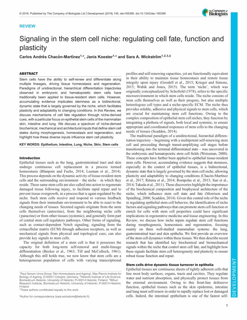
REVIEW
Signaling in the stem cell niche: regulating cell fate, function andplasticityCarlos Andres Chacon-Martınez1,*, Janis Koester1,* and Sara A. Wickstrom1,2,3,‡
ABSTRACTStem cells have the ability to self-renew and differentiate alongmultiple lineages, driving tissue homeostasis and regeneration.Paradigms of unidirectional, hierarchical differentiation trajectoriesobserved in embryonic and hematopoietic stem cells havetraditionally been applied to tissue-resident stem cells. However,accumulating evidence implicates stemness as a bidirectional,dynamic state that is largely governed by the niche, which facilitatesplasticity and adaptability to changing conditions. In this Review, wediscuss mechanisms of cell fate regulation through niche-derivedcues, with a particular focus on epithelial stem cells of themammalianskin, intestine and lung. We discuss a spectrum of niche-derivedbiochemical, mechanical and architectural inputs that define stem cellstates during morphogenesis, homeostasis and regeneration, andhighlight how these diverse inputs influence stem cell plasticity.
KEY WORDS: Epithelium, Intestine, Lung, Niche, Skin, Stem cells
IntroductionEpithelial tissues such as the lung, gastrointestinal tract and skinundergo continuous cell replacement in a process termedhomeostasis (Blanpain and Fuchs, 2014; Leeman et al., 2014).This process depends on the dynamic activity of tissue-resident stemcells and the surrounding environment – the niche – in which theyreside. These same stem cells are also called into action to regeneratedamaged tissue following injury, to facilitate rapid repair and toprevent tissue overgrowth, in a process that is again regulated by theniche. Such stem cells receive and respond to various feedbacksignals from their immediate environment to be able to react to thechanging needs of tissues. Secreted signals originate from the stemcells themselves (autocrine), from the neighboring niche cells(paracrine) or from other tissues (systemic), and generally form partof central stem cell regulatory pathways. Other forms of signaling,such as contact-dependent Notch signaling, signaling from theextracellular matrix (ECM) through adhesion receptors, as well asmechanical signals from physical and topological cues, can alsoprovide key signals to stem cells.The original definition of a stem cell is that it possesses the
capacity for both long-term self-renewal and multi-lineagedifferentiation (Becker et al., 1963; Till and McCulloch, 1961).Although this still holds true, we now know that stem cells are aheterogeneous population of cells with varying transcriptional
profiles and self-renewing capacities, yet are functionally equivalentin their ability to maintain tissue homeostasis and restore tissueintegrity upon injury (Goodell et al., 2015; Krieger and Simons,2015; Wabik and Jones, 2015). The term ‘niche’, which wasoriginally conceptualized by Schofield (1978), refers to the specificmicroenvironment in which stem cells reside. The niche consists ofstem cells themselves as well as their progeny, but also multipleheterologous cell types and a niche-specific ECM. The niche thusprovides soluble, adhesive and physical signals to stem cells, whichare crucial for maintaining stem cell functions. Owing to thecomplex composition of epithelial stem cell niches, they function byintegrating a plethora of signals, both local and systemic, to ensureappropriate and coordinated responses of stem cells to the changingneeds of tissues (Scadden, 2014).
The traditional paradigm of a unidirectional, hierarchal differen-tiation trajectory – beginning with a multipotent self-renewing stemcell and proceeding through transit-amplifying cell stages beforetransitioning into the terminal differentiated state – was uncovered inthe embryonic and hematopoietic stem cell fields (Weissman, 2000).These concepts have further been applied to epithelial tissue-residentstem cells. However, accumulating evidence suggests that stemness,especially in the context of epithelial tissues, is a bidirectional,dynamic state that is largely governed by the stem cell niche, allowingplasticity and adaptability to changing conditions (Chacón-Martínezet al., 2017; Ritsma et al., 2014; Rompolas et al., 2013; Sun et al.,2014; Takeda et al., 2011). These discoveries highlight the importanceof the biochemical composition and biophysical architecture of theniche, which influence stem cell state and fate (Morrison andSpradling, 2008; Scadden, 2014). Given this central role of the nichein regulating epithelial stem cell behavior, the identification of nichefactors and signaling pathways that can promote stem cell function orenrich for cells with stem cell properties could have significantimplications in regenerative medicine and tissue engineering. In thisReview, we discuss how niche inputs regulate stem cell functionsduring morphogenesis, homeostasis and regeneration, focusingmainly on three well-studied mammalian systems: the lung,gastrointestinal tract and skin epithelia. We first provide an overviewof the stem cell dynamics within these tissues.We then describe recentresearch that has identified key biochemical and biomechanicalsignals within the niche that control stem cell fate, and highlight howthese signals facilitate stem cell heterogeneity and plasticity to ensurerobust tissue function and repair.
Stem cells drive dynamic tissue turnover in epitheliaEpithelial tissues are continuous sheets of tightly adherent cells thatline most body surfaces, organs, tracts and cavities. They regulatewater and nutrient absorption, and physically protect tissues fromthe external environment. Owing to this front-line defensivefunction, epithelial tissues such as the skin epidermis, intestineand lung must self-renew in order to rapidly replace lost or damagedcells. Indeed, the intestinal epithelium is one of the fastest self-
1Paul Gerson Unna Group ‘Skin Homeostasis and Ageing’, Max Planck Institute forBiology of Ageing, D-50931 Cologne, Germany. 2Helsinki Institute of Life Science,Biomedicum Helsinki, University of Helsinki, FI-00014 Helsinki, Finland. 3WihuriResearch Institute, Biomedicum Helsinki, University of Helsinki, FI-00014 Helsinki,Finland.*These authors contributed equally to this work
‡Author for correspondence ([email protected])
1
© 2018. Published by The Company of Biologists Ltd | Development (2018) 145, dev165399. doi:10.1242/dev.165399
DEVELO
PM
ENT
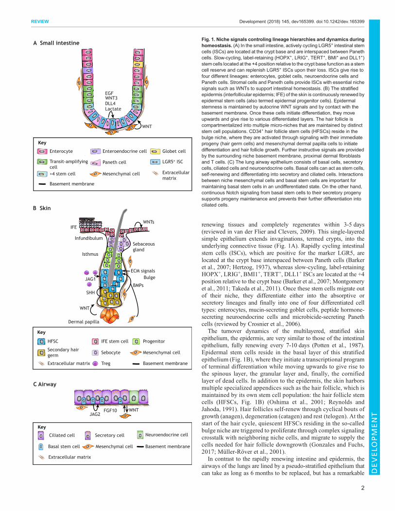
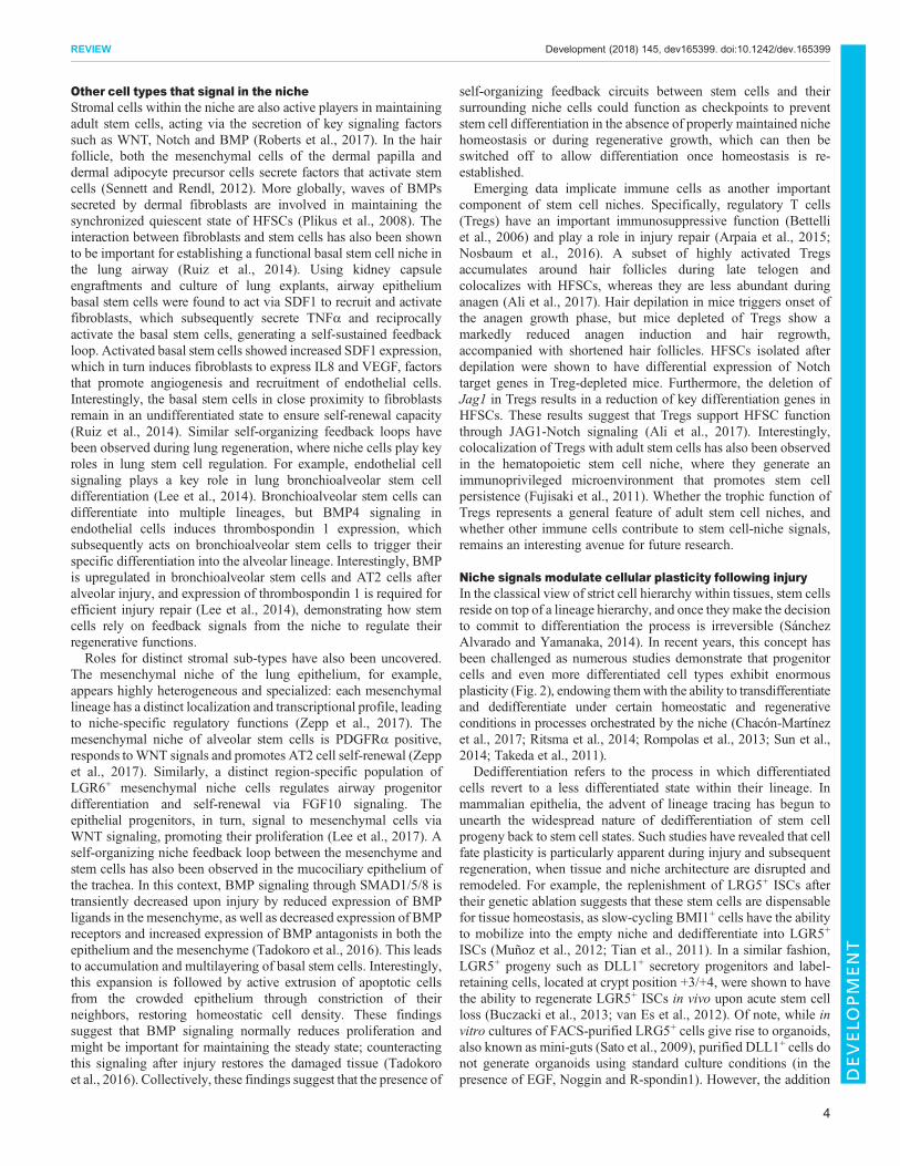
renewing tissues and completely regenerates within 3-5 days(reviewed in van der Flier and Clevers, 2009). This single-layeredsimple epithelium extends invaginations, termed crypts, into theunderlying connective tissue (Fig. 1A). Rapidly cycling intestinalstem cells (ISCs), which are positive for the marker LGR5, arelocated at the crypt base interspaced between Paneth cells (Barkeret al., 2007; Hertzog, 1937), whereas slow-cycling, label-retainingHOPX+, LRIG+, BMI1+, TERT+, DLL1+ ISCs are located at the +4position relative to the crypt base (Barker et al., 2007; Montgomeryet al., 2011; Takeda et al., 2011). Once these stem cells migrate outof their niche, they differentiate either into the absorptive orsecretory lineages and finally into one of four differentiated celltypes: enterocytes, mucin-secreting goblet cells, peptide hormone-secreting neuroendocrine cells and microbicide-secreting Panethcells (reviewed by Crosnier et al., 2006).
The turnover dynamics of the multilayered, stratified skinepithelium, the epidermis, are very similar to those of the intestinalepithelium, fully renewing every 7-10 days (Potten et al., 1987).Epidermal stem cells reside in the basal layer of this stratifiedepithelium (Fig. 1B), where they initiate a transcriptional programof terminal differentiation while moving upwards to give rise tothe spinous layer, the granular layer and, finally, the cornifiedlayer of dead cells. In addition to the epidermis, the skin harborsmultiple specialized appendices such as the hair follicle, which ismaintained by its own stem cell population: the hair follicle stemcells (HFSCs, Fig. 1B) (Oshima et al., 2001; Reynolds andJahoda, 1991). Hair follicles self-renew through cyclical bouts ofgrowth (anagen), degeneration (catagen) and rest (telogen). At thestart of the hair cycle, quiescent HFSCs residing in the so-calledbulge niche are triggered to proliferate through complex signalingcrosstalk with neighboring niche cells, and migrate to supply thecells needed for hair follicle downgrowth (Gonzales and Fuchs,2017; Müller-Röver et al., 2001).
In contrast to the rapidly renewing intestine and epidermis, theairways of the lungs are lined by a pseudo-stratified epithelium thatcan take as long as 6 months to be replaced, but has a remarkable
A Small intestine
EGFWNT3DLL4Lactate
WNT
Globet cell
Paneth cellTransit-amplifyingcell
LGR5+ ISC
Enteroendocrine cell Enterocyte
+4 stem cell Mesenchymal cell
Key
Basement membrane
Extracellularmatrix
C Airway
Neuroendocrine cellSecretory cell
Basal stem cell
Ciliated cell
Mesenchymal cell Basement membrane
Extracellular matrix
Key
JAG2FGF10 WNT
B Skin
Bulge
Isthmus
Infundibulum
Dermal papilla
IFE
Sebaceous gland
BMPsJAG1
WNTs
SHH
WNT
ECM signals
Progenitor
Secondary hairgerm Sebocyte
Treg
IFE stem cellHFSC
Mesenchymal cell
Basement membrane
Key
Extracellular matrix
Fig. 1. Niche signals controling lineage hierarchies and dynamics duringhomeostasis. (A) In the small intestine, actively cycling LGR5+ intestinal stemcells (ISCs) are located at the crypt base and are interspaced between Panethcells. Slow-cycling, label-retaining (HOPX+, LRIG+, TERT+, BMI+ and DLL1+)stem cells located at the +4 position relative to the crypt base function as a stemcell reserve and can replenish LGR5+ ISCs upon their loss. ISCs give rise tofour different lineages: enterocytes, goblet cells, neuroendocrine cells andPaneth cells. Stromal cells and Paneth cells provide ISCs with essential nichesignals such as WNTs to support intestinal homeostasis. (B) The stratifiedepidermis (interfollicular epidermis; IFE) of the skin is continuously renewed byepidermal stem cells (also termed epidermal progenitor cells). Epidermalstemness is maintained by autocrine WNT signals and by contact with thebasement membrane. Once these cells initiate differentiation, they moveupwards and give rise to various differentiated layers. The hair follicle iscompartmentalized into multiple micro-niches that are maintained by distinctstem cell populations. CD34+ hair follicle stem cells (HFSCs) reside in thebulge niche, where they are activated through signaling with their immediateprogeny (hair germ cells) and mesenchymal dermal papilla cells to initiatedifferentiation and hair follicle growth. Further instructive signals are providedby the surrounding niche basement membrane, proximal dermal fibroblastsand T cells. (C) The lung airway epithelium consists of basal cells, secretorycells, ciliated cells and neuroendocrine cells. Basal cells can act as stem cells,self-renewing and differentiating into secretory and ciliated cells. Interactionsbetween niche mesenchymal cells and basal stem cells are important formaintaining basal stem cells in an undifferentiated state. On the other hand,continuous Notch signaling from basal stem cells to their secretory progenysupports progeny maintenance and prevents their further differentiation intociliated cells.
2
REVIEW Development (2018) 145, dev165399. doi:10.1242/dev.165399
DEVELO
PM
ENT
ability to regenerate after injury (reviewed by Rock and Hogan,2011). The airway epithelium consists of basal cells, secretory cells,ciliated cells and neuroendocrine cells (Fig. 1C), while the alveolarepithelium, which facilitates gas exchange, contains alveolar type 1(AT1) and alveolar type 2 (AT2) cells. In mice, basal cells have beenshown to act as the main stem cell population in the proximal airwayepithelium, with the ability to both self-renew and give rise tomultiple cell types, and there is evidence that the same may be truein humans (Tata and Rajagopal, 2017). AT2 cells can both self-renew and give rise to AT1 cells, and thus are considered the stemcell of the alveolar epithelium. Recently, a newly identified subsetof AXIN2+ AT2 cells was shown to constitute a major progenitorpool in the distal lung and to effectively regenerate the alveolarepithelium upon injury (Zacharias et al., 2018). It should be noted,however, that these lineage hierarchies can vary under differentconditions and in different locations of the lung, and care must thusbe taken when discussing and interpreting data relating to thesedifferent cell populations (Chen and Fine, 2016).
Niche signals that originate from stem cell progenyA major theme that has developed in recent years is the crucial roleof stem cell-to-daughter cell crosstalk in regulating homeostasis andthe appropriate response to injury across multiple tissues. This iswell illustrated by recent research on the regulation of ISC function.As mentioned above, LGR5+ ISCs reside at the bottom of intestinalcrypts interspersed between their own specialized progeny, thePaneth cells (Barker et al., 2007; Hertzog, 1937). Paneth cells play arole in immunity and host-defense, but also secrete importantsignaling molecules such as WNT3, EGF and Notch ligand DLL4(Ganz, 2000; Sato et al., 2011), suggesting that they might signal toISCs. In line with this, it has been shown that co-culturing LGR5+
ISCs with a Paneth cell-enriched population or adding exogenousWNT3A, enhances the efficiency of LGR5+ ISCs in formingdifferentiated intestinal organoids in vitro (Sato et al., 2011).Consistently, depletion of Paneth cells in vivo using three differentgenetic mouse models leads to reduced stem cell numbers (Satoet al., 2011), indicating that daughter cells of LGR5+ ISCs provideessential niche signals for these stem cells. However, there iscontroversy about the role of Paneth cells as two subsequent studiesshowed that complete ablation of Paneth cells does not affectLGR5+ ISC maintenance and proliferation (Durand et al., 2012;Kim et al., 2012), challenging the initial findings. However, in theselater studies, alternative pathways that upregulate WNT/β-Cateninsignaling are observed. Consistently, later reports showed thatdeletion of epithelial Wnt3, although necessary for organoidcultures, has no effect on stem cell function in adult mice, asstromal secretion of WNTs could fully support intestinalhomeostasis (Farin et al., 2012; Kabiri et al., 2014; San Romanet al., 2014). Interestingly, WNT alone is not sufficient to promoteLGR5+ ISC self-renewal, but additional signals from R-spondinsare required.WNT stabilizes R-spondin receptor expression (LGR4,LGR5, LGR6), enabling R-spondin to drive stem cell expansion(Yan et al., 2017). Collectively this indicates that Paneth cells are adispensable source of WNT in vivo, and thus the outcome of Panethcell depletion might be dependent on the approach used and itsindirect impact on the WNT signaling status of the niche. A recentpaper has provided alternative mechanisms to explain the functionof Paneth cells in the ISC niche, albeit only in an in vitro organoidsystem. Comparative metabolomics of the two cell types revealedthat LGR5+ ISCs display higher mitochondrial activity comparedto Paneth cells (Rodríguez-Colman et al., 2017). It has previouslybeen suggested that efficient oxidative metabolism and low ROS
levels are crucial processes for stem cell self-renewal andquiescence, whereas mitochondrial status, aerobic glycolysis andROS production are associated with differentiation (Ho et al., 2017;Khacho et al., 2016). Following on from this, it was shown thatPaneth cells provide lactate to LGR5+ ISCs, which fuels oxidativephosphorylation leading to production of ROS and subsequentenhanced differentiation (Rodríguez-Colman et al., 2017).Together, these findings suggest that stem cell progeny within theniche support stem cell functions through multiple mechanisms, butit seems likely that several niche-resident cells, acting in a redundantfashion, provide the most crucial signals such as WNT. This wouldfacilitate robust niche function, ensuring that no particular niche cellpopulation is indispensable for proper stem cell activities.
A similar feedback mechanism – from progeny back to stem cells– is seen in the hair follicle, where early HFSC progenitors signalback to HFSCs to promote their activity during hair regeneration. Inthis context, progenitor cell-derived sonic hedgehog (SHH) sustainsHFSC activation during the hair follicle growth phase for as long asthe progeny and the dermal papilla are in close proximity to thebulge HFSC niche (Hsu et al., 2014). This provides a self-organizing feedback loop to precisely scale HFSC activation to thedegree of hair follicle growth.
Signals that originate from stem cellsBesides daughter cells sending feedback signals to their parent stemcells, stem cells themselves signal to their progeny, as exemplified bya recent study in the lung epithelium (Pardo-Saganta et al., 2015b).Basal stem cells in the lung continuously signal through the Notchligand JAG2 to secretory daughter cells, thereby supporting theirmaintenance.Without this signal, a large proportion of secretory cellsterminally differentiate into ciliated cells. Therefore, basal stem cellsregulate homeostasis of their daughter cells, providing an elegantfeedback loop to control the balance between the number of stem cellsand their progeny (Pardo-Saganta et al., 2015b).
Besides paracrine signaling with niche cells, stem cellsparticipate in self-signaling loops. Stem cells in the epidermis, forexample, express several Wnt genes, and inhibition of WNTsecretion leads to their premature differentiation, indicating thatautocrine WNT signaling maintains the undifferentiated stem cellstate during homeostasis (Lim et al., 2013). During lung injury,upon the loss of differentiated luminal cells, the epithelium isrestored by basal stem cells (Rock et al., 2009). Subsequently, twodistinct basal cell subpopulations emerge – one defined byexpression of the intracellular domain of Notch2 (N2ICD) andanother by expression of MYB, a transcription factor actingdownstream of Notch signaling (Tan et al., 2013). N2ICD+ cellsdifferentiate into secretory cells, while MYB+ cells differentiateinto ciliated cells. Consequently, blocking Notch signaling leadsto increased numbers of MYB+ basal stem cells (Pardo-Sagantaet al., 2015a). Similarly, in the intestine, Notch signaling promotesLGR5+ stem cell proliferation, while preventing differentiationinto to the secretory cell lineage. Accordingly, deletion of Notchleads to secretory cell hyperplasia (Fre et al., 2005; Stanger et al.,2005; van Es et al., 2005; VanDussen et al., 2012). Interestingly,blocking WNT signaling in the intestine rescues this secretory cellhyperplasia (Tian et al., 2015), indicating that Notch signalingtunes local WNT activity, thereby coordinating balanced self-renewal and differentiation within the niche. Collectively, thisintricate complexity of the sources, factors and contexts of nichesignals is beginning to reveal how stem cell behavior is adjusted toensure precise lineage output responses to maintain or restoretissue integrity.
3
REVIEW Development (2018) 145, dev165399. doi:10.1242/dev.165399
DEVELO
PM
ENT
Other cell types that signal in the nicheStromal cells within the niche are also active players in maintainingadult stem cells, acting via the secretion of key signaling factorssuch as WNT, Notch and BMP (Roberts et al., 2017). In the hairfollicle, both the mesenchymal cells of the dermal papilla anddermal adipocyte precursor cells secrete factors that activate stemcells (Sennett and Rendl, 2012). More globally, waves of BMPssecreted by dermal fibroblasts are involved in maintaining thesynchronized quiescent state of HFSCs (Plikus et al., 2008). Theinteraction between fibroblasts and stem cells has also been shownto be important for establishing a functional basal stem cell niche inthe lung airway (Ruiz et al., 2014). Using kidney capsuleengraftments and culture of lung explants, airway epitheliumbasal stem cells were found to act via SDF1 to recruit and activatefibroblasts, which subsequently secrete TNFα and reciprocallyactivate the basal stem cells, generating a self-sustained feedbackloop. Activated basal stem cells showed increased SDF1 expression,which in turn induces fibroblasts to express IL8 and VEGF, factorsthat promote angiogenesis and recruitment of endothelial cells.Interestingly, the basal stem cells in close proximity to fibroblastsremain in an undifferentiated state to ensure self-renewal capacity(Ruiz et al., 2014). Similar self-organizing feedback loops havebeen observed during lung regeneration, where niche cells play keyroles in lung stem cell regulation. For example, endothelial cellsignaling plays a key role in lung bronchioalveolar stem celldifferentiation (Lee et al., 2014). Bronchioalveolar stem cells candifferentiate into multiple lineages, but BMP4 signaling inendothelial cells induces thrombospondin 1 expression, whichsubsequently acts on bronchioalveolar stem cells to trigger theirspecific differentiation into the alveolar lineage. Interestingly, BMPis upregulated in bronchioalveolar stem cells and AT2 cells afteralveolar injury, and expression of thrombospondin 1 is required forefficient injury repair (Lee et al., 2014), demonstrating how stemcells rely on feedback signals from the niche to regulate theirregenerative functions.Roles for distinct stromal sub-types have also been uncovered.
The mesenchymal niche of the lung epithelium, for example,appears highly heterogeneous and specialized: each mesenchymallineage has a distinct localization and transcriptional profile, leadingto niche-specific regulatory functions (Zepp et al., 2017). Themesenchymal niche of alveolar stem cells is PDGFRα positive,responds toWNT signals and promotes AT2 cell self-renewal (Zeppet al., 2017). Similarly, a distinct region-specific population ofLGR6+ mesenchymal niche cells regulates airway progenitordifferentiation and self-renewal via FGF10 signaling. Theepithelial progenitors, in turn, signal to mesenchymal cells viaWNT signaling, promoting their proliferation (Lee et al., 2017). Aself-organizing niche feedback loop between the mesenchyme andstem cells has also been observed in the mucociliary epithelium ofthe trachea. In this context, BMP signaling through SMAD1/5/8 istransiently decreased upon injury by reduced expression of BMPligands in the mesenchyme, as well as decreased expression of BMPreceptors and increased expression of BMP antagonists in both theepithelium and the mesenchyme (Tadokoro et al., 2016). This leadsto accumulation and multilayering of basal stem cells. Interestingly,this expansion is followed by active extrusion of apoptotic cellsfrom the crowded epithelium through constriction of theirneighbors, restoring homeostatic cell density. These findingssuggest that BMP signaling normally reduces proliferation andmight be important for maintaining the steady state; counteractingthis signaling after injury restores the damaged tissue (Tadokoroet al., 2016). Collectively, these findings suggest that the presence of
self-organizing feedback circuits between stem cells and theirsurrounding niche cells could function as checkpoints to preventstem cell differentiation in the absence of properly maintained nichehomeostasis or during regenerative growth, which can then beswitched off to allow differentiation once homeostasis is re-established.
Emerging data implicate immune cells as another importantcomponent of stem cell niches. Specifically, regulatory T cells(Tregs) have an important immunosuppressive function (Bettelliet al., 2006) and play a role in injury repair (Arpaia et al., 2015;Nosbaum et al., 2016). A subset of highly activated Tregsaccumulates around hair follicles during late telogen andcolocalizes with HFSCs, whereas they are less abundant duringanagen (Ali et al., 2017). Hair depilation in mice triggers onset ofthe anagen growth phase, but mice depleted of Tregs show amarkedly reduced anagen induction and hair regrowth,accompanied with shortened hair follicles. HFSCs isolated afterdepilation were shown to have differential expression of Notchtarget genes in Treg-depleted mice. Furthermore, the deletion ofJag1 in Tregs results in a reduction of key differentiation genes inHFSCs. These results suggest that Tregs support HFSC functionthrough JAG1-Notch signaling (Ali et al., 2017). Interestingly,colocalization of Tregs with adult stem cells has also been observedin the hematopoietic stem cell niche, where they generate animmunoprivileged microenvironment that promotes stem cellpersistence (Fujisaki et al., 2011). Whether the trophic function ofTregs represents a general feature of adult stem cell niches, andwhether other immune cells contribute to stem cell-niche signals,remains an interesting avenue for future research.
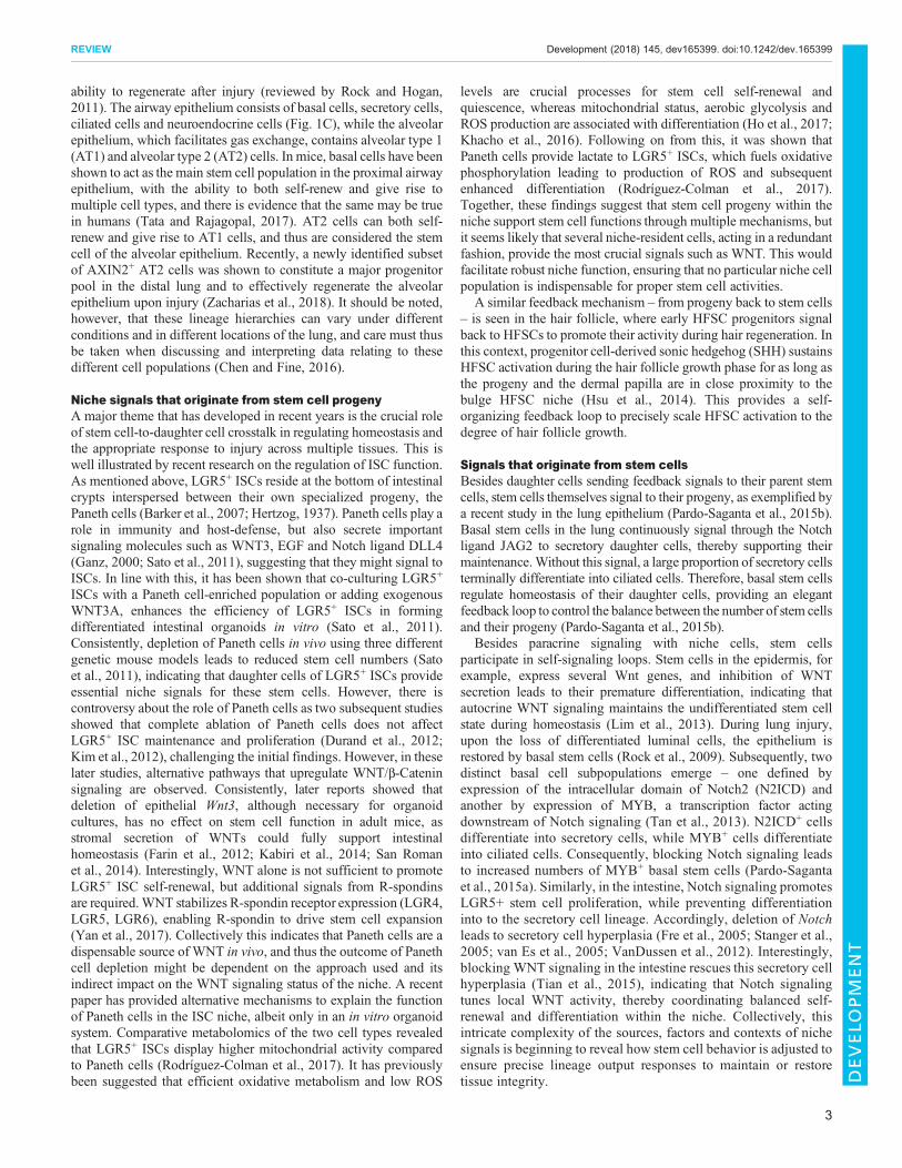
Niche signals modulate cellular plasticity following injuryIn the classical view of strict cell hierarchy within tissues, stem cellsreside on top of a lineage hierarchy, and once they make the decisionto commit to differentiation the process is irreversible (SánchezAlvarado and Yamanaka, 2014). In recent years, this concept hasbeen challenged as numerous studies demonstrate that progenitorcells and even more differentiated cell types exhibit enormousplasticity (Fig. 2), endowing themwith the ability to transdifferentiateand dedifferentiate under certain homeostatic and regenerativeconditions in processes orchestrated by the niche (Chacón-Martínezet al., 2017; Ritsma et al., 2014; Rompolas et al., 2013; Sun et al.,2014; Takeda et al., 2011).
Dedifferentiation refers to the process in which differentiatedcells revert to a less differentiated state within their lineage. Inmammalian epithelia, the advent of lineage tracing has begun tounearth the widespread nature of dedifferentiation of stem cellprogeny back to stem cell states. Such studies have revealed that cellfate plasticity is particularly apparent during injury and subsequentregeneration, when tissue and niche architecture are disrupted andremodeled. For example, the replenishment of LRG5+ ISCs aftertheir genetic ablation suggests that these stem cells are dispensablefor tissue homeostasis, as slow-cycling BMI1+ cells have the abilityto mobilize into the empty niche and dedifferentiate into LGR5+
ISCs (Muñoz et al., 2012; Tian et al., 2011). In a similar fashion,LGR5+ progeny such as DLL1+ secretory progenitors and label-retaining cells, located at crypt position +3/+4, were shown to havethe ability to regenerate LGR5+ ISCs in vivo upon acute stem cellloss (Buczacki et al., 2013; van Es et al., 2012). Of note, while invitro cultures of FACS-purified LRG5+ cells give rise to organoids,also known as mini-guts (Sato et al., 2009), purified DLL1+ cells donot generate organoids using standard culture conditions (in thepresence of EGF, Noggin and R-spondin1). However, the addition
4
REVIEW Development (2018) 145, dev165399. doi:10.1242/dev.165399
DEVELO
PM
ENT
of WNT3 to the culture medium allows full organoid formation byDLL1+ cells, suggesting a crucial role of niche-derived WNTsignaling in secretory progenitor dedifferentiation (van Es et al.,2012). Interestingly, the loss of LGR5+ ISCs (induced by diphtheriatoxin administration in a genetic mouse model to lineage traceenterocyte precursors, which are characterized by the expression ofthe alkaline phosphatase intestinal ALPI), revealed the ability ofenterocytes to dedifferentiate into LGR5+ ISCs. Of note, ALPI+
cells lose this plasticity as they differentiate and move out of thecrypt niche. Single-cell gene expression analyses revealed thatregeneration of LGR5+ cells by ALPI+ enterocytes does not involvetransition through a DLL1-expressing progenitor cell state (Tettehet al., 2016), indicating that several routes of dedifferentiation exist.Together, these studies have revealed a high degree of plasticitywithin the intestine in response to injury, which is guided by thepositioning of cells in the crypt niche and the WNT-enrichedsignaling microenvironment of the crypt (Fig. 2A).A similar dedifferentiation phenomenon has been observed in the
gastric epithelium, where Troy+ (TNFRSF19+) and LGR5+
differentiated chief cells populate the base of gastric glands.These cells do not have stem cell functions during homeostasis.However, upon damage-induced loss of stem cells in the isthmusregion adjacent to the opening of the gland, chief cells becomeactive and are able to replenish isthmus stem cells as well as parietal,mucous and neuroendocrine cells in a WNT-dependent manner(Leushacke et al., 2017; Stange et al., 2013). Taken together, thesefindings show that, upon stem cell loss after injury, exposure todefined stem cell-niche signals such as WNT instructs multipleclasses of early progeny to re-acquire a stem cell state.Similar roles of differentiated cells as stem cell reservoirs have
been identified in other tissues such as the skin interfollicular
epidermis (IFE) and the hair follicle during regeneration followinginjury (Fig. 2B). Unlike HFSCs, IFE stem cells constantlyproliferate to renew the epidermis. Although initially thought tobe maintained by quiescent stem cells and rapidly dividing transit-amplifying cells (Barrandon and Green, 1987; Potten and Morris,1988), a single progenitor population with equal probability to self-renew and differentiate has been suggested to be responsible formaintaining the IFE (Clayton et al., 2007; Mascré et al., 2012;Rompolas et al., 2016). However, the existence of functionallyheterogeneous stem cell populations in specific locations of themouse tail epidermis has been reported (Gomez et al., 2013; Mascréet al., 2012; Sada et al., 2016). In addition, a degree of transcriptionalheterogeneity and the existence of an LGR6+ subpopulation withinthe mouse back skin IFE has been observed (Füllgrabe et al., 2015;Joost et al., 2016; Snippert et al., 2010a), but the functionalsignificance of this heterogeneity is unclear. Thus, although theprecise molecular identity of the IFE stem cell remains elusive,transcriptional and functional heterogeneity is most likely controledby anatomical location and therefore region-specific, currentlyunknown, niche signals.
HFSCs represent further evidence of niche-induced plasticity.These stem cells do not normally contribute to IFE maintenance butmigrate to sites of epidermal wounds, occupy the IFE niche and,through currently unknown signals from this new niche,subsequently adopt IFE fate and contribute to tissue repair afterinjury (Blanpain et al., 2004; Claudinot et al., 2005; Ito et al., 2005).Interestingly, in vivo two-photon imaging coupled to lineage tracingand laser ablation revealed that HFSCs are in fact dispensable forhair regeneration; their committed, immediate progeny, as well asother K14+ cells located in the IFE, infundibulum and sebaceousglands, can repopulate the empty stem cell niche, adopt stem cell
Differentiation
Key
De-/trans-differentiation ? Unknown signaling pathways
C Proximal airway
Basalcell
Ciliatedcell
Secretorycell
YAPcell-cellcontact
B Skin
HFSC IFE SC
Infundibulum cellor sebocyte
Progeny
SHH
?
?
DifferentiatedIFE cell
?
?
A Intestine
LGR5+ ISC +4 SC
Paneth cell
Transit-amplifying cell
Enterocyte Globet cell
Enteroendocytecell
?
WNTs
WNTs
Fig. 2. Niche-controled differentiation trajectories and plasticity in epithelia. (A) In the intestine, progenitors and also enterocytes can dedifferentiate toLGR5+ intestinal stem cells (ISCs) throughWNT-mediated niche signals. (B) In the skin, the immediate progeny of hair follicle stem cells (HFSCs) as well as moredistant populations located in the interfollicular epidermis (IFE), infundibulum and sebaceous glands can repopulate the bulge stem cell niche upon HFSC loss.The precise signals that control this plasticity are unclear but in vitro studies implicate sonic hedgehog (SHH) signaling in this process. In response to wounding,HFSCs are able to migrate into the IFE to regenerate the epidermis and, vice versa, IFE stem cells can generate hair follicles upon transplantation. Althoughexperimental evidence for many dedifferentiation events is compelling (solid arrows) for others it is preliminary (broken arrows). (C) In the lung proximal airwayepithelium, secretory cells can dedifferentiate into basal cells through signals that involve direct cell-cell contact and the transcription factor YAP.
5
REVIEW Development (2018) 145, dev165399. doi:10.1242/dev.165399
DEVELO
PM
ENT
features and actively contribute to subsequent hair regenerationcycles (Rompolas et al., 2013). Similarly, diphtheria toxin-mediatedablation of the LGR5+ subpopulation of HFSCs initially results inabrogation of hair follicle regeneration, but eventually the LGR5+
stem cells as well as normal hair growth is restored by the CD34+
HFSC population (Hoeck et al., 2017). Niche-derived signalscontributing to this phenomenon could emanate from thesurrounding ECM (Morgner et al., 2015), from contiguous stemcell progeny (Hsu et al., 2011; van Es et al., 2012) and/or fromneighboring mesenchymal cells (Chi et al., 2013). For example, ithas been shown that the hair follicle bulge ECM niche has a specificcomposition, with very low levels of the basement membraneprotein laminin 511 compared with the adjacent hair germ thatharbors the activated progeny, and that contact of stem cells withlaminin 511 induces WNT signaling (Morgner et al., 2015). Thisestablishes a mechanism by which a niche-specific ECM couldcontrol stem cell differentiation. Intriguingly, disrupting thephysical contiguity between the epidermal and mesenchymalniche components impairs the dedifferentiation of committedepidermal cells into HFSCs and subsequent hair regeneration(Rompolas et al., 2013). This indicates that direct epidermal-dermalproximity within the niche is indispensable for specifying the HFSCfate during dedifferentiation. On the other hand, exogenousmanipulation of key niche factors such as SHH is sufficient totrigger dedifferentiation of progenitors to the HFSC state in vitro(Chacón-Martínez et al., 2017).Cellular plasticity within the lung has also been reported
(Fig. 2C). In the proximal airway, committed secretory cells candedifferentiate into basal stem cells upon acute loss of these stemcells (Tata et al., 2013). Using diphtheria toxin to specifically ablateCK5+ basal stem cells, secretory cells were found to proliferaterapidly to compensate for basal stem cell loss. Around 8% of thesecells lost markers of secretory cells, while gaining expression ofbasal stem cell markers (Tata et al., 2013). Interestingly, thededifferentiation process of secretory cells is modulated by directcell-cell contact with basal stem cells (Tata et al., 2013) and by themechanosensitive transcription factor YAP (Zhao et al., 2014),which points to a reciprocal mechanism that relies on single-celllevel interactions and sensing the density of the stem cell layer toensure tissue integrity.Collectively, these findings imply that localized signals from the
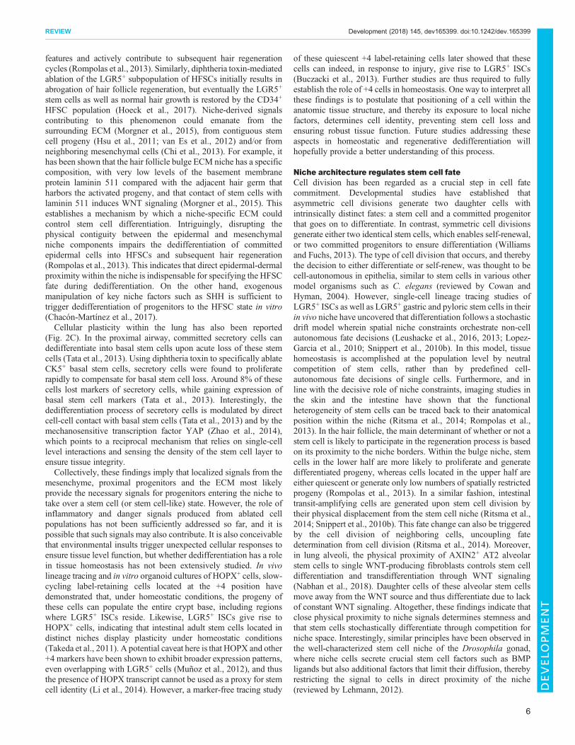
mesenchyme, proximal progenitors and the ECM most likelyprovide the necessary signals for progenitors entering the niche totake over a stem cell (or stem cell-like) state. However, the role ofinflammatory and danger signals produced from ablated cellpopulations has not been sufficiently addressed so far, and it ispossible that such signals may also contribute. It is also conceivablethat environmental insults trigger unexpected cellular responses toensure tissue level function, but whether dedifferentiation has a rolein tissue homeostasis has not been extensively studied. In vivolineage tracing and in vitro organoid cultures of HOPX+ cells, slow-cycling label-retaining cells located at the +4 position havedemonstrated that, under homeostatic conditions, the progeny ofthese cells can populate the entire crypt base, including regionswhere LGR5+ ISCs reside. Likewise, LGR5+ ISCs give rise toHOPX+ cells, indicating that intestinal adult stem cells located indistinct niches display plasticity under homeostatic conditions(Takeda et al., 2011). A potential caveat here is that HOPX and other+4 markers have been shown to exhibit broader expression patterns,even overlapping with LGR5+ cells (Muñoz et al., 2012), and thusthe presence of HOPX transcript cannot be used as a proxy for stemcell identity (Li et al., 2014). However, a marker-free tracing study
of these quiescent +4 label-retaining cells later showed that thesecells can indeed, in response to injury, give rise to LGR5+ ISCs(Buczacki et al., 2013). Further studies are thus required to fullyestablish the role of +4 cells in homeostasis. One way to interpret allthese findings is to postulate that positioning of a cell within theanatomic tissue structure, and thereby its exposure to local nichefactors, determines cell identity, preventing stem cell loss andensuring robust tissue function. Future studies addressing theseaspects in homeostatic and regenerative dedifferentiation willhopefully provide a better understanding of this process.
Niche architecture regulates stem cell fateCell division has been regarded as a crucial step in cell fatecommitment. Developmental studies have established thatasymmetric cell divisions generate two daughter cells withintrinsically distinct fates: a stem cell and a committed progenitorthat goes on to differentiate. In contrast, symmetric cell divisionsgenerate either two identical stem cells, which enables self-renewal,or two committed progenitors to ensure differentiation (Williamsand Fuchs, 2013). The type of cell division that occurs, and therebythe decision to either differentiate or self-renew, was thought to becell-autonomous in epithelia, similar to stem cells in various othermodel organisms such as C. elegans (reviewed by Cowan andHyman, 2004). However, single-cell lineage tracing studies ofLGR5+ ISCs as well as LGR5+ gastric and pyloric stem cells in theirin vivo niche have uncovered that differentiation follows a stochasticdrift model wherein spatial niche constraints orchestrate non-cellautonomous fate decisions (Leushacke et al., 2016, 2013; Lopez-Garcia et al., 2010; Snippert et al., 2010b). In this model, tissuehomeostasis is accomplished at the population level by neutralcompetition of stem cells, rather than by predefined cell-autonomous fate decisions of single cells. Furthermore, and inline with the decisive role of niche constraints, imaging studies inthe skin and the intestine have shown that the functionalheterogeneity of stem cells can be traced back to their anatomicalposition within the niche (Ritsma et al., 2014; Rompolas et al.,2013). In the hair follicle, the main determinant of whether or not astem cell is likely to participate in the regeneration process is basedon its proximity to the niche borders. Within the bulge niche, stemcells in the lower half are more likely to proliferate and generatedifferentiated progeny, whereas cells located in the upper half areeither quiescent or generate only low numbers of spatially restrictedprogeny (Rompolas et al., 2013). In a similar fashion, intestinaltransit-amplifying cells are generated upon stem cell division bytheir physical displacement from the stem cell niche (Ritsma et al.,2014; Snippert et al., 2010b). This fate change can also be triggeredby the cell division of neighboring cells, uncoupling fatedetermination from cell division (Ritsma et al., 2014). Moreover,in lung alveoli, the physical proximity of AXIN2+ AT2 alveolarstem cells to single WNT-producing fibroblasts controls stem celldifferentiation and transdifferentiation through WNT signaling(Nabhan et al., 2018). Daughter cells of these alveolar stem cellsmove away from the WNT source and thus differentiate due to lackof constant WNT signaling. Altogether, these findings indicate thatclose physical proximity to niche signals determines stemness andthat stem cells stochastically differentiate through competition forniche space. Interestingly, similar principles have been observed inthe well-characterized stem cell niche of the Drosophila gonad,where niche cells secrete crucial stem cell factors such as BMPligands but also additional factors that limit their diffusion, therebyrestricting the signal to cells in direct proximity of the niche(reviewed by Lehmann, 2012).
6
REVIEW Development (2018) 145, dev165399. doi:10.1242/dev.165399
DEVELO
PM
ENT
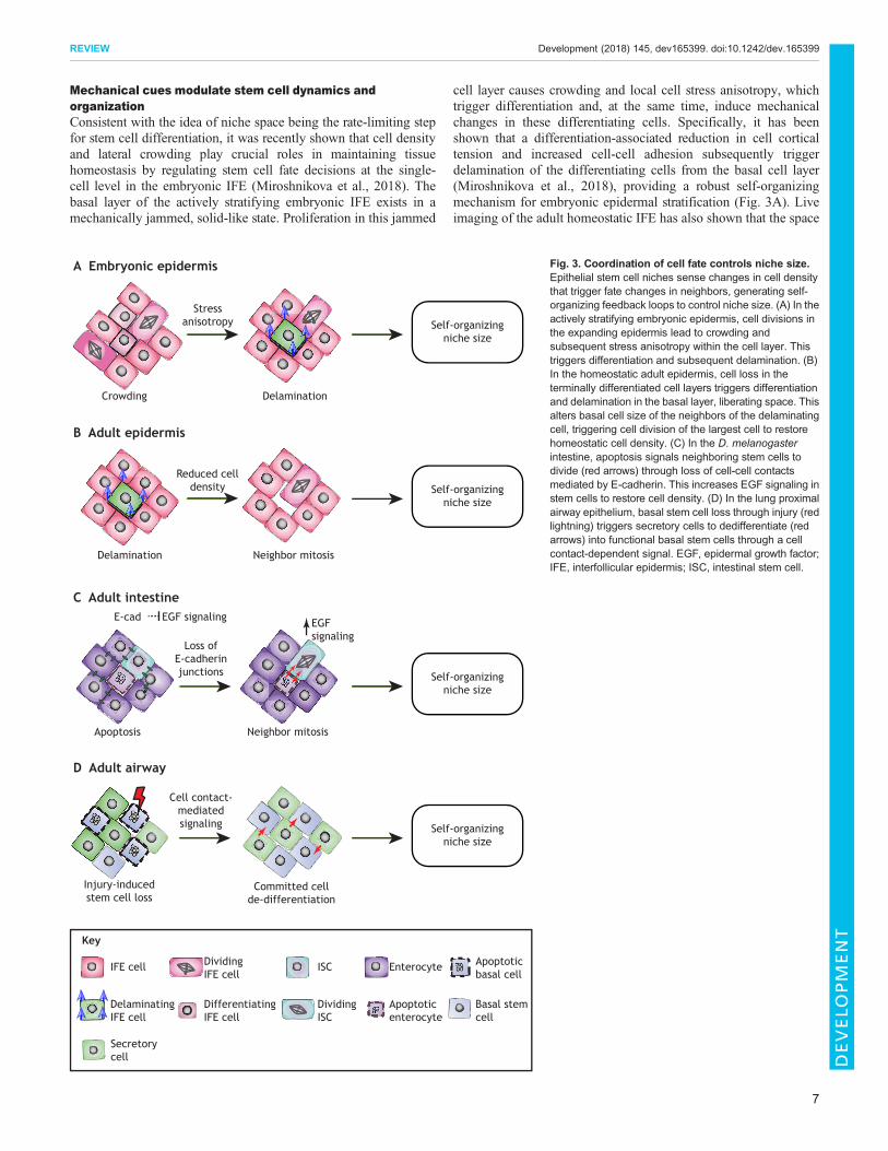
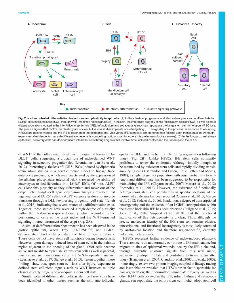
Mechanical cues modulate stem cell dynamics andorganizationConsistent with the idea of niche space being the rate-limiting stepfor stem cell differentiation, it was recently shown that cell densityand lateral crowding play crucial roles in maintaining tissuehomeostasis by regulating stem cell fate decisions at the single-cell level in the embryonic IFE (Miroshnikova et al., 2018). Thebasal layer of the actively stratifying embryonic IFE exists in amechanically jammed, solid-like state. Proliferation in this jammed
cell layer causes crowding and local cell stress anisotropy, whichtrigger differentiation and, at the same time, induce mechanicalchanges in these differentiating cells. Specifically, it has beenshown that a differentiation-associated reduction in cell corticaltension and increased cell-cell adhesion subsequently triggerdelamination of the differentiating cells from the basal cell layer(Miroshnikova et al., 2018), providing a robust self-organizingmechanism for embryonic epidermal stratification (Fig. 3A). Liveimaging of the adult homeostatic IFE has also shown that the space
Crowding Delamination
Stressanisotropy
A Embryonic epidermis
Self-organizingniche size
Delamination Neighbor mitosis
Reduced celldensity
B Adult epidermis
Self-organizingniche size
Apoptosis Neighbor mitosis
Loss ofE-cadherinjunctions
C Adult intestineE-cad EGF signaling EGF
signaling
Self-organizingniche size
Injury-inducedstem cell loss
Committed cellde-differentiation
Cell contact-mediatedsignaling
D Adult airway
Self-organizingniche size
Key
IFE cell
DelaminatingIFE cell
DividingISC
ISC Enterocyte
Apoptoticenterocyte
Basal stemcell
Apoptoticbasal cell
Secretorycell
DividingIFE cell
DifferentiatingIFE cell
Fig. 3. Coordination of cell fate controls niche size.Epithelial stem cell niches sense changes in cell densitythat trigger fate changes in neighbors, generating self-organizing feedback loops to control niche size. (A) In theactively stratifying embryonic epidermis, cell divisions inthe expanding epidermis lead to crowding andsubsequent stress anisotropy within the cell layer. Thistriggers differentiation and subsequent delamination. (B)In the homeostatic adult epidermis, cell loss in theterminally differentiated cell layers triggers differentiationand delamination in the basal layer, liberating space. Thisalters basal cell size of the neighbors of the delaminatingcell, triggering cell division of the largest cell to restorehomeostatic cell density. (C) In the D. melanogasterintestine, apoptosis signals neighboring stem cells todivide (red arrows) through loss of cell-cell contactsmediated by E-cadherin. This increases EGF signaling instem cells to restore cell density. (D) In the lung proximalairway epithelium, basal stem cell loss through injury (redlightning) triggers secretory cells to dedifferentiate (redarrows) into functional basal stem cells through a cellcontact-dependent signal. EGF, epidermal growth factor;IFE, interfollicular epidermis; ISC, intestinal stem cell.
7
REVIEW Development (2018) 145, dev165399. doi:10.1242/dev.165399
DEVELO
PM
ENT
liberated by delamination of a epidermal cell triggers the divisionof its neighboring cell (Mesa et al., 2017 preprint), which isconsistent with the idea that basal cell density affects stem cell fatedecisions in the epidermal layer. Interestingly, whereas in theactively stratifying embryonic epidermis cell divisions triggercrowding, in the homeostatic adult tissue cell divisions do notimpact delamination but divisions occur in response to delamination(Fig. 3B). This could indicate that delamination and stem celldivisions are mechanically coupled in both the embryonic andadult epidermis, but that stem cells in the embryo are constantlycycling to provide sufficient material for the growing tissue,whereas adult stem cells divide only upon demand to replaceterminally differentiated dying cells. A similar mechanism of celldensity-driven homeostasis seems to operate in the Drosophilamidgut (Fig. 3C), where stem cell division is triggered by removalof apoptotic cells in a process dependent on cell-cell contacts andtheir ability to coordinate EGF signaling (Liang et al., 2017).Another recent study in the adult Drosophila midgut furtherhighlights the importance of mechanical signals, in particularmechanical stress in epithelial stem cell homeostasis. Here, aspecific population of enteroendocrine precursor cells sensesmechanical stimuli that regulate their differentiation in a processmediated by intracellular calcium increase through the stretch-activated calcium channel Piezo, allowing these cells to respondto gut compression or extension (He et al., 2018). Similarly,mechanical tension in response to injury has been shown topromote alveolar regeneration in the lung (Liu et al., 2016.Fig. 3D).Mechanical signals are involved not only in determining the fate
of individual cells, but also in establishing and positioning stemcell niches themselves. In the developing chick gut, buckling forcesfold the epithelium, causing local tissue folds with high SHHconcentrations. These high SHH microenvironments alter adjacentmesenchymal cell fate to form local signaling centers that signal backto the epithelium to establish intestinal crypts as stem cell niches(Shyer et al., 2015). Similarly, force-driven structural rearrangementsare not only crucial for creating the hair follicle shape but also triggerlineage commitment in the developing avian skin. Here, spontaneousdermal cell aggregation and contractility compress the epidermisfocally, leading to the mechanosensitive activation of β-catenin andsubsequent initiation of the hair follicle-specific transcriptionalprogram (Shyer et al., 2017). Early studies of follicle patterninghypothesized that a molecular pre-pattern emerges first and changescellular behaviors, which then cause changes in tissue structure(Noramly and Morgan, 1998; Widelitz and Chuong, 1999).However, the study by Shyer and co-workers now indicates thatinitial follicle fate markers, such as β-catenin, are turned onsimultaneously with architectural changes, instead of precedingthem, and that the developing skin is able to form spaced aggregateswithout β-catenin activation, suggesting that pre-patterning does notoccur (Shyer et al., 2017). Collectively, these studies highlight thatthemechanical regulation of stem cell positioning, density, contractilityand compression within the niche provide key instructive signals thatcontribute to providing robust, self-organizing principles thatcontrol stem cell functions (Fig. 3).
Conclusions and perspectivesRecent developments in the fields of single cell sequencing, highresolution imaging and bioengineering have made it possible tomake significant leaps in our understanding of adult stem cellbiology. It has become evident that the stem cell niche provides aspectrum of cues that ensure plasticity of the stem cell compartment
and prevent stem cell loss. Furthermore, the precise location ofstem cells within their niche provides additional fine-tuning oftranscriptional programs, thereby facilitating stem cell heterogeneity,which is important for regulating the appropriate responses to therapidly changing needs of the tissue. A key emerging paradigm is thepresence of self-organizing feedback loops that are based on directcell-cell contact and density sensing. These self-organizingcircuits allow coordinated behaviors of stem cells within a niche.Coupling stem cell loss to neighbor division, or vice versa,provides simple and efficient control of niche size and tissuearchitecture, and could represent a universal feature of stem cellregulation, at least in epithelia.
Despite these recent advances, several key questions remainunanswered regarding the precise molecular mechanisms of stemcell heterogeneity and plasticity. In particular, it will be important tounderstand which regulatory mechanisms and phenomena arerelevant for homeostasis and which of them are used primarily oreven exclusively in response to stress or damage. In addition, morecomprehensive characterization of stem cell niches is still required,as the roles of inflammatory cells, neuronal cells and other niche-proximal cell types are only beginning to emerge. Moreover, manytissues show a decline in regenerative potential during agingcoupled with a loss of stem cell homeostasis and function (López-Otín et al., 2013; Signer and Morrison, 2013); in fact, a directmechanistic link between organismal lifespan and stem cell activityhas been demonstrated, pioneered by work in Drosophila ISCs(Biteau et al., 2010). Thus, understanding how aging affects thecomposition and function of stem cell niches and whether niche-targeted therapies could be used to enhance tissue homeostasis andregeneration potential during aging will be exciting avenues offuture research.
Our increasing understanding of the regulatory networks andenvironmental factors dictating stem cell functions has alreadyenabled the design of in vitro stem cell models, such as organoids(Latil et al., 2017; Nichane et al., 2017; Sato et al., 2009; Yin et al.,2014) and other self-organizing stem cell cultures (Chacón-Martínez et al., 2017, Harrison et al., 2017; Tewary et al., 2017;Turner et al., 2017). These in vitro tools enable mechanisticinterrogation of key components of niches and their contribution(chemical, mechanical and physical) to self-renewal, plasticity anddifferentiation, as well as precise manipulation of both stem cellsand niche components through new technologies such as geneediting. These models are rapidly advancing basic and appliedresearch (Huch et al., 2017), and are also being expanded to diseasemodels and predictive tools of drug-treatment outcomes, asexemplified by the recently established human cancer organoidbiobanks (Sachs et al., 2018; Vlachogiannis et al., 2018). The betterwe understand the complexity of niches and how they regulate stemcell plasticity and self-organization, the better we will be able toharness the potential of stem cells for use in regenerative therapiesand personalized medicine.
AcknowledgementsWe thank members of the Wickstrom lab for discussions and critical reading of themanuscript.
Competing interestsThe authors declare no competing or financial interests.
FundingResearch in the Wickstrom lab is supported by the Helsingin Yliopisto, the Jenny jaAntti Wihurin Rahasto, the Jane ja Aatos Erkon saatio, the Max-Planck-Gesellschaftand the Deutsche Forschungsgemeinschaft through SFB 829 A11.
8
REVIEW Development (2018) 145, dev165399. doi:10.1242/dev.165399
DEVELO
PM
ENT
ReferencesAli, N., Zirak, B., Rodriguez, R. S., Pauli, M. L., Truong, H. A., Lai, K., Ahn, R.,Corbin, K., Lowe, M. M., Scharschmidt, T. C. et al. (2017). Regulatory T cells inskin facilitate epithelial stem cell differentiation. Cell 169, 1119-1129.e1111.
Arpaia, N., Green, J. A., Moltedo, B., Arvey, A., Hemmers, S., Yuan, S., Treuting,P. M. and Rudensky, A. Y. (2015). A distinct function of regulatory T cells in tissueprotection. Cell 162, 1078-1089.
Barker, N., van Es, J. H., Kuipers, J., Kujala, P., van denBorn, M., Cozijnsen, M.,Haegebarth, A., Korving, J., Begthel, H., Peters, P. J. et al. (2007).Identification of stem cells in small intestine and colon by marker gene Lgr5.Nature 449, 1003-1007.
Barrandon, Y. and Green, H. (1987). Three clonal types of keratinocyte withdifferent capacities for multiplication. Proc. Natl. Acad. Sci. USA 84, 2302-2306.
Becker, A. J., McCulloch, E. A. and Till, J. E. (1963). Cytological demonstration ofthe clonal nature of spleen colonies derived from transplanted mouse marrowcells. Nature 197, 452-454.
Bettelli, E., Carrier, Y., Gao, W., Korn, T., Strom, T. B., Oukka, M., Weiner, H. L.and Kuchroo, V. K. (2006). Reciprocal developmental pathways for thegeneration of pathogenic effector TH17 and regulatory T cells. Nature 441,235-238.
Biteau, B., Karpac, J., Supoyo, S., Degennaro, M., Lehmann, R. and Jasper, H.(2010). Lifespan extension by preserving proliferative homeostasis in Drosophila.PLoS Genet. 6, e1001159.
Blanpain, C. and Fuchs, E. (2014). Stem cell plasticity. Plasticity of epithelial stemcells in tissue regeneration. Science 344, 1242281.
Blanpain, C., Lowry, W. E., Geoghegan, A., Polak, L. and Fuchs, E. (2004). Self-renewal, multipotency, and the existence of two cell populations within anepithelial stem cell niche. Cell 118, 635-648.
Buczacki, S. J. A., Zecchini, H. I., Nicholson, A. M., Russell, R., Vermeulen, L.,Kemp, R. and Winton, D. J. (2013). Intestinal label-retaining cells are secretoryprecursors expressing Lgr5. Nature 495, 65-69.
Chacon-Martınez, C. A., Klose, M., Niemann, C., Glauche, I. and Wickstrom,S. A. (2017). Hair follicle stem cell cultures reveal self-organizing plasticity of stemcells and their progeny. EMBO J. 36, 151-164.
Chen, F. and Fine, A. (2016). Stem cells in lung injury and repair.Am. J. Pathol. 186,2544-2550.
Chi, W., Wu, E. andMorgan, B. A. (2013). Dermal papilla cell number specifies hairsize, shape and cycling and its reduction causes follicular decline. Development140, 1676-1683.
Claudinot, S., Nicolas, M., Oshima, H., Rochat, A. and Barrandon, Y. (2005).Long-term renewal of hair follicles from clonogenic multipotent stem cells. Proc.Natl. Acad. Sci. USA 102, 14677-14682.
Clayton, E., Doupe, D. P., Klein, A. M., Winton, D. J., Simons, B. D. and Jones,P. H. (2007). A single type of progenitor cell maintains normal epidermis. Nature446, 185-189.
Cowan, C. R. and Hyman, A. A. (2004). Asymmetric cell division in C. elegans:cortical polarity and spindle positioning. Annu. Rev. Cell Dev. Biol. 20, 427-453.
Crosnier, C., Stamataki, D. and Lewis, J. (2006). Organizing cell renewal in theintestine: stem cells, signals and combinatorial control. Nat. Rev. Genet. 7,349-359.
Durand, A., Donahue, B., Peignon, G., Letourneur, F., Cagnard, N., Slomianny,C., Perret, C., Shroyer, N. F. and Romagnolo, B. (2012). Functional intestinalstem cells after Paneth cell ablation induced by the loss of transcription factorMath1 (Atoh1). Proc. Natl. Acad. Sci. USA 109, 8965-8970.
Farin, H. F., van Es, J. H. and Clevers, H. (2012). Redundant sources of Wntregulate intestinal stem cells and promote formation of Paneth cells.Gastroenterology 143, 1518-1529.e1517.
Fre, S., Huyghe, M., Mourikis, P., Robine, S., Louvard, D. and Artavanis-Tsakonas, S. (2005). Notch signals control the fate of immature progenitor cells inthe intestine. Nature 435, 964-968.
Fujisaki, J., Wu, J., Carlson, A. L., Silberstein, L., Putheti, P., Larocca, R., Gao,W., Saito, T. I., Lo Celso, C., Tsuyuzaki, H. et al. (2011). In vivo imaging of Tregcells providing immune privilege to the haematopoietic stem-cell niche. Nature474, 216-219.
Fullgrabe, A., Joost, S., Are, A., Jacob, T., Sivan, U., Haegebarth, A.,Linnarsson, S., Simons, B. D., Clevers, H., Toftgård, R. et al. (2015).Dynamics of Lgr6(+) progenitor cells in the hair follicle, sebaceous gland, andinterfollicular epidermis. Stem Cell Rep. 5, 843-855.
Ganz, T. (2000). Paneth cells–guardians of the gut cell hatchery. Nat. Immunol. 1,99-100.
Gomez, C., Chua, W., Miremadi, A., Quist, S., Headon, D. J. and Watt, F. M.(2013). The interfollicular epidermis of adult mouse tail comprises two distinct celllineages that are differentially regulated by Wnt, Edaradd, and Lrig1. Stem CellRep. 1, 19-27.
Gonzales, K. A. U. and Fuchs, E. (2017). Skin and its regenerative powers: analliance between stem cells and their niche. Dev. Cell 43, 387-401.
Goodell, M. A., Nguyen, H. and Shroyer, N. (2015). Somatic stem cellheterogeneity: diversity in the blood, skin and intestinal stem cellcompartments. Nat. Rev. Mol. Cell Biol. 16, 299-309.
Harrison, S. E., Sozen, B., Christodoulou, N., Kyprianou, C. and Zernicka-Goetz, M. (2017). Assembly of embryonic and extraembryonic stem cells to mimicembryogenesis in vitro. Science 356, eaal1810.
He, L., Si, G., Huang, J., Samuel, A. D. T. and Perrimon, N. (2018). Mechanicalregulation of stem-cell differentiation by the stretch-activated Piezo channel.Nature 555, 103-106.
Hertzog, A. J. (1937). The Paneth cell. Am. J. Pathol. 13, 351-360.Ho, T. T., Warr, M. R., Adelman, E. R., Lansinger, O. M., Flach, J., Verovskaya,
E. V., Figueroa, M. E. and Passegue, E. (2017). Autophagy maintains themetabolism and function of young and old stem cells. Nature 543, 205-210.
Hoeck, J. D., Biehs, B., Kurtova, A. V., Kljavin, N. M., de Sousa eMelo, F., Alicke,B., Koeppen, H., Modrusan, Z., Piskol, R. and de Sauvage, F. J. (2017). Stemcell plasticity enables hair regeneration following Lgr5(+) cell loss. Nat. Cell Biol.19, 666-676.
Hsu, Y.-C., Pasolli, H. A. and Fuchs, E. (2011). Dynamics between stem cells,niche, and progeny in the hair follicle. Cell 144, 92-105.
Hsu, Y.-C., Li, L. and Fuchs, E. (2014). Transit-amplifying cells orchestrate stemcell activity and tissue regeneration. Cell 157, 935-949.
Huch, M., Knoblich, J. A., Lutolf, M. P. and Martinez-Arias, A. (2017). The hopeand the hype of organoid research. Development 144, 938-941.
Ito, M., Liu, Y., Yang, Z., Nguyen, J., Liang, F., Morris, R. J. and Cotsarelis, G.(2005). Stem cells in the hair follicle bulge contribute to wound repair but not tohomeostasis of the epidermis. Nat. Med. 11, 1351-1354.
Joost, S., Zeisel, A., Jacob, T., Sun, X., La Manno, G., Lonnerberg, P.,Linnarsson, S. and Kasper, M. (2016). Single-cell transcriptomics reveals thatdifferentiation and spatial signatures shape epidermal and hair follicleheterogeneity. Cell Syst. 3, 221-237.e229.
Kabiri, Z., Greicius, G., Madan, B., Biechele, S., Zhong, Z., Zaribafzadeh, H.,Edison, Aliyev, J., Wu, Y., Bunte, R. et al. (2014). Stroma provides an intestinalstem cell niche in the absence of epithelial Wnts. Development 141, 2206-2215.
Khacho, M., Clark, A., Svoboda, D. S., Azzi, J., MacLaurin, J. G., Meghaizel, C.,Sesaki, H., Lagace, D. C., Germain, M., Harper, M.-E. et al. (2016).Mitochondrial dynamics impacts stem cell identity and fate decisions byregulating a nuclear transcriptional program. Cell Stem Cell 19, 232-247.
Kim, T.-H., Escudero, S. and Shivdasani, R. A. (2012). Intact function of Lgr5receptor-expressing intestinal stem cells in the absence of Paneth cells. Proc.Natl. Acad. Sci. USA 109, 3932-3937.
Krieger, T. and Simons, B. D. (2015). Dynamic stem cell heterogeneity.Development 142, 1396-1406.
Latil, M., Nassar, D., Beck, B., Boumahdi, S., Wang, L., Brisebarre, A., Dubois,C., Nkusi, E., Lenglez, S., Checinska, A. et al. (2017). Cell-type-specificchromatin states differentially prime squamous cell carcinoma tumor-initiatingcells for epithelial to mesenchymal transition. Cell Stem Cell 20, 191-204.e195.
Lee, J.-H., Bhang, D. H., Beede, A., Huang, T. L., Stripp, B. R., Bloch, K. D.,Wagers, A. J., Tseng, Y.-H., Ryeom, S. and Kim, C. F. (2014). Lung stem celldifferentiation in mice directed by endothelial cells via a BMP4-NFATc1-thrombospondin-1 axis. Cell 156, 440-455.
Lee, J. H., Tammela, T., Hofree, M., Choi, J., Marjanovic, N. D., Han, S., Canner,D., Wu, K., Paschini, M., Bhang, D. H. et al. (2017). Anatomically andfunctionally distinct lung mesenchymal populations marked by Lgr5 and Lgr6.Cell170, 1149-1163.e1112.
Leeman, K. T., Fillmore, C. M. and Kim, C. F. (2014). Lung stem and progenitorcells in tissue homeostasis and disease. Curr. Top. Dev. Biol. 107, 207-233.
Lehmann, R. (2012). Germline stem cells: origin and destiny. Cell Stem Cell 10,729-739.
Leushacke, M., Ng, A., Galle, J., Loeffler, M. and Barker, N. (2013). Lgr5(+)gastric stem cells divide symmetrically to effect epithelial homeostasis in thepylorus. Cell Rep. 5, 349-356.
Leushacke, M., Barker, N. and Pin, C. (2016). Quantifying Lgr5-positive stem cellbehaviour in the pyloric epithelium. Sci. Rep. 6, 21923.
Leushacke, M., Tan, S. H., Wong, A., Swathi, Y., Hajamohideen, A., Tan, L. T.,Goh, J., Wong, E., Denil, S. L. I. J., Murakami, K. et al. (2017). Lgr5-expressingchief cells drive epithelial regeneration and cancer in the oxyntic stomach. Nat.Cell Biol. 19, 774-786.
Li, N., Yousefi, M., Nakauka-Ddamba, A., Jain, R., Tobias, J., Epstein, J. A.,Jensen, S. T. and Lengner, C. J. (2014). Single-cell analysis of proxy reporterallele-marked epithelial cells establishes intestinal stem cell hierarchy. Stem CellRep. 3, 876-891.
Liang, J., Balachandra, S., Ngo, S. andO’Brien, L. E. (2017). Feedback regulationof steady-state epithelial turnover and organ size. Nature 548, 588-591.
Lim, X., Tan, S. H., Koh, W. L. C., Chau, R. M. W., Yan, K. S., Kuo, C. J., vanAmerongen, R., Klein, A. M. andNusse, R. (2013). Interfollicular epidermal stemcells self-renew via autocrine Wnt signaling. Science 342, 1226-1230.
Liu, Z., Wu, H., Jiang, K., Wang, Y., Zhang, W., Chu, Q., Li, J., Huang, H., Cai, T.,Ji, H. et al. (2016). MAPK-mediated YAP activation controls mechanical-tension-induced pulmonary alveolar regeneration. Cell Rep. 16, 1810-1819.
Lopez-Garcia, C., Klein, A. M., Simons, B. D. and Winton, D. J. (2010). Intestinalstem cell replacement follows a pattern of neutral drift. Science 330, 822-825.
Lopez-Otın, C., Blasco, M. A., Partridge, L., Serrano, M. andKroemer, G. (2013).The hallmarks of aging. Cell 153, 1194-1217.
9
REVIEW Development (2018) 145, dev165399. doi:10.1242/dev.165399
DEVELO
PM
ENT
Mascre, G., Dekoninck, S., Drogat, B., Youssef, K. K., Brohee, S., Sotiropoulou,P. A., Simons, B. D. and Blanpain, C. (2012). Distinct contribution of stem andprogenitor cells to epidermal maintenance. Nature 489, 257-262.
Mesa, K. R., Kawaguchi, K., Gonzalez, D. G., Cockburn, K., Boucher, J., Xin, T.,Klein, A. M. and Greco, V. (2017). Epidermal stem cells self-renew uponneighboring differentiation. bioRxiv doi:10.1101/155408.
Miroshnikova, Y. A., Le, H. Q., Schneider, D., Thalheim, T., Rubsam, M.,Bremicker, N., Polleux, J., Kamprad, N., Tarantola, M., Wang, I. et al. (2018).Adhesion forces and cortical tension couple cell proliferation and differentiation todrive epidermal stratification. Nat. Cell Biol. 20, 69-80.
Montgomery, R. K., Carlone, D. L., Richmond, C. A., Farilla, L., Kranendonk,M. E. G., Henderson, D. E., Baffour-Awuah, N. Y., Ambruzs, D. M., Fogli, L. K.,Algra, S. et al. (2011). Mouse telomerase reverse transcriptase (mTert)expression marks slowly cycling intestinal stem cells. Proc. Natl. Acad. Sci.USA 108, 179-184.
Morgner, J., Ghatak, S., Jakobi, T., Dieterich, C., Aumailley, M. and Wickstrom,S. A. (2015). Integrin-linked kinase regulates the niche of quiescent epidermalstem cells. Nat. Commun. 6, 8198.
Morrison, S. J. and Spradling, A. C. (2008). Stem cells and niches: mechanismsthat promote stem cell maintenance throughout life. Cell 132, 598-611.
Muller-Rover, S., Handjiski, B., van der Veen, C., Eichmuller, S., Foitzik, K.,McKay, I. A., Stenn, K. S. and Paus, R. (2001). A comprehensive guide for theaccurate classification of murine hair follicles in distinct hair cycle stages. J. Invest.Dermatol. 117, 3-15.
Mun oz, J., Stange, D. E., Schepers, A. G., van de Wetering, M., Koo, B.-K.,Itzkovitz, S., Volckmann, R., Kung, K. S., Koster, J., Radulescu, S. et al.(2012). The Lgr5 intestinal stem cell signature: robust expression of proposedquiescent ‘+4’ cell markers. EMBO J. 31, 3079-3091.
Nabhan, A. N., Brownfield, D. G., Harbury, P. B., Krasnow, M. A. and Desai, T. J.(2018). Single-cell Wnt signaling niches maintain stemness of alveolar type 2cells. Science 359, 1118-1123.
Nichane, M., Javed, A., Sivakamasundari, V., Ganesan, M., Ang, L. T., Kraus, P.,Lufkin, T., Loh, K. M. and Lim, B. (2017). Isolation and 3D expansion ofmultipotent Sox9(+) mouse lung progenitors. Nat. Methods 14, 1205-1212.
Noramly, S. and Morgan, B. A. (1998). BMPs mediate lateral inhibition atsuccessive stages in feather tract development. Development 125, 3775-3787.
Nosbaum, A., Prevel, N., Truong, H.-A., Mehta, P., Ettinger, M., Scharschmidt,T. C., Ali, N. H., Pauli, M. L., Abbas, A. K. and Rosenblum, M. D. (2016). Cuttingedge: regulatory T cells facilitate cutaneous wound healing. J. Immunol. 196,2010-2014.
Oshima, H., Rochat, A., Kedzia, C., Kobayashi, K. and Barrandon, Y. (2001).Morphogenesis and renewal of hair follicles from adult multipotent stem cells. Cell104, 233-245.
Pardo-Saganta, A., Law, B. M., Tata, P. R., Villoria, J., Saez, B., Mou, H., Zhao, R.and Rajagopal, J. (2015a). Injury induces direct lineage segregation offunctionally distinct airway basal stem/progenitor cell subpopulations. Cell StemCell 16, 184-197.
Pardo-Saganta, A., Tata, P. R., Law, B. M., Saez, B., Chow, R. D.-W., Prabhu, M.,Gridley, T. and Rajagopal, J. (2015b). Parent stem cells can serve as niches fortheir daughter cells. Nature 523, 597-601.
Plikus, M. V., Mayer, J. A., de la Cruz, D., Baker, R. E., Maini, P. K., Maxson, R.and Chuong, C.-M. (2008). Cyclic dermal BMP signalling regulates stem cellactivation during hair regeneration. Nature 451, 340-344.
Potten, C. S. and Morris, R. J. (1988). Epithelial stem cells in vivo. J. Cell Sci. 1988Suppl. 10, 45-62.
Potten, C. S., Saffhill, R. and Maibach, H. I. (1987). Measurement of the transittime for cells through the epidermis and stratum corneum of the mouse andguinea-pig. Cell Tissue Kinet. 20, 461-472.
Reynolds, A. J. and Jahoda, C. A. (1991). Hair follicle stem cells? A distinctgerminative epidermal cell population is activated in vitro by the presence of hairdermal papilla cells. J. Cell Sci. 99, 373-385.
Ritsma, L., Ellenbroek, S. I. J., Zomer, A., Snippert, H. J., de Sauvage, F. J.,Simons, B. D., Clevers, H. and van Rheenen, J. (2014). Intestinal crypthomeostasis revealed at single-stem-cell level by in vivo live imaging. Nature 507,362-365.
Roberts, K. J., Kershner, A. M. and Beachy, P. A. (2017). The stromal niche forepithelial stem cells: a template for regeneration and a brake on malignancy.Cancer Cell 32, 404-410.
Rock, J. R. and Hogan, B. L. M. (2011). Epithelial progenitor cells in lungdevelopment, maintenance, repair, and disease. Annu. Rev. Cell Dev. Biol. 27,493-512.
Rock, J. R., Onaitis, M. W., Rawlins, E. L., Lu, Y., Clark, C. P., Xue, Y., Randell,S. H. and Hogan, B. L. M. (2009). Basal cells as stem cells of the mouse tracheaand human airway epithelium. Proc. Natl. Acad. Sci. USA 106, 12771-12775.
Rodrıguez-Colman, M. J., Schewe, M., Meerlo, M., Stigter, E., Gerrits, J., Pras-Raves, M., Sacchetti, A., Hornsveld, M., Oost, K. C., Snippert, H. J. et al.(2017). Interplay betweenmetabolic identities in the intestinal crypt supports stemcell function. Nature 543, 424-427.
Rompolas, P., Mesa, K. R. and Greco, V. (2013). Spatial organization within aniche as a determinant of stem-cell fate. Nature 502, 513-518.
Rompolas, P., Mesa, K. R., Kawaguchi, K., Park, S., Gonzalez, D., Brown, S.,Boucher, J., Klein, A. M. and Greco, V. (2016). Spatiotemporal coordination ofstem cell commitment during epidermal homeostasis. Science 352, 1471-1474.
Ruiz, E. J., Oeztuerk-Winder, F. and Ventura, J.-J. (2014). A paracrine networkregulates the cross-talk between human lung stem cells and the stroma. Nat.Commun. 5, 3175.
Sachs, N., de Ligt, J., Kopper, O., Gogola, E., Bounova, G., Weeber, F.,Balgobind, A. V., Wind, K., Gracanin, A., Begthel, H. et al. (2018). A livingbiobank of breast cancer organoids captures disease heterogeneity. Cell 172,373-386.e310.
Sada, A., Jacob, F., Leung, E., Wang, S., White, B. S., Shalloway, D. andTumbar, T. (2016). Defining the cellular lineage hierarchy in the interfollicularepidermis of adult skin. Nat. Cell Biol. 18, 619-631.
San Roman, A. K., Jayewickreme, C. D., Murtaugh, L. C. and Shivdasani, R. A.(2014). Wnt secretion from epithelial cells and subepithelial myofibroblasts is notrequired in the mouse intestinal stem cell niche in vivo. StemCell Rep. 2, 127-134.
Sanchez Alvarado, A. and Yamanaka, S. (2014). Rethinking differentiation: stemcells, regeneration, and plasticity. Cell 157, 110-119.
Sato, T., Vries, R. G., Snippert, H. J., van de Wetering, M., Barker, N., Stange,D. E., van Es, J. H., Abo, A., Kujala, P., Peters, P. J. et al. (2009). Single Lgr5stem cells build crypt-villus structures in vitro without a mesenchymal niche.Nature 459, 262-265.
Sato, T., van Es, J. H., Snippert, H. J., Stange, D. E., Vries, R. G., van den Born,M., Barker, N., Shroyer, N. F., van de Wetering, M. and Clevers, H. (2011).Paneth cells constitute the niche for Lgr5 stem cells in intestinal crypts. Nature469, 415-418.
Scadden, D. T. (2014). Nice neighborhood: emerging concepts of the stem cellniche. Cell 157, 41-50.
Schofield, R. (1978). The relationship between the spleen colony-forming cell andthe haemopoietic stem cell. Blood Cells 4, 7-25.
Sennett, R. and Rendl, M. (2012). Mesenchymal-epithelial interactions during hairfollicle morphogenesis and cycling. Semin. Cell Dev. Biol. 23, 917-927.
Shyer, A. E., Huycke, T. R., Lee, C. H., Mahadevan, L. and Tabin, C. J. (2015).Bending gradients: how the intestinal stem cell gets its home. Cell 161, 569-580.
Shyer, A. E., Rodrigues, A. R., Schroeder, G. G., Kassianidou, E., Kumar, S. andHarland, R. M. (2017). Emergent cellular self-organization andmechanosensation initiate follicle pattern in the avian skin. Science 357, 811-815.
Signer, R. A. J. and Morrison, S. J. (2013). Mechanisms that regulate stem cellaging and life span. Cell Stem Cell 12, 152-165.
Snippert, H. J., Haegebarth, A., Kasper, M., Jaks, V., van Es, J. H., Barker, N.,van de Wetering, M., van den Born, M., Begthel, H., Vries, R. G. et al. (2010a).Lgr6 marks stem cells in the hair follicle that generate all cell lineages of the skin.Science 327, 1385-1389.
Snippert, H. J., van der Flier, L. G., Sato, T., van Es, J. H., van den Born, M.,Kroon-Veenboer, C., Barker, N., Klein, A. M., van Rheenen, J., Simons, B. D.et al. (2010b). Intestinal crypt homeostasis results from neutral competitionbetween symmetrically dividing Lgr5 stem cells. Cell 143, 134-144.
Stange, D. E., Koo, B.-K., Huch, M., Sibbel, G., Basak, O., Lyubimova, A.,Kujala, P., Bartfeld, S., Koster, J., Geahlen, J. H. et al. (2013). DifferentiatedTroy+ chief cells act as reserve stem cells to generate all lineages of the stomachepithelium. Cell 155, 357-368.
Stanger, B. Z., Datar, R., Murtaugh, L. C. and Melton, D. A. (2005). Directregulation of intestinal fate by Notch. Proc. Natl. Acad. Sci. USA 102,12443-12448.
Sun, J., Ramos, A., Chapman, B., Johnnidis, J. B., Le, L., Ho, Y.-J., Klein, A.,Hofmann, O. and Camargo, F. D. (2014). Clonal dynamics of nativehaematopoiesis. Nature 514, 322-327.
Tadokoro, T., Gao, X., Hong, C. C., Hotten, D. and Hogan, B. L. M. (2016). BMPsignaling and cellular dynamics during regeneration of airway epithelium frombasal progenitors. Development 143, 764-773.
Takeda, N., Jain, R., LeBoeuf, M. R., Wang, Q., Lu, M. M. and Epstein, J. A.(2011). Interconversion between intestinal stem cell populations in distinct niches.Science 334, 1420-1424.
Tan, F. E., Vladar, E. K., Ma, L., Fuentealba, L. C., Hoh, R., Espinoza, F. H.,Axelrod, J. D., Alvarez-Buylla, A., Stearns, T., Kintner, C. et al. (2013). Mybpromotes centriole amplification and later steps of the multiciliogenesis program.Development 140, 4277-4286.
Tata, P. R. and Rajagopal, J. (2017). Plasticity in the lung: making and breaking cellidentity. Development 144, 755-766.
Tata, P. R., Mou, H., Pardo-Saganta, A., Zhao, R., Prabhu, M., Law, B. M.,Vinarsky, V., Cho, J. L., Breton, S., Sahay, A. et al. (2013). Dedifferentiation ofcommitted epithelial cells into stem cells in vivo. Nature 503, 218-223.
Tetteh, P. W., Basak, O., Farin, H. F., Wiebrands, K., Kretzschmar, K., Begthel,H., van den Born, M., Korving, J., de Sauvage, F., van Es, J. H. et al. (2016).Replacement of lost Lgr5-positive stem cells through plasticity of their enterocyte-lineage daughters. Cell Stem Cell 18, 203-213.
Tewary, M., Ostblom, J., Prochazka, L., Zulueta-Coarasa, T., Shakiba, N.,Fernandez-Gonzalez, R. and Zandstra, P. W. (2017). A stepwise model ofreaction-diffusion and positional information governs self-organized human peri-gastrulation-like patterning. Development 144, 4298-4312.
10
REVIEW Development (2018) 145, dev165399. doi:10.1242/dev.165399
DEVELO
PM
ENT
Tian, H., Biehs, B., Warming, S., Leong, K. G., Rangell, L., Klein, O. D. and deSauvage, F. J. (2011). A reserve stem cell population in small intestine rendersLgr5-positive cells dispensable. Nature 478, 255-259.
Tian, H., Biehs, B., Chiu, C., Siebel, C. W., Wu, Y., Costa, M., de Sauvage, F. J.and Klein, O. D. (2015). Opposing activities of Notch and Wnt signaling regulateintestinal stem cells and gut homeostasis. Cell Rep. 11, 33-42.
Till, J. E. and McCulloch, C. E. (1961). A direct measurement of the radiationsensitivity of normal mouse bone marrow cells. Radiat. Res. 14, 213-222.
Turner, D. A., Girgin, M., Alonso-Crisostomo, L., Trivedi, V., Baillie-Johnson,P., Glodowski, C. R., Hayward, P. C., Collignon, J., Gustavsen, C., Serup, P.et al. (2017). Anteroposterior polarity and elongation in the absence of extra-embryonic tissues and of spatially localised signalling in gastruloids: mammalianembryonic organoids. Development 144, 3894-3906.
van der Flier, L. G. and Clevers, H. (2009). Stem cells, self-renewal, anddifferentiation in the intestinal epithelium. Annu. Rev. Physiol. 71, 241-260.
van Es, J. H., van Gijn, M. E., Riccio, O., van den Born, M., Vooijs, M., Begthel,H., Cozijnsen, M., Robine, S., Winton, D. J., Radtke, F. et al. (2005). Notch/gamma-secretase inhibition turns proliferative cells in intestinal crypts andadenomas into goblet cells. Nature 435, 959-963.
van Es, J. H., Sato, T., van de Wetering, M., Lyubimova, A., Yee Nee, A. N.,Gregorieff, A., Sasaki, N., Zeinstra, L., van den Born, M., Korving, J. et al.(2012). Dll1+ secretory progenitor cells revert to stem cells upon crypt damage.Nat. Cell Biol. 14, 1099-1104.
VanDussen, K. L., Carulli, A. J., Keeley, T. M., Patel, S. R., Puthoff, B. J.,Magness, S. T., Tran, I. T., Maillard, I., Siebel, C., Kolterud, A. et al. (2012).Notch signaling modulates proliferation and differentiation of intestinal crypt basecolumnar stem cells. Development 139, 488-497.
Vlachogiannis, G., Hedayat, S., Vatsiou, A., Jamin, Y., Fernandez-Mateos, J.,Khan, K., Lampis, A., Eason, K., Huntingford, I., Burke, R. et al. (2018).
Patient-derived organoids model treatment response of metastaticgastrointestinal cancers. Science 359, 920-926.
Wabik, A. and Jones, P. H. (2015). Switching roles: the functional plasticity of adulttissue stem cells. EMBO J. 34, 1164-1179.
Weissman, I. L. (2000). Stem cells: units of development, units of regeneration, andunits in evolution. Cell 100, 157-168.
Widelitz, R. B. and Chuong, C.-M. (1999). Early events in skin appendageformation: induction of epithelial placodes and condensation of dermalmesenchyme. J. Investig. Dermatol. Symp. Proc. 4, 302-306.
Williams, S. E. and Fuchs, E. (2013). Oriented divisions, fate decisions.Curr. Opin.Cell Biol. 25, 749-758.
Yan, K. S., Janda, C. Y., Chang, J., Zheng, G. X. Y., Larkin, K. A., Luca, V. C.,Chia, L. A., Mah, A. T., Han, A., Terry, J. M. et al. (2017). Non-equivalence ofWntand R-spondin ligands during Lgr5(+) intestinal stem-cell self-renewal. Nature545, 238-242.
Yin, X., Farin, H. F., van Es, J. H., Clevers, H., Langer, R. and Karp, J. M. (2014).Niche-independent high-purity cultures of Lgr5+ intestinal stem cells and theirprogeny. Nat. Methods 11, 106-112.
Zacharias, W. J., Frank, D. B., Zepp, J. A., Morley, M. P., Alkhaleel, F. A., Kong,J., Zhou, S., Cantu, E. and Morrisey, E. E. (2018). Regeneration of the lungalveolus by an evolutionarily conserved epithelial progenitor. Nature 555,251-255.
Zepp, J. A., Zacharias, W. J., Frank, D. B., Cavanaugh, C. A., Zhou, S., Morley,M. P. and Morrisey, E. E. (2017). Distinct mesenchymal lineages and nichespromote epithelial self-renewal and myofibrogenesis in the lung. Cell 170,1134-1148.e1110.
Zhao, R., Fallon, T. R., Saladi, S. V., Pardo-Saganta, A., Villoria, J., Mou, H.,Vinarsky, V., Gonzalez-Celeiro, M., Nunna, N., Hariri, L. P. et al. (2014). Yaptunes airway epithelial size and architecture by regulating the identity,maintenance, and self-renewal of stem cells. Dev. Cell 30, 151-165.
11
REVIEW Development (2018) 145, dev165399. doi:10.1242/dev.165399
DEVELO
PM
ENT