Embed Size (px)
Citation preview

Assembly of Polyelectrolyte Multilayer Films on Supported LipidBilayers To Induce Neural Stem/Progenitor Cell Differentiation intoFunctional NeuronsI-Chi Lee*,† and Yu-Chieh Wu†
†Graduate Institute of Biochemical and Biomedical Engineering, Chang-Gung University, No. 259, Wenhua First Road, GuishanTownship, Taoyuan County, 33302, Taiwan (R.O.C.)
*S Supporting Information
ABSTRACT: The key factors affecting the success of neuralengineering using neural stem/progenitor cells (NSPCs) arethe neuron quantity, the guidance of neurite outgrowth, andthe induction of neurons to form functional synapses atsynaptic junctions. Herein, a biomimetic material comprising asupported lipid bilayer (SLB) with adsorbed sequentialpolyelectrolyte multilayer (PEM) films was fabricated toinduce NSPCs to form functional neurons without the needfor serum and growth factors in a short-term culture. SLBs aresuitable artificial substrates for neural engineering due to theirstructural similarity to synaptic membranes. In addition, PEMfilm adsorption provides protection for the SLB as well as theability to vary the surface properties to evaluate the effects ofphysical and mechanical signals on NSPC differentiation. Our results revealed that NSPCs were inducible on SLB−PEM filmsconsisting of up to eight alternating layers. In addition, the process outgrowth length, the percentage of differentiated neurons,and the synaptic function were regulated by the number of layers and the surface charge of the outermost layer. The averageprocess outgrowth length was greater than 500 μm on SLB-PLL/PLGA (n = 7.5) after only 3 days of culture. Moreover, thequantity and quality of the differentiated neurons were obviously enhanced on the SLB−PEM system compared with those onthe PEM-only substrates. These results suggest that the PEM films can induce NSPC adhesion and differentiation and that anSLB base may enhance neuron differentiation and trigger the formation of functional synapses.
KEYWORDS: supported lipid bilayer (SLB), synapse-triggering base, polyelectrolyte multilayer (PEM) films,neural stem/progenitor cells (NSPCs), functional neuron
1. INTRODUCTION
The guidance of neurite outgrowth, the regulation of stem cellniches, and the induction of functional neurons are the criticalcomponents in neural regenerative medicine and neuralengineering.1−7 On the basis of these requirements, asupported lipid bilayer (SLB) was introduced in this study asa biomimetic platform for studying the behavior of neuralstem/progenitor cells (NSPCs) and as a new approach for thedesign of functional susbtrates that can induce the formation ofneurons.Recently, stem cell engineering has focused on the
development of model systems that can direct stem cell fateby providing biomaterials that mimic the native microenviron-ment and allow organized tissue regeneration.2,4,5 However, theinduction of NSPCs may not be optimized due to the poorviability and functionality of the induced cells4 as well as thedifficulty of inducing NSPCs to differentiate into nonglial celltypes.8−10 Therefore, inducing NSPC differentiation intospecific neural types and ensuring neuron functionality areimportant research directions. However, the complexity of stem
cell niches is challenging to reproduce. The in vitrodifferentiation of NSPCs into neurons has been examinedusing a variety of methods to control the extrinsic micro-environmental variation and to regulate the protein expression;these methods include supplementation with growth factors,cytokines, biophysical stimuli, and variations in surfaceproperties.11−17 Nevertheless, the substrate effects on theNSPC niche have rarely been discussed.It has been shown that a synthetic SLB is a nonfouling
surface that resists protein adsorption and prevents nonspecificmolecular and cellular interactions. Among synthetic materials,SLBs demonstrate a fluidity that most closely mimics the livecell surface and retains the functional aspects of natural proteinbehavior in vitro.18−22 SLBs that incorporate biospecific ligandsor are functionalized with adsorbed biomacromolecules couldprovide a valuable system for studying cell−macromolecule
Received: June 12, 2014Accepted: August 11, 2014
Research Article
www.acsami.org
© XXXX American Chemical Society A dx.doi.org/10.1021/am503750w | ACS Appl. Mater. Interfaces XXXX, XXX, XXX−XXX

interactions and cell−cell interactions.18,21 SLBs are alsoconsidered to be suitable candidates for neural systems dueto their structural similarity to synaptic membranes, theasymmetric sites of cell−cell contact between neurons andtheir targets.23−26 Lucido et al. revealed that poly-D-lysine(PDL)-coated beads could induce the formation of functionalpresynaptic boutons.25 Therefore, SLBs could provide newinsights regarding the role of the physical and mechanicalproperties of cell membranes in triggering synapses and havethe potential to induce the differentiation of NSPCs intofunctional neurons. In addition, it is well-known that astrocytesare one of the main components of the central nervous systemand are tightly connected to neurons during embryonicdevelopment and adulthood.27 It is possible that an SLB witha biomimetic and fluidic structure may also provide supportsimilar to that provided by astrocytes.Layer-by-layer assembled polyelectrolyte multilayer (PEM)
films have been widely studied in the past decade, and theyoffer a simple and versatile tool for controlling surfaceproperties.28−33 Among the materials that have been used tomake PEM films, native polypeptides, poly-L-lysine (PLL),poly-L-glutamic acid (PLGA), hyaluronic acid, and chitosanhave been most commonly used to study the biological effectsof film properties on cells.29,30,33−35 It has been shown that theelectrolyte composition and assembly conditions can be variedto regulate the properties of the films, such as the thickness,surface topography, surface charge, and stiffness, all of whichmay alter cell adhesion, protein adsorption, and cell differ-entiation.28−38 Herein, PEM films were incorporated into SLBsto produce layered microenvironmental substrates for NSPCinduction and neural engineering optimization. Due to both themobility of the fluidized polypeptide materials and the closedsynaptic structure, cells can be mobilized on the basis of notonly the diffusivity but also their synapse-triggering interactions.In this study, we explored the potential of using SLBs as a
platform for NSPC induction and the formation of functionalneurons by adsorption with PEM films as adhesive substratesfor NSPC differentiation in an in vitro culture. The followingbiological responses were measured for the NSPCs on theSLB−PEM and PEM substrates: neurite outgrowth length,differentiation lineage, and the synapse functionality ofdifferentiated neurons. In vitro differentiation assays areimportant for characterizing cells, assaying novel instructivesubstrates, and generating specific cell types. The biomimeticSLB structure may provide the mobility and high structuralsimilarity to synaptic membranes necessary to trigger synapsefunctionality.
2. EXPERIMENTAL SECTIONPreparation of SLB. All chemicals were purchased from
commercial sources and used without further purification. Water wasdeionized and purified using a Milli-Q unit (Milli-Q plus, Millipore).The preparation of N-glutarylphosphatidylethanolamine (NGPE)-doped vesicles and the SLB formation were previously described.21
NGPE, 10% (Avanti Polar Lipids, Alabaster, AL), mixed with 1-palmitoyl-2-oleoyl-sn-glycero-3-phosphocholine (POPC) (AvantiPolar Lipids) by weight was dissolved in chloroform and then driedunder a gentle stream of nitrogen to form a thin lipid film on the wallof a tube. Next, the tube was placed in a vacuum for 3 h. After vesiclereconstitution in 1.0 mL of pH 5.5 Tris buffer solution with 10 mMTris and 100 mM NaCl, the vesicle solution was extruded through a100 nm filter (Avanti Polar Lipids), followed by extrusion through a 30nm filter (Avanti Polar Lipids). Subsequently, the vesicle solution wastransferred to hydrophilic glass for cell culture to form lipid bilayers.
Preparation of PEM Films on the SLB. An SLB with adsorbedPLL/PLGA PEM films was prepared according to a proceduredescribed in the literature, with some modifications.39 The physicaladsorption of PEM films was performed using batch and staticconditions. Initially, all polypeptides were dissolved in 10 mM Tris-HCl buffer with 0.15 M NaCl at pH 7.4. The SLB substrates were thenimmersed in PLL (MW 15 000−30 000; Sigma, St. Louis, MO)solution (1 mg/mL) for 10 min at room temperature, followed byrinsing with 1 mL of Tris-HCl buffer for 1 min. To couple PLGA, theSLB−PLL-coated slides were subsequently immersed in a PLGAsolution (MW 3000−15 000, Sigma, St. Louis, MO, 1 mg/mL) for 10min, followed by rinsing with 1 mL of Tris-HCl buffer for 1 min.Lastly, the substrates were cleaned with fresh PBS to removeuncoupled polypeptides. The resulting substrates were named SLB−(PLL/PLGA) (n), where n denotes the number of polyelectrolytepairs generated by repeating the above steps; for example, an n of 0.5refers to SLB−PLL only, and an n of 1 refers to SLB-(PLL/PLGA) (n= 1).
FRAP (Fluorescence Recovery after Photobleaching) Meas-urement. Before FRAP measurements, Texas Red-DHPE (TR-PE,0.5%, w/w) (Molecular Probe, Eugene, OR) was doped intophospholipid mixtures to form liposomes the recovery of whichcould be traced by the FRAP images. The phospholipid vesiclesolution was dropped onto cleaned, hydrophilic glass for fusion of theSLB. All fluorescence experiments were performed with confocalmicroscopy. The following image data were collected: a bleachedimage, an image immediately after bleaching, and a recovered image;the images were then analyzed using MetaMorph software (MolecularDevices, Downingtown, PA).
Isolation and Culture of Cortical NSPCs. Cerebral corticalNSPCs were isolated from ED 14−15 Wistar rat embryos using apreviously described protocol with modification.40 The animal work inthis study was performed in strict accordance with the recommenda-tions from the Institutional Animal Care and Use Committee at ChangGung University (IUPAC permit number CGU12-084). Ratembryonic cerebral cortices were dissected, cut into small pieces,and mechanically triturated in cold Hank’s balanced salt solution(HBSS). After dissociation, the cells were collected by centrifugationand resuspended in serum-free Dulbecco’s modified eagle’s medium(DMEM)-F12 and N2 supplement (100 mg/mL human apotransfer-rin, 25 mg/mL insulin, 30 nM sodium selenite, and 20 nMprogesterone; pH 7.2). Quantification of live cells was performedusing a trypan blue exclusion assay with a hemocytometer. NSPCswere purified and cultured in T25 culture flasks (Corning, NY) at adensity of 50 000 cells/cm2 in the medium described above, with theaddition of 20 ng/mL basic fibroblast growth factor (bFGF). Thecultures were maintained at 37 °C in a humidified 5% CO2 incubator.After 1−3 days, the proliferating cells formed neurospheres, whichwere suspended in the medium. Then, the suspended neurosphereswere collected by centrifugation, mechanically dissociated, andsubcultured in a new T25 culture flask at the same density in freshmedium. The resulting cells grew into new neurospheres over the next2−3 days, and subculturing was repeated to obtain purified NSPCs.
Immunocytochemistry. For immunocytochemical characteriza-tion, cells cultured for 5 days in vitro were fixed in ice-cold 4%paraformaldehyde in PBS for 20 min and washed three times withPBS. After fixation, the cells were incubated with the primaryantibodies; the following antibodies were diluted in PBS containing10% bovine serum albumin (BSA) and 0.3% Triton X-100 for 2 h at37 °C: rabbit anti-microtubule associated protein 2 (MAP-2) (1:1000dilution; Chemicon), rabbit anti-glial fibrillary acidic protein (GFAP)(1:1000 dilution; Chemicon), and synapsin I (1:1000 dilution;Chemicon). The cells were then incubated with FITC- andrhodamine-conjugated secondary antibodies for 30 min at roomtemperature to visualize the signal. The secondary antibodies and theirdilutions were FITC-conjugated donkey anti-rabbit IgG (preabsorbedwith rabbit and rat serum protein; 1:250; Chemicon), FITC-conjugated goat anti-mouse IgG (preabsorbed with rabbit and ratserum protein; 1:250; Chemicon), and rhodamine-conjugated goatanti-mouse IgG (preabsorbed with rabbit and rat serum protein;
ACS Applied Materials & Interfaces Research Article
dx.doi.org/10.1021/am503750w | ACS Appl. Mater. Interfaces XXXX, XXX, XXX−XXXB

1:250; Chemicon). The immunostained cells were visualized usingconfocal microscopy. The antibodies used in this study had beenpreviously tested and characterized in preliminary studies.Analysis of Neurite Outgrowth. Digital photomicrographs were
taken from random fields of view of cultured neurospheres at theindicated time points. The lengths of 10−15 of the longest processeson each neurosphere were measured from the edge of the neurosphereto the tip of the processes, i.e., the end-to-end distance. The processlengths were measured by tracing the processes using ImageJ. Thelengths of processes from more than 15 independent neurosphereswere calculated for each experiment, and the means and standarderrors of the mean (SEM) were calculated.Analysis of the Differentiation Percentage of Neural Cells.
Quantitative analysis of the immunocytochemistry was performedusing a microscope equipped with a standard fluorescence illuminatorand a digital camera. At least three microscopic fields of view fromeach group were randomly taken. To calculate the percentage ofdifferentiated cells of each phenotype, the area of glial fibrillary acidprotein (GFAP) and microtubule associated protein 2 (MAP2)-positive cells was divided by the total number of cells in theneurospheres to determine the astrocyte and neuron percentages ineach field, respectively. The ratios of neurons/astrocytes in each of theculture conditions were also calculated. Data were collected from threeindependent experiments from three different culturing sessions andanalyzed using imaging software.
Labeling of Active Synapses after Culture on the DifferentSubstrates. FM1-43 labeling of functional synapses was performedaccording to standard procedures described in the literature.41 After 5days of culture, a 90 mM KCl solution containing 2 μM of thefluorescent styryl membrane probe FM1-43 (Invitrogen) was addedfor 60 s, after which the cells were washed three times in normal salinefor 5 min to remove surface-bound FM1-43. The normal salineconsisted of 137 mM NaCl, 5.4 mM KCl, 1.3 mM CaCl·2H2O, 0.4mM KH2PO4, 0.8 mM MgSO4, 4 mM NaHCO3, 5.6 mM D-glucose,0.3 mM Na2HPO4, and 20 mM HEPES, with the pH adjusted to 7.2using NaOH. To determine the release and turnover of synapticvesicles, the synapses were loaded with FM1-43 and destained by 150 sstimulation with a 90 mM KCl solution in the absence of FM1-43.Fluorescence images of the FM1-43-loaded synapses were obtainedusing a fluorescence microscope.
Western Blot Assay. Cells were collected by gentle shaking of thewells and washed twice with PBS. The cells were lysed by treatmentwith ice-cold lysis buffer (20 mM Tris, pH 7.5, 150 mM NaCl, 1 mMEDTA, 1% Triton X-100, 10% glycerol, 1 mM NaF, 1 mM Na3VO4,and a 1:200 dilution of Protease Inhibitor Cocktail II; Calbiochem) for30 min, followed by sonication at 4 °C for 15 s. The lysates weresubsequently clarified by centrifugation at 10 000 rpm for 30 min at 4°C, and the resulting supernatant was saved for protein analysis andWestern blot analysis. The protein concentration was measured usingcommercial protein assay reagents (Bio-Rad, Hercules, CA). For
Figure 1. Schematic illustrations (not to scale) of the layer-by-layer assembly of polypeptides adsorbed onto the SLB, the NSPC seeding, and thedirected NSPC differentiation. The chemical structures of POPC and NGPE are shown at the bottom. The SLB structure is similar to a synapse, andthis similarity may induce the differentiation of NSPCs into functional neurons.
ACS Applied Materials & Interfaces Research Article
dx.doi.org/10.1021/am503750w | ACS Appl. Mater. Interfaces XXXX, XXX, XXX−XXXC

Western blotting, the supernatant was added to an equal volume ofLaemmli sample buffer (62.5 mM Tris, pH 6.8, 25% glycerol, 2% SDS,0.01% bromophenol blue, and 5% β-mercaptoethanol) and heated to95 °C for 5 min. Proteins (50 μg total protein per lane) were thenseparated by SDS−PAGE on 10% polyacrylamide gels and transferredto PVDF membranes. The membranes were blocked with 5% nonfatmilk in TBST buffer (Bio-Rad, Hercules, CA). The following primaryantibodies were used: rabbit MAP2 antibody (1:1000), rabbit GFAPantibody (1:2000), mouse synapsin I antibody (1:1000), and mouseGAPDH antibody (1:5000; GeneTex). The membranes wereincubated with primary antibody at 4 °C overnight. After washing,the blots were incubated with HRP secondary antibodies (1:10 000;
BD) at room temperature for 2−3 h. The reaction products were
visualized using an enhanced chemiluminescence (ECL) Western Blot
Detection Kit (Amersham Pharmacia Biotech). Densitometric
quantification of the Western blots was performed using ImageJ
software.Statistical Analysis. The data are presented as the mean ±
standard deviation (SD) of four to six independent experiments. The
results were analyzed by Student’s t test. Statistical significance is
indicated as *, #, and & for p < 0.05; **, ##, and && for p < 0.01; ***and &&& for p < 0.005; and ****, ####, and ΔΔΔΔ for p < 0.001.
Figure 2. Phase-contrast images of the phenotypes of differentiated cells from embryonic cerebral cortical neurospheres cultured on SLB−PEM filmsafter 2 and 5 days. (a) SLB−PEM (n = 0.5), (b) SLB−PEM (n = 1), (c) SLB−PEM (n = 7.5), and (d) SLB−PEM (n = 8).
ACS Applied Materials & Interfaces Research Article
dx.doi.org/10.1021/am503750w | ACS Appl. Mater. Interfaces XXXX, XXX, XXX−XXXD

3. RESULTS AND DISCUSSION
SLB−PEM Film Formation, Surface Structure, AndDiffusivity. The procedure of layer-by-layer PEM filmadsorption on SLB−glass is illustrated in Figure 1. Theformation of PEM films was achieved through electrostatic
interactions between PLL as a polycation and PLGA as apolyanion. After the formation of the PEM structure, NSPCswere seeded and cultured on the layered SLB−PEM films.AFM was also used to examine the surface morphology of theSLB−PEM films on mica. A defect of the lipid bilayer on themica surface was imaged, permitting us to measure the
Figure 3. (A) Representative images showing the morphologies of cell processes on (a) SLB−PEM (n = 0.5), (b) SLB−PEM (n = 1), (c) SLB−PEM (n = 7.5), and (d) SLB−PEM (n = 8). (B) Quantification of the lengths of the processes extending from the neurospheres on the PEM andSLB−PEM film substrates under serum-free conditions at 250 neurospheres per cm2 after 3 days in culture. The lengths of the 10−15 longestprocesses per neurosphere were estimated linearly from the edge of the neurospheres to the tip of the processes. The values represent the means ±SEM for six independent neurospheres. Asterisks denote significant differences in the average length of processes on the different PEM films(****,####,ΔΔΔΔp < 0.001), as determined by Student’s t test.
ACS Applied Materials & Interfaces Research Article
dx.doi.org/10.1021/am503750w | ACS Appl. Mater. Interfaces XXXX, XXX, XXX−XXXE

thickness of the lipid bilayer, which was ∼4 nm (Figure S1A,Supporting Information), consistent with the QCM-D data.AFM was used to characterize the surface structure and
roughness of the SLB−PEM films. Figure S1 (SupportingInformation) shows the“heterogeneous” film topography, andthe roughness increased with the number of deposited PEMfilm layers. In addition, the surface morphology showed some
aggregation as the film thickness increased. Furthermore,because the microdomains and diffusivity of lipids are believedto play an important role in controlling cellular effects andneuronal behavior, the mobility of SLB−PEM was alsoconfirmed here. Lateral lipid diffusivity in the SLB−PEMfilms was evaluated by fluorescence recovery after photo-bleaching (FRAP). For the SLBs without PEM, the recovery
Figure 4. Fluorescence photomicrographs showing the phenotypes of the cells that differentiated from embryonic cerebral cortical neurospheresafter 3 days in culture. Anti-MAP-2 (red) and anti-GFAP (green) antibodies show the immunoreaction of differentiated neurons and astrocytes,respectively. (A) Images of seeded NSPCs on PEM films. (a) PEM (n = 3.5), (b) PEM (n = 7.5), (c) PEM (n = 8). (B) Fluorescencephotomicrographs of NSPCs cultured on SLB−PEM films. (a) SLB−PEM (n = 0.5), (b) SLB−PEM (n = 1), (c) SLB−PEM (n = 3.5), (d) SLB−PEM (n = 4), (e) SLB−PEM (n = 7.5), and (f) SLB−PEM (n = 8).
ACS Applied Materials & Interfaces Research Article
dx.doi.org/10.1021/am503750w | ACS Appl. Mater. Interfaces XXXX, XXX, XXX−XXXF
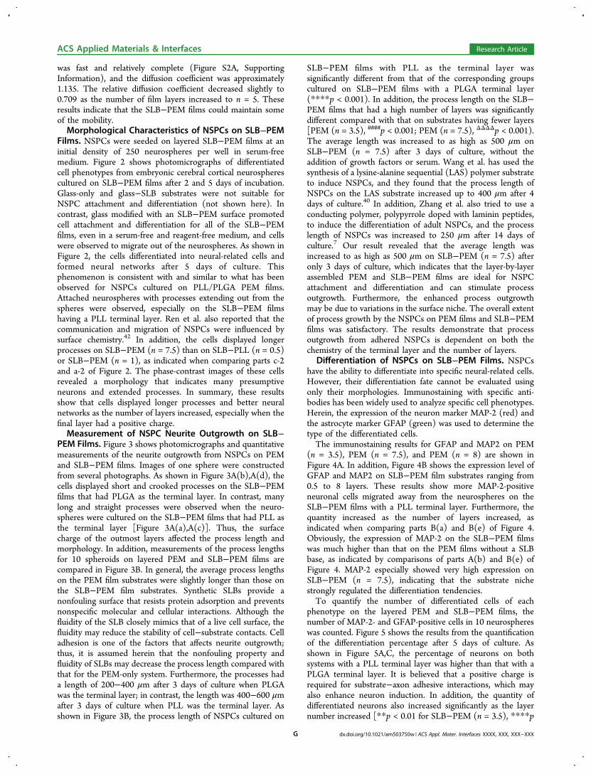
was fast and relatively complete (Figure S2A, SupportingInformation), and the diffusion coefficient was approximately1.135. The relative diffusion coefficient decreased slightly to0.709 as the number of film layers increased to n = 5. Theseresults indicate that the SLB−PEM films could maintain someof the mobility.Morphological Characteristics of NSPCs on SLB−PEM
Films. NSPCs were seeded on layered SLB−PEM films at aninitial density of 250 neurospheres per well in serum-freemedium. Figure 2 shows photomicrographs of differentiatedcell phenotypes from embryonic cerebral cortical neurospherescultured on SLB−PEM films after 2 and 5 days of incubation.Glass-only and glass−SLB substrates were not suitable forNSPC attachment and differentiation (not shown here). Incontrast, glass modified with an SLB−PEM surface promotedcell attachment and differentiation for all of the SLB−PEMfilms, even in a serum-free and reagent-free medium, and cellswere observed to migrate out of the neurospheres. As shown inFigure 2, the cells differentiated into neural-related cells andformed neural networks after 5 days of culture. Thisphenomenon is consistent with and similar to what has beenobserved for NSPCs cultured on PLL/PLGA PEM films.Attached neurospheres with processes extending out from thespheres were observed, especially on the SLB−PEM filmshaving a PLL terminal layer. Ren et al. also reported that thecommunication and migration of NSPCs were influenced bysurface chemistry.42 In addition, the cells displayed longerprocesses on SLB−PEM (n = 7.5) than on SLB−PLL (n = 0.5)or SLB−PEM (n = 1), as indicated when comparing parts c-2and a-2 of Figure 2. The phase-contrast images of these cellsrevealed a morphology that indicates many presumptiveneurons and extended processes. In summary, these resultsshow that cells displayed longer processes and better neuralnetworks as the number of layers increased, especially when thefinal layer had a positive charge.Measurement of NSPC Neurite Outgrowth on SLB−
PEM Films. Figure 3 shows photomicrographs and quantitativemeasurements of the neurite outgrowth from NSPCs on PEMand SLB−PEM films. Images of one sphere were constructedfrom several photographs. As shown in Figure 3A(b),A(d), thecells displayed short and crooked processes on the SLB−PEMfilms that had PLGA as the terminal layer. In contrast, manylong and straight processes were observed when the neuro-spheres were cultured on the SLB−PEM films that had PLL asthe terminal layer [Figure 3A(a),A(c)]. Thus, the surfacecharge of the outmost layers affected the process length andmorphology. In addition, measurements of the process lengthsfor 10 spheroids on layered PEM and SLB−PEM films arecompared in Figure 3B. In general, the average process lengthson the PEM film substrates were slightly longer than those onthe SLB−PEM film substrates. Synthetic SLBs provide anonfouling surface that resists protein adsorption and preventsnonspecific molecular and cellular interactions. Although thefluidity of the SLB closely mimics that of a live cell surface, thefluidity may reduce the stability of cell−substrate contacts. Celladhesion is one of the factors that affects neurite outgrowth;thus, it is assumed herein that the nonfouling property andfluidity of SLBs may decrease the process length compared withthat for the PEM-only system. Furthermore, the processes hada length of 200−400 μm after 3 days of culture when PLGAwas the terminal layer; in contrast, the length was 400−600 μmafter 3 days of culture when PLL was the terminal layer. Asshown in Figure 3B, the process length of NSPCs cultured on
SLB−PEM films with PLL as the terminal layer wassignificantly different from that of the corresponding groupscultured on SLB−PEM films with a PLGA terminal layer(****p < 0.001). In addition, the process length on the SLB−PEM films that had a high number of layers was significantlydifferent compared with that on substrates having fewer layers[PEM (n = 3.5), ####p < 0.001; PEM (n = 7.5), ΔΔΔΔp < 0.001).The average length was increased to as high as 500 μm onSLB−PEM (n = 7.5) after 3 days of culture, without theaddition of growth factors or serum. Wang et al. has used thesynthesis of a lysine-alanine sequential (LAS) polymer substrateto induce NSPCs, and they found that the process length ofNSPCs on the LAS substrate increased up to 400 μm after 4days of culture.40 In addition, Zhang et al. also tried to use aconducting polymer, polypyrrole doped with laminin peptides,to induce the differentiation of adult NSPCs, and the processlength of NSPCs was increased to 250 μm after 14 days ofculture.7 Our result revealed that the average length wasincreased to as high as 500 μm on SLB−PEM (n = 7.5) afteronly 3 days of culture, which indicates that the layer-by-layerassembled PEM and SLB−PEM films are ideal for NSPCattachment and differentiation and can stimulate processoutgrowth. Furthermore, the enhanced process outgrowthmay be due to variations in the surface niche. The overall extentof process growth by the NSPCs on PEM films and SLB−PEMfilms was satisfactory. The results demonstrate that processoutgrowth from adhered NSPCs is dependent on both thechemistry of the terminal layer and the number of layers.
Differentiation of NSPCs on SLB−PEM Films. NSPCshave the ability to differentiate into specific neural-related cells.However, their differentiation fate cannot be evaluated usingonly their morphologies. Immunostaining with specific anti-bodies has been widely used to analyze specific cell phenotypes.Herein, the expression of the neuron marker MAP-2 (red) andthe astrocyte marker GFAP (green) was used to determine thetype of the differentiated cells.The immunostaining results for GFAP and MAP2 on PEM
(n = 3.5), PEM (n = 7.5), and PEM (n = 8) are shown inFigure 4A. In addition, Figure 4B shows the expression level ofGFAP and MAP2 on SLB−PEM film substrates ranging from0.5 to 8 layers. These results show more MAP-2-positiveneuronal cells migrated away from the neurospheres on theSLB−PEM films with a PLL terminal layer. Furthermore, thequantity increased as the number of layers increased, asindicated when comparing parts B(a) and B(e) of Figure 4.Obviously, the expression of MAP-2 on the SLB−PEM filmswas much higher than that on the PEM films without a SLBbase, as indicated by comparisons of parts A(b) and B(e) ofFigure 4. MAP-2 especially showed very high expression onSLB−PEM (n = 7.5), indicating that the substrate nichestrongly regulated the differentiation tendencies.To quantify the number of differentiated cells of each
phenotype on the layered PEM and SLB−PEM films, thenumber of MAP-2- and GFAP-positive cells in 10 neurosphereswas counted. Figure 5 shows the results from the quantificationof the differentiation percentage after 5 days of culture. Asshown in Figure 5A,C, the percentage of neurons on bothsystems with a PLL terminal layer was higher than that with aPLGA terminal layer. It is believed that a positive charge isrequired for substrate−axon adhesive interactions, which mayalso enhance neuron induction. In addition, the quantity ofdifferentiated neurons also increased significantly as the layernumber increased [**p < 0.01 for SLB−PEM (n = 3.5), ****p
ACS Applied Materials & Interfaces Research Article
dx.doi.org/10.1021/am503750w | ACS Appl. Mater. Interfaces XXXX, XXX, XXX−XXXG

< 0.001 for SLB−PEM (n = 7.5), and &&p < 0.01 for SLB−PEM (n = 8)]. Most importantly, comparing the SLB−PEMsystem and the PEM-only system indicates that the expressionof MAP-2 and the ratio of MAP2/GFAP were significantlydifferent (#p < 0.05, ##p < 0.01, ###p < 0.005, and ####p < 0.001).In a previous study, PLL-coated latex beads and PDL were usedto form presynaptic-like endings and functional synapses withhippocampal neurons.25,43 Because this study provides a verysimple and pure culture condition, without requiring theaddition of serum or growth factors, these results suggests thatSLB-based substrates may affect the differentiation tendencyand trigger NSPCs to differentiate into neurons.
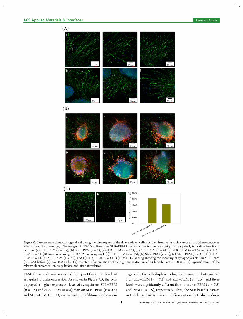
Functionality of Differentiated Neurons: Immunos-taining of Synapsin I and Analysis of Functionally ActiveSynapses. One of the key functionalities of mature CNSneurons is their ability to form synapses. To determine whetherthe differentiated neurons from NSPCs on the SLB−PEM filmsdisplayed synapse function, immunostaining of synapsin I andan analysis of functionally active synapses were performed.Punctate staining of synapsin I revealed that the synaptic vesicleprotein was concentrated at synapses in the neurons differ-entiated from the NSPCs on SLB−PEM films (Figure 6A).Furthermore, the staining was more obvious on the substrateswith a positively charged terminal layer. Coexpression ofsynapsin I and MAP-2, indicated by double staining, is alsoshown in Figure 6B. Because synapsin I is a marker ofpresynaptic terminals, which occur in axons, and MAP-2 is amarker for dendrites, the expression of synapsin I was observedas long tracks that were far from the cell body. In contrast, theexpression of MAP-2 displayed a more branched appearanceand was near the cell body. The patterns of differentiated cellson the SLB−PEM films were also observed. The results showedthat the neurons formed dense networks with a large number ofneurites and neural filaments.Moreover, Figure 6C shows the functionality test for active
synapses on SLB−PEM (n = 7.5) and the results quantifyingthe relative fluorescence intensities. The membranes of synapticvesicles were stained with the FM1-43 lipid dye. Afterstimulation by a high potassium solution, the fluorescenceintensity of the lipid dye decreased after the second stimulationby the high potassium solution without lipid dye. This resultindicated that the synaptic vesicles were functional andrecyclable, and it supports our hypothesis that SLB-basedPEM films can stimulate the differentiation of NSPCs intoneurons and trigger synaptic function by providing adhesivecontacts.
Quantification of Protein Expression of NSPCs onSLB−PEM Films by Western Blot. To quantitatively evaluatethe protein expression of NSPCs on layered SLB−PEM filmsafter 5 days of culture, Western blot analysis was used todetermine the expression levels of MAP2, GFAP, and synapsinI. Figure 7 shows that the MAP2 expression was higher onSLB−PEM (n = 7.5) than on SLB−PEM (n = 0.5) and that theexpression on SLB−PEM (n = 8) was higher than that onSLB−PEM (n = 1). The SLB−PEM films with PLL terminallayers displayed higher expression of MAP2 compared with thefilms with PLGA terminal layers. These results were consistentwith the quantification of the fluorescence intensity, which isshown in Figure 5A. Moreover, the protein concentration andfluorescence intensity results for GFAP expression (Figure 7B)and the relative ratio of MAP2/GFAP (Figure 7C) alsocorrelated well (Figures 5 and 7). Furthermore, the neuronfunctionality of NSPCs cultured on SLB−PEM (n = 0.5) and
Figure 5. Quantification of the percentage of differentiation into (A)neurons and (B) astrocytes and (C) the ratio of neurons/astrocytes forneurospheres on the PEM and SLB−PEM substrates. The cells werecultured under serum-free conditions at 250 neurospheres per cm2 for5 days. # denotes significant differences in the neuron percentagebetween the PEM and SLB−PEM systems (#,&p < 0.05; **,##,&&p <0.01; ###,&&&p < 0.005; ####,****p < 0.001), as determined byStudent’s t test.
ACS Applied Materials & Interfaces Research Article
dx.doi.org/10.1021/am503750w | ACS Appl. Mater. Interfaces XXXX, XXX, XXX−XXXH
PEM (n = 7.5) was measured by quantifying the level ofsynapsin I protein expression. As shown in Figure 7D, the cellsdisplayed a higher expression level of synapsin on SLB−PEM(n = 7.5) and SLB−PEM (n = 8) than on SLB−PEM (n = 0.5)and SLB−PEM (n = 1), respectively. In addition, as shown in
Figure 7E, the cells displayed a high expression level of synapsinI on SLB−PEM (n = 7.5) and SLB−PEM (n = 0.5), and theselevels were significantly different from those on PEM (n = 7.5)and PEM (n = 0.5), respectively. Thus, the SLB-based substratenot only enhances neuron differentiation but also induces
Figure 6. Fluorescence photomicrographs showing the phenotypes of the differentiated cells obtained from embryonic cerebral cortical neurospheresafter 3 days of culture. (A) The images of NSPCs cultured on SLB−PEM films show the immunoreactivity for synapsin I, indicating functionalneurons. (a) SLB−PEM (n = 0.5), (b) SLB−PEM (n = 1), (c) SLB−PEM (n = 3.5), (d) SLB−PEM (n = 4), (e) SLB−PEM (n = 7.5), and (f) SLB−PEM (n = 8). (B) Immunostaining for MAP2 and synapsin I. (a) SLB−PEM (n = 0.5), (b) SLB−PEM (n = 1), (c) SLB−PEM (n = 3.5), (d) SLB−PEM (n = 4), (e) SLB−PEM (n = 7.5), and (f) SLB−PEM (n = 8). (C) FM1−43 labeling showing the recycling of synaptic vesicles on SLB−PEM(n = 7.5) before (a) and 100 s after (b) the start of stimulation with a high concentration of KCl. Scale bars = 100 μm. (c) Quantification of therelative fluorescence intensity before and after stimulation.
ACS Applied Materials & Interfaces Research Article
dx.doi.org/10.1021/am503750w | ACS Appl. Mater. Interfaces XXXX, XXX, XXX−XXXI

synaptic function. Furthermore, as shown in Figure 5C, theneuron percentage on SLB−PEM (n = 7.5) and SLB−PEM (n= 8) was higher than that on SLB−PEM (n = 0.5) and SLB−PEM (n = 1), which is consistent with the result of the synapsinexpression level. This result further confirmed that SLB−PEM(n = 7.5) films could induce the differentiation of NSPCs into
functional neurons and encourage strong axonal growth andsynaptogenesis.These findings suggest that there are three factors affecting
the differentiation tendency of NSPCs in this system: theterminal layer, the number of layers, and the presence of anSLB base. Collectively, these results suggested that the SLB
Figure 7. Western blots were performed with anti-MAP2, anti-GFAP, anti-synapsin I, and anti-actin antibodies for NSPCs cultured on SLB−PEMfilms for 5 days. (A) Relative intensity of MAP2 expression on the SLB−PEM films. (B) Relative intensity of GFAP expression on the SLB−PEMfilms. (C) Relative ratio of MAP2/GFAP intensity. (D) Relative intensity of synapsin I expression on the SLB−PEM films. (E) Relative intensity ofsynapsin I expression on the PEM and SLB−PEM substrates. The intensities were determined by band densitometry analysis. Asterisks denotesignificant differences in neuron percentage between the different PEM film conditions (*,#p < 0.05), as determined by Student’s t test.
ACS Applied Materials & Interfaces Research Article
dx.doi.org/10.1021/am503750w | ACS Appl. Mater. Interfaces XXXX, XXX, XXX−XXXJ

base may trigger the differentiation of the neurons and that thePEM films may consolidate the film structure and enhance thecell migration. In addition, the SLB−PEM films with PLLterminal layers provide a positive charge to enhance celladhesion, and the primary amine moieties may contribute to achemoselective process. Furthermore, previous studies haveverified that the stiffness of PEM films decreases as the numberof layers increases, and many researchers have reported thatsofter materials greatly favor neurons, while stiffer surfacespromote glial cultures, which may explain the high enhancedneuron differentiation ratio on SLB−PEM (n = 7.5).2,4
4. CONCLUSIONSIn conclusion, a neuron induction system was fabricated in thisstudy by the layer-by-layer assembly of PLL/PLGA PEM filmsand an SLB base to regulate the adhesion of NSPCs and thedifferentiation of NSPCs into functional neurons. PEM filmswere used to produce a layered microenvironment of surfacesthat promote cell adhesion and differentiation; the neuriteoutgrowth, the types of differentiated cells, and the synapticfunction were all regulated by the PEM layer number and thecomposition of the outmost layer. The SLB base may triggerneuron differentiation, as this approach resulted in enhanceddifferentiation of NSPCs into neurons on SLB−PEM films.This study provides a biomimetic system that stimulates NSPCdifferentiation and is drastically different from other syntheticmaterials, and the results indicate that key targets of neuralengineering were achieved, including long processes, a largeneural network size, and a large number of functional neurons.These findings may provide a useful tool, as well as newstrategies for surface modification, to create and optimize stemcell niches for neural engineering.
■ ASSOCIATED CONTENT*S Supporting InformationFigure S1 shows TM-AFM height images of adsorbed PEMfilms on SLB, and Figure S2 shows quantitative traces of thenormalized fluorescence intensity across the bleached spot after30 min. This material is available free of charge via the Internetat http://pubs.acs.org/.
■ AUTHOR INFORMATIONCorresponding Author*Phone: +886 2 2118800, ext 5985. E-mail: [email protected] authors declare no competing financial interest.
■ ACKNOWLEDGMENTSWe thank Chang Gung University for providing financialsupport. We also received a Summit Project grant from theNational Science Council (grant NSC 101-2221-E-182-012 toI.-C.L.). We also thank Dr. Ying-Chih Chang for instrumentsupport.
■ REFERENCES(1) Beduer, A.; Vieu, C.; Arnauduc, F.; Sol, J. C.; Loubinoux, I.;Vaysse, L. Engineering of Adult Human Neural Stem CellsDifferentiation through Surface Micropatterning. Biomaterials 2012,33, 504−514.(2) Camci-Unal, G.; Cuttica, D.; Annabi, N.; Demarchi, D.;Khademhosseini, A. Synthesis and Characterization of Hybrid
Hyaluronic Acid−Gelatin Hydrogels. Biomacromolecules 2013, 14,1085−1092.(3) Johnson, P. J.; Wood, M. D.; Moore, A. M.; Mackinnon, S. E.Tissue Engineered Constructs for Peripheral Nerve Surgery. EuropeanSurgery: ACA: Acta Chirurgica Austriaca 2013, 45, 122−135.(4) Li, X.; Liu, X.; Zhang, N.; Wen, X. Engineering in SituCrosslinkable, Injectable, and Neurocompatible Hydrogels. J. Neuro-trauma 2014, DOI: 10.1089/neu.2013.3215.(5) Matyash, M.; Despang, F.; Mandal, R.; Fiore, D.; Gelinsky, M.;Ikonomidou, C. Novel Soft Alginate Hydrogel Strongly SupportsNeurite Growth and Protects Neurons against Oxidative Stress. TissueEng. Part A 2012, 18, 55−66.(6) Xie, H.; Li, J.; Li, L.; Dong, Y.; Chen, G. Q.; Chen, K. C.Enhanced Proliferation and Differentiation of Neural Stem CellsGrown on PHA Films Coated with Recombinant Fusion Proteins.Acta Biomater. 2013, 9, 7845−7854.(7) Zhang, L.; Stauffer, W. R.; Jane, E. P.; Sammak, P. J.; Cui, X. T.Enhanced Differentiation of Embryonic and Neural Stem Cells toNeuronal Fates on Laminin Peptides Doped Polypyrrole. Macromol.Biosci. 2010, 10, 1456−1464.(8) Kim, Y. S.; Park, C. H. Dopamine Neuron Generation fromHuman Embryonic Stem Cells. Int. J. Stem Cells 2011, 4, 85−87.(9) Ko, J. Y.; Lee, J. Y.; Park, C. H.; Lee, S. H. Effect of Cell-Densityon in-Vitro Dopaminergic Differentiation of Mesencephalic PrecursorCells. Neuroreport 2005, 16, 499−503.(10) Li, Y. C.; Lin, Y. C.; Young, T. H. Combination of Media,Biomaterials and Extracellular Matrix Proteins To Enhance theDifferentiation of Neural Stem/Precursor Cells into Neurons. ActaBiomater. 2012, 8, 3035−3048.(11) Li, Y.; Weiss, M.; Yao, L. Directed Migration of EmbryonicStem Cell-Derived Neural Cells in an Applied Electric Field. Stem CellRev. 2014, DOI: 10.1007/s12015-014-9518-z.(12) Yuan, T.; Liao, W.; Feng, N. H.; Lou, Y. L.; Niu, X.; Zhang, A. J.;Wang, Y.; Deng, Z. F. Human Induced Pluripotent Stem Cell-DerivedNeural Stem Cells Survive, Migrate, Differentiate, and ImproveNeurological Function in a Rat Model of Middle Cerebral ArteryOcclusion. Stem Cell Res. Ther. 2013, 4, 73−82.(13) Lim, S. H.; Liu, X. Y.; Song, H.; Yarema, K. J.; Mao, H. Q. TheEffect of Nanofiber-Guided Cell Alignment on the PreferentialDifferentiation of Neural Stem Cells. Biomaterials 2010, 31, 9031−9039.(14) Francis, K. R.; Wei, L. Human Embryonic Stem Cell NeuralDifferentiation and Enhanced Cell Survival Promoted by HypoxicPreconditioning. Cell Death Dis. 2010, 1, e22.(15) Kim, M.; Habiba, A.; Doherty, J. M.; Mills, J. C.; Mercer, R. W.;Huettner, J. E. Regulation of Mouse Embryonic Stem Cell NeuralDifferentiation by Retinoic Acid. Dev. Biol. 2009, 328, 456−471.(16) Tan, J. C.; Li, Y.; Qu, W. Y.; Liu, L. Y.; Jiang, L.; Sun, K. L.Derivation of Embryonic Stem Cell Line from Frozen HumanEmbryos and Neural Differentiation. Neuroreport 2008, 19, 1451−1455.(17) Daadi, M. M. In Vitro Assays for Neural Stem CellDifferentiation: Induction of Dopaminergic Phenotype. Methods Mol.Biol. 2008, 438, 205−212.(18) Kam, L.; Boxer, S. G. Cell Adhesion to Protein-Micropatterned-Supported Lipid Bilayer Membranes. J. Biomed. Mater. Res. 2001, 55,487−495.(19) Wu, J. C.; Tseng, P. Y.; Tsai, W. S.; Liao, M. Y.; Lu, S. H.; Frank,C. W.; Chen, J. S.; Wu, H. C.; Chang, Y. C. Antibody ConjugatedSupported Lipid Bilayer for Capturing and Purification of ViableTumor Cells in Blood for Subsequent Cell Culture. Biomaterials 2013,34, 5191−5199.(20) Hain, N.; Gallego, M.; Reviakine, I. Unraveling Supported LipidBilayer Formation Kinetics: Osmotic Effects. Langmuir 2013, 29,2282−2288.(21) Huang, C. J.; Cho, N. J.; Hsu, C. J.; Tseng, P. Y.; Frank, C. W.;Chang, Y. C.; Type, I. Collagen-Functionalized Supported LipidBilayer as a Cell Culture Platform. Biomacromolecules 2010, 11, 1231−1240.
ACS Applied Materials & Interfaces Research Article
dx.doi.org/10.1021/am503750w | ACS Appl. Mater. Interfaces XXXX, XXX, XXX−XXXK

(22) Jonsson, M. P.; Jonsson, P.; Dahlin, A. B.; Hook, F. SupportedLipid Bilayer Formation and Lipid-Membrane-Mediated Biorecogni-tion Reactions Studied With a New Nanoplasmonic Sensor Template.Nano Lett. 2007, 7, 3462−3468.(23) Gedeon, P. C.; Indarte, M.; Surratt, C. K.; Madura, J. D.Molecular Dynamics of Leucine and Dopamine Transporter Proteinsin a Model Cell Membrane Lipid Bilayer. Proteins 2010, 78, 797−811.(24) Sato, M.; Inoue, K.; Kasai, M. Ion Channels on Synaptic VesicleMembranes Studied by Planar Lipid Bilayer Method. Biophys. J. 1992,63 (6), 1500−1505.(25) Lucido, A. L.; Suarez Sanchez, F.; Thostrup, P.; Kwiatkowski, A.V.; Leal-Ortiz, S.; Gopalakrishnan, G.; Liazoghli, D.; Belkaid, W.;Lennox, R. B.; Grutter, P.; Garner, C. C.; Colman, D. R. RapidAssembly of Functional Presynaptic Boutons Triggered by AdhesiveContacts. J. Neurosci. 2009, 29, 12449−12466.(26) Pautot, S.; Lee, H.; Isacoff, E. Y.; Groves, J. T. Neuronal SynapseInteraction Reconstituted between Live Cells and Supported LipidBilayers. Nat. Chem. Biol. 2005, 1, 283−289.(27) Jones, E. V.; Cook, D.; Murai, K. K. A Neuron−Astrocyte Co-Culture System To Investigate Astrocyte-Secreted Factors in MouseNeuronal Development. Methods Mol. Biol. 2012, 814, 341−352.(28) Davila, J.; Chassepot, A.; Longo, J.; Boulmedais, F.; Reisch, A.;Frisch, B.; Meyer, F.; Voegel, J. C.; Mesini, P. J.; Senger, B.; Metz-Boutigue, M. H.; Hemmerle, J.; Lavalle, P.; Schaaf, P.; Jierry, L. Cyto-Mechanoresponsive Polyelectrolyte Multilayer Films. J. Am. Chem. Soc.2012, 134, 83−86.(29) Yamanlar, S.; Sant, S.; Boudou, T.; Picart, C.; Khademhosseini,A. Surface Functionalization of Hyaluronic Acid Hydrogels byPolyelectrolyte Multilayer Films. Biomaterials 2011, 32, 5590−5599.(30) Wilson, J. T.; Cui, W.; Kozlovskaya, V.; Kharlampieva, E.; Pan,D.; Qu, Z.; Krishnamurthy, V. R.; Mets, J.; Kumar, V.; Wen, J.; Song,Y.; Tsukruk, V. V.; Chaikof, E. L. Cell Surface Engineering withPolyelectrolyte Multilayer Thin Films. J. Am. Chem. Soc. 2011, 133,7054−7064.(31) Chien, H. W.; Wu, S. P.; Kuo, W. H.; Wang, M. J.; Lee, C.; Lai,J. Y.; Tsai, W. B. Modulation of Hemocompatibility of Polysulfone byPolyelectrolyte Multilayer Films. Colloids Surf., B 2010, 77, 270−278.(32) Tsai, W. B.; Chen, Y. H.; Chien, H. W. Collaborative Cell-Resistant Properties of Polyelectrolyte Multilayer Films and SurfacePEGylation on Reducing Cell Adhesion to Cytophilic Surfaces. J.Biomater. Sci. Polym. Ed. 2009, 20, 1611−1628.(33) Picart, C. Polyelectrolyte Multilayer Films: From Physico-Chemical Properties to the Control of Cellular Processes. Curr. Med.Chem. 2008, 15, 685−697.(34) Tsai, H. A.; Wu, R. R.; Lee, I. C.; Chang, H. Y.; Shen, C. N.;Chang, Y. C. Selection, Enrichment, And Maintenance of Self-RenewalLiver Stem/Progenitor Cells Utilizing Polypeptide PolyelectrolyteMultilayer Films. Biomacromolecules 2010, 11, 994−1001.(35) Boudou, T.; Crouzier, T.; Ren, K.; Blin, G.; Picart, C. MultipleFunctionalities of Polyelectrolyte Multilayer Films: New BiomedicalApplications. Adv. Mater. 2010, 22, 441−467.(36) Gribova, V.; Gauthier-Rouviere, C.; Albiges-Rizo, C.; Auzely-Velty, R.; Picart, C. Effect of RGD Functionalization and StiffnessModulation of Polyelectrolyte Multilayer Films on Muscle CellDifferentiation. Acta Biomater. 2013, 9, 6468−6480.(37) Thebaud, N. B.; Bareille, R.; Daculsi, R.; Bourget, C.; Remy, M.;Kerdjoudj, H.; Menu, P.; Bordenave, L. Polyelectrolyte MultilayerFilms Allow Seeded Human Progenitor-Derived Endothelial Cells ToRemain Functional under Shear Stress In Vitro. Acta Biomater. 2010, 6,1437−1445.(38) Moby, V.; Kadi, A.; de Isla, N.; Stoltz, J. F.; Menu, P.Polyelectrolyte Multilayer Films: Effect of the Initial Anchoring Layeron the Cell Growth. Bio-Med. Mater. Eng. 2008, 18, 199−204.(39) Richert, L.; Lavalle, P.; Vautier, D.; Senger, B.; Stoltz, J. F.;Schaaf, P.; Voegel, J. C.; Picart, C. Cell Interactions withPolyelectrolyte Multilayer Films. Biomacromolecules 2002, 3, 1170−1178.
(40) Wang, J. H.; Hung, C. H.; Young, T. H. Proliferation andDifferentiation of Neural Stem Cells on Lysine-Alanine SequentialPolymer Substrates. Biomaterials 2006, 27, 3441−3450.(41) Ma, W.; Fitzgerald, W.; Liu, Q. Y.; O’Shaughnessy, T. J.; Maric,D.; Lin, H. J.; Alkon, D. L.; Barker, J. L. CNS Stem and ProgenitorCell Differentiation Into Functional Neuronal Circuits in Three-Dimensional Collagen Gels. Exp. Neurol. 2004, 190, 276−288.(42) Ren, Y. J.; Zhang, H.; Huang, H.; Wang, X. M.; Zhou, Z. Y.; Cui,F. Z.; An, Y. H. In Vitro Behavior of Neural Stem Cells In Response toDifferent Chemical Functional Groups. Biomaterials 2009, 30, 1036−1044.(43) Burry, R. W. Formation of Apparent Presynaptic Elements InResponse to Poly-Basic Compounds. Brain Res. 1980, 184, 85−98.
ACS Applied Materials & Interfaces Research Article
dx.doi.org/10.1021/am503750w | ACS Appl. Mater. Interfaces XXXX, XXX, XXX−XXXL