Embed Size (px)
Citation preview

Copyright is owned by the Author of the thesis. Permission is given for a copy to be downloaded by an individual for the purpose of research and private study only. The thesis may not be reproduced elsewhere without the permission of the Author.
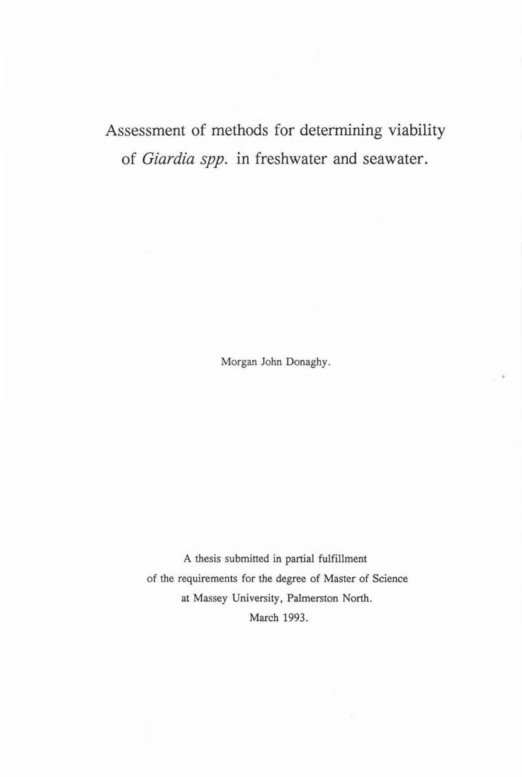
Assessment of methods for determining viability
of Giardia spp. in freshwater and seawater.
Morgan John Donaghy.
A thesis submitted in partial fulfillment
of the requirements for the degree of Master of Science
at Massey University, Palmerston North.
March 1993.
ii
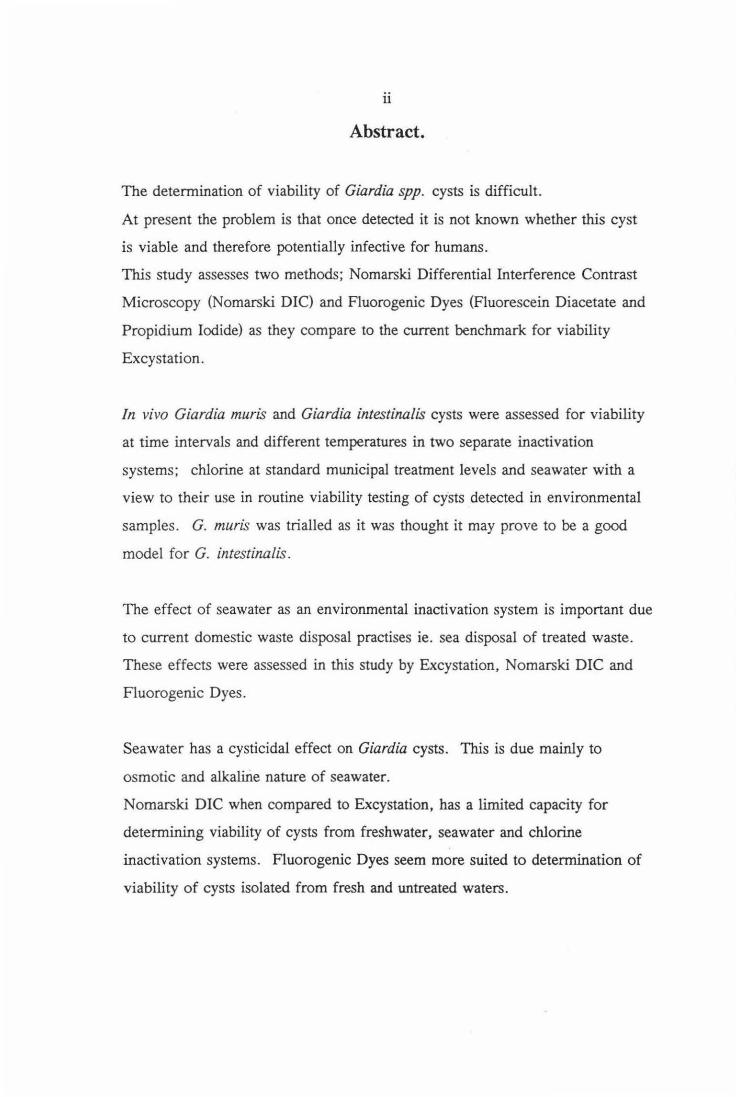
Abstract.
The determination of viability of Giardia spp. cysts is difficult.
At present the problem is that once detected it is not known whether this cyst
is viable and therefore potentially infective for humans.
This study assesses two methods; Nomarski Differential Interference Contrast
Microscopy (Nomarski DIC) and Fluorogenic Dyes (Fluorescein Diacetate and
Propidium Iodide) as they compare to the current benchmark for viability
Excystation.
In vivo Giardia muris and Giardia intestinalis cysts were assessed for viability
at time intervals and different temperatures in two separate inactivation
systems; chlorine at standard municipal treatment levels and seawater with a
view to their use in routine viability testing of cysts detected in environmental
samples. G. muris was trialled as it was thought it may prove to be a good
model for G. intestinalis.
The effect of seawater as an environmental inactivation system is important due
to current domestic waste disposal practises ie. sea disposal of treated waste.
These effects were assessed in this study by Excystation, Nomarski DIC and
Fluorogenic Dyes.
Seawater has a cysticidal effect on Giardia cysts. This is due mainly to
osmotic and alkaline nature of seawater.
Nomarski DIC when compared to Excystation, has a limited capacity for
determining viability of cysts from freshwater, seawater and chlorine
inactivation systems. Fluorogenic Dyes seem more suited to determination of
viability of cysts isolated from fresh and untreated waters.

iii
Acknowledgements.
I would like to thank Massey University and the Department of Microbiology
and Genetics for the use of their facilities, without which I wouldn't have
completed this thesis.
The now Professor Tim Brown whose unfailing keenness and enthusiasm
prompted me to take this project to task.
My flatmates of many great years Pete and Chris, and Tracey-Ann, Lisa,
Jodie, Juliette, and Suzanne.
My friends at Massey; Sheree, Merie, Paul, Terrence and Shalome who
provided me with inspiration and entertainment when it was needed and often
when it wasn't. Much appreciated anyway ! Thanks guys.
'Doctor' Phil Kelly for teaching me that the 19th hole is actually the hardest
hole to make par.
Dr Charlie O'Kelly for teaching me how to 'drive' the Nomarski DIC
microscope.
Mrs . Shirley Gainsford (Valley Diagnostics Ltd. , Lower Hutt) for sending
samples by courier as regularly as they were 'regularly' produced.
All the staff in the Microbiology and Genetics department and fellow Staff
Clubbers, for help and assistance however small.
The people and equipment at AgResearch Wallaceville, who without which I
wouldn't have had the chance to work and write at the same time.
My family who pushed me to finish the project and get this thesis written.
iv
Contents.
Page.
Chapter One : The Organism. 1
1.1 Discovery. 1
1.2 Taxonomy. 2
1.3 Biology of Giardia . 3
1.4 The Disease. 4 1.4.1 Introduction. 4 1.4.2 Pathogenesis. 4 1.4.3 The Symptoms. 5 1.4.4 Diagnosis. 5 1.4.5 Treatment. 6
1.5 Transmission. 7 1.5.1 Direct/Fecal-Oral Transmission. 8 1.5.2 Indirect/Waterborne Transmission. 9
1.6 Giardia cysts in drinking water. 9 1.6.1 Incidence. 9 1.6.2 Detection. 10
1.7 Viability. 11 1.7.1 Methods for determining viability of Giardia. 11 1.7.2 Giardia viability in seawater. 13
Chapter Two : Materials. 14
2.1 Viability Determination. 14 2.1.1 Giardia muris excystation. 14 2.1.2 Giardia intestinalis excystation. 14 2.1.3 Vital Stains. 15 2.1.4 Nomarski Differential Interference
Contrast Microscopy. 15
2.2 Giardia cysts. 15 2.2.1 Giardia muris. 15 2.2.2 Giardia intestinalis. 15

2.3
2.4 2.4.l 2.4.2 2.4.3 2.4.4 2.4.5
Chapter Three :
3.1 3.1.1 3.1.2 3.1.3 3 .1.4
3 .2
3.3
Chapter Four :
Chapter Five :
Chapter Six :
Chapter Seven :
v
D.P.D. Colorimetric Method for chlorine determinations.
General Media, Reagents and Equipment. Hank's Balanced Salt Solution (HBSS). Phosphate Buffered Saline. Tyrode's Salt Solution. Milli-Q™-water. Seawater.
Methods.
Methods for determination of viability. Excystation Giardia muris. Excystation Giardia intestinalis. Vital Stains. Nomarski Differential Interference Contrast Microscopy.
Seawater Inactivation.
Chlorine Inactivation.
Results.
Discussion.
Conclusions.
Bibliography.
Page.
16
17 17 18 19 19 19
20
20 20 21 22
22
23
23
25
71
83
85

Vl
List of Figures~
Page. Figure 1 Viability of Giardia muris in
freshwater and seawater at S°C. 26
Figure 2 Viability of G. muris in freshwater and seawater at 10°C. 27
Figure 3 Viability of G. muris in freshwater and seawater at 1S°C. 28
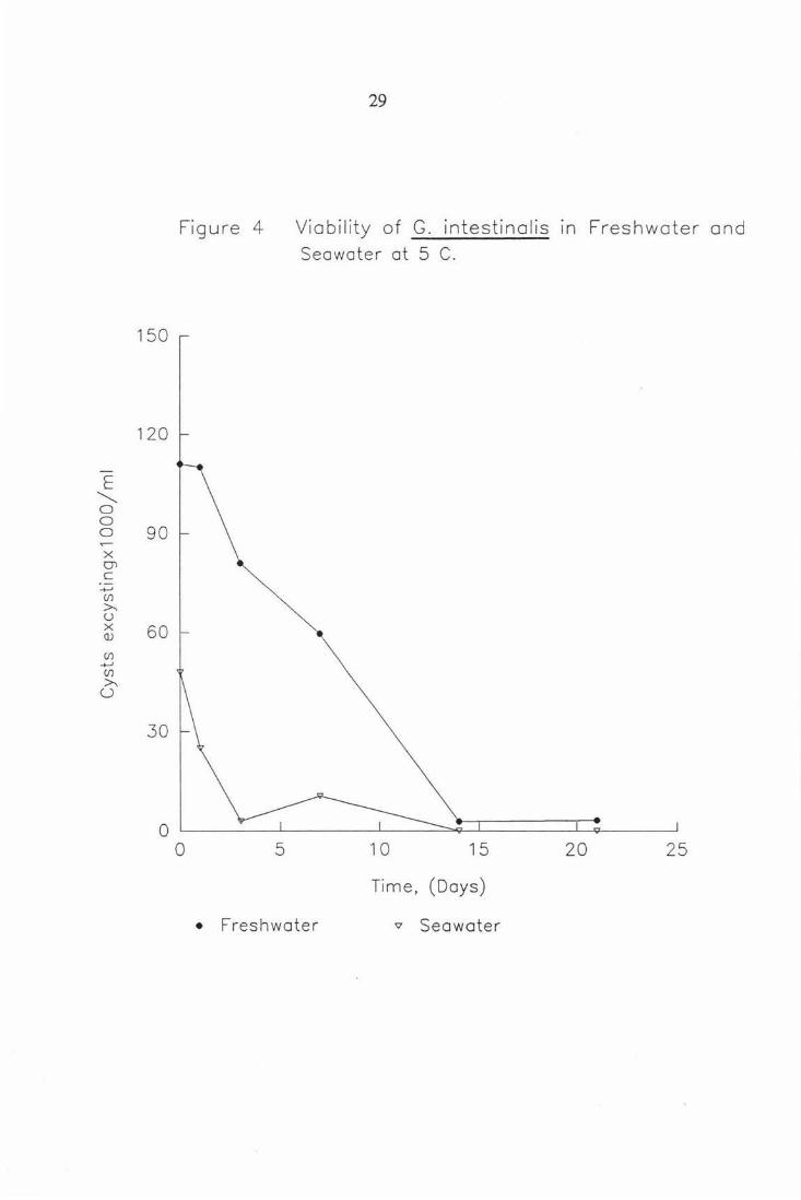
Figure 4 Viability of Giardia intestinalis in freshwater and seawater at S°C. 29
Figure S Viability of G. intestinalis in freshwater and seawater at 10°C. 30
Figure 6 Viability of G. intestinalis in freshwater and seawater at 1S0 C. 31
Figure 7 Nt/No vs Time for G. muris in freshwater and seawater at S°C. 33
Figure 8 Nt/No vs Time for G. muris in freshwater and seawater at 10°C. 34
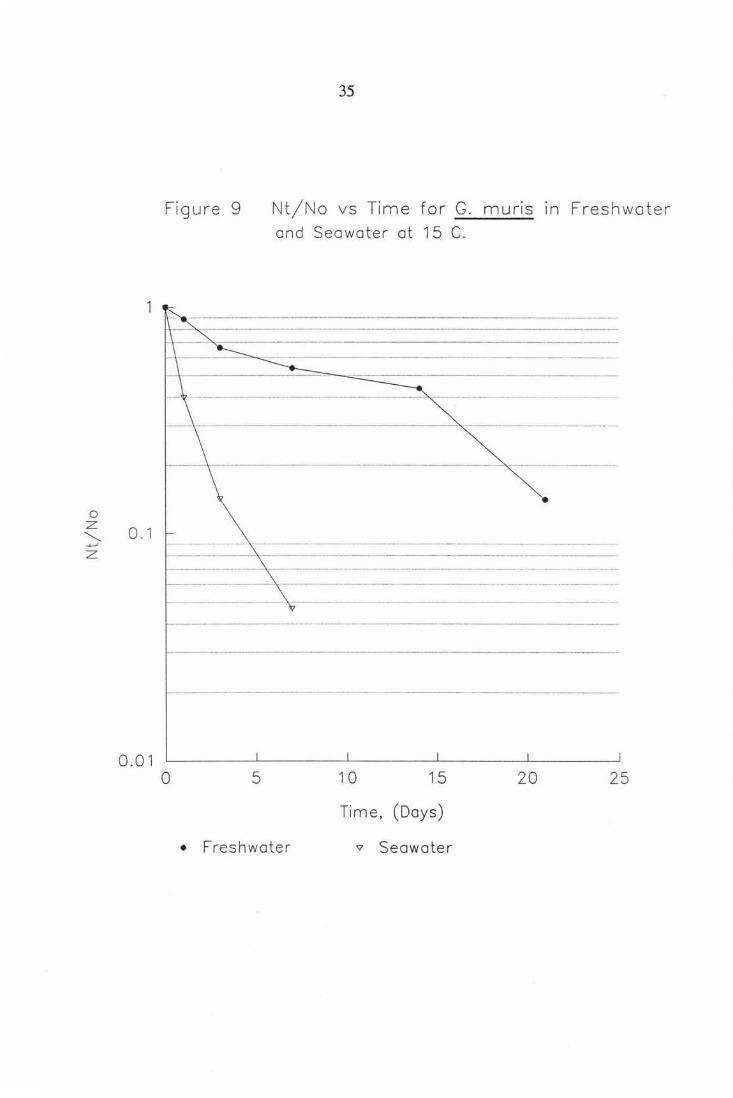
Figure 9 Nt/No vs Time for G. muris in freshwater and seawater at 1S°C. 3S
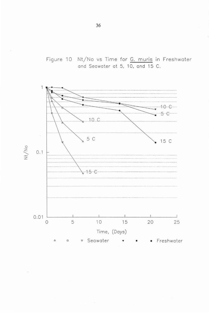
Figure 10 Nt/No vs Time for G. muris in freshwater and seawater at S, 10, 1S°C. 36
Figure 11 Nt/No vs Time for G. intestinalis in freshwater and seawater at S°C. 38
Figure 12 Nt/No vs Time for G. intestinalis in freshwater and seawater at 10°C. 39
Figure 13 Nt/No vs Time for G. intestinalis in freshwater and seawater at 1S°C. 40
Figure 14 Nt/No vs Time for G. intestinalis in freshwater and seawater at S, 10, 1S°C. 41

vii
Page. Figure lS The percent viable cysts as determined
by Nomarski DIC, FDA, PI, and Excystation for G. muris at S°C in freshwater. 46
Figure 16 The percent viable cysts as determined by Nomarski DIC, FDA, PI, and Excystation for G. muris at 15°C in freshwater. 47
Figure 17 The percent viable cysts as determined by Nomarski DIC, FDA, PI, and Excystation for G. muris at 5°C in seawater. 48
Figure 18 The percent viable cysts as detennined by Nomarski DIC, FDA, PI, and Excystation for G. muris at 1S°C in seawater. 49
Figure 19 The percent viable cysts as detennined by Nomarski DIC for G. muris showing temperature and seawater effect. so
Figure 20 The percent viable cysts as detennined by Excystation for G. muris showing temperature and seawater effect. 51
Figure 21 The percent viable cysts as determined by FDA for G. muris showing temperature and seawater effect. S2
Figure 22 The percent viable cysts as determined by PI for G. muris showing temperature and seawater effect. S3
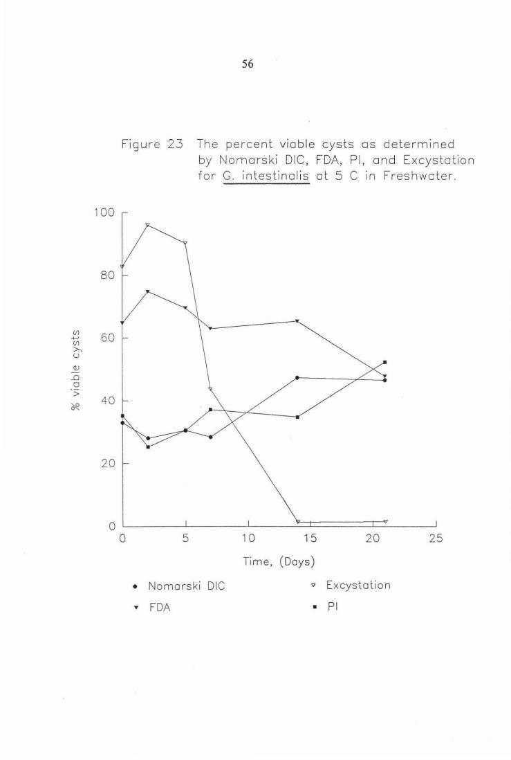
Figure 23 The percent viable cysts as determined by Nomarski DIC, FDA, PI, and Excystation for G. intestinalis at S°C in freshwater. 56
Figure 24 The percent viable cysts as determined by Nomarski DIC, FDA, PI, and Excystation for G. intestinalis at 15°C in freshwater. S7
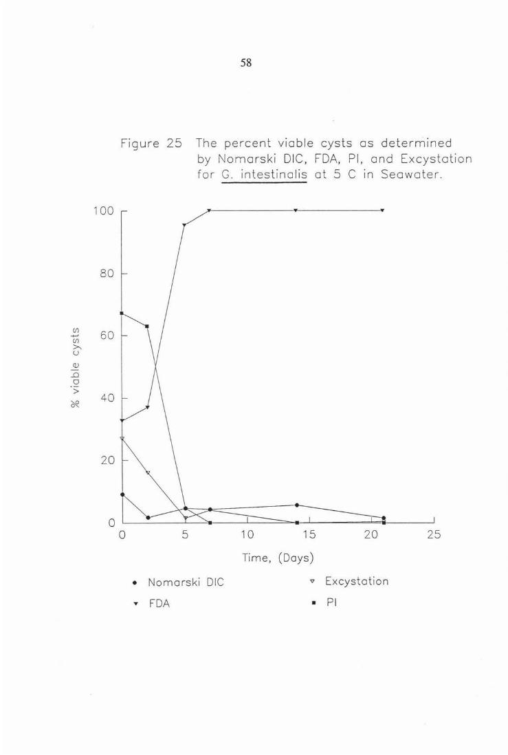
Figure 25 The percent viable cysts as determined by Nomarski DIC, FDA, PI, and Excystation for G. intestinalis at S°C in seawater. 58

Vlll
Page. Figure 26 The percent viable cysts as determined
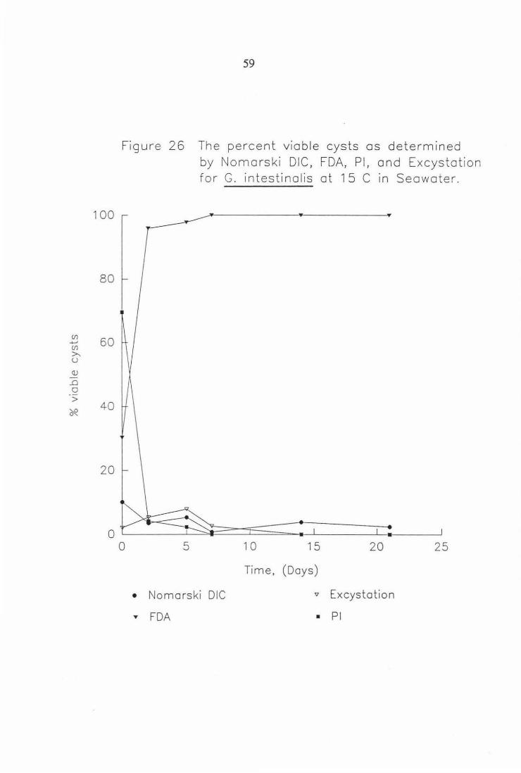
by Nomarski DIC, FDA, PI, and Excystation for G. intestinalis at 15°C in seawater. 59
Figure 27 The percent viable cysts as determined by Nomarski DIC for G. intestinalis showing temperature and seawater effect. 60
Figure 28 The percent viable cysts as determined by Excystation for G. intestinalis showing temperature and seawater effect. 61
Figure 29 The percent viable cysts as determined by FDA for G. intestinalis showing temperature and seawater effect. 62
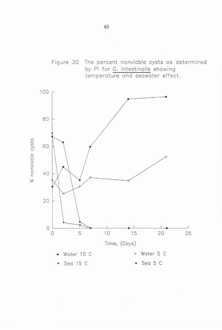
Figure 30 The percent viable cysts as determined by PI for G. intestinalis showing temperature and seawater effect. 63
Figure 31 Chlorine inactivation of G. muris as determined by Nomarski DIC. 65
Figure 32 Chlorine inactivation of G. muris as determined by Excystation. 66
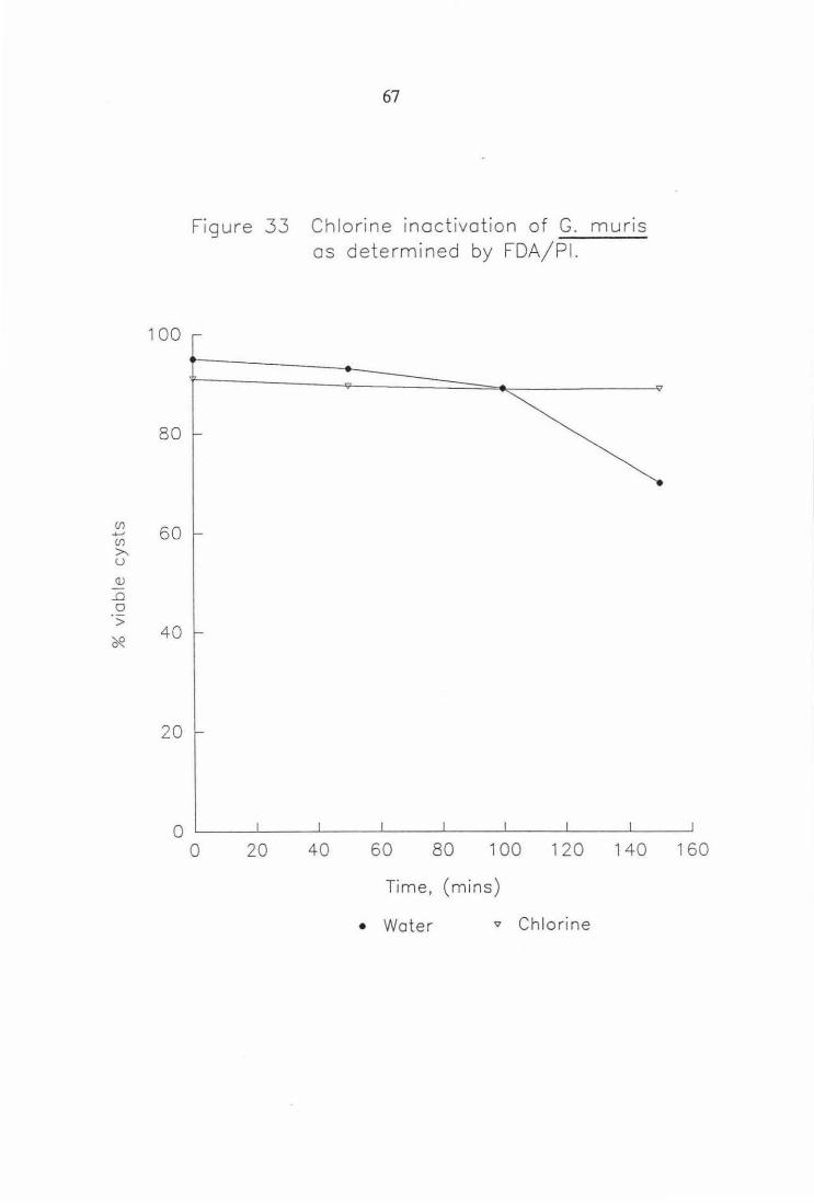
Figure 33 Chlorine inactivation of G. muris as determined by FDA/PI. 67
Figure 34 Chlorine inactivation of G. muris log data for Excystation. 68

Table 1
Table 2
Table 3
Table 4
ix
List of Tables.
Giardia spp. cyst inactivation at t = 0, showing difference between freshwater and seawater.
G. muris log Nt/No gradients.
Inactivation of G. muris using various chlorine treatments.
Top ten ion concentrations. in seawater.
Page.
32
37
64
73
x
List of Plates.
Page. Plate 1 In vivo G. muris cyst showing
non-viable morphology under Nomarski DIC. 42
Plate 2 In vivo G. muris cyst showing viable morphology under Nomarski DIC. 43
Plate 3 In vivo G. intestinalis cyst showing non-viable morphology under Nomarski DIC. 44
Plate 4 In vivo G. intestinalis cyst showing viable morphology under Nomarski DIC. 45
Plate 5 Fully excysted G. intestinal is trophozoites. 54
Plate 6 Seawater inactivated Giardia cyst. 55
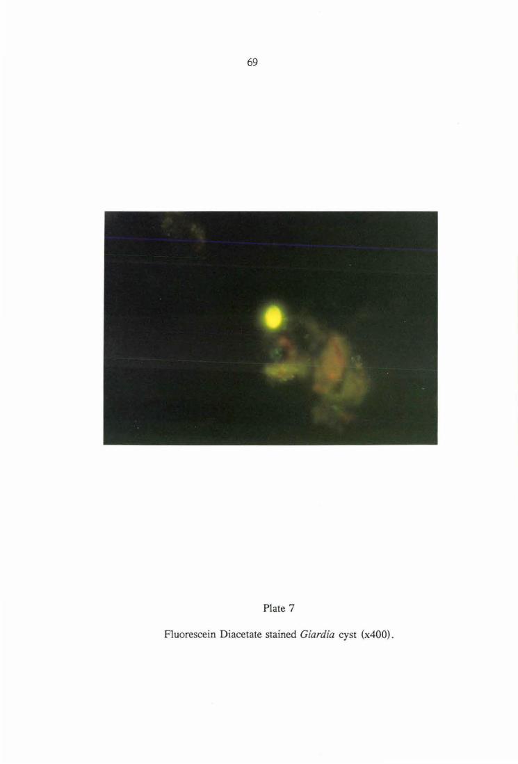
Plate 7 Fluorescein Diacetate stained Giardia ·cyst. 69
Plate 8 Propidium Iodide stained Giardia cyst. 70

1
Chapter One The Organism.
1.1 Discovery.
Van Leeuwenhoek an amateur lens maker from Delft, Holland, was the first
person to describe the microbial world. Using a microscope with only a single
lens, he discovered a wide variety of micro-organisms in materials ranging
from hay infusions to tooth scrapings. His success in seeing cells as small as
bacteria can be _attributed to the exceptionally high quality of his lenses, his
technique of mounting and lighting the specimen and his insatiable curiosity.
It was probably this curiosity that prompted him to examine his own diarrhoeic
stool.
He described many microorganisms, and in a letter dated 4 November 1681,
he stated (in translation)
"Their bodies were somewhat longer than broad, and their belly
which was flat like, furnisht with sundry little paws, where with
they made such a stir in the clean medium and among the
globules, that you might e'en fancy you saw a woodlouse
running up a wall; and albeit they made such a commotion with
their paws, yet for all that they made but slow progress."
(Dobell 1932)
From this statement many scientists today believe Van Leeuwenhoek to have
been the first to observe Giardia trophozoites .
..• Vilem Lambl, a Czech physician is credited with the discovery of the
organism, which he named Lamblia. Van Leeuwenhoek and Lambl saw and
described Giardia trophozoites, but there is no evidence that either man saw or
recognised the relationship of the trophozoite to the cyst form of the parasite.
Giardia cysts were first noted by Grassi. He first thought they might be
2
coccidia, but in 1888 he concluded that they represented the flagellated form of
the organism (Grassi 1888) .
1.2 Taxonomy.
The taxonomy and nomenclature of organisms in this group presented problems
in the late 19th and early 20th century. Two generic names were applied to
these protozoa. Kunstler (1882), isolated Giardia from the tadpole and
Blanchard (1888), Lamblia from mammals. The organism these scientists
described were of the Genus as demonstrated by Hegner (1922).
Today the name Giardia is given to all representatives of this group and the
name giardiasis given to the disease they cause.
To date the assignment of Giardia isolates to a specific species has undergone
much review. Until the middle of this century it was common practise to
assign Giardia species names on the basis of host specificity (Woo et al 1984) ,
and on the differences in various trophozoite dimensions (Filice 1952). Kulda
and Nohynkova (1978) were able to list references to more than forty Giardia
"species", demonstrating that representatives of this genus appear to exist
intestinally in virtually every mammalian species examined. Filice (1952) took
a more objective view and concluded that the use of host specificity and body
measurement was an unreliable method of species classification. His work
assigned Giardia from different sources to a given species on the basis of
median body shape. This he thought made use of "constant biological
differences" which are visible in all trophozoite isolates of Giardia. His
classification put Giardia of the median body type that parasitise humans and
other mammals into the species Giardia duodenalis. The median bodies of this
type are paired, lie transversely across the trophozoite body, and resemble the
claw of a hammer, hence the description "claw shaped median bodies".
The median bodies of Giardia muris are small and rounded, and Giardia agilis
teardrop shaped. This thesis has determined to use the name Giardia

3
intestinalis put forward by Boreham et al (1984) to describe species
demonstrated as infecting humans and non-rodent animals. Since the proposal
of this classification 23 of the 40 "species" listed by Kulda and Nohynkova in
their 1978 review have been classed as Giardia intestinalis having claw shaped
median bodies .
1. 3 Biology of Giardia.
Giardia is a flagellated protozoan belonging to the classification:
Class Zoomastigophorasidan,
Order Diplomonadida,
Family Hexamitidae.
It is found in two morphologically distinct forms: in fresh faeces as the
infective cyst and in the intestine of infected humans and animals as the
reproductive trophozoite.
The trophozoite found living in the duodenum, is responsible for the
pathogenesis of the disease . They are bilaterally symmetrical organisms with
rather a pear shape. Trophozoites have four pairs of flagella and attach to the
microvilli of the duodenum and jejunum by means of an anteriorly situated
ventral sucking disc. Possession of this disc differentiates Giardia from other
members of the family Hexamitidae (Feely et al 1984).
The cyst form is oval in shape and ranges in size from 8-12 µm long and 7-10
µm wide. They consist typically of four nuclei, intracytoplasmic axonemes
and median bodies encased within a hyaline cyst wall. The cyst forms during
passage through the jejunum (Burke 1977; Lambert 1970), and passes out in
the faeces. This is predominant in the faeces, as the cyst is the means of
transmission and survival outside the body.
The trophozoite and cyst forms of Giardia do share some internal organelles.
-Axonemes,

4
-median bodies, and
-nuclei are often immediately obvious as being common, with the
adhesive disc and flagella being the exclusive property of the trophozoite.
1.4 The Disease.
1.4. l Introduction.
G. intestinalis is the etiological agent of giardiasis. This was not immediately
clear due to the range of conditions the disease manifests, ranging from
asymptomatic carriage through to chronic diarrhoeic malabsorption. But now
as stated by Kulda and Nohnykova (1978), "there is no doubt that the organism
is capable of causing disease in man". This was concluded on the basis of
symptoms such as malabsorption, and histopathology. Further, reports of
epidemic outbreaks of giardiasis particularly in the case of Aspen, Colorado,
gave foundation to these claims. Here during the 1965-66 ski-season, 123 out
of 1094 skiers tested, developed symptoms characteristic of Giardiasis, and on
pathological examination of all those infected revealed no other pathogens apart
from G. intestinalis.
1.4.2 Pathogenesis
At the outset, giardiasis begins as an acute disease. This is preceded by a
prepatent period of between nine and twenty days (Brodsky et al 1974,
Rebdtorff 1954, Walzer et al 1971). Prepatent periods shorter than this and
followed by similar symptoms can often be attributed to Cryptosporidiosis
(Jokipii et al 1985).
The acute infection lasts only a few days. During this phase there is dense
colonisation of the jejuna! epithelium with adherence to columnar epithelial
cells at the base of the villi (Owen et al 1979). Trophozoites are also
commonly detected in the duodenum of infected patients. Trophozoites attach
but do not normally penetrate the intestinal epithelium. However if this does
5
occur then it is usually due to necrosis or mechanical trauma (Owen et al 1979)
of the mucosa.
The trophozoite is normally adherent to the epithelial cell surface by means of
its ventral adhesive disc and cysts are passed in the faeces in large numbers (in
the order of 108 I gram faeces/day) . The acute phase usually resolves
spontaneously to give rise to either a sub-acute or chronic stage. Cysts may
disappear from faeces and the patient becomes an asymptomatic cyst-passer;
others may have periodic brief recurrences of the acute symptoms.
Pathologically the sheer number of trophozoites colonising the intestine causes
a problem in itself. Wright et al (1977) found evidence of Vitamin B12
malabsorption in 20 of 40 patients with giardiasis. This can also cause
absorption of fats to be inhibited and a reduced surface area means a much
lower level of fat is uptaken.
1.4.3 The Symptoms
A variety of symptoms may signal the onset of giardiasis; these include nausea,
anorexia, a feeling of queasiness in the upper intestine, malaise, and perhaps
low grade fever and chills. Then in the acute phase usually the sudden onset
of explosive watery foul smelling diarrhoea, associated with foul flatulence and
regular abdominal cramps. Foul belching often called "purple burps" may also
occur. Mucus is rarely found in the stool and blood if present is only
indicative of anal irritation from the diarrhoea. If acute infection is prolonged
as is the case in many immuno-compromised persons and small children, then
the patient may suffer from malabsorption, steatorrhoea, debility and weight
loss (Meyer 1990).
1.4.4 Diagnosis
The term Giardiasis as an infection is often used to wrongly describe any

6
infection that causes the above symptoms. Chronic diarrhoea caused by
giardiasis must be differentiated from that caused by Cryptosporidium,
Dientamoeba jragilis, inflammatory bowel disease and irritable colon.
Giardiasis should be suspected in the absence of blood or mucus in the stool,
and in the presence of upper abdominal cramps, distention and foul smelling
flatulence and faeces .
The unequivocal diagnosis of giardiasis involves the examination of a series of
at least three stool specimens taken over a period of one week. This is due to
the intermittent nature of excretion of Giardia cysts. Cysts are detected most
commonly by a direct faecal smear and suspension in saline or Lugol's Iodine
may be sufficient to see cysts if they are particularly numerous.
If diagnosis by this means fails to find parasites, and symptoms are indicative
of a Giardia infection, then a small bowel biopsy or intestinal fluid
examination may prove to be more effective (Meyer 1990).
1.4.5 Treatment
A number of drugs are available for treatment of patients with giardiasis.
These include Quinacrine, the Nitroimidazoles Metronidazole and Tinidazole,
and Furazolidone.
Quinacrine is a highly effective agent for treatment of giardiasis when given
for a 5-10 day period. However gastro-intestinal side effects are common, and
rarely toxic psychosis and haemolysis in
glucose-6-phosphate-dehydrogenase-deficient patients may occur. It is thought
that Quinacrine' s action is due to action on the flavoprotein and quinone
components of respiration (Paget et al 1989). The nitroimidazoles are highly
effective for the treatment of giardiasis. They have a broad spectrum of

7
activity against anaerobic bacteria and protozoans.
Metronidazole (Flagyl™) is widely used in the USA and New Zealand for
treatment, and is more than 90% effective when given for a 5-day course.
Side effects such as nausea and general malaise are common during therapy,
and serious side effects are rare . Use of Metronidazole is effective but it has
been shown to be a carcinogen in mice and is suspected to be similar in
humans (although not proven).
Tinidazole (Fasigyn TM) is effective when given as a single dose. A single 2
gram dose, is usually effective in clearing infection in an otherwise healthy
individual. This drug is well tolerated and its use in New Zealand is now
common place, although in the USA it has not been approved for use.
Furazolidone, although less effective than Quinacrine and Metronidazole, is
used in small children as the bitter taste and gastro-intestinal side effects of
quinacrine make its use less than suitable. It is not known to be a carcinogen
of man, but like metronidazole it is a carcinogen in rats.
Patients who fail to respond to treatment usually respond to a second course of
treatment with the same drug or with a different treatment. In the case of
asymptomatic giardiasis, treatment is often conducted on the ground of general
public health. This person obviously poses a risk to the well being of those
surrounding them with the risk of transmission; either fecal-oral or water-borne
becoming more probable.
1.5 Transmission of Giardiasis.
Historically diseases of man such as Vibrio cholera and Hepatitis A were
spread as a result of the poor hygiene standards of the time. The common
factor in many outbreaks such as these is that there is usually gross fecal-oral
transmission compounded by contaminated water supplies. Giardiasis similarly

8
is transmitted in the same way; directly by the fecal-oral route and indirectly
by waterborne means.
1. 5 .1 Direct/Fecal-Oral Transmission.
The transmission of Giardia by the fecal-oral route is a well documented
means of Giardia transmission. This is particularly so in persons
living/working in close confines such as at day care centres and institutions
(Brown 1948, Black et al 1977, Woo and Patterson 1986). The level of
infection in day care centres has been seen to be about 17-55% in some cases
(Bartlett et al 1985, Black et al 1977, Boreham and Shepherd 1984, Keystone
et al 1978, , Pickering 1986).
This mode of transmission can also implicate domestic animals. Meyer
(1979), showed this by infecting dogs and cats with a human strain of
Giardia. This addresses the issue of whether Giardia is a zoonoses. Faubert
(1988); and Bemrick and Erlandsen (1988) proposed that wild animals and
perhaps birds which do not contain more than a single morphological or
serological type within the G. intestinalis group, may occasionally have the
potential to serve as a reservoir of human giardiasis in nature, but when this
happens they are simply functioning as biological intermediaries for the human
Giardia, having obtained their original infections from man. One should
perhaps consider wild and domestic animals to be potential human parasitic
reservoirs rather than sources of a zoonoses. Marino et al (1992), showed that
Giardia was indeed endemic in populations of domestic animals such as ship
rats and opossums.
1. 5. 2 lndirect/W aterborne Transmission.
Waterborne giardiasis contrary to public opinion accounts for a much lower
proportion of giardiasis cases than fecal-oral transmission. Media coverage of
waterborne outbreaks has been seen to emphasize this mode of transmission
9
(eg. Paekakariki, Linton outbreaks), and often when the case is examined
epidemiologically many cases are found to be due to fecal-oral transmission.
Giardia cysts are deposited in fresh faeces into water by infected animals or
humans. The number of cysts excreted may be of the order of 108/gram of
faeces/day . Research into the sources of contamination of water has indicated
in the United States of America (USA) that animals and humans infected with
Giardia using the catchment as their natural habitat, or for recreational uses,
are responsible for contamination. In some areas of the USA the beaver is
responsible. The beaver has its habitat high in the hills which may be the
water catchment for a local town. The species of Giardia beavers maintain has
been shown to be infective for dogs and cats (Jakubowski 1988, Meyer 1979),
and is thought to be infective for humans (Davies and Hibler 1979).
Humans using the catchment area for activities such as tramping climbing and
skiing, are at risk of acquiring giardiasis as well as being a potential
contaminator (asymptomatic or symptomatic). These activities by their very
nature mean use of natural fresh-water supplies for daily use is unavoidable.
The risk that this poses to these people is reduced when fresh-water is taken
above the beaver line in the USA (Hibler and Hancock 1989) . In New
Zealand is thought to be a risk in many rivers.
1. 6 Giardia Cysts in Drinking Water.
1.6.1 Incidence.
Giardia cysts are not normally found in drinking water supplied by local
authorities. Fresh-water sources are more likely to be contaminated if they
originate from surface waters than from an artesian supply. Contamination of
ground waters usually results from human sewage (Craun 1985) .
Obviously the presence of Giardia cysts in drinking water is unacceptable.
Thus water treatment plants must be aware of the risks Giardia can pose to a
community and operate treatment plants in such a way that cysts are effectively
MASSEY UNIVERSITY I I LIB~ARY -

10
removed or inactivated. The treatment process should consist of an effective
particulate removal process (flocculation/ coagulation), and an effective
disinfection process (A.W.W.A. 1985).
Evaluation of outbreaks indicates that they have been associated with one or
more of the following when treating surface waters or polluted ground waters:
1. lack of or failure of the filtration process;
2. lack of or failure of the disinfection process;
3. lack of or failure of the chemical coagulation process.
These processes are required even when source waters are low in turbidity and
have low levels of coliform fecal indicator bacteria. This derives from the fact
that the presence of Giardia cysts in water sources is not correlated with either
of these water quality parameters.
1. 6. 2 Detection
Detection of Giardia cysts in fresh-water sources is conducted under the
guidelines set down by the U.S.E.P.A. in 1978, and was subsequently
modified (Jakubowski 1979). Briefly the test involves filtering a minimum of
380 litres through a 23 cm long nylon wound 1 µm porosity cartridge filter.
The filter is subsequently processed by shredding the fibres and washing
thoroughly to remove cysts from fibres. The eluent is then pooled and
concentrated by centrifugation or settling overnight. The supernatant is
discarded and pellet resuspended in a minimum of 10% formalin. Cysts
present in the suspension are concentrated by lM Sucrose flotation
(Roberts-Thomsen et al 1976), and harvested, then examination of this sample
with specific immunoflourescent stains is conducted. A positive sample
contains a cyst which exhibits typical apple-green fluorescence when examined
under U. V. light.

11
In New Zealand the testing of waterways for Giardia has been conducted since
March 1990 and is continuing at present. The distribution in New Zealand is
fairly widespread as described by Brown et al (1992).
1. 7 Viability.
1. 7 .1 Methods for Determining Viability of Giardia.
The problem at present is that once detected; it is not known whether this cyst
is viable and therefore potentially infective for humans.
Many researchers have addressed this problem (Sauch 1988, Sauch et al 1991,
Hoff et al 1985, Feely 1986, Shupp and Erlandsen 1987, Shupp et al 1988),
and agree that excystation is the benchmark for viability testing of Giardia.
This is because it mimics the in vivo conditions found in the intestine. A wide
variety of excystation methods are available and all seem to have been based
around a common protocol ie. acid incubation, reducing solution, excystation
medium (Sauch 1988; Rice and Schaefer 1981; Rice and Schaefer 1984; Bhatia
and Warhurst 1981; Buchel et al 1987; Meyer 1987; Feely 1986). This
method has been used in various Giardia inactivation studies involving
disinfectants (Leahy et al 1987; Neuwirth et al 1988; Hoff et al., 1985; Rice et
al 1982) drugs and different environmental treatments (Deng et al 1992) .
Other methods for determining viability of Giardia have been used with
varying success. Vital staining with Eosin (Kasprzak and Majewska
1983,1987) and Trypan Blue (Kaur et al 1986) as compared to excystation
shows these stains consistently overestimate viabilitY.
The fluorescent stains Fluorescein Diacetate and Propidium Iodide have been
used to assess viability as compared to excystation. Fluorescein Diacetate
(FDA) is the stain used to assess the relative viability of a cyst population.
12
The viable cyst actively uptakes the molecule and once internal, enzymes
cleave the acetate groups off yielding the fluorescent Flourescein molecule.
The reliability of this method is dependent on the integrity of the cyst wall.
This is in contrast to Propidium Iodide (PI) which detects 'non-viable' cysts.
Its action depends once again on the integrity of the cyst wall. The stain
diffuses into the cyst through the damaged cyst wall, where it concentrates and
fluoresces under UV light.
Shupp and Erlandsen (1987) showed that they were able to differentiate
between viable and non-viable cysts as determined by mouse infectivity. Sauch
(1988) and Labatavik et al (1991) examined the effectiveness of Propidium
Iodide to determine the inactivation of Giardia muris in chlorine and ozone
disinfection. Further results suggest that these vital stains may not be a
satisfactory viability test for chemically killed cysts and that they may be more
suited to an environmental inactivation system (Sauch 1991).
The use of morphological methods for viablity testing has been used by Shupp
and Erlandsen (1987) . N omarski Differential Interference Contrast
Microscopy or Nomarski DIC, has been used to assess viability of Giardia
cysts. A viable cyst is characterised by having a clearly defined cyst wall and
peritrophic space. The non-viable cyst has a granular appearing cytoplasm,
intact cyst wall, cytoplasmic contents appear shrunken and peritrophic space
enlarged (Shupp et al 1988). A good correlation between excystation and
Nomarski DIC was found by these researchers.
This study will assess Nomarski DIC and Fluorogenic Dyes as they compare
with excystation. The viability of Giardia cysts will be assessed by these three
methods at time intervals in two inactivation systems,
-chlorine at normal municipal levels and
-seawater.
It is hypothesized that the fluorogenic dyes and Nomarski DIC optics be used

13
to test viability of cysts isolated from treated and raw water and also seawater.
This thesis should address some of these questions.
1. 7. 2 Giardia viability in seawater.
Treatment of sewerage in New Zealand is a controversial topic. The lack of
flat land and proximity to the coast in places like Wellington has meant a close
examination of the way in which we dispose of this waste. Methods used at
present remove most of the coarse unsightly material such as toilet paper, but
many pathogenic microorganisms are still present at completion of treatment.
Hepatitis virus, Escherischia coli and cysts of Protozoa such as Giardia spp.
are able to pass through the treatment and thus enter the environment. One
such environment is the sea. Treated and sometimes untreated waste is
pumped out into the sea, and it is not known if or for how long Giardia cysts
remain viable in seawater. Three scenario's have been hypothesized with
respect to freshwater:
1. it may be that the sea will act as a buffer due to the very balanced range of
salts present, and therefore prolong the viability of the cyst when compared to
fresh-water;
2. the seawater may have a highly corrosive and degrading effect rendering the
cyst non-viable after a very short period of time. Osmotic shock may play an
important part in providing a killing effect;
3. the seawater may have much the same effect that fresh-water has on cysts
over time.

14
Chapter Two Materials
2.1 Viability Determination
2.1.1 Giardia muris Excystation.
1. Reducing Solution.
Hank's Balanced Salt Solution supplemented with 32 mM Glutathione
(Sigma) and 57 mM L-Cysteine-HCl.
0 .1 M Sodium Bicarbonate
Making a final pH of 4.7.
2. Excystation Medium.
0.5 % Proteose Peptone Solution, made by 10 fold dilution of 5 %
Stock Proteose Solution in PBS.
Stock Proteose Peptone Solution (5 % w/v), made by gently boiling the
solution for 10 minutes to destroy any remaining enzymes, then storing in
small aliquots at 4°C until used.
2.1.2 Giardia intestinalis Excystation.
1. Low pH Induction Step.
Aqueous HCl (pH 2.0)
Hank's Balanced Salt Solution supplemented with 29 mM
L-Cysteine-HCl and 67 mM Glutathione.
0 .1 M Sodium Bicarbonate.
15
2. Excystment Step.
Excystment Medium.
0.5 % w/v Trypsin (1:100) dissolved in lX Tyrodes Solution.
To prepare this, Trypsin was dissolved in the Tyrodes Solution by
vigorous shaking for 30 minutes and then pH adjusted to 8.0 by addition of 7.5
% Sodium Bicarbonate.
This was stored in small aliquots at -200C until used.
2.1.3 Vital Stains.
Both stains were made according to the method of Jones and Sneft (1985).
a) Flourescein Diacetate (FDA).
Stock solution of FDA (Sigma) made by dissolving 10 mg of anhydrous FDA
powder in 1 ml of acetone. This solution was stored in the dark at 4°C until
used.
Working solution was made up prior to each experiment by diluting the stock
solution 1: 250 (Shupp and Erlandsen 1979) in PBS.
b) Propidium Iodide (Pl).
Working solution of Propidium Iodide was made by dissolving 1 mg of
anhydrous PI in 50 ml PBS. This solution was stored at 4°C until used.
2.1.4 Nomarski Differential Interference Contrast Microscopy.
A Zeiss Axiophot Microscope equipped with Differential Interference Contrast
Optics was used.
2.2 Giardia cysts.
2.2.1 Giardia muris.
Giardia muris cysts were obtained from Swiss mice that had been infected with
a pure strain of cysts sent from U.S.A. kindly by Walter Jakubowski

16
(U.S.E.P.A.; Environmental Monitoring Systems Laboratory, Cincinnati).
The cysts were isolated from faeces by washing faecal material three times
with 0. 01 % Tween 20, and then 1 M Sucrose Flotation (Schaefer et al.,
1986). Following harvest of cysts from sucrose/water interface cysts were
washed three times in 0.01 % Tween 20 by centrifugation to remove sucrose.
The cysts were enumerated using a Zeiss microscope equipped with phase
contrast optics.
2.2.2 Giardia intestinalis.
These cysts were obtained from Giardia positive human faecal samples kindly
sent by Shirley Gainsford of Valley Diagnostics Ltd. in Lower Hutt. On
arrival in the laboratory samples were moistened with 0.01 % Tween 20 and
left overnight at 4°C to soften. The following day the sample was washed and
cysts harvested in the same manner as for Giardia muris above.
2.3 D.P .D Colorimetric Method for chlorine determinations.
The level of free available chlorine was calculated using this very sensitive
method.
Three stock solutions were needed:
a) Phosphate Buffer Solution.
b) N,N-Diethyl-p-phenylenediamine (DPD) indicator solution.
c) Standard Ferrous ammonium sulphate (FAS) titrant.
a) P.B.S.
Dissolve 24 grams of Anhydrous disodium hydrogen phosphate (Na2HPO.J,
and 46 grams of Anhydrous potassium dihydrogen phosphate (KH2PO.J, in 100
mls of Milli-Q Water. Combine this solution with 100 mis of Milli-Q water in
which 800 mg of Disodium ethylene diamine tetraacetate dihydrate (EDT A)
have been dissolved. Dilute to 1 litre with Milli-Q water and add 20 mg
Mercuric chloride to prevent mould growth, and to prevent interference in the

17
free available chlorine test caused by any trace amounts of iodide in the
reagents.
b) N ,N-DPD Indicator Solution.
Dissolve 1.5 grams of p-amino-N:N-diethylaniline sulphate, in Milli-Q water
containing 8 mls of 1 + 3 sulphuric acid and 200 mg of EDT A. Make up to 1
litre, store in brown glass stoppered bottle, and discard when discoloured.
c) Standard FAS Titrant.
Dissolve 1.106 grams of Mohr's Salt, Fe(NH.J2(SO.Jr6H20 in Milli-Q water
containing 1 ml of 1+3 sulphuric acid and make up to 1 litre with Milli-Q
water. This primary standard may be used for 1 month, and the titer checked
with potassium dichromate.
One ml of FAS titrant is equivalent to a chlorine concentration of 100 µg
chlorine per 1.0 ml ( = lmg/l = 1 ppm)
Free Chlorine determination procedure.
Place 5 ml each of buffer reagent and DPD Indicator solution in the titration
flask and mix. Add 100 ml of sample and mix.
Titrate rapidly with Standard FAS titrant until red colour is discharged.
This method is suitable for chlorine concentrations of up to 4 mg/1, above this
and the sample must be diluted to a total volume of 100 ml.
2.4 General Media. Reagents and Equipment.
2.4.1 Hank's Balanced Salt Solution (HBSS).
Solution A:
1 NaCl
KCl
MgS04.7H20
MgC12.6H20
Milli-Q water
160 g
8g
2g
2g
800 ml
2 CaCl2
Milli-Q water
18
2.8g
100 ml
Mix solutions 1 and 2 and make up tp 1000 ml with Milli-Q water. Add 2 ml
chloroform and store at 4°C.
Solution B: Na2P04.12H20
KH2P04
Glucose
Milli-Q water
3.04 g
1.2 g
20.0 g
800ml
Mix solution B with 100 ml of 0.4% phenol red in NaOH and make up to
1000 ml with Milli-Q water. Add 2 ml of chloroform and store at 4°C.
To make working strength HBSS.
Add 100 ml of solution A to 100 ml of solution Band make up to 1 litre with
Milli-Q water. Sterilise either by membrane filtration or autoclaving at 121°C
for 15 minutes.
2.4. 2 Phosphate Buffered Saline (Jakubowski 1990).
Prepare a lOX stock solution by dissolving:
NaCl
KH2P04
Na2HP04-12H20
KCl
80 g
2g
29 g
2g
in a sufficient quantity of water to produce a final volume of 1000 ml. Dilute
to 1 in 10 with water and adjust to pH 7 .2 before use.

19
2.4.3 Tyrode's Salt Solution (Parker 1950).
To make up one litre of lX Tyrodes dissolve the following in a small quantity
of water:
CaClr2H20
MgC12-6H20 0.214g
KCl
NaHC03
NaCl
Na2HP04 (monobasic)
Glucose
0.265g
0.200g
1.000g
8.000g
0.050g
1.000g
Make up to 1 litre with water and store at 4°c until used.
2.4.4 Milli-Q-Water (Millipore Corporation™)
For all inactivation experiments Milli-Q-water was used as the control
solution. This is because Reverse-Osmosis and Tap water have a chlorine
concentration at levels that may affect viability of cysts in the experiment. The
pH of Milli-Q-Water is 6.5.
2.4.5 Seawater.
It was decided that in vivo seawater would be used for all seawater inactivation
experiments. A 20 litre sample was collected from a coastal spot on
Tangimoana beach on the west coast of the North Island. The sample was
subsequently filtered through a 5 µm porosity filter by positive pressure
membrane filtration to remove large suspended matter. The seawater was then
stored at 4°C until used. The pH of the seawater was between 7.85 and 8.47.

20
Chapter Three Methods.
3.1 Methods for Determination of Viability.
3 .1.1 Excystation for Giardia muris
This was carried out as reported by Sauch (1988), with minor modifications.
Briefly 1.0 ml of cyst suspension was transferred to clean dry 12 ml glass
screw cap Kimax tubes. To this was added 5 mis of Reducing solution and 5
mls of 0.1 M Sodium bicarbonate (final pH=4.7) .
This suspension was mixed by vigorous vortexing and incubated in a 37°C
waterbath for 30 minutes. The tubes were then centrifuged at 600 g for 5
minutes and washed once in 5 mls of prewarmed excystation medium by
centrifugation at 600 g, 5 minutes.
Cysts were finally suspended in 0.5 mls of prewarmed excystation medium and
incubated at 37°C for 1.5 hours in a water bath.
Samples were then removed and counted in a Neaubauer haemocytometer,
using a Zeiss phase contrast microscope.
For correlation with stains/Nomarski DIC the Excystation percentage was
calculated by counting a minimum of 100 forms and calculating the %
Excystment using the following formula.
% Excystation = (TET/2 + PEC)/(TET/2 + PEC + IC)Xl00/1
(Bingham et al; 1979)
where TET = Totally Excysted Trophozoite.
PEC = Partially Excysted Cyst.
IC = Intact Cyst.
For seawater inactivations the number of cysts excysting per ml was
calculated, using the following formula.
(TET/2 + PEC) x 104 = Number of Cysts Excysted per ml.
(From N aeuerbauer Haemocytometer).

21
3 .1. 2 Excystation for Giardia intestinalis.
This was carried out as reported by Rice and Schaefer (1981) with minor
modifications. This excystation procedure involves two steps: a low pH
induction phase using three separate solutions, and an excystation phase using
an excystation medium.
The excystation for all samples was carried out in clean dry 12 ml glass screw
capped Kimax tubes containing 1 ml of cyst suspension.
The induction step: 5 mis of prewarmed aqueous HCI
(pH 2.0) , 2.5 ml of lX HBSS supplemented with 29mM L-cysteine-HCl and
67 mM Glutathione, 2.5 mis of 0.1 M Sodium bicarbonate.
These solutions were added to the cyst suspension in this order and all were
prewarmed to 37°C. Once NaHC03 is added cap the tube tightly and vortex
briefly to resuspend cysts, and ensure adequate mixing of the three solutions.
The tubes were incubated at 37°C in a water bath for 30 minutes to complete
the induction step. Once induced the cysts were centrifuged at 600 g for 5
minutes to pellet the cysts and the supernatant discarded.
The excystation step: 5 mls of excystment medium (0.5% Trypsin in lX
Tyrodes Solution) was added to resuspend and wash the cysts. The cysts were
then centrifuged for 5 minutes at 600 g and the supernatant discarded. They
were resuspended in 0.5 mls of fresh excystation medium, and then incubated
in a 37°C waterbath for 1 hour. Samples were then taken and number of cysts
excysting per ml and % excystment calculated as follows:
(TET/2 + PEC) x 1 x 104 = No.of cysts excysted/ml.
(From Neauerbauer haemocytometer)

22
3.1.3 Vital Stains.
Propidium Iodide (PI) and Fluorescein Diacetate (FDA) solutions were made as
follows.
Add 10 µl of of each working solution to 50 µl of cyst suspension (Smith and
Smith 1990). Cysts were stained for 10 minutes at room temperature before
examination using a Reichart-Jung epifluorescent microscope equipped with
epifluorescence at excitation wavelength of 455-490 nm for both FDA and PI.
Fluorescein stained cysts appear apple green and are scored 'viable', whereas
Propidiurn Iodide stained cysts appear red-orange and are scored 'non-viable'.
Viabilities by this method were determined by counting both green and red
cysts as seen under epifluorescence, a minimum of one hundred forms was
counted for each sample taken.
3.1.4 Nomarski Differential Interference Contrast Microscopy (NomarskiDIC).
Samples of 10 µl of cyst suspension were placed on a slide and viewed under
optimum conditions using Nomarski Optics (Zeiss Axiophot microscope
equipped with full complement of Nomarski prisms). A minimum of one
hundred forms was counted and scored as viable or non-viable as determined
by the method of Shupp et al; (1988).
A viable cyst is one showing the following characteristics; a clearly defined
cyst wall and peritrophic space, hyaline appearance of the cytoplasm. A
non-viable cyst has a granular appearing cytoplasm a deterioration of cellular
organisation, and the peritrophic space is enlarged (see Plate 1 and Plate 2).

23
3.2 Seawater Inactivation.
Suspend 1 ml of 1-2 x 10⁶ cysts in 9 mls of pre-chilled suspension medium
(seawater or Milli-Q water as a control). Pre-chilling temperature is
determined by the temperature of storage and is either 10°C or 15°C. The
suspension is vortexed vigorously for 15 seconds. A 1 ml is sample
immediately removed and called time 0 (zero). Subsequent 1 ml samples are
taken at day 1, 3, 7, 14, 21.
The samples taken were immediately excysted by the methods of Rice and
Schaefer (1981) for Giardia intestinal is and Sauch (1988) for Giardia muris .
The number of cysts excysted per ml and percent excystment was calculated in
each case. The experiment was conducted in duplicate.
3.3 Chlorine Inactivation.
1 ml of approxiamately 1 x 10⁶ Giardia muris cysts was inoculated into 200
mls of cold (4°C) Milli-Q Water and 200 mls of cold (4°C) Milli-Water made 1
ppm Free Available Chlorine with stock hypochlorite (Sodium Hypochlorite
13.5% w/w, Ryan Chemicals Ltd., Palmerston North).
These conical flasks were incubated at 4°C in the absence of U.V. light for 149
minutes on a shaking water bath.
50 ml samples were removed for determination of viability by Excystation,
Nomarski DIC and Fluorogenic dye assessment, into 50 ml conical centrifuge
tubes. Chlorine action was stopped by addition of 0.5 ml of 0.1 M Sodium
thiosulphate, and vigorous vortexing.
The samples were centrifuged for 5 minutes at 600 g in a Haraeus Christ
swinging bucket centrifuge. The supernatant was discarded and the pellet
quantitatively transferred to 1.5 ml Eppendorf tubes using a small amount of

24
Milli-Q water (less than 1 ml). Eppendorf tubes were centrifuged at 600 g to
pellet cysts, and the supernatant discarded. Cysts were resuspended in 1 ml of
Milli-Q water.
This was divided into three portions for viability testing.
500 µl Excystation
250 µl Fluorogenic Dye staining
250 µl Nomarski DIC
The pH and Free Available Chlorine (F AC) levels of these flasks was
monitored throughout the experiment. Chlorine determinations were done
using D.P.D. Colorimetric Method, and varied from 0.75 to 1.05 ppm FAC.
The pH was measured using an Expandable ionAnalyzer (EA 920, Orion
Research Ltd.), after temperature equilibration. Two separate and independent
trials were conducted.

25
Chapter Four Results.
Figures 1 - 6 Graphs for G. muris and G. intestinalis incubated in
freshwater and seawater at 5, 10, 15°C.
Figures 7 - 14 Log data for G. muris and G. intestinalis in freshwater and
seawater at 5, 10, 15°C.
Figures 15 - 30 Graphs for G. muris and G. intestinalis showing the various
methods of determining viability being trialled and their
effectiveness.
Figures 31 - 34 Graphs for chlorine inactivation of G. muris.
Table 1
Table 2
Table 3
Giardia spp. cyst inactivation at t = 0, showing difference
between freshwater and seawater.
G. muris log Nt/No gradients.
Inactivation of G. muris using various chlorine treatments.

E
" 0 0 0
x CJ) c
_...., Ul >-u x Q)
Ul ..._, Ul >-u
Figure 1
1 oo I
80
60
40
20
0 0 5
26
Viability of G. muris 1n Freshwater and Seawater at 5 C.
10 15 20 25
Time, (Days)
• Freshwater 'Q Seawater

250
200
E "'-0 0 0 150 x CJ)
c +-' (/)
>.. u x 100 Q)
(/) +-' (/)
>.. u
50
0
Figure 2
0 5
27
Viabil ity of G. muris in Freshwater and Seawater at 1 0 C.
10 15 20 25
Time, (Days)
• Freshwater -v Seawater

250
200
E "'-... 0 0 0 150 x 01 c
-+-' (J)
>-. u x 100 QJ
(J) -+-' (J)
>-. u
50
0
Figure 3
0 5
28
Viability of G. muns 1n Freshwater and
Seawater at 15 C.
10 15 20 25
Time, (Days)
• Freshwater -v Seawater
150
120
E '-...._ 0 0 0 90 x CJl c
....... (/) >, u x 60 Q)
(/) ....... (/) >, u
30
0
Figure 4
0 5
29
Viability of G. intestinalis 1n Freshwater and Seawater at 5 C.
10 15 20 25
Time, (Days)
• Freshwater "' Seawater
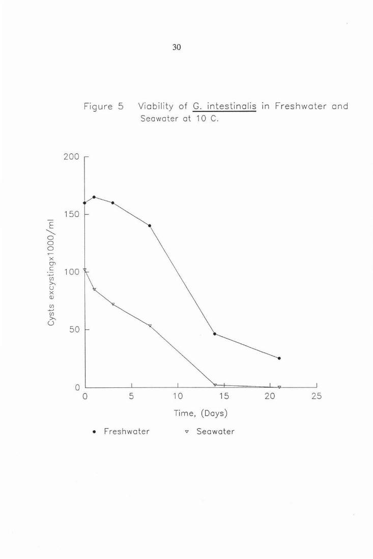
200
150 E
"-.. 0 0 0
x V> c 100 +-' en >-. u x GJ
en +-' en >-. u
50
0
Figure 5
0 5
30
Viability of G. intestinolis in Freshwater and Seawater at 1 0 C.
1 0 15 20 25
Time, (Days)
• Freshwater 'V Seawater

150
E "- 100 0 0 0
x CJ) c (/)
>-u x (I)
(/)
50 ..._, (/)
>-u
0
Figure 6
0 5
31
Viability of G. in t estinalis in Freshwater and Seawater at 15 C.
10 15 20 25
Time, (Days)
• Freshwater v Seawater

32
Table 1 Giardia spp. cyst inactivation at t = 0, showing differences between freshwater and seawater.
Species Temp. (oc) Difference (%) Average
G. muris 5 29%
10 28%
15 43.5% 33.5%
G. intestinalis 5 57%
10 37%
15 59.5% 51%

0 z -........... _,__, z
0.1
0.01
Figure 7
0 5
33
Nt/No vs Time for G. muns in Freshwater and Seawater at 5 C.
10 15 20 25
Time, (Days)
• Freshwater " Seawater

0 z
""' -+-'
z 0. 1
0.01
Figure 8
0 5
34
Nt /N o vs Time for G. muris 1n Freshwater and Seawater at 10 C.
·---···---·-----·-··-·-··-···· .... ·-·--·-----·--··-··-···--···-···--···
10 15 20 25
Time, (Days)
• Freshwater " Seawater
0 z '---... +-'
z
1
0 . 1
0.01
Figure 9
0 5
35
Nt/No vs Time for G. muns in Freshwater and Seawater at 15 C.
1 0 15 20 25
Time, (Days)
• Freshwater v Seawater
0 z "-.. 0 .1 -+-'
z
0 .01
36
Figure 10 Nt/No vs Time for G. muns in Freshwater and Seawater at 5, 10, and 15 C.
···· ·····-·-·--·-·-······ ····-·-·····------------ ····--·····-·- -------------------------- -------········-------------- ---------s ·--·--e--__________ _ ___ 1_n __ c_____________ ---------------·····-----------------------·-
5 c 15 c
········-····-··························---------------- -·---·i -5 ----G -----··--------··-----······-··------------ --------------------------········· ·
0 5 10 15 20 25
Time, (Days)
0 'V Seawater • • Freshwater

37
Table 2 Giardia muris Log Nt/No Gradients.
Temperature°C Gradient Water Gradient Seawater
5 0.0300 0.106
10 0.0257 0.0875
15 0.0404 0.180
Data for this table from Figures 7-9, and 11-13.

0 z ~ -+-'
z 0 .1
0.01
38
Figure 11 Nt/No vs Time for G. intestinalis 1n
Freshwater and Seawater at 5 C.
-------------·-- ··-··--·····-·········
·----· - --------------------········-.. ·-····-·····------·
0 5 10 15 20 25
Time, (Days)
• Freshwater "" Seawater

0 z ""- 0 . 1 ......, z
0 .01
39
Figure 12 Nt / No vs Time for G. intestinalis in
Freshwater and Seawater at 10 C.
·-----·--··--··-·-···-··-·····-·········-········
0 5 10 15 20
Time, (Days)
• Freshwater v Seawater
25

0 z ~ _....., z
0. 1
0 .01
40
Figure 13 Nt/ No vs Time for G. intestinalis in
Freshwater and Seawater at 1 5 C.
0 5 10 15 20
Time, (Days)
• Freshwater '<:] Seawater
25

0 z ""' 0. 1 -+-'
z
0.01
41
Figure 14 Nt / No vs Time for G. intestinalis in Freshwater and Seawater at 5, 10, and 15 C.
10 c
· ··· ·······················-······-·····--·········-- · ··················-············-···--·······---······-·-·-- ····--·---·-··-····-···-···-····-···-·---·····--·--·-- -·--5··--·G·········· · ··
10 c
5 c
0 5 10 15 20 25
Time, (Days)
0 'V Seawater .., • • Freshwater
42
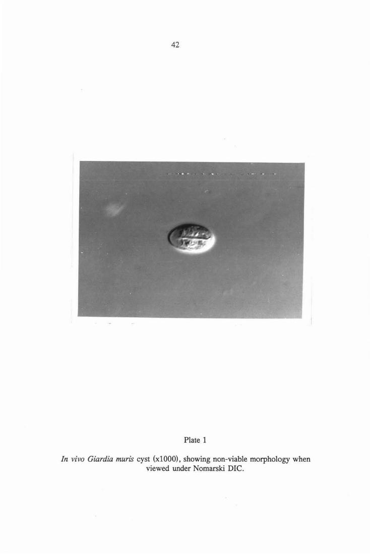
Plate 1
In vivo Giardia muris cyst (xlOOO), showing non-viable morphology when viewed under Nomarski DIC.

43
Plate 2
In vivo Giardia muris cyst (xlOOO), showing viable morphology when viewed under Nomarski DIC.

44
Plate 3
In vivo Giardia intestinalis cyst (x7200), showing non-viable morphology when viewed under Nomarski DIC.

45
Plate 4
In vivo Giardia intestinalis cyst (x7200), showing viable morphology when viewed under Nomarski DIC.

100
80
U1 60 +-' U1 >.. u Q)
..a 0 >
40 ~
20
0
46
Figure 15 The percent viable cysts as determined
0
• ....
5
by Nomarski DIC , FDA, Pl, and Excystotion for G. muris at 5 C in Freshwater.
10 15 20 25
Time, (Days)
Nomarski DIC v Excystation
FDA • Pl
100
80
(/)
60 +-' (/)
>---u Q)
_o 0 > ~
40
20
0
47
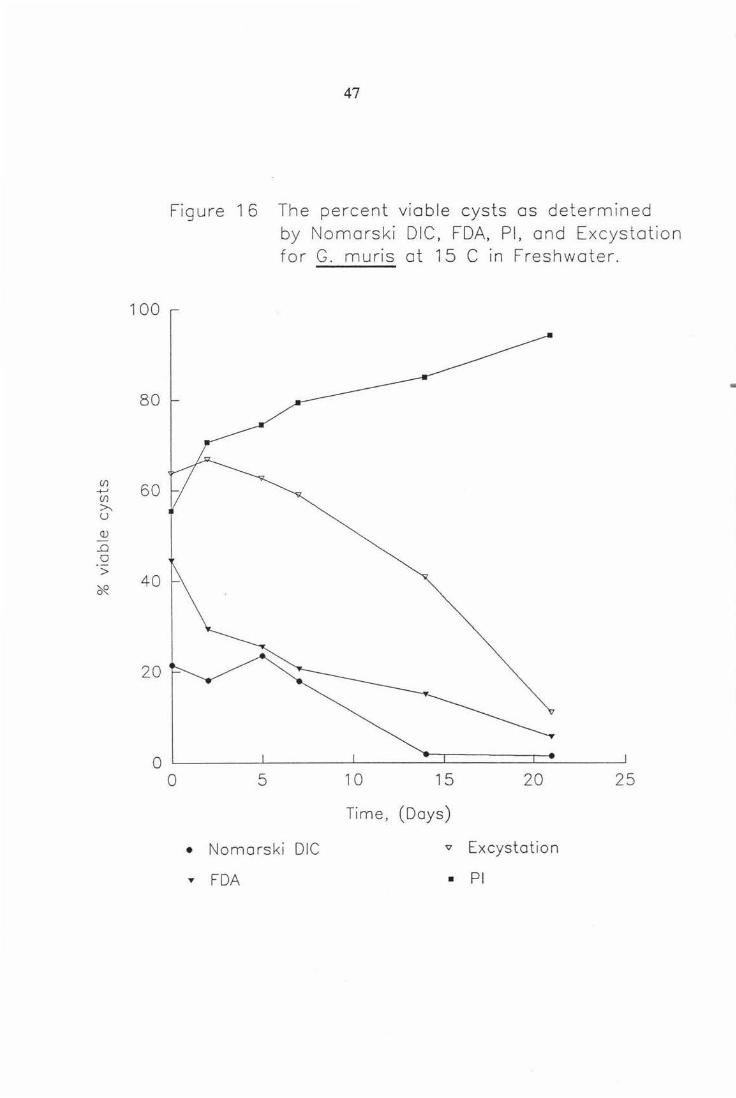
Figure 1 6 The percent viable cysts as determined
0
• y
5
by Nomorski DIC, FDA, Pl, and Excystation for G. muris at 15 C in Freshwater.
10 15 20 25
Time, (Days)
Nomarski DIC v Excystation
FDA • Pl
-

100
80
(/)
60 ......., (/)
>. u Q)
.D 0 > ~
40
20
0
48
Figure 17 The percent viable cysts as determined
0
• ...
5
by Nomarski DIC, FDA, Pl, and Excystation for G. muris at 5 C in Seawater.
10 15 20 25
Time, (Days)
Nomarski DIC v Excystation
FDA • Pl
1 00
80
(f)
60 +-' (f) >-, u (!)
-_Q
0 > ~
40
20
0
49
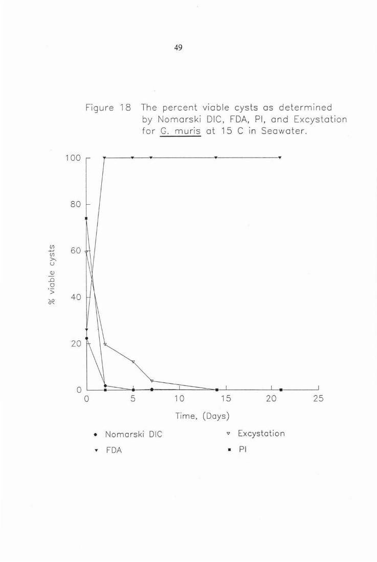
Figure 18 The percent viable cysts as determined
0
• ...
5
by Nomarski DIC, FDA, Pl, and Excystot ion for G. muris at 15 C in Seawater.
10 15 20 25
Time, (Days)
Nomarski DIC " Excystation
FDA • Pl

(j) -+-' (j)
>--u Q)
_Q
0
> ~
100
80
60
40
20
50
Figure 19 The percent viable cysts as determined by Nomarski DIC for G. muris showing temperature and seawater effect.
0 L_-=:=::::::===....j,__.=:l!====d::==~_;-=r::=======:I==-~~_J
0 5
• Water 15 C
" Sea 1 5 C
1 0 15 20 25
Time, (Days)
~ Water 5 C
• Sea 5 C
(/) +-' (/)
:>--u (])
-...0 0
> ~
100
80
60
40
20
51
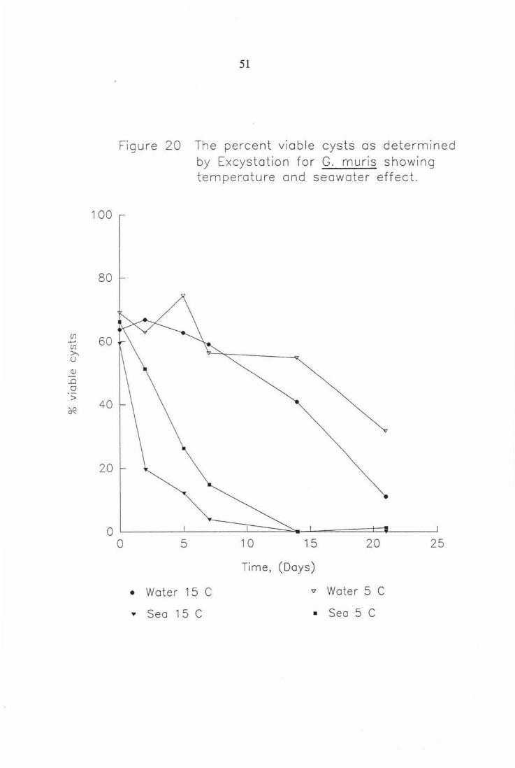
Figure 20 The percent viable cysts as determined by Excys tation for G. muris showing temperature and seawater effect.
0 L_~~~..l.._~~~__L_-=====~.......L-=======±:=l--~~__J 0 5 10 15 20 25
Time, (Days)
• Water 15 C '7 Water 5 C
,. Sea 1 5 C • Sea 5 C

(f) +-' (f)
>.. u Q)
...0 0 > ~
100
80
60
40
20
52
Figure 21 The percent viable cysts as determined by FDA for G. muris showing temperature and seawater effect.
0 L-~~~---'-~~~--.J'--~~~-'-~~~---'-~~~--' 0 5 10 15 20 25
Time, (Days)
• Water 15 C v Water 5 C
.,. Sea 15 C • Sea 5 C

100
80
(/) -+--' (/)
60 >--u (J)
-..0 0
> c 0 40 c
~
20
0
53
Figure 22 The percent nonviable cysts as determined by Pl for G. muris showing
0
temperature and seawater effect.
5
• Water 15 C
"' Sea 15 C
10 15 20
Time, (Days)
"' Water 5 C
• Sea 5 C
25
54
Plate 5
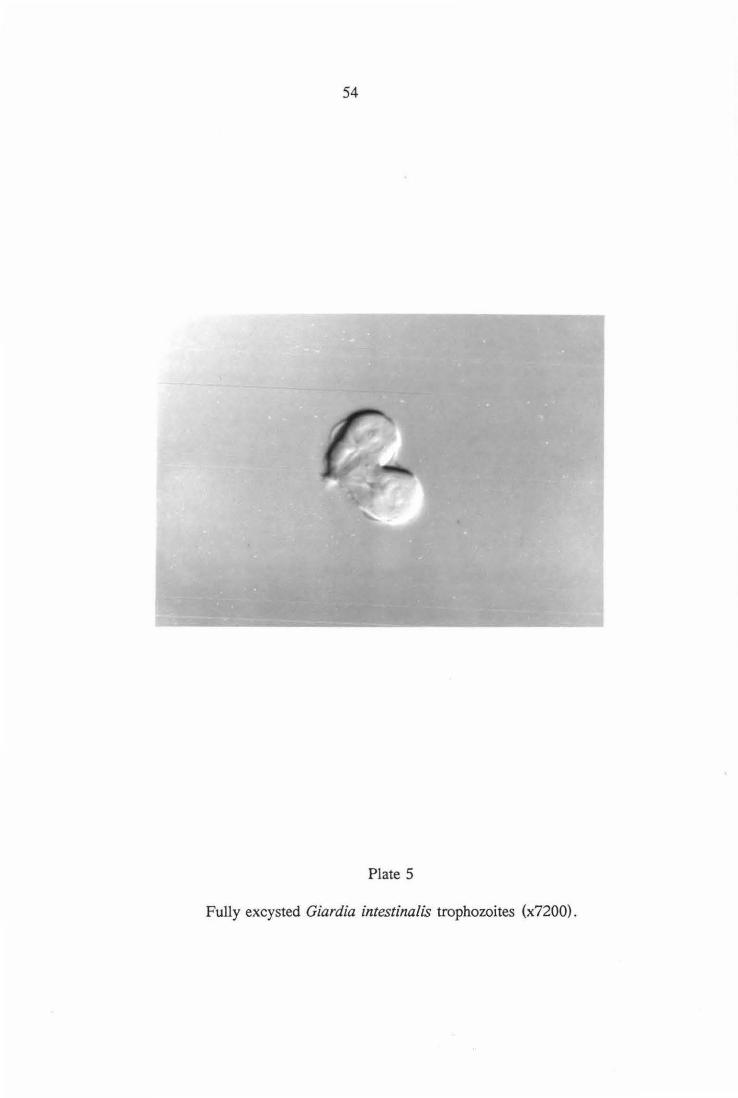
Fully excysted Giardia intestinalis trophozoites (x7200) .

55
Plate 6
Seawater inactivated Giardia cyst (x7200).
100
80
I.fl 60 _...,
I.fl >.. u Q)
..0 0 > ~
40
20
0
56
Figure 23 The percent viable cysts as determined
0
• ..,.
5
by Nomarski DIC, FDA, Pl, and Excystation for G. intestinalis at 5 C in Freshwater .
10 15 20 25
Time, (Days)
Nomarski DIC v Excystation
FDA • Pl

100
80
(/)
60 +-' (/)
>... u Q)
_Q
0 > ~
40
20
0
57
Fig ure 24 The percent viable cysts as determined
0 5
by Nomarski DIC, FDA, Pl, and Excystation for G. intestin olis at 15 C in Freshwater.
10 15 20 25
Time, (Days)
• Nomarski DIC "' Excystation
• Pl T FDA
100
80
(f)
60 ......... (f)
>-. u Q)
...0 0 > ~
40
20
0
58
Figure 25 The percent viable cysts as determined
0
• "'
5
by Nomarski DIC, FDA, Pl, and Excystation for G. intestinolis at 5 C in Seawater .
10 15 20 25
Time, (Days)
Nomarski DIC '<:! Excystat ion
FDA • Pl
100
80
UJ 60 _._, UJ >-u Q)
.D 0 > ~
40
20
0
59
Figure 26 The percent viable cysts as determined
0
• T
5
by Nomarski DIC, FDA, Pl, and Excystation for G. intestinalis at 15 C in Seawater.
1 0 15 20 25
Time, (Days)
Nomarski DIC v Excystation
FDA • Pl

100
80
(/}
60 ..._, (/}
~ u Q)
..0 0
> ~
40
20
0
60
Figure 27 The percent viable cysts as determined
0
by Nomarski DIC for G. intest inolis showing tern perotu re and seowate r effect.
5 10 15 20 25
Time, (Days)
• Water 15 C v Water 5 C
• Seo 15 C • Sea 5 C

100
80
U1 60 +-'
U1 >-. u Q)
..0 0 > ~
40
20
0
61
Figure 28 The percent viable cysts as determined by Excys tation for G. intestinalis showing temperature and seawater effect.
0 5 10 15 20 25
Time, (Days)
• Water 15 C v Water 5 C
• Sea 15 C • Sea 5 C

100
80
(/)
60 ........ (/)
>-u Q)
_o 0 >
40 ~
20
0
62
Figure 29 The percent viable cysts as determined by FDA for G. intestinalis showing temperature and seawater effect.
0 5 10 15 20 25
Time, (Days)
• Water 15 C v Water 5 C
... Sea 15 C • Seo 5 C
100
80
(/] +-' (/]
60 >---u Q)
-..Q
0
> c 0 40 c
~
20
0
63
Figure 30 The percent non viab le cysts as determined by Pl for G. intestinolis showing temperature and seawater effect.
0 5 10 15 20 25
Time, (Days)
• Water 15 C '<l Water 5 C
"' Sea 15 C • Sea 5 C

64
Table 3 Inactivation of Giardia muris using various chlorine treatments.
Temp°C pH FAC Time Inactivation Reference. min. efficiency.
5 7.0 1 30 90% Rice et al., 1982
6 8.2 1.3 30 88.4% Hoff et al., 1985
5 7.0 1.5 30 97% Sauch et al., 1991
5 7.0 31.4 30 99% Leahy et al., 1987
5 6.0 0.95 62* 90% This thesis.
*derived from log expressed data for excystation in Figure 34.

100
80
(/)
60 +-' (/)
>.. u Q)
_o 0
> ~
40
20
0
65
Figure 31 Chlorine inactivation of G. muris as determined by Nomorski DIC.
0 20 40 60 80 1 00 1 20 1 40 1 60
Time, (mins)
• Water ~ Chlorine

U1 +-' U1 >---u Q)
-_Q
0
> ~
100
80
60
40
20
66
Figure 32 Chlorine inactivation of G. muris as determined by Excystation.
0 '--~--'-~~~~~-'---~~-'--~---'~~--'-~~-'--~~ 0 20 40 60 80 1 00 1 20 1 40 1 60
Time, (mins)
• Water -v Chlorine
(/) ....... (/)
>-. u <l.J
_o 0 > ~
100
80
60
40
20
67
Figure 33 Chlorine inactivation of G. muns as determined by FDA/Pl.
0 '--~-'-~~...J-~~-'--~--L~~--'-~~-'--~--'~~-' 0 20 40 60 80 100 120 140 160
Time, (mins)
• Water v Chlorine

(/) .+-J (/)
>(.)
...a 0
>
100
10
1
68
Figure 34 Chlorine inactivation of G. muns log data for Excystation.
--------
0 20 40 60 80 1 00 1 20 1 40 1 60
Time, (mins)
• Wate r v Chlorine
69
Plate 7
Fluorescein Diacetate stained Giardia cyst (x400).

70
Plate 8
Propidium Iodide stained Giardia cyst (x400) .

71
Chapter Five Discussion.
Seawater Inactivation.
Very few areas of New Zealand are far from the sea. This enables many New
Zealanders to enjoy the many recreational attractions that the sea offers, such
as fishing, boating, swimming, surfing and seafood gathering.
Cities and towns produce large volumes of sewage. The disposal and
treatment which, is expensive and sometimes controversial for many borough
councils.
Historically proximity to the coast has meant local authorities opting for the
infinitely cheaper option of disposal into the sea. In today's social climate this
practice is rapidly becoming unacceptable and the move is towards 100% land
based treatment. (eg. Wellington City proposed Milliscreening plant at Moa
Point).
Seaborne waste disposal results in microorganisms such as viruses
(eg. infectious hepatitis), bacteria (eg. Escherichia coli) and protozoa such as
Giardia spp. being introduced into seawater. The length of time such•micro
organisms remain viable in seawater has only received moderate attention.
E.coli and virus survival in seawater (Akin et al., 1975; Carlucci and Pramer
1959; Komek 1926; Nusbaum and Garver 1955; Zobell 1936), and Giardia
cyst survival in seawater (Fontaine et al., 1984), are important considerations
when evaluating the effects of a sewage outfall. Limited information
concerning Giardia cyst viability in seawater prompted this study examining
firstly whether G. muris cysts are able to be used as a model for G. intestinalis
cyst inactivation and secondly whether seawater has any effect on the viability
of cysts when compared to freshwater.
In vivo derived cysts were used for all experiments.
G. intestinalis cysts from human faecal samples excysted with a consistently
high level of 90% in contrast to the findings of Bingham and Meyer (1990);

72
Buchel et al., (1987); and Smith and Smith (1989) who showed rates of
excystation from 15 - 60%.
The level obtained here was ideally suited to this study.
G. muris cysts as reported by Roberts-Thomsen et al., (1976) showed
excystation rates greater than 90% when cysts were freshly harvested from
mouse faeces .
Experiments examining the seawater effect at 3 different temperatures (5, 10,
and 15°C) with respect to a water control, using G. intestinalis and G. muris
were conducted. Results at each temperature and for each species of Giardia
reveals a significant difference (Analysis of Variance, ANOVA p < 0.0001)
between viability of seawater and freshwater incubated cysts (Figs. 1-6) .
G. muris follows a clear exponential decrease in viability with respect to time
for each temperature, and G. intestinalis follows ·a similar pattern. These
differences maybe species related.
The lower viability seen in seawater incubated cysts is probably due to some
cysticidal factor in the seawater. Carlucci and Pramer (1959), stated "that on
the basis of concentrations, inorganic salts are the most potentially toxic
substance present in the sea" . It is postulated that this will have an osmotic
effect or perhaps exert a specific ion toxicity. Analysis of the composition of
seawater suggests that seawater does indeed have the capacity to cause these
effects.

73
Table 4 Top ten ion concentrations in seawater.
Rank Element [ion] mg/l
1 Chlorine 18800
2 Sodium 10700
3 Magnesium 1290
4 Sulpher 905
5 Calcium 412
6 Potassium 399
7 Bromine 67
8 Carbon 28
9 Strontium 7.9
10 Boron 4.5
([ion] based on 1 litre of 35% seawater = 1.028 kg)
(From Marine Pollution, Diagnosis and Therapy. Ed: A. Sebastion)
Results (Figs. 1-6) with both G. muris and G. intestinalis show a decrease in
viability consistent with the statement of Carlucci and Pramer (1959).
At t = 0, when cyst samples were in seawater and freshwater for less than one
minute before viability determination, both species showed a marked difference
in viability at all temperatures (Figs. 1-6; Table 1). (A difference being
calculated as viability of cysts in freshwater at t = 0 less viability of cysts in
seawater at t = O) The differences shown in Table 1 illustrate what is thought
to be the effect of osmotic shock. Giardia spp inoculated into seawater at a

74
sewage outfall would therefore show an almost instantaneous decrease in
viability compared to the same population of cysts in freshwater. The
magnitude of this viability loss in seawater is in the order of 33.5% for G.
muns and 51 % for G. intestinalis (with respect to water) such that seawater
can be said to be cysticidal. The differences seen in Table 1 are illustrative of
species differences between G. muris and G. intestinal is.
The argument that osmotic shock is the primary inactivation factor is
strengthened by the photographic evidence in Plate 6. This Nomarski DIC
photo shows a seawater inactivated cyst with morphology characteristic of a
cell that has undergone plasmolysis in hypotonic solution. The cyst wall is
intact and internal structures are concentrated in a central position in the cyst .
The term "specific ion toxicity" relates to the effect heavy metals such as
Mercury, Lead, Arsenic, and Aluminium have. The seawater used in this
study was collected from a remote coastal location on the West Coast of the
North Island, New Zealand-Tangimoana Beach. The levels of other ions in
seawater as illustrated previously may exert an effect other than osmosis on the
cyst, contributing to the cysticidal effect of seawater ie. there may be unknown
cysticidal radicals present.
Seawater pH is known to have an effect on the survival of Escherichia coli in
seawater (Carlucci and Pramer 1959). Incubation of E. coli for 48 hrs in
seawater at pH 5 yields a survival of 58%, compared with incubation in
seawater at pH 8.0 (pH most commonly found in oceans, Walker 1975),
yielding a survival of just 0.4%. Thus Giardia cysts may be similarly
affected, and could contribute to the viability difference seen at t = 0 (Table 1)
and general decrease in viability seen with respect to time (Figs. 1-6). The
high pH of seawater is due to the high concentration of salts present (Marine
Pollution, Diagnosis and Therapy 1976) Tangimoana seawater has a pH of
between 7.45 - 8.47, easily in the range to give a killing effect.

75
Log plots of Nt/No for G. muris illustrate a linear trend in the decrease of
viability in freshwater and seawater (Figs. 7-10). This is most pronounced for
G. muris at all temperatures, whereas G. intestinalis (Figs. 11-14) does not
show the same degree of linearity. This is probably species related.
An average of data combining the rates of inactivation for the three
temperatures shows G. muns is inactivated at a slower rate in seawater than
fresh water (Table 2) . G. intestinalis does not show this same linear trend and
we can say that there is a species difference in the pattern of inactivation in
seawater and freshwater. This difference is significant (ANO VA p < 0.0001).
By comparing the inactivation trends of G. muris and G. intestinalis we can
see that the animal strain has a greater resistance to inactivation by seawater.
This effect may be compared to the observations of Jarell (1988) for chlorine
inactivation of G. muns. They observed that G. muris cysts have a greater
resistance to chlorine inactivation. This study agrees with these observations
with respect to seawater such that one can suggest that G. muns is a more
hardy species of cyst. This is maybe due to structural differences in the cyst
giving it greater resistance.
In these comparisons the temperature effect stated in the literature (Bingham et
al., 1979), does not seem to be evident, ie. one would have expected to see an
increase in inactivation rate with an increase in temperature. With both G.
muris and G. intestinalis the relationship between temperature and increased
inactivation rate is unusual with greater inactivation being shown at 5 and 15°C
than at 10°C. There is no obvious explanation for this but it could be that the
cysts are being influenced by a temperature dependent enzyme effect
ie. enzymes concerned with the viability of the resting cyst and the cysts ability
to undergo excystation may have optima which support these observations.

76
Assessment of Methods of Determining Viability of Giardia cysts.
1. Seawater inactivation.
Investigations into the methods for determining Giardia spp. cyst viability have
been in progress for some time now (Belosevic et al., 1983; Bingham et al.,
1979; Feely 1986; Feely et al., 1991; Filice 1952; Hoff et al., 1984; Hoff et
al., 1985; Labutuik et al., 1991; Leahy et al., 1987; Rendtorff 1879; Rice and
Schaefer 1981; Roberts-Thomsen et al., 1976; Sauch et al., 1991; Schaefer et
al. , 1991; Schupp and Erlandsen 1987; Schupp et al. , 1988). The accepted
methods used for viability testing are few at present and consist mainly of
excystation, fluorogenic dye staining, infectivity, and morphological
determination using Nomarski Differential Interference Contrast Microscopy
(Nomarski DIC), (Belosevic et al., 1983; Bingham et al., 1979; Feely 1986;
Feely et al., 1991; Filice 1952; Hoff et al., 1984; Hoff et al., 1985; Labutuik
et al., 1991; Leahy et al., 1987; Rendtorff 1879; Rice and Schaefer 1981;
Roberts-Thomsen et al., 1976; Sauch et al., 1991; Schaefer et al., 1991;
Schupp and Erlandsen 1987; Schupp et al., 1988).
Excystation is considered the "benchmark" for viability by many researchers.
But complex media, length of time needed to conduct this assay and the need
for > 105 cysts per ml to test precludes its use in water testing, where
frequently < 10 cysts are found. Therefore the alternatives are investigated in
this study .
The effective merits of these methods have been examined in what is mainly
"time independent" studies using single samples and single field of view
experiments (Schupp and Erlandsen 1987; Smith and Smith 1989).
Investigators using time dependent studies (Sauch et al., 1991; Labutuik 1991),
use cyst inactivations that are not capable of showing trends over a time period
because serial sampling and testing was not prac~ised. They involve a single
set exposure time. This study differs majorly from previous work as follows :
the methodology involved taking samples at time intervals over a period of 21
77
days for seawater inactivation and 150 minutes for chlorine inactivation, then
analyzing these samples using
a) Excystation,
b) Nomarski DIC,
c) Fluorogenic Dyes.
Infectivity (Roberts-Thomsen et al., 1976), was not used as a measure of
viability because of major problems obtaining specific pathogen free mice.
Inactivation using seawater and chlorine was trialled because of previous
reports that fluorogenic dye staining was thought- not to be effective in a
chlorine inactivation system, and that dyes would be better suited to an
environmental inactivation system (Sauch et al., 1991).
Bearing these points in mind an environmental system using seawater as the
inactivating agent was used. This was shown earlier in this study to be an
effective inactivation agent for both G. muris and G. intestinalis .
Verification of the observations of Sauch et al., (1991) with chlorine
inactivation was conducted by performing a similar experiment. The
experiment was conducted using G. muris as at the time in vivo
G. intestinalis were difficult to source.
Chlorine levels for the inactivation were determined by the N. Z. Health
Department as being suitable for 99.9% inactivation of G. intestinalis cysts.
The experiment entailed; 1 ppm Free Available Chlorine, 149 minutes,
pH 6-7, and 5°C (Ampofo et al., 1991).
'Black cysts' as seen by (Sauch et al., 1991; and Shupp and Erlandsen 1987),
were not observed in any of the samples tested using the fluorogenic dyes, and
were therefore not considered in the conclusions stated here.

78
Examination of figures for G. muris incubated in freshwater at 5 and 15°C
(Figs. 15-16), shows Fluorescein Diacetate (FDA) and Nomarski DIC account
for similar levels of viability with FDA recording a slightly higher proportion
at each sampling time. The level of viability determined by the 'benchmark'
excystation is considerably greater (at all data points), and thus Nomarski DIC
and FDA although very similar to each other both underestimate viability with
regard to excystation. Propidium Iodide (PI) staining represents the proportion
of non-viable cysts and was calculated from the same data as that for FDA
viability. ie . Total number in the sample = FDA positive scored cysts + PI
positive scored cysts. This explains the mirrored pattern seen in these
experiments with respect to the fluorogenic dyes . PI under estimates the level
of viability.
In seawater at 5 and 15 °C (Figs. 17-18), G. muris cyst staining initially (t=O)
showed indications of following the same trends as in water. But after 1 - 2
days results were very unusual. ie . FDA displays 100% viability and PI 0%
viability for each temperature. This effect was not seen in freshwater and is
also observed for the same experiment conducted with G. intestinalis (Figs. 25-
26). This effect was probably due to a) the cysticidal effect of the seawater or
b) the seawater possible osmotic effect on the action of the stains. Plate 6
shows the apparent effect seawater has on Giardia cysts, in that it seems the
primary cause of death is plasmolysis.
FDA action as a vital stain means that only viable actively metabolising cysts
are able to fluoresce green at an excitation wavelength 490nm. PI relies on
diffusion into the cyst where it intercalates nucleic acids yielding a red/orange
fluorescing cyst at excitation wavelength 490nm (Schupp and Erlandsen 1987).
Thus if cysts have been metabolically altered by the action of the seawater and
show characteristics such as those in this experiment then the reliability of the
use of these fluorogenic dyes for estimating viability of seawater inactivated
cysts must be questioned.
79
The response the stains give in freshwater, although underestimating the
viability with respect to excystation seem to behave 'normally'. Their use in
classifying cysts isolated from freshwater samples as either viable or non-viable
may be possible.
Commercially available and highly specific immunofluorescent stains (using
Fluorescein label), have been shown to be compatible with Propidium Iodide
(Sauch et al., 1991) and they could therefore be used together to detect and
determine viability of Giardia cysts isolated from freshwater.
The viability as assessed by excystation and Nomarski DIC in seawater, is
much lower than that seen in freshwater for the same population of cysts, this
is due to the seawater as stated earlier (osmotic and other unknown effects).
Nomarski DIC consistently under estimated viability when compared to the
'benchmark' excystation.
A similar experiment conducted using G. intestinalis varies from those given
by G. muris (Figs. 23-24). Incubation in water and for both temperature cases
(5, 15°C) shows Nomarski DIC under estimates viability when compared with
FDA. This agrees with the pattern for G. muris . Pl staining under estimates
the viable proportion of cysts compared to Nomarski DIC. Excystation shows
a different pattern to that for G. muris decreasing at a faster rate and
suggesting a species difference.

80
In seawater at S and 1S°C the pattern of inactivation for G. intestinalis (Figs.
2S-26), as shown by FDA and PI, is identical to that shown by G. muris (Figs.
17-18). Excystation and Nomarski DIC do give a reading but this does not
rise above 10%, suggesting that G. intestinal is is a more susceptible species to
inactivation by seawater. This contrasts with G. muris where viability by these
methods was measured initially at above 20% in each case, and drops in the
same exponential fashion seen in Figs. 1-3.
The unusual results for FDA and PI may be hypothesized as being due to the
same effects discussed for G. muris (Plasmolysis and seawater having an effect
on their ability to stain the cyst).
A re-emphasis of the data allows an examination of each method of
determining viability, both with respect to temperature (S, l S°C) and treatment
(freshwater/seawater). Viability as determined by excystation in Figure 20
for G. muns and Figure 28 for G. intestina/is show a clear difference between
viability in freshwater and seawater for each species. The lower temperature
of S°C appears to "protect" the cysts in each treatment yielding a higher
viability over the time period. This effect is less evident in previous seawater
inactivations (this study). The lethal temperature dependent killing effect seen
in freshwater is documented (Bingham et al., 1979), and seems to apply to
seawater.
Viability as determined by Nomarski DIC (Figs. 19 and 27) shows clear
differences when compared to excystation, ie. at t = 0, the viability being
approximately SO% lower. The difference between freshwater and seawater
treatments is also extremely clear. Temperature as seen for excystation,
'protects' cysts at the lower temperature of S0, with 1S°C. showing a faster rate
of inactivation.
81
2. Chlorine Inactivation.
In the supply of freshwater to townships and cities, effective disinfection is
critical for the safety and well being of the inhabitants. Filtration, coagulation,
and sedimentation although necessary are not always 100% effective and a
subsequent disinfection programme must be effective. The New Zealand
Health Department has set guidelines for disinfection of domestic supply water.
A chlorine concentration of 1 mg/l Free Available Chlorine for 149 minutes, at
pH 6-7, and 6°C, is deemed to give a 99.9% inactivation of G. intestinalis
(Ampofo et al., 1991).
Detection of Giardia cysts either pre or post treatment is indicative of a
potential problem. ie. Giardia cysts in the water supply. Unfortunately the
potential infectivity cysts is not presently assessable. Many researchers have
addressed the problem of viability post disinfection (Leahy et al., 1987;
Neuwirth et al., 1988; Hoff, Rice and Schaefer 1985; Rice et al., 1982) using
excystation as their measure of viability.
Water testing for Giardia cysts is laborious and time consuming. The
methodology involved (Standard Methods), results in approximately 50% of
the cysts present being recovered (Jakubowski 1987). The laboriousness of the
testing combined with the complex materials and methodology needed for
excystation, makes Nomarski DIC, and Fluorogenic Dye Staining more
appealing as an alternative for determining viability. Cysts isolated are able to
be kept on slides for detection, then perhaps viewed with Nomarski DIC or
stained with a vital stain.
With the testing in mind methods for determining viability such as Fluorogenic
Dye Staining and Nomarski DIC are less complex both in materials used and
the methodology followed when compared to excystation, and also take
considerably less time to complete.

82
The experiments show both Nomarski DIC and FDA/PI over estimate viability
when compared to the 'benchmark' for viability excystation. This occurs in
freshwater and to a greater extent in chlorine treated water.
The actual response to chlorine is minimal when compared to excystation
which shows a large decrease. This is the 'benchmark' for viability and thus
in interpreting these results we would be more inclined to believe excystation.
Comparison of the data here with the observations made by other researchers
in Table 3, shows viability as assessed by excystation here follows a similar
trend to their experiments (References in Table 3). These researchers achieved
a 90% inactivation under conditions similar to those used here.
The response shown by Nomarski DIC and FDA/PI does show signs of
decreasing, and it may be that given more time these methods will show an
appreciable decrease in viability. Unfortunately in the disinfection of large
volumes of water with high flow rates, this doesn't allow a lengthening of
retention time to enable the viability of cysts detected to be determined.
The use of Nomarski DIC and FDA/PI in determining viability of pretreated
water would give an exaggerated estimate of the viability of the cysts tested.
This in itself would not be an unwarranted use of Nomarski DIC or the stains
as any cyst detected should prompt a thorough examination of water treatment
practises and make staff wary of the risk that Giardia poses. The same theory
can be applied post-chlorine treatment.

83
Chapter Six Conclusions.
In Figs. 19-22 & 27-30, the effect of temperature is evident more in some
cases than others. G. muris and G. intestinalis both exhibit considerably faster
rates of inactivation in seawater as compared to freshwater. The pattern of
inactivation (Figs. 1-3) for G. muris is different to that for G. intestinalis
(Figs. 4-6) and therefore does not form a good model for G. intestinalis.
The cysticidal effect that seawater has on cysts has been postulated as being
due to the salt in seawater and the alkaline pH. Plate 6 shows a seawater
inactivated cyst which has the morphological character of a plasmolysed cell.
Thus the seawater exerts an osmotic effect on the cysts and in all cases this
effect yields an almost instantaneous drop in viability.
In conjunction with other factors determining the fate of sewage pumped into
the sea, dilution. This is a variable factor and is dependent on tidal patterns
and the direction and rate of currents (H yashi and Ito 1975). It can be
assumed that in the case of outfalls pumped directly into the sea as opposed to
harbours or estuaries that the effect of dilution would be massive. Further,
sedimentation of Giardia cysts physically associated with relatively large
particulate matter and with the flocculating effect of seawater (Mitchell and
Chamberlain 1975), absorption to marine sediments stimulates effective cyst
reduction . Thus we can say the risk of contracting a giardiasis infection from
contaminated sea would be much reduced and significantly less than fresh
water.
The use of Fluorogenic Dyes as an alternative methods of determining viability
in seawater is not possible because of the unusual and as yet unexplained
results in Figs . 17-18, 21-22, 25-26, 29-30. Nomaiski DIC is able to be used
in a limited capacity to determine viability of cysts isolated from seawater, but
one must remember that this method under estimates viability as compared to

84
the benchmark method excystation. In freshwater the effective merits of the
Fluorogenic Dyes are more useful with FDA/Pl only slightly under estimating
viability as compared to Nomarski DIC, but under estimating viability to a
larger extent when compared to excystation.
In chlorine inactivation the use of Nomarski DIC and Fluorogenic Dyes to
determine viability of cysts isolated from water samples would seem feasible at
first. The relative ease with which these methods can be conducted when
compared to excystation means that little training would be required to enable
their use in treatment plants. The cost of the microscopes needed is
unfortunately quite inhibitory to their set up.
An over estimation of viability when determined by Nomarski DIC and
Fluorogenic Dyes means that a greater proportion of the cysts detected would
be classed as viable. This could lead to heightened awareness of the risk that
Giardia poses to the wider community.
Anomalies in the results concerning an underestimation of viability by
Nomarski DIC when compared to excystation in seawater and an over
estimation when inactivated by chlorine is probably due in part to the highly
osmotic nature of seawater. The much greater chlorine ion concentration in
seawater in conjunction with the many other ions present produces more non
viable Nomarski DIC cyst morphology's and therefore greater inactivation.
When acted on solely by chlorine, it maybe that this does not provide an effect
great enough to change the morphology as seen by Nomarski DIC. Thus
giving an over estimation of viability.

85
Chapter Seven Bibliography.
American Waterworks Association. (1982) Giardia Task Group. Review of
giardiasis in water supplies - Detection, Ocurrence and Removal. Technical
Resource Book.
Akin, E .W., Hill, W.F., and Clarke, N.A. (1975). Mortality of enteric
viruses in marine and other waters. In. Discharge of Sewage from Sea
Outfalls, Supplement to progress in Water Technology, DSSO International
Symposium, Water Research Centre-Stevenage Laboratory, Pergamon Press
Ltd., New York, USA. (Ed: A. L. H. Gameson).
Bartlett, A.V., Moore, M., Gary, G.W., Stark, K.M., Erben, J .J ., and
Meredith, B.A. (1985) Diarrheal illness among infants and toddlers in daycare
centres. I. Epidemiology and Pathogens. Journal of Pediatrics . 107, 495-502.
Belosevic, M., Faubert, G.M., MacLean, J.D., Law, C., Croll, N.A. (1983)
Giardia lamblia infections in Mongolian gerbils: an animal model. Journal of
Infection and Disinfection. 147, p222-226.
Bemrick, W.J., and Erlandsen, S .L. (1988) Giardiasis is it really a zoonosis?
Parasitology Today. 4(3), p69-71 .
Bhatia, V.N., and Warhurst, D.C. , (1981) Hatching and subsequent
cultivation of cysts of Giardia intestinalis in Daimonds Medium. Journal of
Tropical Medicine and Hygiene. 84, p45.
Bingham,A.K., Jarroll,E.L., and Meyer,E.A., (1979) Giardia spp: Physical
factors of excystation in vitro and excystation vs eosin exclusion as
determinants of viability. Experimental Parasitology. 47, p284-291.

86
Bingham, A.K., and Meyer, E.A. (1979) Giardia excystation can be induced
in vitro in acidic solutions. Nature. 277 (25 Jan), p301-302.
Black, R.E., Dykes, A.C., Sinclair, S.P., and Wells, J.G. (1977) Giardiasis in
day care centres: evidence of person to person transmission. Pediatrics. 60,
p486-491.
Blanchard, R., (1888). Remarques sur le Megastome intestinal. Bull. Soc.
Zoo/. France. 30, p18-19.
Boucher, S-E.M., and Gillin, F .D., (1990) Excystation of In vitro derived
Giardia lamblia cysts. Injection and Immunity. 58(11), p3516-3522.
Boreham, P.L.F. , and Shepherd, R.W. (1984) Giardiasis in childcare centres.
Medical Journal of Australia. 141, p263. (letter).
Brodsky, R.E., Spencer, H.C. , and Shultz, M. (1974) Giardiasis in American
travellers to the Soviet Union. Journal of Infection and Disinfection. 30,
p319-323.
Brown, E.H. (1948) Giardia lamblia. he incidence and results of
infestations of children in residential nurseries. Archives of Diseases of
Children. 23, p119.
Brown, T.J., Hastie, J.C., Kelly, P.J., Van Duivenboden, R., Ainge, J.,
Jones, N., Walker, N.K. , Till, D.G., Sillars, H., and F. Lemmon. (1992)
Prescence and distribution of Giardia cysts in New Zealand waters. New
Zealand Journal of Marine and Freshwater Research. 26, p279-282.
Black, R.E., Dykes, A.C., Sinclair, M.S., and Wells, J.G. (1977) Giardiasis
in daycare centres: Evidence of person to person transmission. Pediatrics.
60, p486-491.

87
Buchel, L. A., Gorenflot, A., Chochillon, C., Sarel, J., and Gobert, J.G.
(1987) Invitro excystation of Giardia from humans : A scanning electron
microscopy study. Journal of Parasitology. 73(3), p487-493.
Burke, J.A. (1977) The clinical and laboratory diagnosis of giardiasis.
British Medical Journal. 292, p992.
Carlucci, A.F., and Pramer, D . (1959). Factors affecting the survival of
bacteria in seawater. Applied Microbiology. 7, p388-392.
Craun, G. F., (1985) A summary of waterbourne illness transmitted through
contaminated ground water. Journal of Environmental Health. 48, p122-217.
Davies, R. B. , and Hibler, C. P. (1979) Animal reservoirs and cross species
transmission of giardiasis. In . W. Jakubowski and J.C. Hoff. (Eds).,
Waterbourne Transmission of giardiasis. Environmental Protection Agency,
Cincinnatti, Ohio, p104-106.
Debell , L. , (1932) Antony van Leuwenhoek and his "little animals".
Constable and Company Ltd., London.
Debell, L. and O' Connor, F.W. (1921) The intestinal protozoa of man,
Williamwood and Company, New York.
Deng, M.Y.,and Oliver, D.O. (1992) . Degradation of Giardia lamblia cysts
in mixed human and swine wastes. Applied and Environmental Microbiology.
58(8), p2368-74.
Faubert, G.M. (1988) Evidence that giardiasis is a zoonosis. Parasitology
Today. 4(3) , p66-68.

88
Feely, D.E., (1986) A simplified method for in vitro excystation of Giardia
muris. Journal of Parasitology. 72(3), p474-475.
Feely, D .E., Erlandsen, S.L., and Chase, D.G. (1984) Structure of the
trophozoite and cyst. In Giardia and giardiasis. Biology, Pathogenesis, and
Epidemiology. Ed: Erlandsen, S.L. and Meyer, E.A.) Published by Plenum
Press, New York. p3-32.
Feely, D.E., Gardner, M.D., and Hardin, E.L., (1991) Excystation of
Giardia muris induced by a Phosphate-Bicarbonate medium: Localization of
Acid Phosphatase. Journal of Parasitology. 77(3) , p441-448.
Filice, F.P., (1952) Studies on the cytology and life history of a Giardia from
the laboratory rat. University of California Publishing Zoology. 57, p53-143.
Fontaine, J.P., Delage, A., and M. CI. Lauaire. (1984) On the Epidemiology
of Giardiasis. Anna/es de Parasitologie Humaine et Comparee. 59, p541-554.
Grant, D.R., and P.K. Woo. (1978) Comparative studies of Giardia spp. in
small animals in Southern Ontario.II. Host specificity and infectivity of stored
cysts. Canadian Journal of Zoology. 56, p1360-1366.
Grassi, B., (1888) Morfologia e sistematica di alcuni protozoi parassiti. A/ti
R. Accad. Lince. 1 Rendiconti (4 ser.)IV, (I sem.) pS.
Grieg, M.J., Ridgeway, N.M., and Shakespeare, B.S. (1988) Sea surface
temperature variations at coastal sites around New Zealand. New Zealand
Journal of Marine and Freshwater Research. 22, p391-400.
Hegner, R. W., (1922) The systematic relationship of Giardia lamblia Stiles,
1915, from man and Giardia agilis Kunstler, 1882, from the tadpole.
American Journal of Hygeine. 5, p250-257.

89
Hibler, C.P., and Hancock, C.M. (1989) Waterboume giardiasis. Jn:
Drinking Water Microbiology. (Ed. G. McFetter). New Zealand Drinking
Water Standard World Health, Springer-Verlab.
Hoff, J.C. , Rice, E.W., and Schaefer, F.W. III. (1985) Comparison of
animal infectivity and excystation as measures of Giardia muris cyst
inactivation by chlorine. Applied and Environmental Microbiology. 50(4),
p1115-7.
Isaac-Renton, J., Proctor, E.M., Prameya, R., and Wong, Q. (1986) A
method of excystation and culture of Giardia lamblia Transactions of the
Royal Society of Tropical Medicine and Hygiene. 80, p989.
Jakubowski, W. (1988) Purple burps and the filtration of drinking water
supplies. American Journal of Public Health. 78, p123-125.
Jakubowski, W. and Ericksen, T.H. (1979) Methods for detection of Giardia
cysts in Water Supplies. In Waterboume Transmission of giardiasis. W.
Jakubowski and J.C. Hoff (Eds). Environmental Protection Agency,
Cincinnatti, Ohio. (June 1979).
Jarrell, E. A., (1988) Effect of disinfectants on Giardia cysts. In: CRC
Critical Reviews in Environmental Control. 18(1), pl-28.
Jokipii, A.M.M., Hemila, M., Jokipii, L. (1985) Prospective study of
acquisition of Cryptosporidium Giardia lamblia and Gastrointestinal illness.
Lancet. 2, p487-489.
Jones, K.H., and Sneft, J.A. (1985) An Improved method to determine cell
viability by simultaneous staining with Fluorescein Diacetate and Propidium
Iodide. Journal of Histochemistry and Cytochemistry. 33(1), p77-79.
90
Kasprzak, W. and Majewska, A.C. (1983). Infectivity of Giardia sp. cysts in
relation to eosin exclusion and excystation in vitro. Journal of Tropical
Medicine and Parasitology. 34, p70-72.
Keystone, J.S. , Krajden, S., Warren, M.R. (1978) Person to person
transmission of Giardia lamblia in daycare nurseries. Canadian Medical
Association Journal. 119, p241-248.
Korinek, J. (1926). Uber Susswasser-bakterion im Meere. Zentr. Bakteriol.
Parasitenk., Abt.II. 66, p500-505.
Kulda,J . , and Nohynkova,E., (1978) Flagellates of the human intestine and of
intestines of other species, pl-138. In J.P. Kreier. (ed). Parasitic Protozoa.
vol.II. Intestinal flagellates, histomonads, trichomonads, amoeba, opalinids,
and ciliates. Academic Press , Inc., New York.
Kumath, K.R., and R. Murugasu. (1974) A comparitive study of four methods
for detecting Giardia lamblia in children with diarrheal disease and
malabsorption. Gastroenterology. 66, p16-21.
Kunstler J., (1882). Sur cinq protozaires parasites nouveaus. C.R. Seances.
Soc. Biol. 95, p347-349.
Kaur, M., Bajeva, U.K., Jyotin, A.S. and Anand, B.S. (1986). Axenic
cultures from excysted trophozoites of Giardia lamblia and their viability.
Indian Journal of Medical Research. March 1986, p265-267.
Labutuik, C.W., Schaefer, F.W.Ill., Finch, G.R., Belosevic, M. (1991).
Comparison of animal infectivity, excystation, and fluorogenic dye as measures
of Giardia muris cyst inactivation by ozone. Applied and Environmental
Microbiology. 57(11), p3187-3192.

91
Lambert, R.A. (1970) Parasitology : Identification of Protozoa. Published by
Butterworth, London.
Leahy, J.G., Rubin, A.J., and Sproul, D.J. (1987) Inactivation of Giardia
muris cysts by free chlorine . Applied and Environmental Microbiology.
53(7) J p1448-1453.
Mahbubani, M.H., Bej, A.K., Perlin, M.H., Schaefer, F. W. III., Jakubowski,
W .. and R.M. Atlas. (1992) Differentiation of Giardia duodenalis from other
Giardia spp. by using Polymerase Chain Reaction and Gene Probes. Journal of
Clinical Microbiology. 30(1), p74-78.
Marine Pollution, Diagnosis and Therapy. (1976) (Ed: A. Sebastion).
Springer Verlag , Berlin, Heidelburg.
Marino, M.R., Brown, T.J., Waddington, D.C., Brockie, R.E., and P.J.
Kelly. (1992) Giardia intestinalis in North Island possums, house mice, and
ship rats. New Zealand Veterinary Journal. 40, p24-27.
Meyer, E.A., (1987) Giardia and giardiasis. E. A. Meyer ed. Elsevier
Scientific Publishers, Amsterdam.
Meyer, E.A., (1990) Introduction, pl-9, In E.A. Meyer (ed) Human parasitic
diseases, vol. 3. Giardiasis. Elsevier Scientific Publishers, Amsterdam.
Meyer, E.A., and Radulescu, H., (1979) Giardia and giardiasis. Advances
in Parasitology. 17, pl-47.
92
Mitchell, R. and Chamberlain, C. (1975) Factors influncing the survival of
enteric microorganisms in the sea : An Overview. In Discharge of Sewage
from Sea Outfalls, Supplement to progress in Water Technology, DSSO
International Symposium, Water Research Centre-Stevenage Laboratory,
Pergamon Press Ltd., New York, USA. (Ed: A. L. H. Gameson).
Neuwirth,M., Roach, P.D., Buchanan, M. , and Wallis, P.M. (1988). Effects
of chlorine on the ultrastructure of Giardia cysts. In. Advances in Giardia
research. (Ed: P. M. Wallis and B. R. Hammond) . University of Calgary
Press, Calgary, 1988.
Nusbaum, I., and Garver, R.M. (1955). Survival of coliform organisms in
Pacific Coastal Waters. Sewage and Industrial Wastes. 27, p1383-1390.
Owen, R.L., Nemanic, P.D., and Stevens, D.P. (1979) Ultrastructural
observations of giardiasis in a murine model. Gastroenterology. 76,
p757-769 .
Paget, T.A., Jarroll, E.A., Manning, P., Lindmark, D.G., and Floyd, D.
(1989) Respiration in the cysts and trophozoites of Giardia muris Journal of
Clinical Microbiology. 135, p145-154.
Pickering, L.K. (1986) The day care centre diarrhea dilemma. American
Journal of Public Health. 76, p623-624.
Rendtorff, R.C., (1879). The experimental transmission of Giardia lamblia
among volunteer subjects. In W. Jakubowski and J.C. Hoff (Eds),
Waterbourne Transmission of Giardiasis, U.S. Environmental Protection
Agency. Cincinatti, p64-81.

93
Rendtorff, R.C. (1954) The experimental transmission of human intestinal
protozoan parasites. II. Giardia lamblia given in capsules. American Journal
of Hygeine. 59, p209-220.
Rice, E.W., and Schaefer, F.W.III., (1981) Improved in-vitro Excystation
procedure for Giardia lamblia cysts. Journal of Clinical Microbiology. 14(6),
p709-710.
Rice, E.W., Hoff, J.C., and Schaefer, F.W. (III). (1982). Inactivation of
Giardia cysts by chlorine. Applied and Environmental Microbiology. 43(1),
p250-251.
Roberts-Thomsen, LC., Stevens, D.P., Mahmoud, A.A., and Warren, K.S .
(1976) Giardiasis in the mouse; an animal model. Gastroenterology. 71,
p57-61.
Rosoff, J .D. , and Stibbs, H.H., (1986) Isolation and identification of a Giardia
lamblia specific stool antigen (GSA-65) useful in coprodiagnosis of giardiasis.
Journal of Clinical Microbiology. 23, p905-910.
Sauch, J.F., (1988) A new method for Excystation of Giardia. In. Advances
in Giardia Research. p261-264. (Ed: Wallis,P.M., and Hammond,B.R.),
University of Calgary Press, Calgary 1988.
Sauch, J.F., Flanigan, D., Galvin, M.L., Berman, D., Jakubowski, W. (1991)
Propidium Iodide as an indicator of Giardia cyst viability. Applied and
Environmental Microbiology. 57(11), p3243-3247.
Schaefer, F.W.,111., Johnson, C.H., Hsu, C.H., Rice, E.W. (1991)
Determination of Giardia lamblia cyst infective dose for the Mongolian Gerbil
(Merione unguiculatus Applied and Environmental Microbiology. 57(8),
p2408-2409.

94
Schaefer, F.W.III.,Rice, E.W.,and Hoff, J.C., (1984) Factors promoting
in vitro excystation of Giardia muris cysts. Transactions of the Royal Society of
Tropical Medicine and Hygiene. 78, p795-800.
Schaefer, F.W., Sauch, J.F., Jakubowski, W. (eds). (1986) Proposed methods
for detecting Giardia cysts in large volumes of water. pplS-17. United States
Environmental Protection Agency, Cincinnati.
Schupp, D.G., and Erlandsen, S.L., (1987) A new method to determine
Giardia cyst viability : Correlation of Fluorescein Diacetate and Propidium
Iodide Staining with animal infectivity. Applied and Environmental
Microbiology. 53(4), p704-707.
Schupp, D.G. , and Erlandsen, S.L. (1987) Determination of Giardia muris
viability by differential interference contrast, phase, or bright-field microscopy.
Journal of Parasitology. 73, p723-729.
Schupp, D.G., Januschka, M.M., Shelock, L.F. , Stibbs, H.H., Meyer, E.A.,
Bemrick, W.J., Erlandsen, S.L. (1988) Production of viable Giardia cysts in
vitro: Determination by fluorogenic dye staining, excystation and animal
infectivity in the Mouse and Mongolian Gerbil. Gastroenterology. 95, pl-10.
Shupp, D .G. , Januschka, M.M., and Erlandsen, S.L. (1988). Assessing
Giardia cyst viability with fluorogenic dyes : comparisons to animal infectivity
and cyst morphology by light and electron microscopy. In. Advances in
Giardia research. (Ed: P. M. Wallis and B. R. Hammond). University of
Calgary Press, Calgary, 1988.
Smith, A. L. and Smith, H. V. (1989) A comparison of fluorescein diacetate
and propidium iodide staining and in vitro excystation for determining Giardia
intestinalis cyst viability. Parasitology. 99, p329-331.
95
Standard Methods for the Examination of Water and Waste water. (ed: M. C.
Rand, A. E. Greenburg, M. J. Taras. 14th Edition. American Public Health
Association. A. W. W. A. Water Pollution Control Federation.
Tonks, M.C., Brown, T.J., Ionas, G. (1991) Giardia infection in cats and
dogs in New Zealand. New Zealand Veterinary Journal. 39, p33-34.
Walker, J.D., and Guarraia, L.J., (1975) Factors determining life dependency
of microorganisms in the marine environment. In. Discharge of Sewage from
Sea Outfalls, Supplement to progress in Water Technology, DSSO International
Symposium, Water Research Centre-Stevenage Laboratory, Pergamon Press
Ltd., New York, USA. (Ed: A. L. H. Gameson).
Walzer, P.D., Wolfe, M.S., and Shultz, M.G. (1971) giardiasis in travellers.
Journal of Injection and Disinfection. 124, p235-237.
Wolfe, M.S. (1990) Clinical symptoms and diagnosis by traditional methods.
In Giardia and giardiasis. (Meyer, E.A., Editor) Elsevier Science Publishers
(Biomedical Division) New York.
World Health Organisation Expert Committee. (1981). Intestinal protozoan
and helminthic infections. W.H.O. Technical Report Series 666, World Health
Organisation, Geneva.
Woo, P.K., (1984) Evidence for animal reservoirs and transmission of Giardia
infection between animal species, p341-364. In S.L. Erlandsen and E.A.Meyer
(ed). Giardia and giardiasis. Plenum Press ,New York.
Woo, T.K. and Patterson, W.P. (1986) Giardia lamblia in children in
daycare centres in southern Ontario,
Canada, and susceptibility of animals to G. lamblia. Transcripts of the Royal
Society for Tropical Medicine and Hygeine. 80, p56-59.

96
Wright, R.A., and Vernon, T.M. (1976) Epidemic giardiasis at a resort
lodge. Rocky Mountain Medical Journal. 73, p208-211.
ZoBell, C.E., Bacteriocidal action of seawater. Proceedings of the Society for
Experimental Biology and Medicine. 34, p113-116.

97
![multiplexing stool MD short.ppt [Compatibiliteitsmodus] · 8 0394-0527 Giardia Entamoeba coli Giardia 9 0394-0526 Giardia Entamoeba coli Giardia 10 1032-1221 Giardia negative Giardia](https://img.pdfslide.net/doc/110x75/5e4182dad3a23a3d0b082f2c/multiplexing-stool-md-shortppt-compatibiliteitsmodus-8-0394-0527-giardia-entamoeba.jpg)