Embed Size (px)
Citation preview
Aalborg Universitet
Assessment of sensory convergence in the spinal cord
Mørch, Carsten Dahl
Publication date:2006
Document VersionPublisher's PDF, also known as Version of record
Link to publication from Aalborg University
Citation for published version (APA):Mørch, C. D. (2006). Assessment of sensory convergence in the spinal cord. Center for Sensory-MotorInteraction (SMI), Department of Health Science and Technology, Aalborg University.
General rightsCopyright and moral rights for the publications made accessible in the public portal are retained by the authors and/or other copyright ownersand it is a condition of accessing publications that users recognise and abide by the legal requirements associated with these rights.
- Users may download and print one copy of any publication from the public portal for the purpose of private study or research. - You may not further distribute the material or use it for any profit-making activity or commercial gain - You may freely distribute the URL identifying the publication in the public portal -
Take down policyIf you believe that this document breaches copyright please contact us at [email protected] providing details, and we will remove access tothe work immediately and investigate your claim.
Downloaded from vbn.aau.dk on: March 20, 2022
Assessment of sensory convergence in the spinal cord
1/48
Assessment of sensory convergence in the spinal cord
PhD Thesis by Carsten Dahl Mørch
Center for Sensory-Motor Interaction
Department of Health Science and Technology
Aalborg University
2006
Assessment of sensory convergence in the spinal cord
2/48
ISBN (print edition): 978-87-7094-086-3
ISBN (electronic edition): 978-87-7094-087-0
Assessment of sensory convergence in the spinal cord
3/48
Author Carsten Dahl Mørch
Born 1974
Education
M.Sc. in Biophysics from University of Copenhagen 2002
Publications
Trojan J, Stolle AM, Kleinbohl D, Mørch CD, Arendt-Nielsen L, Holzl R (2006) The saltation
illusion demonstrates integrative processing of spatiotemporal information in thermoceptive and
nociceptive networks. Experimental Brain Research 170:88-96.
You HJ, Mørch CD, Arendt-Nielsen L (2004) Electrophysiological characterization of facilitated
spinal withdrawal reflex to repetitive electrical stimuli and its modulation by central glutamate
receptor in spinal anesthetized rats. Brain Research, 1009: 110-119
You HJ, Mørch CD, Chen J, Arendt-Nielsen L (2003) Role of central NMDA versus non-NMDA
receptor in spinal withdrawal reflex in spinal anesthetized rats under normal and hyperexcitable
conditions. Brain Research, 981: 12-22
You HJ, Mørch CD, Chen J, Arendt-Nielsen L (2003) Differential antinociceptive effects induced
by a selection cyclooxygenase-2 inhibitor (SC-236) on dorsal horn neurons and spinal withdrawal
reflexes in anesthetized spinal rats. Neuroscience 121: 459-472
You H.J., Mørch CD., Chen J., and Arendt-Nielsen L., (2003) Simultaneous recordings of wind-up
of paired spinal dorsal horn nociceptive neuron and nociceptive flexion reflex in rats, Brain
Research, 960: 235-245
Alstrom,P., Beierholm,U., Nielsen,C.D., Ryge,J., and Kiehn,O., Reliability of neural encoding,
Physica A: Statistical Mechanics and its Applications, 314 (2002) 61-68.
You HJ, Chen J, Mørch CD, Arendt-Nielsen L (2002) Differential effect of peripheral glutamate
(NMDA, non-NMDA) receptor antagonists on bee venom-induced spontaneous nociception and
sensitization. Brain Research Bulletin. 58: 561-567.
Beierholm,U., Nielsen,C.D., Ryge,J., Alstrom,P., and Kiehn,O., Characterization of Reliability of
Spike Timing in Spinal Interneurons During Oscillating Inputs, J Neurophysiology, 86 (2001) 1858.
Assessment of sensory convergence in the spinal cord
4/48
Table of Content Assessment of sensory convergence in the spinal cord ....................................................................... 1 Author .................................................................................................................................................. 3 Table of Content................................................................................................................................... 4 Preface .................................................................................................................................................. 5 Acknowledgement ............................................................................................................................... 5
List of abbreviations............................................................................................................................. 6 1 Introduction .................................................................................................................................. 7
1.1 The Nociceptive system ........................................................................................................ 7 1.2 Aim of the Ph.D. project ..................................................................................................... 11
2 Animal models of trigeminal and upper cervical convergence .................................................. 12
2.1 Anatomical investigation of afferent fibers ......................................................................... 12
2.1.1 Horseradish peroxidase ................................................................................................ 13 2.1.2 Fos-like immunoactivity .............................................................................................. 13
2.2 Electrophysiological indications of sensory convergence in the C1 DH ............................ 15 2.2.1 Stimulation and recording of C1 DH neurons ............................................................. 16 2.2.2 Cutaneous afferent input .............................................................................................. 17
2.2.3 Corneal afferent input .................................................................................................. 18 2.2.4 Dural afferent input ...................................................................................................... 18 2.2.5 Deep afferent input ....................................................................................................... 19
2.2.6 Visceral afferent input .................................................................................................. 20 2.2.7 Stimulus-Response relations ........................................................................................ 21
2.2.8 Afferent convergence in the craniofacial area. ............................................................ 22 2.2.9 Lack of afferent input caudal to the cervical area ........................................................ 22 2.2.10 Neuron classification based on deep, dural and visceral input .................................... 23
2.3 Short summary .................................................................................................................... 24
3 Human models of spinal cord convergence ............................................................................... 24 3.1 NWR evoked by natural stimulation ................................................................................... 24
3.1.1 Mechanical stimulation ................................................................................................ 25 3.1.2 Heat stimulation ........................................................................................................... 25
3.1.3 Heat evoked NWR ....................................................................................................... 27 3.2 Organization of the NWR.................................................................................................... 29
3.2.1 Natural stimulation e.g. mechanical and heat. ............................................................. 30 3.3 Convergence of muscle afferents to the reflex pathway ..................................................... 31
3.3.1 Modulation of the NWR by tonic noxious muscle stimulation ................................... 32
3.3.2 Sensory convergence of homotopic transient stimuli .................................................. 34 3.3.3 Modulation of the NWR by phasic conditioning stimulation ...................................... 34 3.3.4 NWR evoked by intramuscular electrical stimulation ................................................. 36
3.4 Short summary .................................................................................................................... 38
4 Future perspectives .................................................................................................................... 38 5 Summary and conclusion ........................................................................................................... 39 6 Dansk sammenfatning (Danish Summary) ................................................................................ 40
7 References .................................................................................................................................. 43
Assessment of sensory convergence in the spinal cord
5/48
Preface This Ph.D.-thesis consists of a literature review and three research papers. Study I was carried out at
the Orofacial Laboratory at University of Toronto, Canada January 2004 to July 2004. Studies II
and III were carried out at Center for Sensory-Motor Interaction at Aalborg University, in the period
from 2002 – 2006.
Study I
Convergence of cutaneous, musculoskeletal, dural and visceral afferents onto nociceptive neurons
in the first cervical dorsal horn. Mørch, CD; Hu, JW; Arendt-Nielsen, L; B. J. Sessle. European
Journal of Neuroscience. Volume: 26. Issue: 1. Pages: 142-154. Jul 2007
doi:10.1111/j.1460-9568.2007.05608.x
Study II
Nociceptive withdrawal reflexes evoked by uniform-temperature laser heat stimulation of large skin
areas in humans. Mørch C.D., Andersen O.K., Graven-Nielsen T., Arendt-Nielsen L. Journal of
Neuroscience Methods. Volume: 160. Issue: 1. Pages: 85-92. Published: Feb 15 2007
doi:10.1016/j.jneumeth.2006.08.014
Study III
Modulation of heat evoked nociceptive withdrawal reflexes by painful intramuscular conditioning
stimulation. Andersen O.K., Mørch C.D., Arendt-Nielsen L. Experimental Brain Research. Volume:
174. Issue: 4. Pages: 775-780. Published: Oct 2006
DOI: 10.1007/s00221-006-0674-5
Acknowledgement I wish to express my deepest and sincere gratitude to several people without whom this thesis had
not come into being. I wish to thank my supervisors Ole Kæseler Andersen Ph.D. and Lars Arendt-
Nielsen Ph.D. for their support, advice, encouragements, and not the least patience with my work.
Many thanks to the people who volunteered for the painful experiments, without such persons our
knowledge would be limited. I would like to thank all my colleagues at SMI; Anne, Anne, Birgitte,
Peter and Susanne for moving the right sheets of papers in the right places, my office neighbors
Kristian and Hau-Jun for small and big talks, Knud and John for guiding the right bits into the
computer, and Rod for taming the CO2-laser. I also would like to express my gratitude to Berry J.
Sessle, Ph.D. and James (Jimmy) W. Hu for the opportunity to visit and work in their laboratory at
University of Toronto and for sharing their vast knowledge. I‟m also thankful for the technical
support of Mr. K. Macleod and Ms. S. Carter. Last but definitely not least, special thanks are
dedicated to my benevolent wife and lovely daughter for their support and love, to my family and
friends for being there.
This Ph.D. project is supported by the Danish National Research Foundation. The experiments
performed at University of Toronto were further supported by CIHR strategy training grant: Cell
Signal and NIH grant DE-015420.
Assessment of sensory convergence in the spinal cord
6/48
List of abbreviations AMH: mechanically and heat-sensitive A-fiber nociceptors
C1: first cervical
C2: second cervical
C2/C3: the neck
CMH: mechanically and heat-sensitive C fiber nociceptors
COR: cornea
DH: dorsal horn
EMG: electromyography
Fos-LI: fos-like immunoreactivity
HRP: horseradish peroxidase
MAS: masseter
NWR: nociceptive withdrawal reflex
PAG: periaqueductal gray
PAW: the forepaw
RF: mechano-receptive field
SHO: shoulder
SLN: superior laryngeal nerve
TMJ: temporomandibular joint
UCC: upper cervical spinal cord
V1: ophthalmic
V2: maxillary
V3: mandibular
Vc: subnucleus caudalis
VPL: ventroposterolateral nucleus
VPM: ventroposteromedial nucleus
XII: hypoglossal
Assessment of sensory convergence in the spinal cord
7/48
1 Introduction
Pain has been defined as “an unpleasant sensory and emotional experience with actual or potential
tissue damage, or described in terms as such” (Merskey and Bogduk, 1994). It has also been
emphasized that pain is a personal experience that depends on genetic differences, past experience,
anxiety, expectation etc. However, specific aspects of pain can be conceptualized (Loeser and
Melzack, 1999).
The difference between the concepts of nociception and the perception of pain must be appreciated.
Nociception is in general thought of as the detection of actual or potential tissue damage by
transducers in the periphery and the transmission of the information through the nervous system.
Perception of pain, on the other hand, is not necessarily evoked by a nociceptive input and
nociceptive input does not necessarily evoke pain. In e.g. phantom limb pain the nociceptors are
transected but years after the amputation 50 % of the patients still report pain (Melzack, 1990). Pain
may be categorized according to the persistence as transient, acute or chronic. Transient pain is
evoked by activity in the nociceptors and prompts the individual to avoid the noxious stimulation.
Acute pain is evoked by a substantial insult to the tissue, but may cease before the tissue is
completely healed. Chronic pain outlasts the healing of the injury and may persist for months and
years (Loeser and Melzack, 1999).
1.1 The Nociceptive system
The stimulation that may cause tissue damage is said to be noxious and the primary afferent fibers
that mediate nociception are termed nociceptors (Willis, 1985). Several different stimulation
modalities can activate the nociceptive system and cause a painful sensation. Thermal stimulation,
either heat over 43 – 45 ºC, or cold at 3 – 20 ºC (Franz and Iggo, 1968), strong mechanical
stimulation, and chemical stimulation by analgesic substances may activate the nociceptive system
and cause pain. Noxious stimulation depolarizes free nerve endings of the afferent fibers in the
tissue. How this depolarization occurs is not well understood, however several trans-membrane
proteins have been shown to cause depolarization of the nerve membrane, e.g. heat-sensitive
vanilloid receptors, (Caterina et al., 1997), cold-sensitive menthol or icilin receptors (Montell,
2003), mechano-sensitive osmotic receptors (Liedtke et al., 2000) etc. If the depolarization is strong
enough to generate action potentials, these travel through the nociceptive nerve fiber, to the central
nervous system.
Assessment of sensory convergence in the spinal cord
8/48
The afferent fibers are classified according to their conduction velocity, which basically reflects the
thickness and degree of myelinization of the fibers. Afferent fibers innervating the skin are Aα/β-
fibers which are thick myalinated fast conducting (36 – 120 m/s), Aδ-fibers which are thin
myalinated slow conducting fibers (4 – 36 m/s) and C- fibers that are thinner unmyalinated even
slower conducting fibers (0.4 – 2.0 m/s) (Burgess and Perl, 1973). Nociceptive information is
mainly transmitted by Aδ- and C-fibers, but not all Aδ- and C-fibers are mediating nociceptive
information as some respond to cold, warm or tactile stimualtion. Afferent fibers innervating
muscle tissue are termed group I/II fibers corresponding to Aα/β-fibers, group III fibers
corresponding to Aδ-fibers and group IV fibers corresponding to C-fibers. Nociceptive heat
sensitive afferents in primate have been classified into three types based on their response properties
to heat stimuli. Two types of mechanically and heat-sensitive A-fiber nociceptors (AMH)
nociceptors have been reported. Type I AMHs are relatively insensitive to heat stimuli (threshold
>53 °C) but respond with a long latency (10 s) (Treede et al., 1995;Treede et al., 1998). Type II
AMHs are sensitive to heat (threshold ~46 °C) and respond briskly with a short latency (0.12 s)
(Treede et al., 1995;Treede et al., 1998). The third group consists of mechanically and heat-
sensitive C fiber nociceptors (CMH) (Tillman et al., 1995) responding to warm stimulation
(threshold 41 °C) with a short response latency (0.10 s). High-threshold mechanoreceptive
nociceptors do not respond to heat stimuli (Treede et al., 1995).
The nucleus of the afferent fibers innervating the somatic tissue of the body is located in the dorsal
root ganglion and project to the dorsal horn (DH) of the spinal cord. Most afferents supplying the
craniofacial tissue have their cell nucleus located in the Gasserian (trigeminal) ganglion but the
nucleus of some of the craniofacial proprioceptors of the muscles and joints have their nucleus
located in the trigeminal mesencephalic nucleus (Sessle, 2000). Thus, sensory information is
mediated through afferent nerve fibers through the spinal or Gasserian ganglion into the spinal cord
or brainstem. Here, the primary afferent fibers may ascend or descend and give off collaterals to
terminate more rostral or caudal in the spinal cord or brainstem. The primary afferent fibers of the
spinal cord ganglion terminate mainly in the DH. The DH is a laminated structure where the Aδ-
and C- primary afferents mainly terminate in laminae I, II, V, and VI (Willis, 1985). Likewise, the
Aδ- and C- primary afferents of the Gasserian ganglion terminate in the trigeminal brainstem
sensory nuclear complex (Sessle, 2000). The trigeminal brainstem sensory nuclear complex is
Assessment of sensory convergence in the spinal cord
9/48
usually subdivided into the principal sensory nucleus and the spinal tract nucleus, which is further
divided into the oralis, interpolaris, and caudalis subnuclei. The most caudal of these, the
subnucleus caudalis (Vc), resembles the laminated structure and function of the spinal cord DH, and
is for therefore often termed the medullary DH. The medullary DH is thought to play a major role in
processing the craniofacial nociception (Hu et al., 1981;Sessle, 2000). The upper cervical spinal
cord (UCC) includes the first cervical (C1) and second cervical (C2) spinal segment (Hu et al.,
2005). The DH of the UCC constitutes a transmission zone between the Vc and the rest of the spinal
cord. The nociceptive primary afferents synapse onto second order interneurons of the spinal and
medullary DH. The interneurons are classified according to their cutaneous response properties into
three main types. The two nociceptive types of neurons are the nociceptive specific (NS) neurons
that only respond to noxious stimuli and the wide dynamic range (WDR) neurons that respond to
both noxious and non-noxious stimuli, with an increasing discharge rate as the intensity of the
stimulus increases (Price et al., 1976;Hu et al., 1981). The third non-nociceptive type is the low-
threshold mechanoreceptive neurons that respond to non-noxious stimuli only. The NS neurons are
driven by nociceptive afferents whereas both nociceptive and non-nociceptive afferents converge
onto the WDR neurons. The medullary and UCC DH receive afferent information from the
trigeminal nerve, from several of the cranial nerves, and from the upper cervical nerves (Hu et al.,
2005). This extensive convergence is a feature that distinguishes the medullary and UCC DH from
the lower spinal DH, where convergence is more sparse, though it is recognized (Sessle et al.,
1986;Hoheisel et al., 1993;Mense, 1994).
Some nociceptive neurons in the spinal cord and trigeminal nucleus send axons that project to
various brain regions. In the medulla, ascending nociceptive projections terminate in the reticular
formation (Peschanski and Besson, 1984;Lima, 1990). Many spinal and trigeminal nociceptive
neurons also project to structures in the midbrain, especially the periaqueductal grey (PAG) and
adjacent regions (Yezierski, 1988).
The thalamus is the main target of ascending nociceptive pathways (Willis, 1985). Many
nociceptive neurons in the spinal cord project to the ventroposterolateral nucleus (VPL), and those
in the trigeminal nucleus project to the ventroposteromedial nucleus (VPM) of the thalamus
(Giesler et al., 1976;Peschanski and Besson, 1984). Nociceptive information is transmitted from the
thalamus to the cerebral cortex. The somatotopically precise input arriving in the VPL and VPM is
Assessment of sensory convergence in the spinal cord
10/48
conveyed specifically to the primary somatosensory cortex, while nociceptive input reaching the
intralaminar nuclei is transmitted to several cortical areas (Willis, 1985).
The noxious afferent information may also result in a withdrawal movement of the stimulated limb.
Such nociceptive withdrawal reflex (NWR) is a spinal reflex that protects the body from possible
traumatic insults. The reflex is mediated through a polysynaptic connection in the spinal cord that
activates motor neurons in the ventral horn and ultimately a set of muscles. The muscles are
activated in such a way that the movement most likely will move the insulted tissue from the
potential harm. The NWR has played a major role in the study of the nociceptive physiology and
pain since their description was presented by Sherrington (1910). Sherrington (1910) described the
NWR as a flexion of the ipsilateral limb and an extention of the contralateral limb following
cutaneous or deep nerve stimulation of the frog, cat, and dog. Several years later the NWR was
studied in humans (Eklund et al., 1959;Kugelberg et al., 1960;Grimby, 1963). The NWR has been
proposed as a tool in the study of pain as several studies have shown a correlation between the
NWR size and the intensity of pain caused by the stimulation eliciting the NWR (Willer,
1977;Willer and Bussel, 1980;Garcia-Larrea et al., 1989) (Study II).
The NWR is modulated by several sources of afferent input e.g. skin, muscles, tendons and joint as
well as supra-spinal control (Schomburg, 1990). The afferents mediating this sensory information
was termed „flexor reflex afferents‟ and are characterized as the afferents that may elicit the flexion
reflex (Lundberg, 1979). This idea was based upon spinal interneuron recordings showing
convergence from different afferents excitatory and inhibitory post synaptic potentials (Hongo et
al., 1966). Therefore, the NWR does not always reflect the perception of pain intensity, but reflects
the convergence of afferent input onto neurons in the reflex pathway. For instance, during walking
the NWRs are dependent on the stage of the gait cycle (Spaich et al., 2004), and in an upper limb
grapping task the NWRs are dependent on the state of motion (Serrao et al., 2006). In animal
studies, it has been shown that there is a correlation between the DH neurons and the single motor
units after noxious electrical stimulation (Jankowska and Roberts, 1972a;Jankowska and Roberts,
1972b;Brink et al., 1983;Morgan, 1998;You et al., 2003). This indicates that the flexor afferents
converge before or at the DH neurons that were recorded in these studies. In the orofacial area, the
general protective reflex in the perioral area is the jaw opening reflex (Dubner et al., 1978) and the
blink reflex that protects the eye (Ellrich et al., 1997a). The jaw-opening reflex is often evaluated as
Assessment of sensory convergence in the spinal cord
11/48
exteroceptive suppression, also called a silent period, of the electromyography (EMG) activity
during sustained voluntary contraction of the masseter (MAS) or temporalis muscle. The jaw-
opening is rarely recorded directly in human studies because opening of the jaws is attained by
gravitational pull in the lower jaw and activation of the jaw opening muscles (e.g. the digastric
muscle) that are difficult to record from. Electrically evoked silent periods are usually divided into a
short latency and long latency reflex (Cruccu et al., 1987). It has been argued that the long latency
jaw-opening reflex is a nociceptive reflex, although not purely nociceptive (Ellrich et al., 1997b).
The DH neuron recordings and NWR recordings prove a valuable window into the nociceptive
physiology and sensory convergence at the level of the spinal cord.
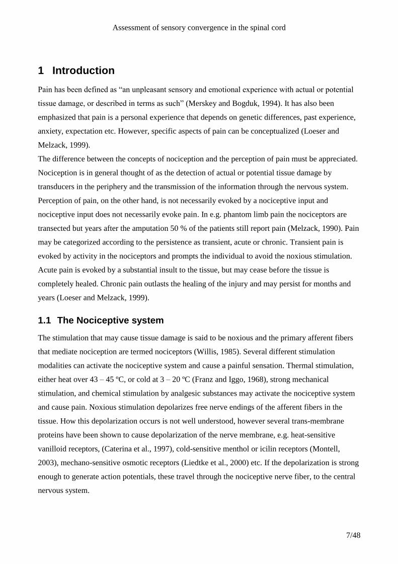
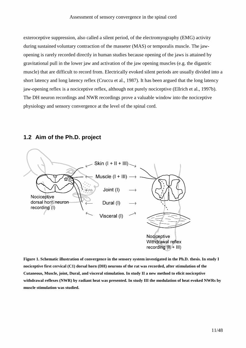
1.2 Aim of the Ph.D. project
Figure 1. Schematic illustration of convergence in the sensory system investigated in the Ph.D. thesis. In study I
nociceptive first cervical (C1) dorsal horn (DH) neurons of the rat was recorded, after stimulation of the
Cutaneous, Muscle, joint, Dural, and visceral stimulation. In study II a new method to elicit nociceptive
withdrawal reflexes (NWR) by radiant heat was presented. In study III the modulation of heat evoked NWRs by
muscle stimulation was studied.
Assessment of sensory convergence in the spinal cord
12/48
The aim of this Ph.D. project was to investigate the sensory convergence in human and animal
experimental settings (Figure 1). Therefore, the convergence pattern of several cutaneous, deep,
dural and visceral afferent sources was investigated in an electrophysiological study of nociceptive
C1 DH neurons in rats (study I). Similar electrophysiological studies of nociceptive DH neurons are
not feasible in human studies. Therefore, two studies of the NWR were performed (study II and III).
Study II was set up to develop a novel approach to evoke NWRs by a natural and nociceptive
specific heat stimulus, and to characterize the stimulus response-relations and the organization of
these NWRs. Transient muscle pain was applied as conditioning stimulus of the NWR at different
time intervals to investigate a possible facilitation of the NWR by muscle afferents caused by a
convergence of cutaneous and muscle afferents (study III).
2 Animal models of trigeminal and upper cervical
convergence
Animal models are often used in research of the nociceptive system (Le Bars et al., 2001). Though
direct inquiry of possible pain is not available, important information about the nervous system still
can be achieved; information that is not accessible in human experimental settings. In the following,
some animal models that have been used to evaluate sensory convergence are discussed. Emphasis
will be on sensory convergence in the craniofacial area, because sensory convergence and central
sensitization has been proposed as the neurophysiologic basis for the spread and referral of pain in
several craniofacial disorders (Sessle, 2000) such as temporomandibular disorder (Dworkin et al.,
1990), whiplash (Munglani, 2000), angina pectoris (cardiac pain) (Foreman, 1999;Foreman, 2000),
and headache (Bogduk, 2001;Bartsch and Goadsby, 2003b). Sensory convergence is pronounced in
the craniofacial area as afferent information from both the trigeminal and cervical area projects both
to the Vc and the UCC.
2.1 Anatomical investigation of afferent fibers
One approach to study sensory convergence is to investigate the projection of afferent input to the
central nervous system. Generally, sensory information is processed at specific locations in the
central nervous system in a somatotopic manner. Hence, adjacent afferent sources are processed at
adjacent locations in the sensory cortex and a map of the peripheral tissue is described in the
sensory cortex accordingly. This map is possibly better known as the homunculus man. A similar
pattern is observed in the spinal and medullary DH.
Assessment of sensory convergence in the spinal cord
13/48
2.1.1 Horseradish peroxidase
Transganglionic transport of horseradish peroxidase (HRP) has been introduced as a method to
trace the projection of afferent nerves and thus to ascertain the central representation of peripheral
receptive fields (Grant et al., 1979;Mesulam and Brushart, 1979). HRP injected to the trigeminal or
C2 ganglions produced labeling in the C1 DH (Pfaller and Arvidsson, 1988), and a systematic study
of the oral and facial nerves confirmed an „onion-like‟ representation of the facial skin in the
trigeminocervical complex, where rostral areas are represented rostrally in the subnucleus
interpolaris, and more caudal areas are represented more caudally in the UCC (Shigenaga et al.,
1986). HRP tracing of the corneal afferents (van Ham and Yeo, 1996), MAS afferents (Dessem and
Luo, 1999), and the superior laryngeal nerve (SLN) afferents (Nomura and Mizuno, 1983) showed
projection to the C1 DH. This indicated a convergence of several trigeminal and cervical afferent
sources in the C1 DH. The HRP technique labels a broad spectrum of afferent fiber thicknesses, and
not specifically the nociceptive afferents. Therefore, a method that labels nociceptive neurons more
specifically may be preferred.
2.1.2 Fos-like immunoactivity
Fos-like immunoreactivity (Fos-LI) was introduced by Hunt et al. (1987) as a method to stain
neurons involved in processing nociceptive input. After a noxious stimulation the immediate early
gene c-fos is expressed in neurons responding to the stimulation (Morgan and Curran, 1989). The c-
fos gene encodes the Fos protein that regulates “downstream” expression of other genes, most likely
the preprodynorphin gene (Draisci and Iadarola, 1989;Hunter et al., 1995). The following increase
in dynorphin probably induces antinociceptive action.
The Fos-LI staining method has been used extensively and proven a valuable tool in the
investigation of the nociceptive system. Fos-LI neurons in the spinal cord DH and medullary DH
are in general located in laminae I-II and V-VI and X (Hunt et al., 1987). However, the specific
location of Fos-LI neurons depends on the stimulated afferent source. In general a somatotopic
organization has been shown in the spinal and medullary DH. But, an overlap of Fos-LI neurons has
been shown in the C1 DH, where stimulation of both trigeminal and cervical afferent sources
produced Fos-LI. Noxious stimulation of the skin innervated by any of the 3 main branches of the
trigeminal nerve, have shown that Fos-LI cells are located mainly in lamina I and V of the C1 DH
(Strassman and Vos, 1993;Zhou et al., 1999). Furthermore, noxious corneal stimulation has shown
Assessment of sensory convergence in the spinal cord
14/48
Fos-LI in the Vc/C1 transition (Lu et al., 1993) and in the C1 (Strassman and Vos, 1993;Meng and
Bereiter, 1996), mainly in the lateral part of lamina I. Noxious mechanical dural stimulation
revealed Fos-LI mainly in laminae I and V of the C1 DH (Strassman et al., 1994), and electrical
stimulation of the dura revealed Fos-LI mainly in lateral part of laminae I and II (Kaube et al.,
1993). Noxious stimulation of deep structures in the trigeminal and cervical area also evoked Fos-
LI in the C1 DH by noxious stimulation of the trapezius and splenius muscles (Kalezic et al., 2004),
the temporomandibular joint (TMJ) (Hathaway et al., 1995;Zhou et al., 1999), the MAS muscle
(Imbe et al., 1999), the tongue (Strassman and Vos, 1993), and the hypoglossal (XII) nerve
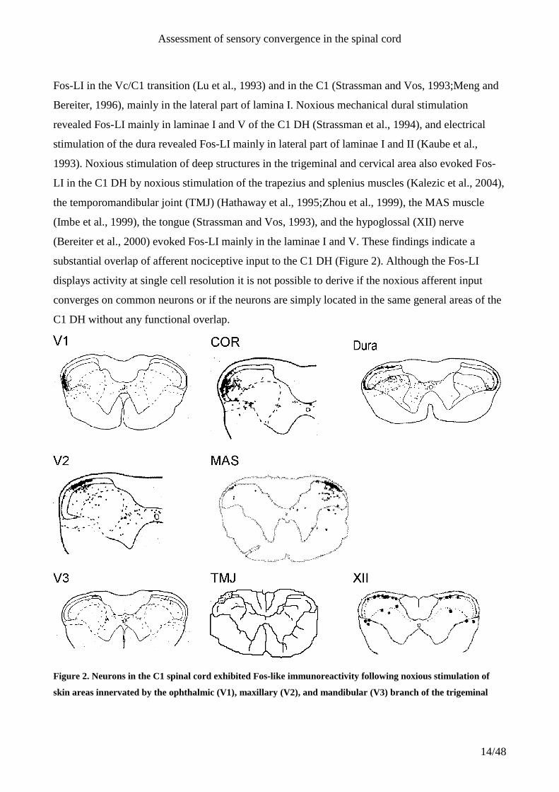
(Bereiter et al., 2000) evoked Fos-LI mainly in the laminae I and V. These findings indicate a
substantial overlap of afferent nociceptive input to the C1 DH (Figure 2). Although the Fos-LI
displays activity at single cell resolution it is not possible to derive if the noxious afferent input
converges on common neurons or if the neurons are simply located in the same general areas of the
C1 DH without any functional overlap.
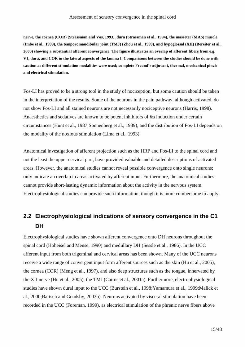
Figure 2. Neurons in the C1 spinal cord exhibited Fos-like immunoreactivity following noxious stimulation of
skin areas innervated by the ophthalmic (V1), maxillary (V2), and mandibular (V3) branch of the trigeminal
Assessment of sensory convergence in the spinal cord
15/48
nerve, the cornea (COR) (Strassman and Vos, 1993), dura (Strassman et al., 1994), the masseter (MAS) muscle
(Imbe et al., 1999), the temporomandibular joint (TMJ) (Zhou et al., 1999), and hypoglossal (XII) (Bereiter et al.,
2000) showing a substantial afferent convergence. The figure illustrates an overlap of afferent fibers from e.g.
V1, dura, and COR in the lateral aspects of the lamina I. Comparisons between the studies should be done with
caution as different stimulation modalities were used; complete Freund’s adjuvant, thermal, mechanical pinch
and electrical stimulation.
Fos-LI has proved to be a strong tool in the study of nociception, but some caution should be taken
in the interpretation of the results. Some of the neurons in the pain pathway, although activated, do
not show Fos-LI and all stained neurons are not necessarily nociceptive neurons (Harris, 1998).
Anaesthetics and sedatives are known to be potent inhibitors of fos induction under certain
circumstances (Hunt et al., 1987;Sonnenberg et al., 1989), and the distribution of Fos-LI depends on
the modality of the noxious stimulation (Lima et al., 1993).
Anatomical investigation of afferent projection such as the HRP and Fos-LI to the spinal cord and
not the least the upper cervical part, have provided valuable and detailed descriptions of activated
areas. However, the anatomical studies cannot reveal possible convergence onto single neurons;
only indicate an overlap in areas activated by afferent input. Furthermore, the anatomical studies
cannot provide short-lasting dynamic information about the activity in the nervous system.
Electrophysiological studies can provide such information, though it is more cumbersome to apply.
2.2 Electrophysiological indications of sensory convergence in the C1
DH
Electrophysiological studies have shown afferent convergence onto DH neurons throughout the
spinal cord (Hoheisel and Mense, 1990) and medullary DH (Sessle et al., 1986). In the UCC
afferent input from both trigeminal and cervical areas has been shown. Many of the UCC neurons
receive a wide range of convergent input form afferent sources such as the skin (Hu et al., 2005),
the cornea (COR) (Meng et al., 1997), and also deep structures such as the tongue, innervated by
the XII nerve (Hu et al., 2005), the TMJ (Cairns et al., 2001a). Furthermore, electrophysiological
studies have shown dural input to the UCC (Burstein et al., 1998;Yamamura et al., 1999;Malick et
al., 2000;Bartsch and Goadsby, 2003b). Neurons activated by visceral stimulation have been
recorded in the UCC (Foreman, 1999), as electrical stimulation of the phrenic nerve fibers above
Assessment of sensory convergence in the spinal cord
16/48
the heart (Razook et al., 1995;Chandler et al., 1998), the vagal and sympathetic fibers (Chandler et
al., 1996) and the SLN which is a branch of the vagal nerve, (Hu et al., 1981;Chandler et al., 1996),
and to intrapericardial injections of algogenic chemicals (Qin et al., 2001) have been shown to
evoke responses in neurons in the UCC.
Although, these studies have shown afferent convergence onto common interneurons in the UCC,
only 2 or 3 afferent sources have been investigated in each study. A systematical investigation of
input form several afferent sources onto interneurons in the in rostral part of the medullary DH of
the cat has been reported (Sessle et al., 1986). Study I of the present thesis aimed at a systematic
investigation of the convergent afferent input from several cutaneous, deep, dural, and visceral
afferent sources onto nociceptive C1 DH neurons in the rat.
2.2.1 Stimulation and recording of C1 DH neurons
In study I, neurons were recorded from the C1 DH and electrolytic lesions confirmed the loci to be
within the C1 DH according to the anatomical landmarks (Molander et al., 1989). The neurons were
classified as nociceptive WDR or NS according to their cutaneous response properties (Price et al.,
1976;Hu et al., 1981;Hu, 1990). Gentle mechanical stimulation (< 5 mg) was applied to the COR
and dura, and noxious mechanical stimulation (~100 g) was applied to the TMJ (Cairns et al.,
2001c). The sizes of the cutaneous mechano-receptive field (RF) were compared before and after
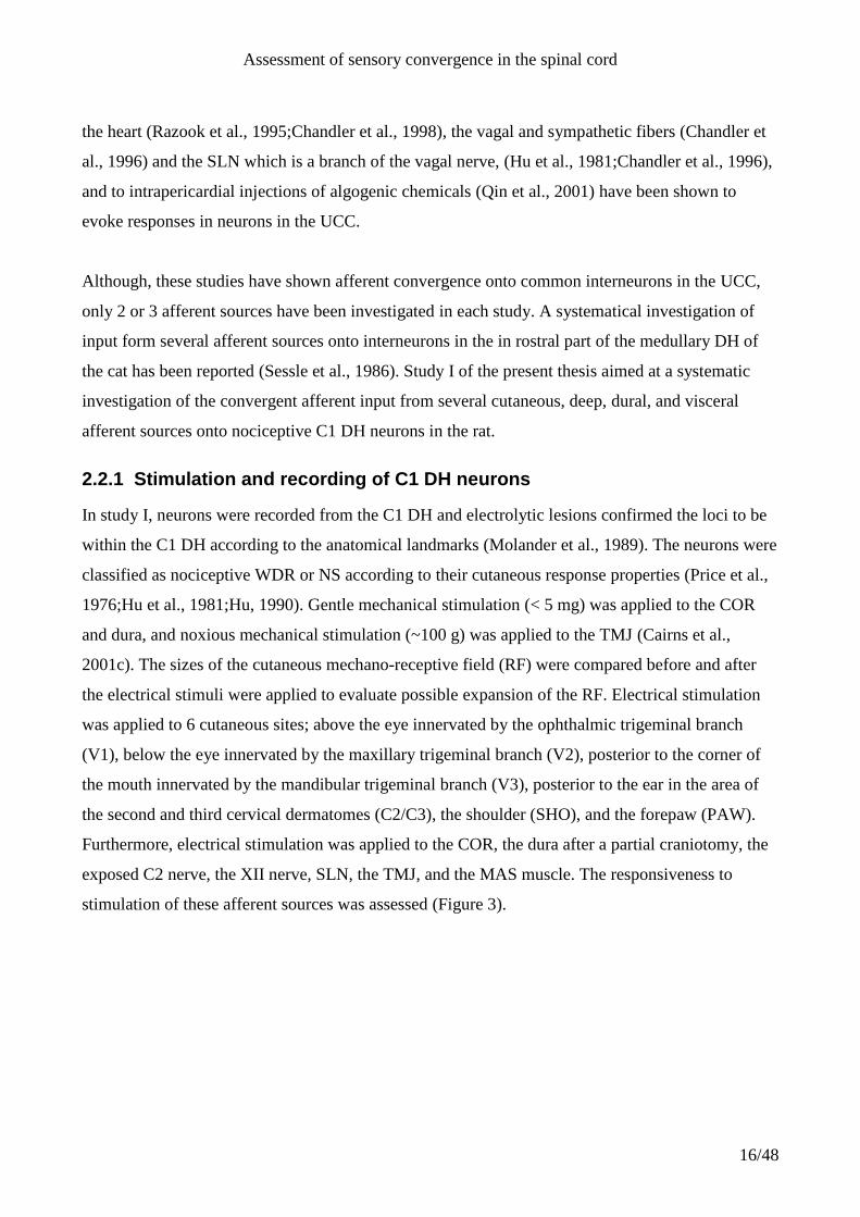
the electrical stimuli were applied to evaluate possible expansion of the RF. Electrical stimulation
was applied to 6 cutaneous sites; above the eye innervated by the ophthalmic trigeminal branch
(V1), below the eye innervated by the maxillary trigeminal branch (V2), posterior to the corner of
the mouth innervated by the mandibular trigeminal branch (V3), posterior to the ear in the area of
the second and third cervical dermatomes (C2/C3), the shoulder (SHO), and the forepaw (PAW).
Furthermore, electrical stimulation was applied to the COR, the dura after a partial craniotomy, the
exposed C2 nerve, the XII nerve, SLN, the TMJ, and the MAS muscle. The responsiveness to
stimulation of these afferent sources was assessed (Figure 3).
Assessment of sensory convergence in the spinal cord
17/48
Figure 3. Electrical stimulation was applied to 6 cutaneous sites (solid arrows); above the eye innervated by the
ophthalmic trigeminal branch (V1), below the eye innervated by the maxillary trigeminal branch (V2), posterior
to the corner of the mouth innervated by the mandibular trigeminal branch (V3), posterior to the ear in the area
of the second and third cervical dermatomes (C2/C3), the shoulder (SHO), and the forepaw (PAW).
Furthermore, electrical stimulation was applied to 7 non-cutaneous sites (dashed arrows); the cornea (COR), the
dura, the exposed second cervical (C2) nerve, the hypoglossal (XII) nerve, superior laryngeal nerve (SLN), the
temporomandibular joint (TMJ), and the masseter (MAS) muscle.
Glutamate (0.5 M, 10 µl, pH 7.0) was injected to the tongue, MAS muscle, neck muscle (splenius
cervicis), or intrapericardially, or dripped onto the dura. Glutamate has been shown to have an
algesic effect (Yu et al., 1996;Cairns et al., 2001c). Furthermore, towards the end of some
experiments, 0.5 ml 3.5% acetic acid was slowly (~5 s) injected intraperitoneally.
2.2.2 Cutaneous afferent input
Several electrophysiological studies have reported C1 DH neurons with cutaneous RF that includes
the facial region and the C2 and C3 dermatomes (Burstein et al., 1998;Yamamura et al.,
1999;Malick et al., 2000;Foreman, 2000) and outside the trigeminal and cervical areas (Smith et al.,
1991;Chandler et al., 1996;Chandler et al., 1998;Clement et al., 2000). However, no neurons were
Assessment of sensory convergence in the spinal cord
18/48
found with RF outside the craniofacial area and only few neurons responded to electrical
stimulation of the SHO and PAW in Study I (Table 1).
2.2.3 Corneal afferent input
Neurons in the C1 DH have been shown to respond to noxious corneal stimulation and that corneal
sensitive neurons also have cutaneous RF (Meng et al., 1997;Hirata et al., 1999;Malick et al.,
2000;Hirata et al., 2004)(study I). Evidently, study I found a significant correlation between the
responsiveness to COR and V1 stimulation, which confirmed previous studies describing neurons
receiving both V1 and COR input (Meng et al., 1997;Hirata et al., 1999;Malick et al., 2000;Hirata et
al., 2004).
2.2.4 Dural afferent input
Electrophysiological studies have indicated an even distribution throughout the C1 DH of neurons
responding to dural stimulation (Burstein et al., 1998;Malick et al., 2000) (study I), though neurons
in the Vc responding to dural stimulation seems to concentrate in the ventrolateral area (Burstein et
al., 1998).
Patients with primary headaches often report pain that involves the front of the head; the V1 nerve
branch territory. However, the pain may in due time exceeds the trigeminal territory to the back of
the head which is innervated by the greater occipital nerve (Anthony, 1992;Bartsch and Goadsby,
2003b). Study I showed correlation between V1 and Dura, which is in accordance with previous
studies showing neurons responding to both dural and cutaneous stimulation of the V1 (Burstein et
al., 1998;Yamamura et al., 1999;Bartsch and Goadsby, 2002;Bartsch and Goadsby, 2003a).
Convergence along with sensitization of central neurons in Vc (Burstein et al., 1998), in C1 (Study
I), and in C2 (Bartsch and Goadsby, 2002) provide a neurophysiologic basis for the clinical
phenomenon of spread and referred pain by which pain originating from an affected tissue is
perceived as originating from a distant area (Ruch, 1947). In cervicogenic headache, pain spreading
to the back of the head may be originating from the structures innervated by the C1 to C3 spinal
nerves, and include the upper cervical synovial joints, the upper cervical muscles, the C2-3 disc, the
vertebral and internal carotid arteries, and the dura mater of the upper spinal cord, and posterior
cranial fossa (Bogduk, 2001). This was supported by study I showing similarities in responsive
areas of the C1 DH following stimulation of the dura and C2/C3.

Assessment of sensory convergence in the spinal cord
19/48
2.2.5 Deep afferent input
C2 nerve
A wide spread distribution of neurons responding to stimulation of the C2 nerve and injection of
glutamate to the splenius cervicis muscle (innervated by the posterior primary rami of the inferior
cervical spinal nerves) evoked responses in nociceptive C1 DH neurons (Study I).
TMJ
Studies have shown that injection of mustard oil into the TMJ evoked Fos-LI mainly in the
superficial layers of the C1 DH but also few neurons in deeper layers (Hathaway et al., 1995;Zhou
et al., 1999), whereas electrophysiological recordings showed a more widespread distribution
throughout the C1 DH (study I). Study I showed correlation between TMJ and V2, although the
TMJ is innervated by the maseteric and auticulotemporal branches of the V3 nerve (Davidson et al.,
2003).
MAS muscle
More than half of the neurons responded to intra muscular electrical stimulation of the MAS
muscle, whereas only one third responded to microinjection of glutamate into the MAS muscle
(study I). It has been shown that injection of glutamate into the MAS muscle activated muscle
afferents in rats, and that the same concentration evoked a painful sensation in humans (Cairns et
al., 2001b). Anatomical studies have shown that the noxious stimulation of MAS projects to the
intermedial part of lamina I of the C1 DH (Imbe et al., 1999;Dessem and Luo, 1999), whereas the
present electrophysiological study found a more widespread distribution of MAS sensitive
nociceptive C1 DH neurons (study I). The responsiveness to V3 cutaneous and MAS muscle
stimulations was found to be correlated and their response pattern to C-fiber input were also similar
(Study I). This complied with the fact that the MAS muscle is innervated by the masseteric branch
of the V3 nerve.
XII nerve
In accordance to anatomical studies (Strassman and Vos, 1993;Carstens et al., 1995;Bereiter et al.,
2000), study I showed neurons responding mainly to C-fiber stimulation of the XII nerve in the
medial part of the C1 DH. Microinjection of glutamate to the tongue only rarely evoked responses
in the C1 DH neurons (7% in both WDR and NS neurons). Furthermore, it has been shown that
Assessment of sensory convergence in the spinal cord
20/48
electrical XII nerve trunk stimulation evoked more Fos-LI lamina I and II of the UCC than
intramuscular tongue injection of mustard oil, thus indicating other afferent than the tongue
musculature afferents travel in the XII nerve (Bereiter et al., 2000). Therefore, the low response rate
found by glutamate injection of the tongue musculature may be caused both by few tongue
glutamate receptors, and that tongue muscle afferents are only a subpopulation of the XII nerve.
The responsiveness to stimulation of the XII nerve and SLN was significantly correlated and the
difference in response patterns was small. The SLN innervates the larynx and the pharynx whereas
the XII nerve innervates mainly the tongue, however these nerves seems to evoke similar responses
in the C1 DH (study I).
2.2.6 Visceral afferent input
Previous electrophysiological studies have shown C1 DH neurons responding to electrical
stimulation of SLN (Hu et al., 1981;Chandler et al., 1996). Approximately one third of the C1 DH
neurons responded to stimulation of the SLN (Study I). Previous studies of neurons in the Vc
showed even lower proportions (Hu et al., 1981).
Although, HRP tracing of left inferior cardiac nerve did not show projection more rostral than C8
(Kuo et al., 1984), stimulation of the vagal nerve afferents originating from the heart or other
visceral structures, and cardiopulmonary sympathetic afferent fibers (Chandler et al., 1996) and the
phrenic nerve (Razook et al., 1995;Chandler et al., 1998) evoked responses in the C1 DH neurons.
However, phrenic fibers innervating diaphragm or abdominal structures either did not affect the C1
DH or to a lesser degree than phrenic input arising from thoracic structures (Razook et al.,
1995;Chandler et al., 1998). Intrapericardial injections of algogenic chemicals has been shown to
activate neurons in the C1 DH (Qin et al., 2001), however intrapericardial injections of glutamate
did not evoke responses in the C1 DH neurons (study 1), indicating that glutamate receptors in the
pericardial tissue either are not present or not sufficient numerous to evoke responses in the C1 DH
neurons. Angina pectoris is often described as retrosternal crushing, burning, or squeezing
characteristic pain, which may radiate to the throat, neck, or ulnar aspect of the left arm, sometimes
reaching to the little finger. Less often, it radiates to the neck and jaw (Foreman, 1999). Stimulation
of the phreneic, vagal or sympathetic afferent fibers and chemical stimulation of the heart excites
spinothalamic tract neurons in the UCC segments of monkeys (Chandler et al., 1996;Chandler et al.,
Assessment of sensory convergence in the spinal cord
21/48
1998) and rats (Razook et al., 1995;Qin et al., 2001), indicating that these neurons may be the
neural basis for the projection of pain to the neck and the jaw areas.
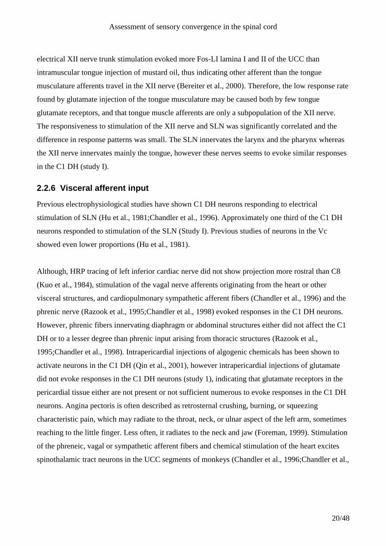
2.2.7 Stimulus-Response relations
Neurons in the C1 DH responded to mechanical and electrical stimulation applied to the skin and to
electrical stimulation of at least one of the deep, dural, and visceral sources (Study I, table 1). Study
I showed stimulus-response relationship between mechanical stimulation applied to the center of the
cutaneous RF for both the WDR and NS neurons (Study I, figure 4) showing the capability of the
neurons to encode sensory information into the noxious range.
Electrical stimulation normalized to the activation threshold showed a double logarithmic
relationship with similar correlation coefficients for all afferent sources (Figure 4). Therefore, the
nociceptive neurons in the C1 DH are capable of encoding afferent information from cutaneous,
deep, dural, and visceral sources.
Figure 4. Stimulus response functions for electrical stimulation of the 6 cutaneous and the 7 non-cutaneous sites.
The stimulation intensity was normalized to activation threshold of A- and C- fibers. The number of spikes to 5
stimuli was considered the response. The activation threshold was set to at least 3 responses to 5 electrical pulses;
therefore some neurons did have few responses below threshold. The stimulus response relation could be
approximated by a double logarithmic function. Linear regression of the double logarithmic transformed data
showed a linear increase (OneWay ANCOVA, P < 0.001). The correlation coefficient was 0.68, 0.65, 0.76, 0.68,
0.64, 0.50, 0.61, 0.79, 0.67, 0.62, and 0.62 for stimulation of the V1, V2, V3, C2/C3, COR, dura, C2, XII, TMJ,
MAS and SLN, respectively.
Assessment of sensory convergence in the spinal cord
22/48
2.2.8 Afferent convergence in the craniofacial area.
Electrophysiological studies of the rostral Vc in cats showed a substantial afferent convergence onto
nociceptive neurons (Sessle et al., 1986). Sessle et al., (1986) showed a larger number of afferent
sources converged onto WDR than onto NS neurons, whereas study I did not find such difference
between WDR and NS neurons in the C1 DH neurons. The reason for the difference between these
two studies may be species differences but also the difference between the rostral Vc and the C1
DH.
Electrophysiological studies have shown that neurons in the UCC responding to deep, visceral, or
dural afferent sources also have cutaneous RFs in the craniofacial area. These neurons may be the
neurophysiological basis for various painful conditions where pain from one afferent source is also
perceived as originating from distant cutaneous, deep, or visceral tissue. In temporomandibular
disorder, pain originating from the TMJ or other deep adjacent areas may spread and be perceived
from other facial areas innervated by all three branches of the trigeminal nerve (Dworkin et al.,
1990). Afferent convergence from blood vessels and skins may play an important role in headache
that often manifested with referral pain to the extracranial skin tissue in periorbital or temporal
region (Piovesan et al., 2003;Bartsch and Goadsby, 2003b). Angina pectoris may result from
nociceptive cardiac afferent input to UCC neurons and may lead to pain sensation in the neck and
jaw areas (Foreman, 1999;Foreman, 2000).
2.2.9 Lack of afferent input caudal to the cervical area
Study I showed that neurons in the C1 DH responded to afferent input from several trigeminal and
cervical afferent sources. All neurons responded to cutaneous stimulation of the facial skin in the
area posterior to the whiskers and generally anterior to the ear. None of the neurons has cutaneous
RF posterior to the ear. Electrical stimulation readily evoked responses from the cutaneous V1, V2,
V3 and C2/C3 areas, but rarely from SHO and PAW. Likewise, intraperitoneal injection of ascetic
acid or intrapericardial microinjection of glutamate did not evoke responses in any of the tested
neurons. Therefore, it seemed that although nociceptive C1 DH neurons received a wide range of
afferent input these sources it did not include input caudal to the cervical areas, which was in
agreement with studies by Hu et al. (2005). However, several previous studies found afferent input
from areas caudal of the cervical region activating neurons in the C1 (Yezierski and Broton,
1991;Smith et al., 1991;Razook et al., 1995;Villanueva et al., 1996;Chandler et al., 1996;Chandler
Assessment of sensory convergence in the spinal cord
23/48
et al., 1998;Ness et al., 1998;Chandler et al., 1999;Qin et al., 2001;Qin et al., 2004). There are
several possible reasons for differences between these studies and the findings by Hu et al. (2005)
and Study I. The first reason could be species differences since Hu et al. (2005) and study I used
rats whereas some of earlier studies used cats (Smith et al., 1991) or monkeys (Chandler et al.,
1996;Chandler et al., 1998;Chandler et al., 1999). However, this possibility seems an unlikely
explanation; although the rat has a limited and unclear C1 dermatomal representation, several of the
earlier electrophysiological studies (Hathaway et al., 1995;Razook et al., 1995;Burstein et al.,
1998;Yamamura et al., 1999;Malick et al., 2000;Qin et al., 2001;Qin et al., 2004) were carried out
in rats. Another possible explanation is that the neuronal sample in some of the earlier studies also
included recordings at sites deeper than laminae I–V of the C1 DH, and sometimes included
neurons in the ventral horn (Qin et al., 2001;Qin et al., 2004), lateral reticular formation (Ness et al.,
1998) including the subnucleus reticularis dorsalis (Villanueva et al., 1996), and lateral cervical
nucleus (Yezierski and Broton, 1991). Many neurons in these deep locations typically have RFs
including half or the whole body (Yezierski and Broton, 1991;Villanueva et al., 1996;Ness et al.,
1998), whereas Vc and C1 DH neurons in lamina I–V have much more restricted RFs (Yamamura
et al., 1999;Malick et al., 2000). The third explanation may be the use of antidromic activation as
the searching stimulus in some of the earlier studies that led to the inclusion neurons with large RF
in the sample. Some of these neurons were indeed recorded outside the DH region (Smith et al.,
1991;Chandler et al., 1996;Chandler et al., 1998;Ness et al., 1998;Chandler et al., 1999;Qin et al.,
2001;Qin et al., 2004).
2.2.10 Neuron classification based on deep, dural and visceral input
Classification of the responsiveness of nociceptive DH neurons is usually based on the response
properties to cutaneous stimulation (Price et al., 1976;Hu et al., 1981;Hu, 1990). Study I revealed a
substantial afferent input from other structures to C1 DH neurons. It therefore seemed obvious to
inquire if there were any natural clustering based on the response properties to deep, dural and
visceral stimulation. Principal component transformation (Hotelling, 1933) and k-means clustering
of the responsiveness revealed two possible groups of neurons. Neurons in group I had in general a
lower responsiveness and contained more NS neurons and fewer WDR neurons than group II. This
indicates that the neurons can be classified according to their deep, dural and visceral
responsiveness properties, and that the cutaneous response properties only to some extend were
reflected in this clustering.
Assessment of sensory convergence in the spinal cord
24/48
2.3 Short summary
Nociceptive neurons in the C1 DH receive excitatory afferent input from trigeminal and cervical
cutaneous areas, but only sparse afferent input from cutaneous areas caudal to the C2/C3
dermatome. Furthermore, the nociceptive neurons receive convergent input from cutaneous,
corneal, deep, dural and visceral sources. Electrical stimulation of several craniofacial structures has
reviled a stimulus-response relationship indicating that these neurons were capable of encoding
noxious information from several afferent sources. This indicates that the nociceptive neurons in the
C1 DH may play an important role in nociceptive integration.
3 Human models of spinal cord convergence
Although animal experiments provide great insight into the physiology and pathology of
nociception the knowledge cannot always be translated directly to the physiology and pathology of
humans. For instance, it is not readily investigated if nociceptive responses in e.g. DH neurons in
fact would cause a painful sensation had the animal been awake. Single firings in single second
order DH neurons are most likely insufficient to evoke a conscious perception of pain. Similar
problems are evident in all animal studies although pain behavior can be studied. Therefore,
experimental methods to investigate the healthy human nociceptive physiology as well as
pathologies of patients are important in order to understand the physiology and treat the pathology
better. Although some of the methods used in animal experiments obviously can not be applied in
human experiments there are several ways to investigate the nociception in humans. Especially,
inquiries about the pain intensity (e.g. VAS) and quality (e.g. McGill questioner) are possible, if
nociception leads to pain. It should be kept in mind that such inquiries are entirely subjective, as
pain is subjective. Furthermore, careful instructions must be given to the subjects as to ensure the
validity of the results. Less subjective methods to study the nociceptive system of humans may be
desirable. For such purpose several imagining and electrophysiological methods are available. To
study sensory convergence at the spinal level the NWR is especially interesting as a window into
nociceptive processing at the spinal level.
3.1 NWR evoked by natural stimulation
Most investigations on NWR have used electrical stimulation to activate afferent responses
(Sandrini et al., 2005). The electrical stimulation can be applied to the surface of the skin in order to
activate the fibers innervating the skin and subcutaneous layers. In animal studies, the nerve can be
Assessment of sensory convergence in the spinal cord
25/48
exposed by dissection and stimulated directly. Electrical stimulation enables a reliable and transient
stimulation, where the parameters such as intensity, pulse width, repetitions and timing are easily
controlled. The advantages of electrical stimulation are numerous, but some disadvantages are also
evident. It is difficult to determine which afferents are activated. It is especially difficult to avoid
activating non-nociceptive cutaneous afferents; therefore, electrical stimulation can not be assumed
to be purely nociceptive. In addition, the electrical stimulation bypasses the receptor endings, so
inquiries about the transduction mechanisms cannot be made. Furthermore, the afferents are
activated simultaneously by the electrical pulse. This does probably not resemble natural
stimulation where the afferent barrage is not synchronized. Therefore, the sensory information
arriving at the spinal cord DH may not resemble natural stimulation, though the NWR is a natural
response to a noxious stimulation.
3.1.1 Mechanical stimulation
Mechanical stimulation has been used as a natural stimulation to evoke NWR in animal studies (Le
Bars et al., 2001). Unlike the manual pinch or pressure algometry often used in animal studies
transient time-locked stimulation must be used to evoke NWR in human subjects. Some methods to
activate the nociceptive system in a transient and time-locked manner have been proposed, but have
some deficits. High-energy ultrasound stimulation is able to activate the nociceptive system, but it is
not known whether the energy is transmitted as mechanical or heat energy (Gavrilov et al., 1977).
Impact with a small metal slug has also been proposed (Kohlloffel et al., 1991), but there is no
evidence that it will evoke NWR. The photoacoustic effect, where short laser pulses are converted
into sound waves in the tissue may be a possible method to apply a transient activation of the
mechanoreceptors (Doukas and Kollias, 2004), however, substantial parts of the energy may be
converted into heat, and again there is no evidence that the stimulus can evoke NWR.
3.1.2 Heat stimulation
Heat is another stimulation modality used to activate the nociceptive system (Julius and Basbaum,
2001). Thermodes have been used to apply contact heat stimulation (Nielsen and Arendt-Nielsen,
1998;Arendt-Nielsen and Chen, 2003), but the mechanical aspect of contact heat has been shown to
alter the perception of pain (Svensson et al., 1997). More importantly the temperature rise-time is
too long to evoke NWR, therefore radiant heat stimulation may be preferable to contact heat
stimulation (Baumgartner et al., 2005). However, temperature at the skin surface can be controlled
by advanced thermodes (Chen et al., 2001;Casey, 2006). Such a temperature control is rarely
Assessment of sensory convergence in the spinal cord
26/48
provided during laser stimulation, though a system has been developed (Meyer et al., 1976).
Therefore it is important to control the delivered energy and the background temperature of the skin
as variation in background temperature may lead to wrong conclusions (Tjolsen et al., 1988).
As a transient stimulation profile is necessary in order to evoke the NWR the temperature increase
of the skin must be fast. High intensity stimulation is generally needed to allow investigations of
transient and time critical responses (e.g. NWR, evoked cortical potentials, reaction time etc.).
Although Hardy (1953) presented a method to evoke reflexes in one paraplegic subject by a focused
light bulb. Experimental setups using high intensity light bulbs, such as a xenon lamp, may provide
shot exposure of heat stimulation at high intensity using a shutter to restrict the stimuli. However,
the broad-spectered visible light is significantly reflected from the skin surface, and blackening of
the skin may be necessary (Andersen et al., 1994). Laser systems are able to deliver very high
intensity radiation in a narrow frequency range, and in well defined areas. Therefore, laser
stimulation seems to be suitable for this purpose (Plaghki and Mouraux, 2003). Each laser is
characterized by the wavelength of the radiated light. Several different lasers are now commercially
available even at high power (~100 W) e.g. diode laser in the visible wavelength range, solid state
lasers such as the YAG and YAP lasers, and gas laser such as the CO2-lasers in the infrared range.
When laser light is emitted to the skin some of the light will be reflected while the rest is
transmitted through the surface. The proportions of transmitted light depend on the wavelength of
the light. The transmitted light is then absorbed in the skin layer or transmitted through the layer.
The energy of the absorbed light is converted into heat and thus contributed to an increase in
temperature. The transmitted light will be passed directly on to deeper layers or scattered along the
path (Hardy et al., 1956). The light will reemerge form the surface if scattering of the light bend the
path more that 90º, and thus add to reflection. In a small volume of the skin the intensity of the
transmitted light IT can be expressed as a function of the absorption coefficient, µ, as
0 expTI I x ,
where I0 is the intensity of the light entering the volume and x is the length of the path that the light
travels through the volume1. In general, µ is a material constant that depends on the wavelength of
the light (Hardy and Muschenheim, 1934). The skin is inhomogeneous and therefore µ is variable
through the skin for light in the visible and near infrared range (< 2 µm). In the infrared range the
1 If the volume is small, x is the dimension of the volume as scattering will be neglectable.
Assessment of sensory convergence in the spinal cord
27/48
light is almost exclusively absorbed in water and since the water content is approximately
homogeneous in the skin below the striatum corneum µ can be approximated to be constant.
Furthermore, in the infrared range the human skin acts almost as a perfect black body (Hardy,
1934), and therefore none of the incident radiation is reflected from the surface.
The CO2-laser emits light at a wavelength of 10.6 µm. At this wavelength µ is approximately
200cm-1
(Haimi-Cohen et al., 1983). From the equation it is seen that the transmission is reduced to
1/e (37%), at a depth of 50µm. Therefore, most of the energy is absorbed in the superficial layers
reasonably close to the nociceptors that are located approximately 200 ± 170 µm below the surface
(Tillman et al., 1995). For these reasons the CO2-laser, as introduced by Mor and Carmon (1975),
has proven suitable for the study of pain (Arendt-Nielsen and Bjerring, 1988a;Arendt-Nielsen and
Bjerring, 1988b). However, solid state lasers such as thulium-YAG (wavelength 2.01µm; µ = 28cm-
1) (Spiegel et al., 2000) or YAP (wavelength 1.34µm; µ < 20cm
-1) (Iannetti et al., 2004) may be
more suitable for activation of nociceptors as the energy from these lasers is absorbed closer to the
nociceptors (Baumgartner et al., 2005). However, assuming that µ is constant throughout the skin
for infrared absorption most energy will be absorbed in the superficial parts of the skin according to
the equation above disregarding the value of µ. For review of heat stimulation please refer to
Plaghki and Mouraux (2003)
Models for heat transfer during and after radiation of infrared pulses have been developed and
tested (Buettner, 1951;Hendler et al., 1958), and has been applied to infrared laser stimulation
(Haimi-Cohen et al., 1983;Bromm and Treede, 1983).
3.1.3 Heat evoked NWR
Heat evoked NWRs have been studies in animal models (e.g. (Burke et al., 1971;Behrends et al.,
1983;Schomburg and Steffens, 1986). However, NWR are not easily evoked by heat stimulation in
human subjects, and therefore the reports of heat evoked NWR in healthy human volunteers are
few. Half a century ago Hardy (1953) presented a study where NWRs were evoked in one
paraplegic subject by radiant heat. Several years later, Willer et al. (1979) reported a study where
the heat was applied to the lateral edge of the dorsum of the foot (sural nerve territory) and the
NWR was assessed as the EMG recorded from the tibialis anterior and biceps femoris muscles.
Although an Argon laser was used, the authors suggested that a CO2-laser would have been
preferable had it been available (Willer et al., 1979). Campbell et al. (1991) reported a study where
Assessment of sensory convergence in the spinal cord
28/48
they applied radiant heat to the dorsal forearm using a tungsten halogen projector lamp and assessed
NWR responses as the EMG response recorded from the biceps muscle. In study II a method was
developed to apply radiant heat supplied by a CO2-laser to a large area and thereby activating a
large number of afferents which utilized spatial summation (Nielsen and Arendt-Nielsen, 1997).
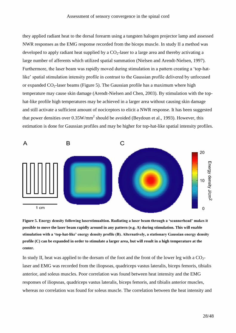
Furthermore, the laser beam was rapidly moved during stimulation in a pattern creating a „top-hat-
like‟ spatial stimulation intensity profile in contrast to the Gaussian profile delivered by unfocused
or expanded CO2-laser beams (Figure 5). The Gaussian profile has a maximum where high
temperature may cause skin damage (Arendt-Nielsen and Chen, 2003). By stimulation with the top-
hat-like profile high temperatures may be achieved in a larger area without causing skin damage
and still activate a sufficient amount of nociceptors to elicit a NWR response. It has been suggested
that power densities over 0.35W/mm2 should be avoided (Beydoun et al., 1993). However, this
estimation is done for Gaussian profiles and may be higher for top-hat-like spatial intensity profiles.
Figure 5. Energy density following laserstimualtion. Radiating a laser beam through a ‘scannerhead’ makes it
possible to move the laser beam rapidly around in any pattern (e.g. A) during stimulation. This will enable
stimulation with a ‘top-hat-like’ energy density profile (B). Alternatively, a stationary Gaussian energy density
profile (C) can be expanded in order to stimulate a larger area, but will result in a high temperature at the
center.
In study II, heat was applied to the dorsum of the foot and the front of the lower leg with a CO2-
laser and EMG was recorded from the iliopsoas, quadriceps vastus lateralis, biceps femoris, tibialis
anterior, and soleus muscles. Poor correlation was found between heat intensity and the EMG
responses of iliopsoas, quadriceps vastus lateralis, biceps femoris, and tibialis anterior muscles,
whereas no correlation was found for soleus muscle. The correlation between the heat intensity and
Assessment of sensory convergence in the spinal cord
29/48
the perceived pain intensity was also significantly, but poorly, correlated (linear regression, P <
0.05, R = 0.53), which resembles the result from Campbell et al. (1991). In contrast to laser
stimulation of the lower limbs it is possible to elicit nociceptive protective craniofacial reflexes
using brief (< 40 ms) pulses applied to a small (< 1 cm2) perioral cutaneous area (Ellrich et al.,
1997b;Cruccu and Romaniello, 1998;Romaniello et al., 2002). The jaw-opening reflex can be
observed as single silent period of ongoing MAS muscle activity following Th:YAG-laser (Ellrich
et al., 1997b) or CO2-laser (Cruccu and Romaniello, 1998) stimulation resembling the long latency
silent period following electrical stimulation, as both are assumed to be mediated by nociceptive
A- afferents. Heat evoked nociceptive reflexes seems to be more readily evoked in the craniofacial
area compared to the extremities. Comparing the laser evoked cortical potential following
stimulation of the foot, hand and perioral skin showed progressively smaller amplitudes at distal
areas (Cruccu et al., 1999). Cruccu et al. (1999) speculated that longer afferent conductance routes
may temporally disperse the afferent barrage of the spinal cord DH nociceptive neurons and thus
cause weaker laser evoked cortical potentials. Furthermore, the perception threshold of laser
stimulation was found to be lower for facial compared to stimulation of the extremities. This was
explained by a possible denser afferent innervation. The temporal dispersion and less dense
innervation of the extremities may explain the difficulties in evoking NWR in the extremities by
heat stimulation. Therefore, larger area was stimulated to compensate for the lower innervation
density in the lower limb compared to the facial areas in study II.
3.2 Organization of the NWR
Already the early studies of the NWR indicated that the reflex may not be a stereotype flexion
reflex as originally described by Sherrington (1910). Hagbarth (1960) observed a flexion reflex
when stimulating the limb electrically, but not when stimulation was applied above the extensor
muscles. Furthermore, Grimby (1963) systematically studied the dependency of the site of
stimulation on the sole of the foot, and observed a dorsiflexion of the ankle when stimulation the
front of the foot, but a plantar flexion when stimulating the foot towards the heel. Similar results
were found by Hagbarth in both humans (Hagbarth, 1960) and cats (Hagbarth, 1952). The
stereotyped flexor reflex organization of NWRs was further challenged by Schouenborg and
Kalliomaki (1990), who showed that muscles in the hindlimb of rats could be activated by noxious
stimulation in a restricted area specific for each muscle. These specific areas were termed „reflex
receptive fields‟, and interestingly not only flexor muscles but also extensor muscles had such
Assessment of sensory convergence in the spinal cord
30/48
reflex receptive fields (Schouenborg et al., 1994). The combination of muscles and their reflex
receptive fields was shown to constitute a system where the reflex movement optimally removed
the stimulated skin area away from the stimulation (Schouenborg and Weng, 1994). Each muscle or
synergistic muscles and their reflex receptive fields were termed a module and the NWR was
proposed to have a modular organization (Schouenborg et al., 1994). Such a modular organization
of the NWRs has also been shown in humans (Andersen et al., 1999;Sonnenborg et al.,
2000;Sonnenborg et al., 2001). The human studies were all performed by electrical stimulation and
showed that the NWR is organized so that a noxious stimulation of the foot would remove the
stimulated area away from harm by an appropriate movement, e.g. plantarflexion for heal
stimulation and dorsiflexion for fore foot stimulation at the sole of the foot.
3.2.1 Natural stimulation e.g. mechanical and heat.
The modular organization has also been shown when reflexes were evoked by natural stimulation in
animal studies. Evidence for a modular organization of reflexes has been obtained in rats for
mechanical (Schouenborg and Kalliomaki, 1990) and radiant heat (Weng and Schouenborg, 1996)
stimulation applied to plantar and to some extend the dorsal side of the hind paw. Furthermore
radiant heat applied to the tail of the rat evoked a movement directed away from the stimulus
(Cleland and Bauer, 2002). A possible modular organization of the NWR in the cat has been
investigated. Heat evoked NWR could be evoked in non-flexor-muscles (Schomburg et al., 2000),
and a modular organization of the NWR was shown following mechanical pinch stimuli (Levinsson
et al., 1999). Study II did not show a modular organization of the heat evoked NWR in humans;
however, the stimulated sites may have been located within the same reflex receptive field. Another
explanation may be that a possible modular organization of the NWR is less pronounced on the
dorsal side compared to the plantar side of e.g. the foot (Clarke and Harris, 2004).
The differences between plantar and dorsal side.
Two similar studies have shown a modular organization of electrically evoked responses both
plantar (Andersen et al., 1999) and dorsal (Sonnenborg et al., 2001) stimulation of the foot.
However, NWR evoked at the dorsal side had lower thresholds, were „smaller‟, and did not show as
clear modular organization as NWR evoked at the plantar side for the foot. Other human studies
have shown differences in reflex activities evoked by laser stimulation of the plantar or dorsal of the
hand (Romaniello et al., 2004), and withdrawal movement analysis showed that modular
organization of reflexes are most readily seen when stimulation was applied to the plantar side of
Assessment of sensory convergence in the spinal cord
31/48
the foot (Clarke and Harris, 2004). In study II, radiant heat stimulation applied to the dorsum of the
foot did not show a modular organization of the NWR, and no plantar flexion as responses in the
soleus muscle was sparse. However, a study investigating the organization of the heat evoked
reflexes evoked from the plantar side of the foot is difficult to design as the plantar skin is glabrous
and therefore contain type I AMHs and CMHs, but not type II AMHs. As described in section 1.1,
type I AMHs have long response delay, and the conduction velocity of the CMHs is low. This
results in a late arrival of afferent information of these nociceptors to the spinal cord that is not
capable of eliciting NWR. The type II AMHs, that are only located in the hairy skin, have short
response latencies and relatively fast conduction velocity, resulting in a sufficiently short response
delay to evoke NWR (study II).
In a study performed to indirectly reveal the role of heat sensitive afferents in the organization of
nociceptive reflexes, a conditioning radiant heat stimulation was applied and the reflexes were
evoked by electrical stimulation of the tibial nerve (Ellrich et al., 2000). Independent of whether the
heel, the forefoot, or the dorsum of the foot was conditioned an increase in the reflex size of both
the biceps femoris and tibialis anterior muscles was observed which is not in compliance with a
modular organization of the reflexes. This may indicate that a modular organization of the NWR
can be demonstrated by electrical stimulation, but not heat stimulation in humans.
3.3 Convergence of muscle afferents to the reflex pathway
Several studies have shown that cutaneous and muscle afferents converge onto common
interneurons in the spinal cord or medullary DH (Kniffki et al., 1981;Sessle et al., 1986;Hoheisel
and Mense, 1990) (Study I). In the craniofacial area, experimental MAS muscle pain reduced the
degree of silent period suppression following perioral cutaneous CO2-laser (Romaniello et al., 2002)
or electrical (Wang et al., 1999) stimulation. The convergence of muscle and e.g. cutaneous
afferents onto common spinal or medullary DH may along with central sensitization form the neural
basis for referred pain. Muscle pain may be associated by referred pain (Sinclair D.C. et al., 1948),
where pain is perceived from the muscle that receives noxious stimulation, but also from distant
parts of the body including the skin. Experimentally, intramuscular injection of hypertonic saline
into the tibilais anterior muscle has been shown to produce pain in the injected muscle but also in
the dorsal aspects of the ankle joint (Graven-Nielsen et al., 1997a).
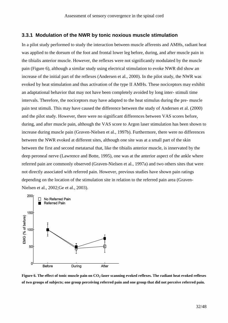
Assessment of sensory convergence in the spinal cord
32/48
3.3.1 Modulation of the NWR by tonic noxious muscle stimulation
In a pilot study performed to study the interaction between muscle afferents and AMHs, radiant heat
was applied to the dorsum of the foot and frontal lower leg before, during, and after muscle pain in
the tibialis anterior muscle. However, the reflexes were not significantly modulated by the muscle
pain (Figure 6), although a similar study using electrical stimulation to evoke NWR did show an
increase of the initial part of the reflexes (Andersen et al., 2000). In the pilot study, the NWR was
evoked by heat stimulation and thus activation of the type II AMHs. These nociceptors may exhibit
an adaptational behavior that may not have been completely avoided by long inter- stimuli time
intervals. Therefore, the nociceptors may have adapted to the heat stimulus during the pre- muscle
pain test stimuli. This may have caused the difference between the study of Andersen et al. (2000)
and the pilot study. However, there were no significant differences between VAS scores before,
during, and after muscle pain, although the VAS score to Argon laser stimulation has been shown to
increase during muscle pain (Graven-Nielsen et al., 1997b). Furthermore, there were no differences
between the NWR evoked at different sites, although one site was at a small part of the skin
between the first and second metatarsal that, like the tibialis anterior muscle, is innervated by the
deep peroneal nerve (Lawrence and Botte, 1995), one was at the anterior aspect of the ankle where
referred pain are commonly observed (Graven-Nielsen et al., 1997a) and two others sites that were
not directly associated with referred pain. However, previous studies have shown pain ratings
depending on the location of the stimulation site in relation to the referred pain area (Graven-
Nielsen et al., 2002;Ge et al., 2003).
Figure 6. The effect of tonic muscle pain on CO2-laser scanning evoked reflexes. The radiant heat evoked reflexes
of two groups of subjects; one group perceiving referred pain and one group that did not perceive referred pain.
Assessment of sensory convergence in the spinal cord
33/48
Radiant heat stimulation was applied before injection of hypertonic saline to the tibialis anterior muscle, during
the perception of pain in the tibialis anterior muscle and 15 minutes after pain perception was no longer
reported. The electromyography (EMG) responses were normalized to the pre-pain response and calculated as
the mean over the four stimulation sites and three stimulation intensities. The group of subjects that experienced
referred pain was compared to the group that did not experience referred pain, however there was no significant
difference between these groups neither when the muscle pain was perceived nor after the perception of pain had
vanished, nor were the any differences before during and after muscle pain (ANOVA; n.s.).
Besides the direct convergence of muscle and cutaneous afferents onto interneurons in the reflex
pathway, two different mechanisms may be involved in muscle pain modulation of the reflexes;
central hyperexcitability and descending inhibition. High intensity (C-fiber) stimulation has been
shown to produce a prolonged increase in the excitability of flexion reflexes in spinalized animals
(Wall and Woolf, 1984;Laird et al., 1995). Central hyperexcitability evoked by chemical (Hu et al.,
1992) or electrical (Cook et al., 1987) noxious muscle stimulation may be associated with an
expansion of cutaneous RF and formation of new deep tissue RFs in rats (Hoheisel et al., 1993).
Therefore, referred pain may be a consequence of convergence of e.g. cutaneous and muscle
afferents onto common interneurons and a hyperexcitability of these interneurons following the
nociceptive barrage from muscle afferents (Mense, 1994;Arendt-Nielsen and Svensson, 2001).
However, the appearance of new cutaneous RFs occurred after minutes in the animal studies
(Steffens and Schomburg, 1993) but referred pain appears after seconds in human experiments
(Graven-Nielsen et al., 1997a), which may be explained by difference between animal and human
experimental models.
Descending pathways inhibit nociceptive responses after long lasting noxious muscle stimulation
(Gjerstad et al., 1999). This decrease in responsiveness may be related to diffuse noxious inhibitory
controls (Le Bars et al., 1979) in which noxious stimulation of distant tissue decreases the
responsiveness of the nociceptive pathway (Schouenborg and Dickenson, 1985;Hu, 1990;Xian-Min
and Mense, 1990;Falinower et al., 1994). In humans, similar depression of the reflex excitability by
remote thermal noxious stimulation has been shown (Willer et al., 1984;Willer et al., 1989).
Therefore, the tendency to reduced size of the NWR in the pilot study may reflect a counteraction of
any central hyperexcitability and an activation of the descending inhibitory pathways both caused
by the induced muscle pain and both acting on neurons in the spinal cord where cutaneous and
muscle afferents converge. The modulation caused by muscle pain seems to depend on the
Assessment of sensory convergence in the spinal cord
34/48
particular experimental setup and the stimulation modality applied. It is likely that the induced pain
affect a dynamic balance between excitatory and inhibitory modulation of the NWR.
3.3.2 Sensory convergence of homotopic transient stimuli
Several studies have shown that radiant heat stimulation may modulate electrically evoked NWR.
Noxious heat stimulation applied by xenon lamp radiation of the blackened sole of the foot has
shown a facilitation of the electrically evoked NWR, and thus showing a convergence between
cutaneous A- and C- fibers (Andersen et al., 1994). In a conceptual similar study, Plaghki et al.
(1998) applied non-noxious heat stimulation at the dorsum of the foot and elicited NWR by
electrical sural nerve stimulation. The NWR was facilitated at latencies that indicated both A and
C fiber mediated heat sensitive afferent convergence onto the spinal cord neurons. Radiant heat
applied to the sole of the foot to condition NWR evoked by electrical stimulation of the medial
plantar (Ellrich et al., 1998) or tibial (Ellrich et al., 2000) nerve also showed a facilitation of the
NWR. However, in the craniofacial area the electrically evoked long latency silent period was
reduced by painful heat conditioning stimulation applied to the cheek (Andersen et al., 1998).
3.3.3 Modulation of the NWR by phasic conditioning stimulation
The design of study III followed the ideas of Andersen et al. (1994) and Plaghki et al. (1998), in that
simultaneous arrival at the spinal cord of afferent input was achieved by varying the conditioning-
test time-interval. To study the influence of muscle pain on the NWR tonic muscle stimulation may
be undesirable due the possible activation of supraspinal control mechanisms and central
hyperexcitability as described above. Phasic activation of muscle nociceptive muscle afferents (type
III and IV) can be achieved by intramuscular electrical stimulation. Although intramuscular
electrical stimulation provides controllable timing and intensity, non-nociceptive muscle afferents
(type I and II) are activated alongside. Furthermore, electrical stimuli evoke muscle contraction that
may evoke a mechanical afferent component. Also, repeated high-intensity intramuscular electrical
stimulation may cause muscle fatigue. Still, conditioning the NWR by intramuscular electrical
stimulation provides better opportunity to control the conditioning-test interval.
In study III, facilitation of the heat evoked reflexes was seen when the conditioning (intramuscular
electrical stimulation) was applied 275 ms and 300 ms after the onset of the test (radiant heat)
stimulation. Thus, at these intervals the heat evoked reflexes were significantly larger than the
unconditioned heat evoked reflexes. The transduction delay of the type II AMHs was estimated to
be 40 ms in accordance to Bromm and Treede (1984), the afferent conduction delay was estimated
Assessment of sensory convergence in the spinal cord
35/48
to 40 ms, and a temperature that could activate the nociceptors were assumed to be achieved at the
end of the 200 ms pulse. Therefore, the earliest possible afferent input was assumed at 280 ms after
stimulation onset. However, the latency of the heat evoked reflexes was seen at 354 ± 9 ms. The 74
ms difference may be assigned to central delay, and efferent conduction time from the ventral horn
to the semitendinosus muscle, but also an overestimate of the conduction velocity of the Type II
AMHs (Burgess and Perl, 1973) or an underestimate of the transduction time as it has been
estimated to 70 ms (Plaghki et al., 1998).
The facilitation found in study III was similar to that observed in studies where electrically evoked
NWR were modulated by painful (Andersen et al., 1994) and non-painful (Plaghki et al., 1998)
radiant heat stimulation. Surprisingly, in a study similar to study III, NWR evoked electrical
stimulation of the sural nerve was depressed by a brief conditioning intramuscular electrical
stimulation of the tibialis anterior when the conditioning was applied 15 ms to 1500 ms before the
test stimulus (Ge et al., 2006). Conditioning stimulation applied to the contralateral tibialis anterior
muscle and the contralateral trapezius muscles also attenuated the NWR, therefore the main reason
for the reflex inhibition was assumed to be activation of descending inhibition (Ge et al., 2006).
Furthermore, presynaptic inhibition induced by activation of group I and II muscle afferents (Rossi
et al., 1999;Knikou and Conway, 2005) by conditioning the ipsilateral tibialis anterior muscle may
also cause inhibition when the test stimulus was applied soon after the conditioning stimulation.
The facilitation observed were at negative conditioning-test time-interval values in study III and
may therefore have occurred before the inhibition reported by Ge et al. (2006). Another reason for
the difference may be that heat stimulation of the dorsum of the foot evoked reflexes in 42 % of the
control trials in study III, whereas electrical stimulation of the sural nerve evoked reflexes following
every stimulation (Ge et al., 2006), even though the stimulation was applied at two times pain
threshold in both studies. Heat stimulation applied at two times pain intensity was just around the
reflex threshold, whereas electrical stimulation at two times pain threshold was well above the
reflex threshold. Therefore, the conditioning may have provided sufficient additional afferent input
to the reflex pathway to facilitate the heat evoked NWR (floor effect). Furthermore, the electrical
stimulation may have evoked a highly synchronized afferent input from a wide range of afferent
fiber, whereas heat stimulation evoked a natural barrage of only heat sensitive afferents.
Assessment of sensory convergence in the spinal cord
36/48
The functional effect of the facilitation of the heat evoked NWR by noxious muscle stimulation,
could be to protect the anterior part of the lower leg. However, the heat evoked reflex was not
facilitated by ongoing muscle pain (pilot study), that presumably resembles muscle soreness more
than the transient electrical stimulation (study III). This may be related to activation of the
inhibitory systems e.g. diffuse noxious inhibitory control (Bars et al., 1979) evoked by tonic but not
transient muscle stimulation as described in section 3.3.1.
A study investigating the central effect of visceral nociception, lower limb NWRs were not affected
by painful dilation of the esophagus (Drewes et al., 2003). However, sensitization of the esophagus
by acid perfusion significantly increased the NWRs. Furthermore, after the sensitization painful
dilation of the esophagus decreased the NWRs showing a complex interaction between visceral and
cutaneous nociception in healthy human subjects. Correspondingly, Bouhassira (1998) showed that
transient rectal stimulation facilitated the NWRs of the lower limb whereas tonic rectal stimulation
inhibited the NWRs in the lower limb. These findings correspond to the present findings where
tonic muscle pain possibly inhibited the NWRs whereas transient muscle pain facilitated the NWRs
(Study III).
Animal studies have shown that stimulation of persistent ongoing pain in deep structures was
associated with activity of the ventrolateral PAG and resulted in passive emotional coping
(hyporeactivity and vasodepression), whereas cutaneous stimulation was associated with activity in
the lateral PAG and resulted in active emotional coping (fight-flight) (Keay and Bandler,
1993;Keay et al., 1994;Clement et al., 2000). However, it has been shown that also noxious
inescapable cutaneous stimulation was associated with activity in the ventrolateral PAG, resembling
the response to deep muscle pain (Keay et al., 2001). Therefore, regional activation of the PAG may
be associated by the escapability of the noxious stimulation rather than the stimulation modality per
se. This may reflect differences between the tonic and transient muscle pain on heat evoked NWR
(Study III).
3.3.4 NWR evoked by intramuscular electrical stimulation
Studies of spinal cord injured subjects has shown that strong phasic activation of the tibialis anterior
muscle afferents itself was capable of evoking withdrawal responses (Hornby et al., 2004), likewise
cutaneous stimulation may also evoke reflexes in areas not observed in spinal intact subjects
Assessment of sensory convergence in the spinal cord
37/48
(Kugelberg et al., 1960;Schmit et al., 2003;Andersen et al., 2004). In study III, significant EMG
response was observed in the semitendinosus muscle in 10 % of the trials following intramuscular
stimulation of the tibialis anterior muscle (Figure 7). The intramuscular electrical stimulation may
directly have activated group III and/or IV muscle afferents thereby evoked the NWR as these
afferents have been reported to belong to the flexor reflex afferents (Eccles and Lundberg, 1959).
The mean latency of the response was 125 ms which was somewhat longer than latencies observed
following cutaneous stimulation e.g. 70 ms (Andersen et al., 2000). This probably indicated that the
group III muscle afferents are slower conducting than the cutaneous A fibers. Alternatively, the
intramuscular stimulation may have evoked the reflex indirectly as the stimulation caused
contraction of the tibialis anterior muscle and therefore a perturbation of the ankle joint. In spinal
cord injured subjects, both plantar- and dorsi- flexion of the ankle has been shown to evoke a multi-
joint reflex resembling the flexor reflex (Schmit et al., 2000). In decerebrated cats, free nerve
endings innervated by group II, III, and IV muscle afferents but neither Golgi tendon organ nor
spindle afferents (Cleland and Rymer, 1990) have been proposed to be responsible for reflexes
(Schmit et al., 2000). Such reflexes is often called the clasp-knife reflex (Rymer et al., 1979), and is
followed by an inhibition at long stretches (Cleland et al., 1990). Whether the reflex response was
evoked directly by electrical stimulation of muscle afferents or indirectly by the joint perturbation,
these findings supported the notion that group II-IV muscle afferents belong to the flexor reflex
afferents.
Figure 7. Electrical intramuscular stimulation of the tibialis anterior evoked withdrawal response. The grand
mean of all trial rectified EMG responses recorded in the semitendinosus muscle is displayed, although only 10%
of the trials showed a significant response as defined in Study III.
Assessment of sensory convergence in the spinal cord
38/48
3.4 Short summary
The NWR has been used to study the nociceptive system at the spinal level in humans. These NWR
have usually been evoked by electrical stimuli which may not resemble natural stimuli. The present
thesis presented a new method where heat was uniformly applied to a large skin area by a scanning
of a CO2-laser beam. This provided a method to activate heat nociceptors sufficiently to evoke
NWR and thus provides a method for investigation NWR to natural noxious stimuli. The method
was used to study the correlation between nociceptive cutaneous afferents and muscle afferents. The
present studies showed a facilitation of the heat evoked NWR by transient but not tonic muscle
pain, indicating afferent convergence at the spinal level that may be modulated by descending
control mechanisms.
4 Future perspectives
The convergence of several afferent sources in the trigeminal and cervical territories onto
nociceptive C1 DH neurons was investigated in the present thesis in rats. Systematical
investigations of DH neurons the remaining of the spinal cord have not been performed. Such
investigations, although cumbersome, may provide information and deeper understanding of the
spinal processing of nociceptive information and the complex nature of pathologic pain. However,
Sessle et al. (1986) systematically investigated the sensory convergence in nociceptive and non-
nociceptive DH neurons cats.
Uniformly application of heat to a large skin area by a scanning of a CO2-laser beam proved to be a
useful method to elicit NWR and to investigate sensory convergence in humans. However, only
evoked reflexes in 1 out of 3 stimuli at the highest intensity that did not cause tissue damage.
Further development of the system may enable even higher response rates. Stimulation by other
infrared laser e.g. YAG, YAP or diode lasers will provide radiation that penetrates deeper into the
skin and may raise the temperature closer to the heat sensitive nociceptors than the CO2-laser
radiation (Plaghki and Mouraux, 2003;Baumgartner et al., 2005). Furthermore, the radiation of the
YAG, YAP or diode lasers may readily be mediated through optic fibers, and thus a more flexible
system may be developed.
The present thesis presented a method to elicit NWR by activation of Type II AMHs. Nilsson et al.
(1997) presented a method to specifically activate nociceptive afferents by intra-epidermal electrical
stimulation applied through small electrodes. This method has been used to treat itch (Nilsson et al.,
1997), to study the analgesic effect (Nilsson and Schouenborg, 1999), and to study long term effects
Assessment of sensory convergence in the spinal cord
39/48
of specific nociceptive stimulation (Klein et al., 2004). The evoked cortical potentials the intra-
epidermal stimulation has also been investigated e.g. (Inui et al., 2002). However, it is not know if
the intra-epidermal electrical stimulation is capable of eliciting NWR.
5 Summary and conclusion
Sensory convergence of afferent input to second order neurons in the spinal and medullary DH has
been shown by anatomical and electrophysiological studies. Animal electrophysiological studies
investigating the input from a larger number of afferent sources have shown that nociceptive
neurons on the medullary (Sessle et al., 1986) and C1 (study I) DH receive afferent input from a
wide range of cutaneous, deep, dural and visceral afferent sources. The central convergence and
hypersentization of the DH neurons may form the neural basis for several craniofacial pain
disorders (Sessle, 2000) such as temporomandibular disorder (Dworkin et al., 1990), whiplash
(Munglani, 2000), angina pectoris (Foreman, 1999;Foreman, 2000), and headache (Bogduk,
2001;Bartsch and Goadsby, 2003b). In order to study indications of sensory convergence in human
subjects, the NWR have been used as a window into the nociceptive human physiology at the spinal
level. A method to evoke pure nociceptive NWR has been developed by applying heat stimulation
to a large area of the skin of the dorsum of the foot by CO2-laser radiation through a scannerhead
(study II). Several studies have shown a modular organization of the NWR animals and electrically
evoked the NWR humans, however a similar modular organization could not be shown for the heat
evoked NWR on the dorsum of the foot and anterior of the lower leg (study II). The NWR is
modulated by numerous afferent sources possibly by sensory convergence of afferent activity onto
second order neurons of the DH (Lundberg, 1979). In animal studies a correlation between DH
neurons and reflex responses have been shown (Jankowska and Roberts, 1972a;Jankowska and
Roberts, 1972b;Brink et al., 1983;Morgan, 1998;You et al., 2003). Study III showed facilatory
modulation of the NWR by varying the time interval between the test stimulus and the conditioning
muscle stimulus. In this way, electrical muscle conditioning stimulation has been shown to facilitate
the heat evoked NWR. This indicated convergence of muscle afferents onto neurons in the NWR
pathway.
Assessment of sensory convergence in the spinal cord
40/48
6 Dansk sammenfatning (Danish Summary)
Undersøgelse af sensorisk konvergens i rygmarven
(Assessment of sensory convergence in the spinal cord)
Smerte er under normale fysiologiske omstændigheder en vigtig oplevelse, som er nødvendig for
individets overlevelse. Smertesystemets perifere del består af afferente nerver, som bringer
informationer om mulige trauma fra periferien til rygmarven. En vigtig funktion af somatiske
smerter er afværgereflekser, hvor den sensoriske information viderebringes til det motoriske
system, her aktiveres muskler, så legemsdelen fjernes fra det mulige traume. Ligeledes bringes
informationen om muligt traume til hjernen, hvor oplevelsen af smerte kan opstå. Imidlertid kan
informationen konvergere på samme neuroner i centralnervesystemet. Denne konvergens har
betydning for smerteopfattelsen og kan resultere i smertespredning og meddelt smerte. Ligeledes
kan konvergens resultere i en ændring i afværgereflekserne. Formålet med dette projekt har været at
undersøge sensorisk konvergens i smertesystemet hos dyr og mennesker.
Afhandlingen bygger på 3 separate videnskabelige arbejder:
I Convergence of cutaneous, visceral and muscle afferents onto nociceptive dorsal horn
neurons at the first cervical level. Mørch C.D., Hu H.W., Arendt-Nielsen L., Sessle
B.J.
II Nociceptive withdrawal reflexes evoked by uniform-temperature laser heat
stimulation of large skin areas in humans. Mørch C.D., Andersen O.K., Graven-
Nielsen T., Arendt-Nielsen L. Accepteret af Journal of Neuroscience Methods
III Modulation of heat evoked nociceptive withdrawal reflexes by painful intramuscular
conditioning stimulation. Andersen O.K., Mørch C.D., Arendt-Nielsen L. Accepteret
af Experimental Brain Research
Assessment of sensory convergence in the spinal cord
41/48
I studie I undersøgtes identificerede nociceptive neuroner i den første cervikale del rygmarvens
baghorn hos rotter. Elektrisk stimulation blev givet til hornhinden, hjernehinderne, den anden
cervikale nerven, nerven som innerverer tungen (hypoglossus), kæbeledet, kæbemusklen (m.
masseter) og nerven, som innerverer struben (n. superiolingualis). Ligeledes blev huden stimuleret i
områderne, som innerveres af første, anden og tredje gren af den trigiminale nerve, huden bag øret
(innerveret af anden og tredje cervikale nerve), huden over skulderen og huden på forpoten.
Yderligere blev mikroinjektioner med glutamat givet i tungen, nakkemusklerne, området omkring
hjertet og dryppet på hjernehinden. Ligeledes blev acetylsyre injiceret i bughulen, for at undersøge
om de nociceptive neuroner i den første cervikale del af rygmarven modtager nociceptive viscerale
informationer fra dette område.
Undersøgelserne viste, at nociceptive neuroner i den første cervikale del af rygmarvens baghorn
modtager nociceptiv afferent information fra kutane og muskelskeletale organer i det trigiminale
område, hjernehinde og n. superiolingualis. Undersøgelsen viste også, at elektrisk stimulation af
disse organer resulterede i en dobbelt logaritmisk stimulus respons funktion, hvilket indikerer, at
disse neuroner er i stand til at kode og transmittere information om smerte fra alle disse organer.
Undersøgelsen viste yderligere, at de nociceptive neuroner modtager afferent information fra
adskillige af disse afferente kilder. Imidlertid modtog de nociceptive neuroner i den første cervikale
del af rygmarvens baghorn kun ganske sporadisk information fra afferente kilder uden for det
trigeminiale område. Den substantielle konvergens af trigiminale afferente kilder kan have
betydning for spredning og meddeling af smerte i forbindelse med smertelidelser i det trigiminale
område, så som migræne, piskesmæld og kæbebetændelses lignende tilstande.
Undersøgelser af enkelte rygmarvsneuroner hos mennesker er imidlertid ikke en simpel opgave.
Derfor blev to yderligere studier planlagt for at se på sensorisk konvergens i forbindelse med
afværgereflekser. Afværgereflekser drives af afferent input gennem et antal neuroner eller netværk
af neuroner i rygmarven til at udløse et muskelrespons. Idet afværgereflekserne drives af en sådan
spinal refleksbue, giver de mulighed for at undersøge smertesystemet og specielt i denne
sammenhæng konvergens i smertesystemet på det spinale niveau. I mange studier udløses
afværgerefleksen ved elektriske stimulation på huden, hvilket direkte aktiverer nociceptive og non-
nociceptive afferente fibre. Yderligere aktiverer den elektriske stimulation de afferente fibre
samtidigt, hvilket givetvis medfører et unaturligt synkroniseret input til centralnervesystemet.
Derfor var formålet med studie II at udvikle en metode til selektivt at stimulere nociceptive
Assessment of sensory convergence in the spinal cord
42/48
receptorer på en måde, der ville udløse afværgereflekser. Til dette formål benyttedes en infrarød
CO2-laser til varmestimulation af oversiden af foden og forsiden af underbenet. Studiet viste, at
varmestimulationen kunne udløse reflekser i iliopsoas, quadriceps vastus lateralis, biceps femoris,
tibialis anterior og soleus musklerne. En lineær stimulus respons sammenhæng sås i alle disse
muskler undtagen soleus musklen. Dette studie påviste en metode til at udløse rent nociceptive
afværgereflekser ved hjælp af varmestimulation.
I studie III blev konvergens mellem smertereceptorer i huden og musklerne undersøgt. Til dette
formål benyttedes metoden til at udløse afværgereflekser (som beskrevet i studie II) ved at stimulere
oversiden af foden og måle afværgerefleksen i semitendinosus musklen. Elektrisk intramuskulær
stimulation af tibialis anterior musklen benyttedes til en transient aktivering af smertereceptorer
muskelen. Tidsintervallet mellem den intramuskulære stimulation og varmestimulationen blev
varieret mellem -400ms (varmestimulationen givet først) til 8s (den intramuskulære givet
stimulation først). Når varmestimulationen blev givet 300ms eller 275ms før den intramuskulære
stimulation, øgedes størrelsen af afværgerefleksen signifikant. Det er derfor sandsynligt, at afferent
information fra huden ankommer til rygmarven samtidig med afferent information fra musklen ved
disse tidsintervaller. Den afferente information fra huden og musklerne konvergerer et sted i
refleksbuen givetvis på nociceptive baghornsneuroner, som beskrevet i studie I. Funktionelt betyder
faciliteringen af afværgerefleksen med transient muskelsmerte en øget afværgereaktion. Med
metoden, som blev benyttet i studie III, er det muligt at undersøge sensorisk konvergens på
rygmarvsniveau hos mennesker. Studier af sensorisk konvergens i smertesystemet kan bidrage til
forståelse reflekssystemet og smertespredning og meddelt smerte i patologiske tilstande.
Assessment of sensory convergence in the spinal cord
43/48
7 References Reference List
1. Andersen OK, Graven-Nielsen T, Matre D, Arendt-Nielsen L, Schomburg ED (2000) Interaction between cutaneous and muscle afferent
activity in polysynaptic reflex pathways: a human experimental study. Pain 84:29-36.
2. Andersen OK, Sonnenborg FA, Arendt-Nielsen L (1999) Modular organization of human leg withdrawal reflexes elicited by electrical stimulation of the foot sole. Muscle & Nerve 22:1520-1530.
3. Andersen OK, Svensson P, Ellrich J, Arendt-Nielsen L (1998) Conditioning of the masseter inhibitory reflex by homotopically applied
painful heat in humans. Electromyography and Motor Control-Electroencephalography and Clinical Neurophysiology 109:508-514. 4. Andersen OK, Finnerup NB, Spaich EG, Jensen TS, Arendt-Nielsen L (2004) Expansion of nociceptive withdrawal reflex receptive fields
in spinal cord injured humans. Clinical Neurophysiology 115:2798-2810.
5. Andersen OK, Jensen LM, Brennum J, Arendt-Nielsen L (1994) Evidence for central summation of C and A[delta] nociceptive activity in man. Pain 59:273-280.
6. Anthony M (1992) Headache and the greater occipital nerve. Clinical Neurology and Neurosurgery 94:297-300.
7. Arendt-Nielsen L, Chen AC (2003) Lasers and other thermal stimulators for activation of skin nociceptors in humans. Neurophysiologie Clinique/Clinical Neurophysiology 33:259-268.
8. Arendt-Nielsen L, Svensson P (2001) Referred muscle pain: basic and clinical findings. Clin J Pain 17:11-19.
9. Arendt-Nielsen L, Bjerring P (1988a) Reaction-times to painless and painful CO2 and argon-laser stimulation. European Journal of
Applied Physiology and Occupational Physiology 58:266-273.
10. Arendt-Nielsen L, Bjerring P (1988b) Sensory and pain threshold characteristics to laser stimuli. Journal of Neurology Neurosurgery and
Psychiatry 51:35-42. 11. Bars DL, Dickenson AH, Besson JM (1979) Diffuse noxious inhibitory controls (DNIC). II. Lack of effect on non-convergent neurones,
supraspinal involvement and theoretical implications. Pain 6:305-327.
12. Bartsch T, Goadsby PJ (2002) Stimulation of the greater occipital nerve induces increased central excitability of dural afferent input. Brain 2002 Jul ;125 (Pt 7 ):1496 -509 125:1496-1509.
13. Bartsch T, Goadsby PJ (2003a) Increased responses in trigeminocervical nociceptive neurons to cervical input after stimulation of the dura
mater. Brain 126:1801-1813. 14. Bartsch T, Goadsby P (2003b) The trigeminocervical complex and migraine: Current concepts and synthesis. Current Pain and Headache
Reports 7:371-376.
15. Baumgartner U, Cruccu G, Iannetti GD, Treede RD (2005) Laser guns and hot plates. Pain 116:1-3. 16. Behrends T, Schomburg ED, Steffens H (1983) Facilitatory interaction between cutaneous afferents from low threshold mechanoreceptors
and nociceptors in segmental reflex pathways to [alpha]-motoneurons. Brain Research 260:131-134.
17. Bereiter DA, Bereiter DF, Hirata H, Hu JW (2000) C-fos expression in trigeminal spinal nucleus after electrical stimulation of the hypoglossal nerve in the rat. Somatosensory and Motor Research 17:229-237.
18. Beydoun A, Morrow TJ, Shen JF, Casey KL (1993) Variability of laser-evoked potentials: attention, arousal and lateralized differences.
Electroencephalography and Clinical Neurophysiology/Evoked Potentials Section 88:173-181. 19. Bogduk N (2001) Cervicogenic headache: Anatomic basis and pathophysiologic mechanisms. Current Pain and Headache Reports 5:382-
386. 20. Bouhassira D, Sabate JM, Coffin B, Le Bars D, Willer JC, Jian R (1998) Effects of rectal distensions on nociceptive flexion reflexes in
humans. Am J Physiol Gastrointest Liver Physiol 275:G410-G417.
21. Brink E, Harrison PJ, Jankowska E, McCrea DA, Skoog B (1983) Post-synaptic potentials in a population of motoneurones following activity of single interneurones in the cat. J Physiol (Lond) 343:341-359.
22. Bromm B, Treede RD (1984) Nerve-fiber discharges, cerebral potentials and sensations induced by CO2-laser stimulation. Human
Neurobiology 3:33-40. 23. Bromm B, Treede RD (1983) CO2 laser radiant heat pulses activate C nociceptors in man. Pfl++gers Archiv European Journal of
Physiology 399:155-156.
24. Buettner K (1951) Effects of extreme heat and cold on human skin.1. analysis of temperature changes caused by different kinds of heat application. J Appl Physiol 3:691-702.
25. Burgess PR, Perl ER (1973) Cutaneous mechanoreceptors and nociceptors. (Iggo A, ed), pp 29-78. Berlin: Springer Verlag.
26. Burke RE, Rudomin P, Vyklicky L, Zajac FE, III (1971) Primary afferent depolarization and flexion reflexes produced by radiant heat stimulation of the skin. J Physiol (Lond) 213:185-214.
27. Burstein R, Yamamura H, Malick A, Strassman AM (1998) Chemical stimulation of the intracranial dura induces enhanced responses to
facial stimulation in brain stem trigeminal neurons. J Neurophysiol 79:964-982. 28. Cairns BE, Sessle BJ, Hu JW (2001a) Temporomandibular-evoked jaw muscle reflex: role of brain stem NMDA and non-NMDA
receptors. Neuroreport 12:1875-1878.
29. Cairns BE, Hu JW, Arendt-Nielsen L, Sessle BJ, Svensson P (2001b) Sex-related differences in human pain and rat afferent discharge evoked by injection of glutamate into the masseter muscle. J Neurophysiol 86:782-791.
30. Cairns BE, Sessle BJ, Hu JW (2001c) Characteristics of glutamate-evoked temporomandibular joint afferent activity in the rat. J
Neurophysiol 85:2446-2454.
31. Campbell IG, Carstens E, Watkins LR (1991) Comparison of human pain sensation and flexion withdrawal evoked by noxious radiant-
heat. Pain 45:259-268.
32. Carstens E, Saxe I, Ralph R (1995) Brainstem neurons expressing c-Fos immunoreactivity following irritant chemical stimulation of the rat's tongue. Neuroscience 69:939-953.
33. Casey KL (2006) Laser guns and hot plates revisited. Pain 120:326-327.
34. Caterina MJ, Schumacher MA, Tominaga M, Rosen TA, Levine JD, Julius D (1997) The capsaicin receptor: a heat-activated ion channel in the pain pathway. Nature 389:816-824.
35. Chandler MJ, Qin C, Yuan Y, Foreman RD (1999) Convergence of trigeminal input with visceral and phrenic inputs on primate C1-C2
spinothalamic tract neurons. Brain Research 829:204-208. 36. Chandler MJ, Zhang JH, Foreman RD (1996) Vagal, sympathetic and somatic sensory inputs to upper cervical (C-1-C-3) spinothalamic
tract neurons in monkeys. J Neurophysiol 76:2555-2567.
Assessment of sensory convergence in the spinal cord
44/48
37. Chandler MJ, Zhang JH, Foreman RD (1998) Phrenic nerve inputs to upper cervical (C1-C3) spinothalamic tract neurons in monkeys.
Brain Research 798:93-100.
38. Chen ACN, Niddam DM, Arendt-Nielsen L (2001) Contact heat evoked potentials as a valid means to study nociceptive pathways in
human subjects. Neuroscience Letters 316:79-82. 39. Clarke RW, Harris J (2004) The organization of motor responses to noxious stimuli. Brain Research Reviews 46:163-172.
40. Cleland CL, Hayward L, Rymer WZ (1990) Neural mechanisms underlying the clasp-knife reflex in the cat. II. Stretch-sensitive muscular-
free nerve endings. J Neurophysiol 64:1319-1330. 41. Cleland CL, Rymer WZ (1990) Neural mechanisms underlying the clasp-knife reflex in the cat. I. Characteristics of the reflex. J
Neurophysiol 64:1303-1318.
42. Cleland CL, Bauer RE (2002) Spatial transformations in the withdrawal response of the tail in intact and spinalized rats. J Neurosci 22:5265-5270.
43. Clement CI, Keay KA, Podzebenko K, Gordon BD, Bandler R (2000) Spinal sources of noxious visceral and noxious deep somatic
afferent drive onto the ventrolateral periaqueductal gray of the rat. J Comp Neurol 425:323-344. 44. Cook AJ, Woolf CJ, Wall PD, McMahon SB (1987) Dynamic receptive-field plasticity in rat spinal-cord dorsal horn following c-primary
afferent input. Nature 325:151-153.
45. Cruccu G, Inghilleri M, Fraioli B, Guidetti B, Manfredi M (1987) Neurophysiologic assessment of trigeminal function after surgery for trigeminal neuralgia. Neurology 37:631-638.
46. Cruccu G, Romaniello A (1998) Jaw-opening reflex after CO2 laser stimulation of the perioral region in man. Experimental Brain Research 118:564-568.
47. Cruccu G, Romaniello A, Amantini A, Lombardi M, Innocenti P, Manfredi M (1999) Assessment of trigeminal small fiber function: Brain and reflex responses evoked by CO2-laser stimulation. Muscle & Nerve 22:508-516.
48. Davidson JA, Metzinger SE, Tufaro AP, Dellon AL (2003) Clinical implications of the innervation of the temporomandibular joint.
Journal of Craniofacial Surgery 14:235-239. 49. Dessem D, Luo P (1999) Jaw-muscle spindle afferent feedback to the cervical spinal cord in the rat. Experimental Brain Research
128:451-459.
50. Doukas AG, Kollias N (2004) Transdermal drug delivery with a pressure wave. Advanced Drug Delivery Reviews 56:559-579. 51. Draisci G, Iadarola MJ (1989) Temporal analysis of increases in c-fos, preprodynorphin and preproenkephalin mRNAs in rat spinal cord.
Molecular Brain Research 6:31-37.
52. Drewes AM, Schipper KP, Dimcevski G, Petersen P, Kaeseler Andersen O, Gregersen H, rendt-Nielsen L (2003) Multi-modal induction and assessment of allodynia and hyperalgesia in the human oesophagus. European Journal of Pain 7:539-549.
53. Dubner R, Sessle BJ, Storey AT (1978) The neutral basis of oral and facial function. New York: Plenum press.
54. Dworkin SF, Huggins KH, Leresche L, Vonkorff M, Howard J, Truelove E, Sommers E (1990) Epidemiology of signs and symptoms in temporomandibular disorders - Clinical signs in cases and controls. Journal of the American Dental Association 120:273-281.
55. Eccles R, Lundberg A (1959) Synaptic actions in motoneurones by afferents which may evoke the flexion reflex. Arch Ital Biol 97:199-
221. 56. Eklund K, Grimby L, Kugelberg E (1959) Nociceptive reflexes of the human foot the plantar responses. Acta Physiologica Scandinavica
47:297-298.
57. Ellrich J, Andersen OK, Treede RD, Arendt-Nielsen L (1998) Convergence of nociceptive and non-nociceptive input onto the medullary dorsal horn in man. Neuroreport 9:3213-3217.
58. Ellrich J, Bromm B, Hopf HC (1997a) Pain-evoked blink reflex. Muscle & Nerve 20:265-270. 59. Ellrich J, Hopf HC, Treede RD (1997b) Nociceptive masseter inhibitory reflexes evoked by laser radiant heat and electrical stimuli. Brain
Research 764:214-220.
60. Ellrich J, Steffens H, Schomburg ED (2000) Neither a general flexor nor a withdrawal pattern of nociceptive reflexes evoked from the human foot. Neuroscience Research 37:79-82.
61. Falinower S, Willer JC, Junien JL, Le Bars D (1994) A C-fiber reflex modulated by heterotopic noxious somatic stimuli in the rat. J
Neurophysiol 72:194-213. 62. Foreman RD (1999) Mechanisms of cardiac pain. Annual Review of Physiology 61:143-167.
63. Foreman RD (2000) Integration of viscerosomatic sensory input at the spinal level. Progress in Brain Research 122:209-221.
64. Franz DN, Iggo A (1968) Conduction failure in myelinated and non-myelinated axons at low temperatures. J Physiol (Lond) 199:319-345. 65. Garcia-Larrea L, Sindou M, Mauguiere F (1989) Nociceptive flexion reflexes during analgesic neurostimulation in man. Pain 39:145-156.
66. Gavrilov LR, Gersuni GV, Ilyinski OB, Tsirulnikov EM, Shchekanov EE (1977) A study of reception with the use of focused ultrasound.
I. Effects on the skin and deep receptor structures in man. Brain Research 135:265-277. 67. Ge HY, Collet T, Arendt-Nielsen L, Andersen OK (2006) The human nociceptive withdrawal reflex is depressed more via segmental than
heterosegmental intramuscular painful conditioning stimulation. Pain Submitted.
68. Ge HY, Madeleine P, Wang K, Arendt-Nielsen L (2003) Hypoalgesia to pressure pain in referred pain areas triggered by spatial summation of experimental muscle pain from unilateral or bilateral trapezius muscles. European Journal of Pain 7:531-537.
69. Giesler J, Menetrey D, Guilbaud G, Besson JM (1976) Lumbar cord neurons at the origin of the spinothalamic tract in the rat. Brain
Research 118:320-324. 70. Gjerstad J, Tjolsen A, Svendsen F, Hole K (1999) Inhibition of evoked C-fibre responses in the dorsal horn after contralateral
intramuscular injection of capsaicin involves activation of descending pathways. Pain 80:413-418.
71. Grant G, Arvidsson J, Robertson B, Ygge J (1979) Transganglionic transport of horseradish peroxidase in primary sensory neurons. Neuroscience Letters 12:23-28.
72. Graven-Nielsen T, Gibson SJ, Laursen RJ, Svensson P, Arendt-Nielsen L (2002) Opioid-insensitive hypoalgesia to mechanical stimuli at
sites ipsilateral and contralateral to experimental muscle pain in human volunteers. Experimental Brain Research 146:213-222. 73. Graven-Nielsen T, Arendt-Nielsen L, Svensson P, Jensen TS (1997a) Experimental muscle pain: A quantitative study of local and referred
pain in humans following injection of hypertonic saline. Journal of Musculoskeletal Pain 5:49-69.
74. Graven-Nielsen T, Arendt-Nielsen L, Svensson P, Staehelin Jensen T (1997b) Stimulus-response functions in areas with experimentally induced referred muscle pain -- a psychophysical study. Brain Research 744:121-128.
75. Grimby L (1963) Normal plantar response - integration of flexor and extensor reflex components. Journal of Neurology Neurosurgery and
Psychiatry 26:39-50. 76. Hagbarth KE (1952) Excitatory and inhibitory skin areas for flexor and extensor motoneurons. Acta Physiol Scand Suppl 26:1-58.
77. Hagbarth KE (1960) Spinal withdrawal reflexes in the human lower limbs. Journal of Neurology Neurosurgery and Psychiatry 23:222-227.
78. Haimi-Cohen R, Cohen A, Carmon A (1983) A model for the temperature distribution in skin noxiously stimulated by a brief pulse of CO2 laser radiation. Journal of Neuroscience Methods 8:127-137.
Assessment of sensory convergence in the spinal cord
45/48
79. Hardy JD (1934) The radiation of heat from the human body III. The human skin as a black-body radiator. J Clin Invest 13:615-620.
80. Hardy JD (1953) Thresholds of pain and reflex contraction as related to noxious stimulation. J Appl Physiol 5:725-739.
81. Hardy JD, Muschenheim C (1934) The radiation of heat from the human body. IV. The emission, reflection, and transmission of infra-red
radiation by the human skin. J Clin Invest 13:817-831. 82. Hardy JD, Hammel HT, Murgatroyd D (1956) Spectral transmittance and reflectance of excised human skin. J Appl Physiol 9:257-264.
83. Harris JA (1998) Using c-fos as a neural marker of pain. Brain Research Bulletin 45:1-8.
84. Hathaway CB, Hu JW, Bereiter DA (1995) Distribution of Fos-like immunoreactivity in the caudal brain-stem of the rat following noxious chemical-stimulation of the temporomandibular-joint. Journal of Comparative Neurology 356:444-456.
85. Hendler E, Crosbie R, Hardy JD (1958) Measurement of heating of the skin during exposure to infrared radiation. J Appl Physiol 12:177-
185. 86. Hirata H, Okamoto K, Tashiro A, Bereiter DA (2004) A novel class of neurons at the trigeminal subnucleus interpolaris/caudalis transition
region monitors ocular surface fluid status and modulates tear production. J Neurosci 24:4224-4232.
87. Hirata H, Hu JW, Bereiter DA (1999) Responses of medullary dorsal horn neurons to corneal stimulation by CO2 pulses in the rat. J Neurophysiol 82:2092-2107.
88. Hoheisel U, Mense S (1990) Response behaviour of cat dorsal horn neurones receiving input from skeletal muscle and other deep somatic
tissues. J Physiol 426:265-280. 89. Hoheisel U, Mense S, Simons DG, Yu XM (1993) Appearance of new receptive fields in rat dorsal horn neurons following noxious
stimulation of skeletal muscle: a model for referral of muscle pain? Neurosci Lett 153:9-12.
90. Hongo T, Jankowska E, Lundberg A (1966) Convergence of excitatory and inhibitory action on interneurones in the lumbosacral cord. Experimental Brain Research 1:338-358.
91. Hornby TG, Tysseling-Mattiace VM, Benz EN, Schmit BD (2004) Contribution of muscle afferents to prolonged flexion withdrawal
reflexes in human spinal cord injury. J Neurophysiol 92:3375-3384. 92. Hotelling H (1933) Analysis of a complex of statistical variables into principal components. J Educ Phych 24:417-441.
93. Hu JW (1990) Response properties of nociceptive and non-nociceptive neurons in the rat's trigeminal subnucleus caudalis (medullary
dorsal horn) related to cutaneous and deep craniofacial afferent stimulation and modulation by diffuse noxious inhibitory controls. Pain 41:331-345.
94. Hu JW, Dostrovsky JO, Sessle BJ (1981) Functional properties of neurons in cat trigeminal subnucleus caudalis (medullary dorsal horn). I.
Responses to oral-facial noxious and nonnoxious stimuli and projections to thalamus and subnucleus oralis. J Neurophysiol 45:173-192. 95. Hu JW, Sessle BJ, Raboisson P, Dallel R, Woda A (1992) Stimulation of craniofacial muscle afferents induces prolonged facilitatory
effects in trigeminal nociceptive brain-stem neurones. Pain 48:53-60.
96. Hu JW, Sun KQ, Vernon H, Sessle BJ (2005) Craniofacial inputs to upper cervical dorsal horn: Implications for somatosensory information processing. Brain Research 1044:93-106.
97. Hunt SP, Pini A, Evan G (1987) Induction of C-Fos-like protein in spinal-cord neurons following sensory stimulation. Nature 328:632-
634. 98. Hunter JC, Woodburn VL, Durieux C, Pettersson EKE, Poat JA, Hughes J (1995) C-fos antisense oligodeoxynucleotide increases
formalin-induced nociception and regulates preprodynorphin expression. Neuroscience 65:485-492.
99. Iannetti GD, Leandri M, Truini A, Zambreanu L, Cruccu G, Tracey I (2004) A-delta nociceptor response to laser stimuli: selective effect of stimulus duration on skin temperature, brain potentials and pain perception. Clinical Neurophysiology 115:2629-2637.
100. Imbe H, Dubner R, Ren K (1999) Masseteric inflammation-induced Fos protein expression in the trigeminal interpolaris/caudalis transition zone: contribution of somatosensory-vagal-adrenal integration. Brain Research 845:165-175.
101. Inui K, Tran TD, Hoshiyama M, Kakigi R (2002) Preferential stimulation of A[delta] fibers by intra-epidermal needle electrode in humans.
Pain 96:247-252. 102. Jankowska E, Roberts WJ (1972a) Electrophysiological demonstration of axonal projections of single spinal interneurones in cat. Journal
of Physiology-London 222:597-&.
103. Jankowska E, Roberts WJ (1972b) Synaptic actions of single interneurones mediating reciprocal Ia inhibition of motoneurones. Journal of Physiology-London 222:623-&.
104. Julius D, Basbaum AI (2001) Molecular mechanisms of nociception. Nature 413:203-210.
105. Kalezic I, Pilyavskii AI, Maisky VA, Kostyukov AI, Windhorst U, Johansson H (2004) Distinctive pattern of c-fos expression in the feline cervico-lumbar spinal cord after stimulation of vanilloid receptors in dorsal neck muscles. Neuroscience Letters 364:94-97.
106. Kaube H, Keay KA, Hoskin KL, Bandler R, Goadsby PJ (1993) Expression of c-Fos-like immunoreactivity in the caudal medulla and
upper cervical spinal cord following stimulation of the superior sagittal sinus in the cat. Brain Research 629:95-102. 107. Keay KA, Clement CI, Depaulis A, Bandler R (2001) Different representations of inescapable noxious stimuli in the periaqueductal gray
and upper cervical spinal cord of freely moving rats. Neurosci Lett 313:17-20.
108. Keay KA, Clement CI, Owler B, Depaulis A, Bandler R (1994) Convergence of deep somatic and visceral nociceptive information onto a discrete ventrolateral midbrain periaqueductal gray region. Neuroscience 61:727-732.
109. Keay KA, Bandler R (1993) Deep and superficial noxious stimulation increases Fos-like immunoreactivity in different regions of the
midbrain periaqueductal grey of the rat. Neuroscience Letters 154:23-26. 110. Klein T, Magerl W, Hopf HC, Sandkuhler J, Treede RD (2004) Perceptual Correlates of Nociceptive Long-Term Potentiation and Long-
Term Depression in Humans. J Neurosci 24:964-971.
111. Kniffki K-D, Schomburg ED, Steffens H (1981) Convergence in segmental reflex pathways from fine muscle afferents and cutaneous or group II muscle afferents to [alpha]-motoneurones. Brain Research 218:342-346.
112. Knikou M, Conway BA (2005) Effects of electrically induced muscle contraction on flexion reflex in human spinal cord injury. Spinal
Cord 43:640-648. 113. Kohlloffel UE, Koltzenburg M, Handwerker HO (1991) A novel technique for the evaluation of mechanical pain and hyperalgesia. Pain
46:81-87.
114. Kugelberg E, Eklund K, Grimby L (1960) An electromyographic study of the nociceptive reflexes of the lower limb - mechanism of the plantar responses. Brain 83:394-&.
115. Kuo DC, Oravitz JJ, DeGroat WC (1984) Tracing of afferent and efferent pathways in the left inferior cardiac nerve of the cat using
retrograde and transganglionic transport of horseradish peroxidase. Brain Research 321:111-118. 116. Laird JMA, de la Rubia PG, Cervero F (1995) Excitability changes of somatic and viscero-somatic nociceptive reflexes in the decerebrate-
spinal rabbit: role of NMDA receptors. J Physiol (Lond) 489:545-555.
117. Lawrence SJ, Botte MJ (1995) The deep peroneal nerve in the foot and ankle - An anatomic study. Foot & Ankle International 16:724-728. 118. Le Bars D, Gozariu M, Cadden SW (2001) Animal models of nociception. Pharmacological Reviews 53:597-652.
Assessment of sensory convergence in the spinal cord
46/48
119. Le Bars D, Dickenson AH, Besson JM (1979) Diffuse noxious inhibitory controls (DNIC). I. Effects on dorsal horn convergent neurones
in the rat. Pain 6:283-304.
120. Levinsson A, Garwicz M, Schouenborg J (1999) Sensorimotor transformation in cat nociceptive withdrawal reflex system. European
Journal of Neuroscience 11:4327-4332. 121. Liedtke W, Choe Y, Marti-Renom MA, Bell AM, Denis CS, AndrejSali, Hudspeth AJ, Friedman JM, Heller S (2000) Vanilloid receptor-
related osmotically activated channel (VR-OAC), a candidate vertebrate osmoreceptor. Cell 103:525-535.
122. Lima D (1990) A spinomedullary projection terminating in the dorsal reticular nucleus of the rat. Neuroscience 34:577-589. 123. Lima D, Avelino A, Coimbra A (1993) Differential activation of C-Fos in spinal neurons by distinct classes of noxious stimuli.
Neuroreport 4:747-750.
124. Loeser JD, Melzack R (1999) Pain: an overview. Lancet 353:1607-1609. 125. Lu J, Hathaway CB, Bereiter DA (1993) Adrenalectomy enhances Fos-like immunoreactivity within the spinal trigeminal nucleus induced
by noxious thermal stimulation of the cornea. Neuroscience 54:809-818.
126. Lundberg A (1979) Multisensory control of spinal reflex pathways. Progress in Brain Research11-28. 127. Malick A, Strassman RM, Burstein R (2000) Trigeminohypothalamic and reticulohypothalamic tract neurons in the upper cervical spinal
cord and caudal medulla of the rat. J Neurophysiol 84:2078-2112.
128. Melzack R (1990) Phantom limbs and the concept of a neuromatrix. Trends in Neurosciences 13:88-92. 129. Meng ID, Bereiter DA (1996) Differential distribution of Fos-like immunoreactivity in the spinal trigeminal nucleus after noxious and
innocuous thermal and chemical stimulation of rat cornea. Neuroscience 72:243-254.
130. Meng ID, Hu JW, Benetti AP, Bereiter DA (1997) Encoding of corneal input in two distinct regions of the spinal trigeminal nucleus in the rat: cutaneous receptive field properties, responses to thermal and chemical stimulation, modulation by diffuse noxious inhibitory controls,
and projections to the parabrachial area. J Neurophysiol 77:43-56.
131. Mense S (1994) Referral of muscle pain. APS J 3:1-9. 132. Merskey H, Bogduk N (1994) Classification of chronic pain: descriptions of chronic pain syndromes and definitions of pain terms. Seattle:
IASP Press.
133. Mesulam M-M, Brushart TM (1979) Transganglionic and anterograde transport of horseradish peroxidase across dorsal root ganglia: A tetramethylbenzidine method for tracing central sensory connections of muscles and peripheral nerves. Neuroscience 4:1107-1117.
134. Meyer RA, Walker RE, Mountcastle VB (1976) Laser stimulator for study of cutaneous thermal and pain sensations. Ieee Transactions on
Biomedical Engineering 23:54-60. 135. Molander C, Xu Q, Riveromelian C, Grant G (1989) Cytoarchitectonic organization of the spinal-cord in the rat. 2. The cervical and upper
thoracic cord. Journal of Comparative Neurology 289:375-385.
136. Montell C (2003) Thermosensation: Hot findings make TRPNs very cool. Current Biology 13:R476-R478. 137. Mor J, Carmon A (1975) Laser emitted radiant heat for pain research. Pain 1:233-237.
138. Morgan JI, Curran T (1989) Stimulus-transcription coupling in neurons: role of cellular immediate-early genes. Trends in Neurosciences
12:459-462. 139. Morgan MM (1998) Direct comparison of heat-evoked activity of nociceptive neurons in the dorsal horn with the hindpaw withdrawal
reflex in the rat. J Neurophysiol 79:174-180.
140. Munglani R (2000) Neurobiological mechanisms underlying chronic whiplash associated pain: The peripheral maintenance of central sensitization. Journal of Musculoskeletal Pain 8:169-178.
141. Ness TJ, Follett KA, Piper J, Dirks BA (1998) Characterization of neurons in the area of the medullary lateral reticular nucleus responsive to noxious visceral and cutaneous stimuli. Brain Research 802:163-174.
142. Nielsen J, Arendt-Nielsen L (1998) The importance of stimulus configuration for temporal summation of first and second pain to repeated
heat stimuli. European Journal of Pain 2:329-341. 143. Nielsen J, Arendt-Nielsen L (1997) Spatial summation of heat induced pain within and between dermatomes. Somatosensory and Motor
Research 14:119-125.
144. Nilsson HJ, Levinsson A, Schouenborg J (1997) Cutaneous field stimulation (CFS): a new powerful method to combat itch. Pain 71:49-55. 145. Nilsson HJ, Schouenborg J (1999) Differential inhibitory effect on human nociceptive skin senses induced by local stimulation of thin
cutaneous fibers. Pain 80:103-112.
146. Nomura S, Mizuno N (1983) Central distribution of efferent and afferent components of the cervical branches of the vagus nerve. Anatomy and Embryology 166:1-18.
147. Peschanski M, Besson J-M (1984) A spino-reticulo-thalamic pathway in the rat: An anatomical study with reference to pain transmission.
Neuroscience 12:165-178. 148. Pfaller K, Arvidsson J (1988) Central distribution of trigeminal and upper cervical primary afferents in the rat studied by anterograde
transport of horseradish peroxidase conjugated to wheat germ agglutinin. J Comp Neurol 268:91-108.
149. Piovesan E, Kowacs P, Oshinsky M (2003) Convergence of cervical and trigeminal sensory afferents. Current Pain and Headache Reports 7:377-383.
150. Plaghki L, Mouraux A (2003) How do we selectively activate skin nociceptors with a high power infrared laser? Physiology and
biophysics of laser stimulation. Neurophysiologie Clinique/Clinical Neurophysiology 33:269-277. 151. Plaghki L, Bragard D, Bars DL, Willer JC, Godfraind JM (1998) Facilitation of a nociceptive flexion reflex in man by nonnoxious radiant
heat produced by a laser. J Neurophysiol 79:2557-2567.
152. Price DD, Dubner R, Hu JW (1976) Trigeminothalamic neurons in nucleus caudalis responsive to tactile, thermal, and nociceptive stimulation of monkey's face. J Neurophysiol 39:936-953.
153. Qin C, Chandler MJ, Jou CJ, Foreman RD (2004) Responses and afferent pathways of C1-C2 spinal neurons to cervical and thoracic
esophageal stimulation in rats. J Neurophysiol 91:2227-2235. 154. Qin C, Chandler MJ, Miller KE, Foreman RD (2001) Responses and afferent pathways of superficial and deeper C1-C2 spinal cells to
intrapericardial algogenic chemicals in rats. J Neurophysiol 85:1522-1532.
155. Razook JC, Chandler MJ, Foreman RD (1995) Phrenic afferent input excites C1-C2 spinal neurons in rats. Pain 63:117-125. 156. Romaniello A, Truini A, Galeotti F, De Lena C, Willer JC, Cruccu G (2004) Cutaneous silent period in hand muscle is evoked by laser
stimulation of the palm, but not the hand dorsum. Muscle & Nerve 29:870-872.
157. Romaniello A, Arendt-Nielsen L, Cruccu G, Svensson P (2002) Modulation of trigeminal laser evoked potentials and laser silent periods by homotopical experimental pain. Pain 98:217-228.
158. Rossi A, Decchi B, Ginanneschi F (1999) Presynaptic excitability changes of group Ia fibres to muscle nociceptive stimulation in humans.
Brain Research 818:12-22.
Assessment of sensory convergence in the spinal cord
47/48
159. Ruch TC (1947) Visceral sensation and referred pain. In: Howell's Textbook of Physiology (Fulton J F, ed), pp 385-401. Philadelphia:
Saunders.
160. Rymer WZ, Houk JC, Crago PE (1979) Mechanisms of the clasp-knife reflex studied in an animal model. Experimental Brain Research
37:93-113. 161. Sandrini G, Serrao M, Rossi P, Romaniello A, Cruccu G, Willer JC (2005) The lower limb flexion reflex in humans. Progress in
Neurobiology 77:353-395.
162. Schmit BD, McKenna-Cole A, Rymer WZ (2000) Flexor reflexes in chronic spinal cord injury triggered by imposed ankle rotation. Muscle & Nerve 23:793-803.
163. Schmit BD, Hornby TG, Tysseling-Mattiace VM, Benz EN (2003) Absence of local sign withdrawal in chronic human spinal cord injury.
J Neurophysiol 90:3232-3241. 164. Schomburg ED (1990) Spinal sensorimotor systems and their supraspinal control. Neuroscience Research 7:265-340.
165. Schomburg ED, Steffens H (1986) Synaptic responses of lumbar alpha-motoneurons to selective stimulation of cutaneous nociceptors and
low threshold mechanoreceptors in the spinal cat. Experimental Brain Research 62:335-342. 166. Schomburg ED, Steffens H, Mense S (2000) Contribution of TTX-resistant C-fibres and A delta-fibres to nociceptive flexor-reflex and
non-flexor-reflex pathways in cats. Neuroscience Research 37:277-287.
167. Schouenborg J, Kalliomaki J (1990) Functional-organization of the nociceptive withdrawal reflexes.1. Activation of hindlimb muscles in the rat. Experimental Brain Research 83:67-78.
168. Schouenborg J, Weng HR (1994) Sensorimotor transformation in a spinal motor system. Experimental Brain Research 100:170-174.
169. Schouenborg J, Weng HR, Holmberg H (1994) Modular organization of spinal nociceptive reflexes - A new hypothesis. News in Physiological Sciences 9:261-265.
170. Schouenborg J, Dickenson A (1985) The effects of a distant noxious stimulation on A and C fibre-evoked flexion reflexes and neuronal
activity in the dorsal horn of the rat. Brain Research 328:23-32. 171. Serrao M, Pierelli F, Don R, Ranavolo A, Cacchio A, Curra A, Sandrini G, Frascarelli M, Santilli V (2006) Kinematic and
electromyographic study of the nociceptive withdrawal reflex in the upper limbs during rest and movement. J Neurosci 26:3505-3513.
172. Sessle BJ (2000) Acute and chronic craniofacial pain: Brainstem mechanisms of nociceptive transmission and neuroplasticity, and their clinical correlates. Critical Reviews in Oral Biology & Medicine 11:57-91.
173. Sessle BJ, Hu JW, Amano N, Zhong G (1986) Convergence of cutaneous, tooth pulp, visceral, neck and muscle afferents onto nociceptive
and non-nociceptive neurones in trigeminal subnucleus caudalis (medullary dorsal horn) and its implications for referred pain. Pain 27:219-235.
174. Sherrington C (1910) Flexion-reflex of the limb, crossed extensionreflex and reflex stepping and standing. Journal of Physiology-
London28-121. 175. Shigenaga Y, Chen IC, Suemune S, Nishimori T, Nasution ID, Yoshida A, Sato H, Okamoto T, Sera M, Hosoi M (1986) Oral and facial
representation within the medullary and upper cervical dorsal horns in the cat. Journal of Comparative Neurology 243:388-408.
176. Sinclair D.C., Weddell G., Feindel W.H. (1948) Referred pain and associated phenomena. Brain 71:184-209. 177. Smith MV, Apkarian AV, Hodge CJ, Jr. (1991) Somatosensory response properties of contralaterally projecting spinothalamic and
nonspinothalamic neurons in the second cervical segment of the cat. J Neurophysiol 66:83-102.
178. Sonnenberg JL, Macgregorleon PF, Curran T, Morgan JI (1989) Dynamic alterations occur in the levels and composition of transcription factor Ap-1 complexes after seizure. Neuron 3:359-365.
179. Sonnenborg FA, Andersen OK, Arendt-Nielsen L (2000) Modular organization of excitatory and inhibitory reflex receptive fields elicited by electrical stimulation of the foot sole in man. Clinical Neurophysiology 111:2160-2169.
180. Sonnenborg FA, Andersen OK, Arendt-Nielsen L, Treede RD (2001) Withdrawal reflex organisation to electrical stimulation of the dorsal
foot in humans. Experimental Brain Research 136:303-312. 181. Spaich EG, Arendt-Nielsen L, Andersen OK (2004) Modulation of lower limb withdrawal reflexes during gait: A topographical study. J
Neurophysiol 91:258-266.
182. Spiegel J, Hansen C, Treede R-D (2000) Clinical evaluation criteria for the assessment of impaired pain sensitivity by thulium-laser evoked potentials. Clinical Neurophysiology 111:725-735.
183. Steffens H, Schomburg ED (1993) Convergence in segmental reflex pathways from nociceptive and nonnociceptive afferents to alpha-
motoneurons in the cat. Journal of Physiology-London 466:191-211. 184. Strassman AM, Mineta Y, Vos BP (1994) Distribution of Fos-like immunoreactivity in the medullary and upper cervical dorsal horn
produced by stimulation of dural blood-vessels in the rat. J Neurosci 14:3725-3735.
185. Strassman AM, Vos BP (1993) Somatotopic and laminar organization of Fos-Like immunoreactivity in the medullary and upper cervical dorsal horn induced by noxious facial stimulation in the rat. Journal of Comparative Neurology 331:495-516.
186. Svensson P, Rosenberg B, Beydoun A, Morrow TJ, Casey KL (1997) Comparative psychophysical characteristics of cutaneous CO2 laser and
contact heat stimulation. Somatosensory and Motor Research 14:113-118. 187. Tillman DB, Treede RD, Meyer RA, Campbell JN (1995) Response of c-fiber nociceptors in the anesthetized monkey to heat stimuli -
Estimates of receptor depth and threshold. Journal of Physiology-London 485:753-765.
188. Tjolsen A, Berge OG, Kristian Eide P, Broch OJ, Hole K (1988) Apparent hyperalgesia after lesions of the descending serotonergic pathways is due to increased tail skin temperature. Pain 33:225-231.
189. Treede RD, Meyer RA, Campbell JN (1998) Myelinated mechanically insensitive afferents from monkey hairy skin: Heat-response
properties. J Neurophysiol 80:1082-1093. 190. Treede RD, Meyer RA, Raja SN, Campbell JN (1995) Evidence for 2 different heat transduction mechanisms in nociceptive primary
afferents innervating monkey skin. Journal of Physiology-London 483:747-758.
191. van Ham JJ, Yeo CH (1996) The central distribution of primary afferents from the external eyelids, conjunctiva, and cornea in the rabbit, studied using WGA-HRP and B-HRP as transganglionic tracers. Experimental Neurology 142:217-225.
192. Villanueva L, Bouhassira D, Le Bars D (1996) The medullary subnucleus reticularis dorsalis (SRD) as a key link in both the transmission
and modulation of pain signals. Pain 67:231-240. 193. Wall PD, Woolf CJ (1984) Muscle but not cutaneous C-afferent input produces prolonged increases in the excitability of the flexion reflex
in the rat. J Physiol 356:443-458.
194. Wang K, Svensson P, Arendt-Nielsen L (1999) Modulation of exteroceptive suppression periods in human jaw-closing muscles by local and remote experimental muscle pain. Pain 82:253-262.
195. Weng HR, Schouenborg J (1996) Cutaneous inhibitory receptive fields of withdrawal reflexes in the decerebrate spinal rat. J Physiol
(Lond) 493:253-265. 196. Willer JC (1977) Comparative-study of perceived pain and nociceptive flexion reflex in man. Pain 3:69-80.
Assessment of sensory convergence in the spinal cord
48/48
197. Willer JC, Boureau F, Berny J (1979) Nociceptive flexion reflexes elicited by noxious laser radiant-heat in man. Pain 7:15-20.
198. Willer JC, De Broucker T, Le Bars D (1989) Encoding of nociceptive thermal stimuli by diffuse noxious inhibitory controls in humans. J
Neurophysiol 62:1028-1038.
199. Willer JC, Roby A, Le Bars D (1984) Psychophysical and electrophysiological approaches to the pain-relieving effects of heterotopic nociceptive stimuli. Brain 107:1095-1112.
200. Willer JC, Bussel B (1980) Evidence for a direct spinal mechanism in morphine-induced inhibition of nociceptive reflexes in humans.
Brain Research 187:212-215. 201. Willis WD (1985) The pain system. (Gildenberg PL, ed), Galveston, Tex: Karger.
202. Xian-Min Y, Mense S (1990) Response properties and descending control of rat dorsal horn neurons with deep receptive fields.
Neuroscience 39:823-831. 203. Yamamura H, Malick A, Chamberlin NL, Burstein R (1999) Cardiovascular and neuronal responses to head stimulation reflect central
sensitization and cutaneous allodynia in a rat model of migraine. J Neurophysiol 81:479-493.
204. Yezierski RP (1988) Spinomesencephalic tract - Projections from the lumbosacral spinal-cord of the rat, cat, and monkey. Journal of Comparative Neurology 267:131-146.
205. Yezierski RP, Broton JG (1991) Functional properties of spinomesencephalic tract (SMT) cells in the upper cervical spinal cord of the cat.
Pain 45:187-196. 206. You HJ, Morch CD, Chen J, Arendt-Nielsen L (2003) Simultaneous recordings of wind-up of paired spinal dorsal horn nociceptive neuron
and nociceptive flexion reflex in rats. Brain Research 960:235-245.
207. Yu X-M, Sessle BJ, Haas DA, Izzo A, Vernon H, Hu JW (1996) Involvement of NMDA receptor mechanisms in jaw electromyographic activity and plasma extravasation induced by inflammatory irritant application to temporomandibular joint region of rats. Pain 68:169-178.
208. Zhou QQ, Imbe H, Dubner R, Ren K (1999) Persistent Fos protein expression after orofacial deep or cutaneous tissue inflammation in rats:
Implications for persistent orofacial pain. Journal of Comparative Neurology 412:276-291.


![Sensory systems in the brain The visual system. Organization of sensory systems PS 103 Peripheral sensory receptors [ Spinal cord ] Sensory thalamus Primary](https://img.pdfslide.net/doc/110x75/56649c755503460f949287a1/sensory-systems-in-the-brain-the-visual-system-organization-of-sensory-systems.jpg)