Embed Size (px)
Citation preview
Chapter 10 Nervous System
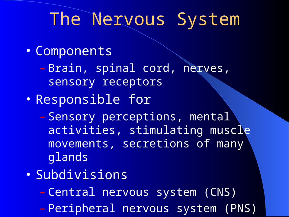
The Nervous System
• Components– Brain, spinal cord, nerves, sensory receptors
• Responsible for– Sensory perceptions, mental activities,
stimulating muscle movements, secretions of many glands
• Subdivisions– Central nervous system (CNS)– Peripheral nervous system (PNS)
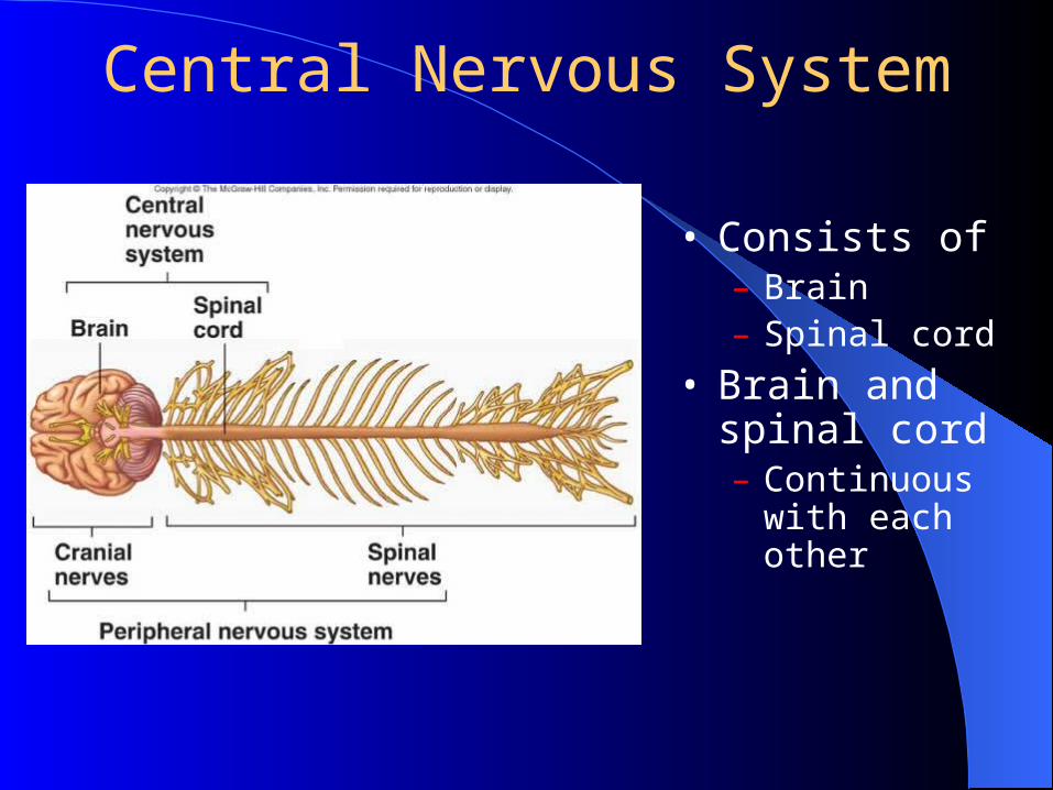
Central Nervous System
• Consists of– Brain– Spinal cord
• Brain and spinal cord– Continuous with
each other

Peripheral Nervous System
• Two subcategories– Sensory or afferent
– Motor or efferent• Divisions
– Somatic nervous system
– Autonomic nervous system (ANS)
» Sympathetic
» Parasympathetic
» Enteric
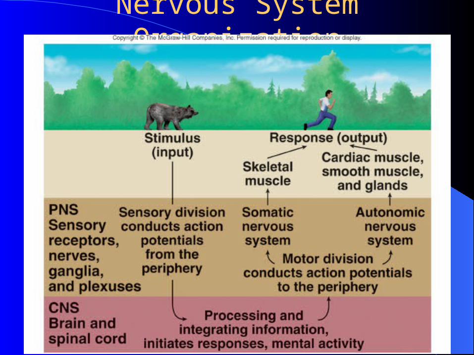
Nervous System Organization
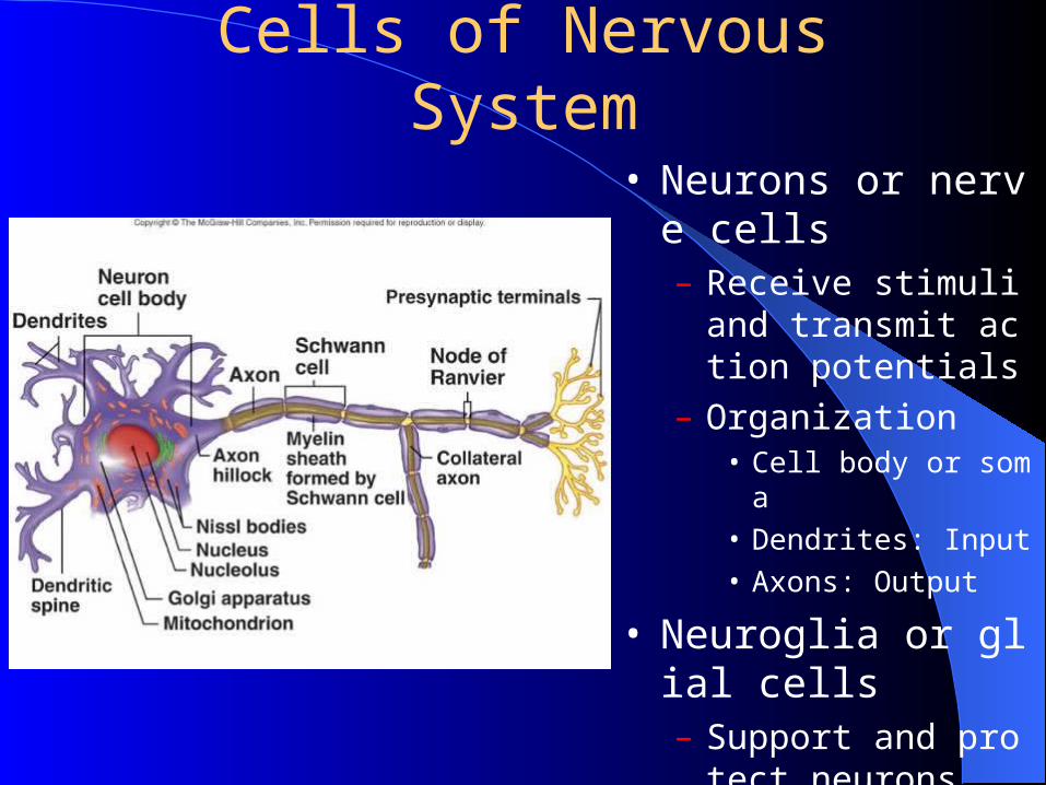
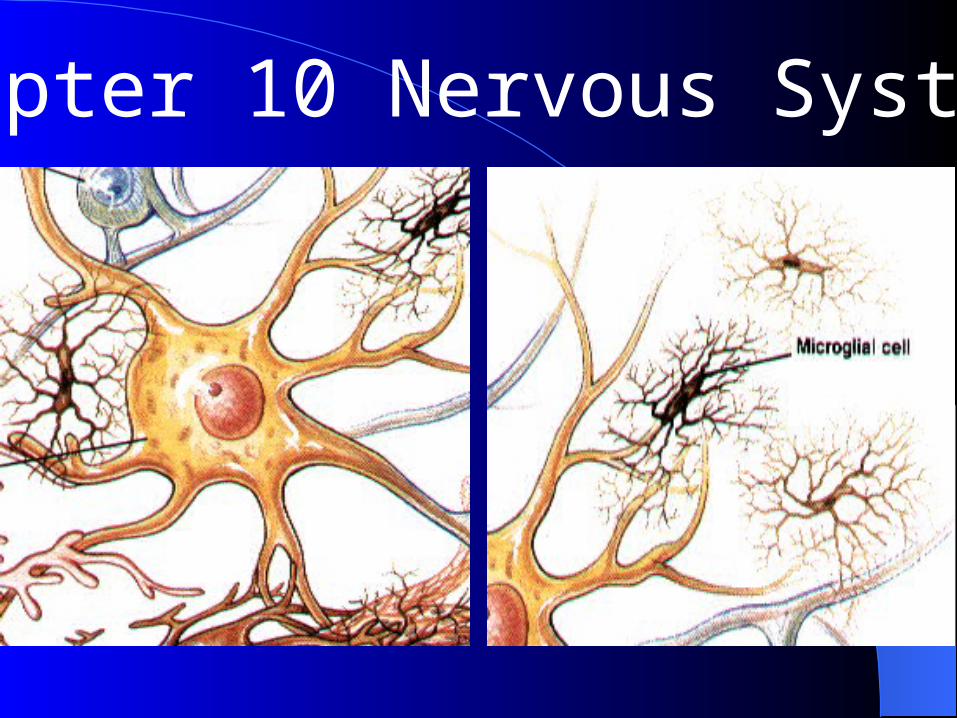
Cells of Nervous System
• Neurons or nerve cells– Receive stimuli and tra
nsmit action potentials
– Organization• Cell body or soma
• Dendrites: Input
• Axons: Output
• Neuroglia or glial cells– Support and protect ne
urons
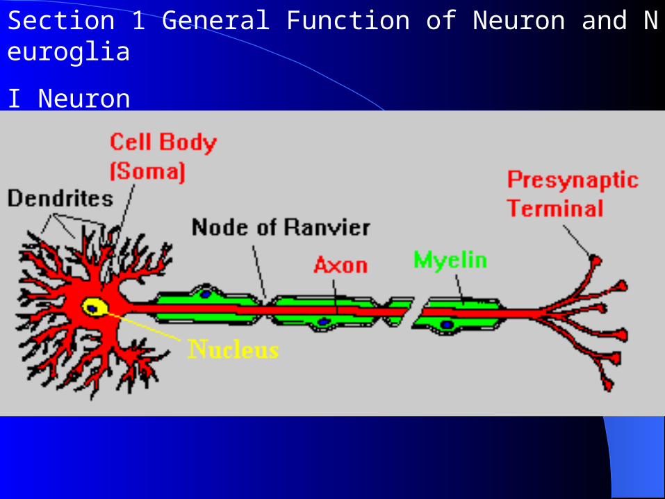
Section 1 General Function of Neuron and Neuroglia
I Neuron
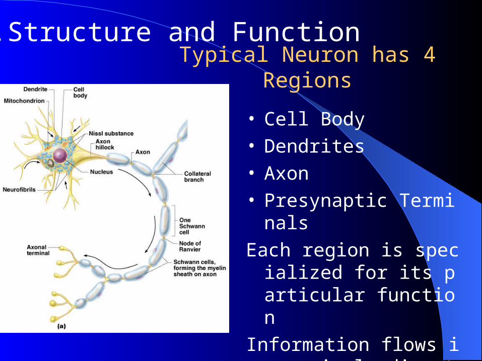
Typical Neuron has 4 Regions
• Cell Body • Dendrites• Axon• Presynaptic Terminals
Each region is specialized for its particular function
Information flows in a single direction
1. Structure and Function
Neuron Cell Body LocationNeuron Cell Body Location
Most are found in the central nervous system
Gray matter – cell bodies and unmylenated fibers
Nuclei – clusters of cell bodies within the white matter of the central nervous system
Ganglia – collections of cell bodies outside the central nervous system
Functional Classification of Functional Classification of NeuronsNeurons
Sensory (afferent) neurons
Carry impulses from the sensory receptors
Cutaneous sense organs
Proprioceptors – detect stretch or tension
Motor (efferent) neurons
Carry impulses from the central nervous system
Functional Classification of Functional Classification of NeuronsNeurons
Interneurons (association neurons)
Found in neural pathways in the central nervous system
Connect sensory and motor neurons
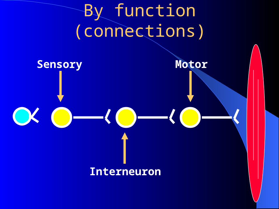
By function (connections)
Interneuron
Sensory Motor
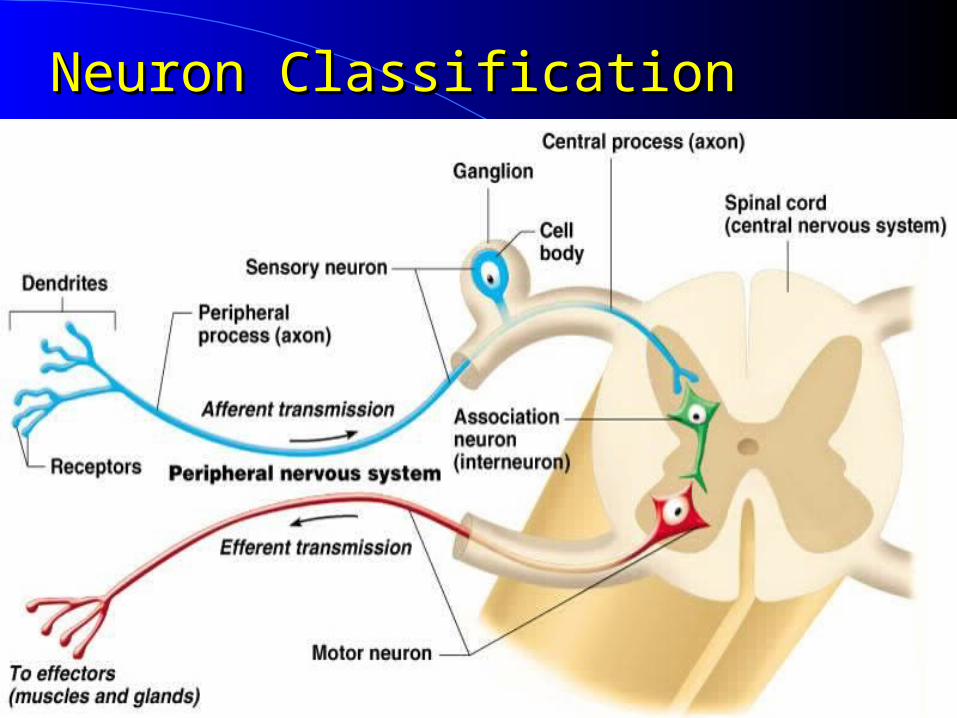
Neuron ClassificationNeuron Classification
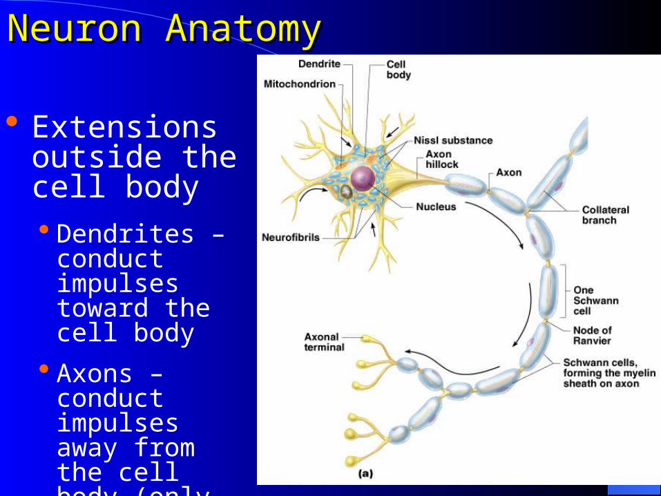
Neuron AnatomyNeuron Anatomy
Extensions outside the cell body Dendrites –
conduct impulses toward the cell body
Axons – conduct impulses away from the cell body (only 1!)
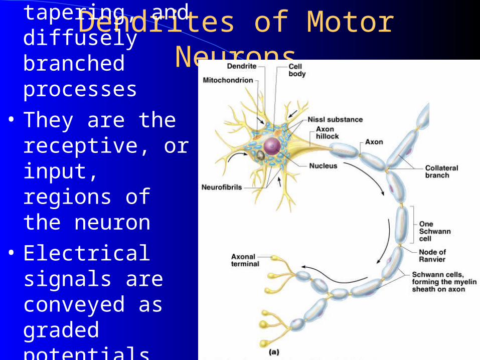
Dendrites of Motor Neurons• Short, tapering, and
diffusely branched processes
• They are the receptive, or input, regions of the neuron
• Electrical signals are conveyed as graded potentials (not action potentials)
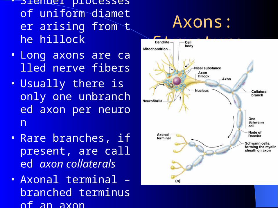
Axons: Structure • Slender processes of uniform diameter arising from the hillock
• Long axons are called nerve fibers
• Usually there is only one unbranched axon per neuron
• Rare branches, if present, are called axon collaterals
• Axonal terminal – branched terminus of an axon
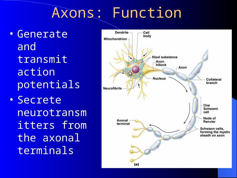
Axons: Function
• Generate and transmit action potentials
• Secrete neurotransmitters from the axonal terminals
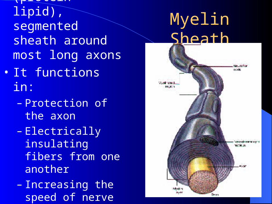
Myelin Sheath• Whitish, fatty (protein-lipid), segmented sheath around most long axons
• It functions in:– Protection of the axon– Electrically insulating
fibers from one another
– Increasing the speed of nerve impulse transmission
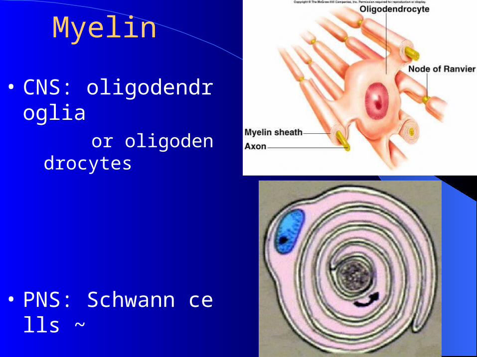
Myelin
• CNS: oligodendrogliaor oligodendroc
ytes
• PNS: Schwann cells ~
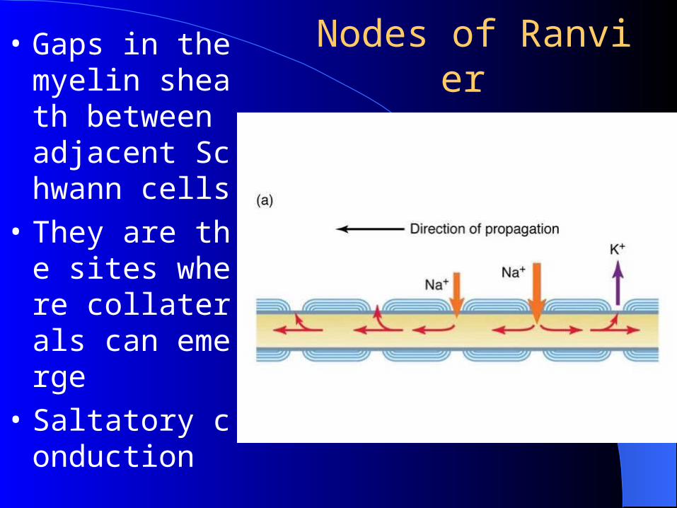
Nodes of Ranvier
• Gaps in the myelin sheath between adjacent Schwann cells
• They are the sites where collaterals can emerge
• Saltatory conduction
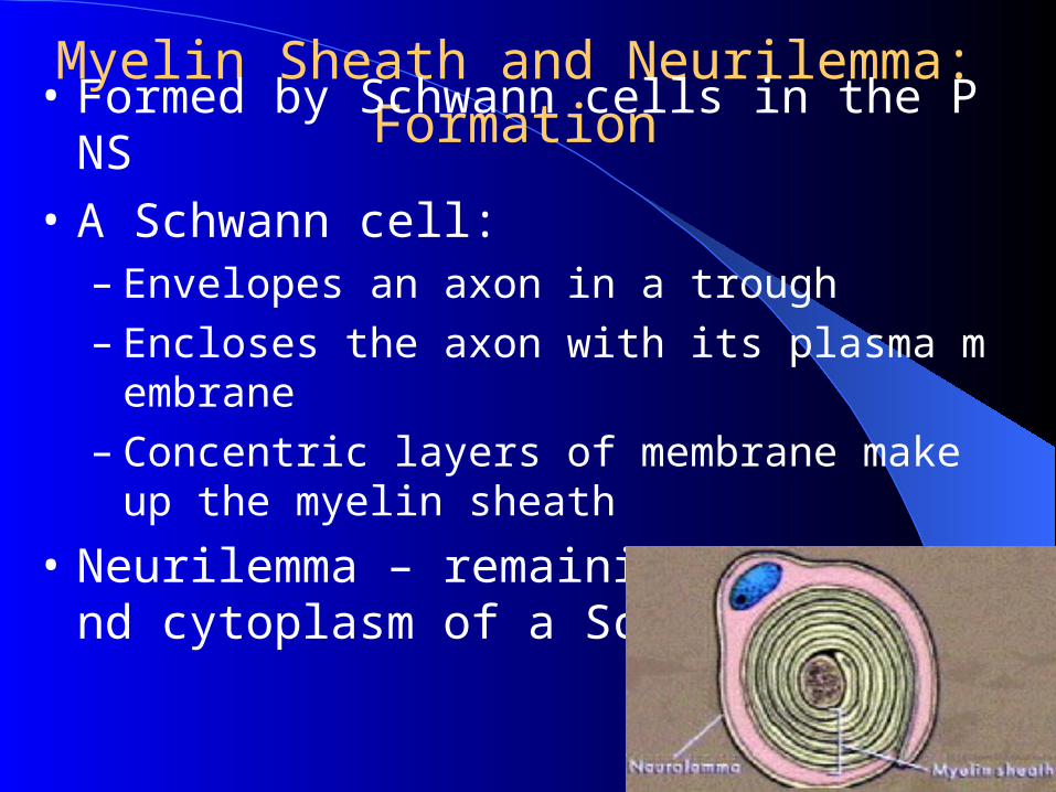
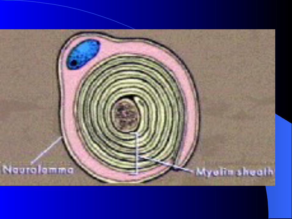
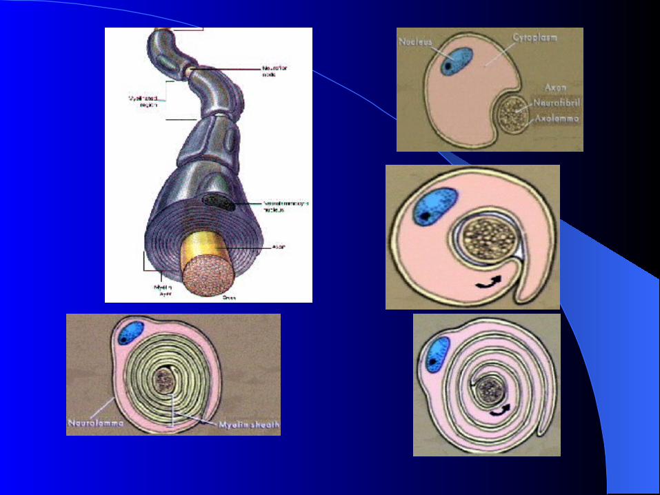
Myelin Sheath and Neurilemma: Formation
• Formed by Schwann cells in the PNS
• A Schwann cell:– Envelopes an axon in a trough– Encloses the axon with its plasma membrane– Concentric layers of membrane make up the myelin s
heath
• Neurilemma – remaining nucleus and cytoplasm of a Schwann cell
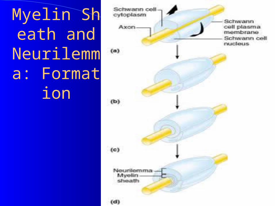
Myelin Sheath and Neurilemma: Format
ion
Figure 11.5a-d
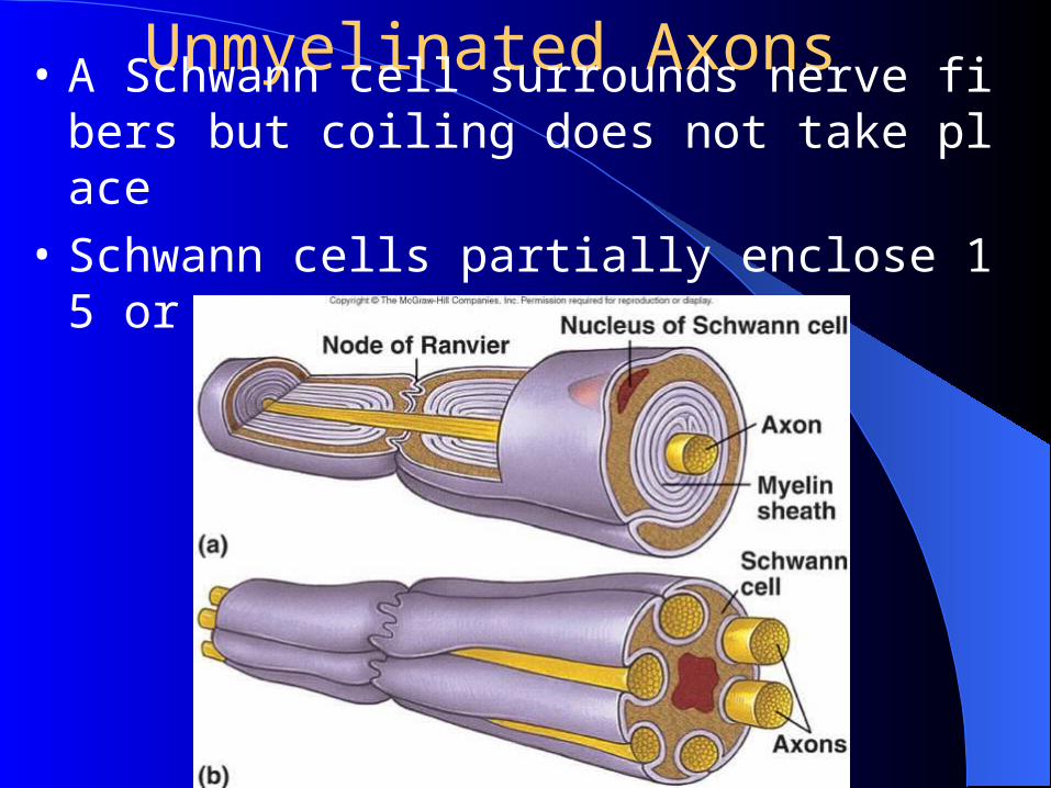
Unmyelinated Axons• A Schwann cell surrounds nerve fibers but coilin
g does not take place
• Schwann cells partially enclose 15 or more axons
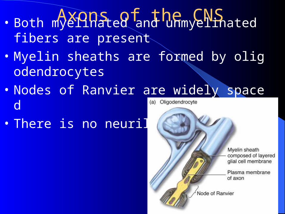
Axons of the CNS• Both myelinated and unmyelinated fibers are pres
ent
• Myelin sheaths are formed by oligodendrocytes
• Nodes of Ranvier are widely spaced
• There is no neurilemma
2. Classification and Function of Nerve Fibers
•Function: conducting action potential
1)Characteristic
physiological integration (anesthetic and tetrodot
oxin, TTX)
insulation,
two direction
no fatigue
2). Conducting velocities of AP propagation:
0.5~120m/s.
The factors that influence the AP propagation:
~The diameter of NF: 0.2 -20 mm, The larger the diameter is , The faster A.P. propagates.
~ Myelin sheath:
~Temperature:
3). The general classification of NF
Electrical physiological classification (efferent nerve): A, A, A, A; B, C.
Morphological classification (afferent nerve): I, II, III, IV.
The concept: Various organelles and materials must be moved from the cell body, where they are made, to the axon and its terminals in order to maintain the structure and function of the cell axon.
The mechanisms: Cytoskeletal filaments in the axon and cell body, which serve as the rails along which the transport occurs, are linked by proteins to the substances and organelles being moved.
4) Axoplasmic transport
Anterograde axoplasmic transport
~ fast axoplasmic transport: 410mm/day, mitochondria, vesicles, secretory granule.
~ slow axoplasmic transport:1-12 mm/d, cytoskeletal elements & soluble proteins
Retrograde axoplasmic transport: Axon transport of certain materials are from the axon terminals to the cell body. 205mm/d, NGF, tetanic toxin, horseradish peroxidase (HRP).
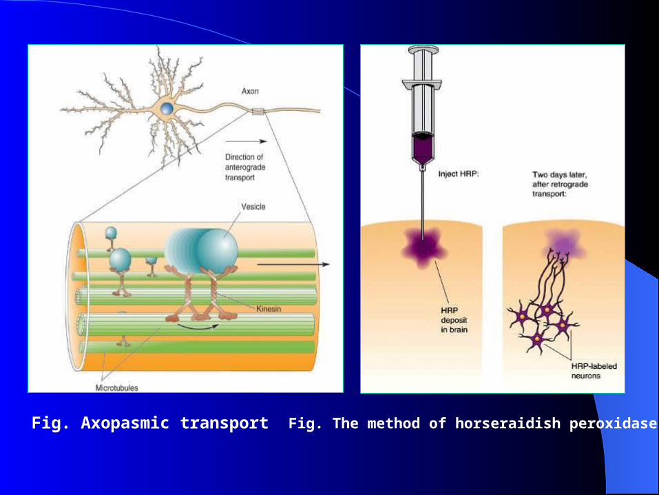
Fig. Axopasmic transport Fig. The method of horseraidish peroxidase
The motor nerve release some substance that has trophic action on the skeletal muscle
The denervated muscle does not receive nerve signals and due to this, muscle atrophy begins.
After two months, the muscle fibers degenerate and denervation atrophy follows.
Fibrous tissue replaces the muscle
5) Trophic action of the nerve to the target
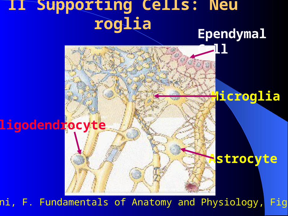
II Supporting Cells: NeurogliaEpendymal Cell
Microglia
Oligodendrocyte
Astrocyte
Martini, F. Fundamentals of Anatomy and Physiology, Fig 12-6.
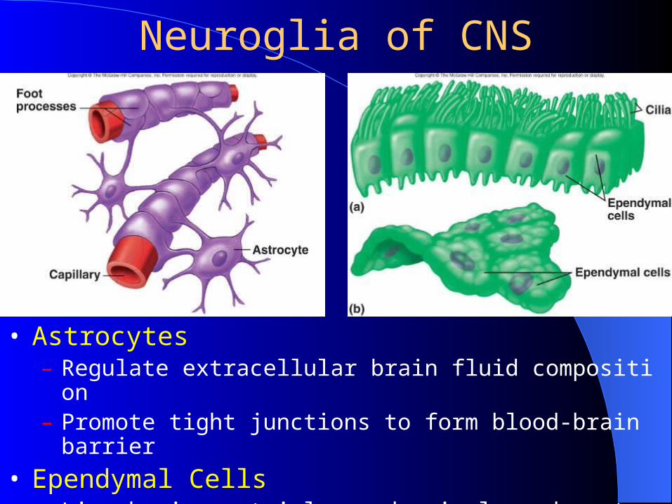
Neuroglia of CNS
• Astrocytes– Regulate extracellular brain fluid composition– Promote tight junctions to form blood-brain barrier
• Ependymal Cells– Line brain ventricles and spinal cord central canal– Help form choroid plexuses that secrete cerebrospinal fluid (CSF)
Neuroglia of CNS
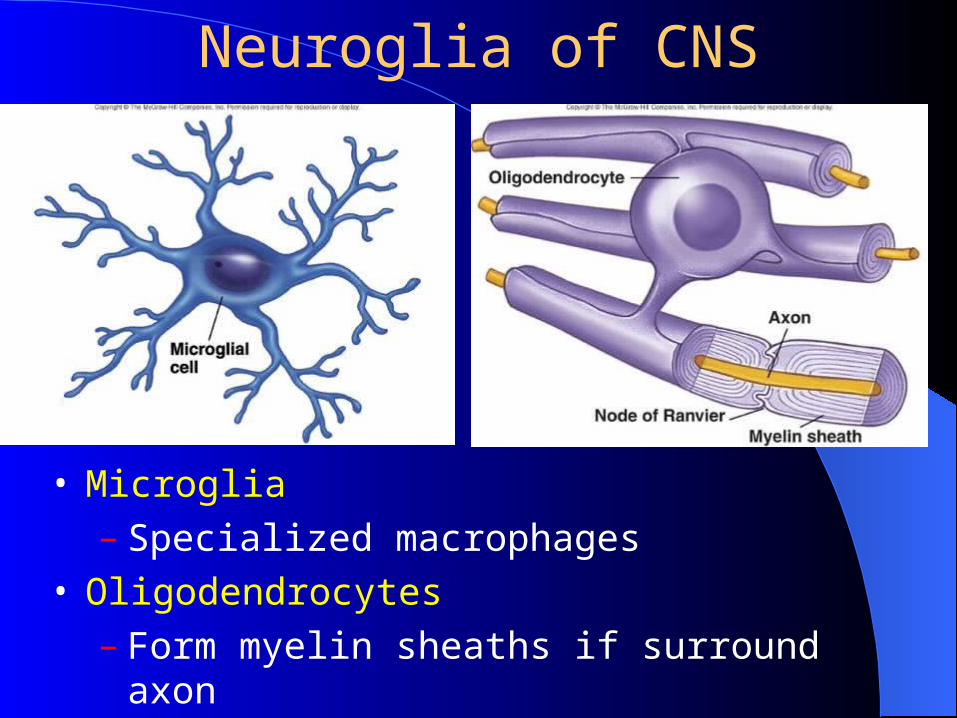
• Microglia– Specialized macrophages
• Oligodendrocytes– Form myelin sheaths if surround axon
Neuroglia of PNS
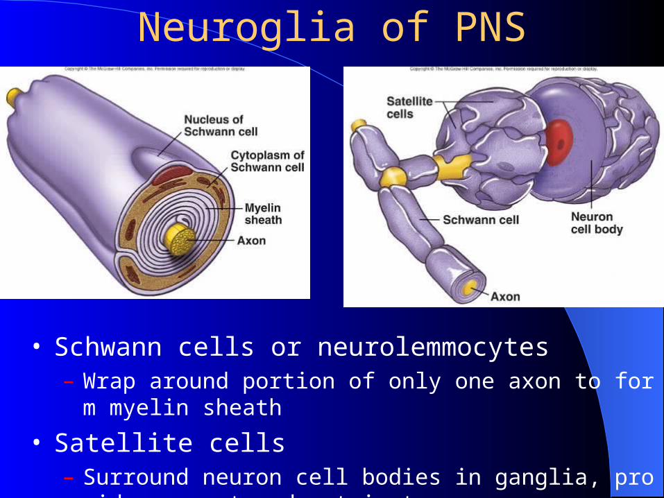
• Schwann cells or neurolemmocytes– Wrap around portion of only one axon to form myelin sheath
• Satellite cells– Surround neuron cell bodies in ganglia, provide support and nutr
ients