Embed Size (px)
Citation preview
ASTRO Refresher Course 2018:
Lymphoma
Rahul R. Parikh, M.D.Department of Radiation Oncology
Rutgers Cancer Institute of New Jersey
Saturday, March 3rd, 2018
Disclosures
• No conflicts of interest to disclose.
Learning Objectives
• Integrate Lugano Classification / Deauville criteria into staging & workup
• Review role of RT in management of lymphoma:• Hodgkin lymphoma• Non-Hodgkin lymphoma
• DLBCL• FL• MALT
• Implement modern radiotherapy techniques (ISRT)
3
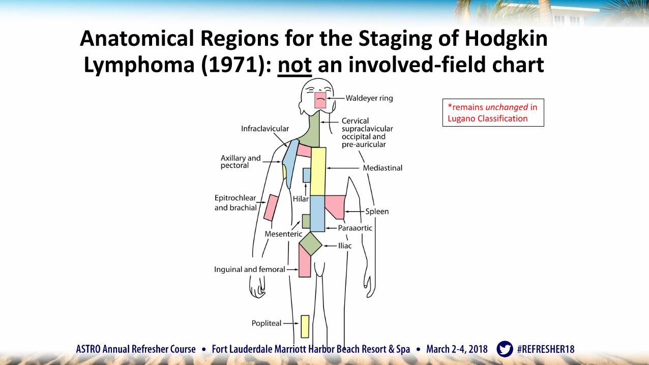
Anatomical Regions for the Staging of Hodgkin Lymphoma (1971): not an involved-field chart
*remains unchanged in Lugano Classification
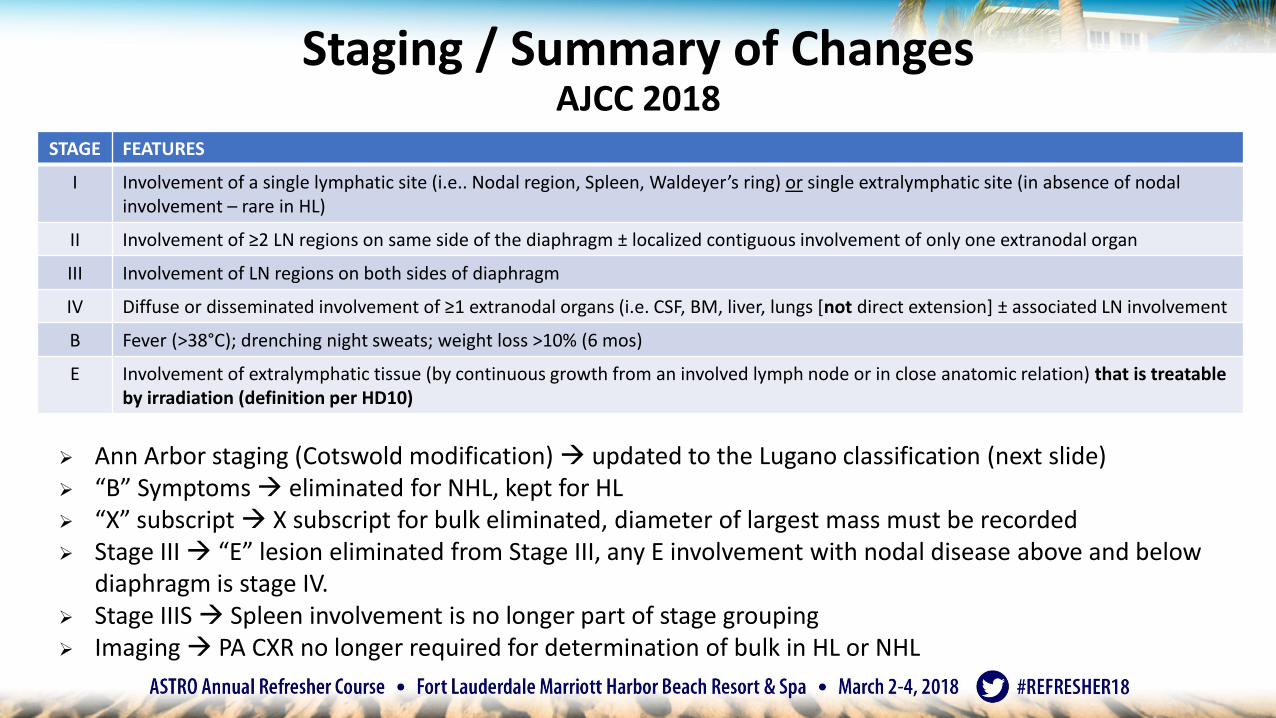
Staging / Summary of ChangesAJCC 2018
➢ Ann Arbor staging (Cotswold modification) updated to the Lugano classification (next slide)➢ “B” Symptoms eliminated for NHL, kept for HL➢ “X” subscript X subscript for bulk eliminated, diameter of largest mass must be recorded➢ Stage III “E” lesion eliminated from Stage III, any E involvement with nodal disease above and below
diaphragm is stage IV.➢ Stage IIIS Spleen involvement is no longer part of stage grouping➢ Imaging PA CXR no longer required for determination of bulk in HL or NHL
STAGE FEATURES
I Involvement of a single lymphatic site (i.e.. Nodal region, Spleen, Waldeyer’s ring) or single extralymphatic site (in absence of nodal involvement – rare in HL)
II Involvement of ≥2 LN regions on same side of the diaphragm ± localized contiguous involvement of only one extranodal organ
III Involvement of LN regions on both sides of diaphragm
IV Diffuse or disseminated involvement of ≥1 extranodal organs (i.e. CSF, BM, liver, lungs [not direct extension] ± associated LN involvement
B Fever (>38°C); drenching night sweats; weight loss >10% (6 mos)
E Involvement of extralymphatic tissue (by continuous growth from an involved lymph node or in close anatomic relation) that is treatable by irradiation (definition per HD10)
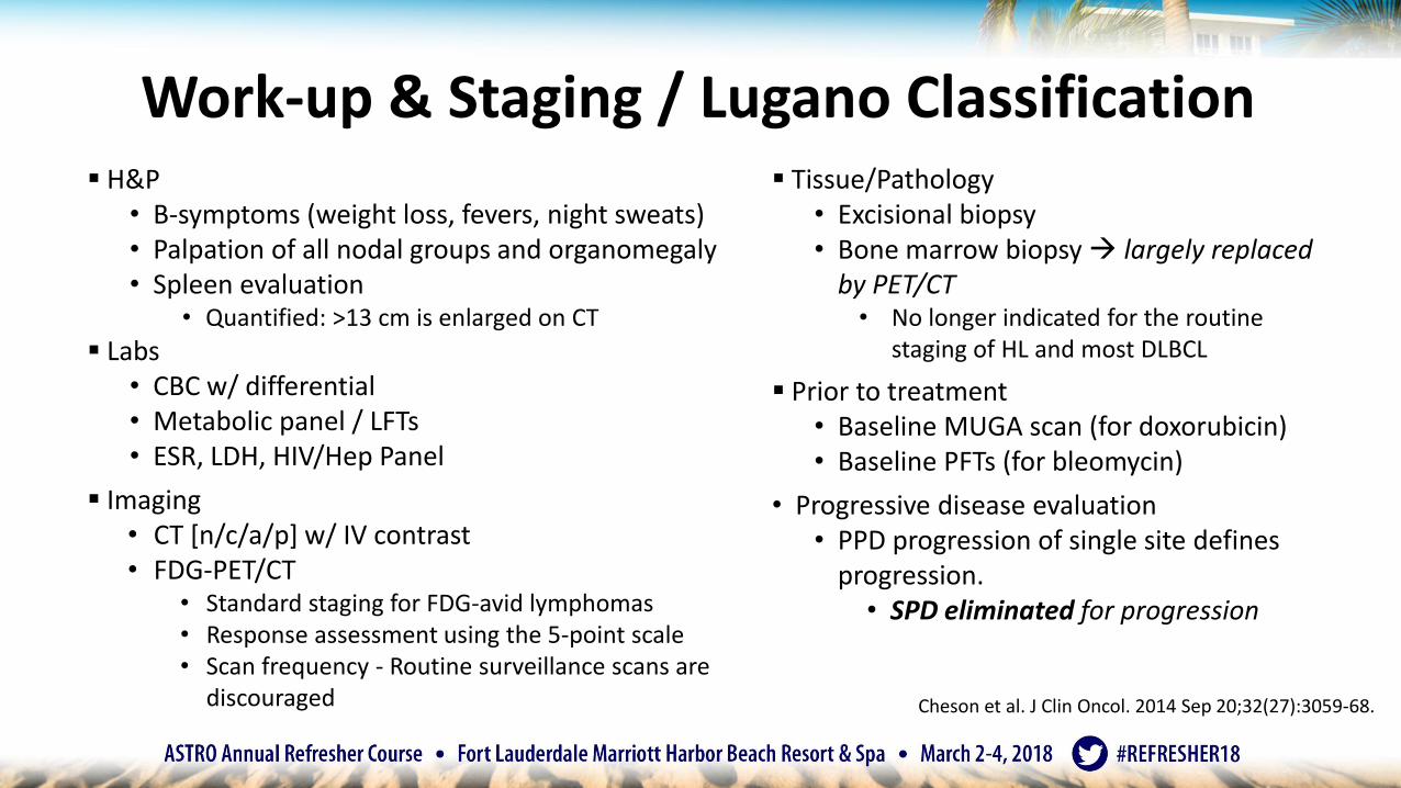
Work-up & Staging / Lugano Classification▪ H&P
• B-symptoms (weight loss, fevers, night sweats)• Palpation of all nodal groups and organomegaly• Spleen evaluation
• Quantified: >13 cm is enlarged on CT
▪ Labs• CBC w/ differential• Metabolic panel / LFTs• ESR, LDH, HIV/Hep Panel
▪ Imaging• CT [n/c/a/p] w/ IV contrast• FDG-PET/CT
• Standard staging for FDG-avid lymphomas• Response assessment using the 5-point scale• Scan frequency - Routine surveillance scans are
discouraged
▪ Tissue/Pathology• Excisional biopsy• Bone marrow biopsy largely replaced
by PET/CT• No longer indicated for the routine
staging of HL and most DLBCL
▪ Prior to treatment• Baseline MUGA scan (for doxorubicin)• Baseline PFTs (for bleomycin)
• Progressive disease evaluation• PPD progression of single site defines
progression. • SPD eliminated for progression
Cheson et al. J Clin Oncol. 2014 Sep 20;32(27):3059-68.
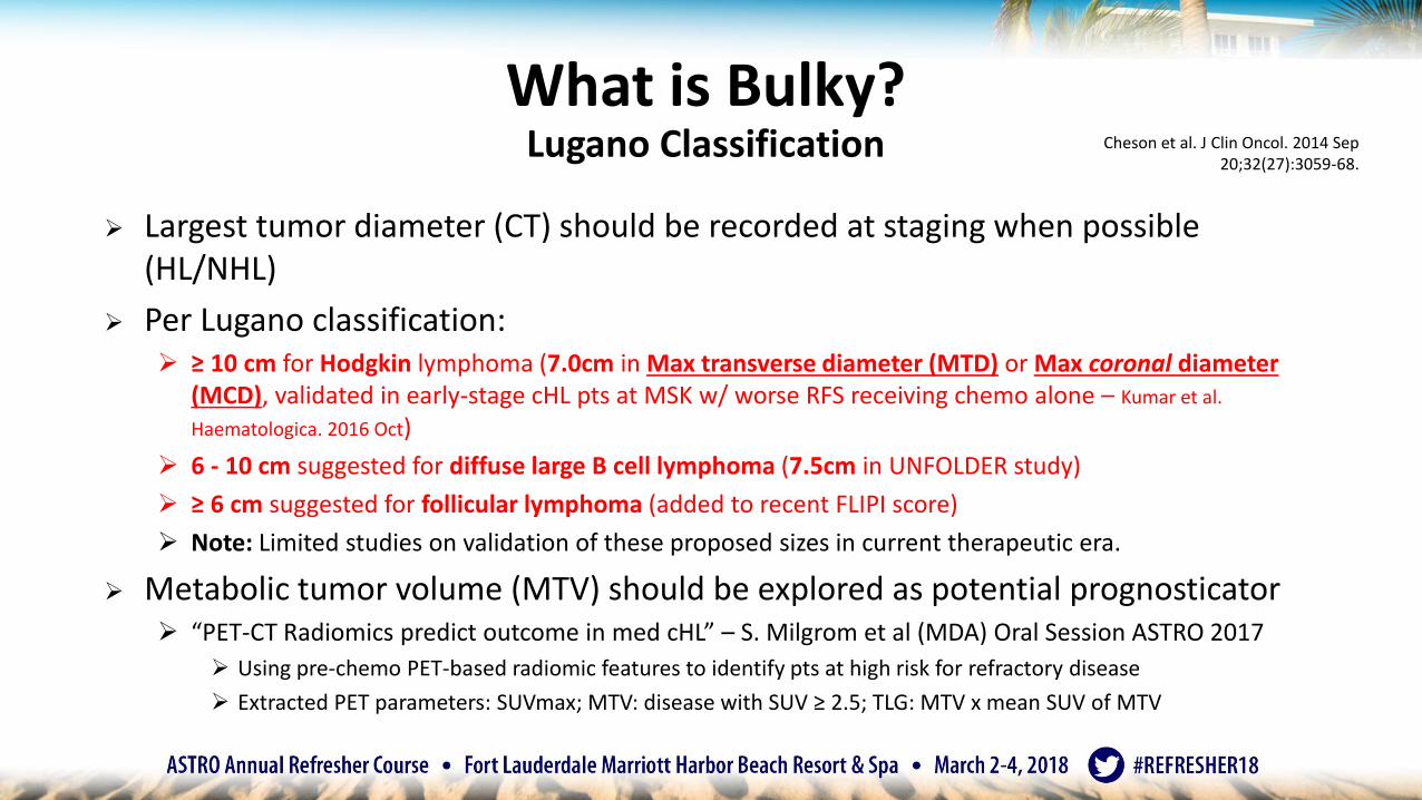
What is Bulky? Lugano Classification
➢ Largest tumor diameter (CT) should be recorded at staging when possible (HL/NHL)
➢ Per Lugano classification: ➢ ≥ 10 cm for Hodgkin lymphoma (7.0cm in Max transverse diameter (MTD) or Max coronal diameter
(MCD), validated in early-stage cHL pts at MSK w/ worse RFS receiving chemo alone – Kumar et al.
Haematologica. 2016 Oct)
➢ 6 - 10 cm suggested for diffuse large B cell lymphoma (7.5cm in UNFOLDER study)
➢ ≥ 6 cm suggested for follicular lymphoma (added to recent FLIPI score)
➢ Note: Limited studies on validation of these proposed sizes in current therapeutic era.
➢ Metabolic tumor volume (MTV) should be explored as potential prognosticator➢ “PET-CT Radiomics predict outcome in med cHL” – S. Milgrom et al (MDA) Oral Session ASTRO 2017
➢ Using pre-chemo PET-based radiomic features to identify pts at high risk for refractory disease
➢ Extracted PET parameters: SUVmax; MTV: disease with SUV ≥ 2.5; TLG: MTV x mean SUV of MTV
Cheson et al. J Clin Oncol. 2014 Sep 20;32(27):3059-68.
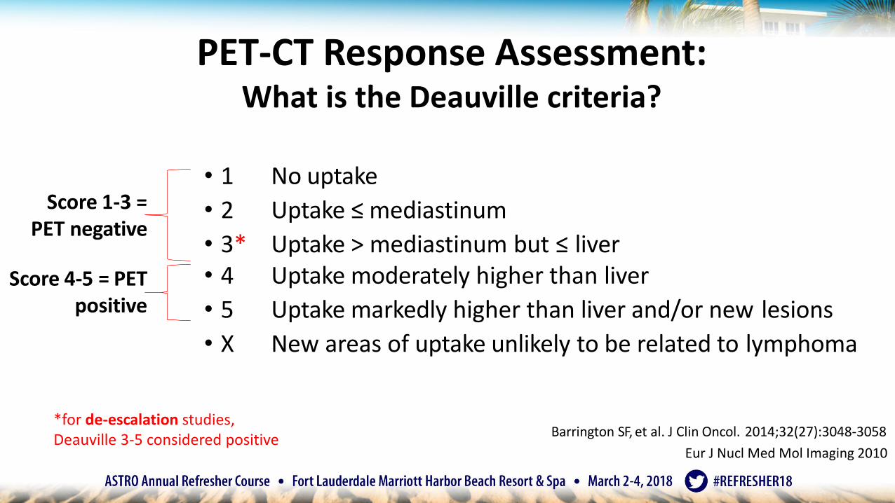
• 1 No uptake
• 2 Uptake ≤ mediastinum
• 3* Uptake > mediastinum but ≤ liver• 4 Uptake moderately higher than liver
• 5 Uptake markedly higher than liver and/or new lesions
• X New areas of uptake unlikely to be related to lymphoma
Barrington SF, et al. J Clin Oncol. 2014;32(27):3048-3058
Score 1-3 = PET negative
Score 4-5 = PETpositive
Eur J Nucl Med Mol Imaging 2010
*for de-escalation studies, Deauville 3-5 considered positive
PET-CT Response Assessment: What is the Deauville criteria?
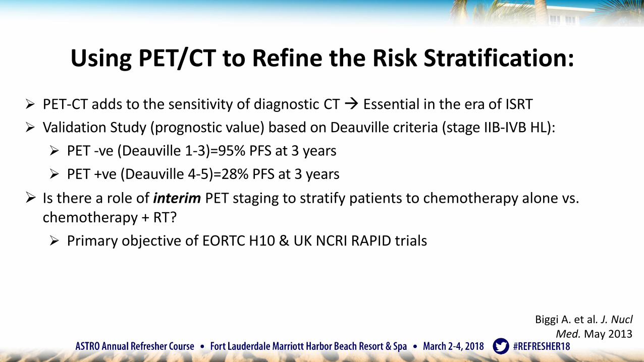
➢ PET-CT adds to the sensitivity of diagnostic CT Essential in the era of ISRT
➢ Validation Study (prognostic value) based on Deauville criteria (stage IIB-IVB HL):
➢ PET -ve (Deauville 1-3)=95% PFS at 3 years
➢ PET +ve (Deauville 4-5)=28% PFS at 3 years
➢ Is there a role of interim PET staging to stratify patients to chemotherapy alone vs. chemotherapy + RT?
➢ Primary objective of EORTC H10 & UK NCRI RAPID trials
Biggi A. et al. J. NuclMed. May 2013
Using PET/CT to Refine the Risk Stratification:
Hodgkin Lymphoma
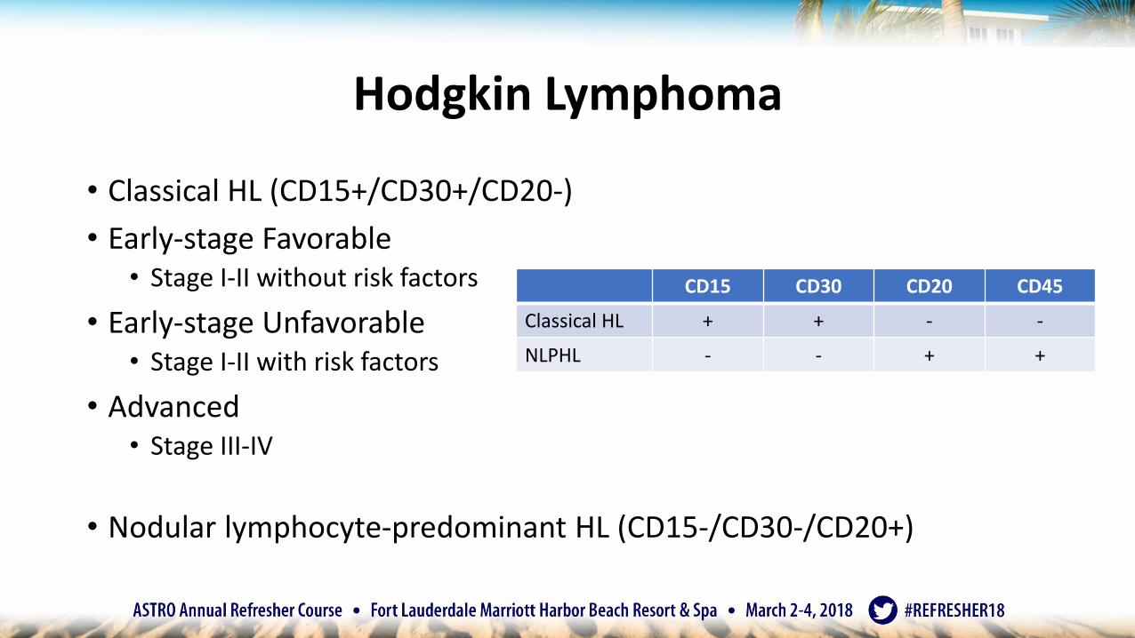
• Classical HL (CD15+/CD30+/CD20-)
• Early-stage Favorable• Stage I-II without risk factors
• Early-stage Unfavorable• Stage I-II with risk factors
• Advanced• Stage III-IV
• Nodular lymphocyte-predominant HL (CD15-/CD30-/CD20+)
CD15 CD30 CD20 CD45
Classical HL + + - -
NLPHL - - + +
Early-stage cHL: Prognostic Classification
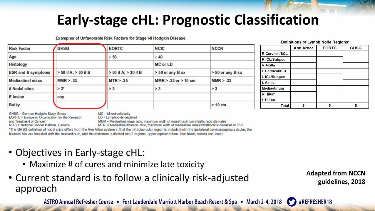
• Objectives in Early-stage cHL:• Maximize # of cures and minimize late toxicity
• Current standard is to follow a clinically risk-adjusted approach
Adapted from NCCN guidelines, 2018
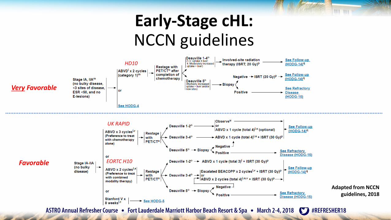
Early-Stage cHL:NCCN guidelines
HD10
UK RAPID
EORTC H10
Adapted from NCCN guidelines, 2018
Very Favorable
Favorable
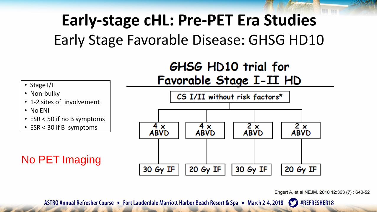
Early-stage cHL: Pre-PET Era StudiesEarly Stage Favorable Disease: GHSG HD10
• Stage I/II• Non-bulky• 1-2 sites of involvement• No ENI• ESR < 50 if no B symptoms• ESR < 30 if B symptoms
No PET Imaging
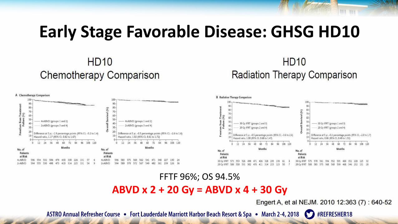
FFTF 96%; OS 94.5%
ABVD x 2 + 20 Gy = ABVD x 4 + 30 Gy
Early Stage Favorable Disease: GHSG HD10
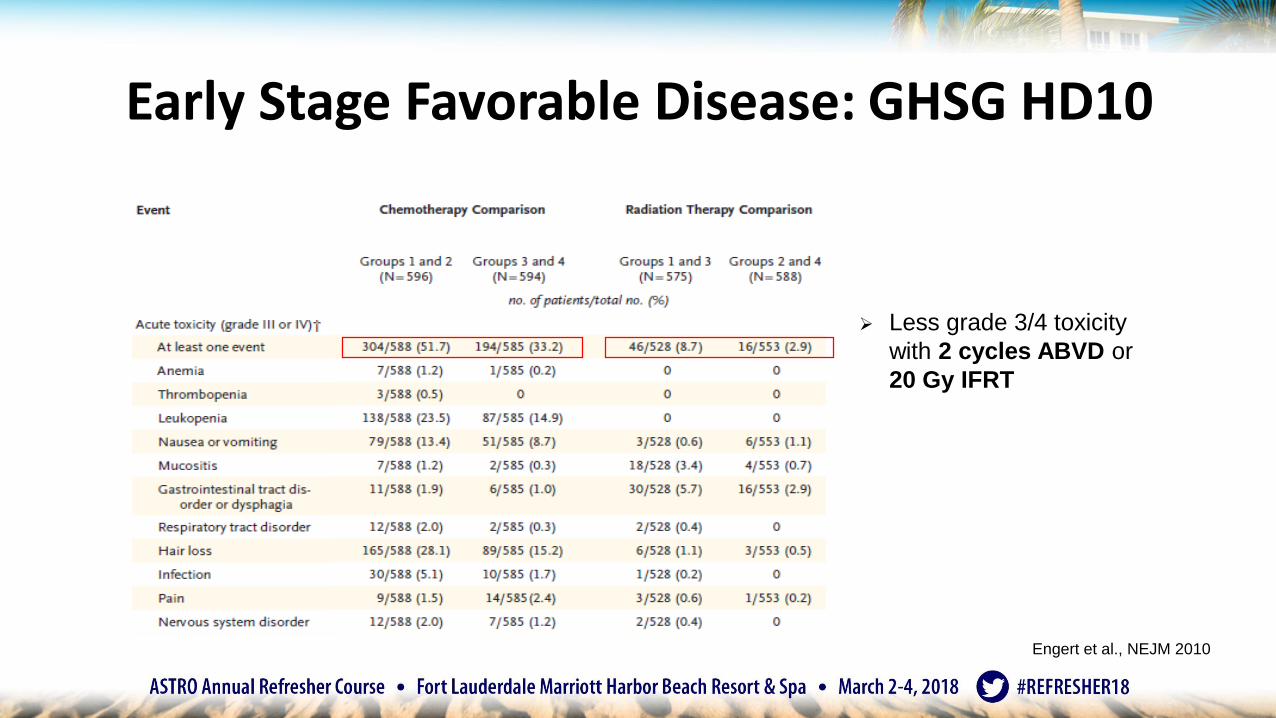
Engert et al., NEJM 2010
➢ Less grade 3/4 toxicity
with 2 cycles ABVD or
20 Gy IFRT
Early Stage Favorable Disease: GHSG HD10
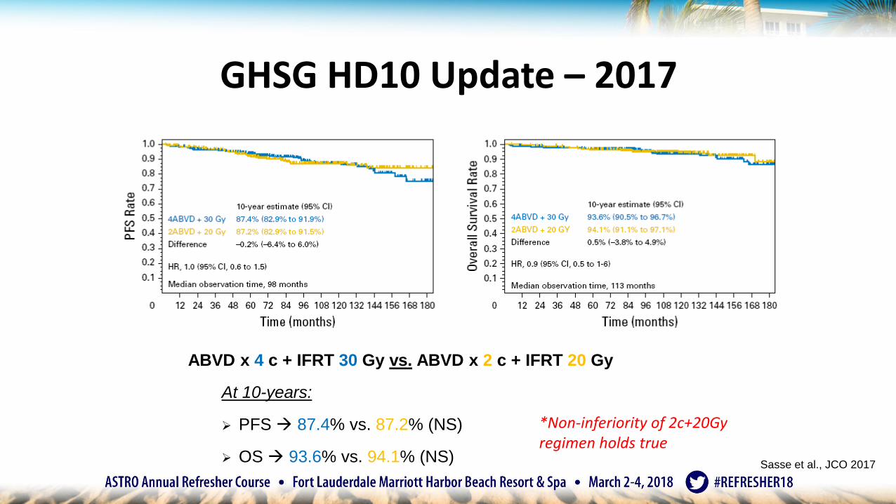
ABVD x 4 c + IFRT 30 Gy vs. ABVD x 2 c + IFRT 20 Gy
At 10-years:
➢ PFS 87.4% vs. 87.2% (NS)
➢ OS 93.6% vs. 94.1% (NS)
*Non-inferiority of 2c+20Gy regimen holds true
Sasse et al., JCO 2017
GHSG HD10 Update – 2017
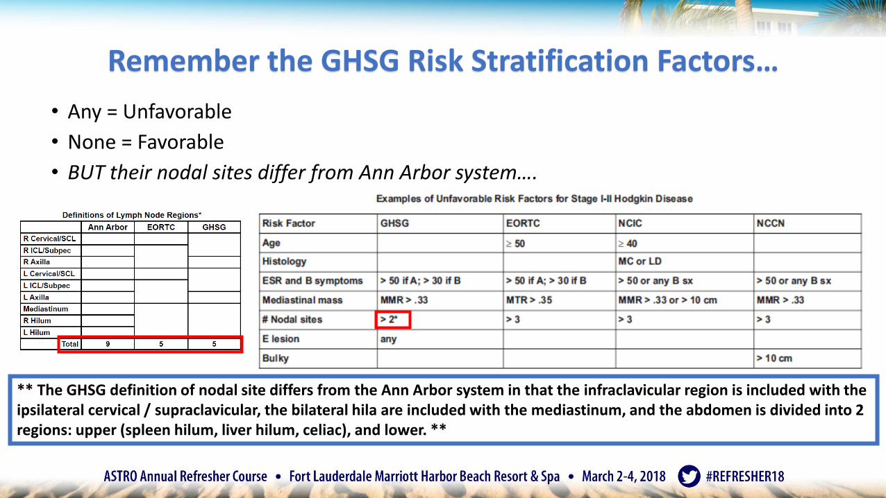
Remember the GHSG Risk Stratification Factors…
• Any = Unfavorable
• None = Favorable
• BUT their nodal sites differ from Ann Arbor system….
** The GHSG definition of nodal site differs from the Ann Arbor system in that the infraclavicular region is included with the ipsilateral cervical / supraclavicular, the bilateral hila are included with the mediastinum, and the abdomen is divided into 2 regions: upper (spleen hilum, liver hilum, celiac), and lower. **
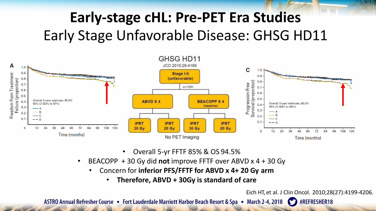
Early-stage cHL: Pre-PET Era StudiesEarly Stage Unfavorable Disease: GHSG HD11
• Overall 5-yr FFTF 85% & OS 94.5%• BEACOPP + 30 Gy did not improve FFTF over ABVD x 4 + 30 Gy
• Concern for inferior PFS/FFTF for ABVD x 4+ 20 Gy arm• Therefore, ABVD + 30Gy is standard of care
Eich HT, et al. J Clin Oncol. 2010;28(27):4199-4206.
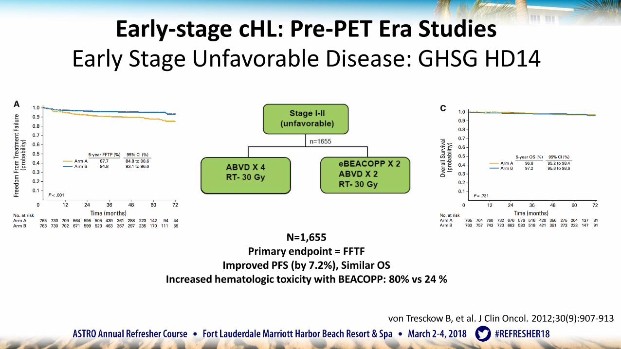
N=1,655Primary endpoint = FFTF
Improved PFS (by 7.2%), Similar OSIncreased hematologic toxicity with BEACOPP: 80% vs 24 %
Early-stage cHL: Pre-PET Era StudiesEarly Stage Unfavorable Disease: GHSG HD14
von Tresckow B, et al. J Clin Oncol. 2012;30(9):907-913
Raemaekers et al., JCO 2014.
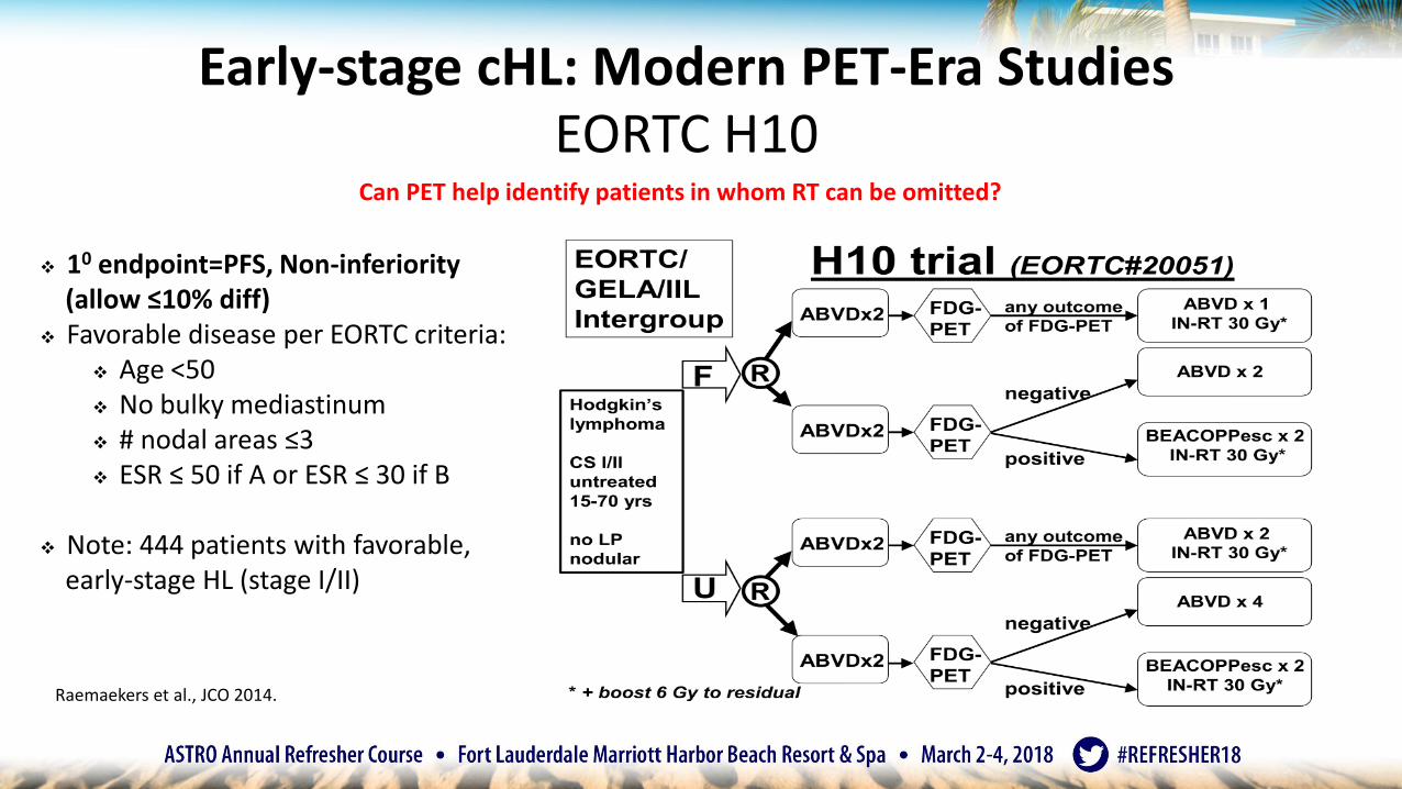
Early-stage cHL: Modern PET-Era StudiesEORTC H10
Can PET help identify patients in whom RT can be omitted?
❖ 10 endpoint=PFS, Non-inferiority (allow ≤10% diff)
❖ Favorable disease per EORTC criteria:❖ Age <50❖ No bulky mediastinum❖ # nodal areas ≤3❖ ESR ≤ 50 if A or ESR ≤ 30 if B
❖ Note: 444 patients with favorable, early-stage HL (stage I/II)
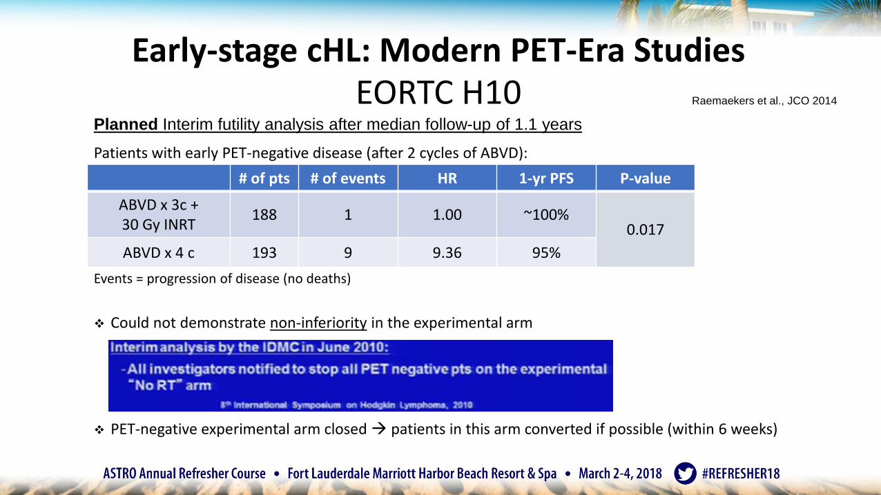
Raemaekers et al., JCO 2014
# of pts # of events HR 1-yr PFS P-value
ABVD x 3c + 30 Gy INRT
188 1 1.00 ~100%0.017
ABVD x 4 c 193 9 9.36 95%
Patients with early PET-negative disease (after 2 cycles of ABVD):
Events = progression of disease (no deaths)
❖ Could not demonstrate non-inferiority in the experimental arm
❖ PET-negative experimental arm closed patients in this arm converted if possible (within 6 weeks)
Planned Interim futility analysis after median follow-up of 1.1 years
Early-stage cHL: Modern PET-Era StudiesEORTC H10
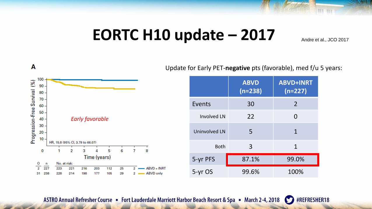
Andre et al., JCO 2017
ABVD(n=238)
ABVD+INRT(n=227)
Events 30 2
Involved LN 22 0
Uninvolved LN 5 1
Both 3 1
5-yr PFS 87.1% 99.0%
5-yr OS 99.6% 100%
Early favorable
EORTC H10 update – 2017
Update for Early PET-negative pts (favorable), med f/u 5 years:
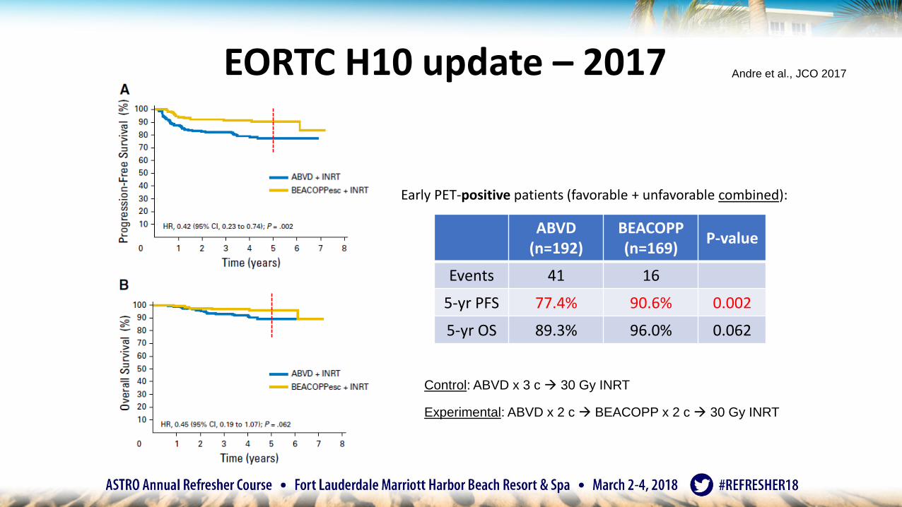
Early PET-positive patients (favorable + unfavorable combined):
Andre et al., JCO 2017
ABVD(n=192)
BEACOPP(n=169)
P-value
Events 41 16
5-yr PFS 77.4% 90.6% 0.002
5-yr OS 89.3% 96.0% 0.062
Control: ABVD x 3 c 30 Gy INRT
Experimental: ABVD x 2 c BEACOPP x 2 c 30 Gy INRT
EORTC H10 update – 2017
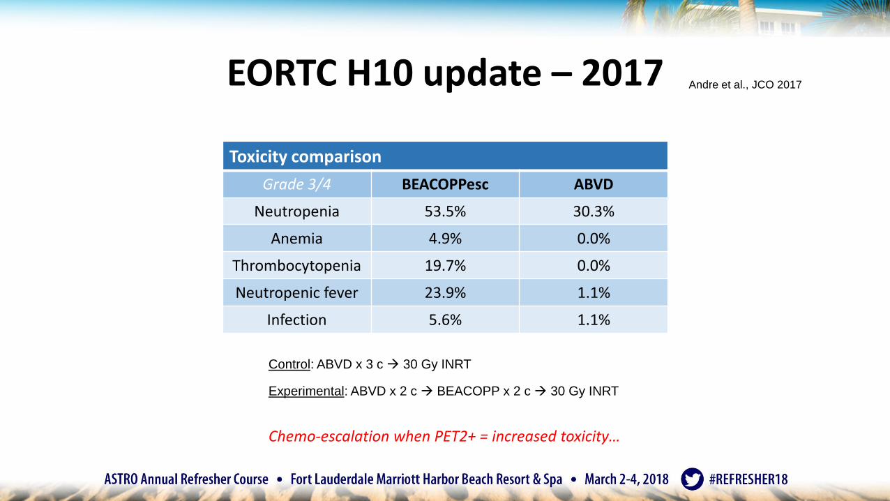
Andre et al., JCO 2017
Toxicity comparison
Grade 3/4 BEACOPPesc ABVD
Neutropenia 53.5% 30.3%
Anemia 4.9% 0.0%
Thrombocytopenia 19.7% 0.0%
Neutropenic fever 23.9% 1.1%
Infection 5.6% 1.1%
Control: ABVD x 3 c 30 Gy INRT
Experimental: ABVD x 2 c BEACOPP x 2 c 30 Gy INRT
Chemo-escalation when PET2+ = increased toxicity…
EORTC H10 update – 2017
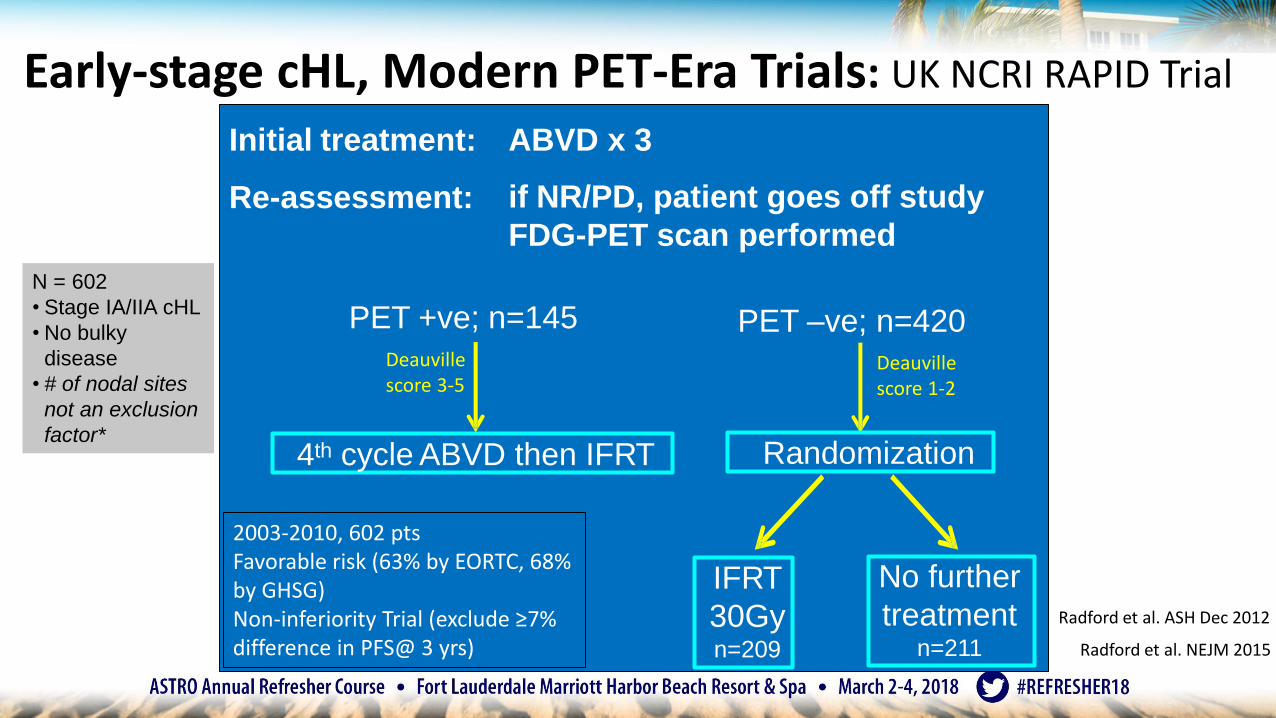
Initial treatment:
Re-assessment:
ABVD x 3
if NR/PD, patient goes off study
FDG-PET scan performed
4th cycle ABVD then IFRT Randomization
IFRT
30Gy n=209
No further
treatmentn=211
PET +ve; n=145 PET –ve; n=420
Radford et al. ASH Dec 2012
2003-2010, 602 ptsFavorable risk (63% by EORTC, 68% by GHSG)Non-inferiority Trial (exclude ≥7% difference in PFS@ 3 yrs) Radford et al. NEJM 2015
Early-stage cHL, Modern PET-Era Trials: UK NCRI RAPID Trial
N = 602
• Stage IA/IIA cHL
• No bulky
disease
• # of nodal sites
not an exclusion
factor*
Deauville score 3-5
Deauville score 1-2
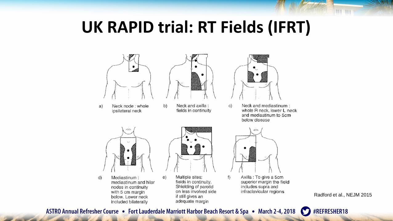
UK RAPID trial: RT Fields (IFRT)
Radford et al., NEJM 2015
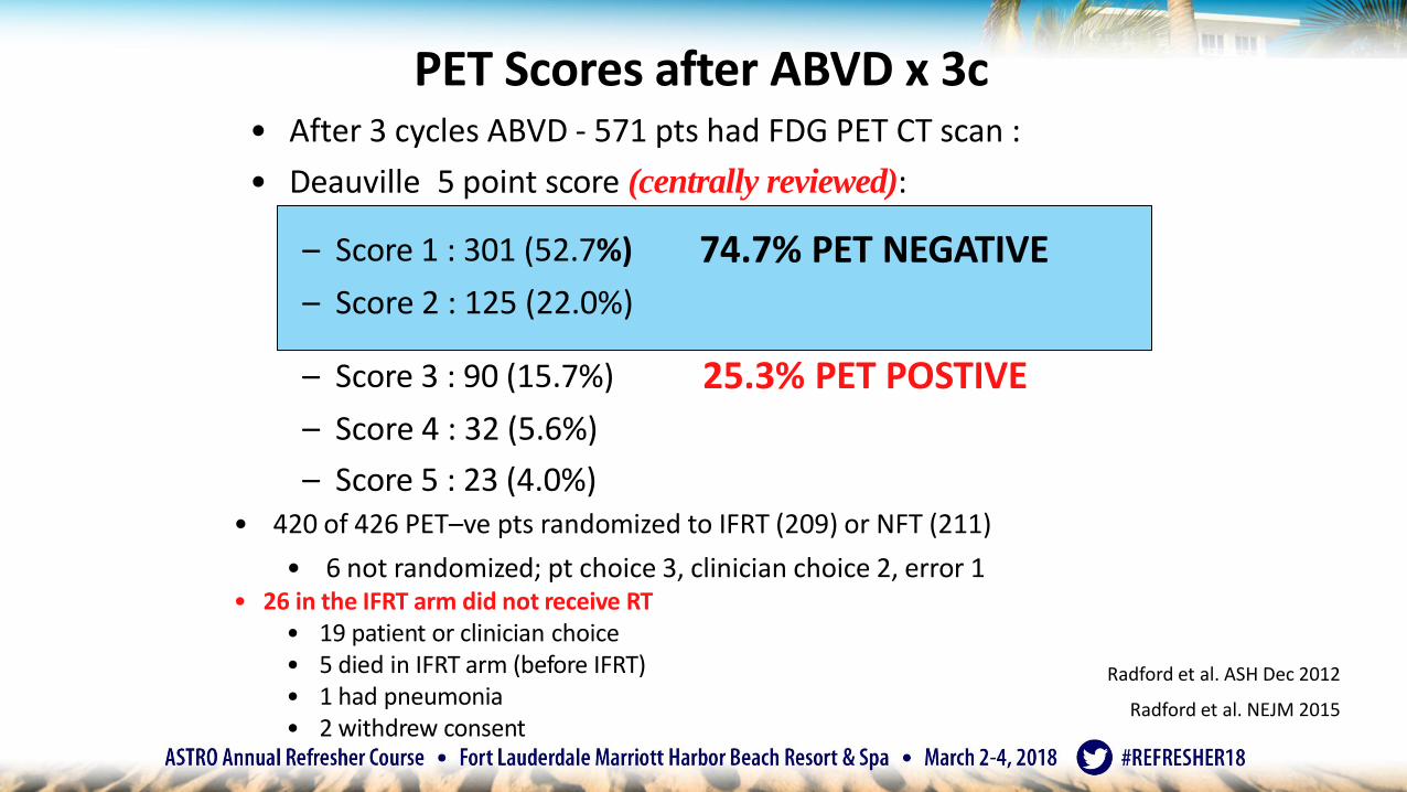
PET Scores after ABVD x 3c• After 3 cycles ABVD - 571 pts had FDG PET CT scan :
• Deauville 5 point score (centrally reviewed):
– Score 1 : 301 (52.7%)
– Score 2 : 125 (22.0%)
74.7% PET NEGATIVE
– Score 3 : 90 (15.7%)
– Score 4 : 32 (5.6%)
– Score 5 : 23 (4.0%)
25.3% PET POSTIVE
• 420 of 426 PET–ve pts randomized to IFRT (209) or NFT (211)
• 6 not randomized; pt choice 3, clinician choice 2, error 1• 26 in the IFRT arm did not receive RT
• 19 patient or clinician choice• 5 died in IFRT arm (before IFRT)• 1 had pneumonia• 2 withdrew consent
Radford et al. ASH Dec 2012
Radford et al. NEJM 2015
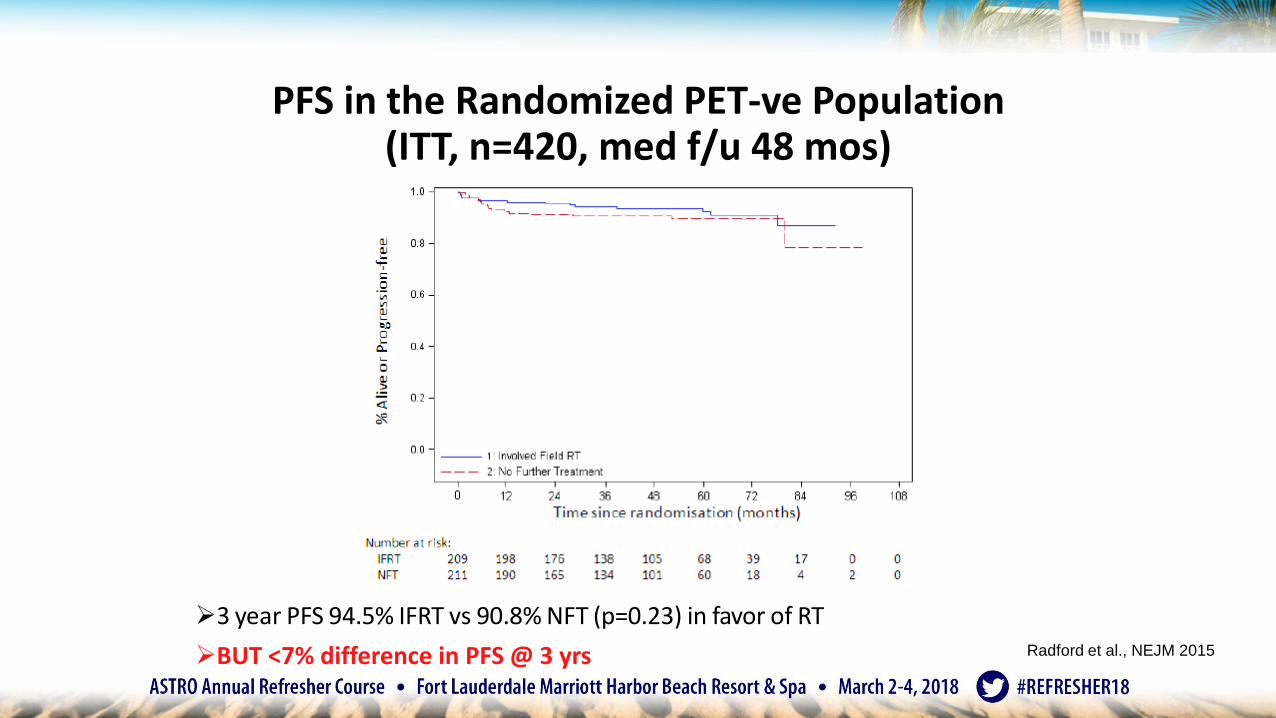
PFS in the Randomized PET-ve Population (ITT, n=420, med f/u 48 mos)
➢3 year PFS 94.5% IFRT vs 90.8% NFT (p=0.23) in favor of RT
➢BUT <7% difference in PFS @ 3 yrs Radford et al., NEJM 2015
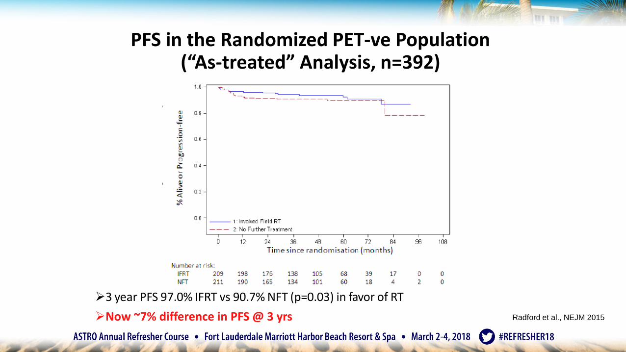
PFS in the Randomized PET-ve Population (“As-treated” Analysis, n=392)
➢3 year PFS 97.0% IFRT vs 90.7% NFT (p=0.03) in favor of RT
➢Now ~7% difference in PFS @ 3 yrs
• 420 of 426 PET–ve pts randomized to IFRT(209) or NFT (211)
• 6 not randomized; pt choice 3, clinicianchoice 2, error 1
• 26 in the IFRT arm did not receive RT• 19 patient or clinician choice• 5 died in IFRT arm (before IFRT)• 1 had pneumonia• 2 withdrew consent
Radford et al., NEJM 2015
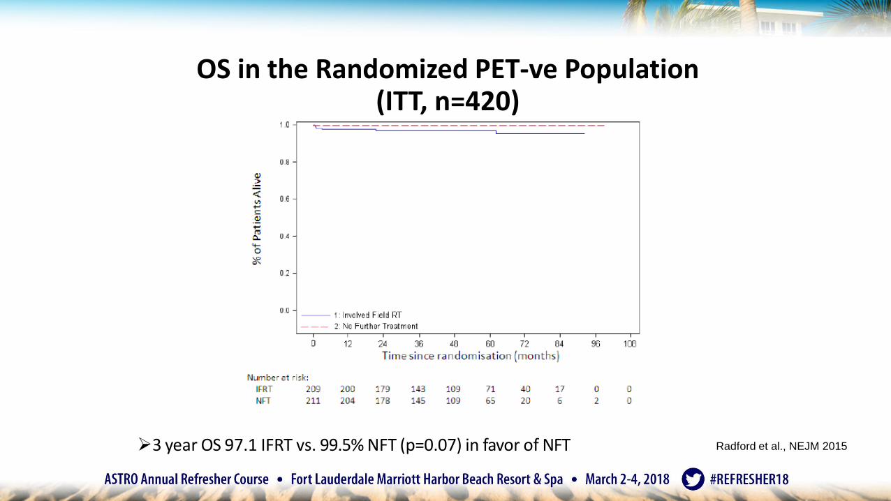
OS in the Randomized PET-ve Population (ITT, n=420)
➢3 year OS 97.1 IFRT vs. 99.5% NFT (p=0.07) in favor of NFT Radford et al., NEJM 2015
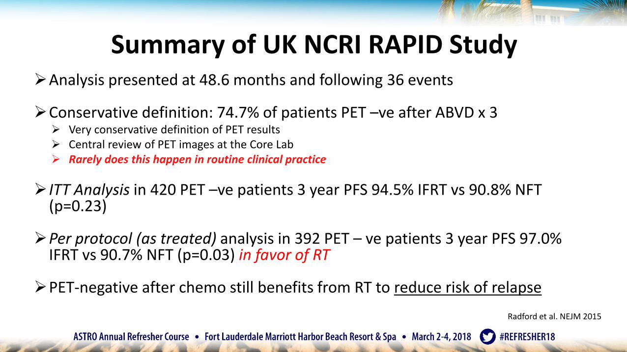
Summary of UK NCRI RAPID Study➢Analysis presented at 48.6 months and following 36 events
➢Conservative definition: 74.7% of patients PET –ve after ABVD x 3➢ Very conservative definition of PET results ➢ Central review of PET images at the Core Lab➢ Rarely does this happen in routine clinical practice
➢ ITT Analysis in 420 PET –ve patients 3 year PFS 94.5% IFRT vs 90.8% NFT(p=0.23)
➢Per protocol (as treated) analysis in 392 PET – ve patients 3 year PFS 97.0%IFRT vs 90.7% NFT (p=0.03) in favor of RT
➢PET-negative after chemo still benefits from RT to reduce risk of relapse
Radford et al. NEJM 2015
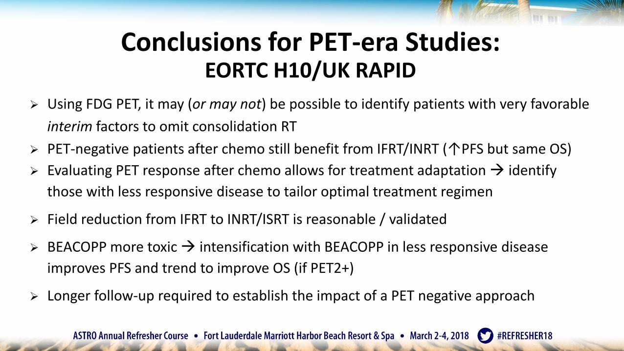
Conclusions for PET-era Studies: EORTC H10/UK RAPID
➢ Using FDG PET, it may (or may not) be possible to identify patients with very favorable
interim factors to omit consolidation RT
➢ PET-negative patients after chemo still benefit from IFRT/INRT (↑PFS but same OS)
➢ Evaluating PET response after chemo allows for treatment adaptation identify
those with less responsive disease to tailor optimal treatment regimen
➢ Field reduction from IFRT to INRT/ISRT is reasonable / validated
➢ BEACOPP more toxic intensification with BEACOPP in less responsive disease
improves PFS and trend to improve OS (if PET2+)
➢ Longer follow-up required to establish the impact of a PET negative approach
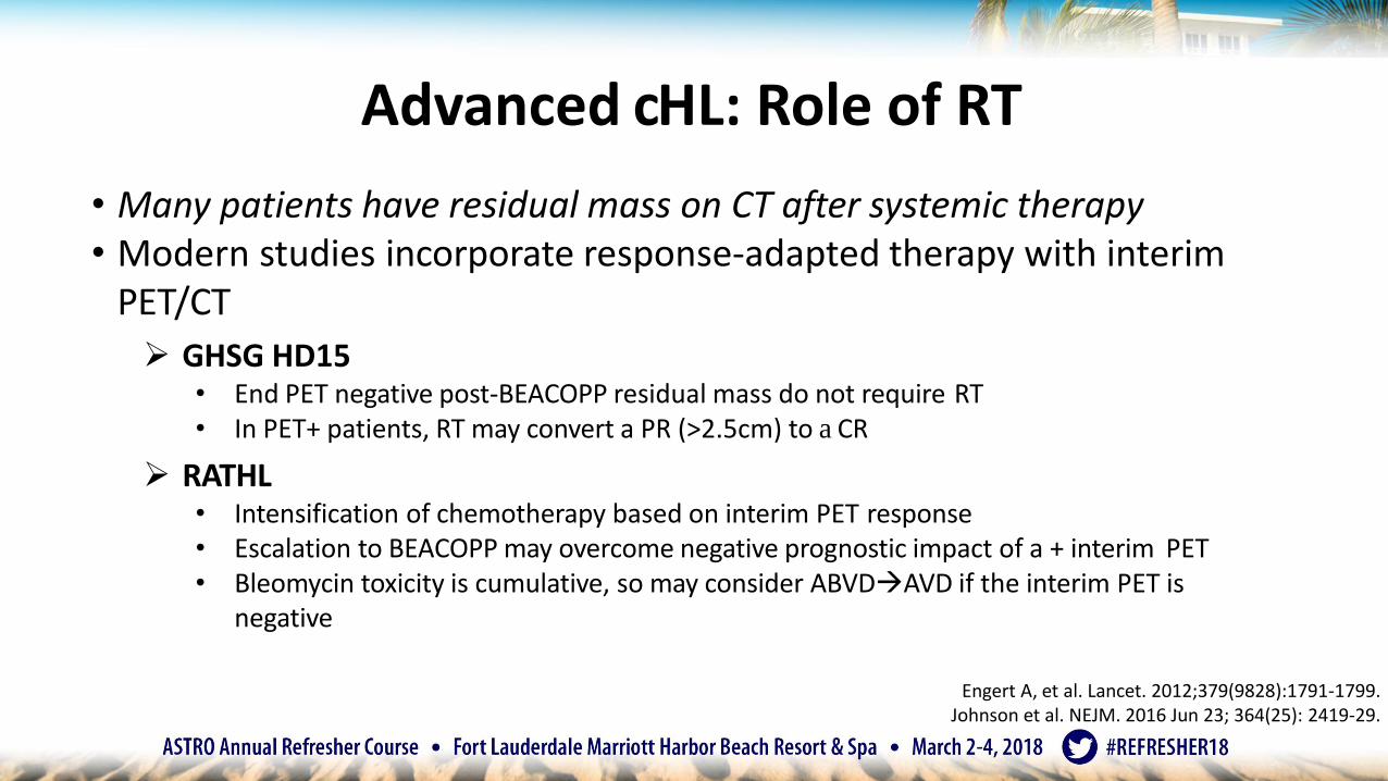
Advanced cHL: Role of RT
• Many patients have residual mass on CT after systemic therapy• Modern studies incorporate response-adapted therapy with interim
PET/CT
➢ GHSG HD15• End PET negative post-BEACOPP residual mass do not require RT• In PET+ patients, RT may convert a PR (>2.5cm) to a CR
➢ RATHL• Intensification of chemotherapy based on interim PET response• Escalation to BEACOPP may overcome negative prognostic impact of a + interim PET• Bleomycin toxicity is cumulative, so may consider ABVDAVD if the interim PET is
negative
Engert A, et al. Lancet. 2012;379(9828):1791-1799.Johnson et al. NEJM. 2016 Jun 23; 364(25): 2419-29.
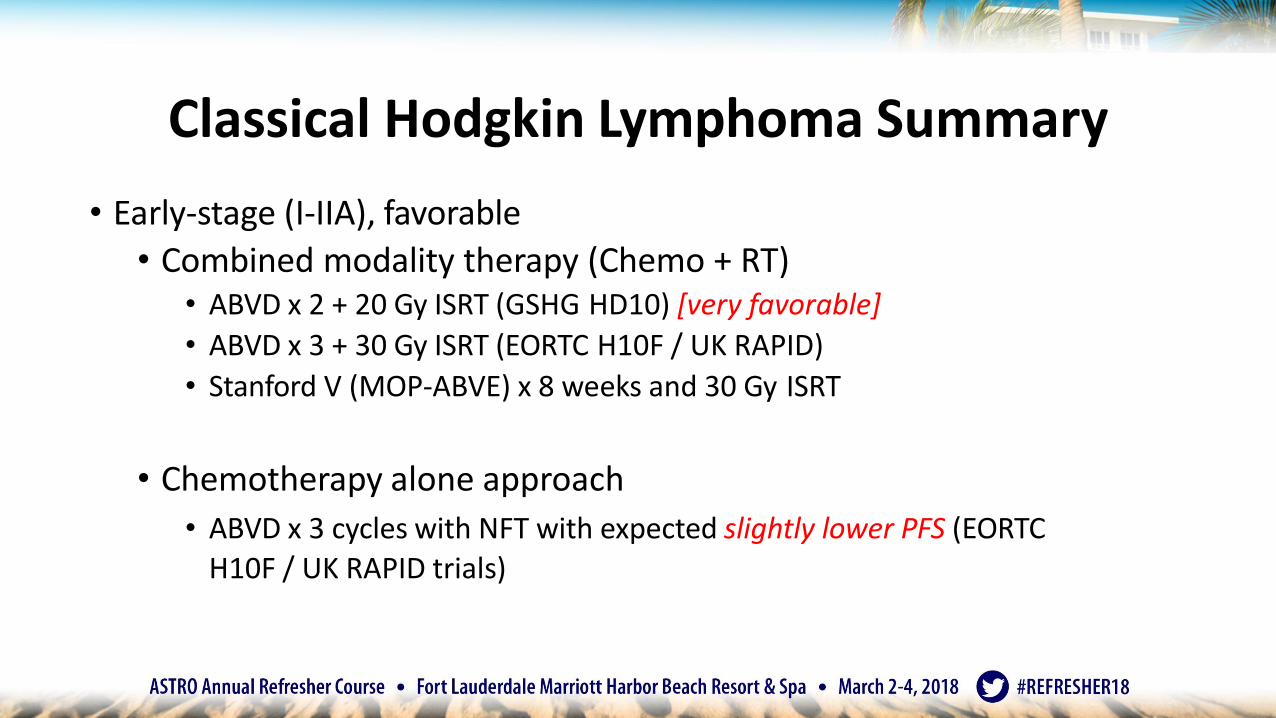
Classical Hodgkin Lymphoma Summary
• Early-stage (I-IIA), favorable
• Combined modality therapy (Chemo + RT)• ABVD x 2 + 20 Gy ISRT (GSHG HD10) [very favorable]
• ABVD x 3 + 30 Gy ISRT (EORTC H10F / UK RAPID)
• Stanford V (MOP-ABVE) x 8 weeks and 30 Gy ISRT
• Chemotherapy alone approach
• ABVD x 3 cycles with NFT with expected slightly lower PFS (EORTCH10F / UK RAPID trials)
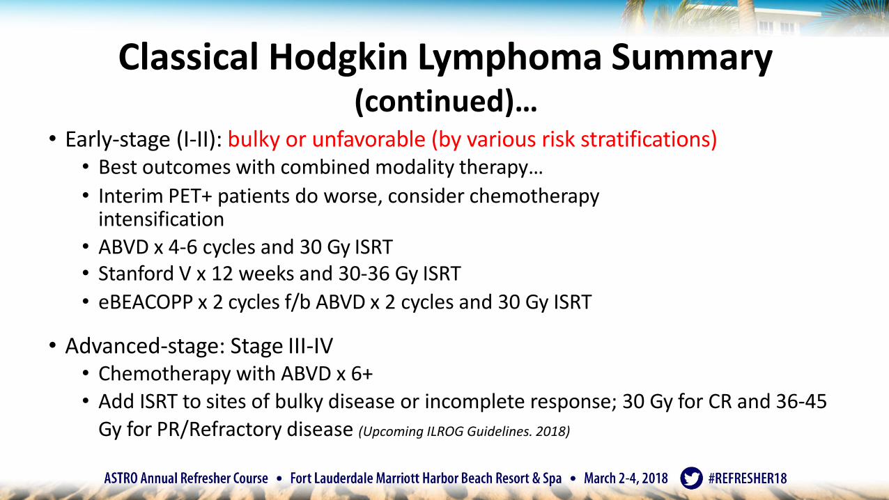
Classical Hodgkin Lymphoma Summary(continued)…
• Early-stage (I-II): bulky or unfavorable (by various risk stratifications)• Best outcomes with combined modality therapy…
• Interim PET+ patients do worse, consider chemotherapyintensification
• ABVD x 4-6 cycles and 30 Gy ISRT• Stanford V x 12 weeks and 30-36 Gy ISRT
• eBEACOPP x 2 cycles f/b ABVD x 2 cycles and 30 Gy ISRT
• Advanced-stage: Stage III-IV• Chemotherapy with ABVD x 6+
• Add ISRT to sites of bulky disease or incomplete response; 30 Gy for CR and 36-45Gy for PR/Refractory disease (Upcoming ILROG Guidelines. 2018)
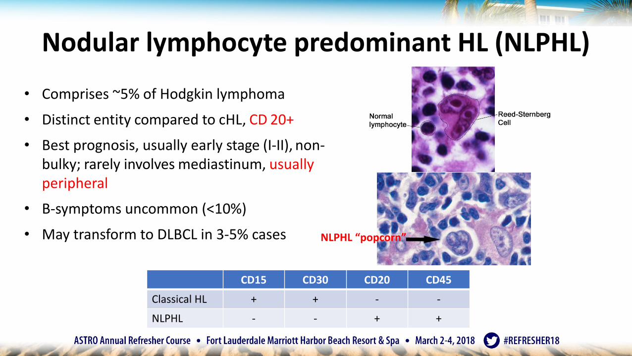
Nodular lymphocyte predominant HL (NLPHL)
• Comprises ~5% of Hodgkin lymphoma
• Distinct entity compared to cHL, CD 20+
• Best prognosis, usually early stage (I-II), non-bulky; rarely involves mediastinum, usually peripheral
• B-symptoms uncommon (<10%)
• May transform to DLBCL in 3-5% cases
CD15 CD30 CD20 CD45
Classical HL + + - -
NLPHL - - + +
NLPHL “popcorn”
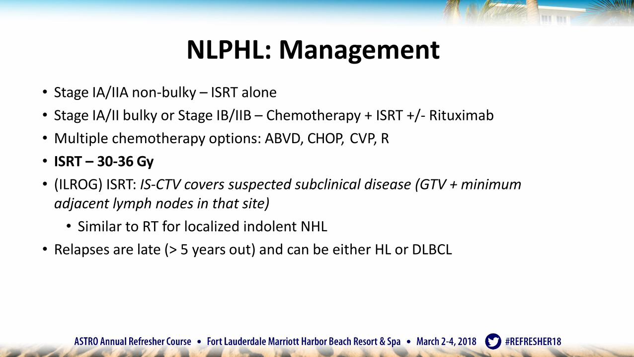
NLPHL: Management
• Stage IA/IIA non-bulky – ISRT alone
• Stage IA/II bulky or Stage IB/IIB – Chemotherapy + ISRT +/- Rituximab
• Multiple chemotherapy options: ABVD, CHOP, CVP, R
• ISRT – 30-36 Gy
• (ILROG) ISRT: IS-CTV covers suspected subclinical disease (GTV + minimum adjacent lymph nodes in that site)
• Similar to RT for localized indolent NHL
• Relapses are late (> 5 years out) and can be either HL or DLBCL
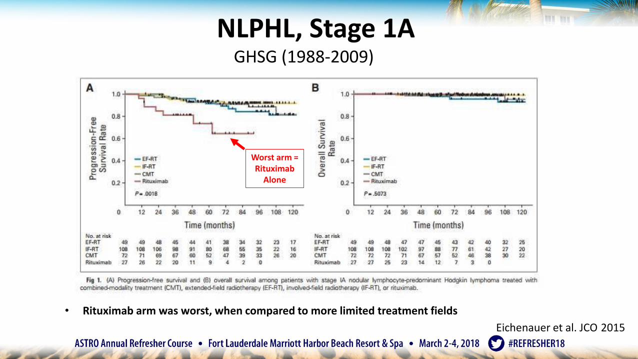
NLPHL, Stage 1AGHSG (1988-2009)
• Rituximab arm was worst, when compared to more limited treatment fields
Eichenauer et al. JCO 2015
Worst arm = Rituximab
Alone
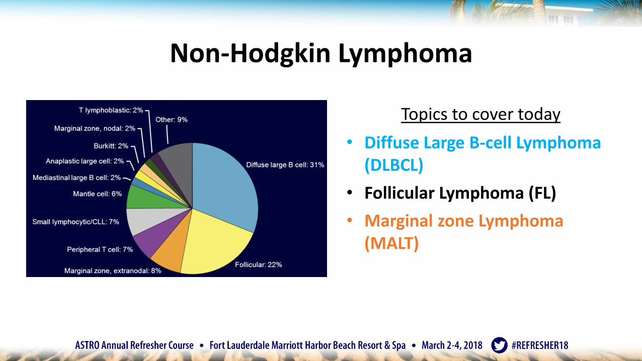
Non-Hodgkin Lymphoma
Topics to cover today
• Diffuse Large B-cell Lymphoma (DLBCL)
• Follicular Lymphoma (FL)
• Marginal zone Lymphoma (MALT)
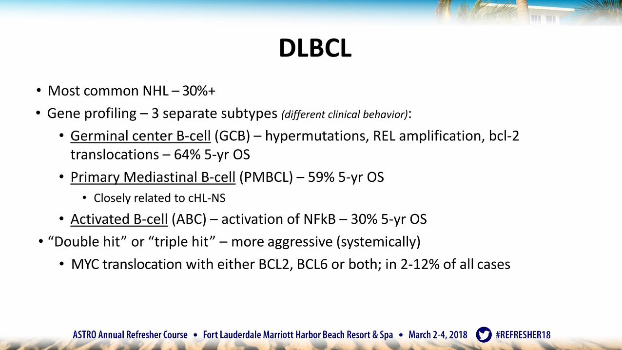
DLBCL
• Most common NHL – 30%+
• Gene profiling – 3 separate subtypes (different clinical behavior):
• Germinal center B-cell (GCB) – hypermutations, REL amplification, bcl-2 translocations – 64% 5-yr OS
• Primary Mediastinal B-cell (PMBCL) – 59% 5-yr OS
• Closely related to cHL-NS
• Activated B-cell (ABC) – activation of NFkB – 30% 5-yr OS
• “Double hit” or “triple hit” – more aggressive (systemically)
• MYC translocation with either BCL2, BCL6 or both; in 2-12% of all cases
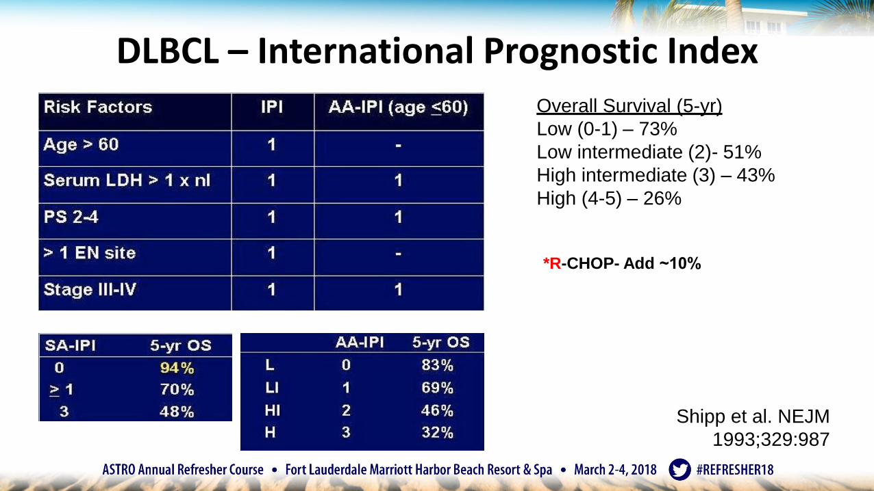
DLBCL – International Prognostic Index
*R-CHOP- Add ~10%
Shipp et al. NEJM
1993;329:987
Overall Survival (5-yr)
Low (0-1) – 73%
Low intermediate (2)- 51%
High intermediate (3) – 43%
High (4-5) – 26%
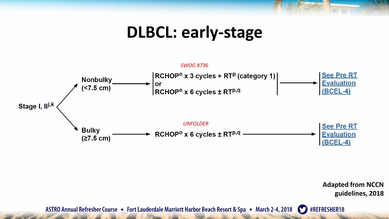
DLBCL: early-stage
SWOG 8736
UNFOLDER
Adapted from NCCN guidelines, 2018
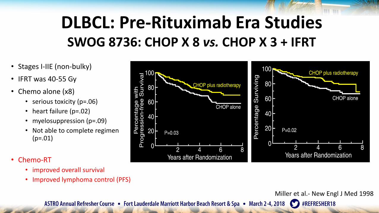
DLBCL: Pre-Rituximab Era StudiesSWOG 8736: CHOP X 8 vs. CHOP X 3 + IFRT
• Stages I-IIE (non-bulky)
• IFRT was 40-55 Gy
• Chemo alone (x8)• serious toxicity (p=.06)
• heart failure (p=.02)
• myelosuppression (p=.09)
• Not able to complete regimen (p=.01)
• Chemo-RT• improved overall survival
• Improved lymphoma control (PFS)
Miller et al.- New Engl J Med 1998
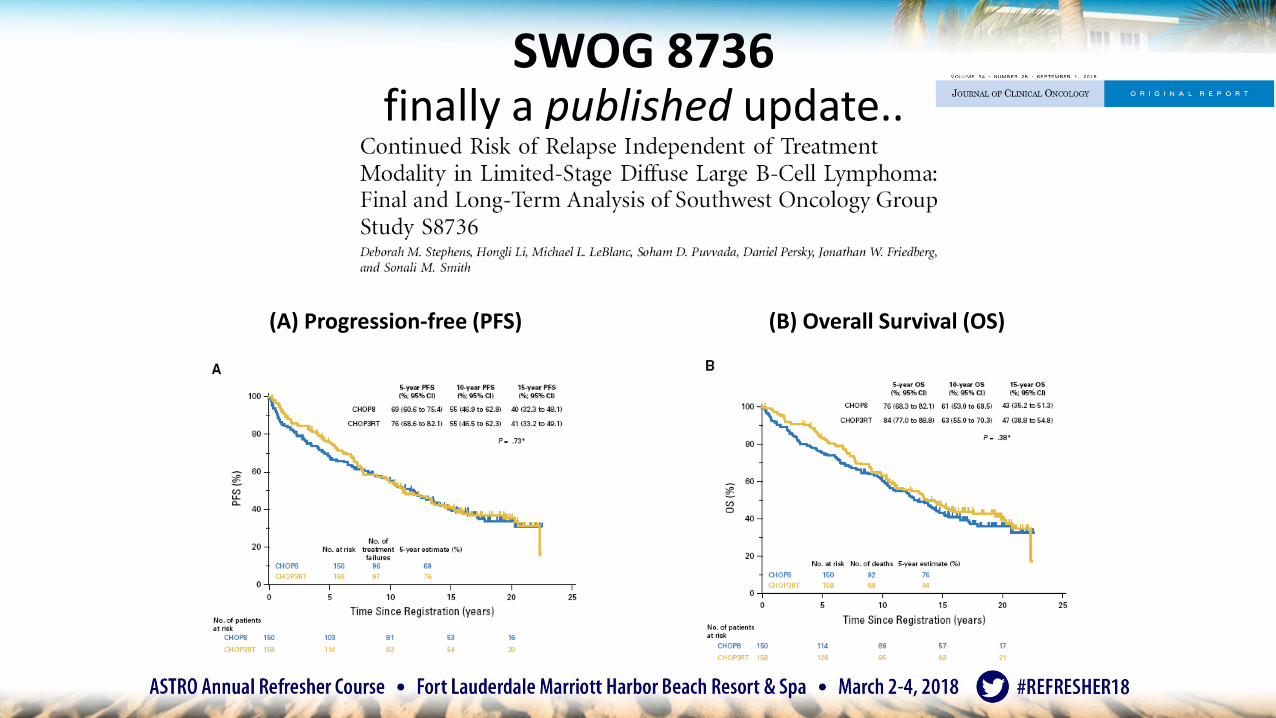
SWOG 8736finally a published update..
(A) Progression-free (PFS) (B) Overall Survival (OS)
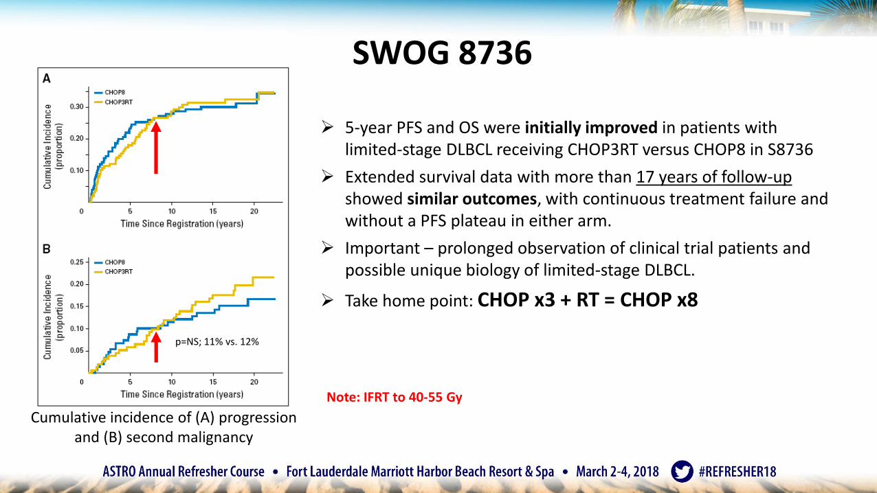
Cumulative incidence of (A) progression and (B) second malignancy
➢ 5-year PFS and OS were initially improved in patients with limited-stage DLBCL receiving CHOP3RT versus CHOP8 in S8736
➢ Extended survival data with more than 17 years of follow-up showed similar outcomes, with continuous treatment failure and without a PFS plateau in either arm.
➢ Important – prolonged observation of clinical trial patients and possible unique biology of limited-stage DLBCL.
➢ Take home point: CHOP x3 + RT = CHOP x8
SWOG 8736
Note: IFRT to 40-55 Gy
p=NS; 11% vs. 12%
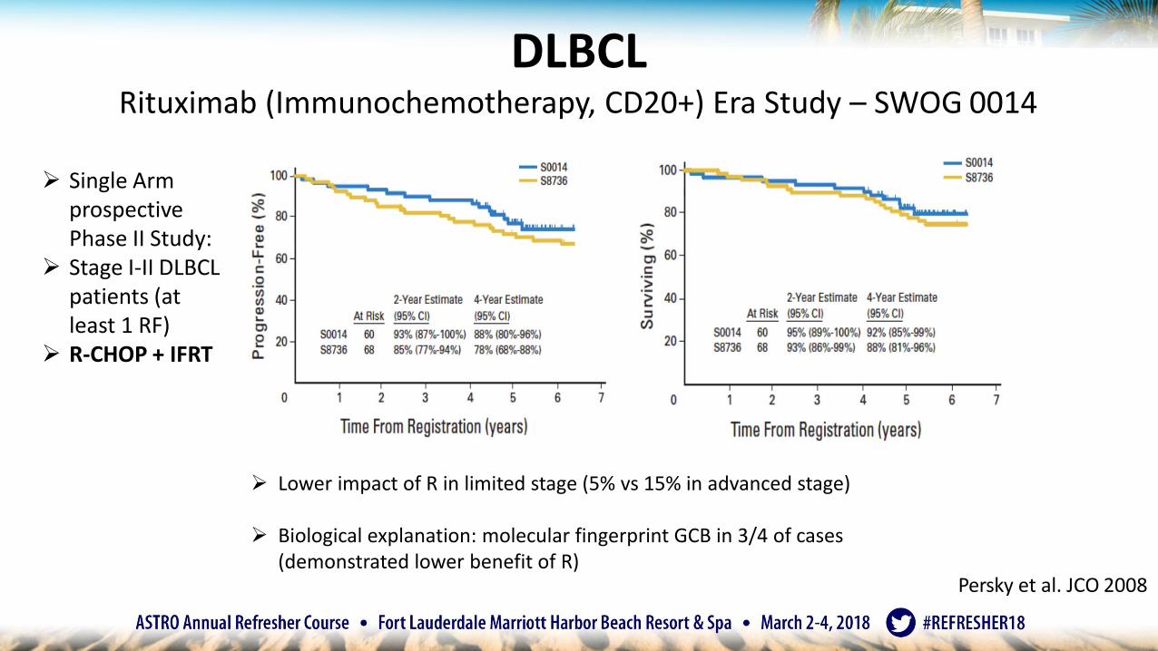
DLBCLRituximab (Immunochemotherapy, CD20+) Era Study – SWOG 0014
➢ Single Arm prospective Phase II Study:
➢ Stage I-II DLBCL patients (at least 1 RF)
➢ R-CHOP + IFRT
Persky et al. JCO 2008
➢ Lower impact of R in limited stage (5% vs 15% in advanced stage)
➢ Biological explanation: molecular fingerprint GCB in 3/4 of cases (demonstrated lower benefit of R)
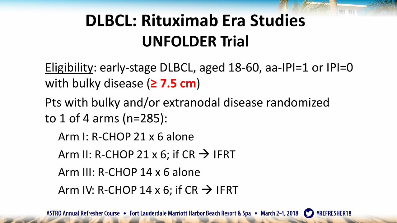
DLBCL: Rituximab Era StudiesUNFOLDER Trial
• Eligibility: early-stage DLBCL, aged 18-60, aa-IPI=1 or IPI=0with bulky disease (≥ 7.5 cm)
• Pts with bulky and/or extranodal disease randomizedto 1 of 4 arms (n=285):
– Arm I: R-CHOP 21 x 6 alone
– Arm II: R-CHOP 21 x 6; if CR IFRT
– Arm III: R-CHOP 14 x 6 alone
– Arm IV: R-CHOP 14 x 6; if CR IFRT
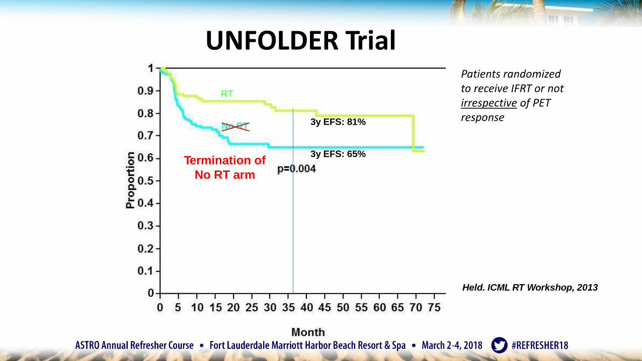
RT
No RT 3y EFS: 81%
3y EFS: 65%
UNFOLDER Trial
Held. ICML RT Workshop, 2013
Termination of
No RT arm
Patients randomizedto receive IFRT or not irrespective of PET response
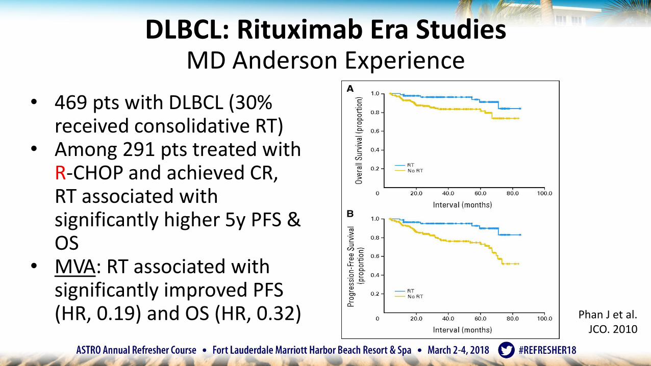
• 469 pts with DLBCL (30%received consolidative RT)
• Among 291 pts treated withR-CHOP and achieved CR,RT associated with significantly higher 5y PFS & OS
• MVA: RT associated with significantly improved PFS(HR, 0.19) and OS (HR, 0.32)
DLBCL: Rituximab Era StudiesMD Anderson Experience
Phan J et al. JCO. 2010
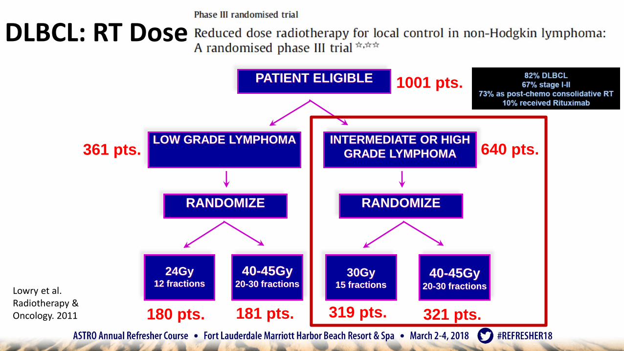
DLBCL: RT Dose
PATIENT ELIGIBLE
RANDOMIZE
LOW GRADE LYMPHOMA INTERMEDIATE OR HIGH
GRADE LYMPHOMA
24Gy12 fractions
40-45Gy20-30 fractions
40-45Gy20-30 fractions
30Gy15 fractions
RANDOMIZE
1001 pts.
180 pts. 181 pts. 321 pts.319 pts.
640 pts.361 pts.
Lowry et al. Radiotherapy & Oncology. 2011
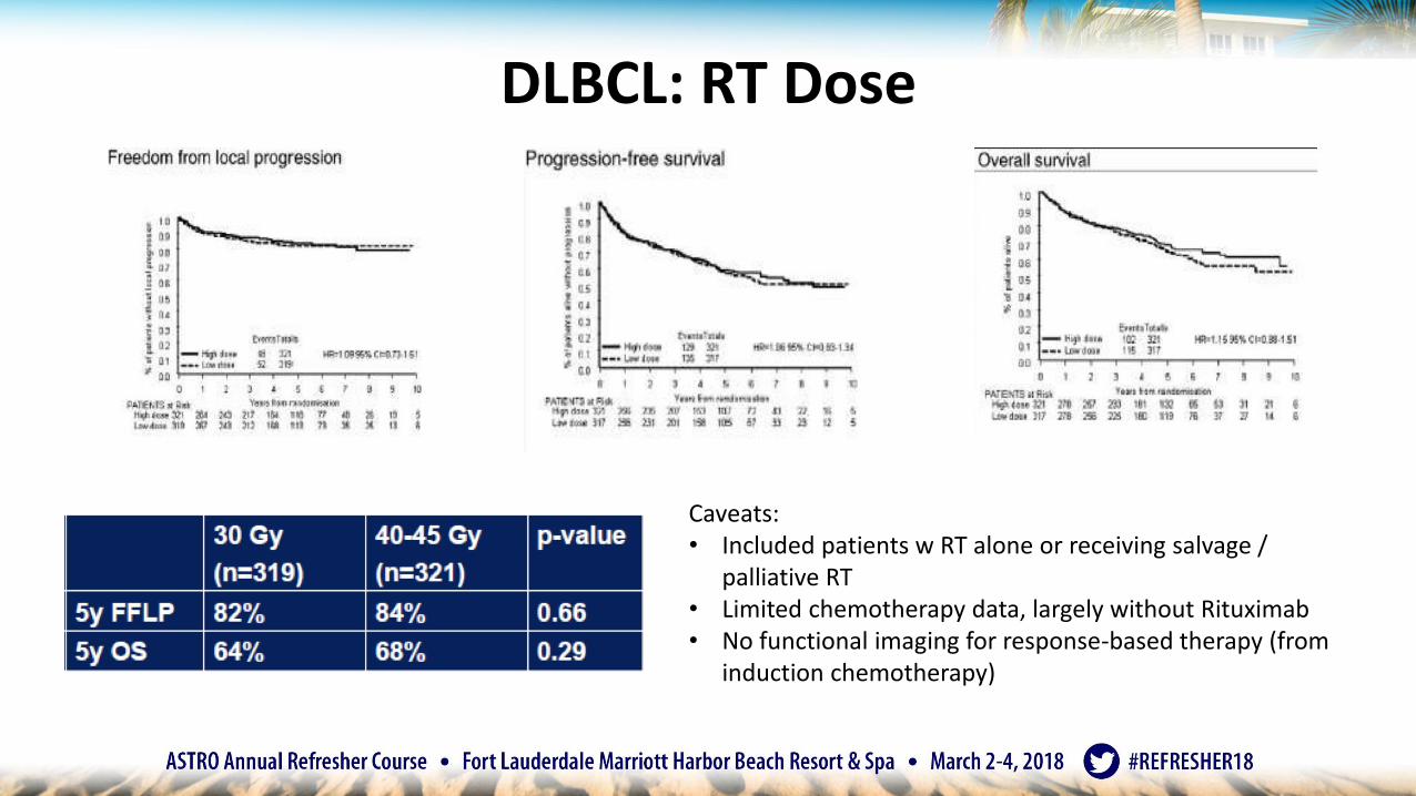
DLBCL: RT Dose
Caveats:• Included patients w RT alone or receiving salvage /
palliative RT• Limited chemotherapy data, largely without Rituximab• No functional imaging for response-based therapy (from
induction chemotherapy)
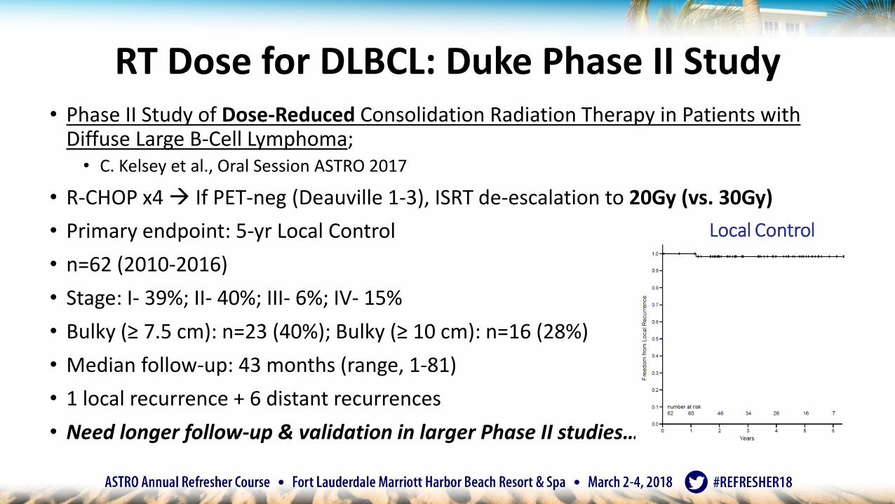
• Phase II Study of Dose-Reduced Consolidation Radiation Therapy in Patients with Diffuse Large B-Cell Lymphoma; • C. Kelsey et al., Oral Session ASTRO 2017
• R-CHOP x4 If PET-neg (Deauville 1-3), ISRT de-escalation to 20Gy (vs. 30Gy)
• Primary endpoint: 5-yr Local Control
• n=62 (2010-2016)
• Stage: I- 39%; II- 40%; III- 6%; IV- 15%
• Bulky (≥ 7.5 cm): n=23 (40%); Bulky (≥ 10 cm): n=16 (28%)
• Median follow-up: 43 months (range, 1-81)
• 1 local recurrence + 6 distant recurrences
• Need longer follow-up & validation in larger Phase II studies…
RT Dose for DLBCL: Duke Phase II Study
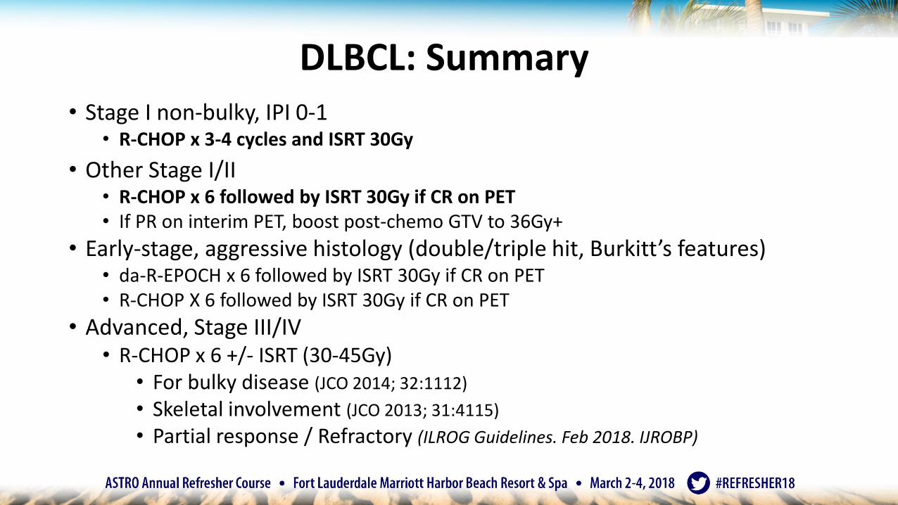
• Stage I non-bulky, IPI 0-1• R-CHOP x 3-4 cycles and ISRT 30Gy
• Other Stage I/II• R-CHOP x 6 followed by ISRT 30Gy if CR on PET• If PR on interim PET, boost post-chemo GTV to 36Gy+
• Early-stage, aggressive histology (double/triple hit, Burkitt’s features)• da-R-EPOCH x 6 followed by ISRT 30Gy if CR on PET• R-CHOP X 6 followed by ISRT 30Gy if CR on PET
• Advanced, Stage III/IV• R-CHOP x 6 +/- ISRT (30-45Gy)
• For bulky disease (JCO 2014; 32:1112)
• Skeletal involvement (JCO 2013; 31:4115)
• Partial response / Refractory (ILROG Guidelines. Feb 2018. IJROBP)
DLBCL: Summary
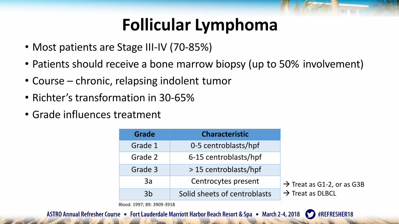
• Most patients are Stage III-IV (70-85%)
• Patients should receive a bone marrow biopsy (up to 50% involvement)
• Course – chronic, relapsing indolent tumor
• Richter’s transformation in 30-65%
• Grade influences treatment
Follicular Lymphoma
Treat as G1-2, or as G3B Treat as DLBCL
Grade Characteristic
Grade 1 0-5 centroblasts/hpf
Grade 2 6-15 centroblasts/hpf
Grade 3 > 15 centroblasts/hpf
3a Centrocytes present
3b Solid sheets of centroblastsBlood. 1997; 89: 3909-3918
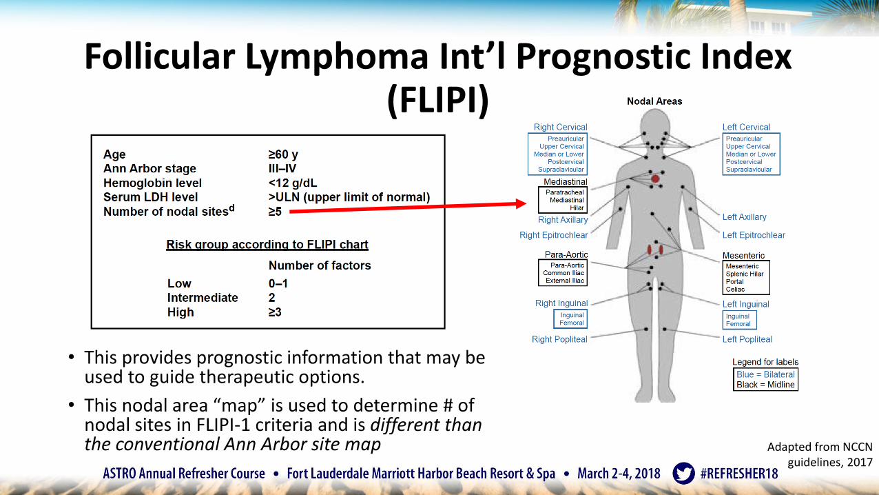
Follicular Lymphoma Int’l Prognostic Index (FLIPI)
• This provides prognostic information that may be used to guide therapeutic options.
• This nodal area “map” is used to determine # of nodal sites in FLIPI-1 criteria and is different than the conventional Ann Arbor site map Adapted from NCCN
guidelines, 2017
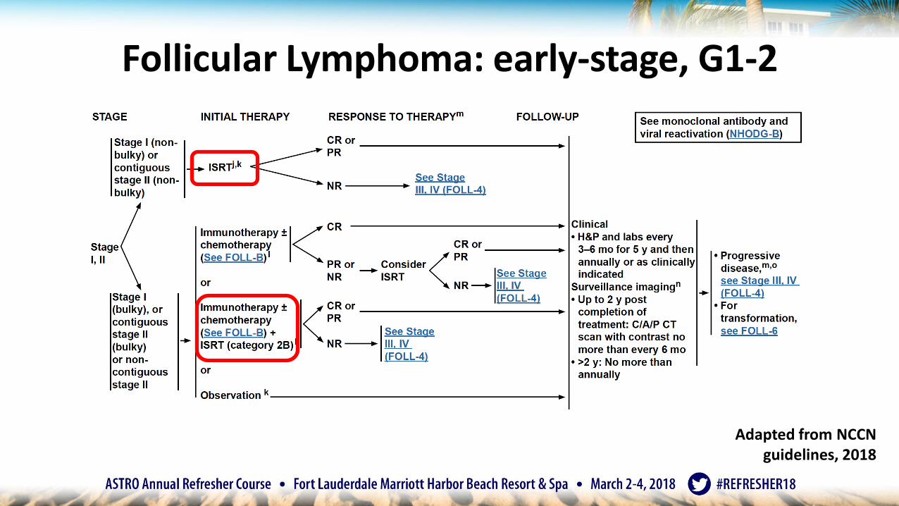
Follicular Lymphoma: early-stage, G1-2
Adapted from NCCN guidelines, 2018
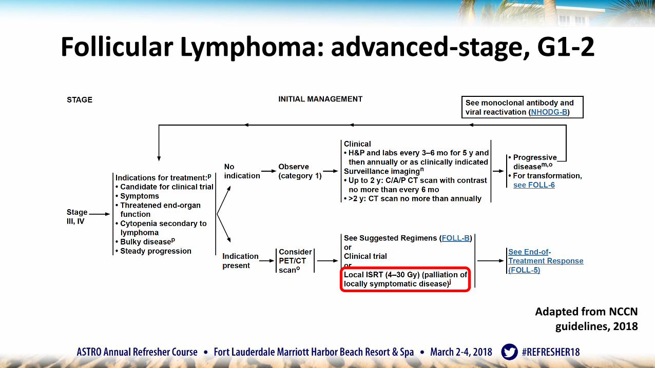
Follicular Lymphoma: advanced-stage, G1-2
Adapted from NCCN guidelines, 2018
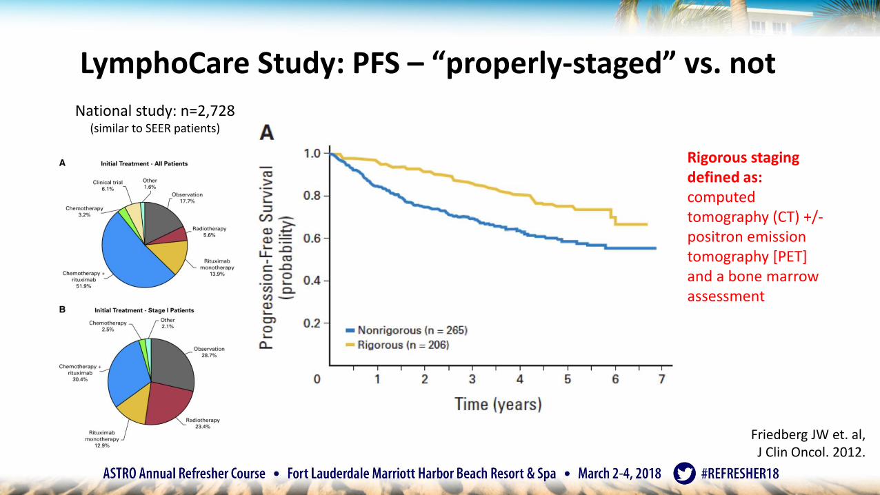
LymphoCare Study: PFS – “properly-staged” vs. not
Rigorous staging defined as:computed tomography (CT) +/-positron emission tomography [PET] and a bone marrow assessment
National study: n=2,728 (similar to SEER patients)
Friedberg JW et. al, J Clin Oncol. 2012.
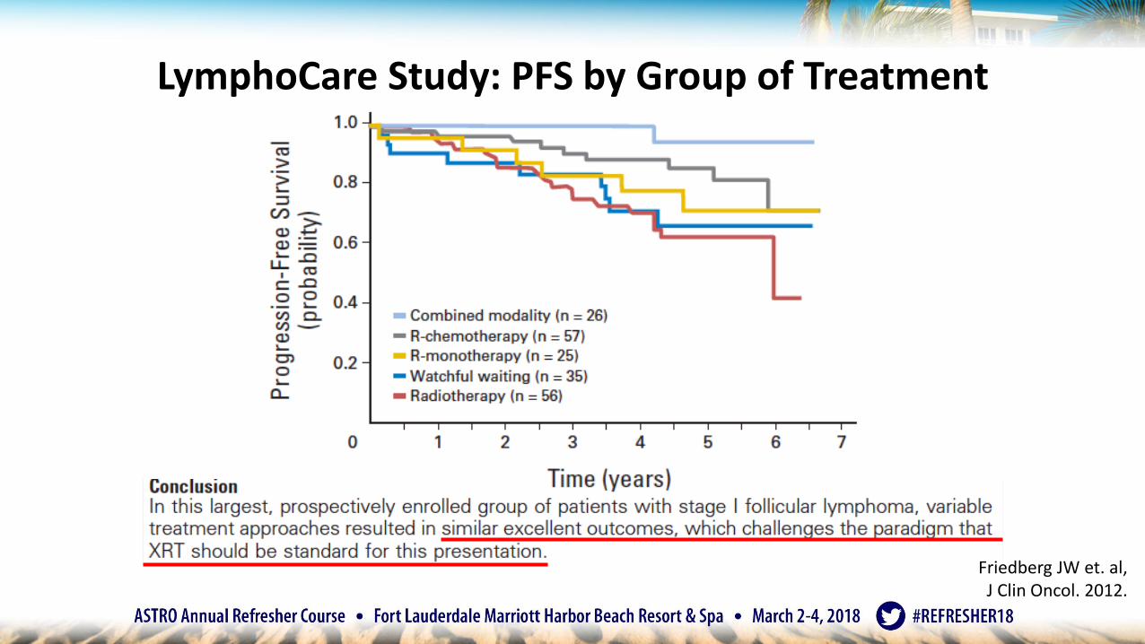
LymphoCare Study: PFS by Group of Treatment
Friedberg JW et. al, J Clin Oncol. 2012.
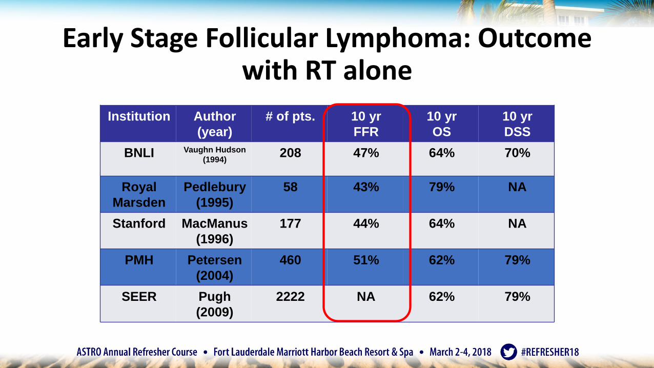
Early Stage Follicular Lymphoma: Outcome with RT alone
Institution Author
(year)
# of pts. 10 yr
FFR
10 yr
OS
10 yr
DSS
BNLI Vaughn Hudson
(1994)208 47% 64% 70%
Royal
Marsden
Pedlebury
(1995)
58 43% 79% NA
Stanford MacManus
(1996)
177 44% 64% NA
PMH Petersen
(2004)
460 51% 62% 79%
SEER Pugh
(2009)
2222 NA 62% 79%
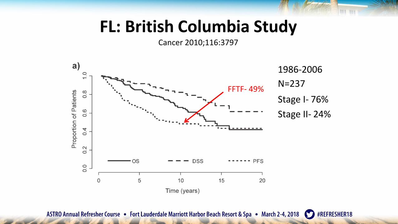
FL: British Columbia StudyCancer 2010;116:3797
1986-2006
FFTF- 49%N=237
Stage I- 76%
Stage II- 24%
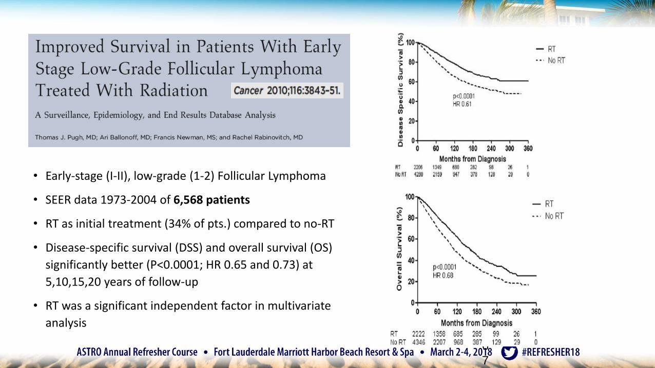
• Early-stage (I-II), low-grade (1-2) Follicular Lymphoma
• SEER data 1973-2004 of 6,568 patients
• RT as initial treatment (34% of pts.) compared to no-RT
• Disease-specific survival (DSS) and overall survival (OS)
significantly better (P<0.0001; HR 0.65 and 0.73) at
5,10,15,20 years of follow-up
• RT was a significant independent factor in multivariate
analysis
6
7
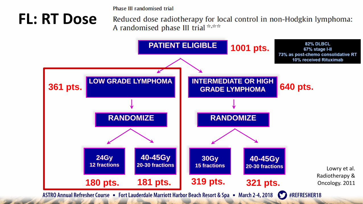
FL: RT Dose
PATIENT ELIGIBLE
RANDOMIZE
LOW GRADE LYMPHOMA INTERMEDIATE OR HIGH
GRADE LYMPHOMA
24Gy12 fractions
40-45Gy20-30 fractions
40-45Gy20-30 fractions
30Gy15 fractions
RANDOMIZE
1001 pts.
180 pts. 181 pts. 321 pts.319 pts.
640 pts.361 pts.
Lowry et al. Radiotherapy & Oncology. 2011
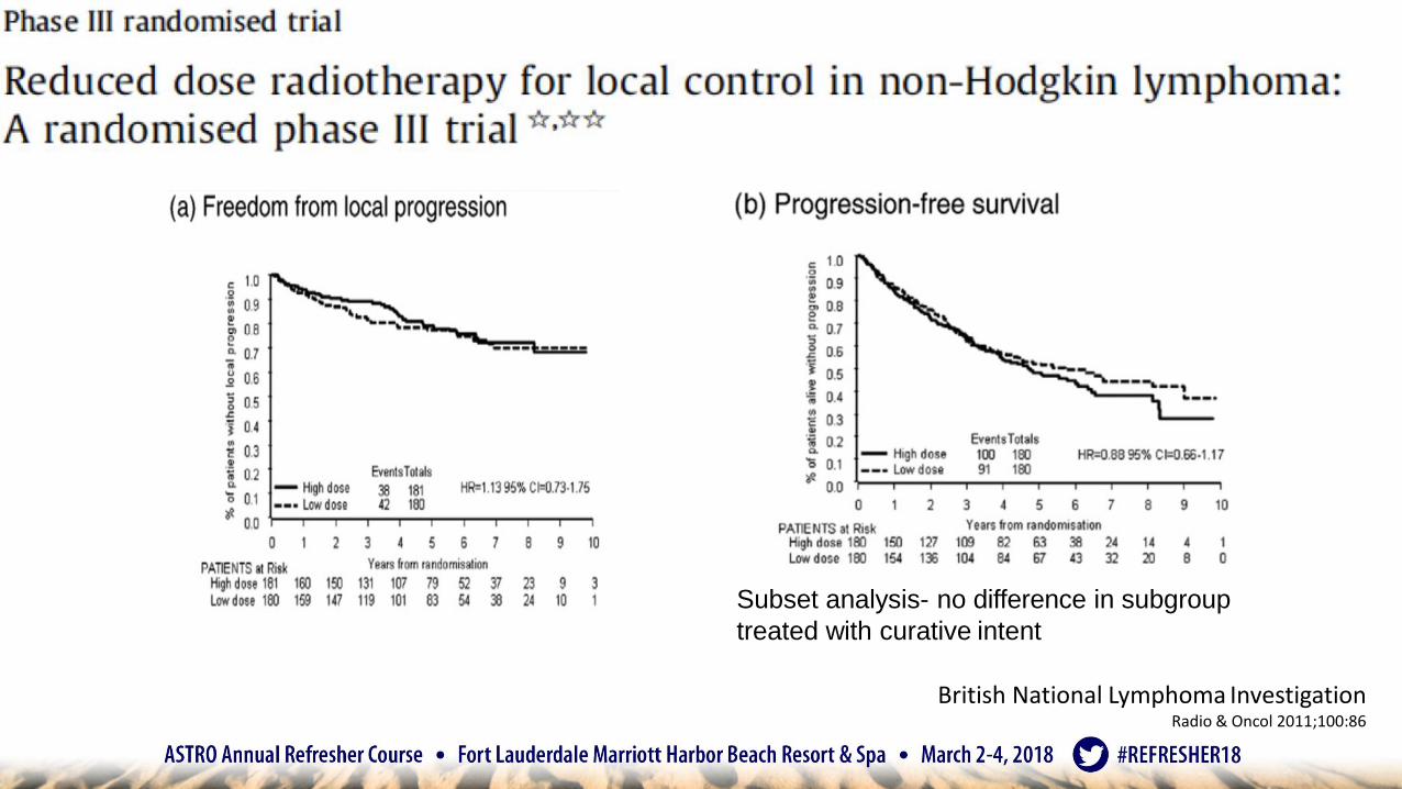
Subset analysis- no difference in subgroup
treated with curative intent
British National Lymphoma InvestigationRadio & Oncol 2011;100:86
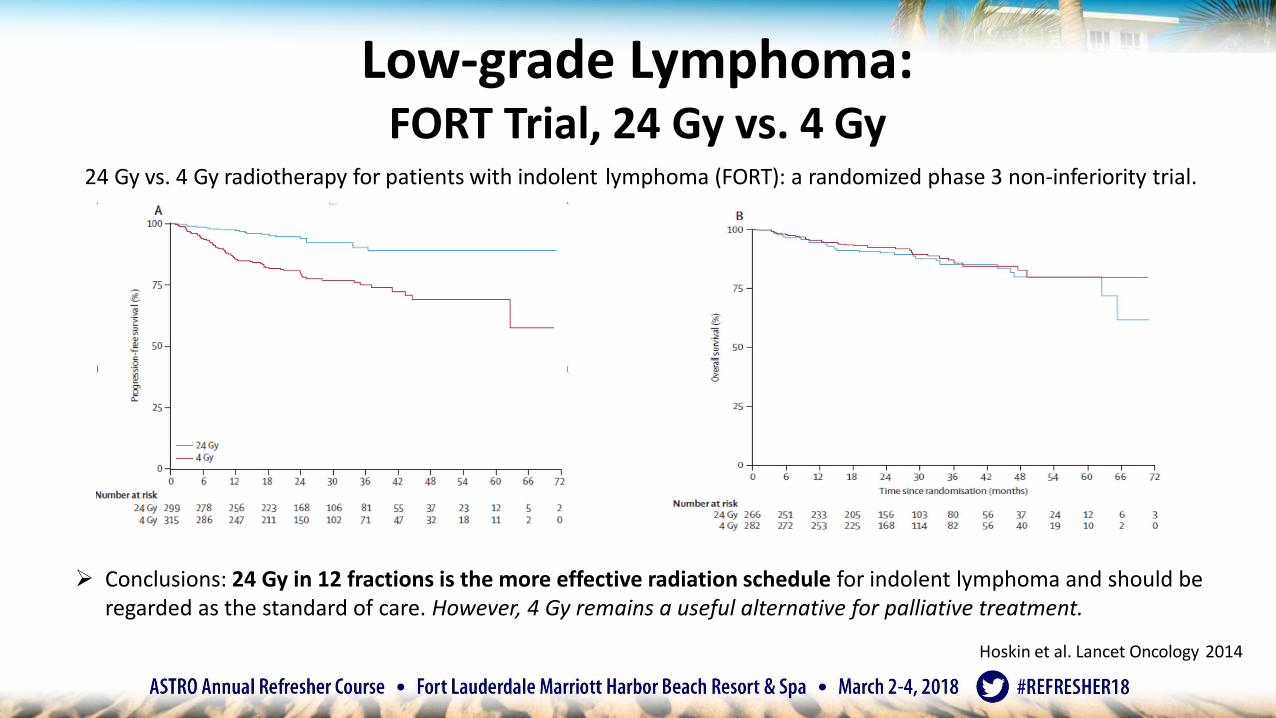
➢ Conclusions: 24 Gy in 12 fractions is the more effective radiation schedule for indolent lymphoma and should be regarded as the standard of care. However, 4 Gy remains a useful alternative for palliative treatment.
Low-grade Lymphoma: FORT Trial, 24 Gy vs. 4 Gy
Hoskin et al. Lancet Oncology 2014
24 Gy vs. 4 Gy radiotherapy for patients with indolent lymphoma (FORT): a randomized phase 3 non-inferiority trial.

Indolent Lymphomas, Stage III-IV:
• Netherlands, n=109; mainly FL (90%), some MALT
• 52% with disease ≥ 5 cm (bulky)
• Prior regimens (median 2, range 0-11)
• IFRT: 4 Gy / 1-2 fx
• ORR 92% (CR 61%, PR 31%, SD 6%) more impressive than most systemic agents (chemo/targeted)!!
• 25 months median time to local progression (42 months in patients with CR)
• Well tolerated and effective palliation using low-dose RTHass et al. J Clin Oncology. 2003 Jul 1;21(13):2474-80.
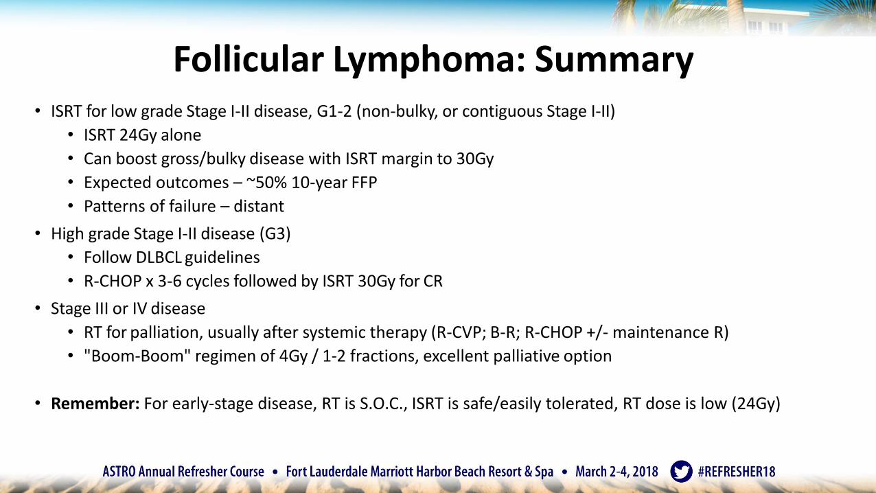
• ISRT for low grade Stage I-II disease, G1-2 (non-bulky, or contiguous Stage I-II)
• ISRT 24Gy alone
• Can boost gross/bulky disease with ISRT margin to 30Gy
• Expected outcomes – ~50% 10-year FFP
• Patterns of failure – distant
• High grade Stage I-II disease (G3)
• Follow DLBCL guidelines
• R-CHOP x 3-6 cycles followed by ISRT 30Gy for CR
• Stage III or IV disease
• RT for palliation, usually after systemic therapy (R-CVP; B-R; R-CHOP +/- maintenance R)
• "Boom-Boom" regimen of 4Gy / 1-2 fractions, excellent palliative option
• Remember: For early-stage disease, RT is S.O.C., ISRT is safe/easily tolerated, RT dose is low (24Gy)
Follicular Lymphoma: Summary
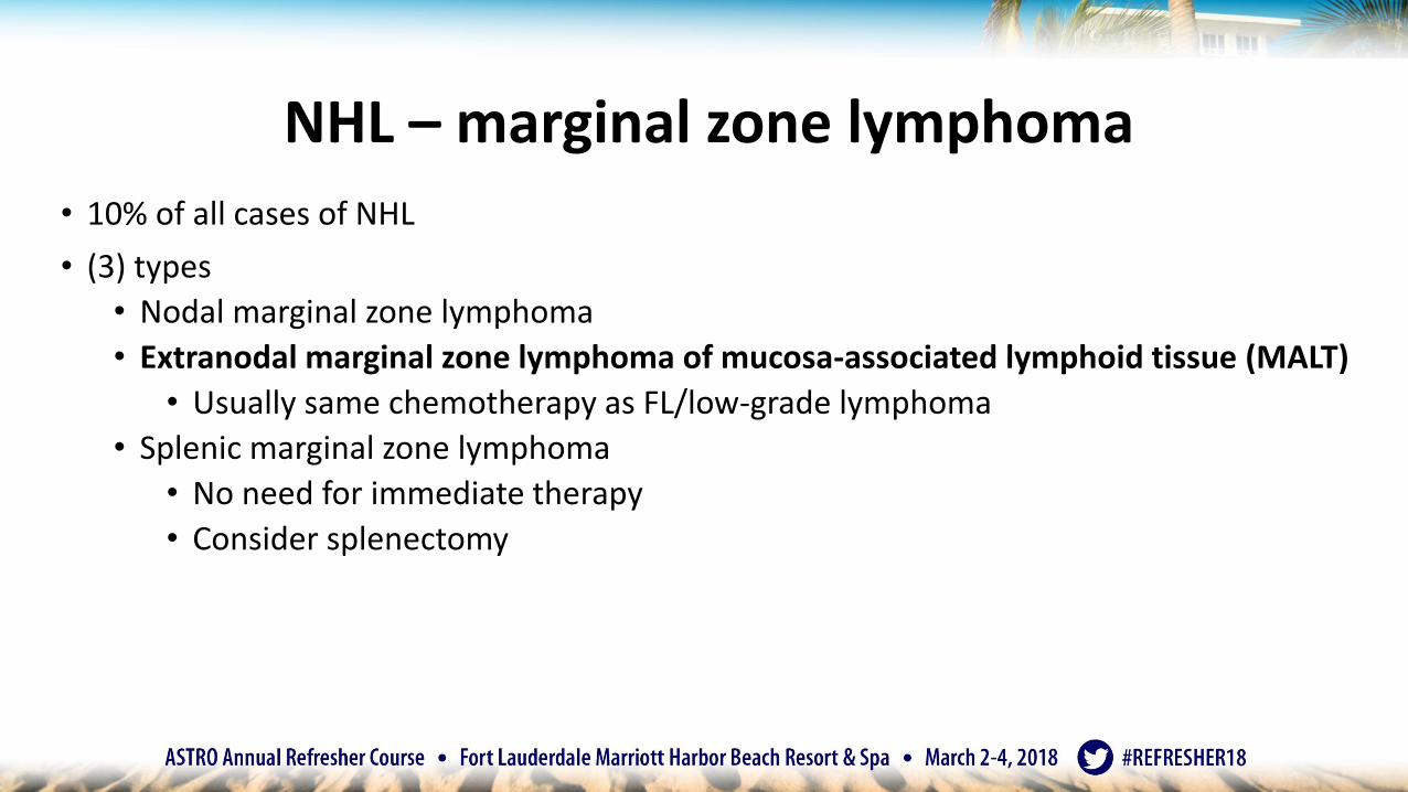
NHL – marginal zone lymphoma
• 10% of all cases of NHL
• (3) types
• Nodal marginal zone lymphoma
• Extranodal marginal zone lymphoma of mucosa-associated lymphoid tissue (MALT)
• Usually same chemotherapy as FL/low-grade lymphoma
• Splenic marginal zone lymphoma
• No need for immediate therapy
• Consider splenectomy
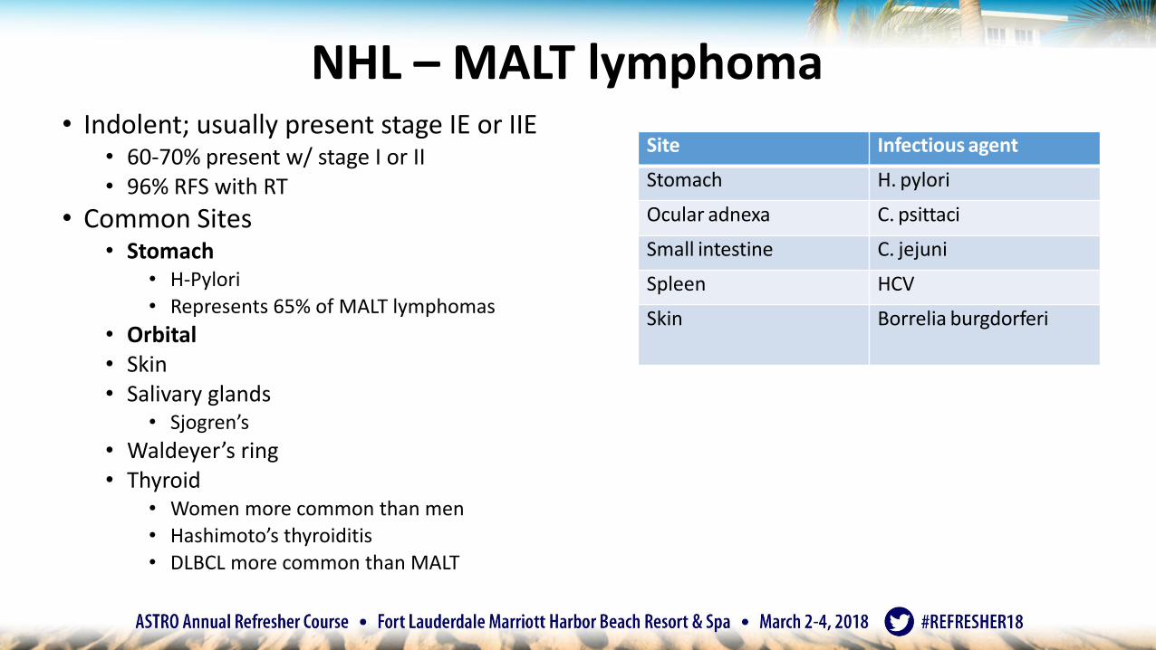
NHL – MALT lymphoma• Indolent; usually present stage IE or IIE
• 60-70% present w/ stage I or II• 96% RFS with RT
• Common Sites• Stomach
• H-Pylori • Represents 65% of MALT lymphomas
• Orbital• Skin • Salivary glands
• Sjogren’s
• Waldeyer’s ring • Thyroid
• Women more common than men • Hashimoto’s thyroiditis • DLBCL more common than MALT
Site Infectious agent
Stomach H. pylori
Ocular adnexa C. psittaci
Small intestine C. jejuni
Spleen HCV
Skin Borrelia burgdorferi

Gastric Lymphoma (MALT)• Presentation
• epigastric pain ~ 72%• weight loss ~ 43%• nausea and vomiting ~ 30%• acute presentations
• bleeding ~ 20%• perforation ~ 1%
• Diagnosis:• Previously – via gastrectomy• Currently – via endoscopy > 90%• Adequate biopsies essential: Often multi-centric in stomach
• transformation seen in deeper layers• multifocal involvement
• Limited to stomach, can involve duodenum and peri-gastric LN
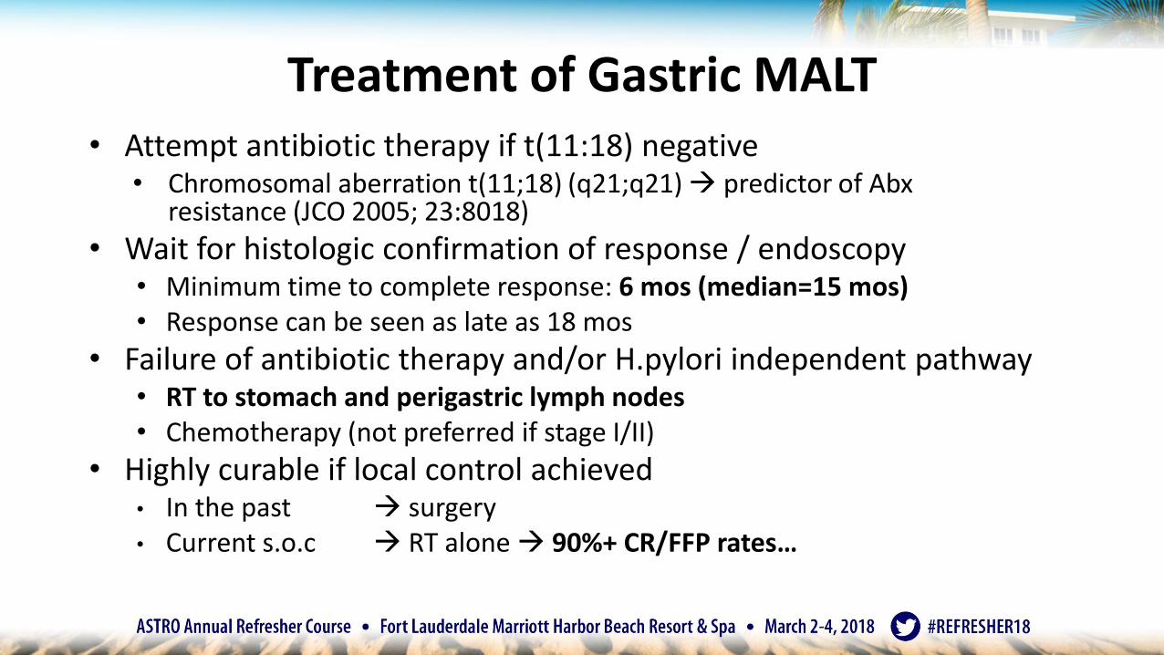
Treatment of Gastric MALT• Attempt antibiotic therapy if t(11:18) negative
• Chromosomal aberration t(11;18) (q21;q21) predictor of Abxresistance (JCO 2005; 23:8018)
• Wait for histologic confirmation of response / endoscopy• Minimum time to complete response: 6 mos (median=15 mos)• Response can be seen as late as 18 mos
• Failure of antibiotic therapy and/or H.pylori independent pathway• RT to stomach and perigastric lymph nodes• Chemotherapy (not preferred if stage I/II)
• Highly curable if local control achieved• In the past surgery• Current s.o.c RT alone 90%+ CR/FFP rates…
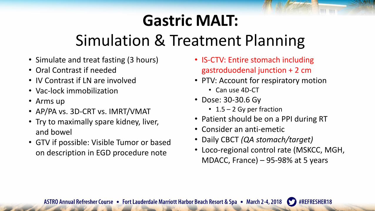
Gastric MALT:Simulation & Treatment Planning
• Simulate and treat fasting (3 hours)• Oral Contrast if needed• IV Contrast if LN are involved• Vac-lock immobilization• Arms up• AP/PA vs. 3D-CRT vs. IMRT/VMAT• Try to maximally spare kidney, liver,
and bowel• GTV if possible: Visible Tumor or based
on description in EGD procedure note
• IS-CTV: Entire stomach including gastroduodenal junction + 2 cm
• PTV: Account for respiratory motion• Can use 4D-CT
• Dose: 30-30.6 Gy• 1.5 – 2 Gy per fraction
• Patient should be on a PPI during RT• Consider an anti-emetic• Daily CBCT (QA stomach/target)• Loco-regional control rate (MSKCC, MGH,
MDACC, France) – 95-98% at 5 years
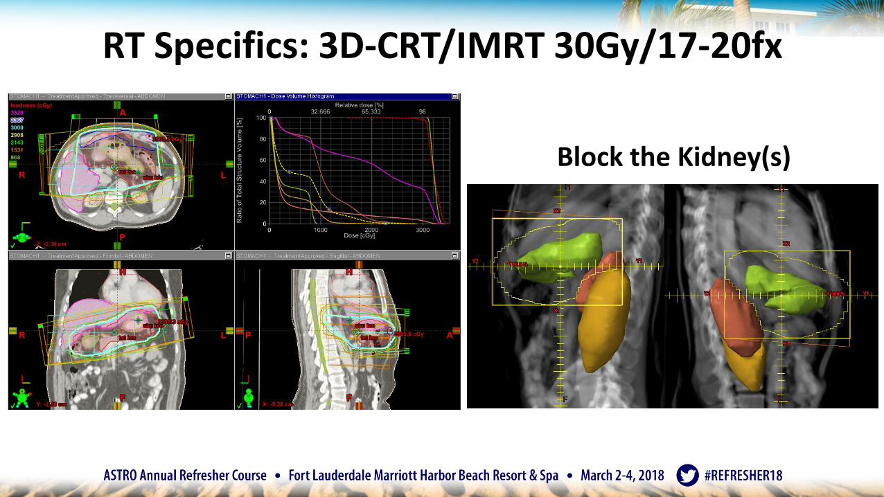
RT Specifics: 3D-CRT/IMRT 30Gy/17-20fx
Block the Kidney(s)
MALT of Non-Gastric sites:
• Ocular Adnexa, Salivary glands, Lung, Skin
• Definitive treatment for early stage disease is RT alone
• Lower doses than gastric MALT
• 24 Gy in 12 fractions
• Can avoid toxicity
• In Stage III/IV patients, can consider 4Gy in 1-2 fractions for local control
• Antibiotic response rates of 35-50% documented
• Takes 6-24 months to see response
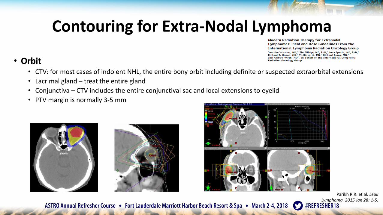
Contouring for Extra-Nodal Lymphoma
• Orbit• CTV: for most cases of indolent NHL, the entire bony orbit including definite or suspected extraorbital extensions
• Lacrimal gland – treat the entire gland
• Conjunctiva – CTV includes the entire conjunctival sac and local extensions to eyelid
• PTV margin is normally 3-5 mm
Parikh R.R. et al. LeukLymphoma. 2015 Jan 28: 1-5.
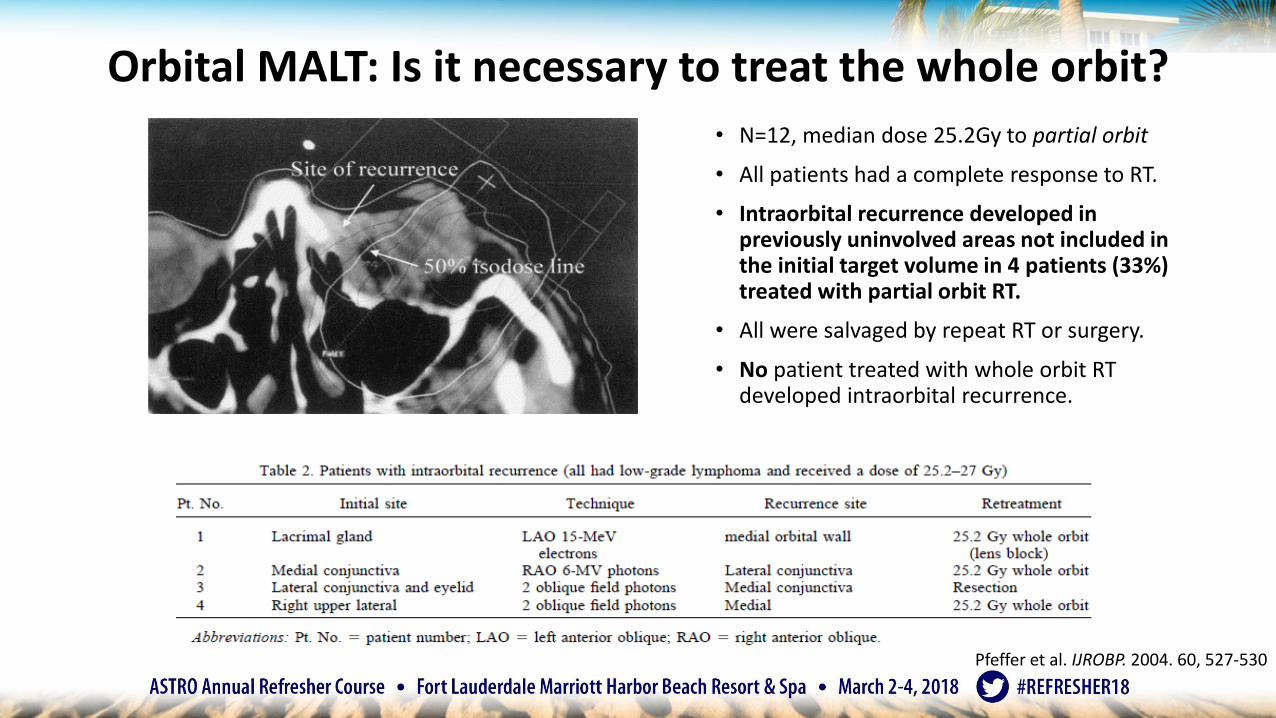
Orbital MALT: Is it necessary to treat the whole orbit?
• N=12, median dose 25.2Gy to partial orbit
• All patients had a complete response to RT.
• Intraorbital recurrence developed in previously uninvolved areas not included in the initial target volume in 4 patients (33%) treated with partial orbit RT.
• All were salvaged by repeat RT or surgery.
• No patient treated with whole orbit RT developed intraorbital recurrence.
Pfeffer et al. IJROBP. 2004. 60, 527-530
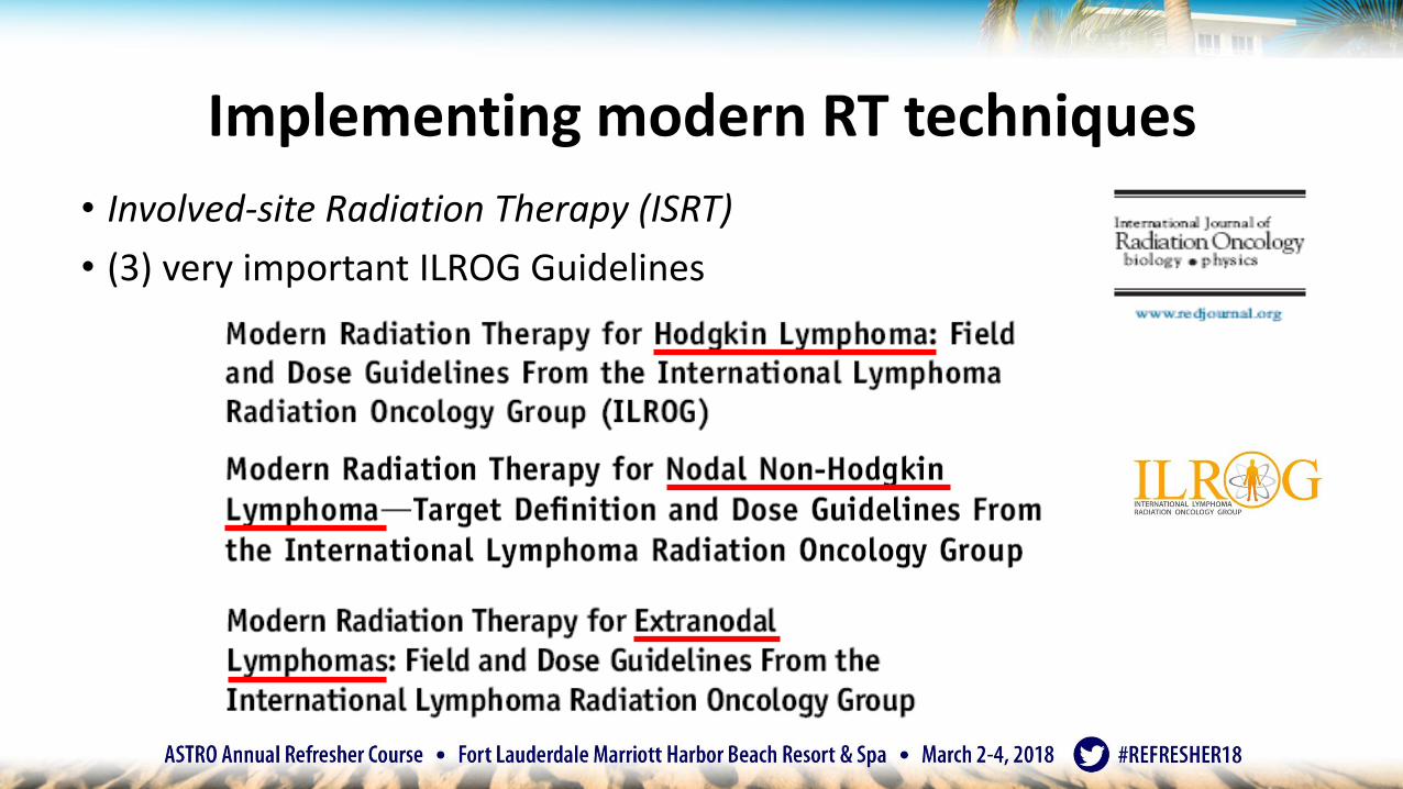
Implementing modern RT techniques
• Involved-site Radiation Therapy (ISRT)
• (3) very important ILROG Guidelines
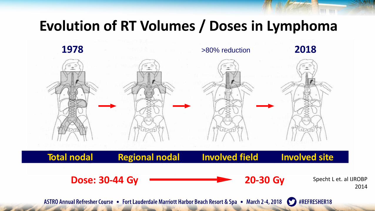
Dose: 30-44 Gy 20-30 Gy
1978 2018
Total nodal Regional nodal Involved field Involved site
Evolution of RT Volumes / Doses in Lymphoma
>80% reduction
Specht L et. al IJROBP 2014
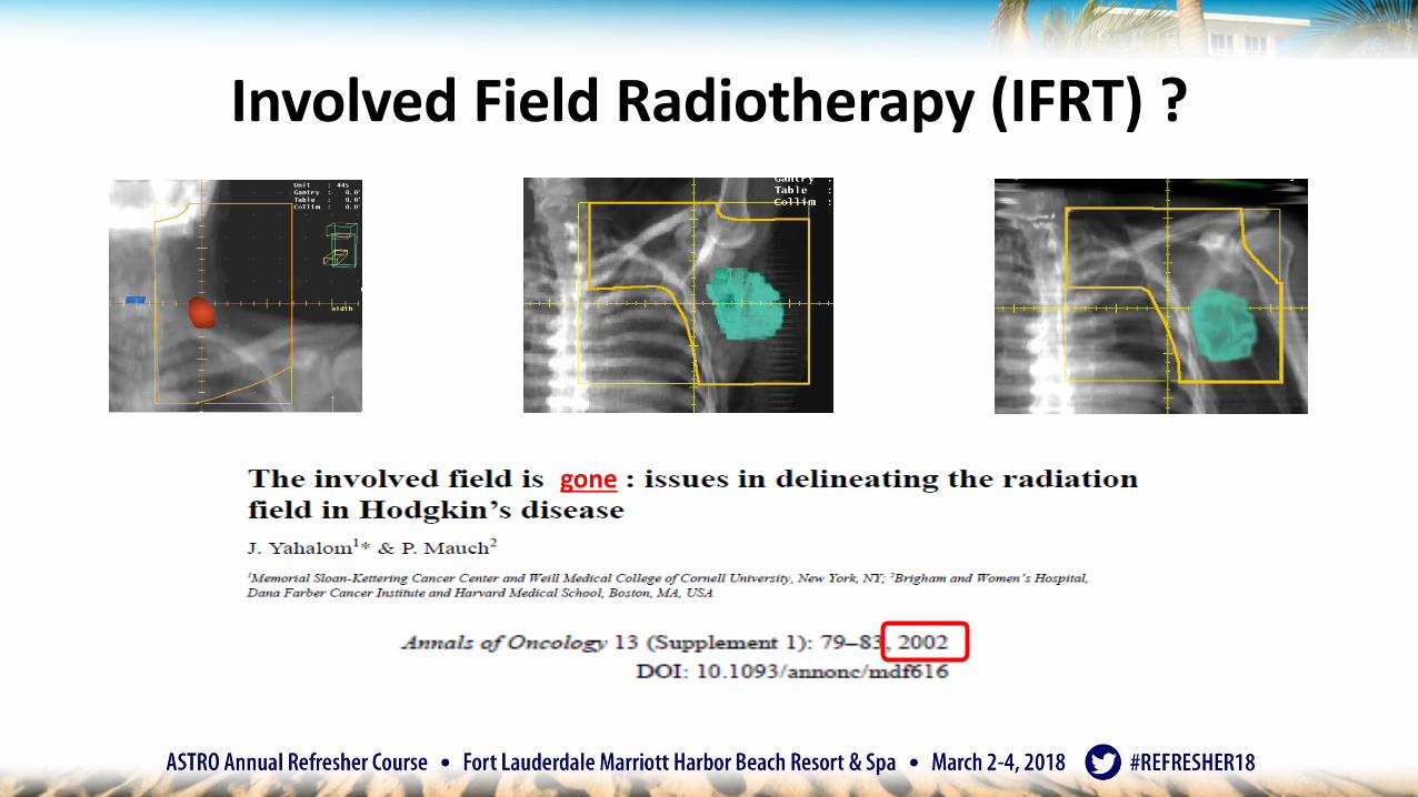
Involved Field Radiotherapy (IFRT) ?
gone
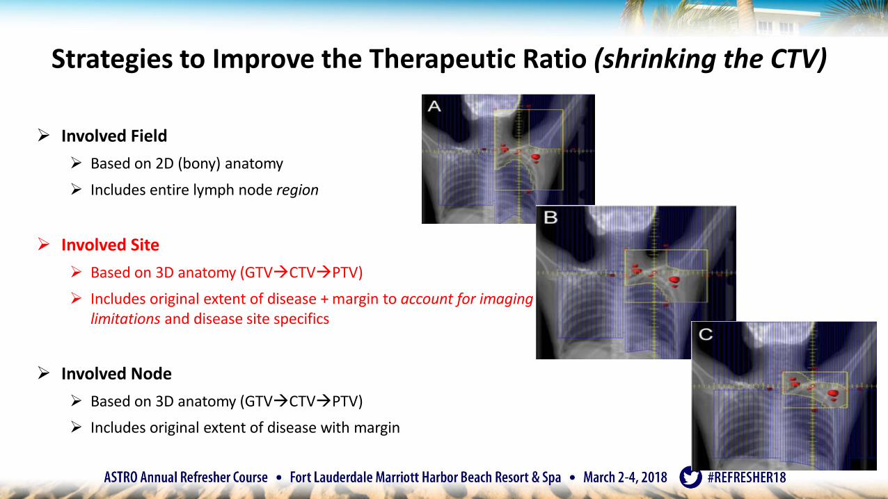
Strategies to Improve the Therapeutic Ratio (shrinking the CTV)
➢ Involved Field
➢ Based on 2D (bony) anatomy
➢ Includes entire lymph node region
➢ Involved Site
➢ Based on 3D anatomy (GTVCTVPTV)
➢ Includes original extent of disease + margin to account for imaginglimitations and disease site specifics
➢ Involved Node
➢ Based on 3D anatomy (GTVCTVPTV)
➢ Includes original extent of disease with margin
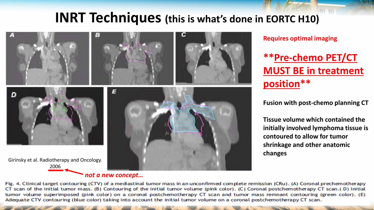
Requires optimal imaging
**Pre-chemo PET/CT MUST BE in treatment position**
Fusion with post-chemo planning CT
Tissue volume which contained the initially involved lymphoma tissue is contoured to allow for tumor shrinkage and other anatomic changes
INRT Techniques (this is what’s done in EORTC H10)
Girinsky et al. Radiotherapy and Oncology. 2006
not a new concept…
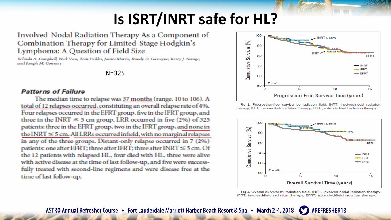
Is ISRT/INRT safe for HL?
N=325
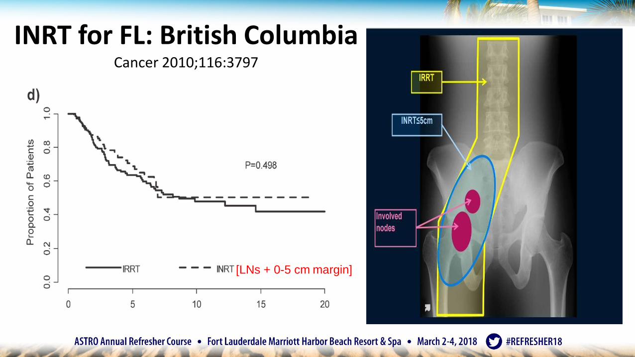
[LNs + 0-5 cm margin]
INRT for FL: British ColumbiaCancer 2010;116:3797
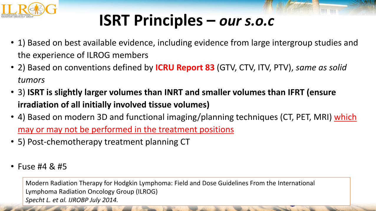
ISRT Principles – our s.o.c
• 1) Based on best available evidence, including evidence from large intergroup studies and
the experience of ILROG members
• 2) Based on conventions defined by ICRU Report 83 (GTV, CTV, ITV, PTV), same as solid
tumors
• 3) ISRT is slightly larger volumes than INRT and smaller volumes than IFRT (ensure
irradiation of all initially involved tissue volumes)
• 4) Based on modern 3D and functional imaging/planning techniques (CT, PET, MRI) which
may or may not be performed in the treatment positions
• 5) Post-chemotherapy treatment planning CT
• Fuse #4 & #5
Modern Radiation Therapy for Hodgkin Lymphoma: Field and Dose Guidelines From the International Lymphoma Radiation Oncology Group (ILROG) Specht L. et al. IJROBP July 2014.
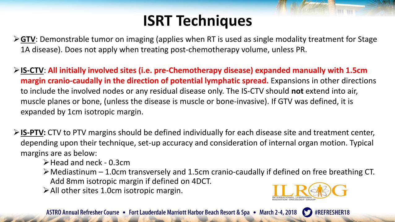
ISRT Techniques➢GTV: Demonstrable tumor on imaging (applies when RT is used as single modality treatment for Stage
1A disease). Does not apply when treating post-chemotherapy volume, unless PR.
➢IS-CTV: All initially involved sites (i.e. pre-Chemotherapy disease) expanded manually with 1.5cm margin cranio-caudally in the direction of potential lymphatic spread. Expansions in other directions to include the involved nodes or any residual disease only. The IS-CTV should not extend into air, muscle planes or bone, (unless the disease is muscle or bone-invasive). If GTV was defined, it is expanded by 1cm isotropic margin.
➢IS-PTV: CTV to PTV margins should be defined individually for each disease site and treatment center, depending upon their technique, set-up accuracy and consideration of internal organ motion. Typical margins are as below:
➢Head and neck - 0.3cm➢Mediastinum – 1.0cm transversely and 1.5cm cranio-caudally if defined on free breathing CT.
Add 8mm isotropic margin if defined on 4DCT.➢All other sites 1.0cm isotropic margin.
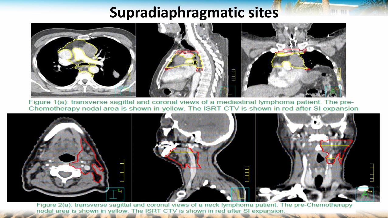
Supradiaphragmatic sites
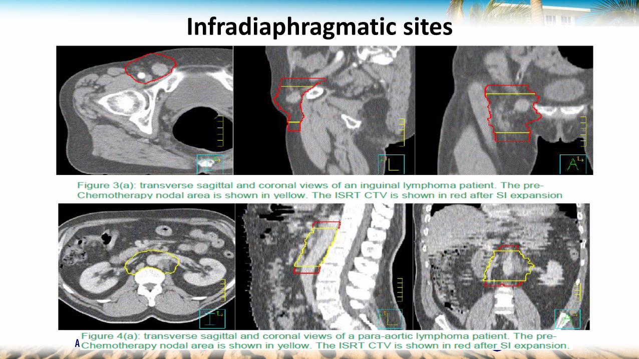
Infradiaphragmatic sites
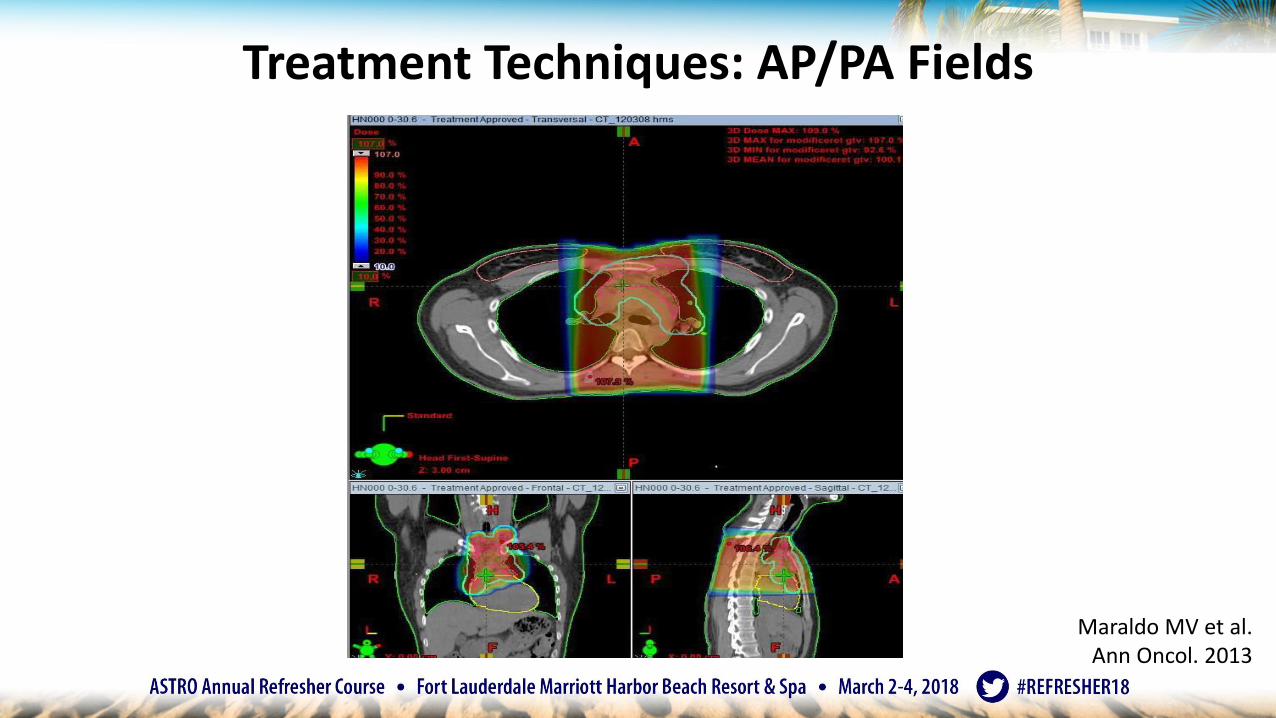
Treatment Techniques: AP/PA Fields
Maraldo MV et al.Ann Oncol. 2013
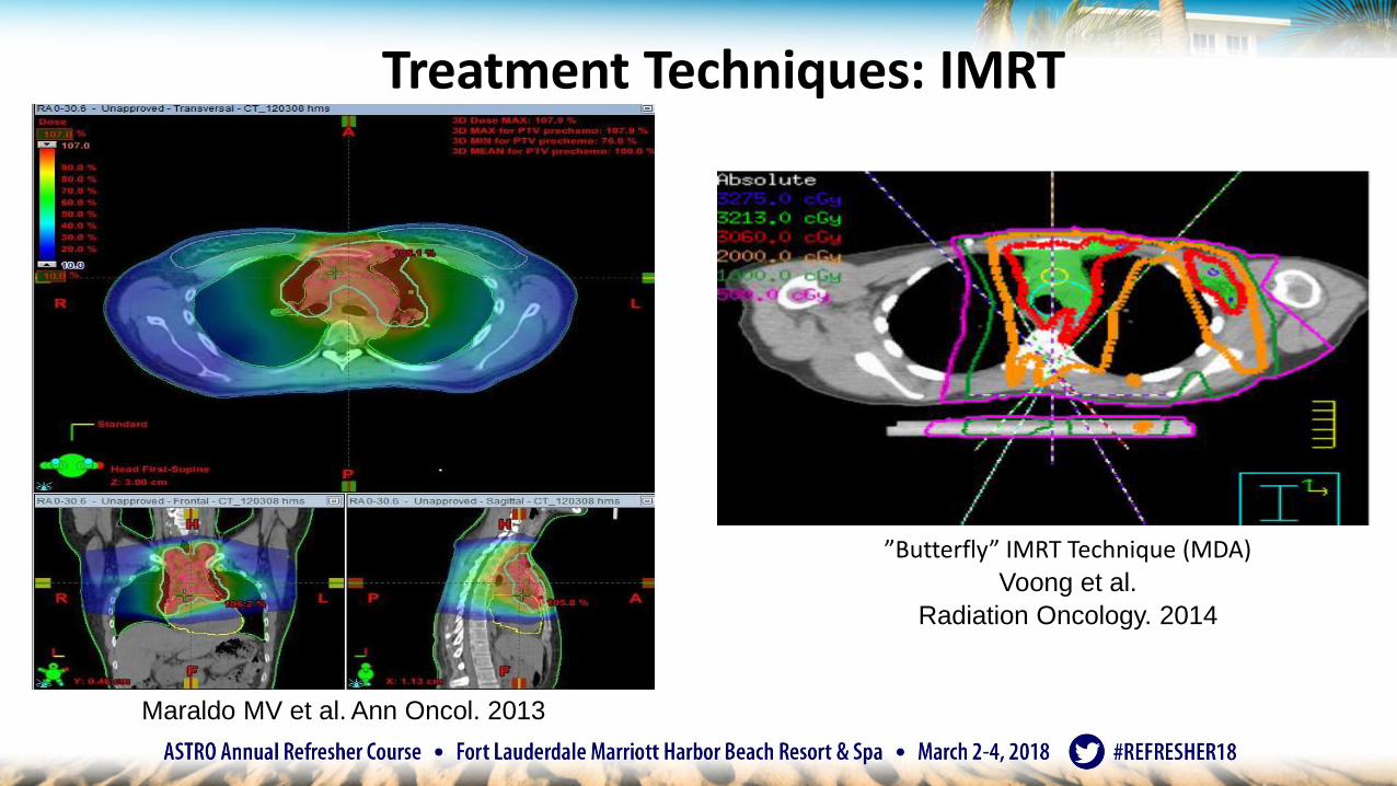
Treatment Techniques: IMRT
Maraldo MV et al. Ann Oncol. 2013
”Butterfly” IMRT Technique (MDA)
Voong et al.
Radiation Oncology. 2014
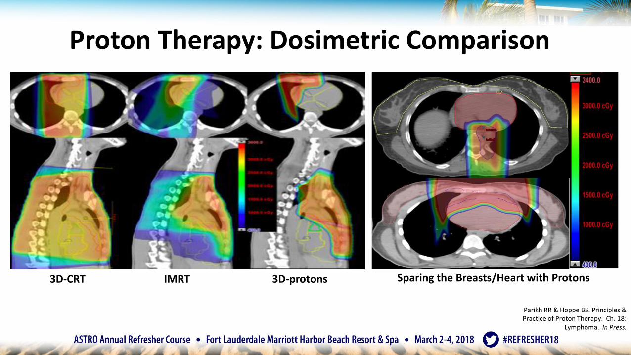
3D-CRT IMRT 3D-protons
Parikh RR & Hoppe BS. Principles & Practice of Proton Therapy. Ch. 18:
Lymphoma. In Press.
Proton Therapy: Dosimetric Comparison
Sparing the Breasts/Heart with Protons
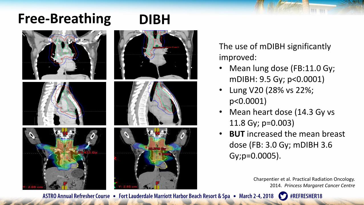
Free-Breathing DIBH
The use of mDIBH significantly improved:• Mean lung dose (FB:11.0 Gy;
mDIBH: 9.5 Gy; p<0.0001)• Lung V20 (28% vs 22%;
p<0.0001)• Mean heart dose (14.3 Gy vs
11.8 Gy; p=0.003)• BUT increased the mean breast
dose (FB: 3.0 Gy; mDIBH 3.6 Gy;p=0.0005).
Charpentier et al. Practical Radiation Oncology. 2014. Princess Margaret Cancer Centre
ISRT Conclusions
• Modern radiotherapy for HL & NHL is highly individualized treatment restrictedto limited treatment volumes
• Significant volume reductions compared to previous IFRT techniques
• Modern imaging and radiotherapy techniques/modalities (IMRT, DIBH, Protons) should be used to limit irradiation of normal tissue, minimizing risk of long-term complications
• Radiation oncologists treating HL & NHL should be involved as part of themultidisciplinary team in the initial treatment program for each patient and attempt to introduce imaging/planning procedures upfront
• Integrated multidisciplinary approach will enable optimal outcome for patients
Summary & Key Points:• Lugano classification / Deauville criteria
• Work with radiology to report/integrate Deauville criteria into management
• Role of consolidation RT in cHL
• ISRT improves recurrence by 50% and PFS/OS by 10%
• Consolidation RT in DLBCL
• ISRT decreases risk of recurrence by 50-60%
• Role of definitive RT in localized FL and MALT lymphoma
– FL: 50% long-term DFS with 24-30Gy ISRT
– MALT: CR and LC > 90% (24-30 Gy)
• Understand and Implement Modern RT techniques – ISRT
– Using solid tumor principles of GTV → CTV → PTV volume design
Thank you!