Embed Size (px)
Citation preview
Atlas of Genetics and Cytogenetics in Oncology and Haematology
OPEN ACCESS JOURNAL AT INIST-CNRS
Scope
The Atlas of Genetics and Cytogenetics in Oncology and Haematology is a peer reviewed on-line journal in
open access, devoted to genes, cytogenetics, and clinical entities in cancer, and cancer-prone diseases.
It presents structured review articles on genes, leukemias, solid tumors, cancer-prone diseases ("cards"), more
traditional review articles on these and also on surrounding topics ("deep insights"), case reports in hematology,
and educational items in the various related topics for students in Medicine and in Sciences.
Editorial correspondance Jean-Loup Huret Genetics, Department of Medical Information,
University Hospital
F-86021 Poitiers, France
tel +33 5 49 44 45 46 or +33 5 49 45 47 67
[email protected] or [email protected]
Staff Mohammad Ahmad, Mélanie Arsaban, Mikael Cordon, Isabelle Dabin, Marie-Christine Jacquemot-Perbal,
Maureen Labarussias, Anne Malo, Sylvie Yau Chun Wan - Senon, Alain Zasadzinski
Philippe Dessen, Database Director of the on-line version
Alain Bernheim, Chairman of the on-line version
The Atlas of Genetics and Cytogenetics in Oncology and Haematology (ISSN 1768-3262) is published 6 times a
year by ARMGHM, a non profit organisation and since 2008 by the INstitute for Scientific and Technical
Information of the French National Center for Scientific Research (INIST-CNRS).
The Atlas is hosted by INIST-CNRS (http://www.inist.fr)
http://AtlasGeneticsOncology.org
© ATLAS - ISSN 1768-3262
Atlas of Genetics and Cytogenetics in Oncology and Haematology
OPEN ACCESS JOURNAL AT INIST-CNRS
Editor-in-Chief
Jean-Loup Huret (Poitiers, France)
Editorial Board
Sreeparna Banerjee (Ankara, Turkey) Solid Tumors Section
Alessandro Beghini (Milan, Italy) Genes Section
Anne von Bergh (Rotterdam, The Netherlands) Genes / Leukemia Sections
Judith Bovée (Leiden, The Netherlands) Solid Tumors Section
Vasantha Brito-Babapulle (London, UK) Leukemia Section
Charles Buys (Groningen, The Netherlands) Deep Insights Section
Anne Marie Capodano (Marseille, France) Solid Tumors Section
Fei Chen (Morgantown, West Virginia) Genes / Deep Insights Sections
Antonio Cuneo (Ferrara, Italy) Leukemia Section
Paola Dal Cin (Boston, Massachussetts) Genes / Solid Tumors Sections
Louis Dallaire (Montreal, Canada) Education Section
Brigitte Debuire (Villejuif, France) Deep Insights Section
François Desangles (Paris, France) Leukemia / Solid Tumors Sections
Enric Domingo-Villanueva (London, UK) Solid Tumors Section
Ayse Erson (Ankara, Turkey) Solid Tumors Section
Richard Gatti (Los Angeles, California) Cancer-Prone Diseases / Deep Insights Sections
Ad Geurts van Kessel (Nijmegen, The Netherlands) Cancer-Prone Diseases Section
Oskar Haas (Vienna, Austria) Genes / Leukemia Sections
Anne Hagemeijer (Leuven, Belgium) Deep Insights Section
Nyla Heerema (Colombus, Ohio) Leukemia Section
Jim Heighway (Liverpool, UK) Genes / Deep Insights Sections
Sakari Knuutila (Helsinki, Finland) Deep Insights Section
Lidia Larizza (Milano, Italy) Solid Tumors Section
Lisa Lee-Jones (Newcastle, UK) Solid Tumors Section
Edmond Ma (Hong Kong, China) Leukemia Section
Roderick McLeod (Braunschweig, Germany) Deep Insights / Education Sections
Cristina Mecucci (Perugia, Italy) Genes / Leukemia Sections
Yasmin Mehraein (Homburg, Germany) Cancer-Prone Diseases Section
Fredrik Mertens (Lund, Sweden) Solid Tumors Section
Konstantin Miller (Hannover, Germany) Education Section
Felix Mitelman (Lund, Sweden) Deep Insights Section
Hossain Mossafa (Cergy Pontoise, France) Leukemia Section
Stefan Nagel (Braunschweig, Germany) Deep Insights / Education Sections
Florence Pedeutour (Nice, France) Genes / Solid Tumors Sections
Elizabeth Petty (Ann Harbor, Michigan) Deep Insights Section
Susana Raimondi (Memphis, Tennesse) Genes / Leukemia Section
Mariano Rocchi (Bari, Italy) Genes Section
Alain Sarasin (Villejuif, France) Cancer-Prone Diseases Section
Albert Schinzel (Schwerzenbach, Switzerland) Education Section
Clelia Storlazzi (Bari, Italy) Genes Section
Sabine Strehl (Vienna, Austria) Genes / Leukemia Sections
Nancy Uhrhammer (Clermont Ferrand, France) Genes / Cancer-Prone Diseases Sections
Dan Van Dyke (Rochester, Minnesota) Education Section
Roberta Vanni (Montserrato, Italy) Solid Tumors Section
Franck Viguié (Paris, France) Leukemia Section
José Luis Vizmanos (Pamplona , Spain) Leukemia Section
Thomas Wan (Hong Kong, China) Genes / Leukemia Sections
11th
European Workshop on Cytogenetics and Molecular Genetics of Solid Tumours
Bilbao, Spain, September 6-9, 2008
Atlas Genet Cytogenet Oncol Haematol 2008, 12: suppl 1
Atlas of Genetics and Cytogenetics in Oncology and Haematology
OPEN ACCESS JOURNAL AT INIST-CNRS
Meeting Report The 11th European Workshop on Cytogenetics and Molecular Genetics of Solid Tumours took place in Bilbao,
Spain, on September 6 to 9, 2008.
The organizer was Luis Antonio Parada, investigator at the Center for Cooperative Research on Biosciences
(CICbioGUNE) and the Scientific Committee was composed of Charles Buys, Juan Cigudosa, Paul Edwards,
Lidia Larizza, Felix Mitelman, Rosa Miro, and Manfred Schwab.
The workshop received generous support from The Spanish Ministry for Education and Science, Genome Spain,
Agilent Technology and MicroBeam Spain. This has allowed us to invite distinguished speakers who are leading
experts in the field.
The workshop was a great success with more than 120 participants from the vast majority of European countries.
There was enthusiastic participation of many researchers, reflecting the fact that the subject evidently keeps
attracting many cytogeneticists and molecular geneticists, in particular many notable young scientists. Of course
nothing better reflects the collective background of the participants, and all the new information they provided,
than the abstracts themselves that are now available as follow.
Luis Antonio Parada
Guest Editor CIC bioGUNE
11th
EWCMGST Organizer
11th European Workshop on Cytogenetics and Molecular Genetics of Solid Tumours
Bilbao, Spain, September 6-9, 2008
Atlas Genet Cytogenet Oncol Haematol 2008, 12: suppl 1
Atlas of Genetics and Cytogenetics in Oncology and Haematology
OPEN ACCESS JOURNAL AT INIST-CNRS
Volume 12, Supplement 1
Table of contents
Session: Genetic changes in solid tumours
High resolution survey of homozygous deletions in cancer 2 GR Bignell, CD Greenman, AP Butler, A Futreal, MR Stratton
Genomic profiling of breast cancers 3 J Adelaide, P Finetti, I Bekhouchi, S Raynaud, F Sircoulomb, J Bonansea, E Charafe-Jauffret, J Jacquemier, P Viens, F Bertucci, D Birnbaum, M Chaffanet
Chromosome translocations in breast cancer 4 K Howarth, K Blood, B Ng, J Beavis, Y Chua, S Cooke, JCM Pole, S Chin, K Ichimura, VP Collins, I Ellis, C Caldas, N Carter, PAW Edwards
Patterns of genomic instability associated with cell cycle and DNA repair in EWING Sarcomas: Gene expression and a-CGH profiling 5 BI Ferreira, J Alonso, J Carrillo, F Acquadro, C Largo, J Suela, MR Teixeira, N Cerveira, A Molares, G Gomez-Lopez, A Pestaña, A Sastre, P Garcia-Miguel, JC Cigudosa
Are ER+PR+ and ER+PR- breast tumours genetically different? An array CGH study 6 A Carracedo, M Salido, JM Corominas, BI Ferreira, I Tusquets, C Corzo, M Segura, B Espinet, JC Cigudosa, J Albanell, S Serrano, F Solé
Genomic aberrations associated with poor survival in Malignant Peripheral Nerve Sheath Tumors 8 HR Brekke, FR Ribeiro, M Eken, GE Lind, M Eknæs, KS Hall, B Bjerkehagen, E van den Berg, S Smeland, MR Teixeira, N Mandahl, RI Skotheim, F Mertens, RA Lothe
Cytogenetic and molecular cytogenetic findings in lipoblastomas 9 H Bartuma, HA Domanski, F Vult Von Steyern, CM Kullendorff, N Mandahl, F Mertens
Characterization of NCI-H69 and NCI-H69AR Small Cell Lung Cancer (SCLC) cell lines by Spectral Karyotype (SKY) 10 M Salido, E Arriola, A Carracedo, A Rovira, B Espinet, F Rojo, M Arumi, I Calzadas, S Serrano, Albanell, F Solé
Identification of novel oncogene candidates present in the highly amplified region 22q11-12 in laryngeal cancer cell lines - preliminary results 12 M Kostrzewska-Poczekaj, M Giefing, M Jarmuz, D Brauze, JI Martin-Subero, R Siebert, R Grenman, K Szyfter
Array-CGH identifies tumor suppressor gene loci in laryngeal cancer cell lines 13 M Giefing, JI Martin-Subero, K Kiwerska, J Malgorzata, R Grenman, R Siebert, K Szyfter
11th European Workshop on Cytogenetics and Molecular Genetics of Solid Tumours
Bilbao, Spain, September 6-9, 2008
Atlas Genet Cytogenet Oncol Haematol 2008, 12: suppl 1
Atlas of Genetics and Cytogenetics in Oncology and Haematology
OPEN ACCESS JOURNAL AT INIST-CNRS
Narrowing the breakpoint in deletion del(8)(q12.1q22.1) detected in cell lines derived from larynx cancer 14 M Jarmuz, A Abramowska, M Giefing, R Grenman, K Szyfter
A papillary thyroid tumor of the follicular variant harboring RET/PTC and PAX8/PPARg gene fusion in different clones 15 P Caria, T Dettori, DV Frau, G Tallini, R Vanni
Chromosome arm 8p shows complex genomic changes in bladder cancer 16 SV Williams, F Platt, C Hurst, J Aveyard, JCM Pole, MJ Garcia, MA Knowles
Geographic heterogeneity of chromosome copy number changes in breast carcinomas 17 B Mesquita, L Torres, D Pereira, C Leal, M Afonso, R Henrique, MR Teixeira
Molecular cytogenetic characterization of an atypical Rhabdoid tumour with a translocation t(1;22)(p36;q11.2) involving rearrangement of EWS1 and a deletion of SNF5/INI1 18 D Bouron-dal Soglio, R Absi, S Barrette, JC Fournet, R Fetni
Novel variant translocation t(12;13;16) and FUS-DDIT3 fusion in infrequent childhood myxoid liposarcoma 19 S Hazourli, J Hébert, H Sartelet, S Barrette, R Fetni
Characterization of primary melanomas using high-resolution array based CGH technology 20 F Acquadro, BI Ferreira, J Suela, JC Cigudosa
Identification of novel genes involved in colorectal cancer predisposition 21 R Venkatachalam, MJL Ligtenberg, EJ Kamping, E Hoenselaar, HK Schakert, A Geurts van Kessel, N Hoogerbrugge, RP Kuiper
Value of combined array CGH and cytogenetic analysis for a precise characterization of childhood and adolescent embryonal tumors and sarcomas 22 E Stejskalova, M Jarosova, H Urbankova, J Malis, K Pycha, L Krskova, R Kodet
Chromosomal instability in Osteosarcoma 23 G Maire, M Yoshimoto, B Sadikovic, S Selvarajah, P Thorner, M Zielenska, JA Squire
Biologically relevant copy number alterations in osteosarcoma cell lines: An array comparative genomic hybridization analysis 24 ML Larramendy, S Kaur, M Gentile, CM Hattinger, M Pasello, T Böhling, K Scotlandi, M Serra, S Knuutila
Wide-genome analysis identified gain of 1q as potential negative prognostic marker for survival in high-risk disseminated neuroblastoma 25 P Scaruffi, S Stigliani, S Coco, S Moretti, K Mazzocco, R Defferrari, C De Vecchi, S Bonassi, GP Tonini
Chromosomal imbalances identified by CGH in invasive versus non-invasive bladder tumours 26 I Ponsa, J del Rey, L Mengual, N Pujol, G Armengol, F Algaba, A Alcaraz, R Miró, E Prat
11th European Workshop on Cytogenetics and Molecular Genetics of Solid Tumours
Bilbao, Spain, September 6-9, 2008
Atlas Genet Cytogenet Oncol Haematol 2008, 12: suppl 1
Atlas of Genetics and Cytogenetics in Oncology and Haematology
OPEN ACCESS JOURNAL AT INIST-CNRS
Is there constitutional chromosome instability in patients affected by two or more primary tumors and/or a family history of cancer? 27 N Pujol, I Ponsa, J del Rey, E Prat, R Miró
Genetic characterization of progression in embryonal rhabdomyosarcoma. Comparative genetic analysis of primary and recurrent or metastatic tumors 28 R Gil-Benso, J Caballero, C López-Ginés, R Callaghan, A Pellín-Carcelén, S Navarro, A Bataller-Calatayud, T Peris, A Llombart-Bosch
Genetic and biological characterization of a novel human melanoma cell line (MEL-RC08) 29 R Gil-Benso, C Monteagudo, M Cerdá-Nicolás, R Callaghan, JC Cigudosa, A Pellín-Carcelén, C López-Ginés
Molecular cytogenetic of hepatic metastasis in colon cancer patients: correlations with the prometastatic and proangiogenic potential Diagnostic-therapeutic implications 30 J Tomé-Garcia, L Mendoza, A Belén de la Hoz
Significance of molecular cytogenetic characterization of glioblastoma for prediction of patient prognosis 32 P Cejpek, P Kuglik, R Veselka, E Necesalova, M Pesakova, V Vranova, T Loja, J Relichova, P Krupa, J Horky, Z Starcuk
Session - Mechanism underlining tumourigenic genetic changes
Comparative cytogenetic analysis of mouse models for breast cancer pinpoints to the amplification of the novel oncogene Septin 9 34 D Connolly, L Tal, M Suzuki, JM Greally, P Verdier-Pinard, C Montagna
Molecular characterization of pediatric medulloblastoma by combining genomic and gene expression profiling 35 S Coco, P Scaruffi, S Moretti, S Bonassi, M Forni, S Aschero, ME Basso, A Sandri, A Oberthuer, J Berthold, M Fischer, I Adolfo, M Zollo, G Cinalli, A Iolascon, GP Tonini
Chromosome instability in bladder cancer: Centrosome abnormalities and CCND1 gene amplification 37 J del Rey, E Prat, I Ponsa, J Camps, J Lloreta, A Gelabert, F Algaba, R Miró
Functional validation of genomic and transcriptome profiles identifies candidate oncogenes in colon cancer 38 J Camps, AB Hummon, M Grade, G Emons, QT Nguyen, BM Ghadimi, NJ Caplen, MJ Difilippantonio, T Ried
Multidisciplinary evaluation of the role of reciprocal translocation and gene fusion in the pathogenesis of solid tumours 39 F Acquadro, G Soler, S Rodriguez Perales, BI Ferreira, JC Cigudosa
11th European Workshop on Cytogenetics and Molecular Genetics of Solid Tumours
Bilbao, Spain, September 6-9, 2008
Atlas Genet Cytogenet Oncol Haematol 2008, 12: suppl 1
Atlas of Genetics and Cytogenetics in Oncology and Haematology
OPEN ACCESS JOURNAL AT INIST-CNRS
The renal cell carcinoma-associated oncogenic fusion protein PRCCTFE3 induces p21
WAF/CIP mediated cell cycle arrest 40
K Medendorp, L Vreede, J van Groningen, H van den Hurk, A Geurts van Kessel
Alternative splicing of ERG and TMPRSS2:ERG rearrangement in prostate cancer cell lines 41 P Paulo, N Cerveira, J Santos, M Pinheiro, V Costa, C Jerónimo, MR Teixeira
Expression profile of significant immortalization genes in colon cancer 42 A Witkowska, J Gumprecht, A Bocianowska, J Glogowska-Ligus, M Stachowicz, A Owczarek, E Nowakowska-Zajdel, U Mazurek
Association of ERBB2 gene status with histopathological parameters and disease-specific survival in gastric carcinoma patients 44 D Barros-Silva, D Leitão, L Afonso, J Vieira, M Dinis-Ribeiro, M Fragoso, MJ Bento, L Santos, P Ferreira, S Rêgo, C Brandão, F Carneiro, C Lopes, F Schmitt, MR Teixeira
Hereditary multiple basal cell at young age in patients without germline PTCH mutations 45 RP Kuiper, MM van Rossum, EJ Kamping, PL Zeeuwen, R de Boer-van Huizen, HG Brunner, MJL Ligtenberg, A Geurts van Kessel, J Schalkwijk, N Hoogerbrugge
Comparative genomic hybridization and cytogenetic studies on the genomic imbalances in the well-differentiated and dedifferentiated liposarcomas 46 J Limon, M Iliszko, J Rys, A Kuzniacka, A Sokolowski, J Lasota, M Miettinen
Analysis of epidermal growth factor receptor gene copy number in glioblastomas and its relation with the protein expression 47 C López-Ginés, R Gil-Benso, R Ferrer-Luna, R Benito, P Roldan, J Gonzalez-Darder, B Celda, M Cerdá-Nicolás
Chromosomal imbalances, metabolic (HR-MAS) and gene expression profiles in bening and atypical meningiomas 48 C López-Ginés, R Gil-Benso, D Monleon, M Mata, JM Morales, P Roldan, J Gonzalez-Darder, B Celda, M Cerdá-Nicolás
Session - Cancer cell biology
Molecular cytogenetic characterization of tumour initiating cells 51 P Gasparini, G Bertolini, A Magnifico, C Casarsa, G Finocchiaro, MG Daidone, S Menard, G Sozzi
Characterization of spontaneously transformed murine epithelial cells 52 HM Padilla-Nash
Genomic alterations and expression profiling in uveal melanoma 53 E Kilic, W van Gils, H Mensink, E Lodder, D Paridaens, H Beverloo, N Mooy, M van Til, N Naus, A de Klein
Relationships among truncating mutations, genomic instability and cancer risk in a molecularly defined Rothmund Thomson case 54 G Roversi, E Colombo, I Magnani, C Pedicelli, M Paradisi, L Larizza
11th European Workshop on Cytogenetics and Molecular Genetics of Solid Tumours
Bilbao, Spain, September 6-9, 2008
Atlas Genet Cytogenet Oncol Haematol 2008, 12: suppl 1
Atlas of Genetics and Cytogenetics in Oncology and Haematology
OPEN ACCESS JOURNAL AT INIST-CNRS
Session - Epigenetic changes in solid tumours
The role of telomere length in telomerase based anticancer therapies 56 I Fernandez-Garcia, A Muñoz-Barruti, LM Montuenga, C Ortiz-de-Solórzano
Genome reorganization during invasive cell growth 57 L Vellón, F Rojo, L Espinosa, R Matthiesen, LA Parada
The epigenetics of human synovial sarcoma: towards novel therapeutic strategies 58 DRH de Bruijn, JM Lubieniecka, L Su, AHA van Dijk, S Subramanian, M van de Rijn, N Poulin, TO Nielsen, A Geurts van Kessel
Unbiased differential methylation screening assay for applications in cancer epigenetic research 59 VV Strelnikov, AS Tanas, VV Shkarupo, EB Kuznetsova, DV Zaletaev
Differential positioning of gene markers in normal and malignant tissues 61 KJ Meaburn, PR Gudla, K Nandy, SJ Lockett, T Misteli
Similar chromosomal expression profiles of genetically favourable and senescent neuroblastoma cells: signs for a common epigenetic pattern? 62 E Bozsaky, C Stock, A Kowalska, IM Ambros, A Luegmayr, B Brunner, D Rieder, Z Trajanoski, G Amann, PF Ambros
Session - Technological advances
HAPPY Mapping: a flexible method for examination of genomic rearrangements in cancer genomes 64 JCM Pole, K Howarth, F McCaughan, P Dear, PAW Edwards
Identification of transcriptional targets by ChIP-Sequencing in t(X;1)-positive renal cell carcinomas 65 L Brugmans, L Hetterschijt, L Vreede, K Medendorp, A Geurts van Kessel
Identification of genes harboring nonsense mutations 66 G Dun, I van Duivenbode, R Hofstra, E van den Berg, K Kok
Array CGH after FISH-MD reveals that typical marker chromosomes in ovarian cancer frequently show fusions between 11q13 and 19p13.3 67 J Weimer, F Micci, R Ullmann, JI Martin-Subero, S Gesk, H Tönnies, R Siebert, S Heim, N Arnold
Design of split-signal FISH probe for the detection of TFE3 translocations in Xp-translocation Renal Cell Carcinoma (RCC) 68 A Galvan, M Salido, J Lloreta, A Padron, B Espinet, O Villa, C Melero, S Serrano, F Solé
Phenotypic and genetic characterization of circulating tumor cells by combining immunomagnetic selection and FICTION techniques 70 M Campos, C Prior, F Warleta, I Zudaire, J Ruiz-Mora, R Catena, A Calvo, J Gaforio
11th European Workshop on Cytogenetics and Molecular Genetics of Solid Tumours
Bilbao, Spain, September 6-9, 2008
Atlas Genet Cytogenet Oncol Haematol 2008, 12: suppl 1
Atlas of Genetics and Cytogenetics in Oncology and Haematology
OPEN ACCESS JOURNAL AT INIST-CNRS
Identification of prostate circulating tumor cells by means of the characterization of the TMPRSS2-ERG fusion gene in peripheral blood 71 A Fernández-Serra, J Rubio, Z García-Casado, A Calatrava, J Maiquez, I Iborra, MA Bonillo,
E Solsona, S Almenar, JA López-Guerrero
Immunodetection and cytogenetic characterization of disseminated tumor cells applied to the clinical management of patients with solid tumors 72 O Crende, J Tomé-Garcia, N Telleria, A Belén de la Hoz, L Mendoza, F Vidal
Session - Clinical impact of gene changes & Future directions
Novel genomic lesions in patients with unexplained microsatellite instable colorectal tumors 74 RP Kuiper, R Venkatachalam, E Hoenselaar, M Goossens, EJ Kamping, SV van Reijmersdal, EFPM Schoenmakers, JH van Krieken, N Hoogerbrugge, A Geurts van Kessel, MJL Ligtenberg
Homozygous deletions may be markers of nearby heterozygous mutations: the complex deletion at FRA16D in the HCT116 colon cancer cell line removes exons of WWOX 75 AE Alsop, K Taylor, J Zhang, H Gabra, AJW Paige, PAW Edwards
Is the DNA damage response a cancer barrier? 76 O Fernández-Capetillo
Prognostic implication of TMPRSS2-ERG fusion gene in patients with prostate cancer operated by radical prostatectomy 77 A Fernández-Serra, J Rubio, A Calatrava, Z García-Casado, I Iborra, MA Bonillo, E Solsona, S Almenar, JA López-Guerrero
Index of authors 79
11th European Workshop on Cytogenetics and Molecular Genetics of Solid Tumours
Bilbao, Spain, September 6-9, 2008
Atlas Genet Cytogenet Oncol Haematol 2008, 12: suppl 1 1
Atlas of Genetics and Cytogenetics in Oncology and Haematology
OPEN ACCESS JOURNAL AT INIST-CNRS
SESSION
Genetic changes
in solid tumours
11th European Workshop on Cytogenetics and Molecular Genetics of Solid Tumours
Bilbao, Spain, September 6-9, 2008
Atlas Genet Cytogenet Oncol Haematol 2008, 12: suppl 1 2
Atlas of Genetics and Cytogenetics in Oncology and Haematology
OPEN ACCESS JOURNAL AT INIST-CNRS
Genetic changes in solid tumours
High resolution survey of homozygous deletions in cancer
GR Bignell, CD Greenman, AP Butler, A Futreal, MR Stratton The Wellcome Trust Sanger Institute, Cancer Genome Project, Hinxton, Cambridge CB10 1SA, UK
Published in Atlas Journal in October 2008
Abstract Oral presentation
Homozygous deletions are often seen in cancer as a means of inactivating tumour suppressor genes. However,
such deletions can also result from genome instability and as such probably represent passenger events that do
not contribute to carcinogenesis. These passenger deletions are often associated with known regions of genomic
fragility (fragile sites). We have analysed approximately 800 cancer cell lines together with 466 normal DNAs
using one of the latest high density SNP arrays (Affymetrix Genome-Wide Human SNP Array 6.0 containing
over 1.8 million loci). This screen provides integrated analysis of copy number and genotyping information and
is probably the largest analysis of its type performed to date. Over 10000 homozygous deletions were identified
within the set of cancer cell lines. Of these >3400 deletions were putatively somatic. These somatic homozygous
deletions collectively accounted for over 552Mb of genomic DNA (>18% of the genome). There were
approximately 300 clusters of two or more overlapping homozygous deletions which accounted for ~2200
deletions. 14 clusters include known tumour suppressor genes and account for ~450 homozygous deletions,
while ~300 deletions are within the defined footprint of the well mapped common fragile sites. Analysing the
deletion patterns seen over known loci provides a means to classify previously unknown homozygous deletion
clusters. There is evidence that a substantial proportion of these may be regions of cancer associated fragility,
although a minority may be novel tumour suppressor genes.
© Atlas of Genetics and Cytogenetics in Oncology and Haematology
11th European Workshop on Cytogenetics and Molecular Genetics of Solid Tumours
Bilbao, Spain, September 6-9, 2008
Atlas Genet Cytogenet Oncol Haematol 2008, 12: suppl 1 3
Atlas of Genetics and Cytogenetics in Oncology and Haematology
OPEN ACCESS JOURNAL AT INIST-CNRS
Genetic changes in solid tumours
Genomic profiling of breast cancers
J Adelaide1*, P Finetti
1*, I Bekhouche
1, S Raynaud
1, F Sircoulomb
1, J Bonansea
1, E
Charafe-Jauffret1,2
, J Jacquemier1,2
, P Viens3, F Bertucci
1,3, D Birnbaum
1, M Chaffanet
1
1Marseille Cancer Institute, Department of Molecular Oncology, UMR599 Inserm and Institut Paoli-Calmettes,
Marseille, France. 2Department of BioPathology, Institut Paoli-Calmettes, Marseille, France.
3Department of Medical Oncology, Institut Paoli-Calmettes, Marseille, France.
* J Adelaide and P. Finetti have equally contributed to this work.
Published in Atlas Journal in October 2008
Abstract Oral presentation
Breast cancer (BC) is a complex, heterogeneous disease at the molecular level. Accumulation and combination
of genetic and epigenetic alterations cause tumorigenesis, genetic instability, and acquisition of an increasingly
invasive and resistant phenotype. High-throughput molecular analyses provide an unprecedented opportunity for
resolving BC heterogeneity and identifying new classes biologically and clinically relevant. We recently
reported the integrated genomic profiling of basal and luminal breast cancers (Adelaide et al, Cancer Res., 2007)
by applying combined high resolution genomic analyses (244K aCGH [Agilent Technologies] + U133 Plus 2.0
human [Affymetrix]). The results support the existence of specific oncogenic pathways in basal and luminal
BCs, involving several potential oncogenes and tumor suppressor genes (TSG). In basal tumors, 73 candidate
oncogenes were identified in chromosome regions 1q21-23, 10p14, and 12p13, and 28 candidate TSG in regions
4q32-34 and 5q11-23. In luminal BCs, 33 potential oncogenes were identified in 1q21-23, 8p12-q21 11q13 and
16p12-13 and 61 candidate TSG in 16q12-13, 16q22-24 and 17p13. HORMAD1 (p=6.5 10-5) and ZNF703 (p=7 10-4) were the most significant basal and luminal potential oncogenes, respectively. Using the same
strategy, we have now extended our study to various molecular subtypes and other types of BC associated with
poor evolution and aggressiveness including inflammatory BC and young women BCs. The integrated genomic
profiles were established to facilitate the search for specific molecular signatures and new cancer-associated
genes. We hope that this analysis of the different aggressive forms of BCs will provide a better understanding of
mammary carcinogenesis to help in the development of appropriate treatments.
© Atlas of Genetics and Cytogenetics in Oncology and Haematology
11th European Workshop on Cytogenetics and Molecular Genetics of Solid Tumours
Bilbao, Spain, September 6-9, 2008
Atlas Genet Cytogenet Oncol Haematol 2008, 12: suppl 1 4
Atlas of Genetics and Cytogenetics in Oncology and Haematology
OPEN ACCESS JOURNAL AT INIST-CNRS
Genetic changes in solid tumours
Chromosome translocations in breast cancer
K Howarth1, K Blood
1, B Ng
2, J Beavis
1, Y Chua
1, S Cooke
1, JCM Pole
1, S Chin
3, K
Ichimura4, VP Collins
4, I Ellis
5, C Caldas
3, N Carter
2, PAW Edwards
1
1Hutchison-MRC Research Institute, Addenbrooke's Site, University of Cambridge, Hills Road, Cambridge, CB2
0XZ, UK 2Wellcome Trust Sanger Institute, Hinxton, Cambridge, UK
3Cancer Research UK Cambridge Research Institute, Cambridge, UK
4Division of Molecular Histopathology, Department of Pathology, University of Cambridge, Addenbrooke’s,
Hills Road, Cambridge, UK 5Department of Histopathology, School of Molecular Medical Sciences, University of Nottingham, Nottingham,
UK
Published in Atlas Journal in October 2008
Abstract Oral presentation
Relatively little is known about chromosome translocations in the common epithelial cancers such as breast
cancer, in spite of the central role played by translocations and consequent gene fusions in haematopoietic
cancers.
We present a comprehensive analysis by array painting of the chromosome translocations of breast cancer cell
lines HCC1806, HCC1187 and ZR-75-30. In array painting, chromosomes are isolated by flow cytometry,
amplified and hybridized to DNA microarrays. A total of 200 breakpoints were identified and all were mapped
to 1Mb resolution on BAC arrays, then 40 selected breakpoints, including all balanced breakpoints, were further
mapped on tiling-path BAC arrays or to around 2kb resolution using oligonucleotide arrays. Many more of the
translocations were balanced than expected, either reciprocal (8 in total) or balanced for at least one participating
chromosome (19 paired breakpoints). Many breakpoints were at genes that are plausible targets of oncogenic
translocation, including CTCF and P300. Two gene fusions were also demonstrated, TAX1BP1-AHCY and RIF1-
PKD1L1. A preliminary screen of paraffin sections of breast tumours revealed breaks in several genes, including
PKD1L1, so the rearrangements are not confined to cell lines.
Our data establishes that array painting is a very effective way to map substantial numbers of translocation
breakpoints and supports the emerging view that chromosome rearrangements that fuse, activate or otherwise
alter genes at their breakpoints may play an important role in common epithelial cancers.
© Atlas of Genetics and Cytogenetics in Oncology and Haematology
11th European Workshop on Cytogenetics and Molecular Genetics of Solid Tumours
Bilbao, Spain, September 6-9, 2008
Atlas Genet Cytogenet Oncol Haematol 2008, 12: suppl 1 5
Atlas of Genetics and Cytogenetics in Oncology and Haematology
OPEN ACCESS JOURNAL AT INIST-CNRS
Genetic changes in solid tumours
Patterns of genomic instability associated with cell cycle and DNA repair in EWING Sarcomas: Gene expression and a-CGH profiling
BI Ferreira, J Alonso, J Carrillo, F Acquadro, C Largo, J Suela, MR Teixeira, N
Cerveira, A Molares, G Gomez-Lopez, A Pestaña, A Sastre, P Garcia-Miguel, JC
Cigudosa Molecular Cytogenetics Group, Centro Nacional de Investigaciones Oncologicas (CNIO), C/ Melchor Fernandez
Almagro, 3, 28029 Madrid, Spain.
Published in Atlas Journal in October 2008
Abstract Oral presentation
Ewing’s sarcoma (ES) is characterized by specific chromosome translocations, the most common being
t(11;22)(q24;q12). Additionally, other type of genetic abnormalities may occur and be relevant for explaining the
variable tumour biology and clinical outcome. We have carried out a high-resolution arrayCGH and expression
profiling on 25 ES tumour samples to characterize the DNA copy number aberrations (CNA) occurring in these
tumours and determine their association with gene-expression profiles and clinical outcome. CNA were observed
in 84% of the cases. We observed a median number of three aberrations per case. Besides numerical
chromosome changes, smaller aberrations were found and defined at chromosomes 5p, 7q and 9p. All CNA were
compiled to define the smallest overlapping regions of imbalance (SORI). A total of 35 SORI were delimited.
Bioinformatics analyses were conducted to identify subgroups according to the pattern of genomic instability.
Unsupervised and supervised clustering analysis (using SORI as variables) segregated the tumours in two
distinct groups: one genomically stable (3 CNA). The genomic unstable group showed a statistically significant
shorter overall survival and was more refractory to chemotherapy. Expression profile analysis revealed
significant differences between both groups. Genes related with chromosome segregation, DNA repair pathways
and cell-cycle control were upregulated in the genomically unstable group. This report elucidates, for the first
time, data about genomic instability in ES, based on CNA and expression profiling, and shows that a
genomically unstable group of Ewing’s tumours is correlated with a significant poor prognosis.
© Atlas of Genetics and Cytogenetics in Oncology and Haematology
11th European Workshop on Cytogenetics and Molecular Genetics of Solid Tumours
Bilbao, Spain, September 6-9, 2008
Atlas Genet Cytogenet Oncol Haematol 2008, 12: suppl 1 6
Atlas of Genetics and Cytogenetics in Oncology and Haematology
OPEN ACCESS JOURNAL AT INIST-CNRS
Genetic changes in solid tumours
Are ER+PR+ and ER+PR- breast tumours genetically different? An array CGH study
A Carracedo1,2,3
, M Salido1,2,3
, JM Corominas4,5
, BI Ferreira6, I Tusquets
7, C Corzo
8, M
Segura9, B Espinet
1,2,3, JC Cigudosa
6, J Albanell
5,7, S Serrano
1,2,3, F Solé
1,2,3
1Servei de Patologia, Laboratori de Citogenetica i Biologia Molecular, Hospital del Mar, IMAS, IMIM,
Universitat Autonoma de Barcelona, Spain. 2Escola de Citologia Hematologica S. Woessner-IMAS, Barcelona, Spain.
3Unitat de Recerca Translacional en Tumors Solidos-PRBB, Barcelona, Spain.
4Unitat de Patologia Mamaria del Servei de Patologia, Hospital del Mar, Barcelona, Spain.
5Biomarkers and molecular therapeutics in breast cancer, Research Cancer Program, IMIM-Hospital del Mar,
Barcelona, Spain. 6Grupo de Citogenética Molecular, CNIO, Madrid, Spain.
7Servei d’Oncologia, Hospital del Mar, Barcelona, Spain.
8Escola Bonanova-IMAS, Barcelona, Spain.
9Unitat de Patologia Mamaria del Servei de Cirurgia, Hospital del Mar, Barcelona, Spain.
Published in Atlas Journal in October 2008
Abstract Oral presentation
Estrogen receptor (ER) is an accepted predictor of response to endocrine therapy. More than 20% of ER+ breast
cancers express progesterone receptor (PR). Clinical observations have indicated that ER+/PR-breast cancers
could present a different pattern of hormone sensitivity than ER+/PR+ ones. Furthermore, ER+/PR-tumors are
more likely to have and aggressive phenotype and it could be a link between their progesterone negativity and an
hypereactive growth factor signaling.
The aim of this study is to investigate the pattern of DNA copy number aberrations (CNAs) of these two
subtypes of breast carcinoma, establish the smallest overlapping regions of imbalance (SORIs), and try to
identify differences between them using a high-resolution array CGH (a-CGH). The agilent 44K oligonucleotide
a-CGH has been applied to 25 ER+/PR+ and 25 ER+/PR-ductal infiltrating carcinomas (DICs) of the breast
(histological grades I, II, and III, negative ERBB2 status). Data analysis and chromosome segmentation was
performed with the InSilicoArray CCGH software. For each sample, FISH was used to validate and to define
cut-off values for gains and losses. Preliminary results show: The total altered genome was 20,3% in ER+/PR+
and 31,7% in ER+/PR-. The % of gained genome was 9,4 in ER+/PR+ and 16,3 in ER+/PR-tumors. The % of
lost genome was 10,9 in ER+/PR+ and 15,3 in ER+/PR-tumors. The most frequently gained chromosomes
(chrs.) in ER+/PR-tumors were 1, 16, 8 and 11, and in ER+/PR-tumors were 1, 17, 11, 8 and 20. The most
frequently lost chrs. in ER+/PR+ tumors were 16, 1, 6 and 11, and in ER+/PR-tumors were 17, 22, 16, 11, 1 and
8. Considering the most frequently altered chrs., we found some SORIs in common between the two subtypes of
breast tumors: gains of 1q25.2-q31.3, 1q32.1, 8q24.3 and 11q13.3; also losses of 1p21.1, 8pter-p21.2, 11q14.2,
11q14.3-qter, and 16q13-qter. Despite their similar CNAs, we found that the ER+/PR-tumors showed some
11
th Workshop on Cytogenetics and Molecular Genetics of Solid Tumours Carracedo A et al
Bilbao, Spain, September 6-9, 2008
Atlas Genet Cytogenet Oncol Haematol 2008, 12: suppl 1 7
frequently altered regions not present in the ER+/PR+ tumors, related to the chrs. 1, 8, 16, 17, 20 and 22. Chrs.
17 and 22 are the most differently altered in the ER+/PR-tumors compared to the ER+/PR+ ones, specifically
gain of 17p13.3-p12 and loss of 22q11.23-qter. In our knowledge, this is one of the first study focused on the
genetic comparison of ER+/PR+ and ER+/PR-breast tumors by aCGH technology. In a global analysis of the 41
DICs, CNAs were consistent with the results previously reported on CGH and aCGH studies. Taking into
account our preliminary results, ER+/PR-tumors present a higher chromosomal instability and have a different
genetic profile compared to the ER+/PR+ ones.
Acknowledgements: Grant PI05/0961 from Ministerio de Sanidad y consumo ISCIII
© Atlas of Genetics and Cytogenetics in Oncology and Haematology
11th European Workshop on Cytogenetics and Molecular Genetics of Solid Tumours
Bilbao, Spain, September 6-9, 2008
Atlas Genet Cytogenet Oncol Haematol 2008, 12: suppl 1 8
Atlas of Genetics and Cytogenetics in Oncology and Haematology
OPEN ACCESS JOURNAL AT INIST-CNRS
Genetic changes in solid tumours
Genomic aberrations associated with poor survival in Malignant Peripheral Nerve Sheath Tumors
HR Brekke, FR Ribeiro, M Eken, GE Lind, M Eknæs, KS Hall, B Bjerkehagen, E van
den Berg, S Smeland, MR Teixeira, N Mandahl, RI Skotheim, F Mertens, RA Lothe
Department of Cancer Prevention, Institute for Cancer Research, The Norwegian Radium Hospital,
Rikshospitalet University Hospital, Montebello, Oslo, Norway.
Published in Atlas Journal in October 2008
Abstract Oral presentation
Background : Malignant Peripheral Nerve Sheath Tumors (MPNSTs) are rare neoplasias associated with a poor
clinical outcome, for which few treatment options exist. Tumors may arise sporadically, but commonly develop
in patients with the hereditary condition neurofibromatosis type1 (NF1). Due to the limited number of reported
cases and the genomic complexity often observed, it is unclear which genetic aberrations are contributing to the
initiation, progression and clinical aggressiveness, and whether MPNST etiology is similar in patients with and
without NF1.
Material and methods : As part of a long-standing international collaboration involving Norway, Sweden and
the Netherlands, a series of fresh-frozen material from 48 MPNSTs and 10 neurofibromas from 51 patients with
(n=31) and without (n=20) NF1 history was obtained. Chromosomal and array-based comparative genomic
hybridization (aCGH) were performed to assess DNA copy number changes, and genome-wide expression data
for a subset of samples (n=30) was later integrated to evaluate candidate target genes within regions recurrently
affected by copy number aberrations.
Results : Forty-four MPNSTs (92%) displayed copy number changes, whereas no aberrations were found in the
nine neurofibromas. A small deletion at 9p was identified as the sole alteration in a plexiform neurofibroma.
Most tumors presented complex profiles with a median of 16 aberrations per sample. Gains at 17q (69%), 8q
(65%) and 7p (56%), and losses at 9p (46%), 11q (46%) and 17p (42%) are the most common. Several
homozygous deletion could also be pinpointed, in particular 9p21 loss seen in five out of 12 samples by aCGH.
Expression data confirmed significant differences in mRNA levels of several candidate genes, such as the lower
expression of CDKN2A and CDKN2B at 9p21 or the very high levels of BIRC5 at 17q25. Interestingly, no
significant differences were found in the genomic profiles of sporadic versus NF1-related MPNSTs. Several
genomic changes showed prognostic significance independently of clinical variables or patient group. In
particular, patients whose tumors displayed concurrent gains at chromosomal regions 7p and 17q, and losses at
9p, displayed a significantly worse prognosis (p=0.00008).
Conclusions: Most MPNSTs display complex profiles that make it difficult to pinpoint primary genetic events,
as virtually all chromosomes show recurrent gains and/or losses of genomic material. No differences could be
found in the genetic profiles of sporadic tumors as compared to those from patients with NF1. However, the
simultaneous occurrence of specific genetic aberrations (gains of 7p and 17q, and loss of 9p) was strongly
associated with an aggressive clinical course independently of patient group or clinical variables. Putative targets
in these regions are CDKN2A (9p) and BIRC5 (17q), confirmed to be differentially expressed in MPNSTs when
compared to their benign counterparts.
© Atlas of Genetics and Cytogenetics in Oncology and Haematology
11th European Workshop on Cytogenetics and Molecular Genetics of Solid Tumours
Bilbao, Spain, September 6-9, 2008
Atlas Genet Cytogenet Oncol Haematol 2008, 12: suppl 1 9
Atlas of Genetics and Cytogenetics in Oncology and Haematology
OPEN ACCESS JOURNAL AT INIST-CNRS
Genetic changes in solid tumours
Cytogenetic and molecular cytogenetic findings in lipoblastomas
H Bartuma1, HA Domanski
2, F Vult Von Steyern
3, CM Kullendorff
4, N Mandahl
1, F
Mertens1
1Department of Clinical Genetics,
2Department of Cytology and Pathology,
3Department of Orthopedics,
4Department of Pediatric Surgery, Lund University Hospital, SE-221 85 Lund, Sweden.
Published in Atlas Journal in October 2008
Abstract Poster presentation
Lipoblastoma is a benign lipomatous tumor, which arises from embryonic adipose tissue and occurs primarily in
children younger than three years of age. Microscopically, it consists of small irregular lobules of adipocytes at
different maturation stages separated by connective tissue septa and primitive mesenchymal areas with a loose
myxoid matrix.
Here, we present a review of available cytogenetic data and the karyotypes of ten new cases of lipoblastoma, of
which seven could be further studied by fluorescence in situ hybridization (FISH) concerning the involvement of
the PLAG1 gene. All seven tumors with clonal aberrations harbored breakpoints in 8q11-13, in agreement with
literature data; including previously published cases, 33 of 40 (82%) lipoblastomas had rearrangement of the
8q11-13 region. These rearrangements target the PLAG1 gene, which becomes up-regulated through promoter
swapping. FISH revealed that five of seven cases in our series had a rearrangement of the PLAG1 gene.
Occasionally, there can be difficulties in distinguishing a lipoblastoma from a conventional lipoma or a myxoid
liposarcoma. As rearrangements of 8q11-13 have only been reported in 3% of conventional lipomas and never in
myxoid liposarcomas, cytogenetic analysis or FISH for the PLAG1 gene can provide useful differential
diagnostic information.
© Atlas of Genetics and Cytogenetics in Oncology and Haematology
11th European Workshop on Cytogenetics and Molecular Genetics of Solid Tumours
Bilbao, Spain, September 6-9, 2008
Atlas Genet Cytogenet Oncol Haematol 2008, 12: suppl 1 10
Atlas of Genetics and Cytogenetics in Oncology and Haematology
OPEN ACCESS JOURNAL AT INIST-CNRS
Genetic changes in solid tumours
Characterization of NCI-H69 and NCI-H69AR Small Cell Lung Cancer (SCLC) cell lines by Spectral Karyotype (SKY)
M Salido1,3
, E Arriola2, A Carracedo
1, A Rovira
4, B Espinet
1,3, F Rojo
4, M Arumi
1,4, I
Calzadas4, S Serrano
1, J Albanell
2,4, F Solé
1,3
1Pathology Department, Cytogenetic and Molecular Laboratory, IMAS, GRETNHE, IMIM-Hospital del Mar,
Barcelona, Spain. 2Servei de Oncologia del Hospital del Mar, Barcelona Spain.
3Escola de Citologia Hematologica S. Woessner-IMAS, Barcelona, Spain.
4Molecular Therapeutics and Biomarkers in Breast Cancer Program, IMIM-Hospital del Mar, Barcelona, Spain.
Published in Atlas Journal in October 2008
Abstract Poster presentation
Small cell lung cancer (SCLC) represents about 15% of all lung cancers, is invariably associated with cigarette
smoking and distant metastases are often present at diagnosis. SCLC shows an excellent sensitivity to the
chemotherapy at the beginning of the treatment, but develops resistance after few months. Research cellular
models in vitro to study SCLC are based on assays using sensitive and resistant to chemotherapy cell lines. NCI-
H69 (sensitive) and NCI-H69AR (resistant) cell lines, purchased from ATCC (American Type Culture
Collection) are used. NCI-H69 cell line was originally obtained from an untreated male patient diagnosed as
SCLC. The NCI-H69AR was derived from the first one and shows resistance to topoisomerase II alpha inhibitors
(doxorubicin). The G-banding karyotype of H69 parental cell line was previously described (Whang-Peng et al,
1982), showing several chromosome aberrations that include del(11)(q23), der(19)t(9;19), del(17)(p12) and
dmins containing N-MYC (2p24.1) amplification. No hsr-bearing chromosomes were identified in parental H69
cell line. In addition, unbalanced t(5;16) was described (Slovak et al, 1991) by G-banding analysis. The analysis
of NCI-H69AR revealed substantial karyotypic changes, including der(16) chromosome, hsr regions and dmins
presented 16p13.1 amplification, which locates multi-drug resistance gene (MRP gene). Multi-color FISH
technique (Spectral Karyotype-SKY) in combination with G-banding was used in order to screen cytogenetic
aberrations of H69 and H69AR cell lines to find marker chromosomes responsible of sensitivity to topoIIa
inhibitors. The current SKY protocol has been used with minor modifications to improve the spectral image.
We have analysed a minimum of ten metaphases per karyotype and they have been described according to the
International System for Human Cytogenetic Nomenclature (ISCN, 2005). The SKY of H69 cell line revealed
translocations not previously defined by conventional cytogenetics [del(5)(q13), add(12)(p?), del(15)(q22qter),
add(18)(q?), der(19)t(12;13;19)(?;?;q12), der(20)t(1;20)(q21;p13), der(20)t(3;20)(?;q11) and
der(22)t(12;22)(p11;q11)]. The analysis of H69AR cell line showed several chromosome translocations (25~30)
that were difficult to define by G-banding, and the SKY technique confirmed structural abnormalities not
observed in H69 karyotype [der(1)t(1;12)(p12;q11), der(3)t(3;4)(p21;?), der(4)t(3;4)(?;p15), der(5)t(5;15)( ?;?),
der(7)t(1;7)(q25;q22), der(7)t(7;14)(p22;?), der(9)t(9;21)(q34;?), der(10)t(4;10)(q21;p11)x2,
der(13)t(13;18)(q?;q?),der(15)t(X;15)(q11;p11),der(16)t(3;16;18;5;18),der(19)t(9;19)(q13;q12) and

11
th Workshop on Cytogenetics and Molecular Genetics of Solid Tumours Salido M et al
Bilbao, Spain, September 6-9, 2008
Atlas Genet Cytogenet Oncol Haematol 2008, 12: suppl 1 11
der(21)t(12;21)]. Of interest, a der(16) with a complex translocation t(3;16;18;5;18) showed a hsr region with
18q amplification, and a hsr in der(13) with 18q amplification was observed. Whole chromosome painting
probes (WCP) were used to confirm the complex translocation t(12;13;19) in the H69 and the structural
abnormalities detected by SKY from the H69ARThe application of SKY technique provides a useful
complementary technique to routine conventional cytogenetics for the accurate characterisation of SCLC cell
lines. This technique allows defining new chromosomal aberrations not well defined previously, in order to set
rules for future studies. The study of chromosomal aberrations in other resistant cell lines like H69AR could help
to find marker regions of chemoresistance.
Acknowledgements: Grant PIOP50961 from Ministerio de Sanidad y consumo ISCIII
© Atlas of Genetics and Cytogenetics in Oncology and Haematology
11th European Workshop on Cytogenetics and Molecular Genetics of Solid Tumours
Bilbao, Spain, September 6-9, 2008
Atlas Genet Cytogenet Oncol Haematol 2008, 12: suppl 1 12
Atlas of Genetics and Cytogenetics in Oncology and Haematology
OPEN ACCESS JOURNAL AT INIST-CNRS
Genetic changes in solid tumours
Identification of novel oncogene candidates present in the highly amplified region 22q11-12 in laryngeal cancer cell lines - preliminary results
M Kostrzewska-Poczekaj1, M Giefing
1, M Jarmuz
1, D Brauze
1, JI Martin-Subero
3, R
Siebert3, R Grenman
4, K Szyfter
1,2
1Institute of Human Genetic, Polish Academy of Sciences, Poznan, Poland.
2Department of Otolaryngology, Poznan University of Medical Sciences, Poland.
3Institute of Human Genetics, University Hospital Schleswig-Holstein Campus Kiel, Germany.
4Department of Otorhinolaryngology -Head and Neck Surgery and Department of Medical Biochemistry, Turku
University Central Hospital and Turku University, Finland.
Published in Atlas Journal in October 2008
Abstract Poster presentation
The diverse group of head and neck cancers includes laryngeal squamous cell carcinomas (LSCC). In the
European Union the average, age adjusted incidence for this cancer is 9.1 / 100 000 for men and 0.6 / 100 000
for women. LSCC is a major health issue in European countries because the average 5-year survival equals only
62%. Amplification of oncogenes is a common event in human cancers, thus the present study was focused on an
identification of the copy number amplifications in LSCC cancer cell lines to delineate novel oncogenes. We
tested three cell lines (UT-SCC 11,22,34) derived from squamous cell carcinoma of the larynx at the University
of Turku with array-CGH technique (Agilent -Human Genome CGH 44A), that spans the human genome with
an average spatial resolution of approximately 75 kb. The obtained copy number profiles showed several copy
number changes including in UT-SCC 11 cell line the highly amplified 22q11-12 region harbouring the putative
oncogene: CRKL. CRKL (v-CRK avian sarcoma virus CT10-homolog-like) has been shown to activate the RAS
and JUN kinase signalling pathways and transform fibroblasts in a RAS-dependent fashion. To determine
possible amplification of CRKL gene in nine further cell lines (UT-SCC 8,18,19A,23,29,35,38,42,50) and three
LSCC cell lines already tested by array-CGH, we applied real-time quantitative PCR both with SYBR Green I
and FRET hydrolysis probes. The beta-2-microglobulin (B2M) gene was used as a reference; neither
amplification nor deletions of B2M DNA were observed. The real-time analysis showed a CRKL gene
amplification in 3 of 12 analyzed cell lines. We identified a gain of 5 copies CRKL gene over the ploidy level in
UT-SCC 11 cell line as compared to the reference B2M gene and a gain of one CRKL copy in the UT-SCC 35
and UT-SCC 18 cell lines. The next step on the way is to check an expression of mRNA for CRKL gene. In any
case, the obtained results are strongly indicative for an oncogenic character of CRKL in LSCC.
© Atlas of Genetics and Cytogenetics in Oncology and Haematology
11th European Workshop on Cytogenetics and Molecular Genetics of Solid Tumours
Bilbao, Spain, September 6-9, 2008
Atlas Genet Cytogenet Oncol Haematol 2008, 12: suppl 1 13
Atlas of Genetics and Cytogenetics in Oncology and Haematology
OPEN ACCESS JOURNAL AT INIST-CNRS
Genetic changes in solid tumours
Array-CGH identifies tumor suppressor gene loci in laryngeal cancer cell lines
M Giefing1,2
, JI Martin-Subero2, K Kiwerska
1, J Malgorzata
1, R Grenman
3, R Siebert
2,
K Szyfter1,4
1Institute of Human Genetics, Polish Academy of Sciences, 60-479 Poznan, Poland.
2Institute of Human Genetics, University Hospital Schleswig-Holstein Campus Kiel, Christian-Albrechts
University 24105 Kiel, Germany. 3Department of Otorhinolaryngology - Head and Neck Surgery and Department of Medical Biochemistry, Turku
University Central Hospital and Turku University, P.O. Box 52, FIN-20521 Turku, Finland. 4Department of Otolaryngology, University of Medical Sciences, 60-355 Poznan, Poland.
Published in Atlas Journal in October 2008
Abstract Poster presentation
Historically, the majority of classical tumor suppressor genes (TSG) like CDKN2A or RB1 were identified by the
delineation of bi-allelic losses called homozygous deletions. To identify small, hitherto undetected homozygous
deletions in laryngeal squamous cell carcinoma cell lines (LSCC) and to unravel novel putative tumor suppressor
genes three LSCC cell lines were screened with the high resolution array Comparative Genomic Hybridization
(array-CGH) technique (Agilent -Human Genome CGH 44B). Altogether, 31 candidate regions for homozygous
deletions were identified by array-CGH. Out of these, 12 regions overlapped with known polymorphic sites
delineated in the Database of Genomic Variants and Human Structural Variation Database and thus were
excluded from the analysis. The remaining 19 candidate regions were further tested by multiplex PCR and 5
regions were verified as homozygous deletions. Among others, these homozygous deletions affected the
apoptosis inducing STK17A gene in one cell line and the tumor suppressor CDKN2A in two cell lines. To assess
the frequency of the identified deletions in a larger panel of samples we investigated the affected sites in 9
additional LSCC cell lines. In 5 of the 9 cell lines the CDKN2A gene was found homozygously lost. Thus,
CDKN2A was homozygously deleted in a total of 7 of 12 analyzed cell lines. No other recurrent homozygous
deletions were found. Summing up, in this study, we show homozygous deletions to be a frequent mechanism of
CDKN2A inactivation in laryngeal cancer cell lines. Moreover, we present several other genes, including the
putative tumor suppressor STK17A, which may be inactivated by homozygous deletions and thus, potentially
implicated in laryngeal squamous cell carcinoma development.
© Atlas of Genetics and Cytogenetics in Oncology and Haematology
11th European Workshop on Cytogenetics and Molecular Genetics of Solid Tumours
Bilbao, Spain, September 6-9, 2008
Atlas Genet Cytogenet Oncol Haematol 2008, 12: suppl 1 14
Atlas of Genetics and Cytogenetics in Oncology and Haematology
OPEN ACCESS JOURNAL AT INIST-CNRS
Genetic changes in solid tumours
Narrowing the breakpoint in deletion del(8)(q12.1q22.1) detected in cell lines derived from larynx cancer
M Jarmuz1,2
, A Abramowska1, M Giefing
1, R Grenman
3, K Szyfter
1,4
1Institute of Human Genetics, Polish Academy of Sciences, 60-479 Poznan, Poland.
2Department of Hematology, University of Medical Sciences, 60-355 Poznan, Poland.
3Department of Otorhinolaryngology - Head and Neck Surgery and Department of Medical Biochemistry, Turku
University Central Hospital and Turku University, P.O. Box 52, FIN-20521 Turku, Finland. 4Department of Otolaryngology, University of Medical Sciences, 60-355 Poznan, Poland.
Published in Atlas Journal in October 2008
Abstract Poster presentation
The head and neck squamous cell carcinomas (HNSCC) are characterized by a huge variety of numerical and
structural chromosome aberrations. At this stage it is a major difficulty to categorize chromosome aberrations
and to select those attributed to HNSCC progression. Ten cell lines derived from laryngeal cancer were analyzed
by GTG banding and comparative genomic hybridization (CGH). The number of chromosomes ranged from
near-diploid to near-tetraploid but most frequently a near-triploid chromosome content was found. In the studied
cell lines the following alterations were established as the most frequent: amplification of 11q13, deletions of
9p21-212, 18q, 3p12-24, gains of 3q, loss of 13q, 14q, loss or rearrangement of chromosome Y. Less frequent
were the gains of 1q, 5p, 7p, 8q, 20q and losses of 8p, 7q22-qter, 22, 21q11-q21. The interstitial deletion of long
arm of chromosome 8 was established by GTG in three cell lines. The aim of this study was focused on
determine if any gene is damaged by this interstitial deletion. In UT-SCC-11 cell line the latter deletion was
confirmed by array CGH. A further narrowing of the breakpoint by FISH using clones (BACs and fosmids)
points to the region where the PDGP gene (plasma glutamate carboxypeptidase) is located. This gene can be
affected by the identified 8q chromosome interstitial deletion. An involvement of this gene in prostate cancer
progression was already suggested. The results of our study seem to suggest its involvement into laryngeal
squamous cell carcinoma as well. We also hypothesize that because of different anatomical locations and
histological types of prostate cancer and HNSCC a function of PDGP gene in carcinogenesis is not restricted to a
particular cancer type or location.
© Atlas of Genetics and Cytogenetics in Oncology and Haematology
11th European Workshop on Cytogenetics and Molecular Genetics of Solid Tumours
Bilbao, Spain, September 6-9, 2008
Atlas Genet Cytogenet Oncol Haematol 2008, 12: suppl 1 15
Atlas of Genetics and Cytogenetics in Oncology and Haematology
OPEN ACCESS JOURNAL AT INIST-CNRS
Genetic changes in solid tumours
A papillary thyroid tumor of the follicular variant harboring RET/PTC and PAX8/PPARg gene fusion in different clones
P Caria1, T Dettori
1, DV Frau
1, G Tallini
2, R Vanni
1
1Dipartimento di Scienze e Tecnologie Biomediche, Università degli Studi di Cagliari, Cagliari, Italia.
2Dipartimento Clinico di Scienze Radiologiche e Istocitopatologiche, Università di Bologna, Bologna, Italia.
Published in Atlas Journal in October 2008
Abstract Poster presentation
Thyroid cancer is the most common endocrine malignancy and accounts for the majority of endocrine cancer
related deaths each year. Recent molecular studies have described several abnormalities associated with the
pathogenesis and the progression of thyroid lesions, leading to a consistent progress in the understanding of the
biology of this tumor. In particular, several chromosomal and molecular mutations have been well-identified in
the two commonest types of differentiated thyroid carcinoma, papillary thyroid carcinoma (PTC) and follicular
thyroid carcinoma (FTC) (1). Activating, mutually exclusive, mutations of the BRAF, RET, or RAS genes are
found in up to 70% of PTC (with almost all RAS mutations in the PTC follicular variant, PTC-FV), whereas
point mutations of the RAS genes and PAX8-PPARg rearrangement are the most frequent genetic alterations in FTC. Due to their almost exclusive association, these molecular changes are making their way
into use for clinical diagnostic testing, by RT-PCR and direct sequencing. Understanding the efficacy level of
these tools may be important. An aggressive thyroid tumor in a 50 years old man was diagnosed as an invasive
PTC-FV, i.e. a PTC showing both areas with papillary thyroid carcinoma type nuclear changes (PTC-NC), and
areas with follicular architectural features. RT-PCR in microdissected areas with PTC-NC showed RET/PTC3
activation (2). Cultured cells from the excised tumor showed normal male karyotype. Nuclei from touch
preparations subjected to fluorescence in situ hybridization with DNA probes on purpose, revealed two separate
clones: one with RET gene disruption and the other with PAX8-PPARg gene fusion. Our results demonstrate that
genetic changes usually associated with different types of thyroid carcinoma, may coexist inside the same tumor
lesion, and are confined to different cell clones. Since RET/PTC and PAX8-PPARg are usually associated with
diverse types of differentiated thyroid lesions, the unexpected finding of the two changes in the same nodule may
be explained by the histological mixed morphology of the lesion, possibly driven by the dissimilar genetic
background developed in different cells.
Acknowledgements: We thank M Rocchi for the DNA probes. Supported by Fondazione Banco Sardegna and FIRB-MIUR project No RBIP0695BB.
References
1. Kondo T et al. Nature 2006. 6:292-306. 2. Fusco A et al. Am J Pathol 2002. 160:2157-2167.
© Atlas of Genetics and Cytogenetics in Oncology and Haematology
11th European Workshop on Cytogenetics and Molecular Genetics of Solid Tumours
Bilbao, Spain, September 6-9, 2008
Atlas Genet Cytogenet Oncol Haematol 2008, 12: suppl 1 16
Atlas of Genetics and Cytogenetics in Oncology and Haematology
OPEN ACCESS JOURNAL AT INIST-CNRS
Genetic changes in solid tumours
Chromosome arm 8p shows complex genomic changes in bladder cancer
SV Williams1, F Platt
1, C Hurst
1, J Aveyard
1, JCM Pole
2, MJ Garcia
2,3, MA Knowles
1
1Leeds Institute of Molecular Medicine, St James Hospital, Beckett Street, Leeds LS9 7TF, UK
2Hutchison-MRC Research Centre, University of Cambridge, Addenbrookes Hospital, Cambridge CB2 2XZ,
UK 3Human Cancer Genetics Programme, Spanish National Cancer, Madrid, Spain.
Published in Atlas Journal in October 2008
Abstract Poster presentation
Loss of chromosome arm 8p in bladder cancer has been shown by loss of heterozygosity (LOH) studies, array
comparative genomic hybridisation (array CGH) and cytogenetics. 8p loss is associated with invasion and poor
prognosis, but the target gene(s) remain elusive.
We carried out array CGH on 40 bladder cancer cell lines and 173 unselected bladder tumours of mixed stage &
grade. Selected samples were analysed in more detail using CGH arrays with tiling path density for proximal 8p.
Any rearrangements on 8p in the cell lines have been further explored using metaphase FISH, to confirm copy
number changes and show the nature of the chromosome changes involved. The cell lines have also been studied
for loss or retention of heterozygosity, and for levels of expression of selected genes. We found 8p loss or
rearrangement in 87% of cell lines and 36% of tumours. Three of the cell lines and 11 of the tumours (7.5% and
6.3% respectively) also showed genomic amplification in proximal 8p. There appear to be several regions of
amplification, which overlap with amplification regions reported in breast tumours. Cell lines and tumours
showed similar profiles. Breakpoints were distributed throughout 8p and no major breakpoint cluster regions
were found. Some of the cytogenetic changes identified in our cell lines were shown to be very complex. We
found more homozygosity on 8p than would be expected by chance. Some regions of homozygosity were
reflected in genomic copy number loss and some were not.
In summary, 8p changes in bladder cancer are complex, with apparently more than one target of loss and more
than one target of gain. Further studies may elucidate the roles of these separate targeted regions.
© Atlas of Genetics and Cytogenetics in Oncology and Haematology

11th European Workshop on Cytogenetics and Molecular Genetics of Solid Tumours
Bilbao, Spain, September 6-9, 2008
Atlas Genet Cytogenet Oncol Haematol 2008, 12: suppl 1 17
Atlas of Genetics and Cytogenetics in Oncology and Haematology
OPEN ACCESS JOURNAL AT INIST-CNRS
Genetic changes in solid tumours
Geographic heterogeneity of chromosome copy number changes in breast carcinomas
B Mesquita1, L Torres
1, D Pereira
2, C Leal
3, M Afonso
3, R Henrique
3, MR Teixeira
1
1Department of Genetics,
2Department of Oncology and
3Department of Pathology, Portuguese Oncology
Institute, Rua Dr António Bernardino de Almeida 4200, 072 Porto, Portugal.
Published in Atlas Journal in October 2008
Abstract Poster presentation
Breast cancer is the most common malignancy in Western women and is a particularly heterogeneous disease.
Our goal was to determine if there is geographic heterogeneity between Portuguese and Danish patients
regarding chromosome copy number changes present in their respective breast carcinomas. We analysed 137
breast carcinomas from Portuguese patients by comparative genomic hybridization (CGH) and compared the
findings with those previously reported by us on 129 Danish breast cancer patients. In order to assess
heterogeneity of the most relevant genetic alterations, we evaluated the most common 11 chromosome changes
in both series, namely 1q+, 8p+, 8p-, 8q+ 11q-, 13q-, 16p-, 16q+, 17p-, 17q+, and 20q+. Fisher’s exact test was
used to compare frequencies of genomic imbalances. Furthermore, chromosomal copy number changes were
subjected to unsupervised hierarchical clustering, time of occurrence and principal component analyses. Three
chromosome copy number changes were significantly more frequent in breast carcinomas from Portuguese
patients, namely 17q (p=0.0016) and 20q (p=0.0164) gains and 11q (p=0.043) loss. Hierarchical cluster, time of
occurrence and principal component analyses of genomic imbalances also revealed differences between the
breast carcinomas of the two populations, indicating partially differing pathogenetic pathways.
We conclude that there is significant geographic heterogeneity between Portuguese and Danish patients
regarding breast carcinoma-associated chromosome copy number changes.
© Atlas of Genetics and Cytogenetics in Oncology and Haematology
11th European Workshop on Cytogenetics and Molecular Genetics of Solid Tumours
Bilbao, Spain, September 6-9, 2008
Atlas Genet Cytogenet Oncol Haematol 2008, 12: suppl 1 18
Atlas of Genetics and Cytogenetics in Oncology and Haematology
OPEN ACCESS JOURNAL AT INIST-CNRS
Genetic changes in solid tumours
Molecular cytogenetic characterization of an atypical Rhabdoid tumour with a translocation t(1;22)(p36;q11.2) involving rearrangement of EWS1 and a deletion of SNF5/INI1
D Bouron-dal Soglio, R Absi, S Barrette, JC Fournet, R Fetni Department. of Pathology, CHU Sainte-Justine, Montreal, Canada.
Published in Atlas Journal in October 2008
Abstract Poster presentation
Malignant rhabdoid tumors are highly malignant pediatric tumors, initially described in kidney then reported in
brain and soft tissue. We describe a pediatric case of subcutaneous atypical rhabdoid tumor occurring in a 2
weeks old boy. Cytogenetic analysis disclosed two translocations t(1;22)(p36;q22.1) and t(18;22), as well as a
deletion del(8p). In addition, a single nucleotide polymorphism (SNP) array revealed a loss of heterozygosity
(LOH) at 1p36, 8p21.2, 8p23, 8p23 and homozygous deletion of 22q11.2 resulting in a loss of SNF5/INI1, a
tumor-suppressor gene involved in the genesis of rhabdoid tumor. Combined results of histology, cytogenetics
and genotyping led us to the diagnosis of a malignant rhabdoid atypical tumor associated with a homozygous
deletion of INI1 along with translocations involving the two chromosomes 22. 3 cases of extra renal and extra
cerebral malignant rhabdoid tumors characterized by t(1;22)(p36;q11.2) with concurrent del(22q11.2) has yet
been reported. One of the three cases reported in literature had a better prognosis than the more frequent poor
prognosis associated with malignant rhabdoid phenotype. To date, there is no available molecular information
about the breakpoints involved in this translocation. FISH characterisation using a EWS1 specific break-apart
probe revealed a rearrangement of this locus. This is the first time that EWS1, classically rearranged in Ewing
sarcoma, is shown to be involved in this translocation.
The present study illustrates the importance of the complementarity of all the techniques available especially
when the histological diagnosis is challenging. Hence, we have characterized a tumoral entity with an atypical
rhabdoid phenotype, a genotype associating a deletion of SNF5/INI1 with a translocation t(1;22)(p36;q22.1)
involving EWS1. This specific rearrangement might explain the Ewing-like phenotype reported in certain
atypical rhabdoid tumors. Further studies will be necessary to verify this hypothesis and to identify the genes
involved on chromosome 1.
© Atlas of Genetics and Cytogenetics in Oncology and Haematology
11th European Workshop on Cytogenetics and Molecular Genetics of Solid Tumours
Bilbao, Spain, September 6-9, 2008
Atlas Genet Cytogenet Oncol Haematol 2008, 12: suppl 1 19
Atlas of Genetics and Cytogenetics in Oncology and Haematology
OPEN ACCESS JOURNAL AT INIST-CNRS
Genetic changes in solid tumours
Novel variant translocation t(12;13;16) and FUS-DDIT3 fusion in infrequent childhood myxoid liposarcoma
S Hazourli1, J Hébert
1, H Sartelet
2, S Barrette
2, R Fetni
1,2
1
Leukemia Cell Bank of Quebec, Maisonneuve-Rosemont Hospital Research Center. 2 Department of pathology, CHU Sainte-Justine, Montreal, Canada.
Published in Atlas Journal in October 2008
Abstract Poster presentation
The translocation t(12;16)(q13;p11) and its associated fusion transcript FUS-DDIT3, characterize more than 95%
of cases myxoid liposarcoma . This cytogenetic finding is used to differentiate myxoid liposarcoma from other
myxoid tumors when pathologic findings are confusing or indecisive. Other chromosomes could be involved in
rare variant forms of the translocation (little data are available).
We report a 15 years old patient with pathologic evidences of a myxoid liposarcoma. Since this tumor is
principally an adult tumor, cytogenetics approach was used to confirm the myxoid liposarcoma diagnosis. GTG
banding karyotype showed the structural rearrangement of chromosomes 12, 13, and 16 suggesting a complex
translocation t(12;13;16). Chromosome painting for chromosomes 12, 13 and 16 confirmed the variant
translocation. Furthermore, using BACs clones covering FUS and DDTI3 genes, FISH experiment demonstrate
both genes rearrangements and revealed the fusion FUS-DDIT3. These results illustrate that in addition to
pathological findings, conventional cytogenetics and FISH provide more complete characterization of an myxoid
liposarcoma in a young patients.
© Atlas of Genetics and Cytogenetics in Oncology and Haematology
11th European Workshop on Cytogenetics and Molecular Genetics of Solid Tumours
Bilbao, Spain, September 6-9, 2008
Atlas Genet Cytogenet Oncol Haematol 2008, 12: suppl 1 20
Atlas of Genetics and Cytogenetics in Oncology and Haematology
OPEN ACCESS JOURNAL AT INIST-CNRS
Genetic changes in solid tumours
Characterization of primary melanomas using high-resolution array based CGH technology
F Acquadro, BI Ferreira, J Suela, JC Cigudosa Molecular Cytogenetics Group, Centro Nacional de Investigaciones Oncológicas (CNIO), Madrid, Spain.
Published in Atlas Journal in October 2008
Abstract Poster presentation
Malignant melanoma is an aggressive heterogeneous disease for which new biomarkers for diagnosis and
clinical outcome are needed. We investigated by array-CGH the presence of DNA gains and losses to provide
better genomic profile of primary malignant melanoma and to explore the possibility to distinguish metastatic
from non metastatic melanomas using this technology.
Experimental Design: High resolution array-CGH (Agilent Technologies, Palo Alto, CA), with more than
40.000 probes, has been used to analyze 20 frozen tissues of vertical growth phase primary melanoma with a
minimum follow-up time of 36 months. Eight patients developed nodal metastatic disease and twelve did not.
For results validation, 83 additional melanoma samples with similar clinical characteristics were analysed by
FISH.
Results: DNA copy number aberrations (CNA) were observed in 19 out of 20 cases. The most frequent changes
were complete or partial losses in chromosomes 9 (12 cases, 60%) and 10 (9 cases, 45%), partial gains or
trisomies of chromosome 7 (10 cases, 50%); and monosomy of chromosome 19 (7 cases, 35%). Sixty-four
recurrent aberrant regions (SORIs) were precisely delimited and used as variables for clustering. Unsupervised
Cluster analysis allowed the segregation of samples into two genomic groups that naturally fitted with the
metastatic condition of the cases. Four of these aberrant regions were chosen for their biological interest and
were confirmed as aberrant using FISH technique on fixed paraffin embedded tissues.
Conclusions: Supervised classification allowed obtaining aberrations useful to separate samples with different
clinical outcome. Obtained results are useful to improve the knowledge about melanoma tumorigenesis but
unfortunately cannot be used as a marker for metastatic progression.
© Atlas of Genetics and Cytogenetics in Oncology and Haematology
11th European Workshop on Cytogenetics and Molecular Genetics of Solid Tumours
Bilbao, Spain, September 6-9, 2008
Atlas Genet Cytogenet Oncol Haematol 2008, 12: suppl 1 21
Atlas of Genetics and Cytogenetics in Oncology and Haematology
OPEN ACCESS JOURNAL AT INIST-CNRS
Genetic changes in solid tumours
Identification of novel genes involved in colorectal cancer predisposition
R Venkatachalam1, MJL Ligtenberg
1,2, EJ Kamping
1, E Hoenselaar
1, HK Schakert
3, A
Geurts van Kessel1, N Hoogerbrugge
1, RP Kuiper
1
1Departments of Human Genetics and
2Pathology, Radboud University Medical Center, Nijmegen Center for
Molecular Life Sciences, Nijmegen, The Netherlands. Department of Surgical Research3, Universitätsklinikum
Carl Gustav Carus an der Technischen Universität, Dresden, Germany.
Published in Atlas Journal in October 2008
Abstract Poster presentation
Colorectal cancer (CRC) is the second most common cancer in the Western world in terms of both incidence and
mortality rate. A positive family history of CRC is observed in about 20 to 25% of the cases. High-penetrant
germline mutations in APC, MUTYH and the mismatch repair genes MLH1, PMS2, MSH2 and MSH6 account
for less than 5% of hereditary cases whereas in the majority of these families the genetic defect is still unknown.
In order to identify novel high-risk mutations contributing to CRC predisposition we employed genome-wide
copy number profiling using high-resolution SNP-based arrayCGH on normal tissue DNA from 32 independent
patients with microsatellite-stable CRC without polyposis. All patients were suspected for hereditary CRC
because of their young age at diagnosis or their positive family history for CRC. We identified small (100-
160kb) copy number anomalies in five independent families (16%), in all the cases affecting only a single gene.
None of the genes had previously been described to be involved in colorectal cancer susceptibility. All genomic
lesions were validated with multiplex ligation-dependent probe amplification (MLPA). In four cases we were
able to establish that the aberrations were inherited from one of the parents. Two of the genomic lesions were
deletions affecting a microRNA gene, illustrating that constitutional defects in these gene expression regulators
might be common. Interestingly, at least two of the identified genes could be linked to pathways involved in
CRC development. By screening a second, much larger cohort of independent families with suspected familial
CRC (n=246) we thus far found at least one of the genes to be recurrently affected, which strongly supports its
role in CRC predisposition.
© Atlas of Genetics and Cytogenetics in Oncology and Haematology
11th European Workshop on Cytogenetics and Molecular Genetics of Solid Tumours
Bilbao, Spain, September 6-9, 2008
Atlas Genet Cytogenet Oncol Haematol 2008, 12: suppl 1 22
Atlas of Genetics and Cytogenetics in Oncology and Haematology
OPEN ACCESS JOURNAL AT INIST-CNRS
Genetic changes in solid tumours
Value of combined array CGH and cytogenetic analysis for a precise characterization of childhood and adolescent embryonal tumors and sarcomas
E Stejskalova1, M Jarosova
2, H Urbankova
2, J Malis
1, K Pycha
4, L Krskova
3, R Kodet
3
1Department of Pediatric Hematology and Oncology, University Hospital Motol-Prague, Czech Republic.
2Department of Hemato-Oncology, Palacky University Hospital Olomouc, Czech Republic.
3Department of Pathology and Molecular Medicine, University Hospital Motol-Prague, Czech Republic.
4Department of Pediatric Surgery, University Hospital Motol-Prague, Czech Republic.
Published in Atlas Journal in October 2008
Abstract Poster presentation
Background: The value of conventional cytogenetics for the diagnosis and prognosis of solid tumors has been
demonstrated. Microarray Comparative Genome Hybridisation (array CGH) allows for the detection of copy
number changes throughout the entire genome at a resolution that far exceeds that of cytogenetics. Array CGH
studies, especially of pediatric solid tumors, are still scarce.
Methods: We analysed 60 patients from a single center with embryonal tumors and sarcomas using conventional
cytogenetics, array CGH and M-FISH. We have correlated our findings with standard morphological
histopathological analysis, clinical features and outcome.
Results: With this combined approach we have detected non random aberrations involving 1q and 2q, +8, +20, a
t(1;6)(q21;q26) and a der(4)t(1;4)(q25;q34) in hepatoblastomas; 1q gain, del(1p), monosomy 22, aberrations
involving 16(q13-qter), i(7)(q10) and a previously not published t(2;8)(p1.5;q22.3) in nefroblastomas (Wilms)
tumors; a del(1p), the typical t(11;22)(q24;q12), secondary aberrations der(16)t(1;16), +8, +12 along with an
atypical t(4;19)(q32;q13.2) in Ewing sarcomas/PNET and a complex karyotype t(2;5),t(12;20),t(X;18;21) along
with the typical t(X;18)(p11.2;q11.2) and a previously not published t(11;22)(q24;q12) in synovialosarcomas.
Conclusions: Our findings bring further evidence regarding typical hepatoblastoma abnormalities, so far
scarcely reported, with array CGH analysis refining the resolution of conventional cytogenetics. The detected
changes in the series of nefroblastoma patients support the already existing evidence concerning prognostic
significance of chromosomal aberrations in this tumor. The impact of secondary aberrations on prognosis in
Ewing sarcomas is still a matter of debate. Altogether, the value of the precise characterisation of pediatric tumor
biology lies in the future possibility to assist the development of more specific targeted drugs. Supported by
grant IGA MZ c.NR/9050-3
© Atlas of Genetics and Cytogenetics in Oncology and Haematology
11th European Workshop on Cytogenetics and Molecular Genetics of Solid Tumours
Bilbao, Spain, September 6-9, 2008
Atlas Genet Cytogenet Oncol Haematol 2008, 12: suppl 1 23
Atlas of Genetics and Cytogenetics in Oncology and Haematology
OPEN ACCESS JOURNAL AT INIST-CNRS
Genetic changes in solid tumours
Chromosomal instability in osteosarcoma
G Maire1, M Yoshimoto
1, B Sadikovic
2, S Selvarajah
2, P Thorner
2,3, M Zielenska
2,3, JA
Squire1
1Department of Pathology and Molecular Medicine, Queen’s University, Kingston, ON, Canada.
2Division of Pathology, Department of Paediatric Laboratory Medicine, Hospital for Sick Children, Toronto,
ON, Canada. 3Department of Laboratory Medicine and Pathobiology, University of Toronto, Toronto, ON, Canada.
Published in Atlas Journal in October 2008
Abstract Poster presentation
Osteosarcoma (OS) is the most common primary bone malignancy, and is characterized by chaotic patterns of
chromosomal aberration with considerable cytogenetic heterogeneity at the cellular level. By integrating
molecular cytogenetics with profiling approaches [comparative genomic hybridization (CGH), spectral
karyotyping (SKY), or multicolor banding (mBAND)], together with classical G-banded cytogenetics analysis of
OS tumors, we and others have been able to better describe consistent features and complexities of the OS
karyotype and genome. Collectively, these studies have shown that all chromosomes could be a target of
chromosomal instability (CIN), but some genomic regions are more prone for rearrangements and copy number
change than others, namely: 1p35-p36; 6p12-p21; 8q23-q24; 17p11-p12 and 19p13. This unstable genome is
considered to provide an adaptive advantage to OS, so that tumors are able to rapidly acquire resistance to
chemotherapy, and similarly clonal selection will favour OS cells with a propensity to metastasize. Many
mechanisms have been proposed to explain the generation of chromosomal diversity, including deregulation of
DNA maintenance, repair deficiencies, defects of check points, centrosomes, spindle attachment, cytokinesis,
and/or other segregation anomalies. We have developed a strategy to rank OS tumors utilizing both their level
and degree of complexity that contributes to CIN. The approach combines relative indices of genomic imbalance
detected using high resolution array-CGH, complemented by cellular measures of CIN determined by interphase
Fluorescence In-Situ Hybridization (FISH). Representative analyses of chromosome 8 using a series of 10 OS
tumors classified and ranked for the CIN phenotype based on a combination of numerical/ structural
chromosomal changes and incorporating the extent of cellular heterogeneity. It is proposed that clustering of
tumors based on relative measures of CIN will be an important step in delineating the critical pathways leading
to OS oncogenesis. This model can be extended to include other types of genome-wide approached such as
epigenetic, gene expression, and miRNA profiling to provide a comprehensive description of the cause and
consequences of chromosomal instability in OS.
Corresponding author: Georges Maire
© Atlas of Genetics and Cytogenetics in Oncology and Haematology
11th European Workshop on Cytogenetics and Molecular Genetics of Solid Tumours
Bilbao, Spain, September 6-9, 2008
Atlas Genet Cytogenet Oncol Haematol 2008, 12: suppl 1 24
Atlas of Genetics and Cytogenetics in Oncology and Haematology
OPEN ACCESS JOURNAL AT INIST-CNRS
Genetic changes in solid tumours
Biologically relevant copy number alterations in osteosarcoma cell lines: An array comparative genomic hybridization analysis
ML Larramendy1,2
, S Kaur1, M Gentile
3, CM Hattinger
4, M Pasello
4, T Böhling
1, K
Scotlandi4, M Serra
4, S Knuutila
1
1Department of Pathology, Haartman Institute and HUSLAB, University of Helsinki and University Central
Hospital, Helsinki, Finland 2Laboratory of Cytogenetics, Faculty of Natural Sciences and Museum, National University of La Plata, La
Plata, Argentina 3Biomedicum Bioinformatics Unit, University of Helsinki, Finland;
4Laboratory of Oncologic Research,
Orthopaedic Rizzoli Institute, Bologna, Italy.
Published in Atlas Journal in October 2008
Abstract Poster presentation
Although cytogenetic analyses in osteosarcoma (OS) revealed complex chromosomal rearrangements with a
markedly cell-to-cell variation, no specific recurrent numerical or structural chromosomal rearrangement has
been identified so far. We employed cDNA microarray containing 16,000 cDNA clones to perform a
genomewide high-resolution analysis of seven human OS-derived cell lines to identify gene copy number
patterns that could be of importance in the tumorigenesis of this neoplasm by comparing our results with to those
previously found in 21 primary OSs in our laboratory. Changes were considered reliable only when five
consecutive clones were altered in 20% of cases. Complex patterns of changes of DNA copy number sequences
were observed in all cell lines (22.6±3.6 amplified/deleted chromosomal locations/sample). Amplicons were
almost as frequent as deletions (amplicons: deletions = 1.0:0.9), with a mean value of 12.0±2.8 of
aberrations/sample. Recurrent amplified genes seen in both cell lines and primary tumors mapped to 1p22.p31,
1q21.q22, 2q11.2.q13, 8q21.1.qter, and 17p11.2.p12 whereas 6q16.q21 was found commonly affected by losses.
Further molecular studies focusing the analyses to those recurrent amplified and deleted chromosomal locations
observed in both cells lines and primary OS will most probably reveal those important target genes involved in
the tumorigenesis of OS.
© Atlas of Genetics and Cytogenetics in Oncology and Haematology
11th European Workshop on Cytogenetics and Molecular Genetics of Solid Tumours
Bilbao, Spain, September 6-9, 2008
Atlas Genet Cytogenet Oncol Haematol 2008, 12: suppl 1 25
Atlas of Genetics and Cytogenetics in Oncology and Haematology
OPEN ACCESS JOURNAL AT INIST-CNRS
Genetic changes in solid tumours
Wide-genome analysis identified gain of 1q as potential negative prognostic marker for survival in high-risk disseminated neuroblastoma
P Scaruffi1, S Stigliani
1, S Coco
1, S Moretti
2, K Mazzocco
3, R Defferrari
3, C De Vecchi
1,
S Bonassi2, GP Tonini
1
1Translational Paediatric Oncology,
2Laboratory of Italian Neuroblastoma Foundation and
3
Molecular
Epidemiology, National Institute for Cancer Research, Genoa, Italy.
Published in Atlas Journal in October 2008
Abstract Poster presentation
Neuroblastoma (NB) is the most common, extra-cranial pediatric malignant solid tumor and it is responsible for
about 15% of childhood cancer-related deaths. NB is characterized by a remarkable heterogeneous clinical
behavior, ranging from aggressive metastatic disease (stage 4), accounting for 50% of all NB patients, to
spontaneous regression, which mainly occurs in infants. Patients with localized disease have good overall
survival (95% for stages 1 and 2, 65% for stage 3) and progression of disease in stage 4 infants is generally
halted by good response to therapy. Conversely, despite multimodal highly intensive chemotherapy, relapse
occurs in more than half of patients older than 1 year of age at diagnosis with stage 4 NB, with a 3 year survival
rate of 25%. In order to identify novel molecular prognostic markers useful to refining current criteria of
patients´ relapse risk estimation, we performed a wide-genome analysis of stage 4 NB tumors of patients over 1
year of age at diagnosis and with at least 3-years of follow-up, who i) died with disease (Group "DWD", n=27)
or ii) survived (Group "S", n=15). NB samples were investigated at diagnosis using array-comparative genomic
hybridization (44k microarrays, Agilent Technologies). Tumors of group S showed 61 numerical aberrations
(average 4/case, range 1-16) and 32 structural aberrations (average 2.1/case, range 011). The most frequent
abnormalities were 17q gain (9/15) and 7 gain (6/15). All tumors of group DWD had at least one structural
aberration (average 7.7/case, range 1-18). The most frequent changes were 17q gain (21/27), 2p gain (14/27),
11q loss (13/27), 1p loss (12/27), 1q gain (10/27), and 3p loss (9/27). In contrast to group S, numerical
aberrations were present with a lower frequency in tumors of group DWD (average 2.9/case, range 0-8), mainly
represented by gains of whole 7 (12/27), 18 (9/27), and 19 (6/27) chromosomes. Hence, although all tumors were
characterized by complex aberration patterns focused on chromosomes 1, 2, 3, 7, 11, and 17, the proportion of
structural copy number alterations was higher in NBs of group DWD compared to group S. Since no reliable
marker is presently able to predict outcome of patients older than 1 year of age suffering from metastatic NB, we
defined correlations among molecular profiles of tumors and clinical features of patients. In our cohort the
strongest indicator for outcome was gain of 1q (minimal common region of gain: 1q24.3-q42.2). Such structural
aberration resulted significantly predictive of OS (P = 0.0012) and EFS (P = 0.0016), suggesting that genes on
1q are related to poor survival in high-risk NB.
Funding sources: Fondazione Italiana per la Lotta al Neuroblastoma, Ministero Italiano per l’Università e la Ricerca Scientifica, Italy. Participation of P.S. to 11th EWCMGST has been partially supported by Agilent Technologies.
© Atlas of Genetics and Cytogenetics in Oncology and Haematology
11th European Workshop on Cytogenetics and Molecular Genetics of Solid Tumours
Bilbao, Spain, September 6-9, 2008
Atlas Genet Cytogenet Oncol Haematol 2008, 12: suppl 1 26
Atlas of Genetics and Cytogenetics in Oncology and Haematology
OPEN ACCESS JOURNAL AT INIST-CNRS
Genetic changes in solid tumours
Chromosomal imbalances identified by CGH in invasive versus non-invasive bladder tumours
I Ponsa1, J del Rey
1, L Mengual
2, N Pujol
1, G Armengol
1, F Algaba
3, A Alcaraz
2, R
Miró1, E Prat
1
1Institut de Biotecnologia i de Biomedicina, Universitat Autònoma de Barcelona, Bellaterra, Spain.
2Departament d’Urologia, Hospital Clínic, Barcelona, Spain.
3Departament de Patologia, Fundació Puigvert, Universitat Autònoma de Barcelona, Barcelona, Spain.
Published in Atlas Journal in October 2008
Abstract Poster presentation
Bladder cancer is the fifth most frequent neoplasm in developed countries. The most common form is bladder
urothelial cell carcinomas (BUC), accounting for approximately 90% of the cases. BUC are usually classified in
two categories: around 70-80% are non muscle invasive (stage Ta and T1) at initial diagnosis and they are
associated with high risk of recurrence (70%) following treatment. The remaining 20-30% of bladder cancers
show muscle invasion at the time of diagnosis (≥ T2).
The objective of the present study was to describe the chromosomal changes associated to invasiveness in BUC.
Comparative Genomic Hybridisation (CGH) was performed on 187 bladder urothelial cell carcinomas (BUC);
82 Ta-T1 low grade, 30 Ta high grade, 25 T1 high grade, 34 T2-T4 no anaplasic and 16 T2-T4 anaplasic.
Statistical analysis was performed using the Fisher exact test. Overall survival was estimated with the Kaplan-
Meier method. The survival curves were statistically compared by a log-rang test. Tumours were organised in
two groups: non muscle invasive (137/187) and muscle invasive (50/187). Five of the chromosomal alterations
observed in both groups showed statistically significant differences (p ≤ 0.05) when compared. Loss of 9q was
found at a frequency of 27.74% in non muscle invasive tumours, in front of the 8% in muscle invasive tumours,
confirming that this lost is an early event in bladder carcinogenesis. The four remaining chromosomal
alterations, -4p, +6p, +10p and +14, were more frequently found in muscle invasive tumours. This suggests that
these chromosome regions could contain genes involved in invasion processes. Interestingly, Kaplan-Meier
analysis showed that gain of 6p was associated with overall poor survival (p = 0.016).
© Atlas of Genetics and Cytogenetics in Oncology and Haematology
11th European Workshop on Cytogenetics and Molecular Genetics of Solid Tumours
Bilbao, Spain, September 6-9, 2008
Atlas Genet Cytogenet Oncol Haematol 2008, 12: suppl 1 27
Atlas of Genetics and Cytogenetics in Oncology and Haematology
OPEN ACCESS JOURNAL AT INIST-CNRS
Genetic changes in solid tumours
Is there constitutional chromosome instability in patients affected by two or more primary tumors and/or a family history of cancer?
N Pujol, I Ponsa, J del Rey, E Prat, R Miró, EPICUR-Red
Institut de Biotecnologia i de Biomedicina, Universitat Autònoma de Barcelona, Bellaterra, Spain.
Published in Atlas Journal in October 2008
Abstract Poster presentation
Chromosome instability in peripheral blood lymphocytes (PBLs) may be associated with cancer predisposition.
Such chromosome instability can be detected by a high rate of spontaneous alterations or an increased sensitivity
to mutagenic or clastogenic agents.
To investigate this hypothesis, we selected 24 bladder cancer patients with two or more primary tumours and/or
a family history of cancer and 14 controls matched to the cases regarding sex, age and smoking status. We
analysed the expression of fragile sites (FS) induced by aphidicolin and specific aberrations of the CCND1 gene
and the 9p21 locus by FISH. The frequency of fragile site expression in PBLs of the patients was higher than in
controls, but differences were not significant (3.39 FS/cell versus 2.97, respectively). Interestingly, a subgroup of
patients showed a frequency of FS expression per cell higher than 5. Significant differences were observed for
the expression of fragile sites between smokers patients and smokers controls [95% confidence interval (95% CI)
1.212 to 2.581] and an odds ratio (OR) of 1.768. These results suggest that the patients present major
susceptibility of the putative effects of tobacco in FS expression. Gain of CCND1 was found in 58.3% of the
patients and in 30.8% of the controls. Loss of 9p21 was detected in 12.5% of the patients and 7.7% of the
controls. We only found significant differences for CCND1 and 9p21 alterations when comparing smokers
versus non smokers, both in patients and in controls. These results not only support the susceptibility of the 9p21
locus to mutagenic effects of tobacco but also suggest a putative effect on the CCND1 gene.
© Atlas of Genetics and Cytogenetics in Oncology and Haematology
11th European Workshop on Cytogenetics and Molecular Genetics of Solid Tumours
Bilbao, Spain, September 6-9, 2008
Atlas Genet Cytogenet Oncol Haematol 2008, 12: suppl 1 28
Atlas of Genetics and Cytogenetics in Oncology and Haematology
OPEN ACCESS JOURNAL AT INIST-CNRS
Genetic changes in solid tumours
Genetic characterization of progression in embryonal rhabdomyosarcoma. Comparative genetic analysis of primary and recurrent or metastatic tumors
R Gil-Benso, J Caballero, C López-Ginés, R Callaghan, A Pellín-Carcelén, S Navarro, A
Bataller-Calatayud, T Peris, A Llombart-Bosch
Department of Pathology, Medical School, University of Valencia, Spain.
Published in Atlas Journal in October 2008
Abstract Poster presentation
Rhabdomyosarcoma (RMS) is one of the most common solid tumours in children. Embryonal and alveolar
subtypes of RMS present completely different genetic abnormalities. Cytogenetically, embryonal RMS
(eRMS)is most frequently hyperdiploid, with extra copies of chromosomes 2, 7, 8, 11, 12, 13, and 20, and is
characterized by loss of heterozygosity on the short arm of chromosome 11 (11p15.5), suggesting inactivation of
a tumour-suppressor gene. No consistent structural chromosomal alteration has been identified. In contrast, the
majority (80-85%) of the alveolar RMS (aRMS) have the reciprocal translocations t(2;13)(q35;q14) or
t(1;13)(p36;q14). t(2;13) appears in approximately 70% of patients with the alveolar subtype. The molecular
counterpart of this translocation consists of the generation of a chimeric fusion gene involving the PAX3 gene,
located in chromosome 2, and a member of the fork-head family, FOXO1 (formerly /FKHR/), located in
chromosome 13. A less frequent variant translocation t(1;13) involves another PAX family gene, PAX7, located
in chromosome 1, and FOXO. Furthermore, a number of oncogenes, primarily MYCN and MDM2 have been
found amplified in RMS. Although embryonal rhabdomyosarcoma represents the most frequent form of RMS,
the karyotypic characterization of this tumor subtype is still incomplete. We report the karyotypic analysis of
three new cases of infant-onset eRMS and include the cytogenetic and molecular changes observed in
recurrences and/or metastasis. The patients were three children (age 8 months to 4 years). The tumors recurred
between 15 and 20 months after diagnosis, and one of them metastatized to lymph nodes. Samples of primary
tumors, recurrences and metastasis were studied by histology, immunohistochemistry and cytogenetics. A
fragment of each sample was cryoconserved for molecular studies. Two cases had a hypotriploid karyotype and
one was hyperdiploid. DNA content was also analyzed by flow cytometry showing DNA indexes concordant
with the cytogenetic analyses. All cases showed in common gain of chromosomes 8 and 11 and the presence of
structural abnormalities, all of these unbalanced rearrangements. One case showed genomic amplification as
dmin. Subsequent molecular analyses showed tumor cells to be negative for FOXO1A, PAX3, or PAX7 gene
locus rearrangements and also negative for PAX3-FOXO1A and PAX7-FOXO1A fusion transcripts by RT-PCR
Analysis of the regulatory genes responsible for G1-to S-phase transition showed a homozygous deletion of the
INK4a and INK4b locus genes in a spindle-cell eRMS. Amplification of Cyclin D1 and MDM2 was also
observed.
This study was supported by: PROTHETS FP6. Contract number 503036. Prognosis and therapeutics targets in the Ewing’s family of tumors.
© Atlas of Genetics and Cytogenetics in Oncology and Haematology
11th European Workshop on Cytogenetics and Molecular Genetics of Solid Tumours
Bilbao, Spain, September 6-9, 2008
Atlas Genet Cytogenet Oncol Haematol 2008, 12: suppl 1 29
Atlas of Genetics and Cytogenetics in Oncology and Haematology
OPEN ACCESS JOURNAL AT INIST-CNRS
Genetic changes in solid tumours
Genetic and biological characterization of a novel human melanoma cell line (MEL-RC08)
R Gil-Benso1, C Monteagudo
1, M Cerdá-Nicolás
1, R Callaghan
1, JC Cigudosa
2, A Pellín-
Carcelén1, C López-Ginés
1
1Department of Pathology, Medical School, University of Valencia, Spain.
2Molecular Cytogenetic Group, Centro Nacional de Investigaciones Oncológicas (CNIO), Madrid, Spain.
Published in Atlas Journal in October 2008
Abstract Poster presentation
Recent investigations have revealed distinct patterns of genetic changes in different groups of cutaneous
melanomas, as defined by anatomical site and pattern of sun exposure. A novel human malignant melanoma cell
line, designated mel-rc08, was established from a cranial metastasis of a malignant melanoma of the skin. Tumor
tissue sample was disaggregated using 0.02% collagenase type II The cells were seeded into culture flasks
containing 5 ml of complete medium (RPMI-1640, supplemented with 10% FBS, 1% L-glutamine, penicillin,
and streptomycin sulfate. A confluent cell layer was obtained at day 7 of culture. Subcultures were then made
with 0.25% trypsin-EDTA Cells of all passages were stored under liquid N2 in culture medium containing 5 %
DMSO. Normal growth kinetics was determined by tripsinizing the cultures in triplicate and resuspending the
cells in medium. The viable cells were counted every 24 hours for 4 days. Doubling time of the cell population
was estimated in a logarithmic growth phase. The cell line was analyzed by histology and cytogenetics methods,
flow cytometry and FISH analysis. The first passage of the cell line took 7 days. The cells were passaged
routinely in vitro for more than 145 generations. They had a slightly elongated shape and grew in monolayer.
The cells showed a saturation density of 8.8 x 10⁴
and a plating efficiency of 95%. The mitotic index was 10
mitoses in 10 LPF. Finally, after single cell cloning, five cell lines showing similar phenotypes were analyzed.
The cells are aneuploid, and the modal chromosomal number is in the hypertriploid range with polysomies of
chromosomes 1, 7, 8, 18, 20, 21, 22, and X. Clonal structural abnormalities affecting chromosomes: 1, 2, 3, 4, 5,
6, 8, 9, 11, 13, 14, 15, 17, 21 and 22 were observed. These rearrangements have been analyzed by conventional
cytogenetics and spectral karyotype (SKY). DNA flow cytometry has demonstrated an aneuploid peak with a
value of 1.51 DI corresponding to a triploid range. Molecular cytogenetics using dual-color mixture consisting of
p53 (17p13)/SE17 and p16 (9p21)/SE9 (KREATECH) showed no deletion of these genes. In conclusion genetic
instability of melanoma was shown by the high percentage of polysomies, the variation of the ploidy status and
the high number of rearrangements found. The mel-rc08 cell line is well characterized and could be a very useful
material for basic research of malignant melanoma. Supported by: FIS07/1203, FISPI070805 and SAF 2005-
04340 (MEC)
© Atlas of Genetics and Cytogenetics in Oncology and Haematology
11th European Workshop on Cytogenetics and Molecular Genetics of Solid Tumours
Bilbao, Spain, September 6-9, 2008
Atlas Genet Cytogenet Oncol Haematol 2008, 12: suppl 1 30
Atlas of Genetics and Cytogenetics in Oncology and Haematology
OPEN ACCESS JOURNAL AT INIST-CNRS
Genetic changes in solid tumours
Molecular cytogenetic of hepatic metastasis in colon cancer patients: correlations with the prometastatic and proangiogenic potential Diagnostic-therapeutic implications
J Tomé-Garcia, L Mendoza, A Belén de la Hoz
Pharmakine S.L. Parque Tecnológico de Bizkaia, Edificio 801A, 1ªPlanta. 48160 Derio, Bizkaia, Spain.
Published in Atlas Journal in October 2008
Abstract Poster presentation
Introduction: Colon cancer is the third most common type of cancer in the world. Several reasons contribute to
its high mortality including the difficulty in determining if patients will develop local recurrences or distant
metastases at the time of diagnosis and knowing whether intermediate staged primary carcinoma patients with no
clinical or histopathological signs of metastases should receive chemotherapy or radiotherapy after surgical
resection. Despite solid references reporting chromosomal alterations for this type of cancer, the prediction of the
illness remains unclear and therefore the search for more reliable novel DNA biomarkers is needed. AIM: The
main goal of the present study is to identify novel prognostic and diagnostic biomarkers which are capable of
making a colorectal cancer diagnosis and predicting the liver metastases commonly associated with it.
Methods: The search for novel gene alterations was carried out using liver metastasis from 10 patients by means
of Comparative Genomic Hybridization arrays (CGHa). The technology used was Agilent Human Genome CGH
Microarray that includes 40000 providing genome-wide coverage with an average spatial resolution of
approximately 75 kb, including coding and non-coding sequences. The pre-selected markers were further
evaluated in 50 different frozen colorectal carcinoma hepatic metastases by quantitative PCR and the results
were analyzed by an absolute method adapted by Moody et al. (2000), capable of determining the gene copy
number in each sample. The validation of the cytogenetic marker candidates was performed in a broad range of
hepatic metastasis samples embedded within a tissue micro array by Fluorescent In Situ Hybridization (FISH)
using self-made DNA probes labelled with Alexa Fluor 488 fluorophore by nick translation methodology. Nuclei
were counterstained with DAPI and samples were analysed using an Olympus BX61 fluorescent microscope.
Results: Following the described methodology, we were able to identify set of ten novel genes with copy
number alterations. Six of them were found amplified in the tumour cells from hepatic metastases of patients
with colon adenocarcinoma in at least 30% of the cases studied. Another alteration was present in 92% of the
cases studied. Another four markers presented deletions, representing one of these up to 43% of the analyzed
patients. All the amplifications found on this study are associated with proliferation, growth and cell signalling
cellular processes, whereas the loss of heterocigosity (LOH) alterations found are involved in cell cycle control
regulations. The number of copies for a particular gene was optimized by using the quantitative PCR method.
Using tissue micro array technology we are currently validating these selected genes in larger populations by
means of FISH technique.
Conclusion: We have been able to corroborate the numerical alterations found by CGHa methodology and the
following validation of pre-selected markers was been carried out using standardised Q-PCR and FISH protocols

11
th Workshop on Cytogenetics and Molecular Genetics of Solid Tumours Tomé-Garcia J et al
Bilbao, Spain, September 6-9, 2008
Atlas Genet Cytogenet Oncol Haematol 2008, 12: suppl 1 31
and tissue microarray technology. The prognostic value of the multi-probe set of genes will be assessed to design
a more predictive multi-FISH approach related to clinical outcome in colorectal cancer.
Corresponding author: Jessica Tome Garcia - Email: [email protected]
© Atlas of Genetics and Cytogenetics in Oncology and Haematology
Bilbao, Spain, September 6-9, 2008
11th European Workshop on Cytogenetics and Molecular Genetics of Solid Tumours
Bilbao, Spain, September 6-9, 2008
Atlas Genet Cytogenet Oncol Haematol 2008, 12: suppl 1 32
Atlas of Genetics and Cytogenetics in Oncology and Haematology
OPEN ACCESS JOURNAL AT INIST-CNRS
Genetic changes in solid tumours
Significance of molecular cytogenetic characterization of glioblastoma for prediction of patient prognosis
P Cejpek, P Kuglik, R Veselka, E Necesalova, M Pesakova, V Vranova, T Loja, J
Relichova, P Krupa, J Horky, Z Starcuk Department of Neurosurgery, Faculty Hospital St.Ann, Brno, Pekarska 53, Czech Republic.
Published in Atlas Journal in October 2008
Abstract Poster presentation
Introduction: In our work we have proved the significance of cytogenetic and molecular genetics for
glioblastoma diagnostics, treatment modifications, and prediction of patients’ prognosis.
Material and Methods: In the years 2001-2006 we carried out molecular cytogenetic examinations of biopsy
samples of 32 patients with GBM The results were correlated with patient survival.
Results: We investigated chromosomal abnormalities in touch preparations of the tumours by interphase-
fluorescence in situ hybridization (I-FISH). The results showed monosomy 10 in 100% of cases, polysomy 7 (3-
5copy of chromosome 7) in 93% of cases, EGFR gene amplification was present in 26% of cases and p53 gene
deletion in 22% of cases. Chromosomal abnormalities of 22 cases of GBM were analyse using CGH. A total of
99 different changes were detected (with a median of 5 changes per case). The most prominent gains were found
in chomosomes 7, 19, 3q, 12, Xp. The losses affected especially the chromosomes 10, 6, 13q, 14q, 1p. Losses of
the chromosomal material (total of 56) were more frequent than gains (total of 43). A total of 172 abnormalities
were found by HR-CGH (with a median of 9 changes per case). Among the most frequent deletions areas was
deletion 10q (78%), 10p (64%), 9p21 (44%), 14q21-q22. Amplifications were frequent in sections 7p (69%), 7q
(64%) a 19q. We proved two locations with significant amplification 7p12 (gene EGFR) and 12q14
(geneM2D2). The shortest survival time was observed in patients where shared chromosome 7 polysomy was
observed with monosomy of chromosome 10 and EGFR gene amplification.
Conclusions: Glioblastoma is a polygenic disease, it is caused by a great number of genes on different
chromosomes.The effects add up and therefore it is more complicated to intervene therapeutically.
This work was supported by: grant from COST OC B 19.001(2001-2006).
Corresponding author: P.Cejpek, P.Kuglik - Email: [email protected]
© Atlas of Genetics and Cytogenetics in Oncology and Haematology
11th European Workshop on Cytogenetics and Molecular Genetics of Solid Tumours
Bilbao, Spain, September 6-9, 2008
Atlas Genet Cytogenet Oncol Haematol 2008, 12: suppl 1 33
Atlas of Genetics and Cytogenetics in Oncology and Haematology
OPEN ACCESS JOURNAL AT INIST-CNRS
SESSION
Mechanism underlining
tumourigenic genetic changes
11th European Workshop on Cytogenetics and Molecular Genetics of Solid Tumours
Bilbao, Spain, September 6-9, 2008
Atlas Genet Cytogenet Oncol Haematol 2008, 12: suppl 1 34
Atlas of Genetics and Cytogenetics in Oncology and Haematology
OPEN ACCESS JOURNAL AT INIST-CNRS
Mechanism underlining tumourigenic genetic changes
Comparative cytogenetic analysis of mouse models for breast cancer pinpoints to the amplification of the novel oncogene Septin 9
D Connolly1, L Tal
1, M Suzuki
1, JM Greally
1, P Verdier-Pinard
2, C Montagna
1
1Albert Einstein College of Medicine, Bronx, New York 10461, USA
2CNRS FRE 2737, Faculté de Pharmacie, 13005 Marseille, France.
Published in Atlas Journal in October 2008
Abstract Oral presentation
In order to identify oncogenes and tumor suppressors important for breast tumorigenesis and maintained trough
evolution we performed comparative cytogenetic analysis on more than 90 primary adenocarcinomas from six
murine breast cancer models transgenic for the oncogenes c-Myc or Her2/Neu, knock-out for the tumor
suppressor gene Brca1 or expressing viral proteins such as polyoma virus middle T antigen or SV40 large T
antigen under control of the mammary gland tissue specific promoter MMTV (MMTV-PyMT, SV40C3(1)/Tag,
BRCA1ko/co
, MMTV-cmyc, MMTV-Notch4 and HER2/neu). Our comparative cytogenetic analysis revealed that
in general, the pattern of genomic imbalances in experimentally induced mouse mammary gland
adenocarcinomas is similar to human breast cancer. In particular we detected high copy number gains of the
Septin 9 (Sept9) locus that occurred in the form of double minute chromosomes, duplications and jumping
translocations all resulting in Sept9 over-expression. Septins are a family of cytoskeleton related proteins that
form a network with actin, tubulin and intermediate filaments. Sept9 is one of 14 members of the septin gene
family that were first identified in budding yeast where they form a highly specialized cortical domain that binds
to microtubules and separates the mother cell and the bud during cytokinesis. Sept9 function in normal and
cancer cells remains largely unknown. In the effort of validating our mouse models findings in human breast
carcinogenesis we now show that Sept9 function as a potential biomarker for tumorigenesis, and that differential
expression of the Sept9 gene is present in polarized epithelial cells of normal breast when compared to DICS
(ductal in situ carcinoma). Additionally using a novel bioinformatic tool to detect high-density CG di-nucleotides
regions in the genome, we identified CG clusters mapping to isoform transcription start sites thereby prompting
the hypothesis that methylation at specific CG di-nucleotides is one of the mechanisms involved in the regulation
of isoform expression. A synergistic approach combining proteomic, genomic and epigenetic analyses was
implemented to decipher the mechanism regulating the expression of the multiple Sept9 isoforms. These
experiments will generate insights into the epigenetic regulation of Sept9 isoform expression and its potential
role in cancer development.
© Atlas of Genetics and Cytogenetics in Oncology and Haematology

11th European Workshop on Cytogenetics and Molecular Genetics of Solid Tumours
Bilbao, Spain, September 6-9, 2008
Atlas Genet Cytogenet Oncol Haematol 2008, 12: suppl 1 35
Atlas of Genetics and Cytogenetics in Oncology and Haematology
OPEN ACCESS JOURNAL AT INIST-CNRS
Mechanism underlining tumourigenic genetic changes
Molecular characterization of pediatric medulloblastoma by combining genomic and gene expression profiling
S Coco1, P Scaruffi
1, S Moretti
2, S Bonassi
2, M Forni
3, S Aschero
4, ME Basso
4, A
Sandri4, A Oberthuer
5, J Berthold
5, M Fischer
5, I Adolfo
6, M Zollo
6, G Cinalli
7, A
Iolascon6, GP Tonini
1
1 2
Translational Paediatric Oncology and 2Molecular Epidemiology, National Institute for Cancer Research,
Genoa, Italy.
3 4Department of Pathology and
4Department of Paediatric Oncology, University of Turin, Turin, Italy.
5Paediatric Oncology Centre, University Children Hospital, Cologne, Germany.
6CEINGE, University Federico II, Naples, Italy.
7Department of Surgery, Santobono Hospital, Naples, Italy.
Published in Atlas Journal in October 2008
Abstract Oral presentation
Purpose: Medulloblastoma (MB) shows both numerical and structural chromosome abnormalities as well as
abnormal gene expression. Combining data from genomic and gene expression microarray analyses could allow
to identify genes whose expression is altered by chromosomal gains or losses. Such overlay analysis is useful to
identify candidate genes associated to MB pathogenesis.
Methods: We performed genome-wide analysis of 24 classical MBs by Agilent 244K oligonucleotide
microarrays. Images were processed by Feature Extraction v.9.5 and data files were visualized by CGHAnalytics
v.3.4 software. In 14 out of 24 samples we used Agilent 44K microarrays to establish gene expression profiles.
Quality control and data normalization were performed using GeneSpring GX v9.0 software. Tumor expression
data were compared to a commercial normal cerebellum RNA pool. Genes whose expressions in at least 13
tumor samples were 3-fold greater or smaller than expression level in normal cerebellum were selected
according to functional annotation analysis.
Results: The most frequent gains occurred at chromosomes 7q (78%), 17q (50%), 1q (47%), whereas 8p21.2-
pter and 17p11.2-pter regions were lost in 39% and 20% of cases respectively. Four MBs showed gain of
chromosome 2 and 3 cases showed amplification restricted at MYCN locus. Gene expression analysis identified
540 and 737 genes that were up-and down-regulated, respectively.
Conclusion: Genome-wide analysis showed complex DNA imbalances with high frequency of 17q and 1q gain.
High TOP2A, BIRC5 and CDC6 expression was found associated with 17q gain. Our data indicate that
expression of MB-associated gene signature is consistent altered by copy number changes. Gene Ontology
analysis indicated a higher cellular proliferation activity in MB cells respect to normal cerebellum. Conversely,
down-regulated genes belong to nervous system development, cell communication and neuron differentiation
pathways, indicating a deregulation of cell differentiation in MB pathogenesis.

11
th Workshop on Cytogenetics and Molecular Genetics of Solid Tumours Coco S et al
Bilbao, Spain, September 6-9, 2008
Atlas Genet Cytogenet Oncol Haematol 2008, 12: suppl 1 36
Funding sources: Fondazione Italiana per la Lotta al Neuroblastoma, Associazione Italiana per la Ricerca sul Cancro, Ministero Italiano per l’Università e la Ricerca Scientifica (NANOMED LAB project), Italy. Participation of P.S. to 11th EWCMGST has been partially supported by Agilent Technologies.
© Atlas of Genetics and Cytogenetics in Oncology and Haematology
11th European Workshop on Cytogenetics and Molecular Genetics of Solid Tumours
Bilbao, Spain, September 6-9, 2008
Atlas Genet Cytogenet Oncol Haematol 2008, 12: suppl 1 37
Atlas of Genetics and Cytogenetics in Oncology and Haematology
OPEN ACCESS JOURNAL AT INIST-CNRS
Mechanism underlining tumourigenic genetic changes
Chromosome instability in bladder cancer: Centrosome abnormalities and CCND1 gene amplification
J del Rey1, E Prat
1, I Ponsa
1, J Camps
1, J Lloreta
3, A Gelabert
2, F Algaba
4, R Miró
1
1Departament de Biologia Celular Fisiologia i Immunologia, Institut de Biotecnologia i de Biomedicina,
Universitat Autònoma de Barcelona, 08193, Bellaterra, Spain. 2Departament d’Urologia, Hospital del Mar, IMAS UAB, Passeig Marítim 25-29 08003, Barcelona, Spain.
3Departament de Patologia, Hospital del Mar, IMAS, Universitat Autònoma de Barcelona, Passeig Marítim 25-
29, Barcelona 08003, Spain. 4Departament de Patologia, Fundació Puigvert, Universitat Autònoma de Barcelona, Cartagena 340-350, 08025
Barcelona, Spain.
Published in Atlas Journal in October 2008
Abstract Oral presentation
Amplification of chromosome band 11q13 is commonly observed in a number of human cancers, including
bladder cancer. Despite a large number of reports regarding abnormalities of this chromosome band, very little
has been done to clarify the behaviour and the fate of the amplicon during tumor development.
In the present study, Fluorescence In Situ Hybridization (FISH) with CCND1 probe and immunolabelling of α, β
and γ tubulin on 22 paraffin-embedded bladder tumors were performed. The careful observation of metaphasic
cells allowed us not only the detection of 11q13 amplification, but also the discrimination between different
types of homogeneously staining regions (hsr). This is the first report in the literature showing that CCND1 gene
amplification can occur on double minutes (dmin). The simultaneous presence of dmin and hsr inside the tumor,
suggests that dmin originates from pre-existing hsr which becomes unstable, giving rise to hsr fragments which,
in turn, derive to dmins, which are spontaneously eliminated by micronuclei extrusion. Chromosome instability
correlated with centrosome abnormalities (p<0.05). Several cytogenetic alterations such as chromosome laggard,
anaphasic bridges and dicentric chromosomes were also observed. Only cases with CCND1 amplification on
dmins showed a short survival. The fact that the non-muscle invasive tumors which actively extrude the dmins
by micronuclei are those from the patients with the worst outcome, stresses the importance of further
investigation of CCND1 amplification form in bladder cancer.
© Atlas of Genetics and Cytogenetics in Oncology and Haematology
11th European Workshop on Cytogenetics and Molecular Genetics of Solid Tumours
Bilbao, Spain, September 6-9, 2008
Atlas Genet Cytogenet Oncol Haematol 2008, 12: suppl 1 38
Atlas of Genetics and Cytogenetics in Oncology and Haematology
OPEN ACCESS JOURNAL AT INIST-CNRS
Mechanism underlining tumourigenic genetic changes
Functional validation of genomic and transcriptome profiles identifies candidate oncogenes in colon cancer
J Camps, AB Hummon, M Grade, G Emons, QT Nguyen, BM Ghadimi, NJ Caplen, MJ
Difilippantonio, T Ried Section of Cancer Genomics, Genetics Branch, Center for Cancer Research, National Cancer Institute, National
Institutes of Health, 50 South Dr, Bethesda, MD 20892, USA
Published in Atlas Journal in October 2008
Abstract Oral presentation
Copy number changes are very common in most solid tumors. Previous conventional CGH studies invariably
showed that the gain of chromosome 13 is a major aneuploidy in colorectal tumorigenesis. However, less is
known about the genes that might lead to the recurrent selection for gain and/or amplification of this
chromosome. The analysis of gene expression along with an RNA interference screening allowed us to describe
potential novel oncogenes on chromosome 13. In order to detect minimal regions of amplification, we performed
high-resolution microarray CGH of 31 colon cancers. For this purpose, we used a microarray with 185K
oligonucleotides that covered the human genome (Agilent Technologies). Minimal regions of recurrent
amplification were detected at 13q12.13-q12.3 and 13q34. A whole genome 44K microarray (Agilent
Technologies) was used to assess the gene expression in a subset of 25 primary colon cancers and 15 colorectal
cancer cell lines in order to identify overexpressed genes that map to the two regions of amplification. A total of
44 genes located at 13q12.13-q12.3 and 13q34 were detected to be overexpressed compared to normal colon.
These results were validated by RT-PCR in 25 colorectal cancer cell lines. Then, a primary RNAi screening for
the 44 candidate genes was performed in triplicate using two different siRNAs per each gene. Functional
validation assessing cell viability after 72 and 96 hours was performed in the aneuploid colon cancer cell lines
SW480 and HT29, and in DLD-1, which is diploid. Cell viability was quantified using CellTiter-Blue.
Remaining RNA after silencing was assessed by RT-PCR Out of the 44 genes, RNA silencing of 10 genes
showed a reduction of at least 20% of viability (up to 70%) in at least two cell lines for at least one siRNA A
secondary screen was performed using two additional siRNAs for those 10 genes in order to confirm the results
of the primary screen. Among these 10 genes, two have an unknown molecular function in the GO database. On
the other hand, we identified two transcription factors, an rRNA processing protein, two receptor activity
proteins, a nuclear pore complex protein, an amino acid transporter, and a protein/DNA binding gene product.
Individual gene silencing for each gene using two different siRNAs, followed by gene expression profiling for
each knockdown product and pathway analysis, will allow us to identify the involved cellular processes and their
potential relevance in colorectal tumorigenesis.
Corresponding author: Jordi Camps: Email: [email protected]
© Atlas of Genetics and Cytogenetics in Oncology and Haematology
11th European Workshop on Cytogenetics and Molecular Genetics of Solid Tumours
Bilbao, Spain, September 6-9, 2008
Atlas Genet Cytogenet Oncol Haematol 2008, 12: suppl 1 39
Atlas of Genetics and Cytogenetics in Oncology and Haematology
OPEN ACCESS JOURNAL AT INIST-CNRS
Mechanism underlining tumourigenic genetic changes
Multidisciplinary evaluation of the role of reciprocal translocation and gene fusion in the pathogenesis of solid tumours
F Acquadro, G Soler, S Rodriguez Perales, BI Ferreira, JC Cigudosa Molecular Cytogenetics Group, Centro Nacional de Investigaciones Oncológicas (CNIO), Madrid, Spain.
Published in Atlas Journal in October 2008
Abstract Oral presentation
Based in the available genetic and cytogenetic literature, it is widely assumed the hypothesis of the chromosome
translocations (CT) and their effect on a specific oncogene expression as being a main player in the development
and progression of all types of cancer. However, our knowledge about CT in solid tumours is clearly less
relevant than in leukaemia and sarcomas.
Purpose: Using literature information and in-silico analysis we have elaborated a list of gene that could be
susceptible to be rearranged in pancreas and bladder cancer.
Experimental Design: (1) From the literature, we have selected a list of 6 genes with a high probability to be involved in RT. We
designed break-apart FISH probes and we have tested the status of theses genes in tissue microarray (TMA)
contain 112 samples of pancreatic carcinoma.
(2) For the in-silico approach, we have adapted a bioinformatics system to find genes that could be involved in
RT, analyzing gene expression array data of bladder and colon cancer. Cancer Outlier Profile Analysis (COPA)
algorithm was used to assign the probability to each found gene to be a partner in the translocation events.
Preliminary results: (1) MLL, ETV6, EWRS1, ERG, ERS, ICAM were selected from the literature and tested in the pancreatic
tumour TMA Eleven per cent of the tumours showed an irregular pattern of signals with the ETV6 probe. These
samples are being currently re-analyzed to confirm the rearrangement and to try to disclose the aberration. MLL
and EWRS1 have not shown any positive result. The remaining genes are under study.
(2) 36 genes were identified by the in silico approach for the bladder cancer study. Break-apart FISH probes are
being designed to test in TMA
© Atlas of Genetics and Cytogenetics in Oncology and Haematology
11th European Workshop on Cytogenetics and Molecular Genetics of Solid Tumours
Bilbao, Spain, September 6-9, 2008
Atlas Genet Cytogenet Oncol Haematol 2008, 12: suppl 1 40
Atlas of Genetics and Cytogenetics in Oncology and Haematology
OPEN ACCESS JOURNAL AT INIST-CNRS
Mechanism underlining tumourigenic genetic changes
The renal cell carcinoma-associated oncogenic fusion protein PRCCTFE3 induces p21WAF1/CIP1 mediated cell cycle arrest
K Medendorp, L Vreede, J van Groningen, H van den Hurk, A Geurts van Kessel Department of Human Genetics, Radboud University Nijmegen Medical Centre, Geert Grooteplein 10 Zuid,
6500HB, Nijmegen, The Netherlands.
Published in Atlas Journal in October 2008
Abstract Oral presentation
As a result of the renal cell carcinoma-associated t(X;1)(p11;q21) translocation the transcription factor TFE3
gene on the X chromosome is fused to PRCC gene on chromosome 1. Previously, we found that the PRCCTFE3
fusion protein, which has retained all known functional domains of TFE3, acts as a more potent transcriptional
activator than wild type TFE3 and that PRCCTFE3 expression leads to in vitro and in vivo transformation of
certain cell types, including those of the kidney. From these results we concluded that PRCCTFE3 acts as an
oncogenic protein. Here we show that the primary response of exogenous PRCCTFE3 expression is cell cycle
arrest. This arrest appears to be mediated by induction of the cyclin-dependent kinase inhibitor p21WAF1/CIP1
. In
addition, we found that this arrest is p53 independent. This observation is in line with several published reports
indicating that the initial response of cells to an oncogenic event is the acquisition of a senescent phenotype
through induction of cell cycle arrest. Subsequently, this senescent phenotype may be numbed, thus leading to
full-blown tumor formation.
Corresponding author : Klaas Medendorp – Email : [email protected]
© Atlas of Genetics and Cytogenetics in Oncology and Haematology
11th European Workshop on Cytogenetics and Molecular Genetics of Solid Tumours
Bilbao, Spain, September 6-9, 2008
Atlas Genet Cytogenet Oncol Haematol 2008, 12: suppl 1 41
Atlas of Genetics and Cytogenetics in Oncology and Haematology
OPEN ACCESS JOURNAL AT INIST-CNRS
Mechanism underlining tumourigenic genetic changes
Alternative splicing of ERG and TMPRSS2:ERG rearrangement in prostate cancer cell lines
P Paulo, N Cerveira, J Santos, M Pinheiro, V Costa, C Jerónimo, MR Teixeira
Department of Genetics, Portuguese Oncology Institute, Porto Rua Dr. António Bernardino Almeida 4200-072 -
Porto, Portugal.
Published in Atlas Journal in October 2008
Abstract Poster presentation
Fusion of the TMPRSS2 gene with the ETS transcription factor ERG is present in more than 50% of the prostate
carcinomas. Despite of the diversity of the alternative splice transcripts described for ERG, the pattern of ERG
transcripts expressed by prostate cancer cells has not yet been determined.
We have evaluated the expression pattern of ERG and TMPRSS2:ERG chimeric transcripts in different prostate
cancer cell lines, positive (VCaP) and negative (LNCaP, DU145 and 22Rv1) for the TMPRSS2:ERG
rearrangement using RT-PCR analysis followed by sequencing of amplification products. Analysis of exons 5 to
11 revealed, in addition to the full-length ERG transcript, expression of one alternative splice ERG transcript in
all cell lines studied, resulting in deletion of exon 8 (del_ex8). A second alternative splice ERG transcript was
expressed only in DU145 cells, with deletion of both exons 7 and 8. In VCaP cells, amplification of
TMPRSS2:ERG transcripts revealed a complete TMPRSS2:ERG type III transcript and three smaller products
that proved to be alternative splice variants of this chimeric transcript. Besides the del_ex8 transcript also
identified in wild-type ERG, two new splice variants were identified, where different fractions of ERG exon 4
were found in-frame with different fractions of ERG exon 10. In both cases the “fusions” occurred within 6 bp
that were common to both exons.
We concluded that the TMPRSS2:ERG fusion transcript shows several alternative splice variants in VCaP
prostate cancer cells and that ERG transcripts missing exon 8 are detected in prostate cancer cells both with and
without the fusion gene.
© Atlas of Genetics and Cytogenetics in Oncology and Haematology
11th European Workshop on Cytogenetics and Molecular Genetics of Solid Tumours
Bilbao, Spain, September 6-9, 2008
Atlas Genet Cytogenet Oncol Haematol 2008, 12: suppl 1 42
Atlas of Genetics and Cytogenetics in Oncology and Haematology
OPEN ACCESS JOURNAL AT INIST-CNRS
Mechanism underlining tumourigenic genetic changes
Expression profile of significant immortalization genes in colon cancer
A Witkowska1, J Gumprecht
1, A Bocianowska
2, J Glogowska-Ligus
2, M Stachowicz
2, A
Owczarek3, E Nowakowska-Zajdel
3, U Mazurek
2
1Department of Internal Medicine, Diabetology and Nephrology in Zabrze, Silesian Medical University in
Katowice, Poland. 2Department of Molecular Biology in Sosnowiec, Silesian Medical University in Katowice, Poland.
3Department of Applied Informatics, WSHE Lodz, Poland.
Published in Atlas Journal in October 2008
Keywords: telomeres, immortalization, colon cancer, genomic instability, microarrays
Abstract Poster presentation
Background and Aim: Cancer is described as a disease of genomic instability, a multistep process involving
multiple mutations and chromosomal aberrations. Telomeres are highly specialized structures at the ends of
chromosomes whose function is to stabilize and protect the ends of linear chromosomes, therefore determining
cell immortalization. Homeostasis of telomere length is a multifactor-dependent process of forming and
maintenance of telomere structure in which telomerase is involved along with other determinants. Since cellular
immortalization is an early and obligatory step towards cancer, the role of telomere dysfunction in initiating and
promoting chromosomal instability is a main point of interest in our study in the context of understanding early
steps of carcinogenesis and searching early clinical markers of cancer.
The aim of our study is to determine immortalization genes that are significant of colon cancer and assess their
usefulness in the early diagnosis of this tumour.
Methods: The study has been divided into two parts: part I -searching for the significant genes of colon cancer
with expression profile examination of immortalization genes, part II -verification of results with quantitative
assessment of the expression of suspected genes (still on-going). In part I the oligonucleotide microarrays
(Affymetrix) methods were applied in 13 probes of colon cancer (low and high clinical staging, all G2 grading,
all adenocarcinoma typing) and 9 probes of controls (normal colon tissue). Expression profiles of 119 transcripts
known to be involved in cell immortalization process were compared between the groups with use of Significant
Analysis Microarray (SAM) software for determining significant genes. A number of the immortalization genes
closely specific to telomere homeostasis were chosen for quantitative verification in part II of our study by QRT-
PCR in a larger population.
Results: Part I: we defined 23 genes with significantly different expression between high clinical stage colon
cancer and control group, and 22 between low clinical stage colon cancer and control group. For further analysis
with QRT-PCR we selected three genes showing altering expression in both low and high clinical stage colon
cancer: ACD (TPP1), DKC1 and ERCC1.
Conclusions: Our findings suggest that expression level of ACD, DKC1 and ERCC1 may serve as early markers
of carcinogenesis in colon cancer. The results were obtained by the applications of statistical analysis of semi-
quantitative microarrays method alone. They should thus be regarded as preliminary and need to be confirmed in
part II of our study and in further trials of larger populations.

11
th Workshop on Cytogenetics and Molecular Genetics of Solid Tumours Witkowska A et al
Bilbao, Spain, September 6-9, 2008
Atlas Genet Cytogenet Oncol Haematol 2008, 12: suppl 1 43
Corresponding author: Agnieszka Witkowska, Department of Internal Medicine, Diabetology and Nephrology
in Zabrze, Silesian Medical University in Katowice, Poland, 3 Maja 13-15, 41-800 Zabrze. Phone: +48 32
3704415, Email [email protected]
© Atlas of Genetics and Cytogenetics in Oncology and Haematology
11th European Workshop on Cytogenetics and Molecular Genetics of Solid Tumours
Bilbao, Spain, September 6-9, 2008
Atlas Genet Cytogenet Oncol Haematol 2008, 12: suppl 1 44
Atlas of Genetics and Cytogenetics in Oncology and Haematology
OPEN ACCESS JOURNAL AT INIST-CNRS
Mechanism underlining tumourigenic genetic changes
Association of ERBB2 gene status with histopathological parameters and disease-specific survival in gastric carcinoma patients
D Barros-Silva1, D Leitão
2, L Afonso
3, J Vieira
1, M Dinis-Ribeiro
4, M Fragoso
5, MJ
Bento6, L Santos
7, P Ferreira
5, S Rêgo
5, C Brandão
5, F Carneiro
2, C Lopes
3, F Schmitt
2,
MR Teixeira1
1Department of Genetics,
3Department of Pathology,
4Deparment of Gastrenterology,
5Department of Oncology,
6Department of Epidemiology, and
7Deparment of Surgery, Portuguese Oncology Institute - Porto, Rua Dr.
António Bernardino de Almeida 4200, 072 Porto, Portugal. 2Institute of Molecular Pathology and Immunology, University of Porto, Rua Dr. Roberto Frias s/n, 4200-465,
Porto, Portugal.
Published in Atlas Journal in October 2008
Abstract Poster presentation
ERBB2 gene amplification and overexpression in breast cancer is a prognostic and predictive factor and also a
therapeutic target. In gastric cancer, there are some studies evaluating ERBB2 overexpression/amplification, but
its clinical significance remains unclear. In this study, we evaluated ERBB2 status in 463 gastric carcinomas
using immunohistochemistry (IHC) and fluorescence in situ hybridization (FISH) and compared the findings
with clinico-pathological characteristics and with disease-specific survival. Among the 463 gastric carcinomas,
43 (9.3%) showed ERBB2 overexpression by IHC and 38 (8.2%) showed ERBB2 amplification by FISH. Perfect
IHC/FISH correlation was found for the 19 cases scored as 0 (all negative by FISH) and also for the 25 cases
scored as 3+ (all positive by FISH). One out of six carcinomas scored as 1+ and 12 out of 18 carcinomas scored
as 2+ were positive by FISH. ERBB2 amplification was found in both histological components of two mixed (F.
Carneiro) carcinomas, suggesting that it occured prior to other genetic and/or epigenetic events that lead to
phenotypical divergence. Interestingly, we have found ERBB2 amplification in two early gastric carcinomas,
strengthening the hypothesis that this is an event that takes place early in gastric carcinogenesis. ERBB2
amplification was more common in intestinal type carcinomas (p = 0.007) and in carcinomas with expansive
growth (p = 0.021). ERBB2 amplification was associated with a trend towards worse survival for patients with
intestinal (p = 0.268) and lymph node negative (p = 0.153) gastric carcinomas. A statistically significant
association was found between ERBB2 amplification and worse survival in patients with expansive gastric
carcinomas (p = 0.014). These data suggests that ERBB2 amplification confers increased malignancy to
carcinomas deemed less aggressive based on growth pattern. Venous invasion assessed through orcein staining
of tissue sections was found to be a marker for worse prognosis (P < 0.001), but no association was found
between venous invasion and ERBB2 amplification.By studying the largest series of gastric cancer patients for
ERBB2 amplification/overexpression, we conclude that ERBB2 status may have clinical significance in subsets
of patients and that further studies are warranted to evaluate whether gastric cancer patients whose tumors
present ERBB2 amplification may benefit from targeted therapy with trastuzumab.
© Atlas of Genetics and Cytogenetics in Oncology and Haematology
11th European Workshop on Cytogenetics and Molecular Genetics of Solid Tumours
Bilbao, Spain, September 6-9, 2008
Atlas Genet Cytogenet Oncol Haematol 2008, 12: suppl 1 45
Atlas of Genetics and Cytogenetics in Oncology and Haematology
OPEN ACCESS JOURNAL AT INIST-CNRS
Mechanism underlining tumourigenic genetic changes
Hereditary multiple basal cell at young age in patients without germline PTCH mutations
RP Kuiper1*, MM van Rossum
2*, EJ Kamping
1, PL Zeeuwen
2, R de Boer-van Huizen
2,
HG Brunner1, MJL Ligtenberg
1, A Geurts van Kessel
1, J Schalkwijk
2, N Hoogerbrugge
1
1Departments of Human Genetics and
2Dermatology, Radboud University Medical Center, Nijmegen Center for
Molecular Life Sciences, Nijmegen, The Netherlands.
*RP. Kuiper
and M M van Rossum
have equally contributed to this work.
Published in Atlas Journal in October 2008
Abstract Poster presentation
Basal cell carcinoma (BCC) of the skin is one of the most common malignancies in elderly people in Western
countries. Early-onset BCC is also common in different syndromes, including Gorlin syndrome (also known as
nevoid basal cell carcinoma syndrome), which is characterized by germline mutations in the PTCH1 gene on
9p22.3. However, a considerable proportion of the multiple BCC patients diagnosed at young age remain
genetically unexplained.
In the present study, we performed high-resolution genomic profiling on normal tissue DNA in patients
diagnosed with multiple BCCs below 40 years of age, and without germline mutations in PTCH1. In one of these
patients we identified a genomic deletion of 1.3 Mb in size, encompassing 4 protein-coding genes. In one of
these genes, encoding a cell cycle regulator, second hit somatic mutations were identified in the remaining allele
in tumor DNA from this patient. Interestingly, normal keratinocytes isolated from this patient showed an
abnormal in vitro growth pattern as compared to those from control individuals, suggesting that mono-allelic loss
of this gene already affects keratinocytic cell division. Currently, we are screening larger cohorts of PTCH-
negative multiple BCC patients to elucidate the incidence of this novel BCC predisposing gene. The results of
this screen will be presented.
Corresponding author: RP. Kuiper: Email [email protected]
© Atlas of Genetics and Cytogenetics in Oncology and Haematology
11th European Workshop on Cytogenetics and Molecular Genetics of Solid Tumours
Bilbao, Spain, September 6-9, 2008
Atlas Genet Cytogenet Oncol Haematol 2008, 12: suppl 1 46
Atlas of Genetics and Cytogenetics in Oncology and Haematology
OPEN ACCESS JOURNAL AT INIST-CNRS
Mechanism underlining tumourigenic genetic changes
Comparative genomic hybridization and cytogenetic studies on the genomic imbalances in the well-differentiated and dedifferentiated liposarcomas
J Limon1, M Iliszko
1, J Rys
2, A Kuzniacka
1, A Sokolowski
3, J Lasota
4, M Miettinen
4
1Department of Biology and Genetics, Medical University in Gdansk, Poland.
2Department of Pathology, Oncological Center in Krakow, Krakow, Poland.
3University of Economics, Krakow, Poland 4-Department of Soft Tissue Pathology AFIP, Washington DC, USA
Published in Atlas Journal in October 2008
Abstract Poster presentation
Well-differentiated liposarcomas (WDLPS) are classified as a local aggressive group of LPS and account for
about 45% of all LPS. The second subtype of these sarcomas is dedifferentiated liposarcomas (DDLPS), which
represent malignant tumors and about 90% of these tumors arise de novo, whereas 10% are recurrences of
WDLPS. Cytogenetically, both subtypes of sarcomas are characterized by the presence of ring chromosomes (r)
and/or giant marker chromosomes (giant mar). Amplification of chromosome region 12q11/13-q21/24 has been
repeatedly established with CGH studies.
The study included 22 WDLPS and 15 DDLPS tumors. CGH and cytogenetic studies were performed on fresh
tumor samples. Chromosomes were obtained from the primary tissue cultures. DNA was isolated from the frozen
tissue samples. The integrated CGH and cytogenetic studies confirmed characteristic chromosome changes in
both subtypes of liposarcoma, especially the amplification of region 12q13-22. We have shown that
amplifications of 12q13-21 in WDLPS tumors and 12q13 and 12q14-22 in DDLPS tumors are correlated with
shorter overall survival of the patients (p=0.003, p=0.004 and p=0.004, respectively). In the WDLPS tumors, the
main genomic imbalances were formed by additional copies of genetic material (in 62% of tumors no losses
were found), whereas in DDLPS tumors both gains and losses were observed. Amplification of chromosome 14q
and additional copies of chromosome 5p were found exclusively in WDLPS tumors, whereas amplification of
chromosome 5q appeared in DDLPS tumors only. Additional copies of chromosome 5p14-15 in WDLPS tumors
and chromosome 5q15-q23 in DDLPS tumors displayed significant correlation with fatal disease outcome
(p=0.012 and p=0.047 in multivariate Cox analysis, respectively).
Our results pointed out the differences in genome balance between WDLPS and DDLPS liposarcomas which
may play a role in biological and clinical differences between both tumor subtypes.
© Atlas of Genetics and Cytogenetics in Oncology and Haematology
11th European Workshop on Cytogenetics and Molecular Genetics of Solid Tumours
Bilbao, Spain, September 6-9, 2008
Atlas Genet Cytogenet Oncol Haematol 2008, 12: suppl 1 47
Atlas of Genetics and Cytogenetics in Oncology and Haematology
OPEN ACCESS JOURNAL AT INIST-CNRS
Mechanism underlining tumourigenic genetic changes
Analysis of epidermal growth factor receptor gene copy number in glioblastomas and its relation with the protein expression
C López-Ginés1, R Gil-Benso
1, R Ferrer-Luna
2, R Benito
1, P Roldan
3, J Gonzalez-
Darder3, B Celda
2, M Cerdá-Nicolás
1
1Department of Pathology, University of Valencia, Spain.
2Department of Physical Chemistry, University of Valencia, Spain.
3Department Neurosurgery, Clinic Universitary Hospital of Valencia, Spain.
Published in Atlas Journal in October 2008
Abstract Poster presentation
Glioblastoma multiforme is the most frequent and most malignant brain tumor. It manifests itself preferentially
in adults, with a peak incidence between 45 and 70 years old. Despite progress in surgical and adjuvant therapy,
the mean survival of patients with this neoplasm is still around one year. Heterogeneous pathological features are
associated with complex molecular heterogeneity. Varying genetic alterations lead to development of malignant
phenotype expression in glioblastoma. Loss of heterozygosity 10q is the most frequent genetic alteration,
followed by amplification of the epidermal growth factor receptor gene (EGFR), TP53 mutations, INK4a
homozygous deletion and PTEN mutations. Epidermal growth factor receptor overexpression occurs in a
significant percentage of cases of glioblastoma multiforme, and amplification has been found in approximately
40% of these neoplasms. Fluorescence in situ hybridization (FISH) is a powerful methodology to detect EGFR
copy number abnormalities, together with real time PCR We performed FISH analysis using EGFR probe in
cultured cells and paraffin sections. EGFR gene amplification/overexpression was determined by RT-PCR and
inmmunohistochemical studies using EGFRvIII, in 40 cases of glioblastomas. The 42% of cases had EGFR
amplification displayed multiple spots suggesting the pattern of double-minute chromosome. In 13 cases of
them, the amplification was present in 50%-100% of cells and in 4 cases in 5%-50% of cells. The 25% of cases
showed a low number of signals for each chromosome 7, appearing in isolated cells or in 10%-70% of tumoral
cells. Trisomy/polysomy of chromosome 7 was found in 70% of cases; in 83% of cases with high amplification
of EGFR, in 70% of cases with low amplification, and in 53% of cases without amplification. The EGFR gene
dosage by q-PCR analysis varied between 1.9 and 227.8 copies. The amplification of EGFR in glioblastomas is
characterized for an important heterogeneity. This event has been correlated with protein expression and
clinicopathological characteristics.
This study was supported by: grant FISPI061134I and eTUMOR: FP6-2002-LIFESCIHEALTH 503094.
© Atlas of Genetics and Cytogenetics in Oncology and Haematology
11th European Workshop on Cytogenetics and Molecular Genetics of Solid Tumours
Bilbao, Spain, September 6-9, 2008
Atlas Genet Cytogenet Oncol Haematol 2008, 12: suppl 1 48
Atlas of Genetics and Cytogenetics in Oncology and Haematology
OPEN ACCESS JOURNAL AT INIST-CNRS
Mechanism underlining tumourigenic genetic changes
Chromosomal imbalances, metabolic (HR-MAS) and gene expression profiles in bening and atypical meningiomas
C López-Ginés1, R Gil-Benso
1, D Monleon
2, M Mata
3, JM Morales
2, P Roldan
4, J
Gonzalez-Darder4, B Celda
5, M Cerdá-Nicolás
1
1Department of Pathology,
2Fundación Investigación,
3Unidad Central de Investigación en Medicinaç,
4Department of Neurosurgery, and
5Departamento de Química, Hospital Clínico Universitario, Valencia, Spain.
Published in Atlas Journal in October 2008
Abstract Poster presentation
Meningiomas are usually benign tumors (grade I) of the Central Nervous System; however, they can recur after
surgical resection and occasionally show histological progression to a higher malignancy grade II and III The
second most frequently reported genetic abnormality after 22q loss is deletion of 1p, although alterations in 9q,
10q, and 14q are also involved in meningioma progression. In contrast to these genetic alterations, relatively
little is known about the changes at the transcript level that are associated with the different meningioma
malignancy grades.
Likewise, high resolution magic angle spinning (HR-MAS) MR spectroscopy of human tissue provides high
metabolic detail of unprocessed tissue samples, and has recently been used in the study of these tumours. One
hundred and twenty tumors comprising 72 benign, 34 atypical, and 14 malignant meningiomas were examined
by means of cytogenetic and fluorescence in situ hybridization analysis. To better understand the molecular basis
of meningioma progression, we performed microarray-based expression profiling and metabolic profiles of 30
meningiomas of different malignancy grades. The cytogenetic and FISH results showed monosomies and
chromosomal imbalances affecting chromosomes 1, 4, 7, 10, 14, 18 and 22, principally in atypical and malignat
meningiomas. The chromosome 1 is implicated at 1p11, 1p13, 1p21, 1p22, 1p32, and 1q21 breakpoints. All but
one of the cases with simultaneous 1p deletion and alterations of chromosome 14 were grade II and grade III; all
the grade III cases were recurrent. These results support the possible association between changes in 1p and
chromosome 14 with the evolution of aggressive meningiomas through tumor progression. Our first approach in
the gene expression profile was performed using the whole data included in the array using non supervised
statistical analyses (PCA and hierarchical clustering). Then we increased the complexity of analysis following
two different approaches: we selected genes according to biological functions involved in tumor progression, and
genes included in chromosomes 1p, 6q, 10, 14q.
Several conclusions were extracted from our analysis. We found a poor correlation between location and
histological type. Benign meningiomas (grade I) conform a more homogeneous group than atypical tumors or
anaplastic tumors. Finally we selected a matrix of genes involved in different biological functions
(differentiation, cell signaling, cellular cycle, apoptosis, structural components of cellular matrix, and
development) which were useful to distinguish and discriminate atypical and benign meningiomas within our
samples. Metabolic differences between meningioma grades include changes in the levels of glutathione.
Glutathione role in cancer is still unclear, as it may act both as protective and pathogenic factor. Glutamine and

11
th Workshop on Cytogenetics and Molecular Genetics of Solid Tumours López-Ginés C et al
Bilbao, Spain, September 6-9, 2008
Atlas Genet Cytogenet Oncol Haematol 2008, 12: suppl 1 49
glutamate, which are related to glutathione metabolism and have been associated with tumor recurrence, are also
increased in atypical meningiomas. Other metabolites associated with tumour malignancy that show statistically
significant differences between benign and atypical meningiomas include phosphocholine and
phosphoethanolamine.
Overall, this work indicates that the additional information obtained by NMR metabolomics applied to biopsies
of human meningiomas may be useful for assessing tumor grade and determining optimum treatment strategies.
This study was supported by: grant FIS PI061134 and eTUMOR: FP6-2002-LIFESCIHEALTH 503094e
© Atlas of Genetics and Cytogenetics in Oncology and Haematology
11th European Workshop on Cytogenetics and Molecular Genetics of Solid Tumours
Bilbao, Spain, September 6-9, 2008
Atlas Genet Cytogenet Oncol Haematol 2008, 12: suppl 1 50
Atlas of Genetics and Cytogenetics in Oncology and Haematology
OPEN ACCESS JOURNAL AT INIST-CNRS
SESSION
Cancer cell biology
11th European Workshop on Cytogenetics and Molecular Genetics of Solid Tumours
Bilbao, Spain, September 6-9, 2008
Atlas Genet Cytogenet Oncol Haematol 2008, 12: suppl 1 51
Atlas of Genetics and Cytogenetics in Oncology and Haematology
OPEN ACCESS JOURNAL AT INIST-CNRS
Cancer cell biology
Molecular cytogenetic characterization of tumour initiating cells
P Gasparini, G Bertolini, A Magnifico, C Casarsa, G Finocchiaro, MG Daidone, S
Menard, G Sozzi
Fondazione IRCSS Istituto Nazionale Tumori, Via Venezian 1, Milano, Italy.
Published in Atlas Journal in October 2008
Abstract Oral presentation
Cancers are notorious for their ability to survive treatment and recur. Despite the clonal origin of many cancers,
a notable characteristic of primary tumors is a marked degree of cellular heterogeneity. There is direct indication
of a tumor-initiating or progenitor cell origin for at least some solid tumors: glioblastoma, breast cancer, colon
carcinoma, melanoma, prostatic carcinoma and ovarian cancer. However, little is known on the genomic
characterization of putative tumor initiating cells and their genetic stability. A thorough chromosomal and
genotypic characterization of stem-like tumor cells is required to truly understand how closely they fulfill the
criteria of cancer stem cells. We isolated and propagated cells cultures growing as spheres in serum-free medium
from 6 established epithelial cell lines (A549, MCF7, 734B, SKOV3, BT474, JR8) and three primary
glioblastoma surgical specimens. Spheres and adherent cells were analyzed for stem-like properties including
sphere-forming efficiency, expression of phenotypic markers and tumorigenisis in vivo. A detailed molecular-
cytogenetic characterization (using Spectral Karyotyping Imaging, SKY, and fluorescent in-situ hybridization,
FISH) of sphere-growing stem-like cancer cells and serum-cultured adherent cells was performed. Overall, SKY
analyses showed that sphere-growing cells, both from primary tumors and long-term cell lines, possess a more
rearranged karyotype then adherent cells and display additional and more complex chromosomal rearrangements
with clear marks of karyotypic evolution. Moreover, a certain degree of genetic instability was observed during
in vitro propagation. Overall, these results suggest the existence of heterogeneous, de-differentiated and
genetically unstable subpopulations of cells within cell-lines and tumors showing markers of karyotypic
evolution. These populations, due to their genetic instability, are endowed of an intrinsic ability to generate
highly tumorigenic cells with stem-like features.
© Atlas of Genetics and Cytogenetics in Oncology and Haematology
11th European Workshop on Cytogenetics and Molecular Genetics of Solid Tumours
Bilbao, Spain, September 6-9, 2008
Atlas Genet Cytogenet Oncol Haematol 2008, 12: suppl 1 52
Atlas of Genetics and Cytogenetics in Oncology and Haematology
OPEN ACCESS JOURNAL AT INIST-CNRS
Cancer cell biology
Characterization of spontaneously transformed murine epithelial cells
HM Padilla-Nash
Genetics Branch, National Cancer Institute, South Drive, Building 50, Room 1408, MSC 8010, Bethesda (MD),
USA Published in Atlas Journal in October 2008
Abstract Oral presentation
The cellular phenotypes of cells undergoing transformation have been extensively studied, predominantly in
fibroblasts; however, the precise genomic changes that initiate immortalization and ultimately tumorigenesis
have yet to be elucidated. In this study, 42 primary epithelial cell cultures, derived from bladder, cervix, colon,
kidney, lung, mammary glands were established from C57Bl6 mice and became spontaneously transformed, i.e.,
no chemical, viral or genetic manipulations in vitro. Morphological changes, centrosomes (numbers and
distribution), telomerase enzyme activity, chromosome complements, genomic imbalances as measured by
CGH/aCGH, and RNA gene expression levels were monitored throughout the transformation process.
We selected three time points referred to herein as: pre-immortal, immortal, and transformation stages. Our
analyses revealed that extensive centrosome instability, formation of bi-nucleate cells, polyploidy, and
chromosome aneuploidy all preceded transformation. SKY analysis revealed recurrent and non-random
chromosome gains and losses, including partial or complete loss of MMU4 (harboring the tumor suppressor gene
p16/Ink4a/Cdkn2a) in 100% of the pre-immortal cultures. Additional transformation-specific rather than tissue-
specific imbalances consisted of gains of chromosomes MMU2, 5, 10, 15, 19, and losses of MMU9, 12, 16, and
Y. FISH analysis detected amplifications (e.g., Myc and Mdm2) in the form of double minutes (Dmin) in 4
bladder, 3 kidney, and one mammary cell line. Telomerase activity was assessed (TRAPP assay) for normal
epithelial cells and two time points (immortal, transformed) for multiple cell lines, revealing variable levels
among all tissue types. Interestingly, 2 bladder cell lines exhibited 100-200 fold increase of telomerase relative
to normal cells and negative controls. Twelve transformed cultures (5 bladder, 5 kidney, 2 mammary) were
tested for tumorigenicity using the nude mouse assay (3 mice/cell line). Two cell lines in particular, expressing
both high telomerase activity, and Myc amplifications, formed tumors within one week, as did another bladder
cell line with low telomerase, and Mdm2 amplifications. One kidney culture, in particular containing
amplifications of a chromosomal region derived from MMU2 also yielded tumors rapidly. The tumorigenic
potential of the cell lines did not always correlate with high telomerase or genomic instability. Array CGH
refined regions of genomic imbalances found by SKY, subsequently confirmed with RNA gene expression
microarrays. Numerous genes are deregulated during spontaneous transformation, many of which are commonly
up-regulated or down-regulated in human bladder and kidney cancers.
This is the first multi-disciplinary analysis of both the phenotypic and genomic changes prevalent in the
spontaneous transformation process of murine epithelial cells, derived from six different tissue types.
Furthermore, we have demonstrated that chromosome aneuploidy, gene amplifications and centrosome
instability always precedes transformation but do not necessarily predicate tumorigenic potential, and increased
telomerase activity is not a requirement for tumor formation.
© Atlas of Genetics and Cytogenetics in Oncology and Haematology
11th European Workshop on Cytogenetics and Molecular Genetics of Solid Tumours
Bilbao, Spain, September 6-9, 2008
Atlas Genet Cytogenet Oncol Haematol 2008, 12: suppl 1 53
Atlas of Genetics and Cytogenetics in Oncology and Haematology
OPEN ACCESS JOURNAL AT INIST-CNRS
Cancer cell biology
Genomic alterations and expression profiling in uveal melanoma E Kilic
1, W van Gils
1,2, H Mensink
3, E Lodder
2, D Paridaens
3, H Beverloo
2, N Mooy
4, M
van Til2, N Naus
1, A de Klein
2
1Departments of Ophthalmology,
2Clinical Genetics
and
4Ophthalmopathology, Erasmus MC and
3The
Rotterdam Eye Hospital, Rotterdam Ocular Melanoma studygroup, PO Box 2040, 3000CA Rotterdam, The
Netherlands.
Published in Atlas Journal in October 2008
Abstract Oral presentation
Uveal melanoma (UM) is the most common primary intraocular malignancy in the western world with an annual
incidence of 7 per million. Approximately 50% of the patients die eventually due to metastatic disease, most
commonly located in the liver. A number of clinical and histopathological parameters have been associated with
development of metastases. Also cytogenetical prognostic factors are reported. Cytogenetically these tumours
are characterized by typical chromosomal losses and gains, such as loss of chromosome 1p, 3, 6q and gain of
chromosome 6p and 8q.
Here we assess the independent value of numerical changes in a large series of UM Furthermore, to gain insight
in the genes responsible for poor prognosis, we have applied gene expression profiling on a selected number of
tumours with extensive clinical, histopathological, cytogenetic and follow-up data.
Patients and methods: 120 tumours from UM patients were analysed for numerical changes of chromosome 1,
3, 6 and 8 with cytogenetic analysis, fluorescent in situ hybridisation and/or comparative genomic hybridisation.
Data were correlated with disease outcome in univariate and multivariate analysis using Kaplan-Meier and Cox
regression analysis. Gene expression profiles of 46 out of the 120 UMs were obtained using Affymetix gene
chips. Data was analysed with Omniviz and PAM software and validated with real-time PCR Furthermore, a
locally adaptive statistical procedure (LAP analysis) was performed to identify differentially expressed
chromosomal regions.
Results: At a mean follow up time of 45 months, 42 patients had died or were suffering from metastatic disease.
In the multivariate analysis, the effect of monosomy 3 on survival was largely modified by changes in
chromosome 1p36. We found that regarding all chromosomal variations that concurrent loss of chromosome
1p36 and 3 is an independent prognostic parameter for disease free survival (p<0.001). On basis of micro-array
expression data the primary UMs could be classified in to two distinct molecular classes with a strong prognostic
value (p<0.001). By combining both data sets using locally adaptive statistical procedure two regions on the
chromosome 3p arm with decreased expression in tumours with a decreased disease free survival could be
identified. The identified regions with lower expression on the chromosome 3p arm could harbour genes
responsible for the poor prognosis.
© Atlas of Genetics and Cytogenetics in Oncology and Haematology
11th European Workshop on Cytogenetics and Molecular Genetics of Solid Tumours
Bilbao, Spain, September 6-9, 2008
Atlas Genet Cytogenet Oncol Haematol 2008, 12: suppl 1 54
Atlas of Genetics and Cytogenetics in Oncology and Haematology
OPEN ACCESS JOURNAL AT INIST-CNRS
Cancer cell biology
Relationships among truncating mutations, genomic instability and cancer risk in a molecularly defined Rothmund Thomson case
G Roversi1, E Colombo
1, I Magnani
1, C Pedicelli
2, M Paradisi
2, L Larizza
1
1Division of Medical Genetics, University of Milan, Italy.
2VII Dermatology Division, IDI-IRCCS, Rome, Italy.
Published in Atlas Journal in October 2008
Abstract Poster presentation
Rothmund-Thomson syndrome (RTS; MIM #268400) is a rare autosomal recessive genodermatosis
characterized by poikiloderma; sparse hair, eyelashes, and/or eyebrows/lashes; small stature; skeletal and dental
abnormalities; cataracts (RTS type I); and increased risk for cancer, especially osteosarcoma (RTS type II).
Mutations in the RECQL4 (MIM #603780) gene, a member of the RecQ helicase family together with those
responsible for Werner (WRN) and Bloom (BML) chromosomal instability syndromes, account for about 66% of
RTS patients. Molecular mechanisms underlying the cancer predisposition in RTS II patients are not fully
understood and no diagnostic tests are nowadays available to assess genomic instability. Moreover the lack of a
clear genotype-phenotype correlation does not allow to identify RTS patients with increased cancer risk. Here
we report the case of a four-years-old RTS patient without cancer carrying two apparently truncating mutations:
the yet undescribed stop mutation at exon 14 (R758X) and the reported frameshift mutation g.4644_4645delAT
at exon 15, leading to a stop codon 51 residues downstream. As it has been proposed that the presence of at least
one truncating mutation is associated to increased cancer risk in RTS patients, we investigated the potential
effect of both RECQL4 inactivating mutations at mRNA level. Transcript analysis by semi quantitative RT-PCR
and cloning revealed that the mRNAs harboring the mutations are not degraded. Moreover, the stop mutation
seems to enhance usage of a never described physiologic splice site predicted to cause an in-frame loss of 66
amino acid at the protein level. The functional significance of this physiologic isoform, detected also in normal
controls, is unknown. The frameshift mutation, besides the expected isoform, yield aberrant spliced transcript
which is predicted to cause an in-frame loss of 74 amino acid at protein level. Here we demonstrate that
characterization of RECQL4 mutations needs more extensive evaluation of physiological and abnormal
transcripts, because a few truncating mutations apparently leading to a complete loss of function may preserve
some RECQL4 protein activity acting on splicing. This represents a crucial step for the establishment of a
correct genotype-phenotype correlation and cancer risk evaluation. Secondly, we investigated whether the
identified mutations mark some kind of genomic instability by applying the cytokinesis-block micronucleous
(CBMN) assay (a comprehensive tool normally used for quantifying instability in cancer cells) on
lymphoblastoid cell lines from the patient and her parents. Results showed a spontaneous marked increase in
chromosome instability in the patient (13,7% of abnormal binucleated cells) relative to normal controls (mean of
5.75% of abnormal binucleated cells). Parents values were similar to those of normal controls (5,7% and 5,1%).
Even if further studies are needed to assess the relationship between genomic instability detected by CBMN
assay and cancer risk, our results indicate that this test may represent a useful tool for detecting biomarkers of
genome damage and for predicting cancer risk in clinical practice.
© Atlas of Genetics and Cytogenetics in Oncology and Haematology
11th European Workshop on Cytogenetics and Molecular Genetics of Solid Tumours
Bilbao, Spain, September 6-9, 2008
Atlas Genet Cytogenet Oncol Haematol 2008, 12: suppl 1 55
Atlas of Genetics and Cytogenetics in Oncology and Haematology
OPEN ACCESS JOURNAL AT INIST-CNRS
SESSION
Epigenetic changes
in
solid tumours
11th European Workshop on Cytogenetics and Molecular Genetics of Solid Tumours
Bilbao, Spain, September 6-9, 2008
Atlas Genet Cytogenet Oncol Haematol 2008, 12: suppl 1 56
Atlas of Genetics and Cytogenetics in Oncology and Haematology
OPEN ACCESS JOURNAL AT INIST-CNRS
Epigenetic changes in solid tumours
The role of telomere length in telomerase based anticancer therapies
I Fernandez-Garcia1,2
, A Muñoz-Barruti1,3
, LM Montuenga1,2
, C Ortiz-de-Solórzano1,2,3
1Division of Oncology, Center for Applied Medical Research (CIMA), University of Navarra, Pamplona, Spain.
2Department of Histology and Pathology, University of Navarra, Pamplona, Spain.
3Electrical, Electronics and Automative Department, TECNUN, University of Navarra, San Sebastián, Spain.
Published in Atlas Journal in October 2008
Abstract Oral presentation
Telomeres protect chromosomes from attrition, irregular recombination and end-to-end fusions, and are
responsible of proliferative senescence. Continuous proliferative telomere attrition leads to critical telomere
length that in turn activates the DNA Damage Response mechanism (DDR), unleashing cellular senescence.
Telomerase, a retrotranscriptasa riboprotein complex prevents cell cycle related telomere attrition by adding
sequences of the microsatellite TTAGGG at the end of single stranded telomeres. This mechanism, a protective
effect in highly proliferative cells, is pathologically activated in genomically unstable cancer cells to become
immortal. It has been postulated that a single, critically short telomere in the cell may be responsible for
triggering the protective effect of telomerase. This seems to be the corollary of several recent studies on
telomerase inhibition and vaccination as cancer treatment. Confirming this hypothesis would be very relevant to
understand the mechanism and improve the efficiency of telomere-related anticancer therapies. This requires
being able to identify and measure the shortest telomere of the cell in situ, knowing the telomerase expression
status of the cell. We have developed a staining protocol that permits simultaneous immunofluorescence of the
human telomerase catalytic subunit (hTERT) and fluorescent in situ hybridization of the telomeres (Telo-FISH).
This, along with novel image analysis tools, allows us to simultaneously measure telomere length and the
expression and spatial distribution of hTERT in intact cells. This way, we can determine cell-by-cell the
relationship between expression of hTERT and the telomere length maintenance in both cancer and normal cells.
Our image analysis protocol proceeds in four steps: epifluorescence 3D image acquisition, deconvolution, linear
unmixing and image segmentation, and allows us to quantify the length of every telomere in an absolute way. To
that end, our telomere quantification measurements are calibrated by correlation with a molecular weight
provided by the Telomere Restriction Fragment method (TRF). We have used our methods to study a small
airway epithelial cell line (SAEC), a normal epithelial cell line immortalized with CdK4 and hTERT and three
well established lung tumour cell lines (A549, H1299 and H157). In all these cell lines, we have measured, cell
by cell the length of the shortest telomere of the cells, the expression of hTERT and the effects of a telomerase
inhibitor (MST-312). Our results indicates that hTERT is preferentially deployed nearly the shortest telomeres of
the cell, and that this correlation increases as telomeres shorten with time. Preliminary results on the inhibitory
effect of MST-312 seem to indicate that it is the relative length of the shortest telomere of the cell, and not the
average telomere length what elicits the activity of telomerase and the inhibitory effect of MST-312. In clinical
practice, should these preliminary results be confirmed, these data suggest that an in situ quantification of
telomere length in cancer biopsies could determine the possible outcome of a telomerase inhibition anticancer
therapy.
© Atlas of Genetics and Cytogenetics in Oncology and Haematology
11th European Workshop on Cytogenetics and Molecular Genetics of Solid Tumours
Bilbao, Spain, September 6-9, 2008
Atlas Genet Cytogenet Oncol Haematol 2008, 12: suppl 1 57
Atlas of Genetics and Cytogenetics in Oncology and Haematology
OPEN ACCESS JOURNAL AT INIST-CNRS
Epigenic changes in solid tumours
Genome reorganization during invasive cell growth
L Vellón, F Rojo, L Espinosa, R Matthiesen, LA Parada
CIC bioGUNE-CIBERehd. Par. Tec. Bizkaia Ed. 801A, 48160-Derio, Spain.
Published in Atlas Journal in October 2008
Abstract Oral presentation
Invasive cell growth is a physiological process executed by stem and progenitor cells during liver embryonic
development and postnatal organ regeneration. Interestingly, this program is usurped by cancer cells during
metastasis. Growing evidence indicates that integrins are responsible for the invasive capacity of progenitor and
cancer cells, enabling them to survive and proliferate in foreign microenvironments. However, it is not known
how the extracellular signals sensed by integrins are converted into responses at the genomic level.
Here, we assessed the genomic events that occur during the invasive growth of hepatic progenitor cells elicited
by β1 integrins-emanating signals. To assess specific signalling stimuli in MLP29 cells, we used an antibody
against the α5β1 integrin, the main fibronectin (FN) receptor in hepatocytes. Functional blocking of α5β1
induced actin cytoskeleton reorganization and a phenomenon of cell spreading similar to that observed during
invasion.To investigate genome organization at a global level, we performed 3D-FISH with a pan-centromeric
probe after α5β1 functional blockade, as well as upon activation with FN. We found that the number of
chromocenters (clusters of centromeres) increase along with a decrease in their volume following α5β1
functional blockade. Conversely, less, but larger, chromocenters were detected after activation of this integrin,
all without changes in the volume of the cell nucleus. These α5β1-mediated effects were further proved by the
observation that the α5β1 blocking antibody drastically increased the level of acetylated histone H3 K9/14 in
MLP29 cells. Furthermore, gene expression profiling revealed that such nuclear architecture remodelling is
associated with functional changes of the genome. Interestingly, classification of the differentially expressed
genes (DEGs) by gene ontology revealed that cell spreading correlates with up-regulation of genes belonging to
the nuclear structure and nucleic acid binding proteins categories. Moreover, we detect that not all chromosomes
are equally affected: In fact, more DEGs were found to map to chromosomes 2, 3, 5, 7, 8 and 11.
Collectively, these results suggest that invasive growth in hepatic progenitor cells is, at least in part, mediated by
the activation or functional blockade of α5β1 integrin, involving remodelling of the nuclear architecture
associated with apparently selective functional changes of the genome.
© Atlas of Genetics and Cytogenetics in Oncology and Haematology

11th European Workshop on Cytogenetics and Molecular Genetics of Solid Tumours
Bilbao, Spain, September 6-9, 2008
Atlas Genet Cytogenet Oncol Haematol 2008, 12: suppl 1 58
Atlas of Genetics and Cytogenetics in Oncology and Haematology
OPEN ACCESS JOURNAL AT INIST-CNRS
Epigenic changes in solid tumours
The epigenetics of human synovial sarcoma: towards novel therapeutic strategies
DRH de Bruijn1, JM Lubieniecka
2, L Su
2, AHA van Dijk
1, S Subramanian
3, M van de
Rijn3, N Poulin
2, TO Nielsen
2, A Geurts van Kessel
1
1Department of Human Genetics, Radboud University Nijmegen Medical Centre, P.O. box 9101, 6500 HB
Nijmegen, The Netherlands. 2The Prostate Centre, Vancouver General Hospital, 2660 Oak Street, V6H 3Z6 Vancouver, British Columbia,
Canada. 3Department of Laboratory Medicine and Pathology, University of Minnesota, 420 Delaware St, Minneapolis,
MN 55455, USA 4Department of Pathology, Stanford University Medical Center, 300 Pasteur Drive Stanford CA, 94305-5243,
USA
Published in Atlas Journal in October 2008
Abstract Oral presentation
Synovial sarcomas (SS) are aggressive soft-tissue tumors which mainly affect children and young adults and
account for up to 10% of all human sarcomas. In over 95% of all cases, the SS18 gene (previously called SYT or
SSXT) is fused to either one of three highly homologous SSX genes (SSX1, SSX2 or SSX4). Since the SS18 and
SSX proteins are involved in opposite epigenetic regulation activities, we hypothesize that the SS18-SSX fusion
proteins act as activator-repressors of transcription, probably in an epigenetic fashion (de Bruijn et al 2007).
Consistent with this notion, it has recently been shown that epigenetic drugs such as histone de-acetylase
(HDAC) inhibitors exhibit a remarkable efficacy in the inhibition of in vitro and in vivo growth capacities of
synovial sarcoma cells. Through microarray-based profiling we found that the SS18-SSX fusion protein affects
the expression of a specific set of target genes, including IGF2, CD44 and EGR1 (de Bruijn et al 2006). The
EGR1 gene, which has been proposed to function as a tumor suppressor, is repressed in primary synovial
sarcomas and in cell lines expressing the SS18-SSX fusion gene. By chromatin immunoprecipitation (ChIP), we
found that the SS18-SSX fusion protein is recruited to the EGR1 promoter. Our ChIP data also demonstrated that
the EGR1 gene is repressed by the Polycomb silencing complex. Finally, we found that HDAC inhibitor-induced
growth arrest of synovial sarcoma cells is associated with up-regulation of the EGR1 gene and abrogation of its
Polycomb-mediated repression. Taken together, our data indicate that the SS18-SSX fusion protein affects
downstream target gene expression via epigenetic promoter modification. HDAC inhibitors effectively reverse
this SS18-SSX induced effect which leads to re-activation of tumor suppressors such as EGR1. This finding may
explain the observed efficacy of HDAC inhibitors on the growth of synovial sarcoma cells and opens up new
avenues for the treatment of this aggressive soft tissue tumor.
References
de Bruijn et al. 2007. Genes Chromosomes Cancer. 46:107-117
de Bruijn et al. 2006. Cancer Res. 66:9474-9482
© Atlas of Genetics and Cytogenetics in Oncology and Haematology
11th European Workshop on Cytogenetics and Molecular Genetics of Solid Tumours
Bilbao, Spain, September 6-9, 2008
Atlas Genet Cytogenet Oncol Haematol 2008, 12: suppl 1 59
Atlas of Genetics and Cytogenetics in Oncology and Haematology
OPEN ACCESS JOURNAL AT INIST-CNRS
Epigenic changes in solid tumours
Unbiased differential methylation screening assay for applications in cancer epigenetic research
VV Strelnikov, AS Tanas, VV Shkarupo, EB Kuznetsova, DV Zaletaev Research Centre for Medical Genetics 115478 Moskvorechie St,1, Moscow, Russia.
Published in Atlas Journal in October 2008
Abstract Oral presentation
The spectrum of differential methylation screening methods is vast, and among the most well known are
methylation sensitive restriction landmark genomic scanning, methylation sensitive representational difference
analysis, methylation sensitive arbitrarily primed PCR, amplification of intermethylated sites (AIMS),
differential methylation microarrays, gene expression analysis following DNA demethylation, methyl-CpG
immunoprecipitation. Still, none of these fulfil all of the requirements to turn it into a universal unbiased
differential methylation screening method. We suggest that these minimal and sufficient requirements are:
unbiased nature of screening, high reproducibility and an option to standardize the experimental results, fast and
easy handling, costeffectiveness, fast and easy mapping of the identified differentially methylated DNA
fragments, and viability for parallel analysis of unlimited number of samples. In an effort to develop a synthetic
unbiased screening technique to serve as a possible basis for cancer epigenome research we adopted AIMS as a
method, which is in best agreement with the above requirements. It should be noted though, that, with high
reproducibility and relative simplicity AIMS possesses serious disadvantages as to fragments resolution and
mapping of the identified differentially methylated loci. As far as AIMS generates significant numbers of PCR
products, the mode of DNA fragments detection has to be optimised to achieve appropriate resolution and easy
mapping to the genome. Single-nucleotide resolution would be preferable, and an obvious way to achieve this is
capillary electrophoresis (CE). Elaboration of CE also makes it unnecessary to label PCR primers radioactively
as far as the very detection method relies on fluorescence. Fluorescent labelling in conjunction with CE
technique has other advantages: there is no further need to use slab gels, and the detection becomes
simultaneous, more reliable and easily archived and annotated. Moreover, representations of compared genomes
may be labelled with different fluorescent dies and thus be analysed in one and the same capillary, which
provides supreme comparison accuracy. At the same time CE does not generally provide preparative option, thus
sequencing of the bands in order to identify the genomic localization of fragments has to be substituted by
another approach, which is advised by the very high resolution of CE DNA analysis. Knowledge of exact
nucleotide length of the predicted and practically obtained AIMS product allows its in silico identification in
genomic context. In order to utilize this option we have designed specific software for automatic identification of
differentially methylated fragments as well as the software capable of identifying the AIMS fragments in silico
by alignment to available genome databases. Additional computer service is available to assist in
experimentation design respective to specific applications. With technical variations this approach may be
elaborated to identify novel targets of DNA methylation/demethylation as well as to characterize epigenomic
status as a whole. We suggest that the assay is universal and can be applied for differential methylation screening
in any disease where epigenetic component may be suspected.

11
th Workshop on Cytogenetics and Molecular Genetics of Solid Tumours Strelnikov V. V. et al
Bilbao, Spain, September 6-9, 2008
Atlas Genet Cytogenet Oncol Haematol 2008, 12: suppl 1 60
Corresponding author: Vladimir V. Strelnikov - E-mail: [email protected]
Acknowledgements: The study is supported by Friends for an Earlier Breast Cancer Test Foundation, USA;
Russian Foundation for Basic Research and Foundation in Support of National Medical Sciences, Russia.
© Atlas of Genetics and Cytogenetics in Oncology and Haematology
Bilbao, Spain, September 6-9, 2008
11th European Workshop on Cytogenetics and Molecular Genetics of Solid Tumours
Bilbao, Spain, September 6-9, 2008
Atlas Genet Cytogenet Oncol Haematol 2008, 12: suppl 1 61
Atlas of Genetics and Cytogenetics in Oncology and Haematology
OPEN ACCESS JOURNAL AT INIST-CNRS
Epigenetic changes in solid tumours
Differential positioning of gene markers in normal and malignant tissues
KJ Meaburn1, PR Gudla
2, K Nandy
2, SJ Lockett
2, T Misteli
1
1National Cancer Institute, NIH, Cell Biology of Genomes Group, Bethesda, MD 20892, USA
2Image Analysis Laboratory, NCI/SAIC-Frederick, Frederick, MD 21702, USA
Published in Atlas Journal in October 2008
Abstract Oral presentation
The genome is non-randomly organized within the three-dimensional space of the cell nucleus. The nuclear
position of many genes and genomic regions changes during physiological processes such as proliferation,
differentiation and, importantly, disease.
We hypothesize that we can exploit the changes in gene positioning patterns as indicators of disease. To this end,
we have analyzed the spatial position of a defined set of cancer-associated genes in an established mammary
epithelial 3D cell culture model of early stages of breast cancer. We find that the genome is globally reorganized
during normal and tumorigenic epithelial differentiation. Systematic mapping of changes in spatial positioning of
cancer-associated genes reveals gene specific positioning behavior and we identify several genes which are
specifically repositioned during tumorigenesis. Alterations of spatial positioning patterns during differentiation
and tumorigenesis were unrelated to gene activity.
Our results demonstrate the existence of activity-independent genome repositioning events in early stages of
tumor formation.
© Atlas of Genetics and Cytogenetics in Oncology and Haematology
Bilbao, Spain, September 6-9, 2008
11th European Workshop on Cytogenetics and Molecular Genetics of Solid Tumours
Bilbao, Spain, September 6-9, 2008
Atlas Genet Cytogenet Oncol Haematol 2008, 12: suppl 1 62
Atlas of Genetics and Cytogenetics in Oncology and Haematology
OPEN ACCESS JOURNAL AT INIST-CNRS
Epigenetic changes in solid tumours
Similar chromosomal expression profiles of genetically favourable and senescent neuroblastoma cells: signs for a common epigenetic pattern?
E Bozsaky1, C Stock
1, A Kowalska
1, IM Ambros
1, A Luegmayr
1, B Brunner
1, D Rieder
2,
Z Trajanoski2, G Amann
3, PF Ambros
1
1CCRI, Children’s Cancer Research Institute, Vienna, Austria.
2Institute for Genomics and Bioinformatics, Graz University of Technology, Graz, Austria.
3Institute of Pathology, General Hospital of Vienna, University of Vienna, Austria.
Published in Atlas Journal in October 2008
Abstract Poster presentation
The tumour regression phenomenon found in patients with localized or 4s neuroblastoma (NB) is linked to a
specific genomic pattern: absence of MYCN amplification, a near-triploid DNA content and absence of
segmental chromosomal aberrations. However, the molecular mechanisms underlying the spontaneous
regression process are still unknown. Another pathway leading to a non aggressive NB phenotype in vitro is the
phenomenon of cellular senescence found in MYCN amplified neuroblastoma cell lines which have expulsed the
amplified gene during the senescence process. Comparative expressed sequence hybridization (CESH) was
applied to visualize the global chromosomal expression profile in both cell types. The expression profiles of
genetically favourable NBs (n=3) and those of senescent NB cell lines (n=4) were compared. In both instances
non senescent, MYCN amplified, NB cell lines were used as reference. The chromosomal hybridization pattern
visualized by CESH displayed global, genome-wide transcriptional differences on the target chromosomes
covering the entire human genome in unbiased fashion. Genetically favourable tumours displayed a uniform
expression pattern. High expression was observed predominantly in AT rich chromatin, while the aggressive
NBs displayed high expression at different loci (including 2p23-24, i.e. MYCN locus) and in predominantly GC
rich chromatin fractions. Surprisingly, when comparing the chromosomal expression profile of genetically
favourable NBs with those of senescent NB cells, a similar expression pattern was found in both cell types.
Despite the fact that the favourable and senescent NB cells are very different concerning their genesis, the global
chromosomal expression profiles are similar. Epigenetic mechanisms are likely to be responsible for these global
expression changes in senescent cells.
© Atlas of Genetics and Cytogenetics in Oncology and Haematology
Bilbao, Spain, September 6-9, 2008
11th European Workshop on Cytogenetics and Molecular Genetics of Solid Tumours
Bilbao, Spain, September 6-9, 2008
Atlas Genet Cytogenet Oncol Haematol 2008, 12: suppl 1 63
Atlas of Genetics and Cytogenetics in Oncology and Haematology
OPEN ACCESS JOURNAL AT INIST-CNRS
SESSION
Technological advances
Bilbao, Spain, September 6-9, 2008
11th European Workshop on Cytogenetics and Molecular Genetics of Solid Tumours
Bilbao, Spain, September 6-9, 2008
Atlas Genet Cytogenet Oncol Haematol 2008, 12: suppl 1 64
Atlas of Genetics and Cytogenetics in Oncology and Haematology
OPEN ACCESS JOURNAL AT INIST-CNRS
Technological advances
HAPPY Mapping: a flexible method for examination of genomic rearrangements in cancer genomes
JCM Pole1, K Howarth
1, F McCaughan
2, P Dear
2, PAW Edwards
1
1Department of Pathology, University of Cambridge, Hutchison/MRC Research Centre, Cambridge CB2 0XZ,
UK 2MRC Laboratory of Molecular Biology, Cambridge CB2 2QH, UK
Published in Atlas Journal in October 2008
Abstract Oral presentation
The importance of chromosomal translocations in the development of epithelial cancers has recently emerged
with the identification of fusion genes in different carcinomas. The most significant example has been identified
in prostate cancer where approximately 70% of cases have gene fusions between ETS transcription factor genes
to the TMPRSS2 gene as a result of translocation or deletion (Tomlins et al., 2005). In order to identify
breakpoints we need to employ technologies used to map the human genome. HAPPY mapping (mapping based
on the analysis of approximately HAPloid DNA samples using the PolYmerase chain reaction) is a means of
making accurate maps of genomes, which has been applied to a wide range of species (Dear & Cook, 1989). It
has enormous potential for mapping structural changes in cancer genomes, such as deletions, inversions and
translocations. Inversions and other balanced rearrangements are difficult to identify by other currently available
methods. The flexibility of this technique allows analysis of any region of the genome and at any resolution, so
that mapping a breakpoint can be refined to a clonable distance.
As a proof of principle experiment we show here the confirmation of a balanced translocation between
chromosome 1 and chromosome 8 in a highly rearranged breast cancer cell line HCC1187 using markers spaced
at 7kb intervals. A rearrangement in another breast line, T-47D, initially mapped using FISH, has also been
HAPPY mapped to 38kb resolution, and turned out to be much more complex than initially thought. We confirm
HAPPY mapping is a viable technique that can be used to examine rearranged cancer genomes.
References
Tomlins SA, Rhodes DR, Perner S, Dhanasekaran SM, Mehra R, Sun XW et al. (2005). Recurrent fusion of TMPRSS2 and ETS transcription factor genes in prostate cancer. Science 310:644-648.
Dear, PH and Cook, PR (1989). Happy Mapping: a proposal for linkage mapping the human genome. Nucleic Acids Research. 17, 6795-6807.
© Atlas of Genetics and Cytogenetics in Oncology and Haematology
Bilbao, Spain, September 6-9, 2008
11th European Workshop on Cytogenetics and Molecular Genetics of Solid Tumours
Bilbao, Spain, September 6-9, 2008
Atlas Genet Cytogenet Oncol Haematol 2008, 12: suppl 1 65
Atlas of Genetics and Cytogenetics in Oncology and Haematology
OPEN ACCESS JOURNAL AT INIST-CNRS
Technological advances
Identification of transcriptional targets by ChIP-Sequencing in t(X;1)-positive renal cell carcinomas
L Brugmans, L Hetterschijt, L Vreede, K Medendorp, A Geurts van Kessel Department of Human Genetics, Radboud University Nijmegen Medical Center, Nijmegen, The Netherlands.
Published in Atlas Journal in October 2008
Abstract Oral presentation
Previously, we and others showed that in a subset of human renal cell carcinomas the bHLH-LZ transcription
factor TFE3 is recurrently fused to a novel protein designated PRCC. Subsequently, we established that the
resulting PRCCTFE3 fusion product acts as an oncogenic protein, both in vitro and in vivo. In addition, we found
that PRCCTFE3 acts as a more potent transcriptional activator than wild-type TFE3. More recently, a functional
cDNA screen revealed that TFE3 over-expression renders cells insensitive to the anti-proliferative effects of the
G1/S cell cycle regulator pRB. We propose that also the PRCCTFE3 fusion protein may act through a cell cycle-
mediated deregulation of proliferation. In order to identify downstream transcriptional targets of the PRCCTFE3
fusion protein, we initiated to use chromatin immunoprecipitation (ChIP). Specifically, we are employing a
recently developed variant of this technology, called ChIP-Sequencing, which combines ChIP with massive
parallel sequencing to identify and quantify in vivo protein-DNA interactions on a genome-wide scale. The
identification of novel PRCCTFE3 transcriptional targets and its implications for our understanding of the role of
cell cycle (de-) regulation in renal tumor development will be discussed.
© Atlas of Genetics and Cytogenetics in Oncology and Haematology
Bilbao, Spain, September 6-9, 2008
11th European Workshop on Cytogenetics and Molecular Genetics of Solid Tumours
Bilbao, Spain, September 6-9, 2008
Atlas Genet Cytogenet Oncol Haematol 2008, 12: suppl 1 66
Atlas of Genetics and Cytogenetics in Oncology and Haematology
OPEN ACCESS JOURNAL AT INIST-CNRS
Technological advances
Identification of genes harboring nonsense mutations
G Dun, I van Duivenbode, R Hofstra, E van den Berg, K Kok
Department of Genetics, University Medical Center Groningen, The Netherlands. [email protected]
Published in Atlas Journal in October 2008
Abstract Oral presentation
According to the classic Knudson two hit model the biallelic inactivation of (a) tumor suppressor gene(s) is a
crucial step in the development of cancer. In fact it was this very model that initiated a hunt for members of this
class of genes. These searches have predominantly been based on LOH approaches, trying to identify so-called
“smallest regions of overlapping deletions (SRO’s)”. Often, these SRO’s turned out to be relatively large,
encompassing many genes. Therefore the search for tumor suppressor genes would highly benefit from a
genome wide mutation screening method that could be used in parallel to the LOH approaches. Noensi et al
(2001) designed a genome wide screening method to identify genes harbouring truncating mutations, called
GINI (Gene Identification by NMD Inhibition). The transcripts of these genes are targets for the NMD
(Nonsense mediated MRNA Decay) pathway and are degraded during a so-called “pioneer round of translation”.
In the GINI approach, this pathway is (pharmacologically) blocked. The resulting accumulation of nonsense
transcripts can be detected using gene expression microarrays and confirmed by RT-PCR This approach has
been used (in modified versions) by several groups to identify mutated genes in cancer cell lines. All methods
deal with the same problem: inhibition of the NMD pathway causes a stress response, resulting in false positives.
We have developed a method that makes it possible to obtain a selective list of nonsense transcripts for each
individual (cancer) cell line by combining three methods of inhibiting the NMD pathway: 1) by using siRNA
against Rent1, a crucial protein in the NMD-pathway, 2) by emetine, which inhibits the movement of the
ribosome along the mRNA, and 3) by caffeine, which inhibits the phosphorylation of Rent 1 by SMG-1.
Inhibiting the NMD pathway in these different ways presumably causes the upregulation of different stress
response genes. Thus, genes upregulated by all three methods are selected for subsequent analysis. We will
present the data obtained with two prostate cancer cell lines harboring known nonsense mutations and show that
indeed combining the three methods gives an enormous reduction of candidate genes. Further we hope to present
some results of our GINI-method on clear cell renal cell carcinoma cell lines.
© Atlas of Genetics and Cytogenetics in Oncology and Haematology
Bilbao, Spain, September 6-9, 2008
11th European Workshop on Cytogenetics and Molecular Genetics of Solid Tumours
Bilbao, Spain, September 6-9, 2008
Atlas Genet Cytogenet Oncol Haematol 2008, 12: suppl 1 67
Atlas of Genetics and Cytogenetics in Oncology and Haematology
OPEN ACCESS JOURNAL AT INIST-CNRS
Technological advances
Array CGH after FISH-MD reveals that typical marker chromosomes in ovarian cancer frequently show fusions between 11q13 and 19p13.3
J Weimer1, F Micci
2, R Ullmann
3, JI Martin-Subero
4, S Gesk
4, H Tönnies
4, R Siebert
4, S
Heim2, N Arnold
1
1Clinic of Gynecology and Obstetrics, University Hospital Schleswig-Holstein, Campus Kiel, Kiel, Germany.
2Department of Medical Genetics, Rikshospitalet-Radiumhospitalet Medical Centre, 0310, Oslo, Norway.
3Max Planck Institute for Molecular Genetics, Berlin, Germany.
4Institute of Human Genetics, University Hospital Schleswig-Holstein, Campus Kiel, Kiel, Germany.
Published in Atlas Journal in October 2008
Abstract Oral presentation
By SKY analysis we identified different marker chromosomes in ovarian cancer cells. One of them, a
translocation product of chromosomes 11 and 19 with unknown breakpoints, has been ascertainable in different
primary cultures and cell lines of ovarian cancer. We micro-dissected the chromosome with the FISH-MD
method to delineate the breakpoint of this recurrent marker chromosome. This was done on FISH labelled
metaphase spreads of the SKOV3 and an ascites ovarian cancer cell line named AON established in our
laboratory. Hybridization with our FISH-MD probes, amplified by DOP-PCR, on different DNA chips (Array
painting) revealed an almost identical fusion region of the isolated marker chromosome of the two cell lines. The
utilized sub-megabase resolution 32k tiling path BAC array as well as the 244k oligo-array from Agilent
permitted the containment of the fusion region on chromosome 19 to 107 kb and on chromosome 11 to 76 kb.
While the BAC-tilling array indicated complete signals in represented areas, the Agilent 244 k-array only
showed a signal in every 16th oligo with an average distance of 97kb. However, according to the results obtained
so far we could design a customized Agilent high density oligo-array with an average oligo distance of 170 bp in
this fusion region. Simultaneously, the existence of a marker chromosome containing a fusion between 11q13.1
and 19p13.13 or around both regions could be confirmed by interphase FISH with probes of selected BAC-and
Fosmid-clones in 16 out of 32 primary ovarian cancer cell cultures.
© Atlas of Genetics and Cytogenetics in Oncology and Haematology
Bilbao, Spain, September 6-9, 2008
11th European Workshop on Cytogenetics and Molecular Genetics of Solid Tumours
Bilbao, Spain, September 6-9, 2008
Atlas Genet Cytogenet Oncol Haematol 2008, 12: suppl 1 68
Atlas of Genetics and Cytogenetics in Oncology and Haematology
OPEN ACCESS JOURNAL AT INIST-CNRS
Technological advances
Design of split-signal FISH probe for the detection of TFE3 translocations in Xp-translocation Renal Cell Carcinoma (RCC)
A Galvan1, M Salido
1,2, J Lloreta
1, A Padron
1, B Espinet
1,2, O Villa
3, C Melero
1, S
Serrano1,2
, F Solé1,2
1Servei de Patologia, Laboratori de Citogenetica i Biologia Molecular, IMAS, GRETNHE, IMIM-Hospital del
Mar, Barcelona, Spain. 2Escola de Citologia Hematologica S. Woessner-IMAS, Barcelona, Spain.
3Departament de Ciencies Experimentals i de la Salut. Universitat Pompeu Fabra, Spain.
Published in Atlas Journal in October 2008
Abstract Poster presentation
The 2004 WHO classification for RCC recognizes several distinct subtypes of RCC based on morphologic
features and genetics. This classification includes a recently recognized family of Xp translocation RCCs. Xp
translocation RCC is associated with rare and juvenile forms of the malignancy. These tumors are characterized
by various translocations involving chromosome Xp11.2, all of them resulting in gene fusions that affect the
transcription factor TFE3 gene. Five distinct gene fusions involving TFE3 have been characterized: ASPL-TFE3
or t(X;17)(p11.2;q25); PRCC-TFE3 or t(X;1)(p11.2;q21); PSF-TFE3 or t(X;1)(p11.2;p34); NoNo-TFE3 or
inv(X)(p11q12) and CLTC-TFE3 or t(X;17)(p11.2;q23). A total of 48 cases have been retrieved from the
Mitelman Database of Chromosome Aberrations in Cancer. The most distinctive immunohistochemical feature
of these neoplasms is nuclear labeling for the TFE3 protein using an antibody that recognizes their C-terminal
portion, which is retained in all of the reported fusion products.
The aim of this study was to develop a genetic tool that allows the detection of tumors with translocations that
involve the TFE3 gene. A split-signal FISH probe has been designed to detect TFE3 translocations
independently of the involved partner gene using labeled BACs (Bacterial Artificial Chromosomes). The BAC
clones used to construct the split probe were obtained from the Human BAC library from the BACPAC
resources (Children’s Hospital Oakland Research Institute, Oakland, CA, http://bacpac.chori.org). The selected
clones flank the TFE3 gene. These BAC clones are: RP11-297F13 (5’ TFE3) and RP11-58H17; RP11-184A10
(3’ TFE3). BAC DNA was isolated using a Plasmid Midi Kit (Qiagen Inc, Valencia, CA), and labeled with the
Vysis Nick Translation Kit. The BAC located at 5’ of the gene were labeled using Spectrum Red dUTP, while
the BAC located at 3’ were labeled with Spectrum Green dUTP. The FISH translocation pattern should be one
fusion signal (normal X chromosome) and one red and one green dot (translocated X chromosome) (FISH
positive pattern: 1F1R1G). The BAC probes were tested separately against normal metaphases from male control
bloods to confirm the probe location. Then, a mixture of selected-labeled BAC probes (TFE3 probe) were tested
in normal metaphases and nuclei in order to demonstrate that two fusion signals were clearly seen. TFE3 probe
was tested in a patient diagnosed as Xp translocation RCC after cytogenetic analysis of the fresh tumor cells. The
karyotype allowed us to detect one metaphase with a t(X;1)(p11.2;p34). Then, we demonstrated a strong nuclear
expression of the TFE3 protein in paraffin embedded tissue sections. Before this finding the patient was

11
th Workshop on Cytogenetics and Molecular Genetics of Solid Tumours Galvan A. et al
Bilbao, Spain, September 6-9, 2008
Atlas Genet Cytogenet Oncol Haematol 2008, 12: suppl 1 69
underdiagnosed as Clear Cell RCC. FISH was performed on the paraffin-embedded tissue because there was not
enough cytogenetic material to apply on metaphase spread. We observed a translocated FISH pattern (1F1R1G)
and complex FISH pattern with 1-3 fusion signals and 1-2 split signals indicating a duplication of the
translocation. FISH using centromeric X probe revealed cells with three-five signals suggesting an aneuploidy of
the X chromosome. We have designed and validated a TFE3 split probe that can be used as a genetic tool in the
differential diagnosis of RCC. FISH could be a gold standard technique to detect cases with TFE3 translocation
that may be underdiagnosed.
Corresponding author : Ana Belén Galván Pérez, Laboratorio de Citogenètica i Biologia Molecular, Servei de Patologia Hospital del Mar, IMAS. Barcelona. [email protected]
© Atlas of Genetics and Cytogenetics in Oncology and Haematology
11th European Workshop on Cytogenetics and Molecular Genetics of Solid Tumours
Bilbao, Spain, September 6-9, 2008
Atlas Genet Cytogenet Oncol Haematol 2008, 12: suppl 1 70
Atlas of Genetics and Cytogenetics in Oncology and Haematology
OPEN ACCESS JOURNAL AT INIST-CNRS
Technological advances
Phenotypic and genetic characterization of circulating tumor cells by combining immunomagnetic selection and FICTION techniques
M Campos1, C Prior
2, F Warleta
1, I Zudaire
2, J Ruiz-Mora
1, R Catena
2, A Calvo
2, J
Gaforio1
1Division of Immunology. Department of Health Sciences. Faculty of Experimental Sciences. University of Jaen,
Spain. 2Division of Oncology. CIMA, University of Navarra, Pamplona, Spain.
Published in Atlas Journal in October 2008
Abstract Poster presentation
The determination of the number of circulating tumor cells from bone marrow or peripheral blood has been
proven to have clinical relevance. However, few studies have analyzed the molecular characteristics of such
cells.
We have developed a new method that combines immunomagnetic selection of circulating tumor cells from
peripheral blood with FICTION (Fluorescence Immunophenotyping and Interphase Cytogenetics). Peripheral
blood (10ml) from healthy individuals was mixed with MCF-7 human breast cancer cells. Tumor-less blood
from healthy individuals was used as negative controls. Samples were centrifuged in a double density-gradient to
recover tumor cells from the mononuclear and granulocyte cell fractions. Immunomagnetic cell separation using
an anti-cytokeratin 7/8 antibody was carried out to isolate tumor cells (CK+ cells). Cells were then placed onto
poly-L-Lysine-coated glass slides by cytospin. A primary monoclonal anti-pancytokeratin cocktail antibody
[AE1 and AE3], followed by two blue-fluorescent Alexa secondary antibodies [Alexa Fluor 350 rabbit anti-
mouse IgG; Alexa Fluor 350 goat anti rabbit IgG] were used to label tumor cells. Then, hybridization with
TOP2A/Her-2/CEP17 multicolor probe was used to detect genetic alterations of isolated cells. The cytoplasm of
MCF-7 immunoselected cells was specifically labeled with the antibodies, thus showing citokeratin expression.
In addition, three centromeric signals belonging to the chromosome 17, two signals for TOP2A, and two signals
for Her-2 were detected in the nuclei of the CK+ MCF-7 cells.
Our results demonstrate that tumor circulating cells can be immunoselected, immunophenotyped and
molecularly characterized by FICTION analysis. This technique could be useful for the better classification of
the patients according to the genetic alterations of the circulating tumor cells, and for the application of targeted-
specific therapies.
© Atlas of Genetics and Cytogenetics in Oncology and Haematology
11th European Workshop on Cytogenetics and Molecular Genetics of Solid Tumours
Bilbao, Spain, September 6-9, 2008
Atlas Genet Cytogenet Oncol Haematol 2008, 12: suppl 1 71
Atlas of Genetics and Cytogenetics in Oncology and Haematology
OPEN ACCESS JOURNAL AT INIST-CNRS
Technological advances
Identification of prostate circulating tumor cells by means of the characterization of the TMPRSS2-ERG fusion gene in peripheral blood
A Fernández-Serra1, J Rubio
2, Z García-Casado
1, A Calatrava
3, J Maiquez
4, I Iborra
2,
MA Bonillo5, E Solsona
2, S Almenar
3, JA López-Guerrero
2
1Laboratory of Molecular Biology, Fundación Instituto Valenciano de Oncología, Spain.
2Department of Urology, Fundación Instituto Valenciano de Oncología, Spain.
3Department of Pathology, Fundación Instituto Valenciano de Oncología, Spain.
4Laboratory of Hemathology and Microbiology, Fundación Instituto Valenciano de Oncología, Spain.
5Department of Urology, Hospital La Fe, Valencia, Spain.
Published in Atlas Journal in October 2008
Key words: Prostate cancer, TMPRSS2-ERG, peripheral blood
Abstract Poster presentation
Introduction: Several chromosomal rearrangements are being identified in prostate cancer (PCa) with already
unknown clinical and pathological significance. In at least a 50% of PCa cases the rearrangement consist in a
microdeletion at 21q22.3 which leads a fusion gene between the TMPRSS2, whose expression is androgen-
dependent, and ERG, which codifies for a member of the ETS family of transcription factors and constitutes one
of the partners of other fusion genes in neoplasias of meshenchymal origin. The aim of this study is to employ
the detection of the TMPRSS2-ERG fusion gene as indicator of circulating tumor cells in patients operated on
PCa by radical prostatectomy.
Material and Methods: The study included a total of 30 patients operated between March and July 2007. RNA
was extracted from the fixed and paraffin-embedded tissue after the histological examination. From the obtained
RNA we proceeded to the TMPRSS2-ERG characterization by means of Q-RT-PCR In parallel, a peripheral
blood sample was taken before prostatectomy and every 4 month during the follow-up. The sensitivity and
specificity of the RT-PCR procedure was studied performing serial dilutions of the VCaP cell line, which
express the fusion gene, in the mononucleated cellular fraction of the peripheral blood of a health volunteer
(range: 106 tumoral cells to 1 cell diluted in 106 normal cells).
Results: The study of sensitivity demonstrated a detection capacity of 10 tumoral cells in one million of normal
cells, and no expression of the TMPRSS2-ERG was observed in the control of normal cells. The TMPRSS2-ERG
expression was detected in 17 out of 30 radical prostactectomies (56%), from which in 4 cases (23.5 %) was also
detected in the peripheral blood prior to operation. No expression of the fusion gene was observed in any of the
blood samples analyzed during the follow-up.
Conclusions: Although the number of cases is still low and the time of followup short, we can say that the
detection of TMPRSS2-ERG in peripheral blood in those patients whose tumors express the fusion gene is
indicative for the presence of circulating tumor cells and could constitute a very useful tool in clinical
management of the disease.
Acknowledgements: This study is financed by the grants FIS PI061619, Madrid and Astra Zeneca, Spain.
© Atlas of Genetics and Cytogenetics in Oncology and Haematology
11th European Workshop on Cytogenetics and Molecular Genetics of Solid Tumours
Bilbao, Spain, September 6-9, 2008
Atlas Genet Cytogenet Oncol Haematol 2008, 12: suppl 1 72
Atlas of Genetics and Cytogenetics in Oncology and Haematology
OPEN ACCESS JOURNAL AT INIST-CNRS
Technological advances
Immunodetection and cytogenetic characterization of disseminated tumor cells applied to the clinical management of patients with solid tumors
O Crende1, J Tomé-Garcia
1, N Telleria
1, A Belén de la Hoz
1, L Mendoza
1, F Vidal
2
1Pharmakine Ltd, Bizkaia Technology Park 801A, Derio, 48160, Spain.
2Basque Country University School of Medicine, Leioa, 48950, Spain.
Published in Atlas Journal in October 2008
Abstract Poster presentation
Aim: The aim of the present study was to show that detection and characterization of dissemianted tumor cells in
peripheral blood can be applied to the clinical management of patients with solid carcinomas.
Materials and Methods: A double gradient of Ficoll allowed the separation of mononuclear cells, granulocytes
and epithelial origin cells from total peripheral blood. Then, for the immunomagnetic selection of the epithelial
cells magnetic particles labeled with antibodies against epithelial markers ( cytokeratin 7/8 or EpCAM ) were
used. Finally the disseminated tumor cells were detected using anti-cytokeratin 7/8/18/19 antibodies followed by
microscopic visualization. Once detected, cells were cytogenetically characterized by means of In Situ
Fluorescent Hybridization ( FISH ), to detect described chromosomic aberrations for each tumor type using
commercially available kits ( Vysis® ). Five diferent cell lines ( A549 lung carcinoma, HT29 colon carcinoma,
J82 bladder carcinoma, LNCaP prostate carcinoma and MCF7 breast carcinoma ) were used to optimize a
method of cell characterization using AMCA immunofluorescence allowing the later visualization of color
variety arising from multiFISH hybridizations.
Results: The described methodology detected circulating tumor cells in the blood of 23%, 30% and 43% of
prostate cancer (n=21), colorectal cancer liver metastasis (n=94) and bone metastasis (n=16) patients
respectively. The number of detected cells ranged from 1 to 16 cells in 6-10ml peripheral blood, with an
exceptional case of 100 disseminated cells in a prostate cancer patient. The neoplasic nature of the identified
cells was verified through cytogenetic characterization, indicating that both metastatic colorectal carcinoma and
bone metastatic breast carcinoma cells show amplification of ZNF217 (20q13) gene. For prostate carcinoma and
lung cancer bone metastasis samples ProVysion and LaVysion kits were used respectively. For the detection of
aneuploidies in disseminated urothelial bladder carcinoma the UroVysion kit was applied. The fluorescence
based detection of disseminated cells made it possible to visualize all the colors of the FISH multiprobe kits,
which was impeded when normal immunocytochemistry was used. This method has demonstrated to be
applicable to the cytogenetic characterization of the circulating tumor cells and capable of confirming that these
cells have the chromosomic alterations that characterize the solid tumos from wich they were released.
Conclusions: The optimized methodology allows the detection and the phenotypical and genotypical
charactrization of disseminated tumor cells in routine peripheral blood samples. Its application offers additional
information to evaluate clinical prognosis and select the most appropriate treatment, and provides a new tool for
the postsurgical monitorization of patients with solid tumors.
Corresponding author: Olatz Crende - Email: [email protected]
© Atlas of Genetics and Cytogenetics in Oncology and Haematology

11th European Workshop on Cytogenetics and Molecular Genetics of Solid Tumours
Bilbao, Spain, September 6-9, 2008
Atlas Genet Cytogenet Oncol Haematol 2008, 12: suppl 1 73
Atlas of Genetics and Cytogenetics in Oncology and Haematology
OPEN ACCESS JOURNAL AT INIST-CNRS
SESSION
Clinical impact of gene changes
&
Future directions
11th European Workshop on Cytogenetics and Molecular Genetics of Solid Tumours
Bilbao, Spain, September 6-9, 2008
Atlas Genet Cytogenet Oncol Haematol 2008, 12: suppl 1 74
Atlas of Genetics and Cytogenetics in Oncology and Haematology
OPEN ACCESS JOURNAL AT INIST-CNRS
Clinical impact of gene changes & Future directions
Novel genomic lesions in patients with unexplained microsatellite instable colorectal tumors
RP Kuiper1, R Venkatachalam
1, E Hoenselaar
1, M Goossens
2, EJ Kamping
1, SV van
Reijmersdal1, EFPM Schoenmakers
1, JH van Krieken
2, N Hoogerbrugge
1, A Geurts van
Kessel1, MJL Ligtenberg
1,2
1Department of Human Genetics,
2Department of Pathology, Radboud University Medical Center, Nijmegen
Center for Molecular Life Sciences, Nijmegen, The Netherlands.
Published in Atlas Journal in October 2008
Abstract Oral presentation
Lynch syndrome (Hereditary Non-Polyposis Colorectal Cancer, HNPCC) is the most common form of
hereditary colorectal cancer and is caused by germline mutations in one of the mismatch repair genes MLH1,
PMS2, MSH2 and MSH6. Lynch syndrome patients are susceptible for colorectal and endometrial cancer, and to
a lesser extent for at least five other carcinomas. Deficiency of mismatch repair in the affected tissues causes
microsatellite instability (MSI) represented by insertions or deletions in short repeated DNA sequences.
Although MSI can occur in sporadic tumors by hypermethylation of the MLH1 promoter, this extreme
manifestation of genomic instability provides an excellent diagnostic marker for hereditary mismatch repair
deficiency.
In fact, in a previous study we have shown that in 78% of the families with an MSI-positive tumor and absence
of hypermethylation of the MLH1 promoter a disease causing germline mutation in one of the mismatch repair
genes could be identified. Nevertheless, in the remaining 22% of the families, the microsatellite instable tumor
remains unexplained. In the current study, we set out to identify novel genomic lesions that could affect DNA
mismatch repair defects in a cohort of 18 patients with unexplained microsatellite instable tumors using two
different genomic profiling strategies. First, we performed high-resolution SNP-based array CGH (Affymetrix)
to screen the entire genome for copy number abnormalities. Furthermore, we reasoned that mismatch repair
deficiency could also be caused by microRNA gene defects which, considering their size, could have escaped
detection with standard high-resolution arrays.
Therefore, we designed a custom-made 385k oligo array (NimbleGen) containing the 695 currently annotated
(predicted) microRNAs (NCBI genome build hg18) in 525 clusters on the entire human genome, which allowed
us to detect copy number aberrations in microRNA genes smaller than 100 bps in size. We identified several
genomic copy number aberrations frequently affecting single genes. One of these lesions directly affects MSH2
expression levels and is present in 40% of the patients with an unexplained MSH2 deficient tumor.
© Atlas of Genetics and Cytogenetics in Oncology and Haematology
11th European Workshop on Cytogenetics and Molecular Genetics of Solid Tumours
Bilbao, Spain, September 6-9, 2008
Atlas Genet Cytogenet Oncol Haematol 2008, 12: suppl 1 75
Atlas of Genetics and Cytogenetics in Oncology and Haematology
OPEN ACCESS JOURNAL AT INIST-CNRS
Clinical impact of gene changes & Future directions
Homozygous deletions may be markers of nearby heterozygous mutations: the complex deletion at FRA16D in the HCT116 colon cancer cell line removes exons of WWOX
AE Alsop1,4
, K Taylor2, J Zhang
3, H Gabra
3, AJW Paige
3, PAW Edwards
1
1Hutchison/MRC Research Centre, Departments of Pathology and Oncology, University of Cambridge, Hills
Road, Cambridge CB2 0XZ, UK 2University of Edinburgh Cancer Research Centre, Edinburgh EH4 2XR, Scotland.
3Ovarian cancer action (HHMT) Research Unit, Department of Oncology, Imperial College, London W12 0NN,
UK 4Current address: Peter MacCallum Cancer Institute, Melbourne, Australia.
Published in Atlas Journal in October 2008
Abstract Oral presentation
Homozygous deletions in cancer cells have been thought to harbour tumour suppressor genes. We show that this
is not necessarily true: we suggest a mechanism that could give a homozygous deletion in a single genetic
accident and describe an example where a heterozygous deletion that flanks a homozygous deletion is almost
certainly the oncogenic event that is selected. The colon cancer cell line HCT116 has two homozygous deletions
of about 25 and 50kb, near each other and within a large intron of the WWOX gene, which spans much of the
fragile site FRA16D
We analysed these deletions to the sequence level, using FISH, high-resolution array-CGH and FISH, and found
that they result from a complex set of heterozygous deletions, some of which overlap to give homozygous loss.
One of the heterozygous deletions has removed exons 6, 7, 8 of one allele of WWOX, resulting in allele-specific
expression of a deleted transcript, which is probably the main biological consequence of the deletions, since such
deleted transcripts have been found in other cases. A simple model will be presented that shows how such a
complex set of deletions could form in a single messy translocation-like event between two homologous
chromosomes, so that the selective advantage of such rearrangements need not be within the homozygous
deletion.
We conclude (i) that the target of deletions in cancers in the FRA16D region is indeed WWOX, the common
outcome being the removal of particular WWOX exons; and (ii) homozygous deletions can be markers of
complex rearrangements that have targets outside the homozygous deletion itself.
Corresponding author: Paul AW. Edwards
© Atlas of Genetics and Cytogenetics in Oncology and Haematology
11th European Workshop on Cytogenetics and Molecular Genetics of Solid Tumours
Bilbao, Spain, September 6-9, 2008
Atlas Genet Cytogenet Oncol Haematol 2008, 12: suppl 1 76
Atlas of Genetics and Cytogenetics in Oncology and Haematology
OPEN ACCESS JOURNAL AT INIST-CNRS
Clinical impact of gene changes & Future directions
Is the DNA damage response a cancer barrier?
O Fernández-Capetillo Genomic Instability Group, Spanish National Cancer Centre (CNIO), Madrid, Spain.
Published in Atlas Journal in October 2008
Abstract Oral presentation
Is the DNA damage response (DDR) a bona fide tumor suppressor barrier? Recent data on oncogene-induced
senescence and the finding of an active DDR in early stages of cancer would suggest so. In particular, the model
postulates that oncogene-induced DDR activation is linked to the generation of “replicative stress”. In this
scenario, oncogenic mutations should mainly impinge on ATR (rather than ATM) activation. However,
understanding the physiological functions of the ATR/Chk1 pathway has been hampered by the early embryonic
lethality of knockout mice. Moreover, no fully deleterious mutations of the pathway have ever been described in
tumous or cancer cell lines, which further underscores the essential nature of this pathway. We have now
developed a couple of genetic tricks to circumvent these problems and generated mouse models in which we
hopefully would be able to address the role of ATR in the context of a mammalian organism. The models, our
initial findings and plans for their use will be discussed.
© Atlas of Genetics and Cytogenetics in Oncology and Haematology
11th European Workshop on Cytogenetics and Molecular Genetics of Solid Tumours
Bilbao, Spain, September 6-9, 2008
Atlas Genet Cytogenet Oncol Haematol 2008, 12: suppl 1 77
Atlas of Genetics and Cytogenetics in Oncology and Haematology
OPEN ACCESS JOURNAL AT INIST-CNRS
Clinical impact of gene changes & Future directions
Prognostic implication of TMPRSS2-ERG fusion gene in patients with prostate cancer operated by radical prostatectomy
A Fernández-Serra1, J Rubio
2, A Calatrava
3, Z García-Casado
1, I Iborra
2, MA Bonillo
4,
E Solsona1, S Almenar
3, JA López-Guerrero
1
1Laboratory of Molecular Biology, Fundacion Instituto Valenciano de Oncologia, Spain.
2Department of Urology, Fundacion Instituto Valenciano de Oncologia, Sapin.
3Department of Pathology, Fundacion Instituto Valenciano de Oncologia, Spain.
4Department of Urology, Hospital La Fe, Valencia, Spain.
Published in Atlas Journal in October 2008
Key words: Prostate cancer, TMPRSS2-ETS, prognosis
Abstract Poster presentation
Introduction: To date any of the described molecular alterations in prostate cancer (PCa) has been clearly
specific for the tumor. Recently, different fusion genes have been identified between TMPRSS2 (21q22.3) with
some of the members of the ETS family of transcription factors [ERG (21q22.3), ETV1 (7p21.3) or ETV4
(17q21)] which specifically are involved in at least 40-50% of PCa. The aim of this study is to determine the
prognostic significance of the expression of the TMPRSS2-ETS fusion gene in a retrospective series of PCa.
Material and Methods: One hundred and twenty-three fixed and paraffin-embedded tissues from radical
prostatectomies comprising the period 1996-2000 (median of follow-up: 95.86 months, range: 0.17-139.03) were
analyzed. Sixty-one out of 123 patients (49.6%) progressed biochemically during the follow-up. After
histopathological examination RNA was extracted and the expression of TMPRSS2, ETV4, ETV1 and ERG was
evaluated by means of quantitative RT-PCR The presence of the TMPRSS2-ERG fusion transcript was also
evaluated using the VCaP cell line, which expresses the fusion gene, as positive control. Univariate (log rank)
and multivariate (Cox regression) analysis were performed to determine the prognostic implication of the
clinical, pathological and molecular variables for the biochemical progression (high serum PSA levels).
Results: The expression of TMPRSS2-ERG was observed in the 44.7 % of PCa and no correlation with any of
the clinical and pathological parameters was observed. Only the expression of ETV4 was inversely correlated
with initial serum PSA levels (p=0.049). For the whole series serum PSA levels (p=0.0003), stage (p=0.0194),
extraprostatic disease (p=0.0004), Gleason (p=0.012) and lymph-node involvement (p=0.0068) had a prognostic
significance for the biochemical progression, the PSA level, the Gleason score and the extraprostatic disease
having an independent prognostic value after the multivariate analysis. When we analyzed the cases separately
according to the TMPRSS2-ERG expression status we observed a different behaviour as is showed in the
following table:
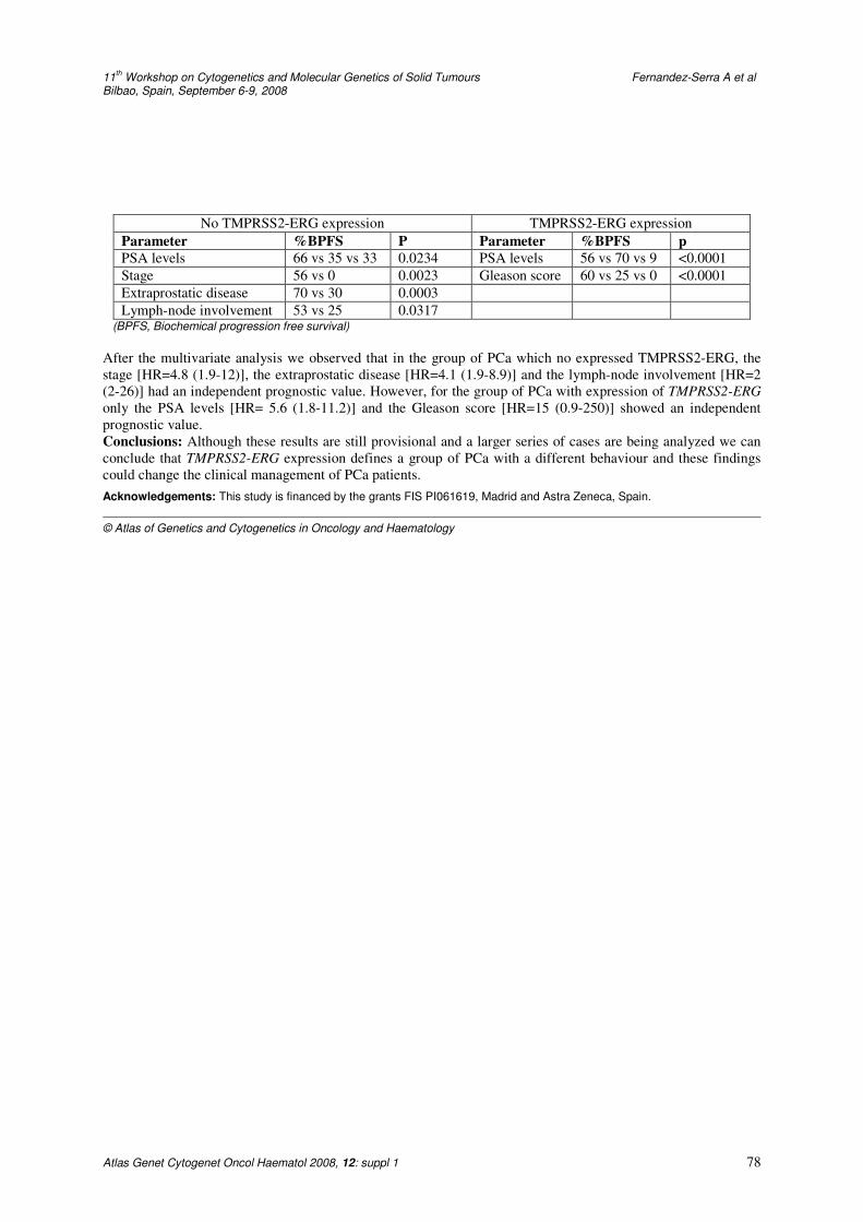
11
th Workshop on Cytogenetics and Molecular Genetics of Solid Tumours Fernandez-Serra A et al
Bilbao, Spain, September 6-9, 2008
Atlas Genet Cytogenet Oncol Haematol 2008, 12: suppl 1 78
No TMPRSS2-ERG expression TMPRSS2-ERG expression
Parameter %BPFS P Parameter %BPFS p
PSA levels 66 vs 35 vs 33 0.0234 PSA levels 56 vs 70 vs 9 <0.0001
Stage 56 vs 0 0.0023 Gleason score 60 vs 25 vs 0 <0.0001
Extraprostatic disease 70 vs 30 0.0003
Lymph-node involvement 53 vs 25 0.0317 (BPFS, Biochemical progression free survival)
After the multivariate analysis we observed that in the group of PCa which no expressed TMPRSS2-ERG, the
stage [HR=4.8 (1.9-12)], the extraprostatic disease [HR=4.1 (1.9-8.9)] and the lymph-node involvement [HR=2
(2-26)] had an independent prognostic value. However, for the group of PCa with expression of TMPRSS2-ERG
only the PSA levels [HR= 5.6 (1.8-11.2)] and the Gleason score [HR=15 (0.9-250)] showed an independent
prognostic value.
Conclusions: Although these results are still provisional and a larger series of cases are being analyzed we can
conclude that TMPRSS2-ERG expression defines a group of PCa with a different behaviour and these findings
could change the clinical management of PCa patients.
Acknowledgements: This study is financed by the grants FIS PI061619, Madrid and Astra Zeneca, Spain.
© Atlas of Genetics and Cytogenetics in Oncology and Haematology
11th European Workshop on Cytogenetics and Molecular Genetics of Solid Tumours
Bilbao, Spain, September 6-9, 2008
Atlas Genet Cytogenet Oncol Haematol 2008, 12: suppl 1 79
Atlas of Genetics and Cytogenetics in Oncology and Haematology
OPEN ACCESS JOURNAL AT INIST-CNRS
Index of authors
Abramowska A 14
Absi R 18
Acquadro F 5, 20, 39
Adelaide J 3
Adolfo I 35
Afonso L 44
Afonso M 17
Albanell J 6, 10
Alcaraz A 26
Algaba F 26, 37
Almenar S 71, 77
Alonso J 5
Alsop AE 75
Amann G 62
Ambros IM 62
Ambros PF 62
Armengol G 26
Arnold N 67
Arriola E 10
Arumi M 10
Aschero S 35
Aveyard J 16
Barrette S 18, 19
Barros-Silva D 44
Bartuma H 9
Basso ME 35
Bataller-Calatayud A 28
Beavis J 4
Bekhouche I 3
Belén de la Hoz A 30, 72
Benito R 47
Bento MJ 44
Berthold J 35
Bertolini G 51
Bertucci F 3
Beverloo H 53
Bignell GR 2
Birnbaum D 3
Bjerkehagen B 8
Blood K 4
Bocianowska A 42
Böhling T 24
Bonansea J 3
Bonassi S 25, 35
Bonillo MA 71, 77
Bouron-dal Soglio D 18
Bozsaky E 62
Brandão C 44
Brauze D 12
Brekke HR 8
Brugmans L 65
Brunner B 62
Brunner HG 45
Butler AP 2
Caballero J 28
Calatrava A 71, 77
Caldas C 4
Callaghan R 28, 29
Calvo A 70
Calzadas I 10
Campos M 70
Camps J 37, 38
Caplen NJ 38
Caria P 15
Carneiro F 44
Carracedo A 6, 10
Carrillo J 5
Carter N 4
Casarsa C 51
Catena R 70
Cejpek P 32
Celda B 47, 48
Cerdá-Nicolás M 29, 47, 48
Cerveira N 41, 5
Chaffanet M 3
Charafe-Jauffret E 3
Chin S 4
Chua Y 4
Cigudosa JC 5, 6, 20, 29, 39
Cinalli G 35
Coco S 25, 35
Collins VP 4
Colombo E 54
Connolly D 34
Cooke S 4
Corominas JM 6
Corzo C 6
Costa V 41
Crende O 72
Daidone MG 51
de Boer-van Huizen R 45
de Bruijn DRH 58
11th European Workshop on Cytogenetics and Molecular Genetics of Solid Tumours
Bilbao, Spain, September 6-9, 2008
Atlas Genet Cytogenet Oncol Haematol 2008, 12: suppl 1 80
Atlas of Genetics and Cytogenetics in Oncology and Haematology
OPEN ACCESS JOURNAL AT INIST-CNRS
de Klein A 53
De Vecchi C 25
Dear P 64
Defferrari R 25
del Rey J 26, 27, 37
Dettori T 15
Difilippantonio MJ 38
Dinis-Ribeiro M 44
Domanski HA 9
Dun G 66
Edwards PAW 4, 64, 75
Eken M 8
Eknæs M 8
Ellis I 4
Emons G 38
Espinet B 6, 10, 68
Espinosa L 57
Fernández-Capetillo O 76
Fernandez-Garcia I 56
Fernández-Serra A 71, 77
Ferreira BI 5, 6, 20, 39
Ferreira P 44
Ferrer-Luna R 47
Fetni R 18, 19
Finetti P 3
Finocchiaro G 51
Fischer M 35
Forni M 35
Fournet JC 18
Fragoso M 44
Frau DV 15
Futreal A 2
Gabra H 75
Gaforio J 70
Galvan A 68
Garcia MJ 16
García-Casado Z 71, 77
Garcia-Miguel P 5
Gasparini P 51
Gelabert A 37
Gentile M 24
Gesk S 67
Geurts van Kessel A 21, 40, 45, 58, 65, 74
Ghadimi BM 38
Giefing M 12, 13, 14
Gil-Benso R 28, 29, 47, 48
Glogowska-Ligus J 42
Gomez-Lopez G 5
Gonzalez-Darder J 47, 48
Goossens M 74
Grade M 38
Greally JM 34
Greenman CD 2
Grenman R 12, 13, 14
Gudla PR 61
Gumprecht J 42
Hall KS 8
Hattinger CM 24
Hazourli S 19
Hébert J 19
Heim S 67
Henrique R 17
Hetterschijt L 65
Hoenselaar E 21, 74
Hofstra R 66
Hoogerbrugge N 21, 45, 74
Horky J 32
Howarth K 4, 64
Hummon AB 38
Hurst C 16
Iborra I 71, 77
Ichimura K 4
Iliszko M 46
Iolascon A 35
Jacquemier J 3
Jarmuz M 12, 14
Jarosova M 22
Jerónimo C 41
Kamping EJ 21, 45, 74
Kaur S 24
Kilic E 53
Kiwerska K 13
Knowles MA 16
Knuutila S 24
Kodet R 22
Kok K 66
Kostrzewska-Poczekaj M 12
Kowalska A 62
Krskova L 22
Krupa P 32
Kuglik P 32
Kuiper RP 21, 45, 74
Kullendorff CM 9
Kuznetsova EB 59
Kuzniacka A 46
Largo C 5
Larizza L 54
Larramendy ML 24
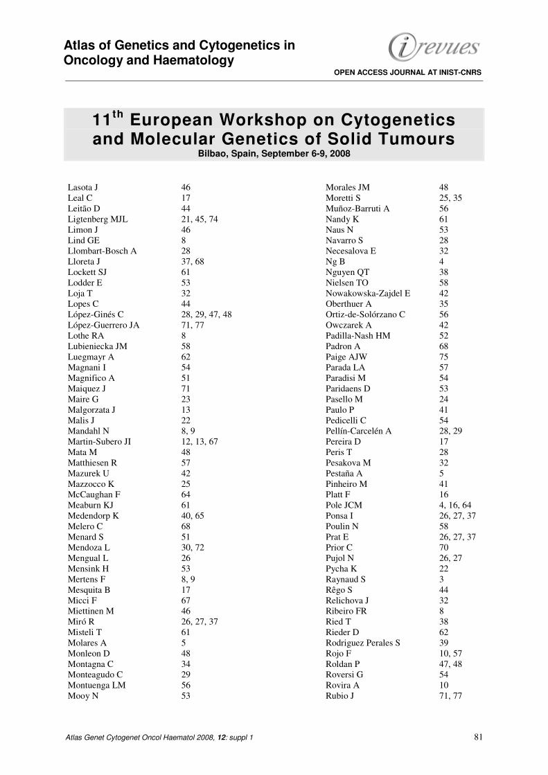
11th European Workshop on Cytogenetics and Molecular Genetics of Solid Tumours
Bilbao, Spain, September 6-9, 2008
Atlas Genet Cytogenet Oncol Haematol 2008, 12: suppl 1 81
Atlas of Genetics and Cytogenetics in Oncology and Haematology
OPEN ACCESS JOURNAL AT INIST-CNRS
Lasota J 46
Leal C 17
Leitão D 44
Ligtenberg MJL 21, 45, 74
Limon J 46
Lind GE 8
Llombart-Bosch A 28
Lloreta J 37, 68
Lockett SJ 61
Lodder E 53
Loja T 32
Lopes C 44
López-Ginés C 28, 29, 47, 48
López-Guerrero JA 71, 77
Lothe RA 8
Lubieniecka JM 58
Luegmayr A 62
Magnani I 54
Magnifico A 51
Maiquez J 71
Maire G 23
Malgorzata J 13
Malis J 22
Mandahl N 8, 9
Martin-Subero JI 12, 13, 67
Mata M 48
Matthiesen R 57
Mazurek U 42
Mazzocco K 25
McCaughan F 64
Meaburn KJ 61
Medendorp K 40, 65
Melero C 68
Menard S 51
Mendoza L 30, 72
Mengual L 26
Mensink H 53
Mertens F 8, 9
Mesquita B 17
Micci F 67
Miettinen M 46
Miró R 26, 27, 37
Misteli T 61
Molares A 5
Monleon D 48
Montagna C 34
Monteagudo C 29
Montuenga LM 56
Mooy N 53
Morales JM 48
Moretti S 25, 35
Muñoz-Barruti A 56
Nandy K 61
Naus N 53
Navarro S 28
Necesalova E 32
Ng B 4
Nguyen QT 38
Nielsen TO 58
Nowakowska-Zajdel E 42
Oberthuer A 35
Ortiz-de-Solórzano C 56
Owczarek A 42
Padilla-Nash HM 52
Padron A 68
Paige AJW 75
Parada LA 57
Paradisi M 54
Paridaens D 53
Pasello M 24
Paulo P 41
Pedicelli C 54
Pellín-Carcelén A 28, 29
Pereira D 17
Peris T 28
Pesakova M 32
Pestaña A 5
Pinheiro M 41
Platt F 16
Pole JCM 4, 16, 64
Ponsa I 26, 27, 37
Poulin N 58
Prat E 26, 27, 37
Prior C 70
Pujol N 26, 27
Pycha K 22
Raynaud S 3
Rêgo S 44
Relichova J 32
Ribeiro FR 8
Ried T 38
Rieder D 62
Rodriguez Perales S 39
Rojo F 10, 57
Roldan P 47, 48
Roversi G 54
Rovira A 10
Rubio J 71, 77
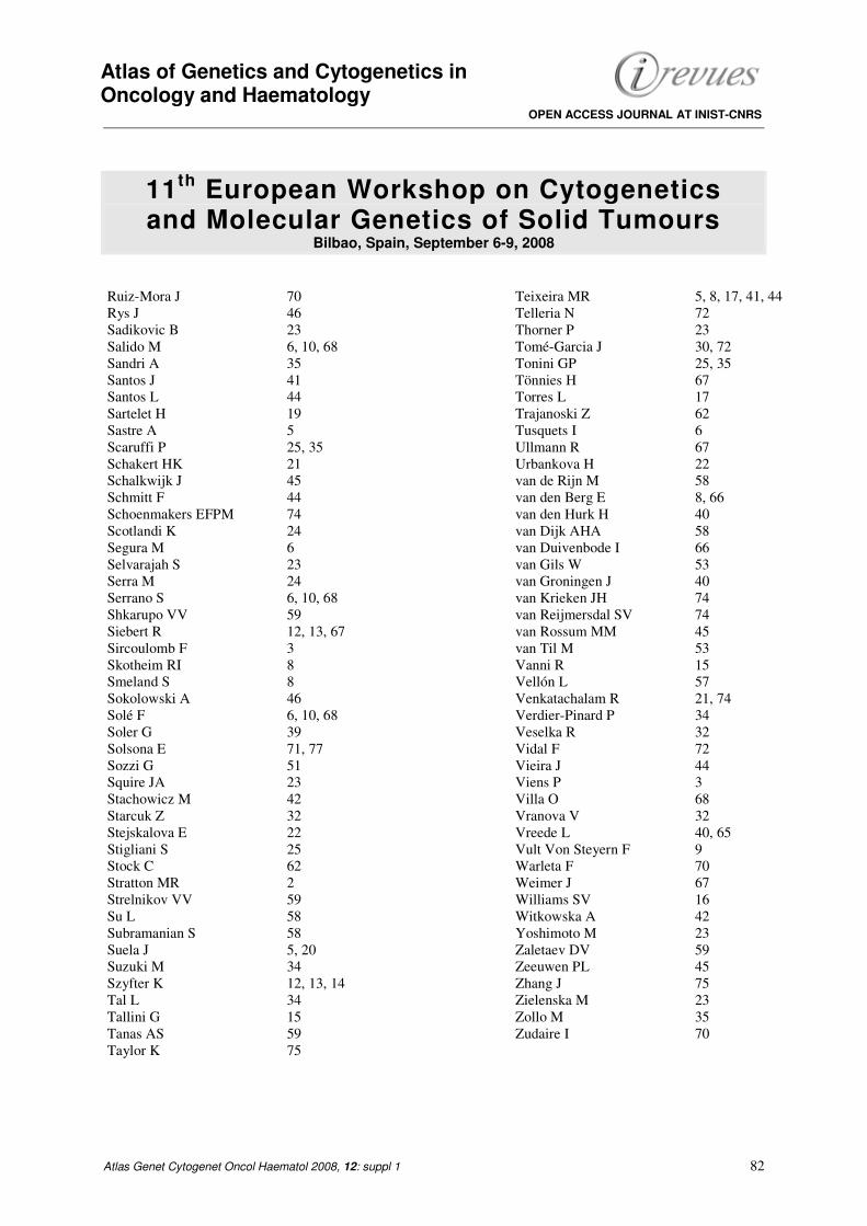
11th European Workshop on Cytogenetics and Molecular Genetics of Solid Tumours
Bilbao, Spain, September 6-9, 2008
Atlas Genet Cytogenet Oncol Haematol 2008, 12: suppl 1 82
Atlas of Genetics and Cytogenetics in Oncology and Haematology
OPEN ACCESS JOURNAL AT INIST-CNRS
Ruiz-Mora J 70
Rys J 46
Sadikovic B 23
Salido M 6, 10, 68
Sandri A 35
Santos J 41
Santos L 44
Sartelet H 19
Sastre A 5
Scaruffi P 25, 35
Schakert HK 21
Schalkwijk J 45
Schmitt F 44
Schoenmakers EFPM 74
Scotlandi K 24
Segura M 6
Selvarajah S 23
Serra M 24
Serrano S 6, 10, 68
Shkarupo VV 59
Siebert R 12, 13, 67
Sircoulomb F 3
Skotheim RI 8
Smeland S 8
Sokolowski A 46
Solé F 6, 10, 68
Soler G 39
Solsona E 71, 77
Sozzi G 51
Squire JA 23
Stachowicz M 42
Starcuk Z 32
Stejskalova E 22
Stigliani S 25
Stock C 62
Stratton MR 2
Strelnikov VV 59
Su L 58
Subramanian S 58
Suela J 5, 20
Suzuki M 34
Szyfter K 12, 13, 14
Tal L 34
Tallini G 15
Tanas AS 59
Taylor K 75
Teixeira MR 5, 8, 17, 41, 44
Telleria N 72
Thorner P 23
Tomé-Garcia J 30, 72
Tonini GP 25, 35
Tönnies H 67
Torres L 17
Trajanoski Z 62
Tusquets I 6
Ullmann R 67
Urbankova H 22
van de Rijn M 58
van den Berg E 8, 66
van den Hurk H 40
van Dijk AHA 58
van Duivenbode I 66
van Gils W 53
van Groningen J 40
van Krieken JH 74
van Reijmersdal SV 74
van Rossum MM 45
van Til M 53
Vanni R 15
Vellón L 57
Venkatachalam R 21, 74
Verdier-Pinard P 34
Veselka R 32
Vidal F 72
Vieira J 44
Viens P 3
Villa O 68
Vranova V 32
Vreede L 40, 65
Vult Von Steyern F 9
Warleta F 70
Weimer J 67
Williams SV 16
Witkowska A 42
Yoshimoto M 23
Zaletaev DV 59
Zeeuwen PL 45
Zhang J 75
Zielenska M 23
Zollo M 35
Zudaire I 70
Atlas of Genetics and Cytogenetics in Oncology and Haematology
OPEN ACCESS JOURNAL AT INIST-CNRS
Instructions to Authors Manuscripts submitted to the Atlas must be submitted solely to the Atlas.
In general, iconography is most welcome: there is no space restriction.
The Atlas publishes "cards", "deep insights", "case reports", and "educational items".
Cards are structured review articles. Detailed instructions for these structured reviews can be found at:
http://AtlasGeneticsOncology.org/Forms/Gene_Form.html for reviews on genes
http://AtlasGeneticsOncology.org/Forms/Leukemia_Form.html for reviews on leukemias,
http://AtlasGeneticsOncology.org/Forms/SolidTumor_Form.html for reviews on solid tumors,
http://AtlasGeneticsOncology.org/Forms/CancerProne_Form.html for reviews on cancer-prone diseases.
According to the length of the paper, cards are divided, into "reviews" (texts exceeding 2000 words), "mini reviews"
(between), and "short communications" (texts below 400 words). The latter category may not be accepted for indexing by
bibliographic databases.
Deep Insights are written as traditional papers, made of paragraphs with headings, at the author's convenience. No length
restriction.
Case Reports in hematological malignancies This section is dedicated to recurrent -but rare- chromosomes abnormalities in
leukemias/lymphomas. Cases of interest shall be: 1- recurrent (i.e. the chromosome anomaly has already been described in at
least 1 case), 2- rare (previously described in less than 20 cases), 3- with well documented clinics and laboratory findings,
and 4- iconography of chromosomes.
It is mandatory that the specific "Submission form for Case reports" is used:
see http://AtlasGeneticsOncology.org/Reports/Case_Report_Submission.html.
Educational Items must be didactic, give full information; accompanied with iconography. Translations into French,
German, Italian, and Spanish are welcome.
Subscription: The Atlas is FREE!
Corporate patronage, sponsorship and advertising Enquiries should be addressed to
Rules, Copyright Notice and Disclaimer Conflicts of Interest Authors must state explicitly whether potential conflicts do or do not exist. Reviewers must disclose to
editors any conflicts of interest that could bias their opinions of the manuscript. The editor and the editorial board members
must disclose any potential conflict. Privacy and Confidentiality – Iconography Patients have a right to privacy. Identifying details should be omitted. If
complete anonymity is difficult to achieve, informed consent should be obtained. Property As "cards" are to evolve with further improvements and updates from various contributors, the property of the
cards belongs to the editor, and modifications will be made without authorization from the previous contributor (who may,
nonetheless, be asked for refereeing); contributors are listed in an edit history manner. Authors keep the rights to use further
the content of their papers published in the Atlas, provided that the source is cited. Copyright The information in the Atlas of Genetics and Cytogenetics in Oncology and Haematology is issued for general
distribution. All rights are reserved. The information presented is protected under international conventions and under
national laws on copyright and neighbouring rights. Commercial use is totally forbidden Information extracted from the Atlas
may be reviewed, reproduced or translated for research or private study but not for sale or for use in conjunction with
commercial purposes. Any use of information from the Atlas should be accompanied by an acknowledgment of the Atlas as
the source, citing the uniform resource locator (URL) of the article and/or the article reference, according to the Vancouver
convention. Reference to any specific commercial products, process, or service by trade name, trademark, manufacturer, or
otherwise, does not necessarily constitute or imply its endorsement, recommendation, or favouring. The views and opinions
of contributors and authors expressed herein do not necessarily state or reflect those of the Atlas editorial staff or of the web
site holder, and shall not be used for advertising or product endorsement purposes. The Atlas does not make any warranty,
express or implied, including the warranties of merchantability and fitness for a particular purpose, or assumes any legal
liability or responsibility for the accuracy, completeness, or usefulness of any information, and shall not be liable whatsoever
for any damages incurred as a result of its use. In particular, information presented in the Atlas is only for research purpose,
and shall not be used for diagnosis or treatment purposes. No responsibility is assumed for any injury and/or damage to
persons or property for any use or operation of any methods products, instructions or ideas contained in the material herein. See also: "Uniform Requirements for Manuscripts Submitted to Biomedical Journals: Writing and Editing for Biomedical
Publication - Updated October 2004": http://www.icmje.org
http://AtlasGeneticsOncology.org
© ATLAS - ISSN 1768-3262