Embed Size (px)
Citation preview
3310 Yu Yao et al. Eur. J. Immunol. 2012. 42: 3310–3321DOI: 10.1002/eji.201142213
ATP conditions intestinal epithelial cells to aninflammatory state that promotes componentsof DC maturation
Yu Yao1, Megan K. Levings2 and Theodore S. Steiner1
1 Department of Medicine, University of British Columbia and Vancouver Coastal HealthResearch Institute, Vancouver, B.C., Canada
2 Department of Surgery, University of British Columbia and Child and Family ResearchInstitute, Vancouver, B.C., Canada
Intestinal epithelial cells (IECs) normally promote the development of gut resident tolero-genic dendritic cells (DCs) and regulatory T cells, but how this process is altered in inflam-matory bowel disease is not well characterized. Recently, we published that the cell injurysignal ATP modulates IEC chemokine responses to the TLR5 ligand flagellin and exacer-bates colitis in the presence of flagellin. We hypothesized that ATP switches these IECsfrom tolerogenic to proinflammatory, enhancing DC activation and immune responsesto commensal antigens. Here, we report that ATP enhanced murine IEC production ofKC, IL-6, TGF-β, and thymic stromal lymphopoietin in response to TLR1/2 stimulation byPam3CSK4 (PAM). Moreover, supernatants from IECs stimulated with ATP+PAM enhancedexpression of CD80 on bone marrow derived dendritic cells, and increased their produc-tion of IL-12, IL-6, IL-23, TGF-β, and aldh1a2, suggesting a Th1/Th17 polarizing envi-ronment. DCs conditioned by stressed IECs stimulated an enhanced recall response toflagellin and supported the expansion of IFN-γ+ and IL-17+ memory T cells. Lastly, colonicadministration of nonhydrolysable ATP increased production of IL-6 and Cxcl1 (KC) byIECs. These findings indicate that ATP influences the response of IECs to TLR ligands andbiases the maturation of DCs to become inflammatory.
Keywords: CD4 T cells � Dendritic cells � Immune regulation � Intestinal immunity � Toll-likereceptors
Supporting Information available online
Introduction
Intestinal epithelial cells (IECs) coordinate the dynamic interac-tions between luminal microbes and local immune cells and rep-resent the front line of enteric defense [1]. In addition to act-ing as a physical barrier to prevent passage of luminal contents,
Correspondence: Dr. Theodore S. Steinere-mail: [email protected]
IECs are crucial for maintaining intestinal homeostasis by sam-pling the luminal microenvironment, integrating signals receivedfrom pattern recognition receptors (PRRs) and local immune cells,and secreting factors that regulate adaptive immunity by primingintestinal dendritic cells (DCs) [2]. Under steady state conditions,IECs maintain a hyporesponsive state to commensal flora by secret-ing factors such as thymic stromal lymphopoietin (TSLP), TGF-β,and retinoic acid (RA), which together facilitate the developmentof tolerogenic DCs and regulatory T (Treg) cells [3, 4]. Duringenteric infection, however, TLR-mediated activation of IECs can
C© 2012 WILEY-VCH Verlag GmbH & Co. KGaA, Weinheim www.eji-journal.eu
Eur. J. Immunol. 2012. 42: 3310–3321 Immunodulation 3311
initiate robust inflammatory responses. How IECs discriminatebetween TLR ligands presented by invasive pathogens versus com-mensal microbes remains largely unknown [5].
One way that IECs may respond to pathogens is by recognizingspecific danger signals such as extracellular ATP [6, 7]. Recently,several groups reported that ATP contributes to the pathogene-sis of a variety of inflammatory disorders, such as asthma, graftversus host disease, and inflammatory bowel disease (IBD), sug-gesting it is an endogenous danger signal that promotes inflam-mation [8–12]. Importantly, low expression of CD39, an enzymethat hydrolyzes ATP, is associated with increased risk for Crohn’sdisease (CD) [12]. Consistent with this finding, CD39-deficientmice have increased susceptibility to dextran sulfate sodium (DSS)induced colitis [12]. The association of ATP with enhanced inflam-mation in the gut has also lead to the recent identification of com-mensal bacteria that actively produce ATP in the lumen [13, 14],providing another important source of ATP in addition to epithelialinjuries as a result of inflammation [14,15]. In the context of IBD,where patients have weaker intestinal barrier function and bacte-rial leakage across the mucus layer, IECs represent the importantfirst responders to cellular stress and microbial ligands. There-fore, it is essential to understand how ATP regulates inflammatoryresponses to TLR activation in IECs.
Previous work in our laboratory showed that ATP regulatesthe inflammatory response to flagellin-TLR5 ligation in humanIECs and that rectal administration of ATP enhanced flagellin-mediated inflammation during DSS-colitis [16]. In this study, weextend our findings to murine IECs and utilize an in vitro coculturemodel with supernatants from IECs and bone marrow deriveddendritic cells (BMDCs) to test how stressed IECs regulate DCmaturation. Furthermore, we explored in vivo whether colorectaladministration of ATP alone, in the absence of DSS colitis, is ableto modulate the how IECs respond to commensal microbes. Wehypothesized that simultaneous exposure to ATP and TLR ligandswould modulate how IECs respond to TLR stimulation, facilitatingthe maturation of DCs and ultimately determining how T cellsrespond to commensal antigens.
Results
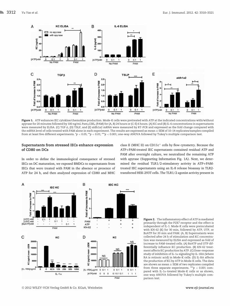
ATP modulates the production of cytokines andchemokines by TLR1/2-activated IECs
During cellular damage and inflammation, ATP can be releasedimmediately into the extracellular space, attaining local concen-trations in the millimolar range. At that concentration, it acts asan important danger signal by activating P2 purinergic receptors,alerting immune cells to the presence of tissue damage and mobi-lizing them to the site of injuries to fight pathogens and clearcellular debris [17, 18]. We previously showed that ATP altersTLR5 signaling in Caco-2 human IECs, and sought to determinewhether this phenomenon applied to murine IECs as well. To testthis, Mode-K IECs were stimulated with different concentrationsof ATP and TLR1/2 agonist Pam3CSK4 (PAM). We focused on
pro-inflammatory mediators and measured secretion of the neu-trophil chemoattractant KC (Cxcl1) and IL-6 by ELISA. As shown inFigure 1A, 1 mM ATP significantly augmented TLR1/2 activation-induced production of KC (from 29.9 ± 1.41 to 73.3 ±2.67 ng/mL), while ATP alone did not induce KC expression. Asimilar increase in IL-6 production was observed with ATP at alower concentration (from 156.4 ± 20.04 to 259.2 ± 9.49 pg/mL,Fig. 1B). Furthermore, the ATP-induced KC secretion was com-pletely blocked by treatment with apyrase, which degrades ATPinto ADP and AMP (from 73.3 ± 2.67 to 32.3 ± 1.97 ng/mL,Fig. 1A), indicating that this effect requires ATP and not itshydrolytic products.
Since IECs are known to produce tolerogenic factors that main-tain immune tolerance to commensal microbes in the intestinaltract [3, 4], we also asked whether ATP modulated the expres-sion of anti-inflammatory mediators. We thus measured expres-sion of TGF-β, TSLP, and aldh1a2, a rate-limiting enzyme in thesynthesis of RA. ATP increased TLR1/2-mediated expression ofmRNA encoding for each of these proteins (Fig. 1C–E), whichwould suggest induction of a tolerant state. However, these tolero-genic factors can promote inflammation in the appropriate milieu[19–21], such as in the presence of large amounts of IL-6 or IL-15.Hence they may act to reduce tolerance in the presence of cellularinjuries.
ATP acts primarily through the P2X7 receptor on IECs
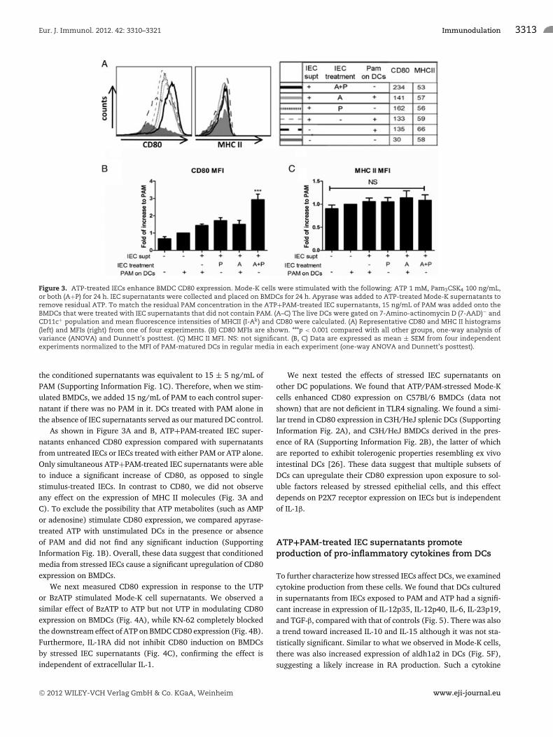
Because most of the biological effects of extracellular ATP aremediated through P2 purinergic receptors [22], we exploredwhich P2 receptors mediated the pro-inflammatory effect onthe IECs. To study this, we first tested two different ATP ana-logues, UTP (uridine 5′-triphosphate), and BzATP (2′(3′)-O-(4-benzoylbenzoyl)adenosine 5′-triphosphate), both of whichhave preferential binding to different P2 receptors. As shownin Figure 2A, BzATP, a preferential P2X7 receptor agonist [23],potently enhanced KC production (BzATP 3.4 ± 0.51 fold com-pared with ATP 3.1 ± 0.57 fold). In contrast, UTP, which is arelatively selective P2Y receptor agonist, only minimally increasedKC production (Fig. 2A, 1.36 ± 1.58 fold). In keeping with theseresults, KN-62, a noncompetitive antagonist for P2X7, completelyblocked the enhancement of KC secretion by ATP (Fig. 2B). Thesedata indicate that the effects of ATP on IECs are largely mediatedthrough the P2X7 receptor and not P2Y receptors.
Since the P2X7 receptor is known for the important down-stream effect of inflammasome activation and IL-1β processing[24], we then asked whether paracrine release of IL-1β is respon-sible for the inflammatory effect of ATP in Mode-K cells, as hasbeen reported in T84 human IECs [25]. We did not detect anyproduction of IL-1β at the protein level as a result of ATP and PAMtreatment (data not shown). Moreover, when we treated Mode-Kcells with IL-1ra in concentrations sufficient to block IL-1 receptorsignaling (Fig. 2C), we did not inhibit KC production by Mode-Ks(Fig. 2D), suggesting that neither extracellular IL-1α nor IL-1β isresponsible for the reported Mode-K phenotype that we observed.
C© 2012 WILEY-VCH Verlag GmbH & Co. KGaA, Weinheim www.eji-journal.eu
3312 Yu Yao et al. Eur. J. Immunol. 2012. 42: 3310–3321
Figure 1. ATP enhances IEC cytokine/chemokine production. Mode-K cells were pretreated with ATP at the indicated concentrations with/withoutapyrase for 20 minutes followed by 100 ng/mL Pam3CSK4 (PAM) for (A, B) 24 hours or (C-E) 6 hours. (A) KC and (B) IL-6 concentrations in supernatantswere measured by ELISA. (C) TGF-β, (D) TSLP, and (E) aldh1a2 mRNA were measured by RT-PCR and expressed as the fold change compared withthe mRNA level of cells treated with PAM alone in each experiment. The results are expressed as mean ± SEM of 10–14 replicates/samples compiledfrom at least five different experiments. *p < 0.05; **p < 0.01; ***p < 0.001; one-way ANOVA followed by Tukey’s multiple comparison test.
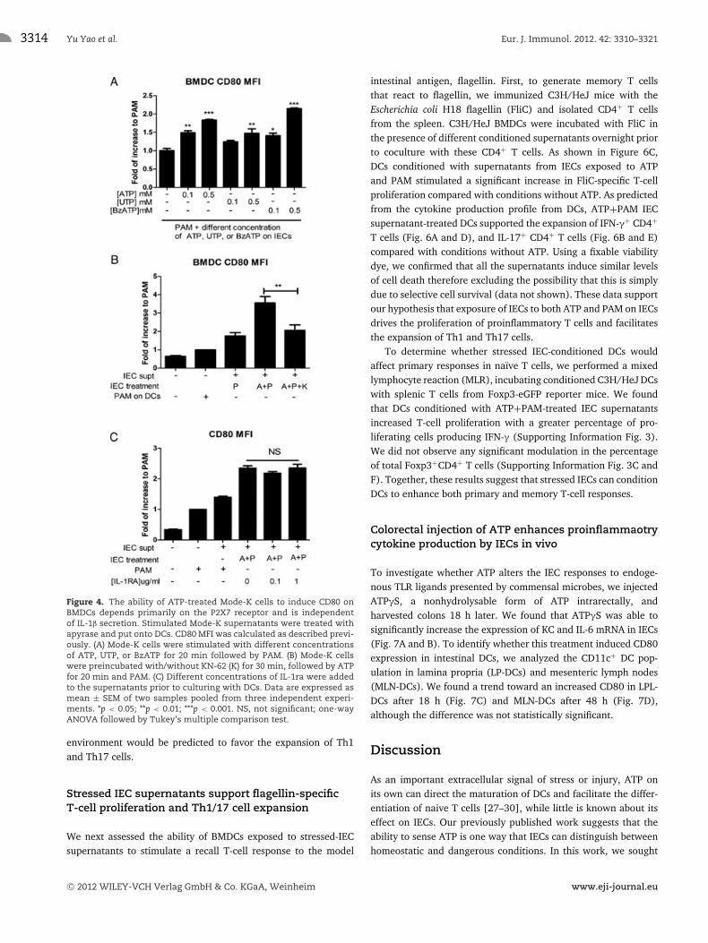
Supernatants from stressed IECs enhance expressionof CD80 on DCs
In order to define the immunological consequence of stressedIECs on DC maturation, we exposed BMDCs to supernatants fromIECs that were treated with PAM in the absence or presence ofATP for 24 h, and then analyzed expression of CD80 and MHC
class II (MHC II) on CD11c+ cells by flow cytometry. Because theATP+PAM-treated IEC supernatants contained residual ATP andPAM after overnight culture, we neutralized the remaining ATPwith apyrase (Supporting Information Fig. 1A). Next, we deter-mined the residual TLR1/2-stimulatory activity in ATP+PAM-treated IEC supernatants using an IL-8 release bioassay in TLR2-transfected HEK-293T cells. The TLR1/2 agonist activity present in
Figure 2. The inflammatory effect of ATP is mediatedprimarily through the P2X7 receptor and the effect isindependent of IL-1. Mode-K cells were preincubatedwith KN-62 (K) for 30 min, followed by ATP, UTP, orBzATP for 20 min and PAM. (A, B) Supernatants werecollected after 24 h of stimulation and KC concentra-tion was measured by ELISA and expressed as fold ofincrease to PAM-treated cells. (A) BzATP and UTP dif-ferentially influence KC production. (B) KN-62 treat-ment affects KC production by ATP. (C) Dose-responsestudy of inhibition of IL-1α signaling by IL-1RA (whereRA is retinoic acid) in Mode-K cells. (D) IL-RA affectsthe production of KC by ATP in Mode-K cells. The dataare shown as mean ± SEM of two replicates compiledfrom three separate experiments. ***p < 0.001 com-pared with IL-1α-treated Mode-K cells or as shown,one-way ANOVA followed by Tukey’s multiple com-parison test.
C© 2012 WILEY-VCH Verlag GmbH & Co. KGaA, Weinheim www.eji-journal.eu
Eur. J. Immunol. 2012. 42: 3310–3321 Immunodulation 3313
Figure 3. ATP-treated IECs enhance BMDC CD80 expression. Mode-K cells were stimulated with the following: ATP 1 mM, Pam3CSK4 100 ng/mL,or both (A+P) for 24 h. IEC supernatants were collected and placed on BMDCs for 24 h. Apyrase was added to ATP-treated Mode-K supernatants toremove residual ATP. To match the residual PAM concentration in the ATP+PAM-treated IEC supernatants, 15 ng/mL of PAM was added onto theBMDCs that were treated with IEC supernatants that did not contain PAM. (A–C) The live DCs were gated on 7-Amino-actinomycin D (7-AAD)− andCD11c+ population and mean fluorescence intensities of MHCII (I-Ak) and CD80 were calculated. (A) Representative CD80 and MHC II histograms(left) and MFIs (right) from one of four experiments. (B) CD80 MFIs are shown. ***p < 0.001 compared with all other groups, one-way analysis ofvariance (ANOVA) and Dunnett’s posttest. (C) MHC II MFI. NS: not significant. (B, C) Data are expressed as mean ± SEM from four independentexperiments normalized to the MFI of PAM-matured DCs in regular media in each experiment (one-way ANOVA and Dunnett’s posttest).
the conditioned supernatants was equivalent to 15 ± 5 ng/mL ofPAM (Supporting Information Fig. 1C). Therefore, when we stim-ulated BMDCs, we added 15 ng/mL of PAM to each control super-natant if there was no PAM in it. DCs treated with PAM alone inthe absence of IEC supernatants served as our matured DC control.
As shown in Figure 3A and B, ATP+PAM-treated IEC super-natants enhanced CD80 expression compared with supernatantsfrom untreated IECs or IECs treated with either PAM or ATP alone.Only simultaneous ATP+PAM-treated IEC supernatants were ableto induce a significant increase of CD80, as opposed to singlestimulus-treated IECs. In contrast to CD80, we did not observeany effect on the expression of MHC II molecules (Fig. 3A andC). To exclude the possibility that ATP metabolites (such as AMPor adenosine) stimulate CD80 expression, we compared apyrase-treated ATP with unstimulated DCs in the presence or absenceof PAM and did not find any significant induction (SupportingInformation Fig. 1B). Overall, these data suggest that conditionedmedia from stressed IECs cause a significant upregulation of CD80expression on BMDCs.
We next measured CD80 expression in response to the UTPor BzATP stimulated Mode-K cell supernatants. We observed asimilar effect of BzATP to ATP but not UTP in modulating CD80expression on BMDCs (Fig. 4A), while KN-62 completely blockedthe downstream effect of ATP on BMDC CD80 expression (Fig. 4B).Furthermore, IL-1RA did not inhibit CD80 induction on BMDCsby stressed IEC supernatants (Fig. 4C), confirming the effect isindependent of extracellular IL-1.
We next tested the effects of stressed IEC supernatants onother DC populations. We found that ATP/PAM-stressed Mode-Kcells enhanced CD80 expression on C57Bl/6 BMDCs (data notshown) that are not deficient in TLR4 signaling. We found a simi-lar trend in CD80 expression in C3H/HeJ splenic DCs (SupportingInformation Fig. 2A), and C3H/HeJ BMDCs derived in the pres-ence of RA (Supporting Information Fig. 2B), the latter of whichare reported to exhibit tolerogenic properties resembling ex vivointestinal DCs [26]. These data suggest that multiple subsets ofDCs can upregulate their CD80 expression upon exposure to sol-uble factors released by stressed epithelial cells, and this effectdepends on P2X7 receptor expression on IECs but is independentof IL-1β.
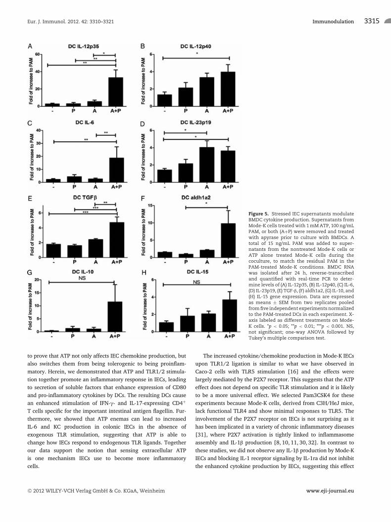
ATP+PAM-treated IEC supernatants promoteproduction of pro-inflammatory cytokines from DCs
To further characterize how stressed IECs affect DCs, we examinedcytokine production from these cells. We found that DCs culturedin supernatants from IECs exposed to PAM and ATP had a signifi-cant increase in expression of IL-12p35, IL-12p40, IL-6, IL-23p19,and TGF-β, compared with that of controls (Fig. 5). There was alsoa trend toward increased IL-10 and IL-15 although it was not sta-tistically significant. Similar to what we observed in Mode-K cells,there was also increased expression of aldh1a2 in DCs (Fig. 5F),suggesting a likely increase in RA production. Such a cytokine
C© 2012 WILEY-VCH Verlag GmbH & Co. KGaA, Weinheim www.eji-journal.eu
3314 Yu Yao et al. Eur. J. Immunol. 2012. 42: 3310–3321
Figure 4. The ability of ATP-treated Mode-K cells to induce CD80 onBMDCs depends primarily on the P2X7 receptor and is independentof IL-1β secretion. Stimulated Mode-K supernatants were treated withapyrase and put onto DCs. CD80 MFI was calculated as described previ-ously. (A) Mode-K cells were stimulated with different concentrationsof ATP, UTP, or BzATP for 20 min followed by PAM. (B) Mode-K cellswere preincubated with/without KN-62 (K) for 30 min, followed by ATPfor 20 min and PAM. (C) Different concentrations of IL-1ra were addedto the supernatants prior to culturing with DCs. Data are expressed asmean ± SEM of two samples pooled from three independent experi-ments. *p < 0.05; **p < 0.01; ***p < 0.001. NS, not significant; one-wayANOVA followed by Tukey’s multiple comparison test.
environment would be predicted to favor the expansion of Th1and Th17 cells.
Stressed IEC supernatants support flagellin-specificT-cell proliferation and Th1/17 cell expansion
We next assessed the ability of BMDCs exposed to stressed-IECsupernatants to stimulate a recall T-cell response to the model
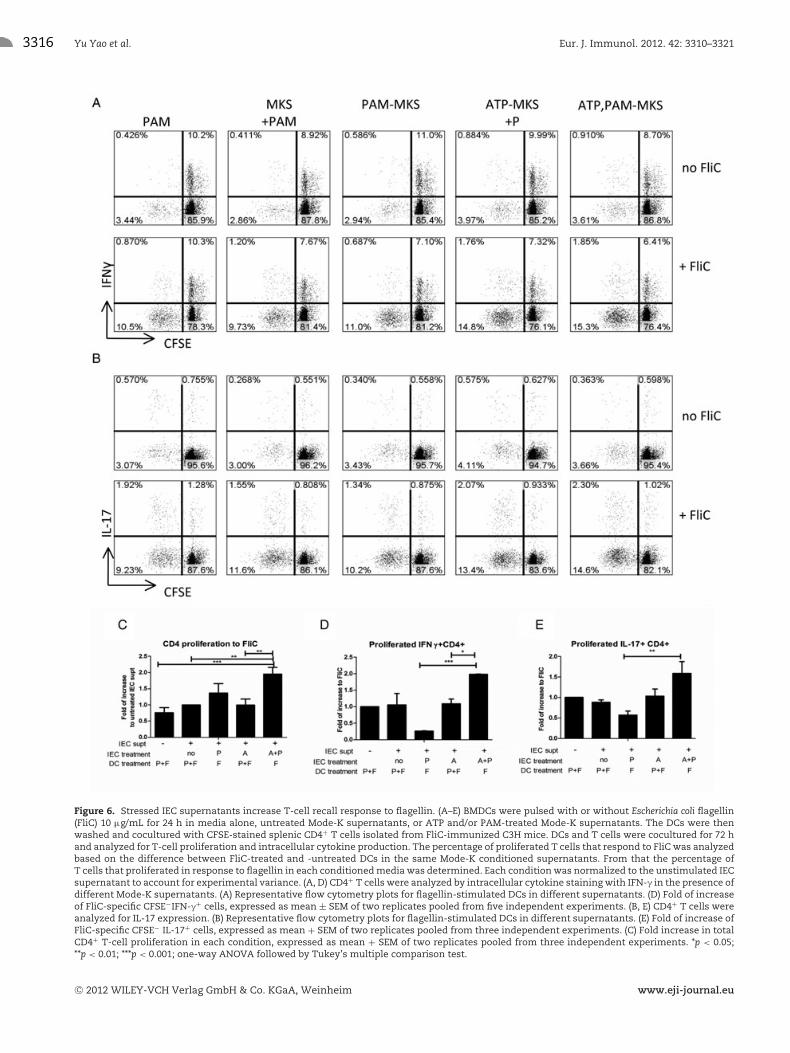
intestinal antigen, flagellin. First, to generate memory T cellsthat react to flagellin, we immunized C3H/HeJ mice with theEscherichia coli H18 flagellin (FliC) and isolated CD4+ T cellsfrom the spleen. C3H/HeJ BMDCs were incubated with FliC inthe presence of different conditioned supernatants overnight priorto coculture with these CD4+ T cells. As shown in Figure 6C,DCs conditioned with supernatants from IECs exposed to ATPand PAM stimulated a significant increase in FliC-specific T-cellproliferation compared with conditions without ATP. As predictedfrom the cytokine production profile from DCs, ATP+PAM IECsupernatant-treated DCs supported the expansion of IFN-γ+ CD4+
T cells (Fig. 6A and D), and IL-17+ CD4+ T cells (Fig. 6B and E)compared with conditions without ATP. Using a fixable viabilitydye, we confirmed that all the supernatants induce similar levelsof cell death therefore excluding the possibility that this is simplydue to selective cell survival (data not shown). These data supportour hypothesis that exposure of IECs to both ATP and PAM on IECsdrives the proliferation of proinflammatory T cells and facilitatesthe expansion of Th1 and Th17 cells.
To determine whether stressed IEC-conditioned DCs wouldaffect primary responses in naıve T cells, we performed a mixedlymphocyte reaction (MLR), incubating conditioned C3H/HeJ DCswith splenic T cells from Foxp3-eGFP reporter mice. We foundthat DCs conditioned with ATP+PAM-treated IEC supernatantsincreased T-cell proliferation with a greater percentage of pro-liferating cells producing IFN-γ (Supporting Information Fig. 3).We did not observe any significant modulation in the percentageof total Foxp3+CD4+ T cells (Supporting Information Fig. 3C andF). Together, these results suggest that stressed IECs can conditionDCs to enhance both primary and memory T-cell responses.
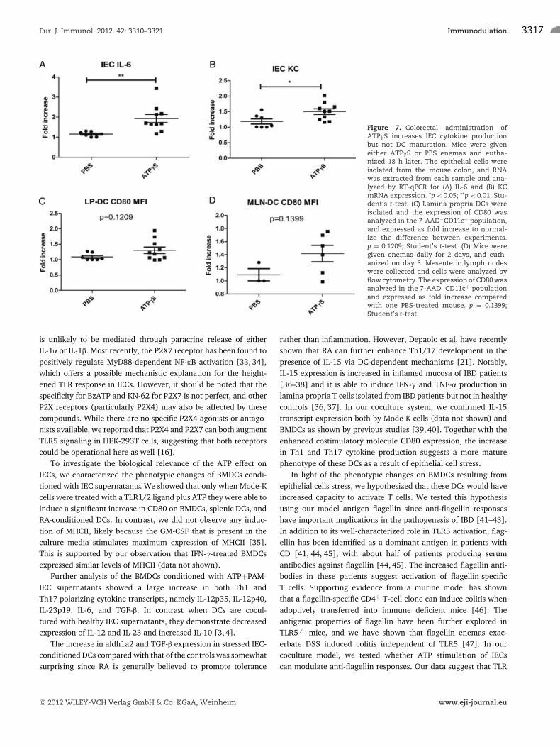
Colorectal injection of ATP enhances proinflammaotrycytokine production by IECs in vivo
To investigate whether ATP alters the IEC responses to endoge-nous TLR ligands presented by commensal microbes, we injectedATPγS, a nonhydrolysable form of ATP intrarectally, andharvested colons 18 h later. We found that ATPγS was able tosignificantly increase the expression of KC and IL-6 mRNA in IECs(Fig. 7A and B). To identify whether this treatment induced CD80expression in intestinal DCs, we analyzed the CD11c+ DC pop-ulation in lamina propria (LP-DCs) and mesenteric lymph nodes(MLN-DCs). We found a trend toward an increased CD80 in LPL-DCs after 18 h (Fig. 7C) and MLN-DCs after 48 h (Fig. 7D),although the difference was not statistically significant.
Discussion
As an important extracellular signal of stress or injury, ATP onits own can direct the maturation of DCs and facilitate the differ-entiation of naive T cells [27–30], while little is known about itseffect on IECs. Our previously published work suggests that theability to sense ATP is one way that IECs can distinguish betweenhomeostatic and dangerous conditions. In this work, we sought
C© 2012 WILEY-VCH Verlag GmbH & Co. KGaA, Weinheim www.eji-journal.eu
Eur. J. Immunol. 2012. 42: 3310–3321 Immunodulation 3315
Figure 5. Stressed IEC supernatants modulateBMDC cytokine production. Supernatants fromMode-K cells treated with 1 mM ATP, 100 ng/mLPAM, or both (A+P) were removed and treatedwith apyrase prior to culture with BMDCs. Atotal of 15 ng/mL PAM was added to super-natants from the nontreated Mode-K cells orATP alone treated Mode-K cells during thecoculture, to match the residual PAM in thePAM-treated Mode-K conditions. BMDC RNAwas isolated after 24 h, reverse-transcribedand quantified with real-time PCR to deter-mine levels of (A) IL-12p35, (B) IL-12p40, (C) IL-6,(D) IL-23p19, (E) TGF-β, (F) aldh1a2, (G) IL-10, and(H) IL-15 gene expression. Data are expressedas means ± SEM from two replicates pooledfrom five independent experiments normalizedto the PAM-treated DCs in each experiment. X-axis labeled as different treatments on Mode-K cells. *p < 0.05; **p < 0.01; ***p < 0.001. NS,not significant; one-way ANOVA followed byTukey’s multiple comparison test.
to prove that ATP not only affects IEC chemokine production, butalso switches them from being tolerogenic to being proinflam-matory. Herein, we demonstrated that ATP and TLR1/2 stimula-tion together promote an inflammatory response in IECs, leadingto secretion of soluble factors that enhance expression of CD80and pro-inflammatory cytokines by DCs. The resulting DCs causean enhanced stimulation of IFN-γ- and IL-17-expressing CD4+
T cells specific for the important intestinal antigen flagellin. Fur-thermore, we showed that ATP enemas can lead to increasedIL-6 and KC production in colonic IECs in the absence ofexogenous TLR stimulation, suggesting that ATP is able tochange how IECs respond to endogenous TLR ligands. Togetherour data support the notion that sensing extracellular ATPis one mechanism IECs use to become more inflammatorycells.
The increased cytokine/chemokine production in Mode-K IECsupon TLR1/2 ligation is similar to what we have observed inCaco-2 cells with TLR5 stimulation [16] and the effects werelargely mediated by the P2X7 receptor. This suggests that the ATPeffect does not depend on specific TLR stimulation and it is likelyto be a more universal effect. We selected Pam3CSK4 for theseexperiments because Mode-K cells, derived from C3H/HeJ mice,lack functional TLR4 and show minimal responses to TLR5. Theinvolvement of the P2X7 receptor on IECs is not surprising as ithas been implicated in a variety of chronic inflammatory diseases[31], where P2X7 activation is tightly linked to inflammasomeassembly and IL-1β production [8, 10, 11, 30, 32]. In contrast tothese studies, we did not observe any IL-1β production by Mode-KIECs and blocking IL-1 receptor signaling by IL-1ra did not inhibitthe enhanced cytokine production by IECs, suggesting this effect
C© 2012 WILEY-VCH Verlag GmbH & Co. KGaA, Weinheim www.eji-journal.eu
3316 Yu Yao et al. Eur. J. Immunol. 2012. 42: 3310–3321
Figure 6. Stressed IEC supernatants increase T-cell recall response to flagellin. (A–E) BMDCs were pulsed with or without Escherichia coli flagellin(FliC) 10 μg/mL for 24 h in media alone, untreated Mode-K supernatants, or ATP and/or PAM-treated Mode-K supernatants. The DCs were thenwashed and cocultured with CFSE-stained splenic CD4+ T cells isolated from FliC-immunized C3H mice. DCs and T cells were cocultured for 72 hand analyzed for T-cell proliferation and intracellular cytokine production. The percentage of proliferated T cells that respond to FliC was analyzedbased on the difference between FliC-treated and -untreated DCs in the same Mode-K conditioned supernatants. From that the percentage ofT cells that proliferated in response to flagellin in each conditioned media was determined. Each condition was normalized to the unstimulated IECsupernatant to account for experimental variance. (A, D) CD4+ T cells were analyzed by intracellular cytokine staining with IFN-γ in the presence ofdifferent Mode-K supernatants. (A) Representative flow cytometry plots for flagellin-stimulated DCs in different supernatants. (D) Fold of increaseof FliC-specific CFSE−IFN-γ+ cells, expressed as mean ± SEM of two replicates pooled from five independent experiments. (B, E) CD4+ T cells wereanalyzed for IL-17 expression. (B) Representative flow cytometry plots for flagellin-stimulated DCs in different supernatants. (E) Fold of increase ofFliC-specific CFSE− IL-17+ cells, expressed as mean + SEM of two replicates pooled from three independent experiments. (C) Fold increase in totalCD4+ T-cell proliferation in each condition, expressed as mean + SEM of two replicates pooled from three independent experiments. *p < 0.05;**p < 0.01; ***p < 0.001; one-way ANOVA followed by Tukey’s multiple comparison test.
C© 2012 WILEY-VCH Verlag GmbH & Co. KGaA, Weinheim www.eji-journal.eu
Eur. J. Immunol. 2012. 42: 3310–3321 Immunodulation 3317
Figure 7. Colorectal administration ofATPγS increases IEC cytokine productionbut not DC maturation. Mice were giveneither ATPγS or PBS enemas and eutha-nized 18 h later. The epithelial cells wereisolated from the mouse colon, and RNAwas extracted from each sample and ana-lyzed by RT-qPCR for (A) IL-6 and (B) KCmRNA expression. *p < 0.05; **p < 0.01; Stu-dent’s t-test. (C) Lamina propria DCs wereisolated and the expression of CD80 wasanalyzed in the 7-AAD−CD11c+ population,and expressed as fold increase to normal-ize the difference between experiments.p = 0.1209; Student’s t-test. (D) Mice weregiven enemas daily for 2 days, and euth-anized on day 3. Mesenteric lymph nodeswere collected and cells were analyzed byflow cytometry. The expression of CD80 wasanalyzed in the 7-AAD−CD11c+ populationand expressed as fold increase comparedwith one PBS-treated mouse. p = 0.1399;Student’s t-test.
is unlikely to be mediated through paracrine release of eitherIL-1α or IL-1β. Most recently, the P2X7 receptor has been found topositively regulate MyD88-dependent NF-κB activation [33, 34],which offers a possible mechanistic explanation for the height-ened TLR response in IECs. However, it should be noted that thespecificity for BzATP and KN-62 for P2X7 is not perfect, and otherP2X receptors (particularly P2X4) may also be affected by thesecompounds. While there are no specific P2X4 agonists or antago-nists available, we reported that P2X4 and P2X7 can both augmentTLR5 signaling in HEK-293T cells, suggesting that both receptorscould be operational here as well [16].
To investigate the biological relevance of the ATP effect onIECs, we characterized the phenotypic changes of BMDCs condi-tioned with IEC supernatants. We showed that only when Mode-Kcells were treated with a TLR1/2 ligand plus ATP they were able toinduce a significant increase in CD80 on BMDCs, splenic DCs, andRA-conditioned DCs. In contrast, we did not observe any induc-tion of MHCII, likely because the GM-CSF that is present in theculture media stimulates maximum expression of MHCII [35].This is supported by our observation that IFN-γ-treated BMDCsexpressed similar levels of MHCII (data not shown).
Further analysis of the BMDCs conditioned with ATP+PAM-IEC supernatants showed a large increase in both Th1 andTh17 polarizing cytokine transcripts, namely IL-12p35, IL-12p40,IL-23p19, IL-6, and TGF-β. In contrast when DCs are cocul-tured with healthy IEC supernatants, they demonstrate decreasedexpression of IL-12 and IL-23 and increased IL-10 [3,4].
The increase in aldh1a2 and TGF-β expression in stressed IEC-conditioned DCs compared with that of the controls was somewhatsurprising since RA is generally believed to promote tolerance
rather than inflammation. However, Depaolo et al. have recentlyshown that RA can further enhance Th1/17 development in thepresence of IL-15 via DC-dependent mechanisms [21]. Notably,IL-15 expression is increased in inflamed mucosa of IBD patients[36–38] and it is able to induce IFN-γ and TNF-α production inlamina propria T cells isolated from IBD patients but not in healthycontrols [36, 37]. In our coculture system, we confirmed IL-15transcript expression both by Mode-K cells (data not shown) andBMDCs as shown by previous studies [39, 40]. Together with theenhanced costimulatory molecule CD80 expression, the increasein Th1 and Th17 cytokine production suggests a more maturephenotype of these DCs as a result of epithelial cell stress.
In light of the phenotypic changes on BMDCs resulting fromepithelial cells stress, we hypothesized that these DCs would haveincreased capacity to activate T cells. We tested this hypothesisusing our model antigen flagellin since anti-flagellin responseshave important implications in the pathogenesis of IBD [41–43].In addition to its well-characterized role in TLR5 activation, flag-ellin has been identified as a dominant antigen in patients withCD [41, 44, 45], with about half of patients producing serumantibodies against flagellin [44, 45]. The increased flagellin anti-bodies in these patients suggest activation of flagellin-specificT cells. Supporting evidence from a murine model has shownthat a flagellin-specific CD4+ T-cell clone can induce colitis whenadoptively transferred into immune deficient mice [46]. Theantigenic properties of flagellin have been further explored inTLR5-/- mice, and we have shown that flagellin enemas exac-erbate DSS induced colitis independent of TLR5 [47]. In ourcoculture model, we tested whether ATP stimulation of IECscan modulate anti-flagellin responses. Our data suggest that TLR
C© 2012 WILEY-VCH Verlag GmbH & Co. KGaA, Weinheim www.eji-journal.eu
3318 Yu Yao et al. Eur. J. Immunol. 2012. 42: 3310–3321
stimulation of stressed IECs leads to further enhancement ofinflammation, resulting in increased flagellin presentation by DCsas shown by increased antigen-induced CD4+ T-cell prolifera-tion. Furthermore, this enhanced T-cell proliferation was alsoobserved in a MLR with naive CD4+ T cells, an effect that couldbe due to the increased production of IL-6 and IL-12 by theDCs.
Our in vitro findings also indicated that ATP stimulated IECscan facilitate the expansion of Th1 and Th17 cells. The increasedTh17 response was consistent with observations by Atarashi etal. [48], who showed that colonic administration of ATP exacer-bated colitis in a T-cell transfer model and that this was medi-ated through increased Th17 polarization and activation of a dis-tinct subset of lamina propria DCs expressing CD11clowCD70high.Increased Th1 and Th17 responses have also been reported inCD39 null mice, which lack efficient ATP degradation [49, 50].Despite seeing an increased percentage of Th1 and Th17 cells,we failed to observe any significant population of Foxp3+ cellsin the proliferated cells due to the low sensitivity of the flagellin-specific antigen response in this nontransgenic system. To furtherinvestigate this question, we used an MLR system with responderFoxp3-eGFP T cells, and found that the proportion of Foxp3+ Tregcells did not change in the presence of stressed IEC supernatant.These data suggest that that ATP causes a skewing toward proin-flammatory cells without a commensurate increase in regulatorycells.
In our in vivo model, we found that ATPγS increased produc-tion of IL-6 and KC mRNA from primary IECs, confirming the invitro data. We also found a trend toward increased expressionof CD80 expression in the lamina propria DCs and later in MLNDCs. The inconsistent ability of ATP alone to induce a signifi-cant increase in CD80 might be due to the insufficient diffusionof ATP across the mucosal barrier when the mice are not colitic,or not enough TLR ligand under the noninflamed state to reachthe epithelial cells to initiate a strong IEC response as seen inan in vitro system. Indeed, we found that IL-6 was more highlyupregulated than KC, consistent with our in vitro data showingIL-6 expression is facilitated at lower concentrations of ATP thanKC. Furthermore, the lamina propria CD11c+ cell populationhas recently been shown to contain gut resident macrophagesthat do not behave the same as conventional DCs [51]. Thesemacrophages may have masked the ability to observe an effect ofATP on intestinal DCs specifically. Nonetheless, our results suggestthat ATP alone is able to modulate how IECs respond to endoge-nous TLR ligands, which may in turn affect the maturation statusand inflammatory activity of DCs.
In conclusion, our studies provide evidence that cellular stresssignals are able to activate epithelial cells, leading to enhancedDC CD80 expression and intensified T-cell responses. Since IBD isdriven partly by overreactive T-cell responses to commensal anti-gens [2,52], and heightened Th1/17 responses are pathogenic inCD [46,53], our results constitute a significant advance in under-standing how the chronic cycle of intestinal inflammation in IBDis maintained.
Materials and methods
Mice
Foxp3-eGFP reporter (generation F11) [54], C3H/HeJ andC57Bl/6 mice were purchased from Jackson Laboratories (BarHarbor, Maine, USA). All mice were bred in-house and maintainedunder specific pathogen-free conditions at the animal facility atthe Jack Bell Research Center. The experiments described in thisstudy were approved by the University of British Columbia (UBC)Animal Care and Use Committee.
Cell culture
Mode-K cells were kindly provided by Dr. Karen Madsen (Uni-versity of Alberta), and cultured from passages 20 to 30 in HyQDMEM/High glucose with 5% heat-inactivated FBS, nonessentialamino acids, penicillin, streptomycin (both at 100 μg/mL andfrom Sigma, St. Louis. MO, USA). For stimulation of cells, Mode-Kswere seeded at 2 × 10⁵/mL in 24-well plates and used for experi-ments after 24 h when the cells were 70% confluent.
BMDCs were generated from C3H/HeJ mice, cultured usinga protocol developed by Lutz et al. [55]. Briefly, BM cells wereflushed out of the femur and tibia, and cultured at a density of2.5 × 105/mL in RPMI-1640 media containing mouse recombinantGM-CSF (a gift from Dr. Alice Mui, University of British Columbia),10% heat-inactivated FBS, 10 mM HEPES (StemCell), 2 mML-glutamine, 50 μM 2-ME (Sigma), penicillin, and streptomycin.The cells were cultured for 7 days with half of the media changedon day 3 and day 6. To stimulate BMDCs with RA, 1 μM RA wasadded to the BMDCs on day 3.
Splenic DCs and CD4+ T cells were isolated using a mouse anti-CD11c-enrichment kit and anti-CD4-enrichment kit, respectively,according to manufacturer’s instructions (StemCell Technologies,Vancouver, BC, Canada) achieving over 90% purity of cells. Allcell culture reagents, except noted, were purchased from Fisher(HyClone, CA, USA).
Stimulation of Mode-K cells and treatment of Mode-Ksupernatants
Mode-K cells were stimulated with freshly prepared solutions ofATP, UTP, or BzATP (2′ (3′)-O-(4-benzoylbenzoyl)adenosine 5′-triphosphate) (all from Sigma) for 20 min followed by 100 ng/mLPam3CSK4 (PAM; InvivoGen, San Diego, CA, USA). In some exper-iments, cells were incubated with KN-62 (Sigma) for 30 min priorto addition of ATP. Supernatants were collected after 24 h of stim-ulation and analyzed for KC and IL-6 by ELISA (OptEIA, BD Bio-sciences, San Jose, CA, USA for IL-6 and Duo-set, R&D, Minneapo-lis, MN, USA for KC) according to the manufacturers’ instructions.Results are expressed as fold increase in cytokine concentration
C© 2012 WILEY-VCH Verlag GmbH & Co. KGaA, Weinheim www.eji-journal.eu
Eur. J. Immunol. 2012. 42: 3310–3321 Immunodulation 3319
compared with PAM alone in each experiment. Total mRNA wasisolated after 6 h of stimulation, and mRNA for TGF-β, TSLP,aldh1a2, and IL-15 were quantified with RT-PCR as describedbelow.
For DC experiments, supernatants from Mode-K cells stimu-lated as above were incubated with 20 U/mL apyrase (Sigma) for30 min at 37◦C to neutralize any residual ATP. ATP concentrationsin Mode-K supernatants before and after apyrase treatment weremeasured using a luminescent ATP assay kit (SUNY, Buffalo, NY,USA) according to the manufacturer’s instructions.
HEK 293T cells were maintained and transfected as described[16] with the following conditions, per well: pEGFP-N1 (Clontech)1 ng, pEF6-hTLR2 or pEF6-hTLR5 5 ng, and salmon-sperm DNAto total 100 ng. The TLR5 construct was a gift from Alan Aderem(University of Washington) and the TLR2 construct was generatedas described previously [56].
BMDC conditioning and activation
BMDCs were incubated for 24 h with medium alone or condi-tioned cell supernatants, with or without 15 ng/mL of PAM. After24 h, cells were harvested for FACS analysis using the follow-ing antibodies: CD80-PE (ebioscience), I-Ak-FITC (Santa Cruz),and CD11c-APC (ebioscience). Cell viability was confirmed using7-AAD. To measure the cytokine responses in BMDCs, total RNAwas isolated after conditioning with Mode-K supernatants for24 h. In some experiments, DCs were pulsed with E. coli H18 flag-ellin (FliC) [57] in the presence of differentially treated Mode-Ksupernatants for 24 h prior to coculture with T cells isolated fromFliC-immunized C3H/HeJ mice.
BMDC and T-cell coculture
To expand flagellin-specific T cells, we injected 10 μg of FliCintraperitoneally into C3H/HeJ mice, followed by two boosterimmunizations with 1 μg of FliC at 2 week intervals. CD4+
splenocytes were isolated from these immunized mice and stainedwith carboxyfluorescein succinimidyl ester (CFSE), or cell pro-liferation dye eFluor R© 760 for MLR experiments (eBioscience)prior to coculture. C3H/HeJ BMDCs were pulsed with or without10 μg/mL of FliC for 24 h in the presence of different conditionedsupernatants as described. DCs were then washed and culturedwith isolated T cells at a ratio of 1:5 (DC/T cell). After 72 hof coculture, cells were stimulated with 10 ng/mL PMA and500 ng/mL ionomycin for 5 h, with 10 mg/mL brefeldin A (allfrom Sigma-Aldrich) added 1 h after PMA/ionomycin addition.After surface staining for CD4 and fixable viability dye eFluor R©
780 (eBioscience), the cells were fixed with 2% formaldehyde andpermeabilized with 0.5% Saponin (Sigma). The antibodies usedfor cytokine stains were IFN-γ-PE-Cy7 and IL-17-allophycocyanin(eBioscience). The cells were then analyzed on a FACS Canto(BD Biosciences) to measure T-cell proliferation and cytokineexpression.
RNA isolation and quantitative reverse-transcriptionpolymerase chain reaction (RT-PCR)
RNA isolation, cDNA synthesis, and quantification were performedas described previously [16]. Primers used are shown in Sup-porting Information Table 1. Each reaction was performed induplicate. The mRNA levels of β-actin for each sample were usedfor normalization and the fold induction for each cytokine com-pared with unstimulated control cells was calculated based on the2−��Ct method. All reagents, except as noted, were obtained fromFermentas (Burlington, ON, Canada).
Intrarectal delivery of ATP
Six- to eight-week-old mice were used for the experiments. Onehundred microliters volumes were administered to isofluorane-anesthetised mice as described previously [16]. ATPγS enemascontained 100 μL of 10 mM ATPγS in 50mM Tris-HCl, pH 7.4,adjusted to 1 mL with PBS. Animals were euthanized after 18 h andcolons (excluding cecum) and MLNs collected. Colonic epithelialcells and lamina propria cells were collected as described [58]. Iso-lated IECs were put into TriZol followed by RNA analysis. Isolatedlamina propria cells and lymphocytes from MLNs were stainedfor 7-AAD, CD11c-PE-Cy7, CD80-APC, and MHC II (I-Ak)-FITCfollowed by FACS analysis.
Statistical analysis
Statistical analyses were performed in GraphPad. Groups wereanalyzed by one-way ANOVA followed by Tukey’s multiple com-parison test, except noted. Significant differences were set at p lessthan 0.05. Results are expressed as mean ± SEM.
Acknowledgments: This work was supported by a New EmergingTeam grant in Autoimmunity from the CIHR (III 84037 93793).Y.Y. holds CIHR Doctoral Research Award, CIHR Transplant Train-ing Award, and UBC Affiliated Scholarship. M.K.L. holds a CanadaResearch Chair in Transplantation.
Conflict of interest: The authors declare no financial or commer-cial conflict of interest.
References
1 Maloy, K. J. and Powrie, F., Intestinal homeostasis and its breakdown in
inflammatory bowel disease. Nature 2011. 474: 298–306.
2 Artis, D., Epithelial-cell recognition of commensal bacteria and main-
tenance of immune homeostasis in the gut. Nat. Rev. Immunol. 2008. 8:
411–420.
C© 2012 WILEY-VCH Verlag GmbH & Co. KGaA, Weinheim www.eji-journal.eu
3320 Yu Yao et al. Eur. J. Immunol. 2012. 42: 3310–3321
3 Iliev, I. D., Mileti, E., Matteoli, G., Chieppa, M. and Rescigno, M., Intesti-
nal epithelial cells promote colitis-protective regulatory T-cell differenti-
ation through dendritic cell conditioning. Mucosal Immunol. 2009. 2: 340–
350.
4 Zeuthen, L. H., Fink, L. N. and Frokiaer, H., Epithelial cells prime the
immune response to an array of gut-derived commensals towards a
tolerogenic phenotype through distinct actions of thymic stromal lym-
phopoietin and transforming growth factor-beta. Immunology 2008. 123:
197–208.
5 Medzhitov, R., Recognition of microorganisms and activation of the
immune response. Nature 2007. 449: 819–826.
6 Matzinger, P., The danger model: a renewed sense of self. Science 2002.
296: 301–305.
7 Kono, H. and Rock, K. L., How dying cells alert the immune system to
danger. Nat. Rev. Immunol. 2008. 8: 279–289.
8 Lister, M., Sharkey, J., Sawatzky, D., Hodgkiss, J., Davidson, D., Rossi, A.
and Finlayson, K., The role of the purinergic P2X7 receptor in inflamma-
tion. J. Inflamm. 2007. 4: 5.
9 Chen, L. and Brosnan, C. F., Exacerbation of experimental autoimmune
encephalomyelitis in P2X7R-/- mice: evidence for loss of apoptotic activ-
ity in lymphocytes. J. Immunol. 2006. 176: 3115–3126.
10 Riteau, N., Gasse, P., Fauconnier, L., Gombault, A., Couegnat, M., Fick,
L., Kanellopoulos, J. et al., Extracellular ATP is a danger signal activating
P2X7 receptor in lung inflammation and fibrosis. Am. J. Respir. Crit. Care
Med. 2010. 182: 774–783.
11 Wilhelm, K., Ganesan, J., Muller, T., Durr, C., Grimm, M., Beilhack, A.,
Krempl, C. D. et al., Graft-versus-host disease is enhanced by extracellu-
lar ATP activating P2X7R. Nat. Med. 2010. 16: 1434–1438.
12 Friedman, D. J., Kunzli, B. M., A-Rahim, Y. I., Sevigny, J., Berberat, P. O.,
Enjyoji, K., Csizmadia, E. et al., CD39 deletion exacerbates experimen-
tal murine colitis and human polymorphisms increase susceptibility to
inflammatory bowel disease. Proc. Natl. Acad. Sci. 2009. 106: 16788–16793.
13 Ivanova, E. P., Alexeeva, Y. V., Pham, D. K., Wright, J. P. and Nicolau,
D. V., ATP level variations in heterotrophic bacteria during attachment
on hydrophilic and hydrophobic surfaces. Int. Microbiol. 2006. 9: 37–
46.
14 Iwase, T., Shinji, H., Tajima, A., Sato, F., Tamura, T., Iwamoto, T.,
Yoneda, M. et al., Isolation and identification of ATP-secreting bacteria
from mice and humans. J. Clin. Microbiol. 2010. 48: 1949–1951.
15 Yin, J., Xu, K., Zhang, J., Kumar, A. and Yu, F. S., Wound-induced ATP
release and EGF receptor activation in epithelial cells. J. Cell. Sci. 2007.
120: 815–825.
16 Ivison, S. M., Himmel, M. E., Mayer, M., Yao, Y., Kifayet, A., Levings, M.
K. and Steiner, T. S., The stress signal extracellular ATP modulates anti-
flagellin immune responses in intestinal epithelial cells. Inflamm. Bowel
Dis. 2011. 17: 319–333.
17 Di Virgilio, F., Purinergic mechanism in the immune system: a signal of
danger for dendritic cells. Purinergic Signal 2005. 1: 205–209.
18 Rock, K. L., Lai, J. J. and Kono, H., Innate and adaptive immune responses
to cell death. Immunol. Rev. 2011. 243: 191–205.
19 Aggarwal, S., Ghilardi, N., Xie, M. H., de Sauvage, F. J. and Gurney, A.
L., Interleukin-23 promotes a distinct CD4 T-cell activation state char-
acterized by the production of interleukin-17. J. Biol. Chem. 2003. 278:
1910–1914.
20 Langrish, C. L., Chen, Y., Blumenschein, W. M., Mattson, J., Basham, B.,
Sedgwick, J. D., McClanahan, T. et al., IL-23 drives a pathogenic T-cell
population that induces autoimmune inflammation. J. Exp. Med. 2005.
201: 233–240.
21 Depaolo, R. W., Abadie, V., Tang, F., Fehlner-Peach, H., Hall, J. A., Wang,
W., Marietta, E. V. et al., Co-adjuvant effects of retinoic acid and IL-15
induce inflammatory immunity to dietary antigens. Nature 2011. 471:
220–224.
22 Bours, M. J., Dagnelie, P. C., Giuliani, A. L., Wesselius, A. and Di Virgilio,
F., P2 receptors and extracellular ATP: a novel homeostatic pathway in
inflammation. Front. Biosci. (Schol Ed) 2011. 3: 1443–1456.
23 North, R. and Surprenant, A., Pharmacology of cloned P2X receptors.
Annu. Rev. Pharmacol. Toxicol. 2000. 40: 563–580.
24 Ferrari, D., Pizzirani, C., Adinolfi, E., Lemoli, R. M., Curti, A., Idzko, M.,
Panther, E. et al., The P2X7 receptor: a key player in IL-1 processing and
release. J. Immunol. 2006. 176: 3877–3883.
25 Cesaro, A., Brest, P., Hofman, V., Hebuterne, X., Wildman, S., Ferrua,
B., Marchetti, S. et al., Amplification loop of the inflammatory process
is induced by P2X7R activation in intestinal epithelial cells in response
to neutrophil transepithelial migration. Am. J. Physiol. Gastrointest. Liver
Physiol. 2010. 299: G32–G42.
26 Feng, T., Cong, Y., Qin, H., Benveniste, E. N. and Elson, C. O., Generation
of mucosal dendritic cells from bone marrow reveals a critical role of
retinoic acid. J. Immunol. 2010. 185: 5915–5925.
27 la Sala, A., Ferrari, D., Corinti, S., Cavani, A., Di Virgilio, F. and
Girolomoni, G., Extracellular ATP induces a distorted maturation of den-
dritic cells and inhibits their capacity to initiate Th1 responses. J. Immunol.
2001. 166: 1611–1617.
28 Bles, N., Horckmans, M., Lefort, A., Libert, F., Macours, P., El Housni,
H., Marteau, F. et al., Gene expression profiling defines ATP as a key
regulator of human dendritic cell functions. J. Immunol. 2007. 179: 3550–
3558.
29 Schnurr, M., Then, F., Galambos, P., Scholz, C., Siegmund, B., Endres,
S. and Eigler, A., Extracellular ATP and TNF-alpha synergize in the acti-
vation and maturation of human dendritic cells. J. Immunol. 2000. 165:
4704–4709.
30 Yip, L., Woehrle, T., Corriden, R., Hirsh, M., Chen, Y., Inoue, Y., Ferrari, V.
et al., Autocrine regulation of T-cell activation by ATP release and P2X7
receptors. FASEB J. 2009. 23: 1685–1693.
31 Keating, C., Pelegrin, P., Martınez, C. M. and Grundy, D., P2X7 receptor-
dependent intestinal afferent hypersensitivity in a mouse model of
postinfectious irritable bowel syndrome. J. Immunol. 2011. 187: 1467–
1474.
32 Muller, T., Paula Vieira, R., Grimm, M., Durk, T., Cicko, S., Zeiser, R.,
Jakob, T. et al., A potential role for P2X7R in allergic airway inflammation
in mice and humans. Am. J. Respir. Cell Mol. Biol. 2010 44: 456–464.
33 Liu, Y., Xiao, Y. and Li, Z., P2X7 receptor positively regulates MyD88-
dependent NF-kappaB activation. Cytokine 2011 55: 229–236.
34 Theatre, E., Bours, V. and Oury, C., A P2X ion channel–triggered NF-κB
pathway enhances TNF-α–induced IL-8 expression in airway epithelial
cells. Am. J. Resp. Cell Mol. Biol. 2009. 41: 705–713.
35 Labeur, M. S., Roters, B., Pers, B., Mehling, A., Luger, T. A., Schwarz, T.
and Grabbe, S., Generation of tumor immunity by bone marrow-derived
dendritic cells correlates with dendritic cell maturation stage. J. Immunol.
1999. 162: 168–175.
36 Liu, Z., Geboes, K., Colpaert, S., D’Haens, G. R., Rutgeerts, P. and Ceup-
pens, J. L., IL-15 is highly expressed in inflammatory bowel disease and
regulates local T cell-dependent cytokine production. J. Immunol. 2000.
164: 3608–3615.
37 Vainer, B., Nielsen, O. H., Hendel, J., Horn, T. and Kirman, I., Colonic
expression and synthesis of interleukin 13 and interleukin 15 in inflam-
matory bowel disease. Cytokine 2000. 12: 1531–1536.
C© 2012 WILEY-VCH Verlag GmbH & Co. KGaA, Weinheim www.eji-journal.eu
Eur. J. Immunol. 2012. 42: 3310–3321 Immunodulation 3321
38 Kirman, I. and Nielsen, O. H., Increased numbers of interleukin-15-
expressing cells in active ulcerative colitis. Am. J. Gastroenterol. 1996. 91:
1789–1794.
39 Bas, A., Swamy, M., Abeler-Dorner, L., Williams, G., Pang, D. J., Barbee,
S. D. and Hayday, A. C., Butyrophilin-like 1 encodes an enterocyte protein
that selectively regulates functional interactions with T lymphocytes.
Proc. Natl. Acad. Sci. USA 2011. 108: 4376–4381.
40 Dubois, S. P., Waldmann, T. A. and Muller, J. R., Survival adjustment
of mature dendritic cells by IL-15. Proc. Natl. Acad. Sci. USA 2005. 102:
8662–8667.
41 Lodes, M. J., Cong, Y., Elson, C. O., Mohamath, R., Landers, C. J., Targan,
S. R., Fort, M. et al., Bacterial flagellin is a dominant antigen in crohn
disease. J. Clin. Invest. 2004. 113: 1296–1306.
42 Lunardi, C., Bason, C., Dolcino, M., Navone, R., Simone, R., Saverino, D.,
Frulloni, L. et al., Antiflagellin antibodies recognize the autoantigens toll-
like receptor 5 and pals 1-associated tight junction protein and induce
monocytes activation and increased intestinal permeability in crohn’s
disease. J. Intern. Med. 2009. 265: 250–265.
43 Vijay-Kumar, M. and Gewirtz, A. T., Flagellin: key target of mucosal
innate immunity. Mucosal Immunol. 2009. 2: 197–205.
44 Sitaraman, S. V., Klapproth, J. M., Moore, D. A., 3rd, Landers, C.,
Targan, S., Williams, I. R. and Gewirtz, A. T., Elevated flagellin-specific
immunoglobulins in Crohn’s disease. Am. J. Physiol. Gastrointest. Liver
Physiol. 2005. 288: G403–G406.
45 Targan, S. R., Landers, C. J., Yang, H., Lodes, M. J., Cong, Y., Papadakis,
K. A., Vasiliauskas, E. et al., Antibodies to CBir1 flagellin define a unique
response that is associated independently with complicated Crohn’s dis-
ease. Gastroenterology 2005. 128: 2020–2028.
46 Elson, C. O., Cong, Y., Weaver, C. T., Schoeb, T. R., McClanahan, T. K.,
Fick, R. B. and Kastelein, R. A., Monoclonal anti-interleukin 23 reverses
active colitis in a T cell-mediated model in mice. Gastroenterology 2007.
132: 2359–2370.
47 Ivison, S. M., Himmel, M. E., Hardenberg, G., Wark, P. A., Kifayet, A., Lev-
ings, M. K. and Steiner, T. S., TLR5 is not required for flagellin-mediated
exacerbation of DSS colitis. Inflamm. Bowel Dis. 2009. 16: 401–409.
48 Atarashi, K., Nishimura, J., Shima, T., Umesaki, Y., Yamamoto, M.,
Onoue, M., Yagita, H. et al., ATP drives lamina propria T(H)17 cell dif-
ferentiation. Nature 2008. 455: 808–812.
49 Kunzli, B. M., Nuhn, P., Enjyoji, K., Banz, Y., Smith, R. N., Csizmadia,
E., Schuppan, D. et al., Disordered pancreatic inflammatory responses
and inhibition of fibrosis in CD39-null mice. Gastroenterology 2008. 134:
292–305.
50 Deaglio, S., Dwyer, K. M., Gao, W., Friedman, D., Usheva, A., Erat, A.,
Chen, J. F., et al, Adenosine generation catalyzed by CD39 and CD73
expressed on regulatory T cells mediates immune suppression. J. Exp.
Med. 2007. 204: 1257–1265.
51 Rivollier, A., He, J., Kole, A., Valatas, V. and Kelsall, B. L., Inflammation
switches the differentiation program of Ly6Chi monocytes from antiin-
flammatory macrophages to inflammatory dendritic cells in the colon.
J. Exp. Med. 2012. 209: 139–155.
52 Niess, J. H., Role of mucosal dendritic cells in inflammatory bowel dis-
ease. World J. Gastroenterol. 2008. 14: 5138–5148.
53 Bouma, G. and Strober, W., The immunological and genetic basis of
inflammatory bowel disease. Nat. Rev. Immunol. 2003. 3: 521–533.
54 Haribhai, D., Lin, W., Relland, L. M., Truong, N., Williams, C. B.
and Chatila, T. A., Regulatory T cells dynamically control the pri-
mary immune response to foreign antigen. J. Immunol. 2007. 178: 2961–
2972.
55 Lutz, M. B., Kukutsch, N., Ogilvie, A. L. J., Roßner, S., Koch, F., Romani,
N. and Schuler, G., An advanced culture method for generating large
quantities of highly pure dendritic cells from mouse bone marrow. J.
Immunol. Methods 1999. 223: 77–92.
56 Ivison, S. M., Graham, N. R., Bernales, C. Q., Kifayet, A., Ng, N., Shobab,
L. A. and Steiner, T. S., Protein kinase D interaction with TLR5 is required
for inflammatory signaling in response to bacterial flagellin. J. Immunol.
2007. 178: 5735–5743.
57 Donnelly, M. A. and Steiner, T. S., Two nonadjacent regions in enteroag-
gregative Escherichia coli flagellin are required for activation of toll-like
receptor 5. J. Biol. Chem. 2002. 277: 40456–40461.
58 Hardenberg, G., Yao, Y., Piccirillo, C. A., Levings, M. K. and Steiner,
T. S., Toll-like receptor 5 deficiency protects from wasting disease in a T-
cell transfer colitis model in T-cell receptor-beta-deficient mice. Inflamm.
Bowel Dis. 2012. 18: 85–93.
Abbreviations: IEC: intestinal epithelial cell · RA: retinoic acid · TSLP:
thymic stromal lymphopoietin
Full correspondence: Dr. Theodore S. Steiner, Rm. D452 HP East, VGH,Vancouver, BC V5Z 3J5, CanadaFax: +1-604-875-4013e-mail: [email protected]
Received: 31/10/2011Revised: 2/8/2012Accepted: 7/9/2012Accepted article online: 14/9/2012
C© 2012 WILEY-VCH Verlag GmbH & Co. KGaA, Weinheim www.eji-journal.eu