Embed Size (px)
Citation preview
ATP: Universal Currency of Cellular Energy
All living things including plants, animals, birds, insects, humans need energy for the proper functioning of cells, tissues and other organ systems. As we are aware that green plants, obtain their energy from the sunlight, and animals get their energy by feeding on these plants. Energy acts as a source of fuel. We, humans, gain energy from the food we eat, but how are the energy produced and stored in our body.
A living cell cannot store significant amounts of free energy. Excess free energy would result in an increase of heat in the cell, which would result in excessive thermal motion that could damage and then destroy the cell. Rather, a cell must be able to handle that energy in a way that enables the cell to store energy safely and release it for use only as needed. Living cells accomplish this by using the compound adenosine triphosphate (ATP). ATP is often called the “energy currency” of the cell, and, like currency, this versatile compound can be used to fill any energy need of the cell. How? It functions similarly to a rechargeable battery.
When ATP is broken down, usually by the removal of its terminal phosphate group, energy is released. The energy is used to do work by the cell, usually by the released phosphate binding to another molecule, activating it. For example, in the mechanical work of muscle contraction, ATP supplies the energy to move the contractile muscle proteins. Recall the active transport work of the sodium-potassium pump in cell membranes. ATP alters the structure of the integral protein that functions as the pump, changing its affinity for sodium and potassium. In this way, the cell performs work, pumping ions against their electrochemical gradients.
ATP – Adenosine triphosphate is called the energy currency of the cell.
It is the organic compound composed of the phosphate groups, adenine, and the sugar ribose. These molecules provide energy for various biochemical processes in the body. Therefore, it is called “Energy Currency of the Cell”. These ATP molecules are synthesized by Mitochondria, therefore it is called powerhouse of the cell.
ATP Structure and Function
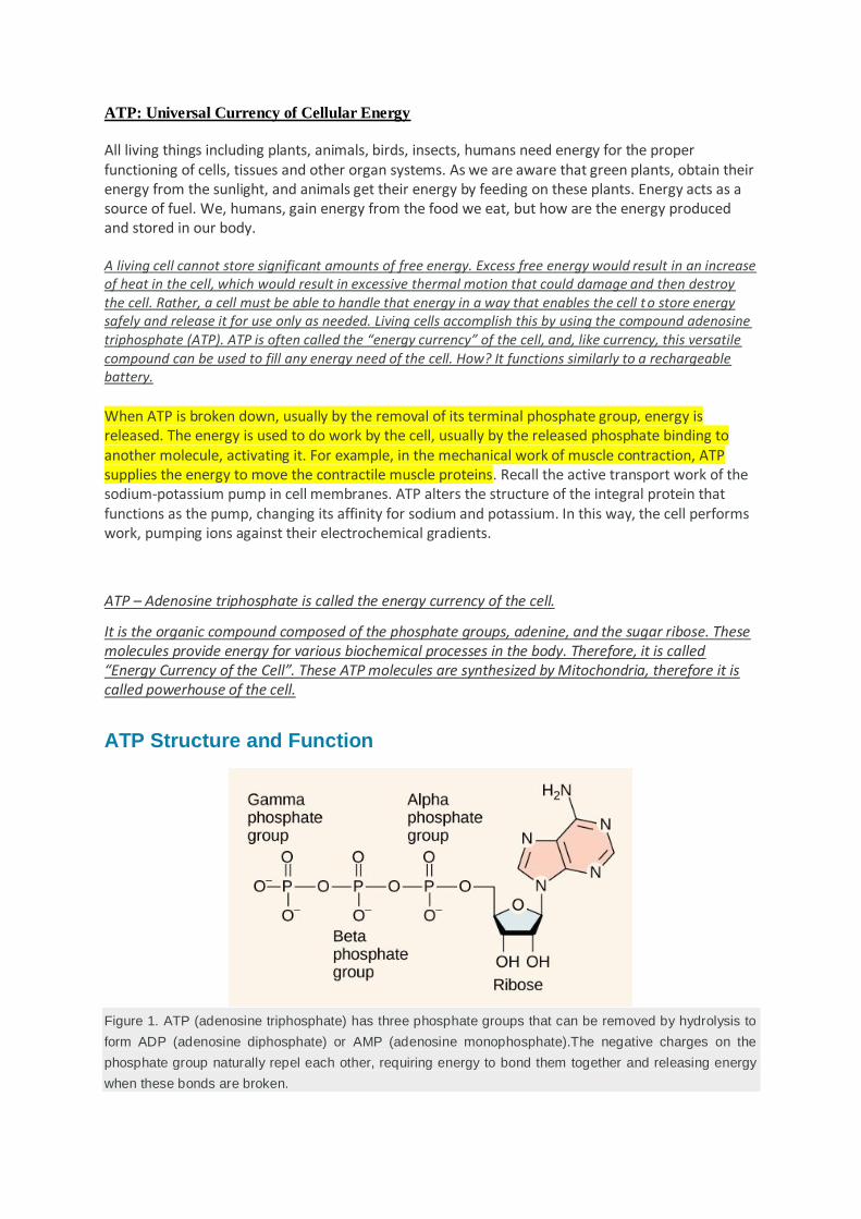
Figure 1. ATP (adenosine triphosphate) has three phosphate groups that can be removed by hydrolysis to
form ADP (adenosine diphosphate) or AMP (adenosine monophosphate).The negative charges on the
phosphate group naturally repel each other, requiring energy to bond them together and releasing energy
when these bonds are broken.
At the heart of ATP is a molecule of adenosine monophosphate (AMP), which is composed of an adenine molecule bonded to a ribose molecule and to a single phosphate group (Figure 1). Ribose is a five-carbon sugar found in RNA, and AMP is one of the nucleotides in RNA. The addition of a second phosphate group to this core molecule results in the formation of adenosine diphosphate (ADP); the addition of a third phosphate group forms adenosine triphosphate (ATP).
The addition of a phosphate group to a molecule requires energy. Phosphate groups are negatively charged and thus repel one another when they are arranged in series, as they are in ADP and ATP. This repulsion makes the ADP and ATP molecules inherently unstable. The release of one or two phosphate groups from ATP, a process called dephosphorylation, releases energy.
The triphosphate tail of ATP is the actual power source which the cell taps. The available energy is contained in the bonds between the phosphates and is released when they are broken or split into molecules. This occurs through the addition of a water molecule (hydrolysis). Usually, only the outer phosphate group is removed from ATP to yield energy; when this occurs, ATP – Adenosine triphosphate is converted into ADP – adenosine diphosphate, it is the form of the nucleotide having only two phosphates.
ATP molecules are largely composed of three essential components.
The pentose sugar molecule i.e. ribose sugar.
Nitrogen base- Adenine, attached to the first carbon of this sugar molecule.
The three phosphate groups which are attached in a chain to the 5th carbon of the pentose sugar. The phosphoryl groups, starting with the group closest to the ribose sugar, are referred to as the alpha, beta, and gamma phosphates. These phosphates play an important role in the activity of ATP.
Energy from ATP
Hydrolysis is the process of breaking complex macromolecules apart. During hydrolysis, water is split, or lysed, and the resulting hydrogen atom (H+) and a hydroxyl group (OH–) are added to the larger molecule. The hydrolysis of ATP produces ADP, together with an inorganic phosphate ion (Pi), and the release of free energy. To carry out life processes, ATP is continuously broken down into ADP, and like a rechargeable battery, ADP is continuously regenerated into ATP by the reattachment of a third phosphate group. Water, which was broken down into its hydrogen atom and hydroxyl group during ATP hydrolysis, is regenerated when a third phosphate is added to the ADP molecule, reforming ATP.
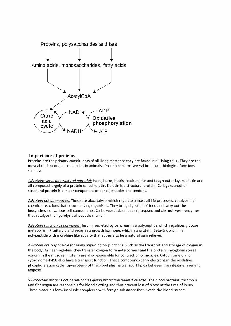
Obviously, energy must be infused into the system to regenerate ATP. Where does this energy come from? In nearly every living thing on earth, the energy comes from the metabolism of glucose. In this way, ATP is a direct link between the limited set of exergonic pathways of glucose catabolism and the multitude of endergonic pathways that power living cells.
How is Energy Produced by the ATP molecules?
The three phosphate groups present in this ATP molecule are called high energy bonds as they are involved in the liberation of a huge amount of energy when they are broken. This molecule provides energy for various life processes without which life cannot exist.
It is used by various enzymes and structural proteins in cellular processes like biosynthetic reactions, cell divisions, etc. This “energy currency of the cell” is produced during cellular respiration where a digested simple molecule of food is utilized.
Once after the energy is produced by the ATP molecules, they are stored in its bonds which are later utilized by the cells by breaking the bonds whenever required
Functions of ATP
The ATP is used for various cellular functions, including transportation of different molecules across cell membranes.
Other functions of ATP include supplying the energy required for the muscle contraction, circulation of blood, locomotion and various body movements.
A significant role of ATP apart from energy production includes: synthesizing the multi -thousand types of macromolecules that the cell requires for their survival. ATP molecule is also used as a switch to control chemical reactions and to send messages.
Importance of ATP Molecule in Metabolism
1. These ATP molecules can be recycled after every reaction. 2. ATP molecule provides energy for both the exergonic and endergonic processes. 3. ATP serves as an extracellular signalling molecule and acts as a neurotransmitter in both central
and peripheral nervous systems. 4. It is the only energy, which can be directly used for different metabolic process. Other forms of
chemical energy need to be converted into ATP before they can be used. 5. It plays an important role in the Metabolism – A life-sustaining chemical reactions including
cellular division, fermentation, photosynthesis, photophosphorylation, aerobic respiration, protein synthesis, exocytosis, endocytosis and motility.
Catabolic pathways of proteins and fats
Fats
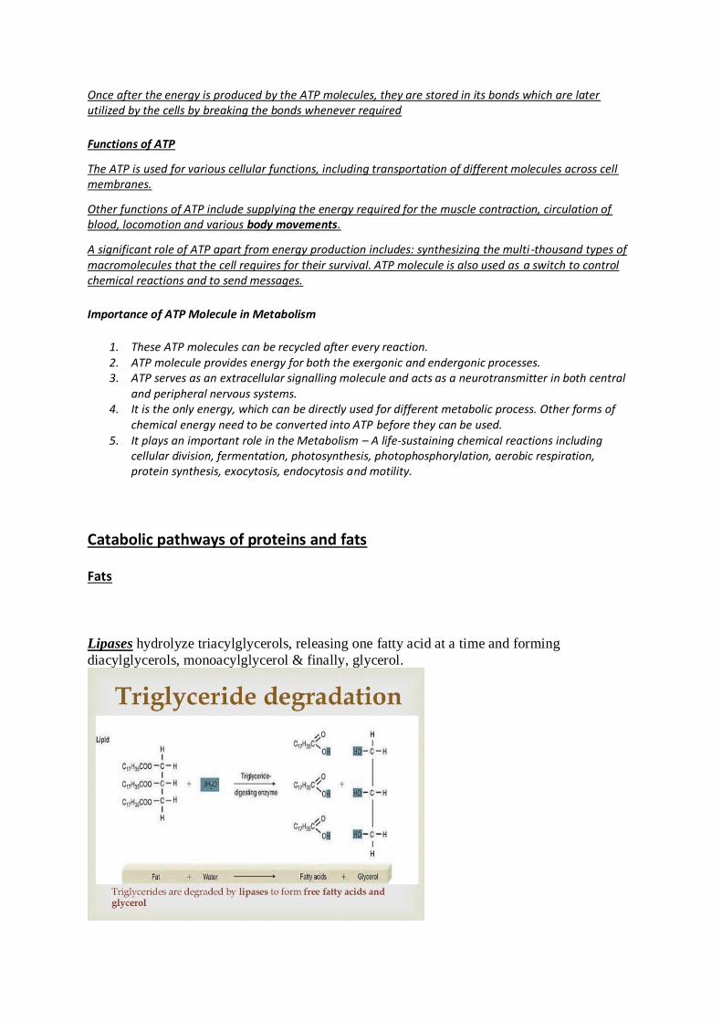
Lipases hydrolyze triacylglycerols, releasing one fatty acid at a time and forming
diacylglycerols, monoacylglycerol & finally, glycerol.
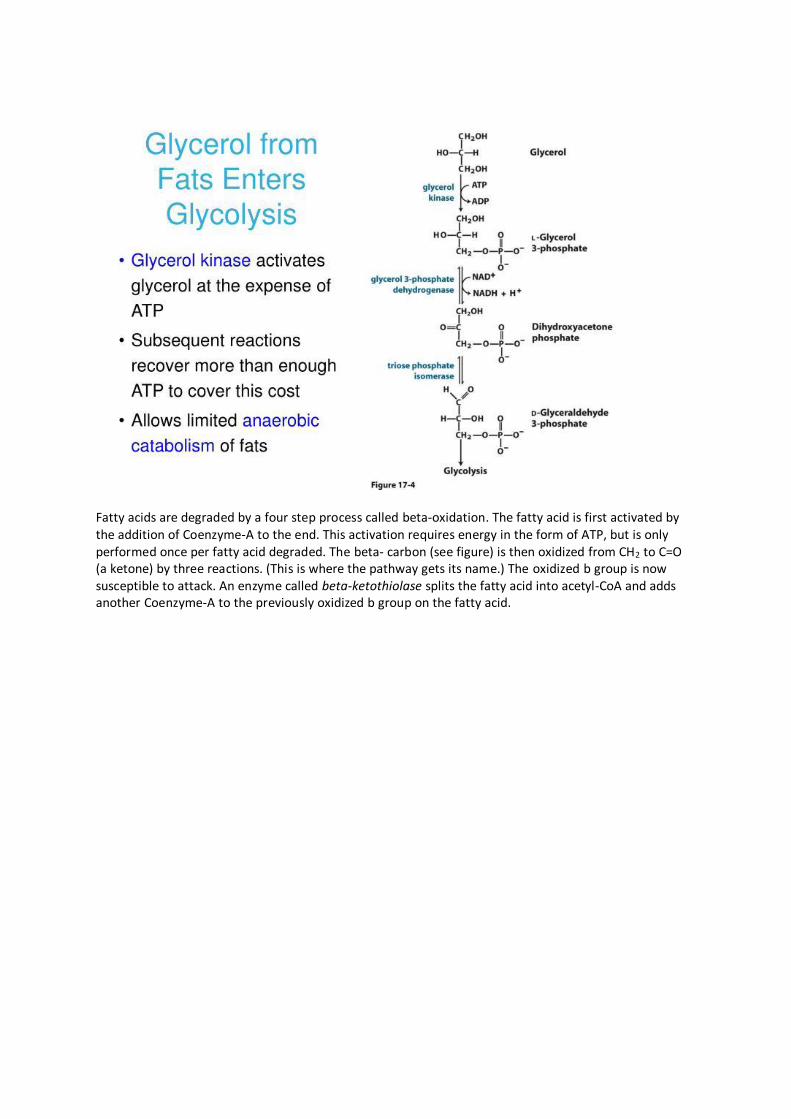
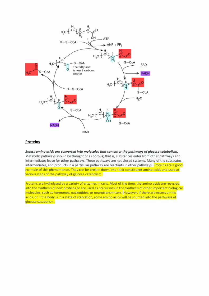
Fatty acids are degraded by a four step process called beta-oxidation. The fatty acid is first activated by the addition of Coenzyme-A to the end. This activation requires energy in the form of ATP, but is only performed once per fatty acid degraded. The beta- carbon (see figure) is then oxidized from CH2 to C=O (a ketone) by three reactions. (This is where the pathway gets its name.) The oxidized b group is now susceptible to attack. An enzyme called beta-ketothiolase splits the fatty acid into acetyl-CoA and adds another Coenzyme-A to the previously oxidized b group on the fatty acid.
Proteins
Excess amino acids are converted into molecules that can enter the pathways of glucose catabolism. Metabolic pathways should be thought of as porous; that is, substances enter from other pathways and intermediates leave for other pathways. These pathways are not closed systems. Many of the substrates, intermediates, and products in a particular pathway are reactants in other pathways. Proteins are a good example of this phenomenon. They can be broken down into their constituent amino acids and used at various steps of the pathway of glucose catabolism.
Proteins are hydrolysed by a variety of enzymes in cells. Most of the time, the amino acids are recycled into the synthesis of new proteins or are used as precursors in the synthesis of other important biological molecules, such as hormones, nucleotides, or neurotransmitters. However, if there are excess amino acids, or if the body is in a state of starvation, some amino acids will be shunted into the pathways of glucose catabolism.
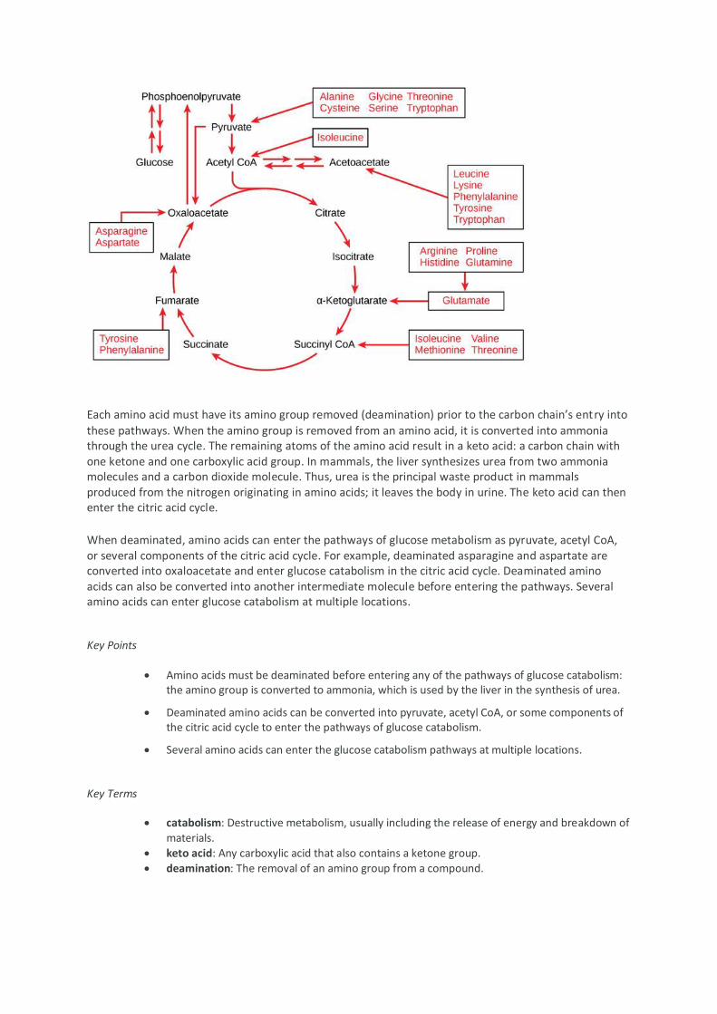
Each amino acid must have its amino group removed (deamination) prior to the carbon chain’s entry into
these pathways. When the amino group is removed from an amino acid, it is converted into ammonia through the urea cycle. The remaining atoms of the amino acid result in a keto acid: a carbon chain with one ketone and one carboxylic acid group. In mammals, the liver synthesizes urea from two ammonia molecules and a carbon dioxide molecule. Thus, urea is the principal waste product in mammals produced from the nitrogen originating in amino acids; it leaves the body in urine. The keto acid can then enter the citric acid cycle.
When deaminated, amino acids can enter the pathways of glucose metabolism as pyruvate, acetyl CoA, or several components of the citric acid cycle. For example, deaminated asparagine and aspartate are converted into oxaloacetate and enter glucose catabolism in the citric acid cycle. Deaminated amino acids can also be converted into another intermediate molecule before entering the pathways. Several amino acids can enter glucose catabolism at multiple locations.
Key Points
Amino acids must be deaminated before entering any of the pathways of glucose catabolism: the amino group is converted to ammonia, which is used by the liver in the synthesis of urea.
Deaminated amino acids can be converted into pyruvate, acetyl CoA, or some components of the citric acid cycle to enter the pathways of glucose catabolism.
Several amino acids can enter the glucose catabolism pathways at multiple locations.
Key Terms
catabolism: Destructive metabolism, usually including the release of energy and breakdown of
materials.
keto acid: Any carboxylic acid that also contains a ketone group. deamination: The removal of an amino group from a compound.
Importance of proteins Proteins are the primary constituents of all living matter as they are found in all living cells . They are the most abundant organic molecules in animals . Protein perform several important biological functions such as: 1.Proteins serve as structural material: Hairs, horns, hoofs, feathers, fur and tough outer layers of skin are all composed largely of a protein called keratin. Keratin is a structural protein. Collagen, another structural protein is a major component of bones, muscles and tendons. 2.Protein act as enzymes: These are biocatalysts which regulate almost all life processes, catalyse the chemical reactions that occur in living organisms. They bring digestion of food and carry out the biosynthesis of various cell components. Carboxypeptidase, pepsin, trypsin, and chymotrypsin-enzymes that catalyse the hydrolysis of peptide chains. 3.Protein function as hormones: Insulin, secreted by pancreas, is a polypeptide which regulates glucose metabolism. Pituitary gland secretes a growth hormone, which is a protein. Beta-Endorphin, a polypeptide with morphine like activity that appears to be a natural pain reliever. 4.Protein are responsible for many physiological functions: Such as the transport and storage of oxygen in the body. As haemoglobins they transfer oxygen to remote corners and the protein, myoglobin stores oxygen in the muscles. Proteins are also responsible for contraction of muscles. Cytochrome C and cytochrome-P450 also have a transport function. These compounds carry electrons in the oxidative phosphorylation cycle. Lipoproteins of the blood plasma transport lipids between the intestine, liver and adipose. 5.Protective proteins act as antibodies giving protection against disease: The blood proteins, thrombin and fibrinogen are responsible for blood clotting and thus prevent loss of blood at the time of injury. These materials form insoluble complexes with foreign substance that invade the blood-stream.
6.Protein are also associated with hereditary characters: Nucleoproteins form chromatin material. During cell division, chromatin condenses to form chromosomes. These are carriers of hereditary characters. 7.Proteins have other protective functions: Snake venoms and plant toxins protect their owners from the species. In addition to snake venom, the toxics proteins of cotton seed and caster bean are highly poisonous to vertebrates. 8.Cell secretions like mucus are glycoproteins: These provide slippery texture and help in protection. 9.Spiders and silkworm secrete a thick solution of protein fibroin: it solidifies into thread of high tensile strength and is used for weaving web or cocoon. We get silk from cocoon. 10.Proteins are stored as reserve food: Some proteins are used for storage. Ovalbumin is employed by nature as a food reservoir in egg white. Casein plays a similar role in mammalian milk. Similarly, proteins are stored as reserve food in various seeds, pulses, rice, maize and peas.
Protein classification based on shape
On the basis of their shape, proteins may be divided into two classes: fibrous and globular.
Fibrous proteins
They have primarily mechanical and structural functions, providing support to the cells as well as the whole organism. These proteins are insoluble in water as they contain, both internally and on their surface, many hydrophobic amino acids. It should be noted that their polypeptide chains form long filaments or sheets, where in most cases only one type of secondary structure, that repeats itself, is found.(Fibrous proteins have long thread like structure and tend to lie side by side to form fibres.) In vertebrates, these proteins provide external protection, support and shape; in fact, thanks to their structural properties, they ensure flexibility and/or strength. Some fibrous proteins, such as α-keratins, are only partially hydrolyzed in the intestine. Here are some examples: 1.Keratins α-Keratins.They constitute almost the entire dry weight of nails, claws, beak, hooves, horns, hair, wool, and a large part of the outer layer of the skin. They consist of 𝛼-helices that are twisted together like the strands of a rope. The different stiffness and flexibility of these structures is a consequence of the number of disulfide bonds that contribute, together with other binding forces, to stabilize the protein structure. And this is the reason why wool keratins, which have a low number of disulfide bonds, are flexible, soft and extensible, unlike claw and beak keratins that are rich in disulfide bonds. 𝜷-Keratins are found in hard tissues such as nails, horns, hooves and bird feathers. They have a 𝛽-pleated sheet structure. 2.Collagens The term “collagen” indicates not a single protein but a family of structurally related proteins (at least 29 different types), which constitute the main protein component of connective tissue, and more generally, the extracellular scaffolding of multicellular organisms. In vertebrates, they represent about 25-30% of all proteins. They are found in different tissues and organs, such as tendons and the organic matrix of bone, where they are present in very high percentages, but also in cartilage and in the cornea of the eye.
In the different tissues, they form different structures, each capable of satisfying a particular need. For example, in the cornea, the molecules are arranged in an almost crystalline array, so that they are virtually transparent, while in the skin they form fibers not very intertwined and directed in all directions, which ensure the tensile strength of the skin itself. The gelatin used in food preparation is a derivative of collagen. 3.Fibroin It is produced by spiders and insects. An example is that produced by the silkworm, Bombyx mori. 4.Elastins This protein provides elasticity to the skin and blood vessels, a consequence of its random coiled structure, that differs from the structures of the α-keratins and collagens. They are also found in yellow elastin tissue like ligaments.
Globular proteins
Most of the proteins belong to this class. They have a compact and more or less spherical structure, more complex than fibrous proteins. In this regard, motifs, domains, tertiary and quaternary structures are found, in addition to the secondary structures. They are generally soluble in water but can also be found inserted into biological membranes (transmembrane proteins), thus in a hydrophobic environment. They are folded into roughly spherical shapes. Unlike fibrous proteins, that have structural and mechanical functions, they act as:
enzymes; hormones; membrane transporters and receptors; transporters of triglycerides, fatty acids and oxygen in the blood; immunoglobulins or antibodies; grain and legume storage proteins.
Examples of globular proteins are insulin, myoglobin, haemoglobin, and cytochrome c, fibrinogen, which is converted into insoluble fibrous protein, fibrin and this causes the clotting of blood. At the intestinal level, most of the globular proteins of animal origin are hydrolysed almost entirely to amino acids.
Classification of Proteins on the basis of structure
based on structure, proteins may be classified as simple, conjugated and derived.
Simple proteins :Simple proteins on hydrolysis yield only amino acids. they do not contain any non-protein part attached to them. most of the simple proteins are global in nature and some fibrous proteins are also simple proteins.
Major groups of simple globalular proteins are:
1.Albumins: These are soluble in water in Aqua solution of acids, bases and salts. They can be precipitated by saturating the solution either with neutral salt such as sodium sulphate in slightly acidic solution or will acidic salts such as ammonium sulphate. On heating , albums gets coagulated. Examples are egg white or ovalbumin, blood serum albumin, so you have been albumin, casein in milk, legumelin(protein of pulses), gliadin(wheat protein) etc. These are stored as food reserves.
2.Globulins: These proteins are insoluble in water but readily dissolves in dilute salt such as sodium chloride. They can be precipitated by half saturation with ammonium sulphate . These copulate on heating and heat calculation is enhanced by addition of dilute acids . They are found in animals and plant issues. Examples are ovoglobulin(eggs), lactoglobulin (milk), serum globulin in blood plasma, myosin muscles are animal globulins. Vegetable globulin include legumin (in peas), tuberin(potatoes) and edestin (wheat and hemp seeds).
3.Protamines:they are basic proteins, highly soluble in water and dilute aqueous solutions of acids, bases and ammonia. They form crystalline salts with mineral acids and are not coagulated by heat. these are simplest of all naturally occurring proteins and have lowest molecular weights. protamine’s occur almost entirely in animals and are main components of sperm cells and certain fishes .
4.Histones: They are also basic proteins of high molecular weight. they are soluble in water dilute acids but not in ammonium hydroxide. they occur as a part of nucleoproteins. Histones are not easily coagulated by heat.
5.Prolamines:These proteins are insoluble in water but are soluble in dilute eco solutions of acids and bases. they are found in plants only and are not coagulated by heat. Gliadin from wheat, zein from maize and hordein from barely are common examples of prolamins.
6.Glutelins: They are insoluble in water, alcohol and neutral salt solutions but are soluble in dilute aqueous solutions of acids and bases. They can be easily coagulated by heat. They are found exclusively in seeds of grains.
Conjugated Proteins: these proteins are composed of a simple globular protein combined with some non-protein substance. this non protein substance is known as prosthetic group. Depending upon the nature of the prosthetic groups, conjugated proteins are divided into following classes:
1.Glycoproteins: In glycoproteins , the simple proteins combined carbohydrates. Example , mucin of saliva, heparin of bile juice, immunoglobulins of plasma , mucopolysaccharides of cartilage and tendon are glycoproteins.
2.Chromoproteins: In chromoproteins, simple proteins are combined with coloured pigments like flavins, carotenoids, porphyrins etc. haemoglobin, myoglobin and cytochromes are some examples of chromoproteins.
3.Phosphoproteins: These proteins contain phosphoric acid as the prosthetic group. These are soluble in dilute alkali and are precipitated by the addition of acids. Casein, the milk protein and Ovovitellin of eggs are examples of phosphoproteins.
4.Metalloproteins: In these proteins, the protein molecule is bound to some metal ion like iron, copper or zinc. Siderophilin, an important plasma protein has a great affinity for iron.
5.Lipoproteins: In these, simple proteins are combined with lipids and have a variable composition. These proteins are present in the brain, blood plasma, milk and egg yolk etc.
6.Flavoproteins: These are enzymes. In these proteins, the protein moiety (apoenzyme) is permanently attached to flavin compound. Flavoproteins are enzymes of Kerb’s Cycle and participate in electron transport system of respiratory chain.
7.Nucleoproteins: In nucleoproteins, the protein molecules are combined with nucleic acid. These proteins are protamine’s or histones. The chromatin material of the nuclei is composed of nucleoproteins.
Derived Proteins :These are degradation products obtained from native proteins either by hydrolysis or coagulation by heat Primary-derived proteins
These protein derivatives are formed by processes causing only slight changes in the protein molecule and its properties
There is little or no hydrolytic cleavage of peptide bonds. Proteans
Proteans are insoluble products formed by the action of water, dilute acids and enzymes. These are particularly formed from globulins but are insoluble in dilute salt solutions.e.g., myosan from myosin, fibrin from fibrinogen
Metaproteins
These are formed by the action of acids and alkalies upon protein. They are insoluble in neutral solvents. Coagulated proteins
Coagulated proteins are insoluble products formed by the action of heat or alcohol on natural proteins,e.g., cooked meat and cooked albumin
Secondary-derived proteins
These proteins are formed in the progressive hydrolytic cleavage of the peptide bonds of protein molecule. They are roughly grouped into proteoses, peptones and peptides according to average molecular weight. Proteoses are hydrolytic products of proteins, which are soluble in water and are not coagulated by heat. Peptones are hydrolytic products, which have simpler structure than proteoses
They are soluble in water and are not coagulated by heat. Peptides are composed of relatively few amino acids. They are water-soluble and not coagulated by heat. The complete hydrolytic decomposition of the natural protein molecule into amino acids generally progresses through successive stages as follows
Protein Protean →Metaprotein Proteoses →Peptones →Peptides →amino acids


![STRUCTURE AND FUNCTION OF PHOTOSYSTEMS I …...by two photosystems [photosystem I (PSI) and photosystem II (PSII)], an ATP synthase (F-ATPase) that produces ATP at the expense of the](https://img.pdfslide.net/doc/110x75/5e6a9bf3b881810a8b6cdf92/structure-and-function-of-photosystems-i-by-two-photosystems-photosystem-i.jpg)