Embed Size (px)
Citation preview
STRUCTURE AND FUNCTION OF A NOVEL TYPE OF ATP-DEPENDENT CLP PROTEASE
Fredrik I Andersson1, Anders Tryggvesson1, Michal Sharon2, Alexander V Diemand3,
Mirjam Classen4, Christoph Best4, Ronny Schmidt5, Jenny Schelin6, Tara M Stanne1, Bernd Bukau5, Carol V Robinson2, Susanne Witt4, Axel Mogk5 and Adrian K Clarke1
1Department of Plant and Environmental Sciences, University of Gothenburg, Box 461, S-405 30 Göteborg, Sweden, 2Department of Chemistry, University of Cambridge, Cambridge, United Kingdom,
3Department of Protein Evolution, Max-Planck-Institute for Developmental Biology, D-72076 Tübingen, Germany, 4Department of Molecular Structural Biology, Max-Planck-Institute of Biochemistry, D-82152 Martinsried, Germany, 5Zentrum fur Molekulare Biologie der Universität Heidelberg, Im Neuenheimer
Feld 282, D-69120 Heidelberg, Germany, 6Department of Applied Microbiology, Lund Institute of Technology, Lund University, Box 124, S-221 00 Lund, Sweden Running title: A novel Clp protease in photosynthetic organisms
Address correspondence to: Adrian K. Clarke, Department of Plant and Environmental Sciences, University of Gothenburg, Box 461, 405 30 Gothenburg, Sweden, Tel. 46 31 7862502, Fax. 46 31 7862626: E-mail: [email protected] The Clp protease is conserved among eubacteria and most eukaryotes, and uses ATP to drive protein substrate unfolding and translocation into a chamber of sequestered proteolytic active sites. The main constitutive Clp protease in photosynthetic organisms has evolved into a functionally essential and structurally intricate enzyme. The model Clp protease from the cyanobacterium Synechococcus consists of the HSP100 molecular chaperone ClpC and a mixed proteolytic core comprised of two distinct subunits, ClpP3 and ClpR. We have purified the ClpP3/R complex, the first for a Clp proteolytic core comprised of heterologous subunits. The ClpP3/R complex has unique functional and structural features, consisting of twin heptameric rings each with an identical ClpP33ClpR4 configuration. As predicted by its lack of an obvious catalytic triad, the ClpR subunit is shown to be proteolytically inactive. Interestingly, extensive modification to ClpR to restore proteolytic activity to this subunit showed that its presence in the core complex is not rate-limiting for the overall proteolytic activity of the ClpCP3/R protease. Altogether, the ClpP3/R complex shows remarkable similarities to the 20S core of the proteasome, revealing a far greater degree of convergent evolution than previously thought between the development of the Clp protease in
photosynthetic organisms and that of the eukaryotic 26S proteasome. Proteases perform numerous tasks vital for cellular homeostasis in all organisms. Much of the selective proteolysis within living cells is performed by multisubunit chaperone-protease complexes. These proteases all share a common two-component architecture and mode of action, with one of the best known examples being the proteasome in archaebacteria, certain eubacteria and eukaryotes (1). The 20S proteasome is a highly conserved cylindrical structure composed of two distinct types of subunits, α and ß. These are organized in four stacked heptameric rings, with two central ß-rings sandwiched between two outer α-rings. Although the α- and ß-protein sequences are similar, it is only the latter that is proteolytic active, with a single Thr active site at the N-terminus. The barrel-shaped complex is traversed by a central channel that widens up into three cavities. The catalytic sites are positioned in the central chamber formed by the ß-rings, adjacent to which are two antechambers conjointly built up by ß- and α-subunits. In general, substrate entry into the core complex is essentially blocked by the α-rings, and thus relies on the associating regulatory partner – PAN and 19S complexes in Archae and eukaryotes, respectively (1). Typically, the archaeal core structure is assembled from only one type of α- and ß-subunit, so that the central proteolytic chamber contains 14 catalytic active
1
http://www.jbc.org/cgi/doi/10.1074/jbc.M809588200The latest version is at JBC Papers in Press. Published on February 23, 2009 as Manuscript M809588200
Copyright 2009 by The American Society for Biochemistry and Molecular Biology, Inc.
at Cam
bridge University Library on M
arch 2, 2009 w
ww
.jbc.orgD
ownloaded from
sites (2). In contrast, each ring of the eukaryotic 20S complex has seven distinct α- and ß-subunits. Moreover, only three of the seven ß-subunits in each ring are proteolytically active (3). Having a strictly conserved architecture, the main difference between the 20S proteasomes is one of complexity. In mammalian cells, the three constitutive active subunits can even be replaced with related subunits upon induction by γ-interferon to generate antigenic peptides presented by the class 1 MHC (4). Two chambered proteases architecturally similar to the proteasome also exist in eubacteria – HslV and ClpP. HslV is commonly thought the prokaryotic counterpart to the 20S proteasome mainly since both are Thr proteases. A single type of HslV protein, however, forms a proteolytic chamber consisting of twin hexameric rather than heptameric rings (5). Also displaying structural similarities to the proteasome is the unrelated ClpP protease. The model Clp protease from Escherichia coli consists of a proteolytic ClpP core flanked on one or both sides by the ATP-dependent chaperones ClpA or ClpX (6). The ClpP proteolytic chamber is comprised of two opposing homo-heptameric rings with the catalytic sites harbored within (7). ClpP alone displays only limited peptidase activity towards short unstructured peptides (8). Larger native protein substrates need to be recognized by ClpA or ClpX and then translocated in an unfolded state into the ClpP proteolytic chamber (9, 10). Inside, the unfolded substrate is bound in an extended manner to the catalytic triads (Ser97, His122, and Asp171) and degraded into small peptide fragments that can readily diffuse out (11). Several adaptor proteins broaden the array of substrates degraded by a Clp protease by binding to the associated HSP100 partner and modifying its protein substrate specificity (12, 13). One example is the adaptor ClpS that interacts with ClpA (EcClpA) and targets N-end rule substrates for degradation by the ClpAP protease (14).
Like the proteasome, the Clp protease is found in a wide variety of organisms. Besides in all eubacteria, the Clp protease also exists in mammalian and plant mitochondria, as well as in various plastids of algae and plants. It also occurs in the unusual plastid in Apicomplexan protozoan (15), a family of parasites responsible for many important medical and veterinary diseases such as
malaria. Of all these organisms, photobionts have by far the most diverse array of Clp proteins. This was first apparent in cyanobacteria, with the model species Synechococcus elongatus having ten distinct Clp proteins – four HSP100 chaperones (ClpB1-2, ClpC and ClpX), three ClpP proteins (ClpP1-3), a ClpP-like protein termed ClpR, and two adaptor proteins (ClpS1-2) (16). Of particular interest is the ClpR variant, which has protein sequence similarity to ClpP but appears to lack the catalytic triad of Ser-type proteases (17). This diversity of Clp proteins is even more extreme in photosynthetic eukaryotes, with at least 23 different Clp proteins in the higher plant Arabidopsis thaliana, most of which are plastid-localized (18).
We have recently shown that two distinct Clp proteases exist in Synechococcus, both of which contain mixed proteolytic cores. The first consists of ClpP1 and ClpP2 subunits, and associates with ClpX, whereas the other has a proteolytic core consisting of ClpP3 and ClpR that binds to ClpC, as do the two ClpS adaptors (19). Of these proteases, it is the more constitutively abundant ClpCP3/R that is essential for cell viability and growth (20, 21). It is also the ClpP3/R complex that is homologous to the single type in eukaryotic plastids, all of which also have ClpC as the chaperone partner (16). In algae and plants, however, the complexity of the plastidic Clp proteolytic core has evolved dramatically. In Arabidopsis, the core complex consists of five ClpP and four ClpR paralogs, along with two unrelated Clp proteins unique to higher plants (22). Like ClpP3/R, the plastid Clp protease in Arabidopsis is essential for normal growth and development, and appears to function primarily as a housekeeping protease (23, 24).
One of the most striking developments in the Clp protease in photosynthetic organisms and Apicomplexan parasites is the inclusion of ClpR within the central proteolytic core. Although this type of Clp protease has evolved into a vital enzyme, little is known about its activity or the exact role of ClpR within the core complex. To address these points we have purified the intact Synechococcus ClpP3/R proteolytic core by co-expression in E. coli. The recombinant ClpP3/R forms a double heptameric ring complex, with each ring having a specific ClpP3/R stoichiometry and arrangement. Together with ClpC the ClpP3/R
2
at Cam
bridge University Library on M
arch 2, 2009 w
ww
.jbc.orgD
ownloaded from
complex degrades several polypeptide substrates, but at a rate considerably slower than that by the E. coli ClpAP protease. Interestingly, although ClpR is shown to be proteolytically inactive, its inclusion in the core complex is not rate-limiting to the overall activity of the ClpCP3/R protease. In general, the results reveal remarkable similarities between the evolutionary development of the Clp protease in photosynthetic organisms and the eukaryotic proteasome relative to their simpler prokaryotic counterparts.
EXPERIMENTAL PROCEDURES Purification of ClpP3/R complex - The
Synechococcus clpP3 and clpR genes were amplified from genomic clones using Pfx DNA polymerase (Invitrogen) and cloned into the pACYC Duet vector (Novagen) for co-expression in E. coli. Another construct was made in which site-directed mutagenesis was used to replace the active site Ser residue in ClpP3 with Ala. A similar approach was used to prepare the chimeric form of ClpR, in which the central region of clpR coding for amino acids 38 to 212 was replaced with the corresponding region from clpP3. In all cases, a His6- tag was added to the 3´end of the clpP3 gene to aid purification, whereas no such tag was included in the clpR gene. All gene constructs were confirmed by sequencing.
Co-expression of ClpP3 and ClpR was performed in E. coli BL21-STAR cells (Invitrogen) grown at 37°C in 10 l LB cultures. Once in mid-exponential growth (i.e., A600 of 0.5), cultures were cooled to 16°C and IPTG added (final concentration 0.5 mM) to induce protein expression. After 6 h, cells were pelleted and then resuspended in buffer A (20 mM Tris/Cl pH 7.5, 400 mM NaCl, 40 mM imidazole, 1 mM DTT). Cells were ruptured using a French Press (1000 atm) followed by centrifugation to remove insoluble cell debris. The soluble protein fraction was loaded onto a Ni2+ affinity column (HisTrap HP, GE Healthcare). After washing the column with buffer A, bound proteins were eluted using buffer B (20 mM Tris/Cl pH 7.5, 400 mM NaCl, 400 mM imidazole, 1 mM DTT). SDS-PAGE revealed that both ClpP3-His6 and ClpR were purified in the same fraction, indicating that the co-expressed proteins formed a single stable oligomer. After dialysis (Slid-A-Lyzer, Pierce) and equilibration in buffer C (20 mM Tris/Cl pH 7.5,
75 mM NaCl and 1 mM DTT), the ClpP3/R oligomer was further purified by gel filtration chromatography using a 16/60 Superdex column (GE Healthcare). MS of the sample revealed no contamination of the purified proteins with E. coli ClpP, as did a lack of peptidase activity as shown in Supplementary Figure 2. Proteins were stored in buffer C with 15% glycerol. Protein concentration was determined using the Bradford assay (Pierce), with the concentration of ClpP3/R used in each assay based on a monomeric conformation.
Purification of ClpC and ClpS1 - Purification of Synechococcus ClpC and ClpS1 was performed as previously described (25). Protein concentration was determined using the Bradford assay, with the concentration of ClpC and ClpS1 used in each assay based on a monomeric conformation.
Native-PAGE - Separation of purified ClpP3/R complexes under non-denaturing conditions was done using a Tris/Borate gel system (19). For recombinant ClpP3/R complexes, 25 µg of protein were resolved on 6-16% polyacrylamide gradient gels. To accurately separate proteins solely on their molecular mass, gels were run at constant current at 4°C as described (26). Ferretin (440 kDa, monomer, 880 kDa, dimer), Urease (270 kDa, trimer) and BSA (132 kDa, dimer) were used as molecular mass markers. Protein complexes were visualized using coomassie-blue staining (Invitrogen). When comparing the size of recombinant and native ClpP3/R complexes, 250 ng of recombinant protein was separated with 20 µg of soluble protein extracted from wild type Synechococcus. The ClpP3/R complex was then detected by immunoblotting using a ClpR-specific antibody (19).
AFM and TEM - AFM was performed with a MultiMode NanoScope IIIa (Veeco) in a commercial glass fluid cell with SiN cantilevers (OMCL-TR-800, normal force constant 0.57 N m-
1, Olympus). For sample preparation, freshly cleaved mica was incubated with buffer solution (20 mM Tris/Cl pH 7.5, 100 mM NaCl, 0.5 mM DTT) containing 7 µg ml-1 ClpP3/R for 10 min. After several washing steps with buffer for removal of excess protein, imaging under native conditions was performed in TappingMode. For negative-stain TEM, samples were prepared on plasma-treated carbon-coated copper grids by incubating a 5 µl droplet of the sample solution for 60 s. Excess suspension was removed by blotting
3
at Cam
bridge University Library on M
arch 2, 2009 w
ww
.jbc.orgD
ownloaded from
with filter paper. After a washing step using 5 µl of buffer, the grid was blotted and stained with 5 µl of 2% UAc for 60 s. Images were recorded using a FEI CM20 FEG at a pixel size of 3.2 Å and a defocus of -2 µm. Candidate particles were selected automatically from electron micrographs using a blob detector. A subset of 1000 picks were subjected to unsupervised clustering based on a full similarity matrix derived by cross-correlating each particle with each other accounting for the rotational and translational degrees of freedom. Good particle views were then selected from the class averages of the resulting clusters, and the complete data set was classified against these templates. The highest-scoring matches for each class were used to create a final set of class averages for visual inspection. The complete processing pipeline was implemented in the in-house Python/C++ package "empi".
Mass spectrometry - To determine the stoichiometry of the ClpP3/R complex electrospray ionization (ESI), MS and tandem MS (MS/MS) experiments were performed on a high mass Q-TOF type instrument adapted for a QSTAR XL platform (27, 28). Prior to MS analysis 300 μl of a 0.6 mg ml-1 solution of ClpP3/R in buffer C was concentrated 6 fold by using a Vivaspin (Vivascience) centrifugal device with a molecular mass cutoff of 5 kDa. The sample was then buffer exchanged twice into different concentrations of ammonium acetate solution (0.1, 0.2 or 1 M) by using Bio-Rad Biospin columns. To induce dissociation of complex-containing solutions methanol was added from 5 to 20%. NanoESI capillaries were prepared in house from borosilicate glass tubes29 and an aliquot of 2 μl solution was loaded for sampling. The conditions within the mass spectrometer were adjusted to preserve non-covalent interactions. The following experimental parameters were used: capillary voltage up to 1.3 kV, declustering potential up to 150 V, focusing potential 250 V, second declustering potential 55 V and focusing rod offset ranging from 20 to 100 V, MCP 2350 V. For tandem MS experiments peaks centered at m/z 5,650 or 5,850 were selected in the quadrupole and collision energy up to 200 V was employed. Argon was used as a collision gas at maximum pressure. All spectra were calibrated externally by using a solution of cesium iodide (100 mg ml-1). Spectra
are shown here with minimal smoothing and without background subtraction.
Homology modeling - The monomeric structure of ClpR has been modeled based on the homologous template EcClpP (2fzs; 30) using MODELLER (31) following the alignment computed by hhsearch (32). Its N-terminus has been remodeled in SPDBV (33) using the EcClpP template 1yg6A (34). Accordingly, the C-terminus was completed with coordinates from the human ClpP structure (1tg6E) (11). The insertions in ClpR were modeled in SPDBV as N-terminal extensions to the helices they are part of. The monomer of ClpP3 has been modeled using the template EcClpP (2fzs). The alignment was computed using PSI-BLAST (35); the catalytic residues are conserved (Fig. 4A). The tetradecameric complex formed by two heptameric rings of ClpP33ClpR4 (Fig. 4C) was built following a previous method (36). Additionally, the final model was selected from a number of alternative models recomputed using MODELLER and energy minimized in SPDBV. The model was analyzed using the iMolTalk server (37) and interface contacts in the complex were detected with a distance threshold of 3.4 Å. Images have been prepared using either Molscript (38) and Raster3D (39), or SPDBV and Povray (www.povray.org).
Peptidase assay - The peptidase activity of ClpP3/R and EcClpP was measured against the fluorogenic peptides N-succinyl (Suc)-Leu-Tyr-7-amido-4-methylcoumarin (AMC), Suc-Val-Lys-Met-AMC and Suc-Ile-Ile-Trp-AMC (Sigma). For each assay, 30 μM of peptide and 1-5 µg ClpP3/R or EcClpP were incubated in buffer D (25 mM Tris/Cl pH 7.5, 75 mM NaCl, 10 mM MgCl2, 1 mM DTT) for 5-20 min at 37°C. Peptide degradation was measured as an increase in relative fluorescence (FluoSTAR; BWG) at 310-380 nm excitation and 460 nm emission wavelengths.
Degradation of α-casein and GFP substrates - For the proteolytic assays, each of the Clp proteins used was diluted to 1 μM final concentration in buffer D together with an ATP-regeneration system (13). For each assay 1 µM α-casein was used, whereas 100 nM was used for FITC-casein and the GFP substrates (FR-GFP and MR-GFP). All reactions were performed at 37°C. Degradation of α-casein was monitored by SDS-PAGE and coomassie-blue staining, whereas FITC-casein
4
at Cam
bridge University Library on M
arch 2, 2009 w
ww
.jbc.orgD
ownloaded from
degradation was measured by fluorescence (FluoSTAR; BWG) at 490 nm excitation and 525 nm emission wavelengths. Degradation of FR- and MR-GFP was determined either by immunoblotting using a GFP-specific antibody or by loss in fluorescence at 400 nm excitation and 510 nm emission wavelengths.
ATPase activity - The ATPase activity of ClpC was measured by the release of inorganic phosphate as previously described (25). In the assays, 0.5 μM ClpC was incubated either alone or with 1 μM wild type or mutated ClpP3/R in buffer D (+ 4 mM ATP) for 20 min at 37°C.
RESULTS Purification of Synechococcus ClpP3 and ClpR
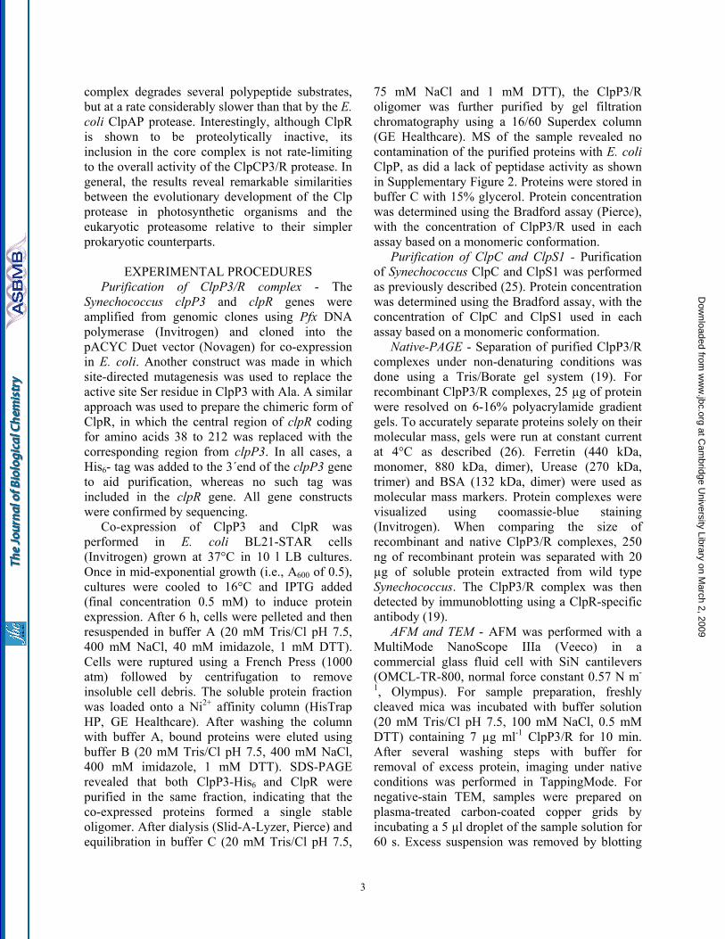
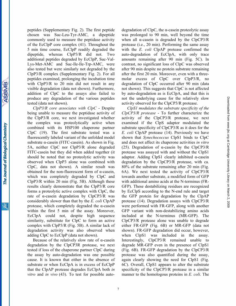
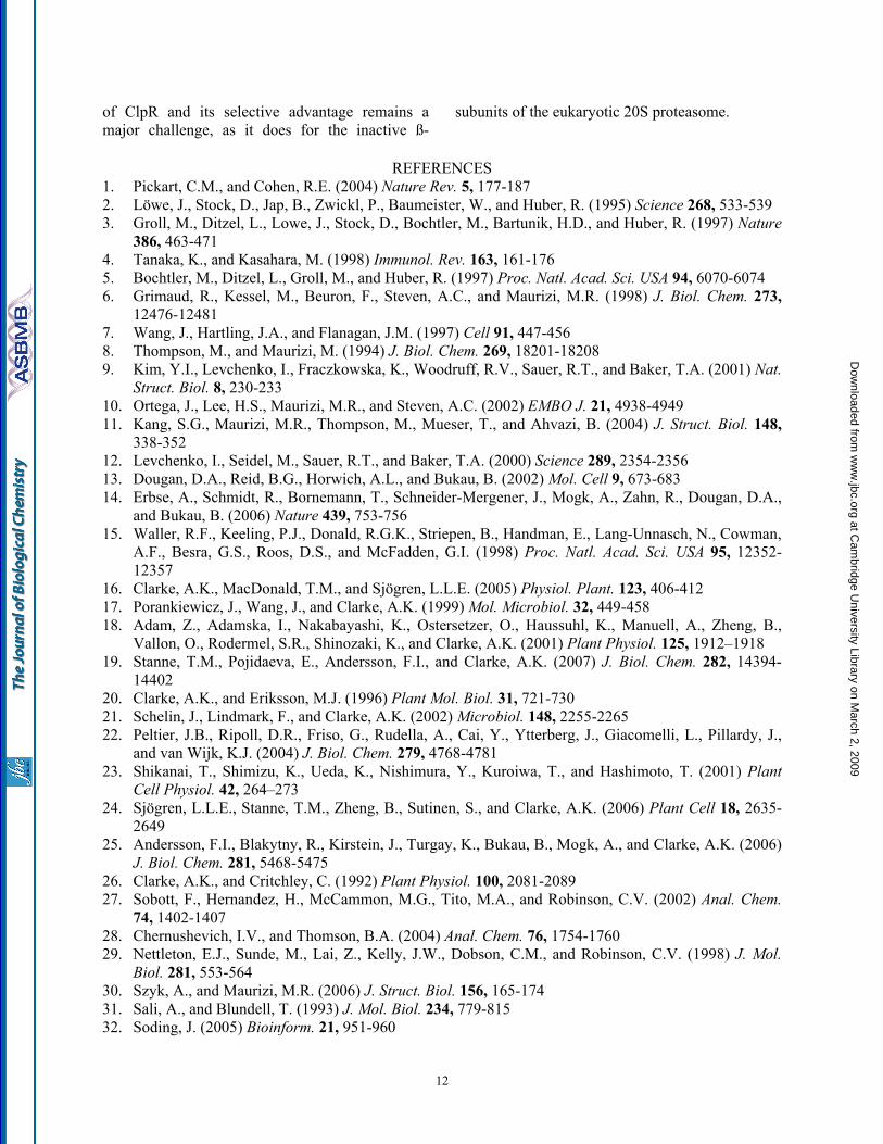
- We have shown that the two constitutively expressed ClpP3 and ClpR proteins in Synechococcus form an essential hetero-oligomeric complex in vivo (19). Initial attempts to reconstitute this core complex by mixing individually purified ClpP3 and ClpR proteins failed (data not shown), and so a new strategy was adopted (note: neither ClpP3 nor ClpR form a proteolytic core complex on their own). Given that both clpP3 and clpR genes in Synechococcus are arranged within a bicistronic operon (21), we used an E. coli expression system in which the genes were co-expressed within the same cell (Fig. 1A). Based on the premise that both proteins would readily oligomerize together once synthesized in E. coli, a His6-tag was included at the C-terminus of only ClpP3 to aid in purification of the core complex. Upon induction with IPTG, soluble cell extracts containing both ClpP3 and ClpR were first passed over a Ni2+-affinity column. Following extensive washing to remove non-specific contaminants, both ClpP3 and ClpR were the principle proteins eluted from the column indicating they form a stable oligomeric complex. The ClpP3/R proteins were then further purified by size-exclusion chromatography (Fig. 1A).
Recombinant ClpP3/R proteins form a core complex - To determine if the recombinant ClpP3 and ClpR proteins formed a proteolytic core complex, we first separated the purified proteins by native-PAGE. As shown in Fig. 1B, the ClpP3/R proteins formed a single oligomer of 270 kDa, the size of which was identical to that formed in vivo by the native ClpP3/R proteins (Fig. 1C) (19). To further analyze the overall structure of the
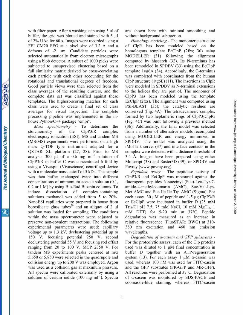
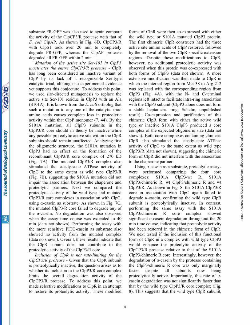
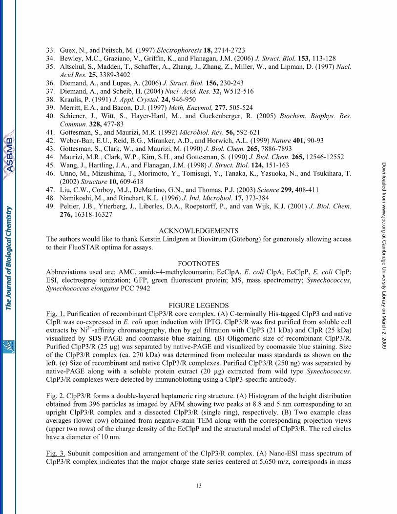
ClpP3/R complex we performed both atomic force microscopy (AFM) and negative-stain transmission electron microscopy (TEM). Using AFM, the ClpP3/R complexes could be successfully imaged under native conditions in buffer solution (Supplementary Fig. 1). Topographical analysis revealed that the imaged particles grouped into two classes, one with a height of 5 nm, the other 8.8 nm (Fig. 2A). Assuming an overall structure of the ClpP3/R complex similar to that of E. coli ClpP (EcClpP), the higher particles most likely correspond to upright standing ClpP3/R, whereas the 5 nm high population might derive from half ClpP3/R where the upper ring has been sheered off by the scanning tip. Such equatorial sectioning of barrel-shaped multisubunit complexes has been previously reported while imaging under similar conditions (40). In addition to the AFM analysis, template-free class averages obtained from single-particle analysis of negative-stain TEM showed a symmetric, seven-pointed object with straight edges, a central hole, and a diameter of ca. 9 nm, compatible with the top view of the structural models for EcClpP and ClpP3/R. The other class averages show a slightly more bulky view with an elongated cleft in the center, compatible with a diagonal view of two sandwiched rings (Fig. 2B). Taken together, the results suggest that the recombinant proteins form a complex matching that of native ClpP3/R in vivo, with a double layered heptameric ring structure consistent with that of a Clp proteolytic core.
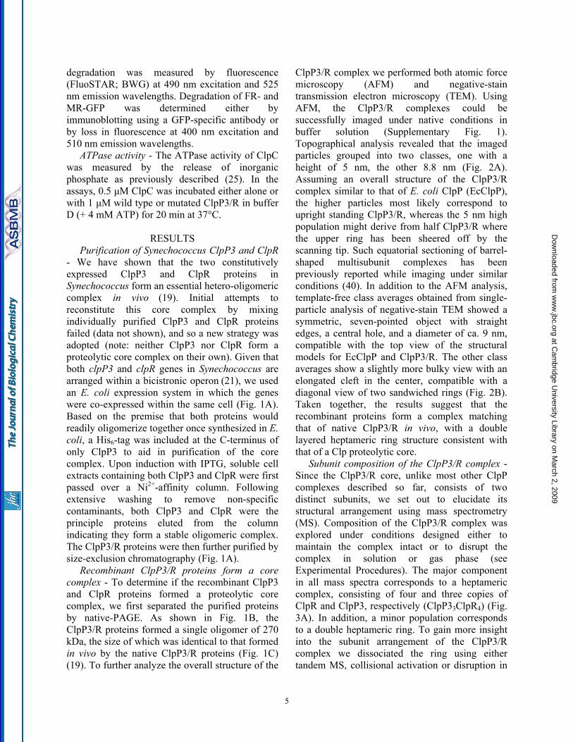
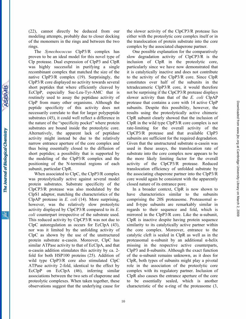
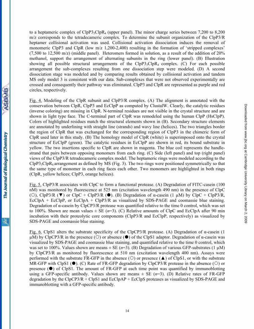
Subunit composition of the ClpP3/R complex - Since the ClpP3/R core, unlike most other ClpP complexes described so far, consists of two distinct subunits, we set out to elucidate its structural arrangement using mass spectrometry (MS). Composition of the ClpP3/R complex was explored under conditions designed either to maintain the complex intact or to disrupt the complex in solution or gas phase (see Experimental Procedures). The major component in all mass spectra corresponds to a heptameric complex, consisting of four and three copies of ClpR and ClpP3, respectively (ClpP33ClpR4) (Fig. 3A). In addition, a minor population corresponds to a double heptameric ring. To gain more insight into the subunit arrangement of the ClpP3/R complex we dissociated the ring using either tandem MS, collisional activation or disruption in
5
at Cam
bridge University Library on M
arch 2, 2009 w
ww
.jbc.orgD
ownloaded from
solution. By collisional activation, acceleration of the complex induces dissociation of both ClpP3 and ClpR subunits and the resulting stripped complexes correspond to different combinations of ClpP3 and ClpR (Fig. 3A, Supplementary Table).
To define the subunit arrangement within the single ClpP3/R heptameric ring, we modeled all possible arrangements with a 3:4 subunit combination (Fig. 3B). From these initial structures we generated all potential sub-complexes formed by dissociation of one (Fig. 3C) and two subunits (Fig. 3D). The sub-complexes generated were then compared with those formed experimentally (Supplementary Table). Only one out of the five possible structural arrangements (Structure 3, Fig. 3B), was consistent with the sub-complexes formed. Further support for this assignment comes from disruption of the complex in solution using sub-denaturing quantities of organic solvent. In 20% methanol the predominant species is the heterodimer together with a heterotrimer and other larger heteromers. The fact that no homodimers are formed under these conditions is consistent with our assignment of structure 3 in which the predominant dissociation products would be heteromers. Overall, by using MS approaches we could not only define the assembly state of the ClpP3/R complex but also define its specific subunit organization within a single heptameric ring.
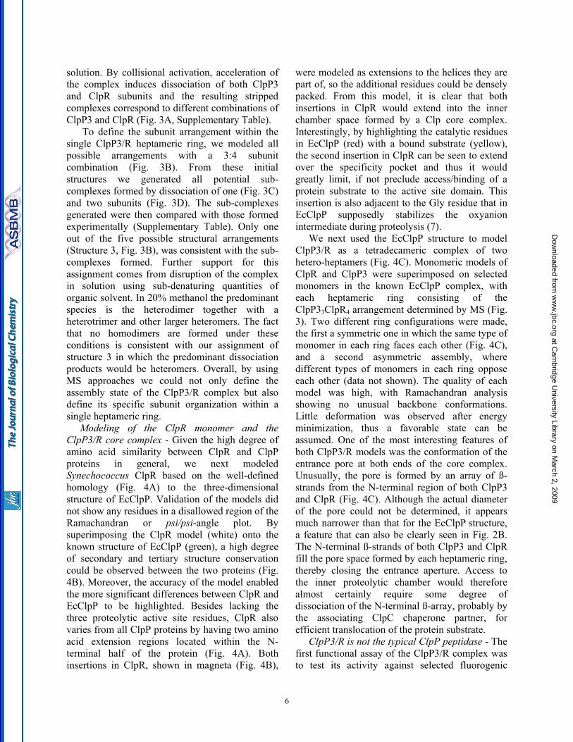
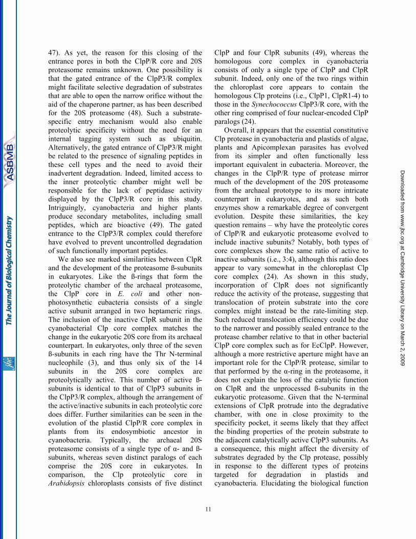
Modeling of the ClpR monomer and the ClpP3/R core complex - Given the high degree of amino acid similarity between ClpR and ClpP proteins in general, we next modeled Synechococcus ClpR based on the well-defined homology (Fig. 4A) to the three-dimensional structure of EcClpP. Validation of the models did not show any residues in a disallowed region of the Ramachandran or psi/psi-angle plot. By superimposing the ClpR model (white) onto the known structure of EcClpP (green), a high degree of secondary and tertiary structure conservation could be observed between the two proteins (Fig. 4B). Moreover, the accuracy of the model enabled the more significant differences between ClpR and EcClpP to be highlighted. Besides lacking the three proteolytic active site residues, ClpR also varies from all ClpP proteins by having two amino acid extension regions located within the N-terminal half of the protein (Fig. 4A). Both insertions in ClpR, shown in magneta (Fig. 4B),
were modeled as extensions to the helices they are part of, so the additional residues could be densely packed. From this model, it is clear that both insertions in ClpR would extend into the inner chamber space formed by a Clp core complex. Interestingly, by highlighting the catalytic residues in EcClpP (red) with a bound substrate (yellow), the second insertion in ClpR can be seen to extend over the specificity pocket and thus it would greatly limit, if not preclude access/binding of a protein substrate to the active site domain. This insertion is also adjacent to the Gly residue that in EcClpP supposedly stabilizes the oxyanion intermediate during proteolysis (7).
We next used the EcClpP structure to model ClpP3/R as a tetradecameric complex of two hetero-heptamers (Fig. 4C). Monomeric models of ClpR and ClpP3 were superimposed on selected monomers in the known EcClpP complex, with each heptameric ring consisting of the ClpP33ClpR4 arrangement determined by MS (Fig. 3). Two different ring configurations were made, the first a symmetric one in which the same type of monomer in each ring faces each other (Fig. 4C), and a second asymmetric assembly, where different types of monomers in each ring oppose each other (data not shown). The quality of each model was high, with Ramachandran analysis showing no unusual backbone conformations. Little deformation was observed after energy minimization, thus a favorable state can be assumed. One of the most interesting features of both ClpP3/R models was the conformation of the entrance pore at both ends of the core complex. Unusually, the pore is formed by an array of ß-strands from the N-terminal region of both ClpP3 and ClpR (Fig. 4C). Although the actual diameter of the pore could not be determined, it appears much narrower than that for the EcClpP structure, a feature that can also be clearly seen in Fig. 2B. The N-terminal ß-strands of both ClpP3 and ClpR fill the pore space formed by each heptameric ring, thereby closing the entrance aperture. Access to the inner proteolytic chamber would therefore almost certainly require some degree of dissociation of the N-terminal ß-array, probably by the associating ClpC chaperone partner, for efficient translocation of the protein substrate.
ClpP3/R is not the typical ClpP peptidase - The first functional assay of the ClpP3/R complex was to test its activity against selected fluorogenic
6
at Cam
bridge University Library on M
arch 2, 2009 w
ww
.jbc.orgD
ownloaded from
peptides (Supplementary Fig. 2). The first peptide chosen was Suc-Leu-Tyr-AMC, a dipeptide commonly used to measure the peptidase activity of the EcClpP core complex (41). Throughout the 5 min time course, EcClpP readily degraded the dipeptide, whereas ClpP3/R did not. Two additional peptides degraded by EcClpP, Suc-Val-Lys-Met-AMC and Suc-Ile-Ile-Trp-AMC, were also tested but were similarly not degraded by the ClpP3/R complex (Supplementary Fig. 2). For all peptides examined, prolonging the incubation time with ClpP3/R to 20 min did not result in any visible degradation (data not shown). Furthermore, addition of ClpC to the assays also failed to produce any degradation of the various peptides tested (data not shown).
ClpP3/R core associates with ClpC - Despite being unable to measure the peptidase activity of the ClpP3/R core, we next investigated whether the complex was proteolytically active when combined with its HSP100 chaperone partner ClpC (19). The first substrate tested was a fluorescently labeled variant of the unfolded model substrate α-casein (FITC-casein). As shown in Fig. 5A, neither ClpC nor ClpP3/R alone degraded FITC-casein but they did when added together (it should be noted that no proteolytic activity was observed when ClpP3 alone was combined with ClpC, data not shown). A similar result was obtained for the non-fluorescent form of α-casein, which was completely degraded by ClpC and ClpP3/R within 20 min (Fig. 5B). Although these results clearly demonstrate that the ClpP3/R core forms a proteolytic active complex with ClpC, the rate of α-casein degradation by ClpCP3/R was considerably slower than that by the E. coli ClpAP protease, which completely degraded the α-casein within the first 5 min of the assay. Moreover, EcClpA could not, despite high sequence similarity, substitute for ClpC to form an active complex with ClpP3/R (Fig. 5B). A similar lack of degradation activity was also observed when adding ClpC to EcClpP (data not shown).
Because of the relatively slow rate of α-casein degradation by the ClpCP3R protease, we next tested if loss of the chaperone partner ClpC during the assay by auto-degradation was one possible cause. It is known that either in the absence of substrate or when EcClpA is in excess of EcClpP that the ClpAP protease degrades EcClpA both in vitro and in vivo (43). To test for possible auto-
degradation of ClpC, the α-casein proteolytic assay was prolonged to 90 min, well beyond the time when all α-casein is degraded by the ClpCP3/R protease (i.e., 20 min). Performing the same assay with the E. coli ClpAP protease confirmed the auto-degradation of EcClpA, with only trace amounts remaining after 90 min (Fig. 5C). In contrast, no significant loss of ClpC was observed after 90 min despite no protein substrate remaining after the first 20 min. Moreover, even with a three-molar excess of ClpC over ClpP3/R, no degradation of ClpC occurred after 90 min (data not shown). This suggests that ClpC is not affected by auto-degradation as is EcClpA, and that this is not the underlying cause for the relatively slow activity observed for the ClpCP3/R protease.
ClpS1 modulates the substrate specificity of the ClpCP3/R protease - To further characterize the activity of the ClpCP3/R protease, we next examined if the ClpS adaptor modulated the substrate specificity of ClpCP3/R as it does for the E. coli ClpAP protease (14). Previously we have shown that Synechococcus ClpS1 binds to ClpC and does not affect its chaperone activities in vitro
(25). Degradation of α-casein by the ClpCP3/R protease was assayed with and without the ClpS1 adaptor. Adding ClpS1 clearly inhibited α-casein degradation by the ClpCP3/R protease, with ca. 80% of the substrate remaining after 20 min (Fig. 6A). We next tested the activity of ClpCP3/R towards another substrate, a modified form of GFP with additional amino acids at the N-terminus (FR-GFP). These destabilizing residues are recognized by EcClpS according to the N-end rule and target the GFP protein for degradation by the ClpAP protease (14). Degradation assays with ClpCP3/R were performed with FR-GFP, along with another GFP variant with non-destabilizing amino acids included at the N-terminus (MR-GFP). The ClpCP3/R protease alone was unable to degrade either FR-GFP (Fig. 6B) or MR-GFP (data not shown). FR-GFP degradation did occur, however, when ClpS1 was included in the assay. Interestingly, ClpCP3/R remained unable to degrade MR-GFP even in the presence of ClpS1 (Fig. 6B). FR-GFP degradation by the ClpCP3/R protease was also quantified during the assay, again clearly showing the need for ClpS1 (Fig. 6C). Overall, ClpS1 appears to alter the substrate specificity of the ClpCP3/R protease in a similar manner to the homologous proteins in E. coli. The
7
at Cam
bridge University Library on M
arch 2, 2009 w
ww
.jbc.orgD
ownloaded from
substrate FR-GFP was also used to again compare the activity of the ClpCP3/R protease with that of E. coli ClpAP. As shown in Fig. 6D, ClpCP3/R with ClpS1 took over 20 min to completely degrade FR-GFP, whereas the ClpAP protease degraded all FR-GFP within 2 min.
Mutation of the active site Ser-101 in ClpP3 inactivates the entire ClpCP3/R protease - ClpR has long been considered an inactive variant of ClpP by its lack of a recognizable Ser-type catalytic triad, although no experimental evidence yet supports this conjecture. To address this point, we used site-directed mutagenesis to replace the active site Ser-101 residue in ClpP3 with an Ala (S101A). It is known from the E. coli ortholog that such a mutation to any one of the catalytic triad amino acids causes complete loss in proteolytic activity within that ClpP monomer (7, 44). By the S101A mutation, all ClpP3 subunits in the ClpP3/R core should in theory be inactive while any possible proteolytic active site within the ClpR subunits should remain unaffected. Analyzing first the oligomeric structure, the S101A mutation in ClpP3 had no effect on the formation of the recombinant ClpP3/R core complex of 270 kD (Fig. 7A). The mutated ClpP3/R complex also stimulated the steady-state ATPase activity of ClpC to the same extent as wild type ClpP3/R (Fig. 7B), suggesting the S101A mutation did not impair the association between the chaperone and proteolytic partners. Next we compared the proteolytic activity of the wild type and mutated ClpP3/R core complexes in association with ClpC, using α-casein as substrate. As shown in Fig. 7C, the mutated ClpP3/R core failed to degrade any of the α-casein. No degradation was also observed when the assay time course was extended to 40 min (data not shown). Performing the assay with the more sensitive FITC-casein as substrate also showed no activity from the mutated complex (data no shown). Overall, these results indicate that the ClpR subunit does not contribute to the proteolytic activity of the ClpP3/R core.
Inclusion of ClpR is not rate-limiting for the ClpCP3/R protease - Given that the ClpR subunit is proteolytically inactive, the question arises as to whether its inclusion in the ClpP3/R core complex limits the overall degradation activity of the ClpCP3/R protease. To address this point, we made selective modifications to ClpR in an attempt to restore its proteolytic activity. These modified
forms of ClpR were then co-expressed with either the wild type or S101A mutated ClpP3 protein. The first chimeric ClpR constructs had the three active site amino acids of ClpP restored, followed by the removal of the two ClpR-specific extension regions. Despite these modifications to ClpR, however, no additional proteolytic activity was observed when this protein was co-expressed with both forms of ClpP3 (data not shown). A more extensive modification was then made to ClpR in which the internal region from Met-38 to Arg-212 was replaced with the corresponding region from ClpP3 (Fig. 4A), with the N- and C-terminal regions left intact to facilitate intra-ring association with the ClpP3 subunit (ClpP3 alone does not form a stable heptameric ring; Schelin, unpublished result). Co-expression and purification of this chimeric ClpR form with either the active wild type or inactive S101A ClpP3 produced a core complex of the expected oligomeric size (data not shown). Both core complexes containing chimeric ClpR also stimulated the steady-state ATPase activity of ClpC to the same extent as wild type ClpP3/R (data not shown), suggesting the chimeric form of ClpR did not interfere with the association to the chaperone partner.
Using α-casein as a substrate, proteolytic assays were performed comparing the four core complexes: S101A ClpP3/wt R, S101A ClpP3/chimeric R, wt ClpP3/chimeric R and wt ClpP3/R. As shown in Fig. 8, the S101A ClpP3/R core in association with ClpC again failed to degrade α-casein, confirming the wild type ClpR subunit is proteolytically inactive. In contrast, performing the same assay with the S101A ClpP3/chimeric R core complex showed significant α-casein degradation throughout the 20 min time course, indicating that proteolytic activity had been restored in the chimeric form of ClpR. We next tested if the inclusion of this functional form of ClpR in a complex with wild type ClpP3 would enhance the proteolytic activity of the ClpCP3/R protease relative to that of the S101A ClpP3/chimeric R core. Interestingly, however, the degradation of α-casein by the protease containing the ClpP3/chimeric R core was only marginally faster despite all subunits now being proteolytically active. Importantly, this rate of α-casein degradation was not significantly faster than that by the wild type ClpP3/R core complex (Fig. 8). This suggests that the wild type ClpR subunit,
8
at Cam
bridge University Library on M
arch 2, 2009 w
ww
.jbc.orgD
ownloaded from

despite being proteolytically inactive, does not limit the overall degradation activity of the ClpCP3/R protease.
DISCUSSION In this study, we have described the
biochemical characteristics of the first Clp proteolytic core complex to contain two distinct subunits, one of which is the unusual ClpP variant, ClpR. The ClpP3/R complex is the main constitutive Clp proteolytic core in cyanobacteria in vivo (19). Besides being essential in Synechococcus, it is homologous to the equally important Clp proteolytic core in eukaryotic plastids (23, 24). Phylogenetically, the ClpR variant appears to have evolved first in cyanobacteria and then through various endosymbiotic events been retained in the sole Clp protease in plastids of photosynthetic eukaryotes and Apicomplexan protozoan. ClpR proteins in general have several signature characteristics; the apparent absence of a catalytic triad characteristic of Ser-type proteases and at least one extension within the N-terminal half that commonly make them larger (2-3 kDa) than ClpP (17). Despite the prevalence of ClpR in many different organisms, nothing was known about their structural or functional significance within the proteolytic core complex prior to this work.
Modeling of ClpP3/R using the known ClpP core structures from E. coli and Streptococcus revealed several key features. Besides the obvious lack of a catalytic triad, ClpR also has two N-terminal extensions that are absent in all ClpP forms. The second of these is the most highly conserved in terms of length and amino acid composition among all ClpR orthologs. According to the model, these additional amino acids extend the relevant α-helix in ClpR so that it protrudes further into the inner cavity space. Importantly, this region extends over the specificity pocket that houses the catalytic triad in ClpP proteins. In the EcClpP structure, the specificity pocket consists of two continuous parallel grooves formed by helices 4 and 9, along which the catalytic triads are arranged in each subunit (7). These grooves, which are also present in ClpP3, are covered in ClpR by the extended α-helix thereby severely restricting access to the pocket domain for unfolded protein substrates. In addition, the second extension in ClpR is in close proximity to the Gly residue
proposed to stabilize the oxyanion intermediate (7), which would further disrupt the specificity pocket; a possibility also proposed for the corresponding extension in the Arabidopsis ClpR proteins (22). As a consequence, there would remain little selection pressure to retain a functional catalytic triad in ClpR, causing the active site residues to eventually be lost over time. Indeed, loss of the catalytic triad appears to be ongoing in certain cyanobacteria, with their ClpR ortholog still retaining one of the active site residues (i.e., His).
The other critical feature arising from the modeling of ClpP3/R was the structure of the region surrounding the entrance pore. Using recent ClpP structures that show more residues at the N-terminus (34), we were able to obtain a detailed and reliable prediction of the conformation of the pore-forming residues in the ClpP3/R complex. Unstructured regions of the N-terminus, particularly for ClpR, extend out of the main body of the core, more so than for the EcClpP core complex. Given the position of these extended “pin” regions, it is likely that they directly affect the association of ClpC to the ClpP3/R core. Moreover, they might well influence the specificity of the chaperone partner, possibly explaining why EcClpA is unable to function with ClpP3/R. Properties of the amino acids lining the pore channel also vary considerably in the ClpP3/R complex, with many more residues with hydrophobic side groups compared to the more hydrophilic ones in EcClpP. The increased hydrophobicity of the ClpP3/R pore might well influence the efficiency in which unfolded proteins are translocated into the degradative chamber. More importantly, however, is that these hydrophobic domains could contribute to the relatively narrow diameter to the ClpP3/R pore entrance, which appears effectively closed. Not only would this sealed entrance prevent inadvertent diffusion of proteins into the inner chamber, it would require significantly more “opening” presumably by the associating chaperone partner to enable translocation of the unfolded protein substrate. Indeed, the ability of the chaperone partner to efficiently perform such conformational changes to the core entrance might well be another difference between the various types of HSP100 proteins. Lateral openings in the complex barrel, as predicted by a previous study
9
at Cam
bridge University Library on M
arch 2, 2009 w
ww
.jbc.orgD
ownloaded from
(22), cannot directly be deduced from our modeling attempts, probably due to closer docking of the monomers in the rings and between the two rings.
The Synechococcus ClpP3/R complex has proven to be an ideal model for this novel type of Clp protease. Dual expression of ClpP3 and ClpR was highly successful in purifying a single recombinant complex that matched the size of the native ClpP3/R complex (19). Surprisingly, the ClpP3/R core displayed no activity towards several short peptides that where efficiently cleaved by EcClpP, especially Suc-Leu-Tyr-AMC that is routinely used to assay the peptidase activity of ClpP from many other organisms. Although the peptide specificity of this activity does not necessarily correlate to that for larger polypeptide substrates (45), it could well reflect a difference in the nature of the “specificity pocket” where protein substrates are bound inside the proteolytic core. Alternatively, the apparent lack of peptidase activity might instead be due to the relatively narrow entrance aperture of the core complex and thus being essentially closed to the diffusion of short peptides; a possibility that is supported by the modeling of the ClpP3/R complex and the positioning of the N-terminal regions of each subunit, particular ClpR.
When associated to ClpC, the ClpP3/R complex was proteolytically active against several model protein substrates. Substrate specificity of the ClpCP3/R protease was also modulated by the ClpS1 adaptor, matching the characteristics of the ClpAP protease in E. coli (14). More surprising, however, was the relatively slow proteolytic activity displayed by ClpCP3/R compared to its E. coli counterpart irrespective of the substrate used. This reduced activity by ClpCP3/R was not due to ClpC autoregulation as occurs for EcClpA (43), nor was it limited by the unfolding activity of ClpC as shown by the use of the unstructured protein substrate α-casein. Moreover, ClpC has similar ATPase activity to that of EcClpA, and that α-casein addition stimulates this activity by ca. 2-fold for both HSP100 proteins (25). Addition of wild type ClpP3/R core also stimulated ClpC ATPase activity 2-fold, identical to the effect by EcClpP on EcClpA (46), inferring similar associations between the two sets of chaperone and proteolytic complexes. When taken together, these observations suggest that the underlying cause for
the slower activity of the ClpCP3/R protease lies either with the proteolytic core complex itself or in the translocation of protein substrate into the core complex by the associated chaperone partner.
One possible explanation for the comparatively slow degradation activity of ClpCP3/R is the inclusion of ClpR in the proteolytic core, particularly since we have now demonstrated that it is catalytically inactive and does not contribute to the activity of the ClpP3/R core. Since ClpR constitutes over half of the subunits in the tetradecameric ClpP3/R core, it would therefore not be surprising if the ClpCP3/R protease displays slower activity than that of the E. coli ClpAP protease that contains a core with 14 active ClpP subunits. Despite this possibility, however, the results using the proteolytically active chimeric ClpR subunit clearly showed that the inclusion of ClpR in the wild type ClpP3/R core complex is not rate-limiting for the overall activity of the ClpCP3/R protease and that available ClpP3 subunits are sufficient for the required degradation. Given that the unstructured substrate α-casein was used in these assays, the translocation rate of substrate into the core complex now appears to be the more likely limiting factor for the overall activity of the ClpCP3/R protease. Reduced translocation efficiency of unfolded substrate by the associating chaperone partner into the ClpP3/R core would again be consistent with the apparently closed nature of its entrance pore.
In a broader context, ClpR is now shown to have characteristics similar to the subunits comprising the 20S proteasome. Proteasomal α- and ß-type subunits are remarkably similar in regards to their sequence and fold, which is mirrored in the ClpP3/R core. Like the α-subunit, ClpR is inactive despite having protein sequence similarity to its catalytically active partner within the core complex. Moreover, entrance to the catalytic cleft is sealed in ClpR as well as in the proteasomal α-subunit by an additional α-helix missing in the respective active counterparts, ClpP3 and ß-subunits. Although the exact function of the α-subunit remains unknown, as it does for ClpR, both types of subunits might play a pivotal role in the association of the proteolytic core complex with its regulatory partner. Inclusion of ClpR also causes the entrance aperture of the core to be essentially sealed, which is another characteristic of the α-ring of the proteasome (3,
10
at Cam
bridge University Library on M
arch 2, 2009 w
ww
.jbc.orgD
ownloaded from
47). As yet, the reason for this closing of the entrance pores in both the ClpP/R core and 20S proteasome remains unknown. One possibility is that the gated entrance of the ClpP3/R complex might facilitate selective degradation of substrates that are able to open the narrow orifice without the aid of the chaperone partner, as has been described for the 20S proteasome (48). Such a substrate-specific entry mechanism would also enable proteolytic specificity without the need for an internal tagging system such as ubiquitin. Alternatively, the gated entrance of ClpP3/R might be related to the presence of signaling peptides in these cell types and the need to avoid their inadvertent degradation. Indeed, limited access to the inner proteolytic chamber might well be responsible for the lack of peptidase activity displayed by the ClpP3/R core in this study. Intriguingly, cyanobacteria and higher plants produce secondary metabolites, including small peptides, which are bioactive (49). The gated entrance to the ClpP3/R complex could therefore have evolved to prevent uncontrolled degradation of such functionally important peptides.
We also see marked similarities between ClpR and the development of the proteasome ß-subunits in eukaryotes. Like the ß-rings that form the proteolytic chamber of the archaeal proteasome, the ClpP core in E. coli and other non-photosynthetic eubacteria consists of a single active subunit arranged in two heptameric rings. The inclusion of the inactive ClpR subunit in the cyanobacterial Clp core complex matches the change in the eukaryotic 20S core from its archaeal counterpart. In eukaryotes, only three of the seven ß-subunits in each ring have the Thr N-terminal nucleophile (3), and thus only six of the 14 subunits in the 20S core complex are proteolytically active. This number of active ß-subunits is identical to that of ClpP3 subunits in the ClpP3/R complex, although the arrangement of the active/inactive subunits in each proteolytic core does differ. Further similarities can be seen in the evolution of the plastid ClpP/R core complex in plants from its endosymbiotic ancestor in cyanobacteria. Typically, the archaeal 20S proteasome consists of a single type of α- and ß-subunits, whereas seven distinct paralogs of each comprise the 20S core in eukaryotes. In comparison, the Clp proteolytic core in Arabidopsis chloroplasts consists of five distinct
ClpP and four ClpR subunits (49), whereas the homologous core complex in cyanobacteria consists of only a single type of ClpP and ClpR subunit. Indeed, only one of the two rings within the chloroplast core appears to contain the homologous Clp proteins (i.e., ClpP1, ClpR1-4) to those in the Synechococcus ClpP3/R core, with the other ring comprised of four nuclear-encoded ClpP paralogs (24).
Overall, it appears that the essential constitutive Clp protease in cyanobacteria and plastids of algae, plants and Apicomplexan parasites has evolved from its simpler and often functionally less important equivalent in eubacteria. Moreover, the changes in the ClpP/R type of protease mirror much of the development of the 20S proteasome from the archaeal prototype to its more intricate counterpart in eukaryotes, and as such both enzymes show a remarkable degree of convergent evolution. Despite these similarities, the key question remains – why have the proteolytic cores of ClpP/R and eukaryotic proteasome evolved to include inactive subunits? Notably, both types of core complexes show the same ratio of active to inactive subunits (i.e., 3:4), although this ratio does appear to vary somewhat in the chloroplast Clp core complex (24). As shown in this study, incorporation of ClpR does not significantly reduce the activity of the protease, suggesting that translocation of protein substrate into the core complex might instead be the rate-limiting step. Such reduced translocation efficiency could be due to the narrower and possibly sealed entrance to the protease chamber relative to that in other bacterial ClpP core complex such as for EcClpP. However, although a more restrictive aperture might have an important role for the ClpP/R protease, similar to that performed by the α-ring in the proteasome, it does not explain the loss of the catalytic function on ClpR and the unprocessed ß-subunits in the eukaryotic proteasome. Given that the N-terminal extensions of ClpR protrude into the degradative chamber, with one in close proximity to the specificity pocket, it seems likely that they affect the binding properties of the protein substrate to the adjacent catalytically active ClpP3 subunits. As a consequence, this might affect the diversity of substrates degraded by the Clp protease, possibly in response to the different types of proteins targeted for degradation in plastids and cyanobacteria. Elucidating the biological function
11
at Cam
bridge University Library on M
arch 2, 2009 w
ww
.jbc.orgD
ownloaded from
12
of ClpR and its selective advantage remains a major challenge, as it does for the inactive ß-
subunits of the eukaryotic 20S proteasome.
REFERENCES
1. Pickart, C.M., and Cohen, R.E. (2004) Nature Rev. 5, 177-187 2. Löwe, J., Stock, D., Jap, B., Zwickl, P., Baumeister, W., and Huber, R. (1995) Science 268, 533-539 3. Groll, M., Ditzel, L., Lowe, J., Stock, D., Bochtler, M., Bartunik, H.D., and Huber, R. (1997) Nature
386, 463-471 4. Tanaka, K., and Kasahara, M. (1998) Immunol. Rev. 163, 161-176 5. Bochtler, M., Ditzel, L., Groll, M., and Huber, R. (1997) Proc. Natl. Acad. Sci. USA 94, 6070-6074 6. Grimaud, R., Kessel, M., Beuron, F., Steven, A.C., and Maurizi, M.R. (1998) J. Biol. Chem. 273,
12476-12481 7. Wang, J., Hartling, J.A., and Flanagan, J.M. (1997) Cell 91, 447-456 8. Thompson, M., and Maurizi, M. (1994) J. Biol. Chem. 269, 18201-18208 9. Kim, Y.I., Levchenko, I., Fraczkowska, K., Woodruff, R.V., Sauer, R.T., and Baker, T.A. (2001) Nat.
Struct. Biol. 8, 230-233 10. Ortega, J., Lee, H.S., Maurizi, M.R., and Steven, A.C. (2002) EMBO J. 21, 4938-4949 11. Kang, S.G., Maurizi, M.R., Thompson, M., Mueser, T., and Ahvazi, B. (2004) J. Struct. Biol. 148,
338-352 12. Levchenko, I., Seidel, M., Sauer, R.T., and Baker, T.A. (2000) Science 289, 2354-2356 13. Dougan, D.A., Reid, B.G., Horwich, A.L., and Bukau, B. (2002) Mol. Cell 9, 673-683 14. Erbse, A., Schmidt, R., Bornemann, T., Schneider-Mergener, J., Mogk, A., Zahn, R., Dougan, D.A.,
and Bukau, B. (2006) Nature 439, 753-756 15. Waller, R.F., Keeling, P.J., Donald, R.G.K., Striepen, B., Handman, E., Lang-Unnasch, N., Cowman,
A.F., Besra, G.S., Roos, D.S., and McFadden, G.I. (1998) Proc. Natl. Acad. Sci. USA 95, 12352-12357
16. Clarke, A.K., MacDonald, T.M., and Sjögren, L.L.E. (2005) Physiol. Plant. 123, 406-412 17. Porankiewicz, J., Wang, J., and Clarke, A.K. (1999) Mol. Microbiol. 32, 449-458 18. Adam, Z., Adamska, I., Nakabayashi, K., Ostersetzer, O., Haussuhl, K., Manuell, A., Zheng, B.,
Vallon, O., Rodermel, S.R., Shinozaki, K., and Clarke, A.K. (2001) Plant Physiol. 125, 1912–1918 19. Stanne, T.M., Pojidaeva, E., Andersson, F.I., and Clarke, A.K. (2007) J. Biol. Chem. 282, 14394-
14402 20. Clarke, A.K., and Eriksson, M.J. (1996) Plant Mol. Biol. 31, 721-730 21. Schelin, J., Lindmark, F., and Clarke, A.K. (2002) Microbiol. 148, 2255-2265 22. Peltier, J.B., Ripoll, D.R., Friso, G., Rudella, A., Cai, Y., Ytterberg, J., Giacomelli, L., Pillardy, J.,
and van Wijk, K.J. (2004) J. Biol. Chem. 279, 4768-4781 23. Shikanai, T., Shimizu, K., Ueda, K., Nishimura, Y., Kuroiwa, T., and Hashimoto, T. (2001) Plant
Cell Physiol. 42, 264–273 24. Sjögren, L.L.E., Stanne, T.M., Zheng, B., Sutinen, S., and Clarke, A.K. (2006) Plant Cell 18, 2635-
2649 25. Andersson, F.I., Blakytny, R., Kirstein, J., Turgay, K., Bukau, B., Mogk, A., and Clarke, A.K. (2006)
J. Biol. Chem. 281, 5468-5475 26. Clarke, A.K., and Critchley, C. (1992) Plant Physiol. 100, 2081-2089 27. Sobott, F., Hernandez, H., McCammon, M.G., Tito, M.A., and Robinson, C.V. (2002) Anal. Chem.
74, 1402-1407 28. Chernushevich, I.V., and Thomson, B.A. (2004) Anal. Chem. 76, 1754-1760 29. Nettleton, E.J., Sunde, M., Lai, Z., Kelly, J.W., Dobson, C.M., and Robinson, C.V. (1998) J. Mol.
Biol. 281, 553-564 30. Szyk, A., and Maurizi, M.R. (2006) J. Struct. Biol. 156, 165-174 31. Sali, A., and Blundell, T. (1993) J. Mol. Biol. 234, 779-815 32. Soding, J. (2005) Bioinform. 21, 951-960
at Cam
bridge University Library on M
arch 2, 2009 w
ww
.jbc.orgD
ownloaded from
33. Guex, N., and Peitsch, M. (1997) Electrophoresis 18, 2714-2723 34. Bewley, M.C., Graziano, V., Griffin, K., and Flanagan, J.M. (2006) J. Struct. Biol. 153, 113-128 35. Altschul, S., Madden, T., Schaffer, A., Zhang, J., Zhang, Z., Miller, W., and Lipman, D. (1997) Nucl.
Acid Res. 25, 3389-3402 36. Diemand, A., and Lupas, A. (2006) J. Struct. Biol. 156, 230-243 37. Diemand, A., and Scheib, H. (2004) Nucl. Acid. Res. 32, W512-516 38. Kraulis, P. (1991) J. Appl. Crystal. 24, 946-950 39. Merritt, E.A., and Bacon, D.J. (1997) Meth, Enzymol, 277. 505-524 40. Schiener, J., Witt, S., Hayer-Hartl, M., and Guckenberger, R. (2005) Biochem. Biophys. Res.
Commun. 328, 477-83 41. Gottesman, S., and Maurizi, M.R. (1992) Microbiol. Rev. 56, 592-621 42. Weber-Ban, E.U., Reid, B.G., Miranker, A.D., and Horwich, A.L. (1999) Nature 401, 90-93 43. Gottesman, S., Clark, W., and Maurizi, M. (1990) J. Biol. Chem. 265, 7886-7893 44. Maurizi, M.R., Clark, W.P., Kim, S.H., and Gottesman, S. (1990) J. Biol. Chem. 265, 12546-12552 45. Wang, J., Hartling, J.A., and Flanagan, J.M. (1998) J. Struct. Biol. 124, 151-163 46. Unno, M., Mizushima, T., Morimoto, Y., Tomisugi, Y., Tanaka, K., Yasuoka, N., and Tsukihara, T.
(2002) Structure 10, 609-618 47. Liu, C.W., Corboy, M.J., DeMartino, G.N., and Thomas, P.J. (2003) Science 299, 408-411 48. Namikoshi, M., and Rinehart, K.L. (1996) J. Ind. Microbiol. 17, 373-384 49. Peltier, J.B., Ytterberg, J., Liberles, D.A., Roepstorff, P., and van Wijk, K.J. (2001) J. Biol. Chem.
276, 16318-16327
ACKNOWLEDGEMENTS The authors would like to thank Kerstin Lindgren at Biovitrum (Göteborg) for generously allowing access to their FluoSTAR optima for assays.
FOOTNOTES
Abbreviations used are: AMC, amido-4-methylcoumarin; EcClpA, E. coli ClpA; EcClpP, E. coli ClpP; ESI, electrospray ionization; GFP, green fluorescent protein; MS, mass spectrometry; Synechococcus, Synechococcus elongatus PCC 7942
FIGURE LEGENDS
Fig. 1. Purification of recombinant ClpP3/R core complex. (A) C-terminally His-tagged ClpP3 and native ClpR was co-expressed in E. coli upon induction with IPTG. ClpP3/R was first purified from soluble cell extracts by Ni2+-affinity chromatography, then by gel filtration with ClpP3 (21 kDa) and ClpR (25 kDa) visualized by SDS-PAGE and coomassie blue staining. (B) Oligomeric size of recombinant ClpP3/R. Purified ClpP3/R (25 µg) was separated by native-PAGE and visualized by coomassie blue staining. Size of the ClpP3/R complex (ca. 270 kDa) was determined from molecular mass standards as shown on the left. (c) Size of recombinant and native ClpP3/R complexes. Purified ClpP3/R (250 ng) was separated by native-PAGE along with a soluble protein extract (20 µg) extracted from wild type Synechococcus. ClpP3/R complexes were detected by immunoblotting using a ClpP3-specific antibody. Fig. 2. ClpP3/R forms a double-layered heptameric ring structure. (A) Histogram of the height distribution obtained from 396 particles as imaged by AFM showing two peaks at 8.8 and 5 nm corresponding to an upright ClpP3/R complex and a dissected ClpP3/R (single ring), respectively. (B) Two example class averages (lower row) obtained from negative-stain TEM along with the corresponding projection views (upper two rows) of the charge density of the EcClpP and the structural model of ClpP3/R. The red circles have a diameter of 10 nm. Fig. 3. Subunit composition and arrangement of the ClpP3/R complex. (A) Nano-ESI mass spectrum of ClpP3/R complex indicates that the major charge state series centered at 5,650 m/z, corresponds in mass
13
at Cam
bridge University Library on M
arch 2, 2009 w
ww
.jbc.orgD
ownloaded from
to a heptameric complex of ClpP33ClpR4 (upper panel). The minor charge series between 7,200 to 8,200 m/z corresponds to the tetradecameric complex. To determine the subunit organization of the ClpP3/R heptamer collisional activation was used. Collisional activation dissociation induces the removal of monomeric ClpP3 and ClpR (low m/z 1,200-2,400) resulting in the formation of ‘stripped complexes’ (7,500 to 12,500 m/z) (middle panel). Heteromers formed in solution, as a result of the addition of 20% methanol, support the arrangement of alternating subunits in the ring (lower panel). (B) Illustration showing all possible structural arrangements of the ClpP33ClpR4 complex. (C) For each possible arrangement the sub-complexes resulting from one dissociation step were modeled. (D) A second dissociation stage was modeled and by comparing results obtained by collisional activation and tandem MS only model 3 is consistent with our data. Sub-complexes that were not observed experimentally are crossed and consequently their pathway was eliminated. ClpP3 and ClpR are represented as purple and red circles, respectively. Fig. 4. Modeling of the ClpR subunit and ClpP3/R complex. (A) The alignment is annotated with the conservation between ClpR, ClpP3 and EcClpP as computed by ClustalW. Clearly, the catalytic residues (inverse coloring) are missing in ClpR. N-terminal residues are not visible in the crystal structure and are shown in light type face. The C-terminal part of ClpR was remodeled using the human ClpP (HsClpP). Colors of highlighted residues match the structural elements shown in (B). Secondary structure elements are annotated by underlining with straight line (strands) and wavy line (helices). The two triangles border the region of ClpR that was exchanged for the corresponding region of ClpP3 in the chimeric form of ClpR used later in this study. (B) The homology model of ClpR (white) is superimposed onto the crystal structure of EcClpP (green). The catalytic residues in EcClpP are shown in red, its bound substrate in yellow. The two insertions specific to ClpR are shown in magenta. The blue coil represents the handle-strand that pairs between opposing monomers from each ring. (C) Side (left panel) and top (right panel) views of the ClpP3/R tetradecameric complex model. The heptameric rings were modeled according to the ClpP33ClpR4 arrangement as defined by MS (Fig. 3). The two rings were positioned symmetrically so that the same type of monomer in each ring faces each other. Two monomers are highlighted in both rings (ClpR, yellow helices; ClpP3, orange helices). Fig. 5. ClpP3/R associates with ClpC to form a functional protease. (A) Degradation of FITC-casein (100 nM) was monitored by fluorescence at 525 nm (excitation wavelength 490 nm) in the presence of ClpC ( ), ClpP3/R (▼) or ClpC + ClpP3/R ( ). (B) Degradation of α-casein (1 µM) by ClpC + ClpP3/R, EcClpA + EcClpP, or EcClpA + ClpP3/R as visualized by SDS-PAGE and coomassie blue staining. Degradation of α-casein by ClpCP3/R protease was quantified relative to the time 0 control, which was set to 100%. Shown are mean values ± SE (n=3). (C) Relative amounts of ClpC and EcClpA after 90 min incubation with their proteolytic core components (ClpP3/R and EcClpP, respectively) as visualized by SDS-PAGE and coomassie blue staining. Fig. 6. ClpS1 alters the substrate specificity of the ClpCP3/R protease. (A) Degradation of α-casein (1 µM) by ClpCP3/R in the presence ( ) or absence ( ) of the ClpS1 adaptor. Degradation of α-casein was visualized by SDS-PAGE and coomassie blue staining, and quantified relative to the time 0 control, which was set to 100%. Values shown are means ± SE (n=3). (B) Degradation of various GFP-substrates (1 µM) by ClpCP3/R as monitored by fluorescence at 510 nm (excitation wavelength 400 nm). Assays were performed with the substrate FR-GFP in the absence ( ) or presence (▲) of ClpS1, or with the substrate MR-GFP with ClpS1 ( ). (C) Rate of FR-GFP degradation by ClpCP3/R protease in the absence ( ) or presence ( ) of ClpS1. The amount of FR-GFP at each time point was quantified by immunoblotting using a GFP-specific antibody. Values shown are means ± SE (n=3). (D) Relative rates of FR-GFP degradation by the ClpCP3/R + ClpS1 and EcClpAP + EcClpS proteases as visualized by SDS-PAGE and immunoblotting with a GFP-specific antibody.
14
at Cam
bridge University Library on M
arch 2, 2009 w
ww
.jbc.orgD
ownloaded from
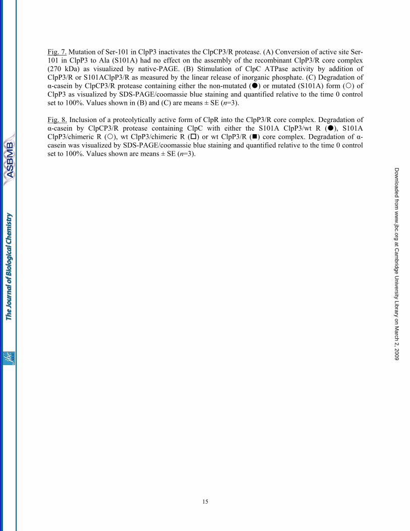
Fig. 7. Mutation of Ser-101 in ClpP3 inactivates the ClpCP3/R protease. (A) Conversion of active site Ser-101 in ClpP3 to Ala (S101A) had no effect on the assembly of the recombinant ClpP3/R core complex (270 kDa) as visualized by native-PAGE. (B) Stimulation of ClpC ATPase activity by addition of ClpP3/R or S101AClpP3/R as measured by the linear release of inorganic phosphate. (C) Degradation of α-casein by ClpCP3/R protease containing either the non-mutated ( ) or mutated (S101A) form ( ) of ClpP3 as visualized by SDS-PAGE/coomassie blue staining and quantified relative to the time 0 control set to 100%. Values shown in (B) and (C) are means ± SE (n=3). Fig. 8. Inclusion of a proteolytically active form of ClpR into the ClpP3/R core complex. Degradation of α-casein by ClpCP3/R protease containing ClpC with either the S101A ClpP3/wt R ( ), S101A ClpP3/chimeric R ( ), wt ClpP3/chimeric R ( ) or wt ClpP3/R ( ) core complex. Degradation of α-casein was visualized by SDS-PAGE/coomassie blue staining and quantified relative to the time 0 control set to 100%. Values shown are means ± SE (n=3).
15
at Cam
bridge University Library on M
arch 2, 2009 w
ww
.jbc.orgD
ownloaded from
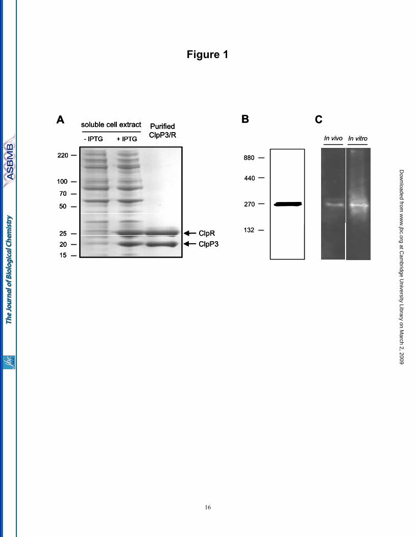
Figure 1
soluble cell extract
220
100
70
50
25
20
15
- IPTG + IPTG
PurifiedClpP3/R
ClpRClpP3
AIn vivo
B
132
270
440
880
CIn vitro
soluble cell extract
220
100
70
50
25
20
15
- IPTG + IPTG
PurifiedClpP3/R
ClpRClpP3
soluble cell extract
220
100
70
50
25
20
15
- IPTG + IPTG
PurifiedClpP3/R
ClpRClpP3
AIn vivo
B
132
270
440
880
CIn vitro
16
at Cam
bridge University Library on M
arch 2, 2009 w
ww
.jbc.orgD
ownloaded from
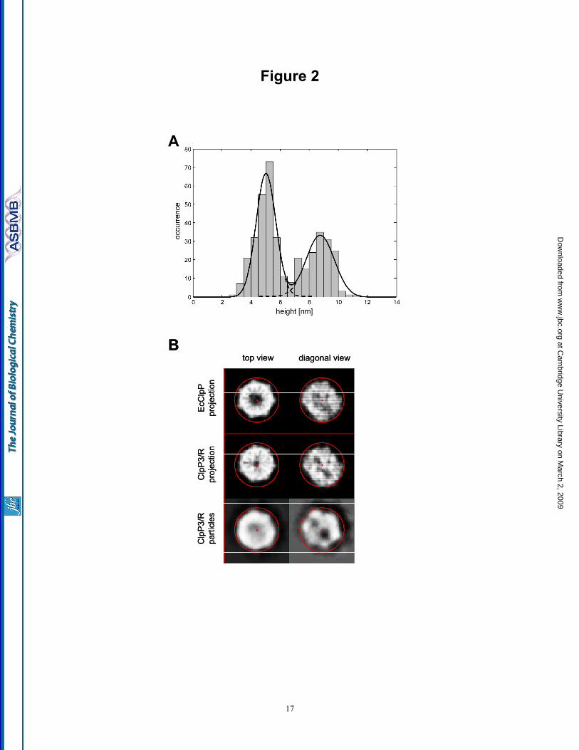
Figure 2
top view diagonal view
Clp
P3/
Rpr
ojec
tion
Clp
P3/
Rpa
rticl
es
A
B
EcC
lpP
proj
ectio
n
top view diagonal view
Clp
P3/
Rpr
ojec
tion
Clp
P3/
Rpa
rticl
es
A
B
EcC
lpP
proj
ectio
n
17
at Cam
bridge University Library on M
arch 2, 2009 w
ww
.jbc.orgD
ownloaded from
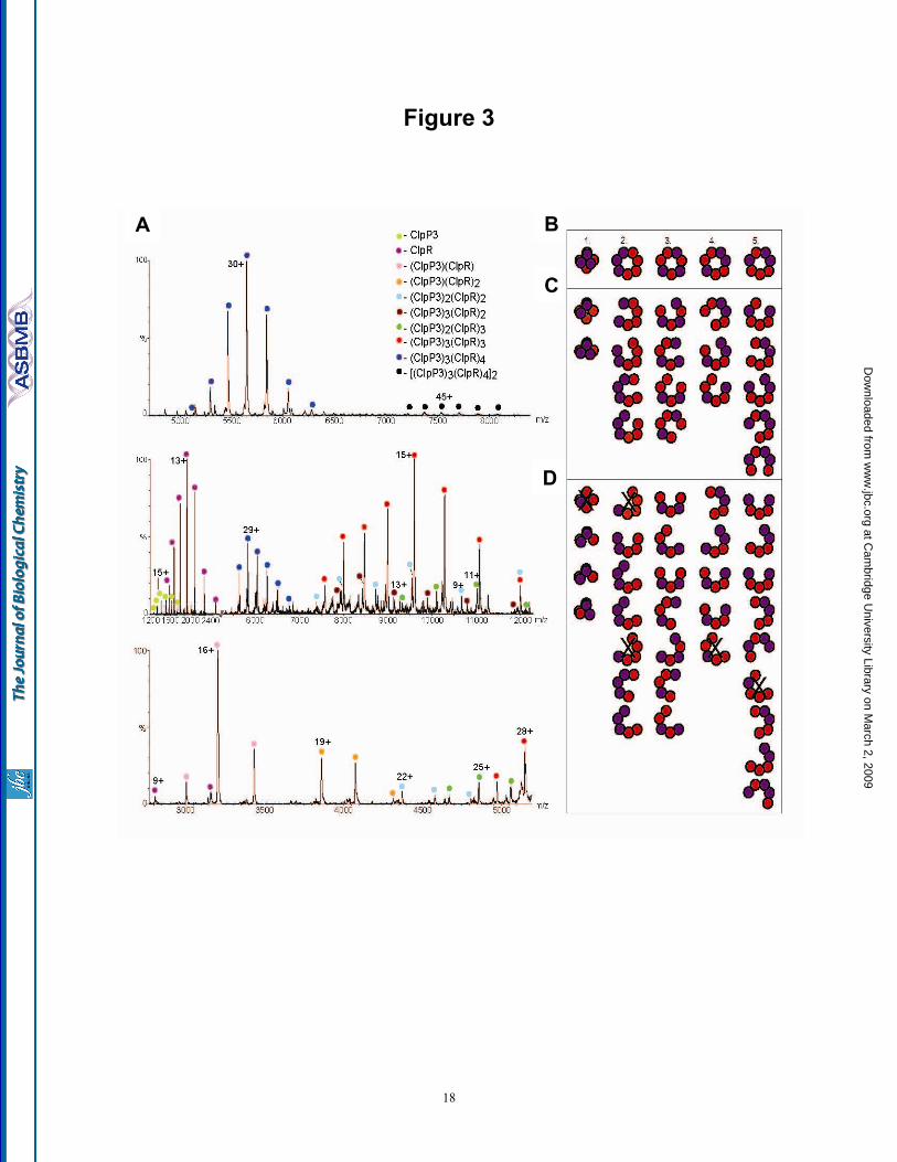
Figure 3
A B
C
D
A B
C
D
18
at Cam
bridge University Library on M
arch 2, 2009 w
ww
.jbc.orgD
ownloaded from
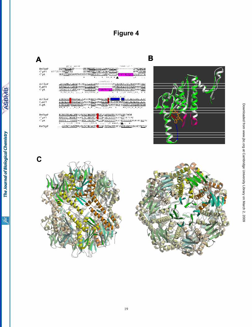
Figure 4
A
C
BA
C
BA
C
B
19
at Cam
bridge University Library on M
arch 2, 2009 w
ww
.jbc.orgD
ownloaded from
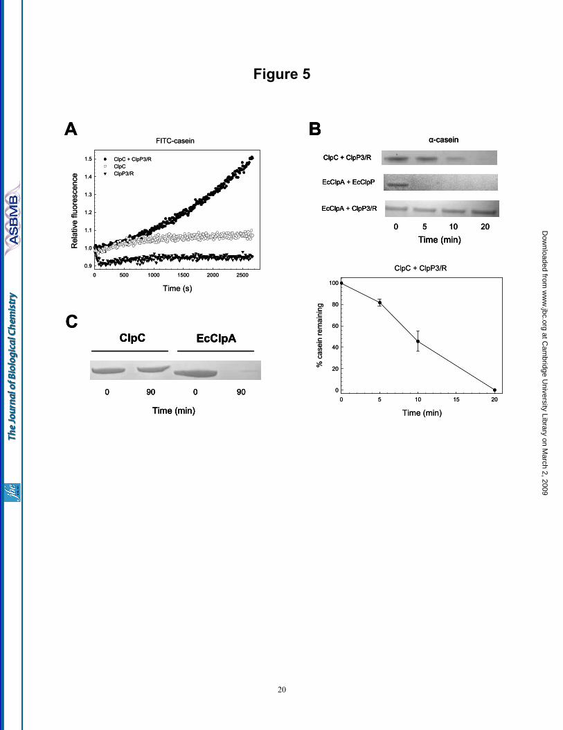
Figure 5
A
EcClpAClpC
0 0 9090
Time (min)
C
FITC-casein
Time (s)
0 500 1000 1500 2000 2500
Rel
ativ
e flu
ores
cenc
e
0.9
1.0
1.1
1.2
1.3
1.4
1.5 ClpC + ClpP3/R ClpC ClpP3/R
Time (min)
BClpC + ClpP3/R
EcClpA + EcClpP
EcClpA + ClpP3/R
0 5 10 20
α-casein
Time (min)
0 5 10 15 20
% c
asei
n re
mai
ning
0
20
40
60
80
100
ClpC + ClpP3/R
A
EcClpAClpC
0 0 9090
Time (min)
EcClpAClpC
0 0 9090
Time (min)
C
FITC-casein
Time (s)
0 500 1000 1500 2000 2500
Rel
ativ
e flu
ores
cenc
e
0.9
1.0
1.1
1.2
1.3
1.4
1.5 ClpC + ClpP3/R ClpC ClpP3/R
Time (min)
BClpC + ClpP3/R
EcClpA + EcClpP
EcClpA + ClpP3/R
0 5 10 20
α-casein
Time (min)
BClpC + ClpP3/R
EcClpA + EcClpP
EcClpA + ClpP3/R
0 5 10 20
α-casein
Time (min)
0 5 10 15 20
% c
asei
n re
mai
ning
0
20
40
60
80
100
ClpC + ClpP3/R
20
at Cam
bridge University Library on M
arch 2, 2009 w
ww
.jbc.orgD
ownloaded from
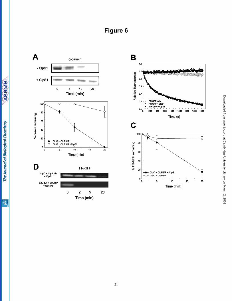
Figure 6
ClpC + ClpP3/R+ ClpS1
EcClpA + EcClpP+ EcClpS
FR-GFP
Time (min)0 2 5 20
C
- ClpS1
+ ClpS1
Time (min)0 5 10 20
A α-casein B
Time (s)
0 200 400 600 800 1000 1200 1400 1600
Rel
ativ
e flu
ores
cenc
e
0.5
0.6
0.7
0.8
0.9
1.0
1.1
FR-GFP onlyFR-GFP + ClpS1MR-GFP + ClpS1
D
Time (min)0 5 10 15 20
% c
asei
n re
mai
ning
0
20
40
60
80
100
ClpC + ClpP3/R ClpC + ClpP3/R +ClpS1
Time (min)0 5 10 15 20
% F
R-G
FP re
mai
ning
0
20
40
60
80
100
ClpC + ClpP3/R + ClpS1ClpC + ClpP3/R
ClpC + ClpP3/R+ ClpS1
EcClpA + EcClpP+ EcClpS
FR-GFP
Time (min)0 2 5 20
ClpC + ClpP3/R+ ClpS1
EcClpA + EcClpP+ EcClpS
FR-GFP
Time (min)0 2 5 20
C
- ClpS1
+ ClpS1
Time (min)0 5 10 20
A α-casein
- ClpS1
+ ClpS1
Time (min)0 5 10 20
A α-casein B
Time (s)
0 200 400 600 800 1000 1200 1400 1600
Rel
ativ
e flu
ores
cenc
e
0.5
0.6
0.7
0.8
0.9
1.0
1.1
FR-GFP onlyFR-GFP + ClpS1MR-GFP + ClpS1
B
Time (s)
0 200 400 600 800 1000 1200 1400 1600
Rel
ativ
e flu
ores
cenc
e
0.5
0.6
0.7
0.8
0.9
1.0
1.1
FR-GFP onlyFR-GFP + ClpS1MR-GFP + ClpS1
D
Time (min)0 5 10 15 20
% c
asei
n re
mai
ning
0
20
40
60
80
100
ClpC + ClpP3/R ClpC + ClpP3/R +ClpS1
Time (min)0 5 10 15 20
% F
R-G
FP re
mai
ning
0
20
40
60
80
100
ClpC + ClpP3/R + ClpS1ClpC + ClpP3/R
21
at Cam
bridge University Library on M
arch 2, 2009 w
ww
.jbc.orgD
ownloaded from
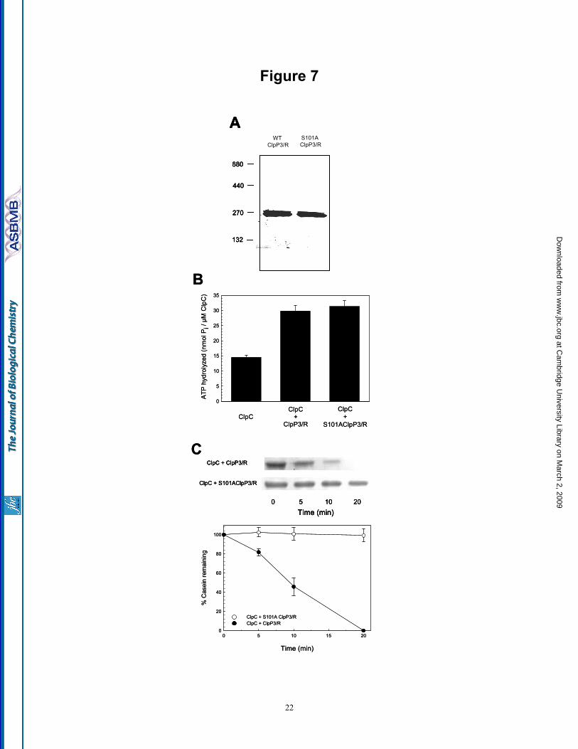
Figure 7
A
132
270
440
880
WT ClpP3/R
S101A ClpP3/R
B
ClpC + ClpP3/R
ClpC + S101AClpP3/R
Time (min)0 5 10 20
ClpCClpC
+ClpP3/R
ClpC+
S101AClpP3/R
0
5
10
15
20
25
30
35
ATP
hyd
roly
zed
(nm
ol P
i / µ
M C
lpC
)
C
Time (min)
0 5 10 15 20
% C
asei
n re
mai
ning
0
20
40
60
80
100
ClpC + S101A ClpP3/R ClpC + ClpP3/R
A
132
270
440
880
WT ClpP3/R
S101A ClpP3/R
A
132
270
440
880
WT ClpP3/R
S101A ClpP3/R
B
ClpC + ClpP3/R
ClpC + S101AClpP3/R
Time (min)0 5 10 20
ClpC + ClpP3/R
ClpC + S101AClpP3/R
Time (min)0 5 10 20
ClpCClpC
+ClpP3/R
ClpC+
S101AClpP3/R
0
5
10
15
20
25
30
35
ATP
hyd
roly
zed
(nm
ol P
i / µ
M C
lpC
)
C
Time (min)
0 5 10 15 20
% C
asei
n re
mai
ning
0
20
40
60
80
100
ClpC + S101A ClpP3/R ClpC + ClpP3/R
22
at Cam
bridge University Library on M
arch 2, 2009 w
ww
.jbc.orgD
ownloaded from
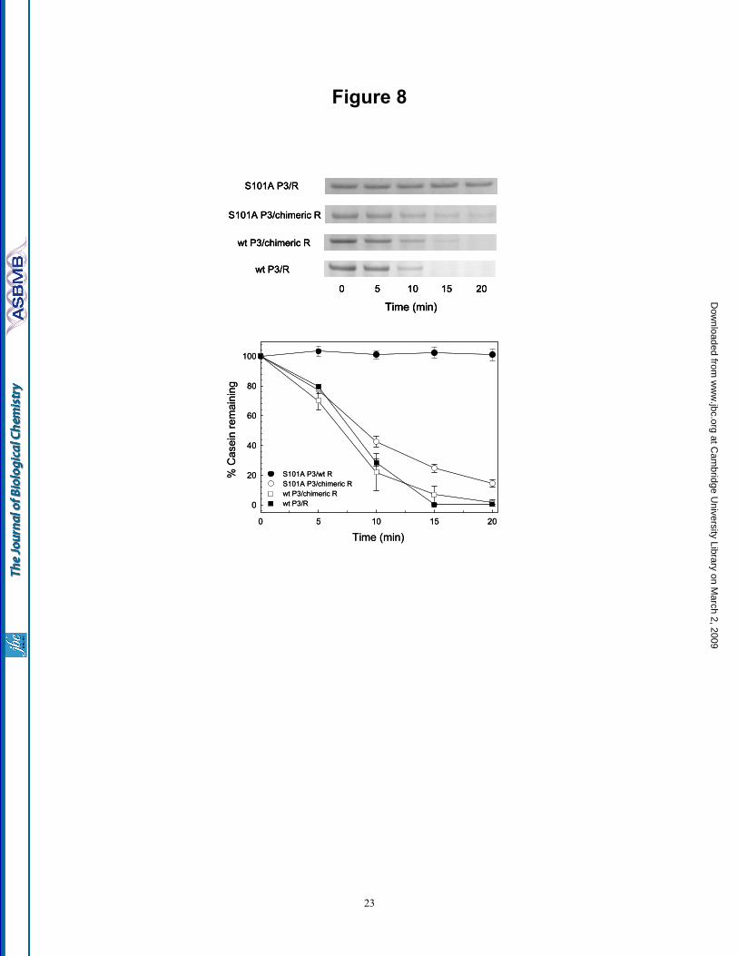
Figure 8
Time (min)0 5 10 15 20
% C
asei
n re
mai
ning
0
20
40
60
80
100
S101A P3/wt R S101A P3/chimeric Rwt P3/chimeric R wt P3/R
S101A P3/R
S101A P3/chimeric R
wt P3/chimeric R
0 5 10 15 20
Time (min)
wt P3/R
Time (min)0 5 10 15 20
% C
asei
n re
mai
ning
0
20
40
60
80
100
S101A P3/wt R S101A P3/chimeric Rwt P3/chimeric R wt P3/R
S101A P3/R
S101A P3/chimeric R
wt P3/chimeric R
0 5 10 15 20
Time (min)
wt P3/R
S101A P3/R
S101A P3/chimeric R
wt P3/chimeric R
0 5 10 15 20
Time (min)
wt P3/R
23
at Cam
bridge University Library on M
arch 2, 2009 w
ww
.jbc.orgD
ownloaded from