Embed Size (px)
Citation preview
RESEARCH ARTICLE Open Access
Attenuation of LPS-induced acute lunginjury by continentalic acid in rodentsthrough inhibition of inflammatorymediators correlates with increased Nrf2protein expressionHassan Ali†, Ashrafullah Khan†, Jawad Ali, Hadayat Ullah, Adnan Khan, Hussain Ali, Nadeem Irshad andSalman Khan*
Abstract
Background: Acute lung injury (ALI) together with acute respiratory distress syndrome (ARDS) are associated withhigh rate of mortality and morbidity in patients. In the current study, the anti-inflammatory effects of continentalicacid (CNT) in LPS-induced acute lung injury model was explored.
Methods: The acute lung injury model was established by administering LPS (5 mg/kg) intraperitonealy. FollowingLPS administration, the survival rate, temperature changes and lung Wet/Dry ratio were assessed. The antioxidants(GSH, GST, Catalase and SOD) and oxidative stress markers (MDA, NO, MPO) were evaluated in all the treatedgroups. Similarly, the cytokines such as IL-1β, IL-6 and TNF-α were analyzed using ELISA assay. The histologicalchanges were determined using H and E staining, while Nrf2 and iNOS level were determined usingimmunohistochemistry analysis. The molecular docking analysis was performed to assess the pharmacokineticsparameters and interaction of the CNT with various protein targets.
(Continued on next page)
© The Author(s). 2020 Open Access This article is licensed under a Creative Commons Attribution 4.0 International License,which permits use, sharing, adaptation, distribution and reproduction in any medium or format, as long as you giveappropriate credit to the original author(s) and the source, provide a link to the Creative Commons licence, and indicate ifchanges were made. The images or other third party material in this article are included in the article's Creative Commonslicence, unless indicated otherwise in a credit line to the material. If material is not included in the article's Creative Commonslicence and your intended use is not permitted by statutory regulation or exceeds the permitted use, you will need to obtainpermission directly from the copyright holder. To view a copy of this licence, visit http://creativecommons.org/licenses/by/4.0/.The Creative Commons Public Domain Dedication waiver (http://creativecommons.org/publicdomain/zero/1.0/) applies to thedata made available in this article, unless otherwise stated in a credit line to the data.
* Correspondence: [email protected]; [email protected]†Hassan Ali and Ashrafullah Khan contributed equally to this work.Pharmacological Sciences Research Lab, Department of Pharmacy, Faculty ofBiological Sciences, Quaid-i-Azam University, Islamabad, Pakistan
Ali et al. BMC Pharmacology and Toxicology (2020) 21:81 https://doi.org/10.1186/s40360-020-00458-7
(Continued from previous page)
Results: The results showed that CNT dose dependently (10, 50 and 100 mg/kg) reduced mortality rate, bodytemperature and lungs Wet/Dry ratio. CNT post-treatment significantly inhibited LPS-induced production of pro-inflammatory cytokines such as IL-1β, IL-6 and TNF-α. The CNT post-treatment markedly improved thehematological parameters, while significantly reduced the MPO (indicator of the neutrophilic infiltration) activitycompared to the LPS treated group. Furthermore, the CNT (100 mg/kg) post-administration remarkably inhibitedthe lung Wet/Dry ratio. The CNT (100 mg/kg) treated group showed marked reduction in the oxidative stressmarkers such as malonaldehyde (MDA) and Nitric oxide (NO) concentration, while induced the level of the anti-oxidant enzymes such as GST, GSH, Catalase and SOD. Similarly, the CNT markedly reduced the iNOS expressionlevel, while induced the Nrf2 protein expression. Additionally, the molecular docking study showed significantbinding interaction with the Nrf2, p65, Keap1, HO-1, IL-1β, IL-6, TNF-α and COX-2, while exhibited excellentphysicochemical properties.
Conclusion: The CNT showed marked protection against the LPS-induced lung injury and improved the behavioral,biochemical and histological parameters. Furthermore, the CNT showed significant interaction with several proteintargets and exhibited better physicochemical properties.
Keywords: Continentalic acid, LPS, Inflammation, Cytokines, Antioxidants
BackgroundAcute lung injury (ALI) together with acute respiratorydistress syndrome (ARDS) are respiratory syndromes as-sociated with high mortality and morbidity caused bypneumonia, trauma and sepsis [1]. ALI is an inflamma-tory disease of lungs which is characterized by the dis-ruption of alveolar endothelial and epithelial barriers,neutrophilic infiltration at pulmonary sites together withnon-cardiogenic edema [2, 3]. Despite improvements intherapies, the morbidity and mortality rates of ALI re-main high i.e. 30–40% [4–6]. Thus, novel therapies withproven safety and efficacy are needed to improve theclinical outcomes of the patients affected with the dis-ease. LPS is a glycolipid that is present in the Gram-negative microbes cell wall, is considered as the corebasis of ALI [7]. LPS induces ALI in animal models byaltering alveolar membrane permeability, recruiting acti-vated neutrophils and macrophages to the lungs [8, 9].These effects compromises the alveolar membrane integ-rity and ultimately impairs the gaseous exchange [8, 9].Furthermore, LPS exposure is associated with exaggeratedproduction of various pro-inflammatory cytokines such astumor necrosis factor (TNF)-α, interleukin (IL)-1β andinterleukin (IL)-6 in lungs [8]. Over production of TNF-α,IL-1β and IL-6 lead to the development of ALI and resultsin poor clinical outcome in patients with ALI [10, 11].The Nrf2 signaling plays an important role in neutralizing
the oxidative stress and oxidative stress mediated damage[12]. The Nrf2 remains dormant within the cytosol underthe inhibitory influence of the Keap1 and translocated tonucleus when become free from the Keap1 influence. Fol-lowing translocation to nucleus, the Nrf2 interact with theantioxidant response element (ARE) to alter the expressionof the genes concerned with the antioxidants and cytokines[13, 14]. Previously reported studies suggest that over
production of inflammatory cytokines and oxidative stresstriggered by LPS leads to neutrophil infiltration in the lungtissue [15]. Persistent activation and migration of neutro-phils into the transepithelial membrane is considered as ahallmark feature of acute lung injury that increases the de-struction of basement membrane with promotion of mem-brane permeability [16]. Significant evidence reported theinvolvement of inflammatory response and oxidative stressin the pathogenesis of ALI [17]. The results observed in-clude decreased level of antioxidant enzymes (GSH, GST,SOD and CAT), abundant production of neutrophils andelevated levels of inflammatory cytokines (IL-1β, TNF-αand IL-6) in plasma and lung tissue of mice [8]. Elevatedplasma levels of cytokines i.e. IL-1β, IL-6 and tumor necro-sis factor-α (TNF-α) strongly predicts the high mortality as-sociated with acute lung injury [10, 18, 19].Currently, numerous studies revealed that bioactive com-
pounds from natural herbs act as potential candidates forthe management of ALI in rodent model [20]. Continenta-lic acid is a diterpene obtained from Aralia continentaliswhich belongs to the family of Araliaceae [21]. Studies havebeen reported that continentalic acid exhibit numerouspharmacological activities such as anti-inflammatory, anti-arthritic, nephroprotective etc. [22]. Therefore, in thepresent study, LPS-induced lung injury model in mice wasused to simulate acute lung injury and observed the effectsthrough the behavioral and biochemical methods. It washypothesized that continentalic acid possesses its anti-inflammatory effect via inhibiting inflammatory responseand oxidative stress in LPS-induced lung injury model.
MethodsChemical and reagentsContinentalic acid (purity 99.9%) was received fromProf. Yeong Shik Kim, Emiritus Professor, College of
Ali et al. BMC Pharmacology and Toxicology (2020) 21:81 Page 2 of 14
Pharmacy, Seoul National University, Korea. All thechemicals and reagent included in this study such asdexamethasone and LPS were obtained from theSigma Aldrich (St. Louis, MO, USA). The cytokineswere analyzed in lung tissue using ELISA Kits ob-tained from the eBioscience (eBioscience, Inc. USA).The primary and secondary antibodies for Nrf2 andiNOS were obtained from the Santa Cruz (Santa CruzBiotechnology, Inc). The DAB reagent was obtainedfrom the Sigma Aldrich (St. Louis, MO, USA). TheNGS (normal goat serum) and AB complex used inthe immunohistochemistry were obtained from(SCBT, U.S.A).
Animals and ethical statementMale albino mice (BALB/c) (22–26 g) were used for theentire study having age of 3 to 4 weeks and purchasedfrom National Institute of Health (NIH) Islamabad,Pakistan. Standard environmental and food conditionswere provided to all the animals i.e. 22 ± 1 °C, 55 ± 5%humidity and 12 h light/dark cycle with free food andwater access. All animal experimentations were carriedout as per Bioethical Committee protocols for laboratoryAnimals Care and Use (Quaid-i-Azam University,Islamabad) under Ethical Committee code (ApprovalNo. BEC-FBS-QAU 2018–86).
Model and groupingAll the animals were randomly and double blindlyassigned to six groups to avoid the experimental biasness(each group contain 8 mice). The continentalic acid anddexamethasone was dissolved in the normal saline (2%DMSO), while the LPS was dissolved in the normal sa-line only.
1. Vehicle control treated with normal saline (i.p)2. Negative control treated with LPS 5 mg/kg only
(i.p)3. LPS (5 mg/kg, i.p) + vehicle4. Positive control treated with the LPS +
dexamethasone (10 mg/kg, i.p)5. Continentalic acid (10 mg/kg, i.p) + LPS 5 mg/kg
(i.p)6. Continentalic acid (50 mg/kg, i.p) + LPS 5 mg/kg
(i.p)7. Continentalic acid (100 mg/kg, i.p) + LPS 5 mg/kg
(i.p)
In-vivo model of ALI and sampling protocolsTo assess the percent survival rate during entire study,LPS 5mg/kg with different doses of continentalic acid(10, 50 and 100 mg/kg body weight dissolved in normalsaline (2% DMSO)) was administered intraperitoneallythirty minutes prior to LPS injection. The mortality of
mice was recorded every 3 h after the LPS injection ineach group for 24 h. At the end of the experiment, theanimals were anesthetized with the combination of Xyla-zine and Ketamine injection (16 mg and 60mg respect-ively, i.p) to make them unconscious and lessen thepainful feeling related with the euthanasia. Once the ani-mals were anesthetized, the CO2 chamber was used toeuthanize the animals. The animal death was confirmedby assessing the heartbeat, respiration, eye reflexes andbody movement. The overall euthanasia process was reg-ulated by the institutional ethical committee.
Temperature assessmentLPS is the endotoxin of the Gram negative bacteria andis associated with increase in the body temperature [23].The pyrexia was measured before and after LPS induc-tion at 0 and 24 h according to the previously describedmethod [24].
Determination of lung wet/dry weight ratioIn order to assess the pulmonary edema following LPSadministration, the wet/dry weight ratio was determined[25]. Mice were sacrificed 24 h after LPS stimulation.Wet weight was determined after excising lung tissues.The wet lung was then placed in the oven at 80 °C for24 h to measure the dried weight. Then W/D weight ra-tio was calculated by dividing dry weight over wetweight [25].
Determination of GSH, GST, catalase and SODconcentrationsThe tissue GSH, GST, Catalase and SOD levels weredetermined according to the previously establishedprotocols [14]. Briefly, lung tissues were homogenizedand spun at 448 RCF (relative centrifugal force) for10 min. The supernatant obtained was then used forthe determination of enzymatic and non-enzymaticantioxidant activity. The concentrations of GSH, GST,Catalase and SOD were detected by monitoring thechange in absorbance using spectrophotometer atλmax 412 nm, λmax 340 nm, λmax 240 nm and λmax413 nm, respectively [14].
Estimation of lipid peroxidationMDA level was determined according to previously re-ported method [26]. Briefly lung tissues were first ho-mogenized and then centrifuged at 448 RCF for 10 min.The supernatant obtained was then used for the deter-mination of antioxidant activity. The presence of thio-barbituric acid reactive substances (TBARS) weredetected by monitoring changes in absorbance using mi-croplate reader at 535 nm [14].
Ali et al. BMC Pharmacology and Toxicology (2020) 21:81 Page 3 of 14

Determination of nitrite concentrationsThe production of nitric oxide in all the treated groupswere measured by Griess assay according to the methoddescribed previously [27]. At the final day of experiment,all the mice were sacrificed and blood collection wasdone through cardiac puncture. The collected blood wasthen centrifuged at 700 RCF for 10 min at 4 °C andplasma was separated from the cellular component forNO determination as reported previously [27].
Cytokines analysisInflammatory cytokines (IL1-β, IL-6 and TNF-α) levelswere determined in lung tissues using ELISA (enzymelinked immunosorbent assay) kits obtained from(eBioscience, Inc., USA). Cytokines level were deter-mined as per manufacturer’s instruction [28, 29].
Complete blood countThe immune cells plays key role during inflammatoryconditions [30]. In order to assess the effect of continen-talic acid on the various immune cells, blood completecount was performed. Following 24 h of LPS administra-tion, the blood sample was collected from the heart ofmice. The collected blood was then utilized inhematological studies by obtaining complete blood pic-ture including major hematological parameters such asWBC, Red blood cells (RBC), platelets, Hemoglobin(Hb), neutrophils and lymphocytes [31].
Tissue processing and sample collectionMice were sacrificed 24 h after LPS administration usingCO2 anesthetization and the entire lung tissue were dis-sected from all the groups. Separated lung tissue werewashed by 0.9% normal saline and then preserved in10% formalin for histopathological examination [32–34].
Histopathological analysisTo illustrate the histological modifications induced byLPS, lungs were kept in formaldehyde solution and thenfixed in PFA solution for 24 h. Subsequently, the tissueswere sectioned at 5 μm and were stained withhematoxylin–eosin using standard histological tech-niques. The lung and tracheal tissues were examinedunder optical microscope, and photos were taken as de-scribed previously by [35, 36]. Results were graded from0 to 4 for each item, as described above, where 0 =min-imal damage, 1 =mild damage, 2 =moderate damage,3 = severe damage and 4 =maximal damage as describedpreviously [37].
Myeloperoxidase assay for the neutrophilic infiltrationThe MPO activity was performed to assess the inhibitoryeffect of the continentalic acid on the LPS-induced neu-trophilic infiltration [38]. The MPO activity was
determined using CTAB and o-dianisidine method as re-ported previously with necessary modification for all thetreated group [38].
Immunohistochemistry studyThe immunohistochemistry was performed to deter-mine the effect of the contenentalic acid on the Nrf2and iNOS proteins [14]. The immunohistochemistrywas performed as reported previously [14]. The tissuewas deparrafinzed using xylene and washed in alcoholsolution of different concentration. Following washingwith alcohol the tissue were treated with the Avidin-biotin complex, followed by treated with NGS for 2 h,primary and secondary antibodies (Nrf2 and iNOS).At the end tissue were placed in the DAB solution,dried and cover slips were applied using mountingmedia. The slides were visualized at 100X microscopeand quantified using Image J software 1.8_172 (NIH,USA) [14].
Assessment of pharmacokinetic and pharmacodynamicsanalysis using docking studiesThe pharmacokinetic parameters of the continentalicacid were assessed using Swiss target prediction soft-ware (http://www.swisstargetprediction.ch/) as re-ported previously [39]. The various parameters thatwere assessed during the current study includes phys-icochemical properties, lipophilicity, aqueous solubil-ity, absorption and pharmacokinetic parameters wereevaluated. The possible metabolic route and metabo-lites were also determined using Glory software(https://nerdd.zbh.uni-hamburg.de/glory/) [40]. Fur-thermore, docking analysis was performed to investi-gate the interaction with various molecular targetsusing AutoDock Vina program. The 3D structure ofthe continentalic acid using Chemdraw_16 and savedas PDB file. Furthermore, the toxicity of the continen-talic acid was evaluated against the various cell linesusing CLC-Pred (http://www.way2drug.com/Cell-line/)software. Similarly, the 3D-structures of target pro-teins (Keap-1 PDB-ID: 4iqk), (Nrf2 PDB-ID: 2flu), (p65 PDB-ID: 1vkx), (HO-1 PDB-ID: 1ubb), (TNF-αPDB-ID: 2az5), (IL-1Β PDB-ID: 1itb), (IL-6 PDB-ID:1p9m) and (COX-2 PDB-ID: 5ikq) were taken fromRCSB protein data bank. Affinity of best docked poseof ligand and protein target complex was determinedby E-value (Kcal/mol). The results of the dockinginteraction were analyzed using discovery studiovisualizer_2016.
Statistical analysisResults were represented as the means (n = 8) ± StandardDeviations (S.D). The data was analyzed using One wayanalysis of variance (ANOVA) followed by Dunnett′s t
Ali et al. BMC Pharmacology and Toxicology (2020) 21:81 Page 4 of 14
test to compare means among groups. The “p” value lessthan 0.05 were considered statistically significant.
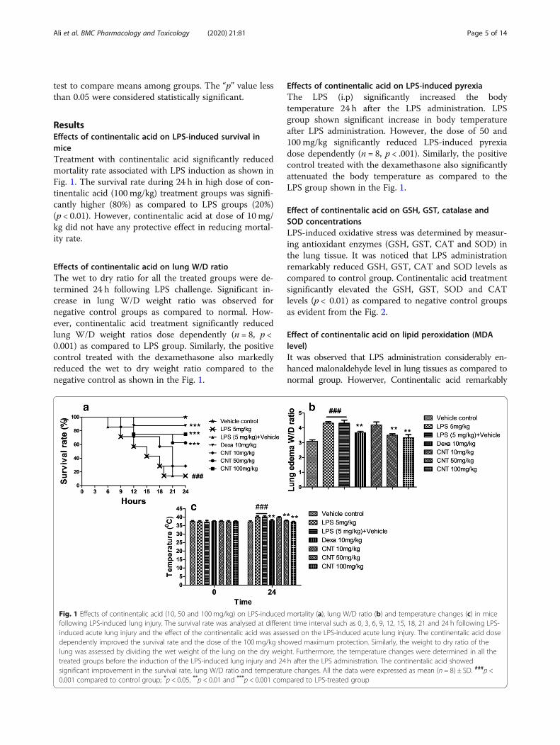
ResultsEffects of continentalic acid on LPS-induced survival inmiceTreatment with continentalic acid significantly reducedmortality rate associated with LPS induction as shown inFig. 1. The survival rate during 24 h in high dose of con-tinentalic acid (100 mg/kg) treatment groups was signifi-cantly higher (80%) as compared to LPS groups (20%)(p < 0.01). However, continentalic acid at dose of 10 mg/kg did not have any protective effect in reducing mortal-ity rate.
Effects of continentalic acid on lung W/D ratioThe wet to dry ratio for all the treated groups were de-termined 24 h following LPS challenge. Significant in-crease in lung W/D weight ratio was observed fornegative control groups as compared to normal. How-ever, continentalic acid treatment significantly reducedlung W/D weight ratios dose dependently (n = 8, p <0.001) as compared to LPS group. Similarly, the positivecontrol treated with the dexamethasone also markedlyreduced the wet to dry weight ratio compared to thenegative control as shown in the Fig. 1.
Effects of continentalic acid on LPS-induced pyrexiaThe LPS (i.p) significantly increased the bodytemperature 24 h after the LPS administration. LPSgroup shown significant increase in body temperatureafter LPS administration. However, the dose of 50 and100 mg/kg significantly reduced LPS-induced pyrexiadose dependently (n = 8, p < .001). Similarly, the positivecontrol treated with the dexamethasone also significantlyattenuated the body temperature as compared to theLPS group shown in the Fig. 1.
Effect of continentalic acid on GSH, GST, catalase andSOD concentrationsLPS-induced oxidative stress was determined by measur-ing antioxidant enzymes (GSH, GST, CAT and SOD) inthe lung tissue. It was noticed that LPS administrationremarkably reduced GSH, GST, CAT and SOD levels ascompared to control group. Continentalic acid treatmentsignificantly elevated the GSH, GST, SOD and CATlevels (p < 0.01) as compared to negative control groupsas evident from the Fig. 2.
Effect of continentalic acid on lipid peroxidation (MDAlevel)It was observed that LPS administration considerably en-hanced malonaldehyde level in lung tissues as compared tonormal group. Howerver, Continentalic acid remarkably
Fig. 1 Effects of continentalic acid (10, 50 and 100mg/kg) on LPS-induced mortality (a), lung W/D ratio (b) and temperature changes (c) in micefollowing LPS-induced lung injury. The survival rate was analysed at different time interval such as 0, 3, 6, 9, 12, 15, 18, 21 and 24 h following LPS-induced acute lung injury and the effect of the continentalic acid was assessed on the LPS-induced acute lung injury. The continentalic acid dosedependently improved the survival rate and the dose of the 100mg/kg showed maximum protection. Similarly, the weight to dry ratio of thelung was assessed by dividing the wet weight of the lung on the dry weight. Furthermore, the temperature changes were determined in all thetreated groups before the induction of the LPS-induced lung injury and 24 h after the LPS administration. The continentalic acid showedsignificant improvement in the survival rate, lung W/D ratio and temperature changes. All the data were expressed as mean (n = 8) ± SD. ###p <0.001 compared to control group; *p < 0.05, **p < 0.01 and ***p < 0.001 compared to LPS-treated group
Ali et al. BMC Pharmacology and Toxicology (2020) 21:81 Page 5 of 14

Fig. 2 Effect of treatment with continentalic acid (10, 50 and 100mg/kg) on levels of antioxidant enzymes such as (a) GSH, (b) GST, (c) Catalase,(d) SOD, (e) MDA in LPS-induced lung tissue. The level of these antioxidants and oxidative stress parameters were markedly compromised.However, continentalic acid post treatment significantly enhanced antioxidant enzymes such as GSH, GST, Catalase, SOD and reduced the level ofMDA. The results were shown in percentage. All data were expressed as mean (n = 8) ± SD. ###p < 0.001 compared to control group; *p < 0.05,**p < 0.01 and ***p < 0.001 compared to LPS-treated group
Fig. 3 Effect of continentalic acid (10, 50 and 100mg/kg) on LPS-induced nitrite concentration. The continentalic acid was assessed against theNO production in both plasma (a) and (b) tissue following establishing ALI. The continentalic acid treatment significantly reduced NO productionin both plasma and tissue dose dependently. Similarly, the dexamethasone also markedly decreased the NO production in the both plasma andtissue. The results were shown in percentage. All data were expressed as mean (n = 8) ± SD. ###p < 0.001 compared to control group; *p < 0.05,**p < 0.01 and ***p < 0.001 compared to LPS-treated group
Ali et al. BMC Pharmacology and Toxicology (2020) 21:81 Page 6 of 14
reduced MDA level dose dependently as compared to LPStreated group (p < 0.001) as evident from the Fig. 2.
Effect of continentalic acid on nitrite concentrationsThe nitrite level was significantly raised (p < 0.001) bothin lung tissue and plasma post 24 h of LPS administra-tion as compared to the control group. Continentalicacid remarkably reduced (p < 0.001) LPS-induced ele-vated nitrite level both in lung tissue and plasma. Simi-larly, dexamethasone also suppress nitrite level Fig. 3.
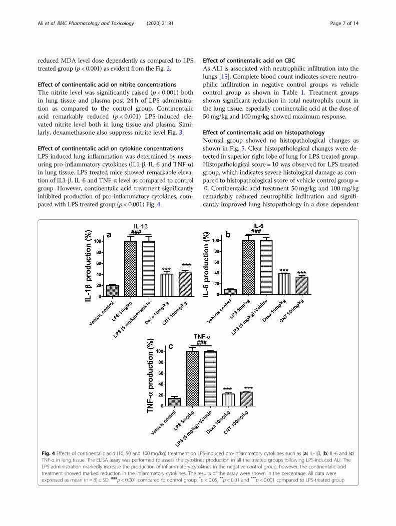
Effect of continentalic acid on cytokine concentrationsLPS-induced lung inflammation was determined by meas-uring pro-inflammatory cytokines (IL1-β, IL-6 and TNF-α)in lung tissue. LPS treated mice showed remarkable eleva-tion of IL1-β, IL-6 and TNF-α level as compared to controlgroup. However, continentalic acid treatment significantlyinhibited production of pro-inflammatory cytokines, com-pared with LPS treated group (p < 0.001) Fig. 4.
Effect of continentalic acid on CBCAs ALI is associated with neutrophilic infiltration into thelungs [15]. Complete blood count indicates severe neutro-philic infiltration in negative control groups vs vehiclecontrol group as shown in Table 1. Treatment groupsshown significant reduction in total neutrophils count inthe lung tissue, especially continentalic acid at the dose of50mg/kg and 100mg/kg showed maximum response.
Effect of continentalic acid on histopathologyNormal group showed no histopathological changes asshown in Fig. 5. Clear histopathological changes were de-tected in superior right lobe of lung for LPS treated group.Histopathological score = 10 was observed for LPS treatedgroup, which indicates severe histological damage as com-pared to histopathological score of vehicle control group =0. Continentalic acid treatment 50mg/kg and 100mg/kgremarkably reduced neutrophilic infiltration and signifi-cantly improved lung histopathology in a dose dependent
Fig. 4 Effects of continentalic acid (10, 50 and 100mg/kg) treatment on LPS-induced pro-inflammatory cytokines such as (a) IL-1β, (b) IL-6 and (c)TNF-α in lung tissue. The ELISA assay was performed to assess the cytokines production in all the treated groups following LPS-induced ALI. TheLPS administration markedly increase the production of inflammatory cytokines in the negative control group, however, the continentalic acidtreatment showed marked reduction in the inflammatory cytokines. The results of the assay were shown in the percentage. All data wereexpressed as mean (n = 8) ± SD. ###p < 0.001 compared to control group; *p < 0.05, **p < 0.01 and ***p < 0.001 compared to LPS-treated group
Ali et al. BMC Pharmacology and Toxicology (2020) 21:81 Page 7 of 14
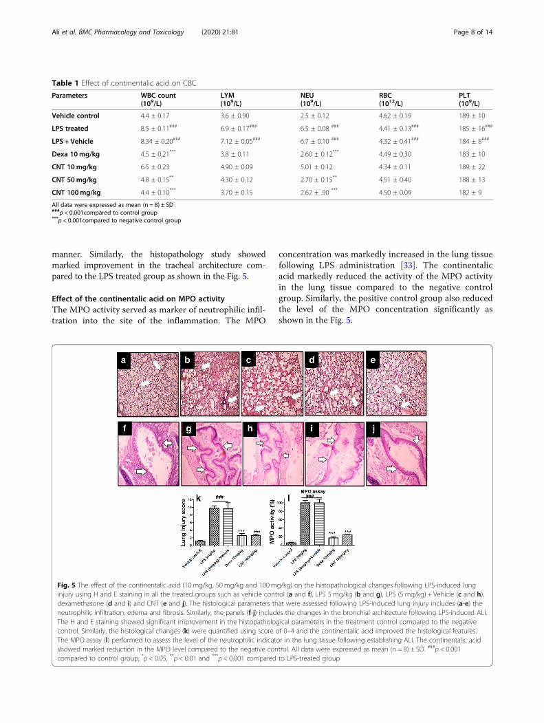
manner. Similarly, the histopathology study showedmarked improvement in the tracheal architecture com-pared to the LPS treated group as shown in the Fig. 5.
Effect of the continentalic acid on MPO activityThe MPO activity served as marker of neutrophilic infil-tration into the site of the inflammation. The MPO
concentration was markedly increased in the lung tissuefollowing LPS administration [33]. The continentalicacid markedly reduced the activity of the MPO activityin the lung tissue compared to the negative controlgroup. Similarly, the positive control group also reducedthe level of the MPO concentration significantly asshown in the Fig. 5.
Table 1 Effect of continentalic acid on CBC
Parameters WBC count(109/L)
LYM(109/L)
NEU(109/L)
RBC(1012/L)
PLT(109/L)
Vehicle control 4.4 ± 0.17 3.6 ± 0.90 2.5 ± 0.12 4.62 ± 0.19 189 ± 10
LPS treated 8.5 ± 0.11### 6.9 ± 0.17### 6.5 ± 0.08 ### 4.41 ± 0.13### 185 ± 16###
LPS + Vehicle 8.34 ± 0.20### 7.12 ± 0.05### 6.7 ± 0.10 ### 4.32 ± 0.41### 184 ± 8###
Dexa 10mg/kg 4.5 ± 0.21*** 3.8 ± 0.11 2.60 ± 0.12*** 4.49 ± 0.30 183 ± 10
CNT 10mg/kg 6.5 ± 0.23 4.90 ± 0.09 5.01 ± 0.12 4.34 ± 0.11 189 ± 22
CNT 50mg/kg 4.8 ± 0.15** 4.30 ± 0.12 2.70 ± 0.15** 4.51 ± 0.40 188 ± 13
CNT 100mg/kg 4.4 ± 0.10*** 3.70 ± 0.15 2.62 ± .90 *** 4.50 ± 0.09 182 ± 9
All data were expressed as mean (n = 8) ± SD###p < 0.001compared to control group***p < 0.001compared to negative control group
Fig. 5 The effect of the continentalic acid (10 mg/kg, 50 mg/kg and 100mg/kg) on the histopathological changes following LPS-induced lunginjury using H and E staining in all the treated groups such as vehicle control (a and f), LPS 5 mg/kg (b and g), LPS (5 mg/kg) + Vehicle (c and h),dexamethasone (d and i) and CNT (e and j). The histological parameters that were assessed following LPS-induced lung injury includes (a-e) theneutrophilic infiltration, edema and fibrosis. Similarly, the panels (f-j) includes the changes in the bronchial architecture following LPS-induced ALI.The H and E staining showed significant improvement in the histopathological parameters in the treatment control compared to the negativecontrol. Similarly, the histological changes (k) were quantified using score of 0–4 and the continentalic acid improved the histological features.The MPO assay (l) performed to assess the level of the neutrophilic indicator in the lung tissue following establishing ALI. The continentalic acidshowed marked reduction in the MPO level compared to the negative control. All data were expressed as mean (n = 8) ± SD. ###p < 0.001compared to control group; *p < 0.05, **p < 0.01 and ***p < 0.001 compared to LPS-treated group
Ali et al. BMC Pharmacology and Toxicology (2020) 21:81 Page 8 of 14
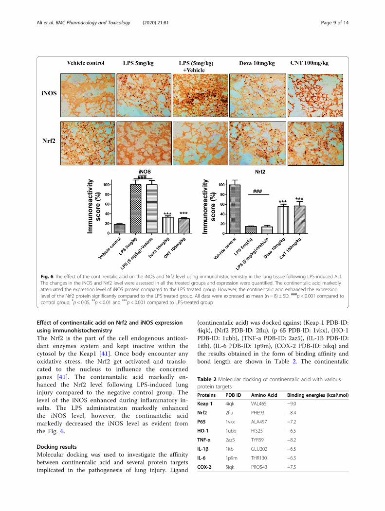
Effect of continentalic acid on Nrf2 and iNOS expressionusing immunohistochemistryThe Nrf2 is the part of the cell endogenous antioxi-dant enzymes system and kept inactive within thecytosol by the Keap1 [41]. Once body encounter anyoxidative stress, the Nrf2 get activated and translo-cated to the nucleus to influence the concernedgenes [41]. The contenantalic acid markedly en-hanced the Nrf2 level following LPS-induced lunginjury compared to the negative control group. Thelevel of the iNOS enhanced during inflammatory in-sults. The LPS administration markedly enhancedthe iNOS level, however, the continantelic acidmarkedly decreased the iNOS level as evident fromthe Fig. 6.
Docking resultsMolecular docking was used to investigate the affinitybetween continentalic acid and several protein targetsimplicated in the pathogenesis of lung injury. Ligand
(continentalic acid) was docked against (Keap-1 PDB-ID:4iqk), (Nrf2 PDB-ID: 2flu), (p 65 PDB-ID: 1vkx), (HO-1PDB-ID: 1ubb), (TNF-a PDB-ID: 2az5), (IL-1Β PDB-ID:1itb), (IL-6 PDB-ID: 1p9m), (COX-2 PDB-ID: 5ikq) andthe results obtained in the form of binding affinity andbond length are shown in Table 2. The continentalic
Fig. 6 The effect of the continentalic acid on the iNOS and Nrf2 level using immunohistochemistry in the lung tissue following LPS-induced ALI.The changes in the iNOS and Nrf2 level were assessed in all the treated groups and expression were quantified. The continentalic acid markedlyattenuated the expression level of iNOS protein compared to the LPS treated group. However, the continentalic acid enhanced the expressionlevel of the Nrf2 protein significantly compared to the LPS treated group. All data were expressed as mean (n = 8) ± SD. ###p < 0.001 compared tocontrol group; *p < 0.05, **p < 0.01 and ***p < 0.001 compared to LPS-treated group
Table 2 Molecular docking of continentalic acid with variousprotein targets
Proteins PDB ID Amino Acid Binding energies (kcal\mol)
Keap 1 4iqk VAL465 −9.0
Nrf2 2flu PHE93 −8.4
P65 1vkx ALA497 −7.2
HO-1 1ubb HIS25 −6.5
TNF-α 2az5 TYR59 −8.2
IL-1β 1itb GLU202 −6.5
IL-6 1p9m THR130 −6.5
COX-2 5iqk PRO543 −7.5
Ali et al. BMC Pharmacology and Toxicology (2020) 21:81 Page 9 of 14
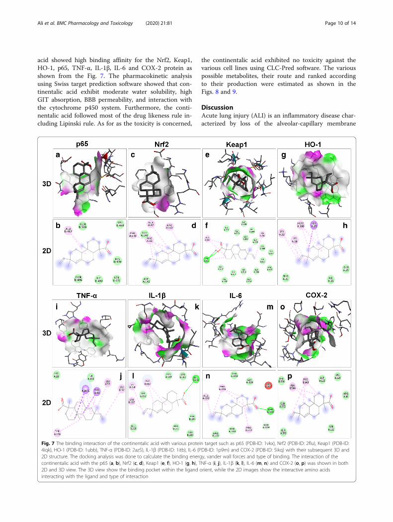
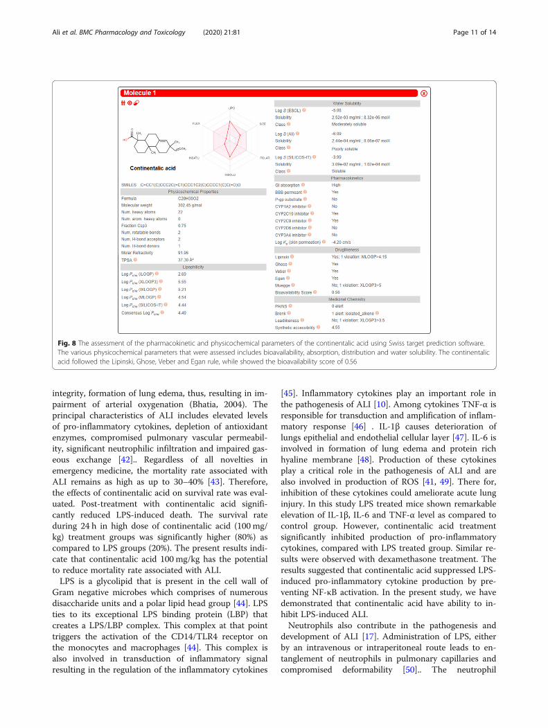
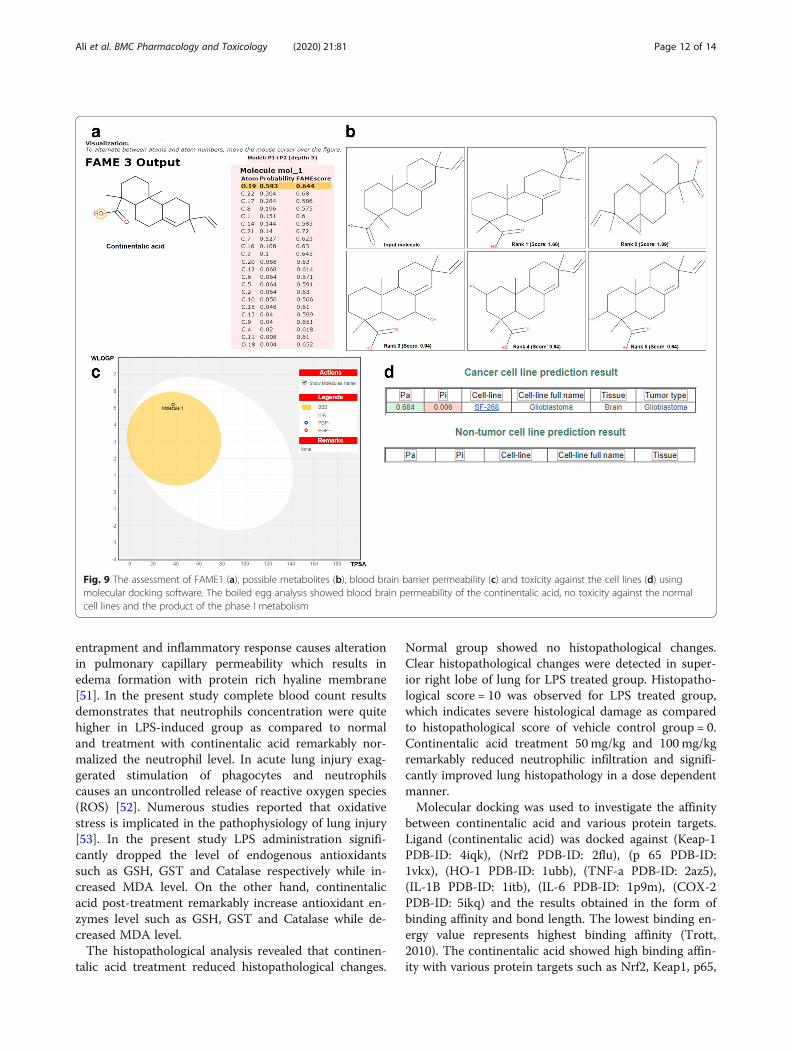
acid showed high binding affinity for the Nrf2, Keap1,HO-1, p65, TNF-α, IL-1β, IL-6 and COX-2 protein asshown from the Fig. 7. The pharmacokinetic analysisusing Swiss target prediction software showed that con-tinentalic acid exhibit moderate water solubility, highGIT absorption, BBB permeability, and interaction withthe cytochrome p450 system. Furthermore, the conti-nentalic acid followed most of the drug likeness rule in-cluding Lipinski rule. As for as the toxicity is concerned,
the continentalic acid exhibited no toxicity against thevarious cell lines using CLC-Pred software. The variouspossible metabolites, their route and ranked accordingto their production were estimated as shown in theFigs. 8 and 9.
DiscussionAcute lung injury (ALI) is an inflammatory disease char-acterized by loss of the alveolar-capillary membrane
Fig. 7 The binding interaction of the continentalic acid with various protein target such as p65 (PDB-ID: 1vkx), Nrf2 (PDB-ID: 2flu), Keap1 (PDB-ID:4iqk), HO-1 (PDB-ID: 1ubb), TNF-α (PDB-ID: 2az5), IL-1β (PDB-ID: 1itb), IL-6 (PDB-ID: 1p9m) and COX-2 (PDB-ID: 5ikq) with their subsequent 3D and2D structure. The docking analysis was done to calculate the binding energy, vander wall forces and type of binding. The interaction of thecontinentalic acid with the p65 (a, b), Nrf2 (c, d), Keap1 (e, f), HO-1 (g, h), TNF-α (i, j), IL-1β (k, l), IL-6 (m, n) and COX-2 (o, p) was shown in both2D and 3D view. The 3D view show the binding pocket within the ligand orient, while the 2D images show the interactive amino acidsinteracting with the ligand and type of interaction
Ali et al. BMC Pharmacology and Toxicology (2020) 21:81 Page 10 of 14
integrity, formation of lung edema, thus, resulting in im-pairment of arterial oxygenation (Bhatia, 2004). Theprincipal characteristics of ALI includes elevated levelsof pro-inflammatory cytokines, depletion of antioxidantenzymes, compromised pulmonary vascular permeabil-ity, significant neutrophilic infiltration and impaired gas-eous exchange [42].. Regardless of all novelties inemergency medicine, the mortality rate associated withALI remains as high as up to 30–40% [43]. Therefore,the effects of continentalic acid on survival rate was eval-uated. Post-treatment with continentalic acid signifi-cantly reduced LPS-induced death. The survival rateduring 24 h in high dose of continentalic acid (100 mg/kg) treatment groups was significantly higher (80%) ascompared to LPS groups (20%). The present results indi-cate that continentalic acid 100 mg/kg has the potentialto reduce mortality rate associated with ALI.LPS is a glycolipid that is present in the cell wall of
Gram negative microbes which comprises of numerousdisaccharide units and a polar lipid head group [44]. LPSties to its exceptional LPS binding protein (LBP) thatcreates a LPS/LBP complex. This complex at that pointtriggers the activation of the CD14/TLR4 receptor onthe monocytes and macrophages [44]. This complex isalso involved in transduction of inflammatory signalresulting in the regulation of the inflammatory cytokines
[45]. Inflammatory cytokines play an important role inthe pathogenesis of ALI [10]. Among cytokines TNF-α isresponsible for transduction and amplification of inflam-matory response [46] . IL-1β causes deterioration oflungs epithelial and endothelial cellular layer [47]. IL-6 isinvolved in formation of lung edema and protein richhyaline membrane [48]. Production of these cytokinesplay a critical role in the pathogenesis of ALI and arealso involved in production of ROS [41, 49]. There for,inhibition of these cytokines could ameliorate acute lunginjury. In this study LPS treated mice shown remarkableelevation of IL-1β, IL-6 and TNF-α level as compared tocontrol group. However, continentalic acid treatmentsignificantly inhibited production of pro-inflammatorycytokines, compared with LPS treated group. Similar re-sults were observed with dexamethasone treatment. Theresults suggested that continentalic acid suppressed LPS-induced pro-inflammatory cytokine production by pre-venting NF-κB activation. In the present study, we havedemonstrated that continentalic acid have ability to in-hibit LPS-induced ALI.Neutrophils also contribute in the pathogenesis and
development of ALI [17]. Administration of LPS, eitherby an intravenous or intraperitoneal route leads to en-tanglement of neutrophils in pulmonary capillaries andcompromised deformability [50].. The neutrophil
Fig. 8 The assessment of the pharmacokinetic and physicochemical parameters of the continentalic acid using Swiss target prediction software.The various physicochemical parameters that were assessed includes bioavailability, absorption, distribution and water solubility. The continentalicacid followed the Lipinski, Ghose, Veber and Egan rule, while showed the bioavailability score of 0.56
Ali et al. BMC Pharmacology and Toxicology (2020) 21:81 Page 11 of 14
entrapment and inflammatory response causes alterationin pulmonary capillary permeability which results inedema formation with protein rich hyaline membrane[51]. In the present study complete blood count resultsdemonstrates that neutrophils concentration were quitehigher in LPS-induced group as compared to normaland treatment with continentalic acid remarkably nor-malized the neutrophil level. In acute lung injury exag-gerated stimulation of phagocytes and neutrophilscauses an uncontrolled release of reactive oxygen species(ROS) [52]. Numerous studies reported that oxidativestress is implicated in the pathophysiology of lung injury[53]. In the present study LPS administration signifi-cantly dropped the level of endogenous antioxidantssuch as GSH, GST and Catalase respectively while in-creased MDA level. On the other hand, continentalicacid post-treatment remarkably increase antioxidant en-zymes level such as GSH, GST and Catalase while de-creased MDA level.The histopathological analysis revealed that continen-
talic acid treatment reduced histopathological changes.
Normal group showed no histopathological changes.Clear histopathological changes were detected in super-ior right lobe of lung for LPS treated group. Histopatho-logical score = 10 was observed for LPS treated group,which indicates severe histological damage as comparedto histopathological score of vehicle control group = 0.Continentalic acid treatment 50 mg/kg and 100 mg/kgremarkably reduced neutrophilic infiltration and signifi-cantly improved lung histopathology in a dose dependentmanner.Molecular docking was used to investigate the affinity
between continentalic acid and various protein targets.Ligand (continentalic acid) was docked against (Keap-1PDB-ID: 4iqk), (Nrf2 PDB-ID: 2flu), (p 65 PDB-ID:1vkx), (HO-1 PDB-ID: 1ubb), (TNF-a PDB-ID: 2az5),(IL-1Β PDB-ID: 1itb), (IL-6 PDB-ID: 1p9m), (COX-2PDB-ID: 5ikq) and the results obtained in the form ofbinding affinity and bond length. The lowest binding en-ergy value represents highest binding affinity (Trott,2010). The continentalic acid showed high binding affin-ity with various protein targets such as Nrf2, Keap1, p65,
Fig. 9 The assessment of FAME1 (a), possible metabolites (b), blood brain barrier permeability (c) and toxicity against the cell lines (d) usingmolecular docking software. The boiled egg analysis showed blood brain permeability of the continentalic acid, no toxicity against the normalcell lines and the product of the phase I metabolism
Ali et al. BMC Pharmacology and Toxicology (2020) 21:81 Page 12 of 14
TNF-α and COX-2. The continentalic acid showedinteraction with the protein tragte such as Nrf2, Keap1,p65, COX-2 and TNF-α more strongly than the otherproteins such as IL-1β, IL-6 and HO-1, which indicatesthat protective activity of the continentalic acid mightinvolve these antioxidant and anti-inflammatoryproteins.
ConclusionIn conclusion, the continentalic acid 100mg/kg has thepotential to reduce mortality rate associated with ALI. Inpresent study, complete blood count results demon-strates that treatment with continentalic acid remarkablynormalized the neutrophil level. In the present studycontinentalic acid remarkably increased level of antioxi-dant enzymes such as GSH, GST, Catalase and SOD,while decreased the MDA, MPO and NO level. Further-more, the continentalic acid improved the histologicalparameters and attenuated the inflammatory cytokines.Furthermore, the docking analysis showed good inter-action with the various protein targets. However, morein depth investigation is still required to explore the mo-lecular mechanism, but the current results showed thatcontinentalic acid might be a candidate for treatment ofacute lung injury.
AbbreviationNrf2: Nuclear factor erythroid 2-related factor 2; TNF-α: Tumor necrosis factor-α; COX-2: Cyclooxygenase-2; NF-κB: Nuclear factor kappa-light-chain-enhan-cer of activated B cells; GST: Glutathione-S-transferases;MPO: Myeloperoxidase; NO: Nitric oxide; SOD: Sulphur oxide dismutase;LPO: Lipid peroxidase; HO-1: Hemeoxygenase-1; MDA: Malonaldehde; IL-6: Interleukin 6; IL-1β: Interleukin-1β; Keap1: Kelch-like ECH-associated protein1; iNOS: Inducible nitric oxide synthase
AcknowledgmentsWe greatly acknowledge Prof Yeong Shik Kim, Emiritus Professor, College ofPharmacy, Seoul National University, Seoul, Korea for providing continentalicacid (purity 99.9%).
Authors’ contributionsHA1, AUK, AK, JA and HU performed animal activities and biochemicalassays. SK and HA2 designed the project. The HA1, AUK, SK and HA2analyzed the data and drafted the manuscript. SK supervised the project. NIperformed the molecular docking analysis. All the authors read themanuscript and approved the final manuscript.
FundingThis work was supported by the Higher Education Commission of Pakistan(HEC, Pakistan) under the SRGP (Start-up Research Grant Program) funding(No. 357 SRGP/HEC/2014).
Availability of data and materialsThe corresponding author will provide the date used in the current studyupon request.
Ethics approval and consent to participateThe animal activities were approved by the ethical committee i.e. Faculty ofbiological sciences, Quaid-i-Azam University, Islamabad, Pakistan under ap-proval No. BEC-FBS-QAU 2018–86.
Consent for publicationNot applicable.
Competing interestsThe author has no conflict of interest.
Received: 14 June 2020 Accepted: 5 November 2020
References1. Kollef MH, Schuster DP. The acute respiratory distress syndrome. N Engl J
Med. 1995;332:27–37.2. Rubenfeld GD, Caldwell E, Peabody E, Weaver J, Martin DP, Neff M, Stern EJ,
Hudson LD. Incidence and outcomes of acute lung injury. N Engl J Med.2005;353:1685–93.
3. Worthen G, Haslett C, Rees A, Gumbay R, Henson J, Henson P. Neutrophil-mediated pulmonary vascular injury. Am Rev Respir Dis. 1987;136:19–28.
4. Diaz A, Chepenik KP, Korn JH, Reginato AM, Jimenez SA. Differentialregulation of cyclooxygenases 1 and 2 by interleukin-1β, tumor necrosisfactor-α, and transforming growth factor-β1 in human lung fibroblasts. ExpCell Res. 1998;241:222–9.
5. Hla T, Neilson K. Human cyclooxygenase-2 cDNA. Proc Natl Acad Sci. 1992;89:7384–8.
6. Masferrer JL, Zweifel BS, Manning PT, Hauser SD, Leahy KM, Smith WG,Isakson PC, Seibert K. Selective inhibition of inducible cyclooxygenase 2in vivo is antiinflammatory and nonulcerogenic. Proc Natl Acad Sci. 1994;91:3228–32.
7. Serou MJ, DeCoster MA, Bazan NG. Interleukin-1 beta activates expression ofcyclooxygenase-2 and inducible nitric oxide synthase in primaryhippocampal neuronal culture: platelet-activating factor as a preferentialmediator of cyclooxygenase-2 expression. J Neurosci Res. 1999;58:593–8.
8. Cuzzocrea S, Mazzon E, Sautebin L, Dugo L, Serraino I, De Sarro A, CaputiAP. Protective effects of Celecoxib on lung injury and red blood cellsmodification induced by carrageenan in the rat. Biochem Pharmacol. 2002;63:785–95.
9. Fukunaga K, Kohli P, Bonnans C, Fredenburgh LE, Levy BD. Cyclooxygenase2 plays a pivotal role in the resolution of acute lung injury. J Immunol. 2005;174:5033–9.
10. Bhatia M, Moochhala S. Role of inflammatory mediators in thepathophysiology of acute respiratory distress syndrome. J Pathol. 2004;202:145–56.
11. Parsons PE, Eisner MD, Thompson BT, Matthay MA, Ancukiewicz M, BernardGR, Wheeler AP. Lower tidal volume ventilation and plasma cytokinemarkers of inflammation in patients with acute lung injury. Crit Care Med.2005;33:1–6.
12. Khan AU, Muhammad A, Khan A, Shal B, Aziz A, Ahmad MN, Khan S. Thenewly synthesized compounds (NCHDH and NTHDH) attenuates LPS-induced septicemia and multi-organ failure via Nrf2/HO1 and HSP/TRVP1signaling in mice. Chem Biol Interact. 2020;329:109220.
13. Khan AM, Khan AU, Ali H, Islam SU, Seo EK, Khan S. Continentalic acidexhibited nephroprotective activity against the LPS and E coli-inducedkidney injury through inhibition of the oxidative stress and inflammation.Int Immunopharmacol. 2020;80:106209.
14. Ali J, Khan AU, Shah FA, Ali H, Islam SU, Kim YS, Khan S. Mucoprotectiveeffects of Saikosaponin-A in 5-fluorouracil-induced intestinal mucositis inmice model. Life Sci. 2019;239:116888.
15. Grommes J, Soehnlein O. Contribution of neutrophils to acute lung injury.Mol Med. 2011;17:293.
16. Yang K-Y, Arcaroli JJ, Abraham E. Early alterations in neutrophil activationare associated with outcome in acute lung injury. Am J Respir Crit CareMed. 2003;167:1567–74.
17. Abraham E. Neutrophils and acute lung injury. Crit Care Med. 2003;31:S195–9.
18. Ma X. TNF-α and IL-12: a balancing act in macrophage functioning.Microbes Infect. 2001;3:121–9.
19. Neurath MF, Fuss I, Pasparakis M, Alexopoulou L, Haralambous S, KHM ZB,Strober W, Kollias G. Predominant pathogenic role of tumor necrosis factorin experimental colitis in mice. Eur J Immunol. 1997;27:1743–50.
20. Lim H, Jung HA, Choi JS, Kim YS, Kang SS, Kim HP. Anti-inflammatoryactivity of the constituents of the roots of Aralia continentalis. Arch PharmRes. 2009;32:1237–43.
21. Yin L-L, Zhu X-Z. The involvement of central cholinergic system in(+)-matrine-induced antinociception in mice. Pharmacol Biochem Behav.2005;80:419–25.
Ali et al. BMC Pharmacology and Toxicology (2020) 21:81 Page 13 of 14
22. Lee IS, Jin WY, Zhang X, Hung TM, Song KS, Seong YH, Bae K. Cytotoxic andCOX-2 inhibitory constituents from the aerial parts ofAralia cordata. ArchPharm Res. 2006;29:548.
23. Kozak W, Conn CA, Kluger MJ. Lipopolysaccharide induces fever anddepresses locomotor activity in unrestrained mice. Am J Phys Regul IntegrComp Phys. 1994;266:R125–35.
24. Ahmad N, Subhan F, Islam NU, Shahid M, Rahman FU, Fawad K. A novelpregabalin functionalized salicylaldehyde derivative afforded prospectivepain, inflammation, and pyrexia alleviating propensities. Arch Pharm. 2017;350.
25. Khan A, Khan S, Ali H, Shah KU, Ali H, Shehzad O, Onder A, Kim YS.Anomalin attenuates LPS-induced acute lungs injury through inhibition ofAP-1 signaling. Int Immunopharmacol. 2019;73:451–60.
26. Atiq A, Shal B, Naveed M, Khan A, Ali J, Zeeshan S, Al-Sharari SD, Kim YS,Khan S. Diadzein ameliorates 5-fluorouracil-induced intestinal mucositis bysuppressing oxidative stress and inflammatory mediators in rodents. Eur JPharmacol. 2019;843:292–306.
27. Khan S, Shin EM, Choi RJ, Jung YH, Kim J, Tosun A, Kim YS. Suppression ofLPS-induced inflammatory and NF-κB responses by anomalin in RAW 264.7macrophages. J Cell Biochem. 2011;112:2179–88.
28. Khan S, Choi RJ, Shehzad O, Kim HP, Islam MN, Choi JS, Kim YS. Molecularmechanism of capillarisin-mediated inhibition of MyD88/TIRAP inflammatorysignaling in in vitro and in vivo experimental models. J Ethnopharmacol.2013;145:626–37.
29. Rasheed H, Afridi R, Khan AU, Ullah MZ, Khalid S, Atiq A, Kashif H, AhmedMN, Kim YS, Khan S. Anti-inflammatory, anti-rheumatic and analgesicactivities of 2-(5-mercapto-1, 3, 4-oxadiazol-2-yl)-N-propylbenzenesulphonamide (MOPBS) in rodents. Inflammopharmacology.2018;26:1037–49.
30. Nowarski R, Gagliani N, Huber S, Flavell RA. Innate immune cells ininflammation and cancer. Cancer Immunol Res. 2013;1:77–84.
31. E-H TM, A-M AA. Effect of prolonged vigabatrin treatment on hematologicaland biochemical parameters in plasma, liver and kidney of Swiss albinomice. Sci Pharm. 2002;70:135–45.
32. Khan S, Shehzad O, Chun J, Choi RJ, Park S, Islam MN, Choi JS, Kim YS. Anti-hyperalgesic and anti-allodynic activities of capillarisin via suppression ofinflammatory signaling in animal model. J Ethnopharmacol. 2014;152:478–86.
33. Khalid S, Ullah MZ, Khan AU, Afridi R, Rasheed H, Khan A, Ali H, Kim YS,Khan S. Antihyperalgesic properties of honokiol in inflammatory painmodels by targeting of NF-κB and Nrf2 signaling. Front Pharmacol. 2018;9:140.
34. Ullah MZ, Khan AU, Afridi R, Rasheed H, Khalid S, Naveed M, Ali H, Kim YS,Khan S. Attenuation of inflammatory pain by puerarin in animal model ofinflammation through inhibition of pro-inflammatory mediators. IntImmunopharmacol. 2018;61:306–16.
35. Tran PL, Weinbach J, Opolon P, Linares-Cruz G, Reynes J-P, Grégoire A,Kremer E, Durand H, Perricaudet M. Prevention of bleomycin-inducedpulmonary fibrosis after adenovirus-mediated transfer of the bacterialbleomycin resistance gene. J Clin Invest. 1997;99:608–17.
36. Afridi R, Khan AU, Khalid S, Shal B, Rasheed H, Ullah MZ, Shehzad O, Kim YS,Khan S. Anti-hyperalgesic properties of a flavanone derivative Poncirin inacute and chronic inflammatory pain models in mice. BMC PharmacolToxicol. 2019;20:1–16.
37. Zhou Z-H, Sun B, Lin K, Zhu L-W. Prevention of rabbit acute lung injury bysurfactant, inhaled nitric oxide, and pressure support ventilation. Am JRespir Crit Care Med. 2000;161:581–8.
38. Shen W, Gan J, Xu S, Jiang G, Wu H. Penehyclidine hydrochloride attenuatesLPS-induced acute lung injury involvement of NF-κB pathway. PharmacolRes. 2009;60:296–302.
39. Ullah H, Khan A, Baig MW, Ullah N, Ahmed N, Tipu MK, Ali H, Khan S.Poncirin attenuates CCL4-induced liver injury through inhibition of oxidativestress and inflammatory cytokines in mice. BMC Complementary Med Ther.2020;20:1–14.
40. de Bruyn KC, Stork C, Šícho M, Kochev N, Svozil D, Jeliazkova N, Kirchmair J.GLORY: generator of the structures of likely cytochrome P450 metabolitesbased on predicted sites of metabolism. Frontiers in chemistry. 2019;7:402.
41. Khan A, Ullah MZ, Afridi R, Rasheed H, Khalid S, Ullah H, Ali H, AlSharari SD,Kim YS, Khan S. Antinociceptive properties of 25-methoxy hispidol A, atriterpinoid isolated from Poncirus trifoliata (Rutaceae) through inhibition ofNF-κB signalling in mice. Phytother Res. 2019;33:327–41.
42. Ware LB, Matthay MA. The acute respiratory distress syndrome. N Engl JMed. 2000;342:1334–49.
43. Ware LB. Matthay MAN: acute pulmonary edema. Engl J Med. 2005;353:2788–96.
44. Wright SD, Ramos RA, Tobias PS, Ulevitch RJ, Mathison JC. CD14, a receptorfor complexes of lipopolysaccharide (LPS) and LPS binding protein. Science.1990;249:1431–3.
45. Yang R-B, Mark MR, Gray A, Huang A, Xie MH, Zhang M, Goddard A, WoodWI, Gurney AL, Godowski PJ. Toll-like receptor-2 mediateslipopolysaccharide-induced cellular signalling. Nature. 1998;395:284.
46. Gouwy M, Struyf S, Proost P, Van Damme J. Synergy in cytokine andchemokine networks amplifies the inflammatory response. Cytokine GrowthFactor Rev. 2005;16:561–80.
47. Kolb M, Margetts PJ, Anthony DC, Pitossi F, Gauldie J. Transient expressionof IL-1β induces acute lung injury and chronic repair leading to pulmonaryfibrosis. J Clin Invest. 2001;107:1529–36.
48. Kubo K, Hanaoka M, Hayano T, Miyahara T, Hachiya T, Hayasaka M, KoizumiT, Fujimoto K, Kobayashi T, Honda T. Inflammatory cytokines in BAL fluidand pulmonary hemodynamics in high-altitude pulmonary edema. RespirPhysiol. 1998;111:301–10.
49. Goodman RB, Pugin J, Lee JS, Matthay MA. Cytokine-mediated inflammationin acute lung injury. Cytokine Growth Factor Rev. 2003;14:523–35.
50. Wiener-Kronish J, Albertine K, Matthay M. Differential responses of theendothelial and epithelial barriers of the lung in sheep to Escherichia coliendotoxin. J Clin Invest. 1991;88:864–75.
51. Kawano T, Mori S, Cybulsky M, Burger R, Ballin A, Cutz E, Bryan A. Effect ofgranulocyte depletion in a ventilated surfactant-depleted lung. J ApplPhysiol. 1987;62:27–33.
52. Esterbauer H, Schaur RJ, Zollner H. Chemistry and biochemistry of 4-hydroxynonenal, malonaldehyde and related aldehydes. Free Radic BiolMed. 1991;11:81–128.
53. Kooy NW, Royall JA, Ischiropoulos H, Beckman JS. Peroxynitrite-mediatedoxidation of dihydrorhodamine 123. Free Radic Biol Med. 1994;16:149–56.
Publisher’s NoteSpringer Nature remains neutral with regard to jurisdictional claims inpublished maps and institutional affiliations.
Ali et al. BMC Pharmacology and Toxicology (2020) 21:81 Page 14 of 14


![Oridonin protects LPS-induced acute lung injury by ......and acute lung injury (ALI) [1, 2]. Lipopolysaccharide (LPS), from the outer membrane of gram-negative bacteria, has been widely](https://img.pdfslide.net/doc/110x75/608e9a4b0654131b49646243/oridonin-protects-lps-induced-acute-lung-injury-by-and-acute-lung-injury.jpg)