Embed Size (px)
Citation preview

Atypical angiopoietin-like protein thatregulates ANGPTL3Fabiana Quagliarinia,b,1, Yan Wanga,b,c,1, Julia Kozlitinaa,b, Nick V. Grishinc,d,e, Rhonda Hydea,b, Eric Boerwinklef,David M. Valenzuelag, Andrew J. Murphyg, Jonathan C. Cohena,b,2, and Helen H. Hobbsa,b,c,2
aDepartment of Internal Medicine, bDepartment of Molecular Genetics, dDepartment of Biophysics, and eDepartment of Biochemistry, cHoward HughesMedical Research Institute, University of Texas Southwestern Medical Center, Dallas, TX 75390; fDivision of Epidemiology School of Public Health, Universityof Texas, Houston, TX 77030; and gRegeneron Pharmaceuticals, Tarrytown, NY 10591
Contributed by Helen H. Hobbs, October 9, 2012 (sent for review August 27, 2012)
Angiopoietin-like proteins (ANGPTLs) play major roles in thetrafficking and metabolism of lipids. Inactivation of ANGPTL3,a gene located in an intron of DOCK7, results in very low levelsof LDL-cholesterol (C), HDL-C and triglyceride (TAG). We identifiedanother ANGPTL family member, ANGPTL8, which is located in thecorresponding intron of DOCK6. A variant in this family member(rs2278426, R59W) was associated with lower plasma LDL-C andHDL-C levels in three populations. ANGPTL8 is expressed in liverand adipose tissue, and circulates in plasma of humans. Expres-sion of ANGPTL8 was reduced by fasting and increased by refeed-ing in bothmice and humans. To examine the functional relationshipbetween the two ANGPTL family members, we expressed ANGPTL3at physiological levels alone or together with ANGPTL8 in livers ofmice. Plasma TAG level did not change in mice expressing ANGPTL3alone, whereas coexpression with ANGPTL8 resulted in hypertrigly-ceridemia, despite a reduction in circulating ANGPTL3. ANGPTL8coimmunoprecipitated with the N-terminal domain of ANGPTL3 inplasma of these mice. In cultured hepatocytes, ANGPTL8 expressionincreased the appearance of N-terminal ANGPTL3 in the medium,suggesting ANGPTL8 may activate ANGPTL3. Consistent with thisscenario, expression of ANGPTL8 in Angptl3−/− mice failed to pro-mote hypertriglyceridemia. Thus, ANGPTL8, a paralog of ANGPTL3that arose through duplication of an ancestral DOCK gene, regulatespostprandial TAG and fatty acid metabolism by controlling activa-tion of its progenitor, and perhaps other ANGPTLs. Inhibition ofANGPTL8 provides a new therapeutic strategy for reducing plasmalipoprotein levels.
The angiopoietin-like (ANGPTL) genes encode a family ofsecreted proteins with pleiotropic effects on vascular cells
(1), lipid metabolism (2), and stem cell biology (3). The familymembers share a common architecture, comprising an extendedN-terminal domain and a C-terminal fibrinogen-like domain.Genetic studies have revealed that two closely related familymembers, ANGPTL3 and ANGPTL4, play pivotal roles in thetrafficking and metabolism of lipids and lipoproteins (4–7).Mutations that disrupt ANGPTL3 are associated with greatlyreduced plasma levels of triacylglycerol (TAG) and cholesterolin mice (5) and in humans (4). Mice lacking ANGPTL4 also havemarkedly reduced levels of plasma TAG and cholesterol (8, 9),and sequence variations in ANGPTL4 are associated with lowerplasma TAG levels in humans (7).TAG synthesized in the gut and liver are incorporated into
chylomicrons and very low density lipoproteins (VLDL), re-spectively, and delivered to peripheral tissues where they interactwith lipoprotein lipase (LPL). LPL hydrolyzes the TAGs, re-leasing fatty acids to the adjacent tissues. Other intravascularlipases, including hepatic lipase and endothelial lipase, furtherremodel lipoprotein particles. Several lines of evidence suggestthat ANGPTL3 and ANGPTL4 contribute to the partitioning ofTAGs among tissues (2). During fasting, ANGPTL4 levels in-crease in adipose tissue (10) where it inhibits LPL, thus pre-venting fatty acid uptake from circulating lipoproteins (2, 11).After a meal, expression of ANGPTL4 is markedly reduced,relieving the inhibition of intravascular lipolysis and promotinguptake of dietary lipids into adipose tissue.
The role of ANGPTL3 is less well understood. ANGPTL3 isexpressed almost exclusively in the liver (5, 6), and is onlymodestly regulated by food intake (12). Angptl3−/− mice haveincreased LPL activity (8, 13, 14), and recombinant ANGPTL3inhibits LPL in vitro (6, 13, 15, 16), leading several investigatorsto propose that ANGPTL3 raises plasma TAG levels by inhib-iting LPL activity (13, 14). However, the concentrations ofANGPTL3 used in these studies were supraphysiological.Moreover in some (14, 17), although not all (8) studies, the in-crease in LPL activity in Angptl3−/− mice has been modest.ANGPTL3 also inhibits the activity of endothelial lipase (18, 19),which catalyzes the hydrolysis of phospholipids in circulatinglipoproteins. Angptl3−/− mice have a ∼50% reduction in HDLlevels and a ∼50% increase in heparin-releasable phospholipaseactivity (19). Thus, ANGPTL3 may raise HDL levels by inhib-iting endothelial lipase.ANGPTL3 is activated by cleavage at a proprotein convertase
consensus site (221RAPR224) to release the N-terminal domain(20). Cleavage is essential for ANGPTL3-mediated inhibition oflipases; disruption of the consensus site markedly reduces theeffect of the recombinant protein on plasma TAG levels (20).Here we show that ANGPTL3 is activated by ANGPTL8,
a paralog of ANGPTL3 that is highly regulated by fasting andrefeeding in mice and humans. We provide biochemical evidencethat ANGPTL8 binds to ANGPTL3 and promotes the appear-ance of the cleaved form, and genetic evidence in mice that thetwo proteins are mechanistically interdependent. We also showthat genetic variation in ANGPTL8 is associated with reductionsin HDL-C and LDL-C in three populations, thus confirminga role for ANGPTL8 in lipoprotein metabolism in humans.
ResultsDegenerate ANGPTL Family Member Evolutionarily Related to ANGPTL3.A database search for proteins related to the ANGPTL familyidentified ANGPTL8, formerly known as TD26, hepatocellularcarcinoma-associated gene, C19orf80, refeeding-induced fat andliver (21), and Lipasin (22). ANGPTL8 shares ∼20% identitywith the N-terminal domains of ANGPTL3 and ANGPTL4. Theprotein terminates at residue 198 and therefore lacks a C-ter-minal fibrinogen-related domain (Fig. 1A). Sequencing of polyARNA from both human and mouse liver revealed only a singlespecies of ANGPTL8 transcript. ANGPTL8 and ANGPTL3 arelocated in corresponding introns of DOCK6 and DOCK7, re-spectively (Fig. 1B), a configuration that was already established
Author contributions: F.Q., Y.W., J.C.C., and H.H.H. designed research; F.Q., Y.W., N.V.G.,R.H., and E.B. performed research; N.V.G., D.M.V., and A.J.M. contributed new reagents/analytic tools; F.Q., Y.W., J.K., R.H., E.B., J.C.C., and H.H.H. analyzed data; and F.Q., Y.W.,R.H., J.C.C., and H.H.H. wrote the paper.
The authors declare no conflict of interest.
Freely available online through the PNAS open access option.1F.Q. and Y.W. contributed equally to this work.2To whom correspondence may be addressed. E-mail: [email protected] or [email protected].
This article contains supporting information online at www.pnas.org/lookup/suppl/doi:10.1073/pnas.1217552109/-/DCSupplemental.
www.pnas.org/cgi/doi/10.1073/pnas.1217552109 PNAS | November 27, 2012 | vol. 109 | no. 48 | 19751–19756
MED
ICALSC
IENCE
S
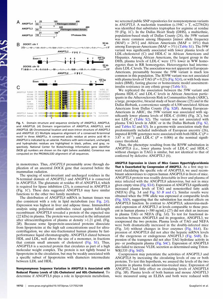
in monotremes. Thus, ANGPTL8 presumably arose through du-plication of an ancestral DOCK gene that occurred before themammalian radiation.The spacing of semi-invariant and uncharged residues in the
N-terminal domain of ANGPTL3 and ANGPTL4 is conservedin ANGPTL8. The glutamate at residue 40 of ANGPTL4, whichis required for lipase inhibition (23), is conserved in ANGPTL8(Fig. 1C). These data suggested ANGPTL8 may have similarfunctions to the other two family members.The distribution of ANGPTL8 mRNA in human tissues was
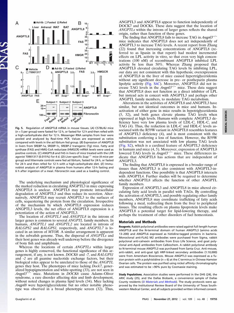
also consistent with a role in lipid metabolism (see Fig. 2A).Expression was highest in liver and adipose tissue. Immunoblotanalysis using polyclonal antibodies raised against full-lengthrecombinant ANGPTL8 revealed a protein of the expected size(22 kDa) in plasma. The protein was recovered in the infranatantafter ultracentrifugation of plasma at density >1.21 g/mL (Fig.2B). Because some lipoprotein-associated proteins dissociatefrom lipoproteins at the high salt concentrations used for ultra-centrifugation, we also size-fractionated human plasma by fast-performance liquid chromatography. ANGPTL8 eluted betweenHDL and LDL, well above the size of a monomer, in fractionsthat contain small amounts of cholesterol (Fig. S1). Thus,ANGPTL8 is a secreted protein that circulates as part of a highmolecular weight complex. The protein is not tightly bound tolipoproteins in the circulation, but may be weakly associated witha specific subset of lipoproteins with diameters intermediatebetween LDL and HDL.
Nonsynonymous Sequence Variation in ANGPTL8 Is Associated withReduced Plasma Levels of LDL-Cholesterol and HDL-Cholesterol. Todetermine if ANGPTL8 plays a role in lipoprotein metabolism,
we screened public SNP repositories for nonsynonymous variantsin ANGPTL8. A nucleotide transition (c.194C > T, rs2278426)was identified that substitutes tryptophan for arginine at residue59 (Fig. 1C). In the Dallas Heart Study (DHS), a multiethnic,population-based study of Dallas County (24), the 59W variantwas more common among Hispanics [minor allele frequency(MAF) = 26%] and African Americans (MAF = 18%) thanamong European-Americans (MAF = 5%) (Table S1). The 59Wvariant was significantly associated with lower plasma levels ofLDL-cholesterol (C) and HDL-C in African Americans andHispanics. Among African Americans, the largest group in theDHS, plasma levels of LDL-C were 15% lower in WW homo-zygotes than in RR homozygotes. Heterozygotes had interme-diate LDL-C levels. The association was not apparent inEuropean-Americans, presumably because the 59W variant is much lesscommon in this population. The R59W variant was not associatedwith plasma levels of TAG (P = 0.23) (Fig. S2A), or with body massindex (BMI), fasting glucose or homeostatic model assessment-insulin resistance in any ethnic group (Table S1).We replicated the association between the 59W variant and
plasma HDL-C and LDL-C levels in African American partic-ipants in the Atherosclerosis Risk in Communities Study (ARIC),a large, prospective, biracial study of heart disease (25) and in theDallas Biobank, a convenience sample of 4,500 unrelated AfricanAmericans from Dallas County (Fig. S2B). Among European-Americans in ARIC, the 59W variant was associated with sig-nificantly lower plasma levels of HDL-C (0.006) (Fig. 2C), butnot LDL-C (Table S2). The variant was not associated withplasma TAG levels in ARIC or in the Dallas Biobank (Fig. S2,and Tables S2 and S3). In a genome-wide association study thatpredominantly included individuals of European ancestry (26),imputed R59W genotypes were associated with both HDL-C (P =3.87 × 10⁻⁷) and LDL-C (P = 0.006) at the nominal signifi-cance threshold.Thus, the phenotype resulting from the R59W substitution in
ANGPTL8 (i.e., lower plasma levels of LDL-C and HDL-Cwithout changes in TAG) is similar, but not identical to that ofconferred by defective ANGPTL3 (6).
ANGPTL8 Expression in Livers of Mice Causes HypertriglyceridemiaThat Is Exacerbated by Coexpression of ANGPTL3. As a first step to-ward elucidating the specific role of ANGPTL8, we used recom-binant adenoviruses to express human ANGPTL8 in livers of mice.ANGPTL8 protein was readily detectable in liver and plasma ofmice infected with the ANGPTL8 adenovirus, but not in micegiven empty virus (Fig. S3A). Expression of ANGPTL8 significantlyincreased plasma levels of TAG and nonesterified fatty acids(NEFA) (Fig. 3A and Fig. S3 B and C). Similar results wereobtained when the 59W allele was expressed at comparable levels(Fig. S3D), suggesting that the substitution has modest effects onANGPTL8 function. In contrast to ANGPTL8, adenovirus-medi-ated expression of ANGPTL3 at levels comparable to those pres-ent in human plasma (∼300 ng/mL) (27) did not elicit an increasein plasma TAG or NEFA (Fig. 3A). To test for functional in-teraction between ANGPTL8 and its progenitor, ANGPTL3, wecoexpressed the two proteins. Plasma levels of TAG and NEFAwere both dramatically increased in the doubly-infected mice(Fig. 3A) without changes in liver enzymes (Fig. S4A). Ex-pression of ANGPTL8 did not alter the hepatic mRNA levelsof the exogenous or endogenous ANGPTL3 (Fig. S4B). Ex-pression of the transgenes did not alter TAG hydrolase activity inpre- or postheparin plasma (Fig. S4C). Expression of ANGPTL8also failed to increase VLDL secretion as determined using Triton-WR1335 (Fig. S4D).Coexpression may potentiate the activities of ANGPTL3 and
ANGPTL8 by increasing the circulating levels of one or bothproteins. To test this hypothesis, we assayed the levels of the twoproteins in plasma from adenovirus-treated mice. Expression ofANGPTL3 had little effect on circulating levels of ANGPTL8(Fig. 3B). Plasma levels of both human and mouse ANGPTL3were measured using an ELISA and both were reduced with
DOCK6: chr 19
ANGPTL3
ANGPTL8
100 bp
500 bp
50 kb
20 kb
5’3’
5’ 3’
3’5’
Exon:
Exon:
1 2
1 2 3 4 5 6 7
3 4
ANGPTL4
ANGPTL3
ANGPTL8
198
460
406
SS
SS
SS
1 25
16
21
245
185
Fibrinogen-related
Fibrinogen-related domain
Protease Site
A
B DOCK7: chr 1
5’3’
C
Fig. 1. Domain structure and sequence similarity of ANGPTL3, ANGPTL4,and ANGPTL8. (A) Domain organization of ANGPTL4, ANGPTL3, andANGPTL8. (B) Chromosomal location and exon-intron structure of ANGPTL3and ANGPTL8. (C) Multiple sequence alignment of a conserved N-terminalmotif in three ANGPTLs. A conserved acidic residue at amino acid 40 inANGPTL4 is indicated by an asterisk. Positions with semi-invariant, uncharged,and hydrophobic residues are highlighted in black, yellow, and gray, re-spectively. National Center for Biotechnology Information gene identifier(NCBI gi) numbers are shown on the right (where available). Consensus wascomputed on the PROMALS3D alignment of all sequences.
19752 | www.pnas.org/cgi/doi/10.1073/pnas.1217552109 Quagliarini et al.

coexpression of ANGPTL8 (Fig. 3C). The reduction in humanANGPTL3 was confirmed by immunoprecipitating the proteinfrom mouse plasma (Fig. 3D). The plasma levels of both full-length ANGPTL3 and the N-terminal fragment were reduced inthe mice coexpressing the two viruses.To test if ANGPTL8 physically interacts with ANGPTL3, mice
were infected with adenoviruses encoding ANGPTL3 and a fusionprotein of ANGPTL8 containing a FLAG epitope tag at theC terminus. ANGPTL8 was immunoprecipitated from the plasmausing an anti-FLAG antibody, and immunoblot analysis of theprecipitated proteins using an anti–N-terminal ANGPTL3 anti-body revealed that both the full-length andN-terminal fragment ofANGPTL3 coimmunoprecipitated with ANGPTL8 (Fig. 3E).Thus, ANGPTL8 interacts with ANGPTL3 in vivo.
ANGPTL8 Requires ANGPTL3 to Promote Hypertriglyceridemia. IfANGPTL8 functionally interacts with ANGPTL3, we would pre-dict that ANGPTL8 would not increase plasma TAG levels whenexpressed in Angptl3−/− mice. In three independent experiments,expression of ANGPTL8 increased plasma TAG levels more thantwofold in wild-type mice, but did not increase TAG or NEFAlevels in Angptl3−/− mice (Fig. 3F). In all three experiments, thelevels of TAG and VLDL-TAG actually fell in Angptl3−/− micereceiving the ANGPTL8-expressing virus despite robust expres-sion of ANGPTL8 in these animals (Fig. 3F and Fig. S5 A and B).Liver enzyme levels were not increased in these mice (Fig. S5C).
ANGPTL8 Promotes Cleavage of ANGPTL3 in Cultured Hepatocytes.The finding that coexpression of ANGPTL8 and ANGPTL3 in-creased plasma TAG and decreased circulating ANGPTL3 levelswas paradoxical, because inactivation of ANGPTL3 in mice
causes a marked reduction in plasma TAG levels (5). Ono et al.reported that ANGPTL3 must undergo cleavage to increaseplasma TAG levels (20). Therefore, a possible explanation forthe anomalous effects of ANGPTL8 on ANGPTL3 levels andactivity is that ANGPTL8 may promote cleavage of ANGPTL3,a process that would simultaneously increase the activity of thepeptide and decrease the amount of full-length ANGPTL3.To test if ANGPTL8 promotes cleavage of ANGPTL3 in
cultured cells, we coexpressed the two proteins in HepG2 cells(Fig. 4A). In medium from cells expressing ANGPTL3 alone, onlyfull-length ANGPTL3 was detected. Coexpression of ANGPTL3with ANGPTL8 resulted in the appearance of a ∼33-kDa band
1.0 Human
0.8
0.6
0.4
0.2
0
adip
ose-w
hite
adrenal gla
nd
colo
n
brain
heart
kid
ney
liver
lung
pla
centa
prosta
teretina
skel m
uscle
sm
all inte
stine
sple
en
testis
thym
us
thyroid
Relative L
evels
AN
GP
TL
8 m
RN
A
A
C
B
35
25
pla
sm
a
supern
ata
nt
infranata
nt
ApoE
ANGPTL8
2578 1086 112 10002 971 16
0
20
40
60
n =
2549 1079 111 9842 951 16
0
50
100
150
n =
ARIC StudyDallas Heart Study
50
100
150
02376 864 82 1160 534 49 908 91 1 308 239 32
LD
L-C
(m
g/d
L)
n =
RR RW WW
African
American
European
American
African
American
European
American
HispanicAll
HD
L-C
(m
g/d
L)
2377 864 82 1161 534 49 908 91 1 308 239 32
0
20
40
60
n =
ANGPTL8 (R59W)
P=0.0006 P=0.005
P=0.001P=0.30 P=0.03
P=0.03
P=2.1x10⁻⁴ P=0.89
P=0.001 P=0.006
P=0.51 P=0.03
Fig. 2. Tissue distribution of ANGPTL8 expression in humans and geneticassociation of sequence variation in ANGPTL8 with plasma lipoprotein levels.(A) ANGPTL8 mRNA levels in human tissues were determined by real-timePCR, and normalized to expression levels of 36B4. Levels were expressed asa ratio relative to liver expression, which was set to 1. (B) Distribution ofANGPTL8 in human plasma. Plasma lipoproteins were isolated by ultracen-trifugation and equal proportions of each fraction were subjected to im-munoblot analysis with antibodies to ANGPTL8 and ApoE. (C) Mean (± SE)LDL-C and HDL-C values in the DHS and ARIC participants.
1000
Trig
lycerid
e
(m
g/d
L)
500
0
P=0.016P=5.1 x 10⁻⁵
P=0.036
P=4.2 x 10⁻⁵
ANGPTL3Control
ANGPTL8 ANGPTL3+ANGPTL8
150
Ch
olestero
l
(m
g/d
L)
100
50
0
0.8
0.6
NE
FA
(m
Eq
/L
)
0.4
0.2
0
A
B
+ANGPTL8
ANGPTL3 ++ANGPTL8
1000
mA
NG
PT
L3
(ng/
ml)
500
0
hA
NG
PT
L3
(ng/
ml)
P=9.6 x 10⁻⁵
P=0.01
400 P=0.014
200
0
ANGPTL8ANGPTL8
ANGPTL3-N
hA
NG
PT
L3
ANGPTL3
25
70
55
35
ANGPTL3
ANGPTL8
Control + + +
+
+ +
+
ANGPTL3
ANGPTL8
Control + + +
+
+ +
+
50
100
150
0
P=0.0165
(n
g/m
l)
F
Trig
lycerid
e (m
g/d
L)
P=0.0416
Ch
olestero
l (m
g/d
L)
NE
FA
(m
Eq
/L
)
D
WT Angptl3-/- WT Angptl3-/- WT Angptl3-/-
600
400
200
0
100
150
50
0
0.6
0.4
0.2
0
P=0.0004
ANGPTL3-N
ANGPTL3
ANGPTL3-mAb
ANGPTL8
ANGPTL3
Control
25
35
55
70
+ +Control
C
E
25
kDa
kDa
Fig. 3. Overexpression of human ANGPTL8 and ANGPTL3 in livers of wild-type and Angptl3−/− mice. (A) Plasma levels of TAG, cholesterol, and NEFAwere measured in 10-wk-old C57Bl6/J male mice (n = 4–5 per group) 3 d afterbeing injected with adenovirus. (B) Immunoblot analysis of plasma ANGPTL8.(C) Human and mouse ANGPTL3 were measured by ELISA, as described inMaterials and Methods. (D) In an independent experiment, ANGPTL3 wasimmunoprecipitated from pooled plasma samples (n = 3 per group) of miceinfected with the indicated viruses using a mouse anti-human ANGPTL3mAb. Immunoprecipates were subjected to immunoblotting with an anti-ANGPTL3 polyclonal antibody. (E) Coimmunoprecipitation of ANGPTL3 withANGPTL8-FLAG from plasma of mice expressing human ANGPTL8-FLAGand ANGPTL3 (n = 6 per group). The immunoprecipitated proteins wereblotted with polyclonal antibodies to human ANGPTL8 and the N terminusof human ANGPTL3 (Left). Plasma levels of ANGPTL3 and ANGPTL8 (input)were assessed by ELISA and by immunoblotting, respectively (Right). (F )Wild-type and Angptl3−/− mice (n = 3–5 per group) were infected withrecombinant ANGPTL8 adenoviruses. After 3 d, the mice were fasted for 4 h,killed, and plasma levels of TAG, cholesterol and NEFA were measured. Theexperiments were all performed three times with similar results and represen-tative data are shown.
Quagliarini et al. PNAS | November 27, 2012 | vol. 109 | no. 48 | 19753
MED
ICALSC
IENCE
S

corresponding to the N-terminal domain of ANGPTL3. In contrast,coexpression of ANGPTL8 with ANGPTL6 did not increase theappearance of the N-terminal domain of ANGPTL6. Thus, theeffect of ANGPTL8 on ANGPTL3 is not a nonspecific effect inthese cells.To determine if cells are required for the generation of the
N-terminal fragment, we added recombinant mouse ANGPTL3to medium of cells infected with adenoviruses expressing eitherANGPTL8 or the vector alone (control) (Fig. 4C). CleavedANGPTL3 was present in the medium of the ANGPTL8-expressing cells, whereas no increase in N-terminal ANGPTL3was detected when the protein was incubated with the samemedium in the absence of cells.These data are consistent with a model in which ANGPTL8
activates ANGPTL3 by binding to the N-terminal domain of thefull-length ANGPTL3 protein and promoting its cleavage, per-haps by changing the conformation of ANGPTL3 to make thecleavage site more accessible or by recruiting a cell-associatedproteases. After cleavage, ANGPTL8 remains bound to the N-terminal domain of ANGPTL3, and may form part of an activecomplex that orchestrates trafficking of lipids among tissues. Theinteraction between ANGPTL8 and ANGPTL3 appears to alsopromote the egress of ANGPTL3 from the circulation.
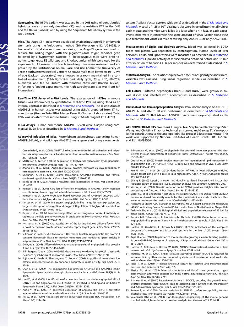
ANGPTL8 Expression Is Regulated by Food Intake in an SREBP-1c–Independent Manner. To elucidate the physiological context inwhich ANGPTL8 and ANGPTL3 interact, we examined thetissue distribution and regulation of ANGPTL8 mRNA in mice.As this manuscript was being prepared, ANGPTL8 mRNA wasreported to be most abundantly expressed in the livers of mice,with lower levels in brown and white adipose tissue (21). Wefound a similar distribution of ANGPTL8 mRNA in mice, but inaddition found substantial expression of the transcript in theadrenals (Fig. S6). In all four tissues, ANGPTL8 mRNA levelswere markedly reduced by fasting and restored by refeeding (Fig.5A). Whereas Ren et al. (21) identified ANGPTL8 as an insulintarget gene in adipocytes, ANGPTL8 expression in liver was notincreased in mice overexpressing sterol regulatory binding pro-tein (SREBP)-1c, which coordinates the lipogenic response to
insulin in the liver (Fig. 5B) (28). Amodest increase in ANGPTL8expression was seen in mice expressing SREBP-1a and SREBP-2.Moreover, a potent agonist of LXR, which is required for theinsulin-induced increase in SREBP-1c (29), only modestly in-creased ANGPTL8mRNA levels (Fig. 5C). The fasting-refeedingresponse of ANGPTL8 was preserved, although attenuated, inmice lacking Scap, a protein required for SREBP activation (30,31) (Fig. 5D).Finally, we examined the regulation of ANGPTL8 levels in
humans in response to food intake. Circulating levels were verylow after a 12-h fast and increased significantly within 3 h offeeding (Fig. 5E). Thus, the fasting-refeeding response in micewas also seen in humans.
DiscussionThe major finding of this study is that a recently identifiedmember of the ANGPTL family, ANGPTL8, plays an importantrole in lipoprotein metabolism through a functional interactionwith ANGPTL3. Our results provide evidence for a close evo-lutionary relationship, direct physical interaction, and functionalinterdependence between the two ANGPTLs. ANGPTL8 andANGPTL3 are located in corresponding introns of two relatedgenes and share significant sequence similarity. The two proteinscoimmunoprecipitated from the plasma of mice when coex-pressed via recombinant adenoviruses, and neither protein wasfully active without the other. ANGPTL3 did not alter plasmalevels of TAG when expressed at physiological concentrations inwild-type mice, whereas coexpression of ANGPTL8 withANGPTL3 markedly increased plasma TAG levels. Conversely,expression of ANGPTL8 increased plasma levels of TAG inwild-type mice but caused a small decrease in plasma TAG inAngptl3−/− animals. In cultured cells, ANGPTL8 stimulated ap-pearance of the N-terminal fragment of ANGPTL3. Taken to-gether, our data indicate that ANGPTL8 is a degenerate paralogof ANGPTL3 that regulates TAG metabolism in concert withANGPTL3.The present study used adenoviral transgenesis to probe the
role of ANGPTL8, but we provide two lines of evidence thatour data are not an artifact of transgene overexpression. First,the ANGPTL8 adenovirus, which caused substantial hyper-triglyceridemia in wild-type mice, did not increase TAG levels inAngptl3−/− mice. Thus, ANGPTL8 requires ANGPTL3 to raiseTAG levels. Second, levels of recombinant ANGPTL3 in miceinjected with the ANGPTL3 adenovirus were comparable tothose observed in humans. At these levels, the recombinantANGPTL protein had no detectable effect on plasma TAG inwild-type mice unless exogenous ANGPTL8 was coexpressed.These data provide direct support for the conclusion thatANGPTL8 normally acts to increase plasma TAG levels and thatit requires ANGPTL3 to do so. Consistent with this notion,Angptl8−/− mice generated as part of a library of mice withmutations in membrane and secreted proteins had markedlydecreased serum TAG levels compared with their sex-matchedwild-type littermates (32).The finding that ANGPTL3 expression did not increase
plasma TAG levels in wild-type mice indicates that ANGPTL3 isusually present in excess and that ANGPTL8 is rate-limiting forANGPTL3 action. Our data are most consistent with a model inwhich ANGPTL8 activates ANGPTL3 by promoting its cleavage,but we cannot exclude alternative explanations. It remains pos-sible that ANGPTL3 activates ANGPTL8, or that neither pro-tein activates the other but instead the two proteins simply forma functional complex. Expression of ANGPTL3 in culturedHepG2 cells increases the level of ANGPTL8 in the medium(Fig. 4A). It is also possible that ANGPTL8 stabilizes the N-terminal domain of the protein. In preliminary studies we did notfind evidence that ANGPTL8 stabilizes ANGPTL3 in culturedcells, but in vivo studies with physiologically relevant concen-trations of both proteins will be required to further elucidate therelationship between ANGPTL3 and ANGPTL8.
A BCell lysate Medium
ANGPTL3-N
ANGPTL3
ANGPTL3
ANGPTL8
Control +
7055
35
25 ANGPTL8
Medium+Cells
70
ANGPTL8
ANGPTL3
ANGPTL3-N
55
3525
Control ANGPTL8
ANGPTL3
25
Control ANGPTL8
Medium
*
kDa
Control
ANGPTL8
ANGPTL6
ANGPTL825
ANGPTL6-N
ANGPTL6
7055
35
Cell Lysate Medium
C
Fig. 4. ANGPTL8 promotes cleavage of ANGPTL3 in cultured hepatocytes.ANGPTL8-FLAG was expressed alone or together with either ANGPTL3 (A) orANGPTL6 (B) in HepG2 cells as described in Materials and Methods. Immu-noblot analysis was performed to detect ANGPTL8, ANGPTL3, and ANGPTL6.(C) Cells are required for cleavage of ANGPTL3. Recombinant mouseANGPTL3 protein (1 μg/mL) was added to the conditioned medium of HuH7cells infected with ANGPTL8 or control adenoviruses. ANGPTL3 was alsoadded to the same medium in the absence of cells. Cells were grown for 16 hat 37 °C, 5% CO2. Medium was subjected to immunoblot analysis with anti-ANGPTL8 and anti-mANGPTL3 antibodies as described in Materials andMethods. The asterisk represents a nonspecific band.
19754 | www.pnas.org/cgi/doi/10.1073/pnas.1217552109 Quagliarini et al.
The underlying mechanism and physiological significance ofthe marked reduction in circulating ANGPTL3 in mice expressingANGPTL8 is unclear. ANGPTL8 may promote intracellulardegradation of ANGPTL3 and thus reduce its secretion. Alter-natively, ANGPTL8 may recruit ANGPTL3 to the surfaces ofcells, sequestering the protein from the circulation. Irrespectiveof the mechanism by which ANGPTL8 expression reducesANGPTL3 levels, the net effect of ANGPTL8 expression is apotentiation of the action of ANGPTL3.The location of ANGPTL3 and ANGPTL8 in the introns of
larger genes is common to several ANGPTL family members. Inhumans, ANGPTL1 and ANGPTL2 are located in introns ofRALGPS2 and RALGPS1, respectively, and ANGPTL7 is lo-cated in an intron of MTOR. A similar arrangement is apparentin the zebrafish genome. Thus, the dispersal of ANGPTLs andtheir host genes was already well underway before the divergenceof bony fish and amphibians.Whereas the locations of certain ANGPTLs within larger
genes is highly conserved, the functional significance of this ar-rangement, if any, is not known. DOCK6 and -7, and RALGPS1and -2 are all guanine nucleotide exchange factors, but theirbiological roles appear to be unrelated to those of the ANGPTLgenes they host. The phenotypes of mice lacking Dock7, gener-alized hypopigmentation and white-spotting (33), are not seen inAngptl3−/− mice. Mutations in DOCK6 cause Adams–Oliversyndrome, a rare disorder affecting skin and limb development,without noted changes in plasma lipid levels (34). Mice lackingAngptl8 were hypotriglyceridemic but no other notable pheno-type was observed in a broad phenotypic screen (32). Thus,
ANGPTL3 and ANGPTL8 appear to function independently ofDOCK7 and DOCK6. These data suggest that the location ofANGPTLs within the introns of larger genes reflects the sharedorigin, rather than function of these genes.The finding that ANGPTL8 fails to increase TAG in Angptl3−/−
mice indicates that ANGPTL8 does not act independently ofANGPTL3 to increase TAG levels. A recent report from Zhang(22) found that increasing concentrations of ANGPTL8 (re-ferred to as lipasin in that report) had modest incrementaleffects on LPL activity in vitro, so that even very high concen-trations (100 nM) of recombinant ANGPTL8 inhibited LPLactivity by less than 50%. Whereas Zhang proposed thatANGPTL8 elevated circulating TAG levels by inhibiting LPL,our data are not consistent with this hypothesis; overexpressionof ANGPTL8 in the liver of mice caused hypertriglyceridemiawithout any significant decrease in pre- or postheparin plasmalipolytic activity (Fig. S4C). Moreover, ANGPTL8 did not in-crease TAG levels in the Angptl3−/− mice. These data suggestthat ANGPTL8 does not function as a direct inhibitor of LPLbut instead acts in concert with ANGPTL3 and perhaps otherANGPTL family members, to modulate TAG metabolism.Alterations in the activities of ANGPTL8 and ANGPTL3 have
similar, but not identical outcomes in mice and humans. In-activation of either gene in mice results in hypotriglyceridemia(5, 32), and both genes elevate plasma TAG levels whenexpressed at high levels. Humans with complete ANGPTL3 de-ficiency have very low plasma levels of LDL-C, HDL-C, andTAG (4). Thus, the reduction in LDL-C and HDL-C levels as-sociated with the R59W variant in ANGPTL8 resembles featuresof ANGPTL3 deficiency (4), and is most consistent with thesubstitution conferring a loss of function. However, the R59Wsubstitution was not associated with low plasma TAG levels(Fig. S2), which is a cardinal feature of ANGPTL3 deficiencyin humans and mice (4, 5). Moreover, expression of ANGPTL8lowered TAG levels in Angptl3−/− mice. These differences in-dicate that ANGPTL8 has actions that are independent ofANGPTL3.The finding that ANGPTL8 is expressed in a broader range of
tissues than ANGPTL3 is also consistent with ANGPTL3-in-dependent functions. One possibility is that ANGPTL8 interactswith ANGPTL4. Further studies will be required to determinewhether ANGPTL8 affects the function of other ANGPTLfamily members.Expression of ANGPTL3 and ANGPTL8 in mice altered cir-
culating fatty acid levels in parallel with TAGs. By controllingthe activation of ANGPTL3, and possibly regulating other familymembers, ANGPTL8 may coordinate trafficking of fatty acidsfollowing a meal, redirecting them from the liver to peripheraltissues. The resulting effects on plasma lipoprotein levels makeANGPTL8 a potential target for lipid-lowering therapy, andperhaps the treatment of other disorders of fuel homeostasis.
Materials and MethodsReagents. Rabbit polyclonal antibodies were raised against full-length humanANGPTL8 and the N-terminal domain of human ANGPTL3 (amino acids13–200) and ANGPTL6 expressed as histidine-tagged proteins in bacteria.Monoclonal anti-FLAG M2 antibodies were purchased from Sigma, rabbitpolyclonal anti-calnexin antibodies from Enzo Life Science, and goat poly-clonal anti-ApoE antibodies from Calbiochem. A rabbit polyclonal antibodyto N-terminal mouse ANGPTL3 was purchased from Santa Cruz. Anti-mouse,anti-rabbit, and anti-goat IgG HRP-linked secondary antibodies (1:10,000)were from Amersham Biosciences. Mouse ANGPTL3 was expressed as a fu-sion protein with a polyhistidine (n = 6) at the C terminus in Chinese HamsterOvarian cells. The protein was purified using nickel affinity chromatographyand was estimated to be >90% pure by Coomassie staining.
Study Populations. Association studies were performed in the DHS (24), theARIC study (25), and the Dallas Biobank, a convenience sample of DallasCounty residents (SI Materials and Methods). The study protocols were ap-proved by the Institutional Review Board of the University of Texas South-western Medical Center, and all subjects provided written informed consent.
1
0
2
1
0
A
C D
Ad-lib Fasted Refed
Ad-lib Fasted Refed
AdrenalLiver BATWAT
FAS HMGCR
WT Scap-/-WT Scap-/-WT Scap-/-
3.0
1.5
0
3.0
1.0
0
5
3
1
0
B
0
0
10
20
30
10
20
30
WT SREBP1a
Tg
SREBP1c
Tg
SREBP2
Tg
6
10
2
0
ANGPTL8
ANGPTL8
ANGPTL8
FAS
FASHMGR
40
T0901317
Vehicle
6
4
2
0
FASANGPTL8
E Subject 1 Subject 2 Subject 3
Fast
Refe
d
Fast
Refe
d
Fast
Refe
d
ANGPTL8
Fibronectin180
26
19
Fig. 5. Regulation of ANGPTL8 mRNA in mouse tissues. (A) C57BL/6J mice(n = 5 per group) were fasted for 12 h, or fasted for 12 h and then refed witha high-carbohydrate diet for 12 h. Messenger RNA samples from liver werepooled and analyzed by Real-time PCR. Values are expressed as ratioscompared with levels in the ad libitum fed group. (B) Expression of ANGPTL8in livers from SREBP-1a, SREBP-1c, SREBP-2 transgenic (Tg) mice. Fatty acidsynthase (FAS) and HMG-CoA reductase (HMGCR) mRNA levels were used aspositive controls. (C) ANGPTL8 and FAS in livers of mice treated with the LXRagonist T0901317 (0.015%) for 4 d. (D) Liver-specific Scap−/− mice (4 mice pergroup) and littermate controls were fed ad libitum, fasted for 24 h, or fastedfor 24 h and then refed for 12 h with a high-carbohydrate diet. (E) Immu-noblot analysis of ANGPTL8 in plasma from humans after 12-h fasting and6 h after ingestion of a meal. Fibronectin was used as a loading control.
Quagliarini et al. PNAS | November 27, 2012 | vol. 109 | no. 48 | 19755
MED
ICALSC
IENCE
S
Genotyping. The R59W variant was assayed in the DHS using oligonucleotidehybridization as previously described (35) and by real-time PCR in the DHSand the Dallas Biobank, and by using the SequenomMassArray system in theARIC cohort.
Mice. The Angptl3−/− mice were developed by ablating Angptl3 in embryonicstem cells using the Velocigene method (36) (Velocigene ID: VG1425). Abacterial artificial chromosome containing the Angptl3 gene was used toreplace the coding region with the β-galactosidase (β-gal) reporter genefollowed by a hygromysin cassette. F1 heterozygous mice were bred to-gether to generate F2 wild-type and knockout mice, which were used for theexperiments. All research protocols involving mice were reviewed and ap-proved by the Institutional Animal Care and Use Committee (University ofTexas Southwestern Medical Center). C57BL/6J male mice between 10–12 wkof age (Jackson Laboratory) were housed in a room maintained in a con-trolled environment (12-h light/12-h dark daily cycle, 23 ± 1 °C, 60–70%humidity), and fed ad libitum with standard chow diet (Harlan Teklad).In fasting-refeeding experiments, the high-carbohydrate diet was from MPBiomedicals.
Real-Time PCR Assay of mRNA Levels. The expression of mRNAs in mousetissues was determined by quantitative real-time PCR (6) using 36B4 as aninternal control as described in SI Materials and Methods. The distribution ofANGPTL8 in human tissues was assayed using cDNAs prepared from 17 hu-man tissues (Human Total RNA Master Panel II; Clontech Laboratories). TotalRNA was isolated from mouse tissues using STAT-60 reagent (TEL-TEST).
ELISA Assays. Human and mouse ANGPTL3 levels were assayed using com-mercial ELISA kits as described in SI Materials and Methods.
Adenoviral Infection of Mice. Recombinant adenoviruses expressing humanANGPTL8-FLAG, and wild-type ANGPTL3 were generated using a commercial
system (AdEasy Vector System; Qbiogene) as described in the SI Materials andMethods. A total of 1.25 × 10¹¹ viral particles were injected into the tail vein ofeach mouse and the mice were killed 3 d later after a 4-h fast. In each exper-iment, mice were injected with the same amount of virus (vector alone virusplus recombinant viruses in mice receiving only ANGPTL3 or only ANGPTL8).
Measurement of Lipids and Lipolytic Activity. Blood was collected in EDTAtubes and plasma was separated by centrifugation. Plasma levels of liverenzymes, lipids, and lipoproteins were measured as described in SI Materialsand Methods. Lipolytic activity of mouse plasma obtained before and 15 minafter injection of heparin (30 U per mouse) was determined as described in SIMaterials and Methods.
Statistical Analysis. The relationship between rs2278426 genotype and clinicalvariables was assessed using linear regression models as described in SIMaterials and Methods.
Cell Culture. Cultured hepatocytes (HepG2 and HuH7) were grown in six-well dishes and infected with adenoviruses as described in SI Materialsand Methods.
Immunoblot and Immunoprecipitation Analysis. Immunoblot analysis of ANGPTL3,ANGPTL6, and ANGPTL8 was performed as described in SI Materials andMethods. ANGPTL8-FLAG and ANGPTL3 were immunoprecipitated as de-scribed in SI Materials and Methods.
ACKNOWLEDGMENTS. We thank Liangcai Nie, Stephanie Blankenship, ZifenWang, and Christina Zhao for technical assistance; and George D. Yancopou-los for contributions to the angiopoietin-like protein-3 knockout mouse. Thiswork was supported by National Institutes of Health Grants P01 HL20948and RL1 HL092550.
1. Camenisch G, et al. (2002) ANGPTL3 stimulates endothelial cell adhesion and migra-
tion via integrin alpha vbeta 3 and induces blood vessel formation in vivo. J Biol Chem
277(19):17281–17290.2. Mattijssen F, Kersten S (2012) Regulation of triglyceride metabolism by Angiopoietin-
like proteins. Biochim Biophys Acta 1821(5):782–789.3. Zhang CC, et al. (2006) Angiopoietin-like proteins stimulate ex vivo expansion of
hematopoietic stem cells. Nat Med 12(2):240–245.4. Musunuru K, et al. (2010) Exome sequencing, ANGPTL3 mutations, and familial
combined hypolipidemia. N Engl J Med 363(23):2220–2227.5. Koishi R, et al. (2002) Angptl3 regulates lipid metabolism in mice. Nat Genet 30(2):
151–157.6. Romeo S, et al. (2009) Rare loss-of-function mutations in ANGPTL family members
contribute to plasma triglyceride levels in humans. J Clin Invest 119(1):70–79.7. Romeo S, et al. (2007) Population-based resequencing of ANGPTL4 uncovers varia-
tions that reduce triglycerides and increase HDL. Nat Genet 39(4):513–516.8. Köster A, et al. (2005) Transgenic angiopoietin-like (angptl)4 overexpression and
targeted disruption of angptl4 and angptl3: Regulation of triglyceride metabolism.
Endocrinology 146(11):4943–4950.9. Desai U, et al. (2007) Lipid-lowering effects of anti-angiopoietin-like 4 antibody re-
capitulate the lipid phenotype found in angiopoietin-like 4 knockout mice. Proc Natl
Acad Sci USA 104(28):11766–11771.10. Kersten S, et al. (2000) Characterization of the fasting-induced adipose factor FIAF,
a novel peroxisome proliferator-activated receptor target gene. J Biol Chem 275(37):
28488–28493.11. Sukonina V, Lookene A, Olivecrona T, Olivecrona G (2006) Angiopoietin-like protein 4
converts lipoprotein lipase to inactive monomers and modulates lipase activity in
adipose tissue. Proc Natl Acad Sci USA 103(46):17450–17455.12. Ge H, et al. (2005) Differential regulation and properties of angiopoietin-like proteins
3 and 4. J Lipid Res 46(7):1484–1490.13. Shimizugawa T, et al. (2002) ANGPTL3 decreases very low density lipoprotein triglyceride
clearance by inhibition of lipoprotein lipase. J Biol Chem 277(37):33742–33748.14. Fujimoto K, Koishi R, Shimizugawa T, Ando Y (2006) Angptl3-null mice show low
plasma lipid concentrations by enhanced lipoprotein lipase activity. Exp Anim 55(1):
27–34.15. Shan L, et al. (2009) The angiopoietin-like proteins ANGPTL3 and ANGPTL4 inhibit
lipoprotein lipase activity through distinct mechanisms. J Biol Chem 284(3):1419–
1424.16. Lee EC, et al. (2009) Identification of a new functional domain in angiopoietin-like 3
(ANGPTL3) and angiopoietin-like 4 (ANGPTL4) involved in binding and inhibition of
lipoprotein lipase (LPL). J Biol Chem 284(20):13735–13745.17. Ando Y, et al. (2003) A decreased expression of angiopoietin-like 3 is protective
against atherosclerosis in apoE-deficient mice. J Lipid Res 44(6):1216–1223.18. Jin W, et al. (2007) Hepatic proprotein convertases modulate HDL metabolism. Cell
Metab 6(2):129–136.
19. Shimamura M, et al. (2007) Angiopoietin-like protein3 regulates plasma HDL cho-lesterol through suppression of endothelial lipase. Arterioscler Thromb Vasc Biol 27(2):366–372.
20. Ono M, et al. (2003) Protein region important for regulation of lipid metabolism inangiopoietin-like 3 (ANGPTL3): ANGPTL3 is cleaved and activated in vivo. J Biol Chem278(43):41804–41809.
21. Ren G, Kim JY, Smas CM (2012) Identification of RIFL, a novel adipocyte-enrichedinsulin target gene with a role in lipid metabolism. Am J Physiol Endocrinol Metab303(3):E334–E351.
22. Zhang R (2012) Lipasin, a novel nutritionally-regulated liver-enriched factor thatregulates serum triglyceride levels. Biochem Biophys Res Commun 424(4):786–792.
23. Yin W, et al. (2009) Genetic variation in ANGPTL4 provides insights into proteinprocessing and function. J Biol Chem 284(19):13213–13222.
24. Victor RG, et al. and Dallas Heart Study Investigators (2004) The Dallas Heart Study: Apopulation-based probability sample for the multidisciplinary study of ethnic differ-ences in cardiovascular health. Am J Cardiol 93(12):1473–1480.
25. Anonymous (1987) ARIC Manual of Operations: No 2, Cohort Component Procedures(ARIC Coordinating Center, School of Public Health, Univ of North Carolina, Chapel Hill).
26. Teslovich TM, et al. (2010) Biological, clinical and population relevance of 95 loci forblood lipids. Nature 466(7307):707–713.
27. Robciuc MR, Tahvanainen E, Jauhiainen M, Ehnholm C (2010) Quantitation of serumangiopoietin-like proteins 3 and 4 in a Finnish population sample. J Lipid Res 51(4):824–831.
28. Horton JD, Goldstein JL, Brown MS (2002) SREBPs: Activators of the completeprogram of cholesterol and fatty acid synthesis in the liver. J Clin Invest 109(9):1125–1131.
29. Repa JJ, et al. (2000) Regulation of mouse sterol regulatory element-binding protein-1c gene (SREBP-1c) by oxysterol receptors, LXRalpha and LXRbeta. Genes Dev 14(22):2819–2830.
30. Horton JD, Goldstein JL, Brown MS (2002) SREBPs: Transcriptional mediators of lipidhomeostasis. Cold Spring Harb Symp Quant Biol 67:491–498.
31. Matsuda M, et al. (2001) SREBP cleavage-activating protein (SCAP) is required forincreased lipid synthesis in liver induced by cholesterol deprivation and insulin ele-vation. Genes Dev 15(10):1206–1216.
32. Tang T, et al. (2010) A mouse knockout library for secreted and transmembraneproteins. Nat Biotechnol 28(7):749–755.
33. Blasius AL, et al. (2009) Mice with mutations of Dock7 have generalized hypo-pigmentation and white-spotting but show normal neurological function. Proc NatlAcad Sci USA 106(8):2706–2711.
34. Shaheen R, et al. (2011) Recessive mutations in DOCK6, encoding the guanidine nu-cleotide exchange factor DOCK6, lead to abnormal actin cytoskeleton organizationand Adams-Oliver syndrome. Am J Hum Genet 89(2):328–333.
35. Romeo S, et al. (2008) Genetic variation in PNPLA3 confers susceptibility to non-alcoholic fatty liver disease. Nat Genet 40(12):1461–1465.
36. Valenzuela DM, et al. (2003) High-throughput engineering of the mouse genomecoupled with high-resolution expression analysis. Nat Biotechnol 21:652–659.
19756 | www.pnas.org/cgi/doi/10.1073/pnas.1217552109 Quagliarini et al.