Embed Size (px)
Citation preview
Aus der Klinik für Radioonkologie und Strahlentherapie der Medizinischen Fakultät Charité – Universitätsmedizin Berlin
DISSERTATION
Akzelerierte hyperfraktionierte Radiotherapie plus Temozolomid beim Glioblastom
zur Erlangung des akademischen Grades Doctor medicinae (Dr. med.)
vorgelegt der Medizinischen Fakultät Charité – Universitätsmedizin Berlin
von
Julian Gabriel Florange
aus Hannover
Datum der Promotion: !!!!!!!!!
2
Inhaltsverzeichnis 1 Abstrakt (deutsch) 3 2 Abstract (englisch) 4 3 Eidesstattliche Versicherung und Anteilserklärung 5 4 Auszug aus der Journal Summary List 6 (ISI Web of KnowledgeSM) 5 Publikation: Accelerated hyperfractionation plus 11 temozolomide in Glioblastoma 6 Lebenslauf 18 7 Publikationsliste 20 8 Danksagung 21 !!!!!!!!!
3
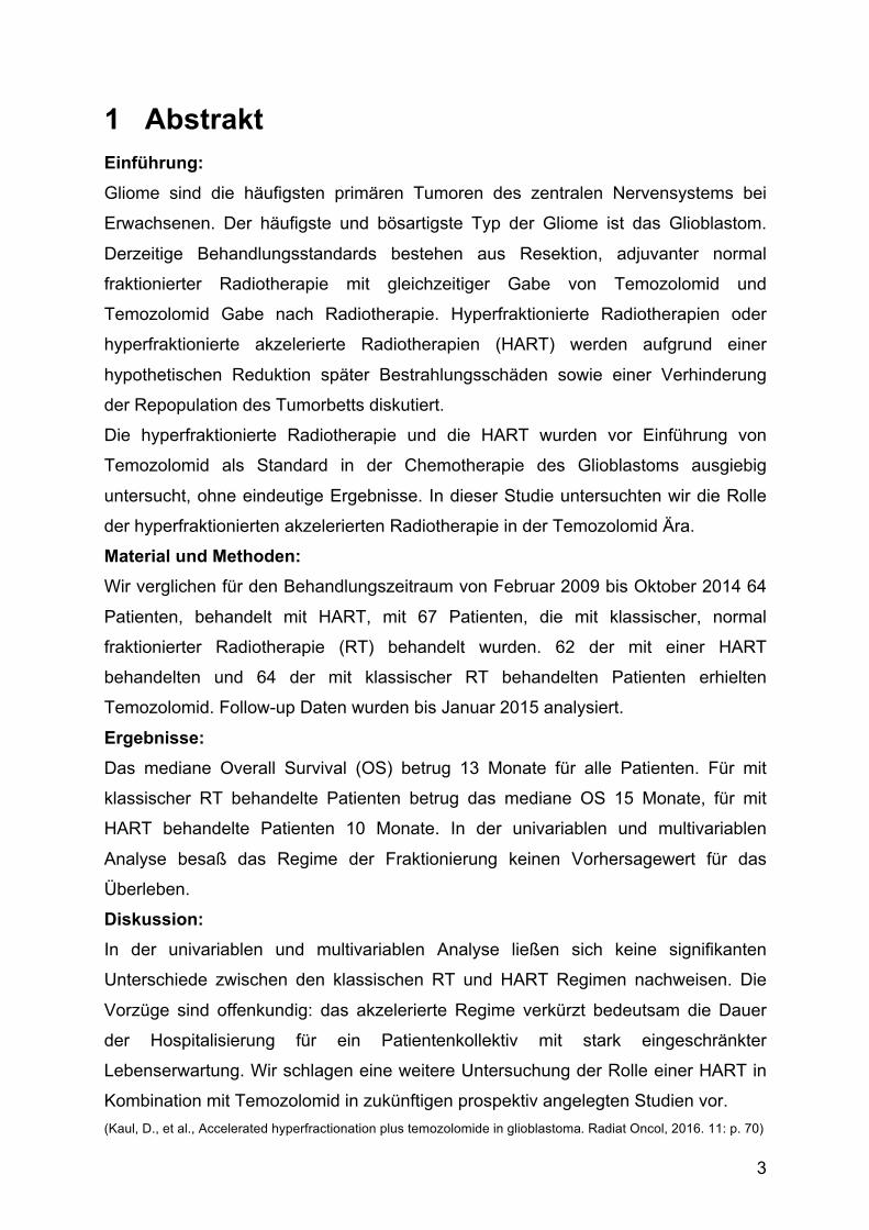
1 Abstrakt Einführung:
Gliome sind die häufigsten primären Tumoren des zentralen Nervensystems bei
Erwachsenen. Der häufigste und bösartigste Typ der Gliome ist das Glioblastom.
Derzeitige Behandlungsstandards bestehen aus Resektion, adjuvanter normal
fraktionierter Radiotherapie mit gleichzeitiger Gabe von Temozolomid und
Temozolomid Gabe nach Radiotherapie. Hyperfraktionierte Radiotherapien oder
hyperfraktionierte akzelerierte Radiotherapien (HART) werden aufgrund einer
hypothetischen Reduktion später Bestrahlungsschäden sowie einer Verhinderung
der Repopulation des Tumorbetts diskutiert.
Die hyperfraktionierte Radiotherapie und die HART wurden vor Einführung von
Temozolomid als Standard in der Chemotherapie des Glioblastoms ausgiebig
untersucht, ohne eindeutige Ergebnisse. In dieser Studie untersuchten wir die Rolle
der hyperfraktionierten akzelerierten Radiotherapie in der Temozolomid Ära.
Material und Methoden:
Wir verglichen für den Behandlungszeitraum von Februar 2009 bis Oktober 2014 64
Patienten, behandelt mit HART, mit 67 Patienten, die mit klassischer, normal
fraktionierter Radiotherapie (RT) behandelt wurden. 62 der mit einer HART
behandelten und 64 der mit klassischer RT behandelten Patienten erhielten
Temozolomid. Follow-up Daten wurden bis Januar 2015 analysiert.
Ergebnisse:
Das mediane Overall Survival (OS) betrug 13 Monate für alle Patienten. Für mit
klassischer RT behandelte Patienten betrug das mediane OS 15 Monate, für mit
HART behandelte Patienten 10 Monate. In der univariablen und multivariablen
Analyse besaß das Regime der Fraktionierung keinen Vorhersagewert für das
Überleben.
Diskussion:
In der univariablen und multivariablen Analyse ließen sich keine signifikanten
Unterschiede zwischen den klassischen RT und HART Regimen nachweisen. Die
Vorzüge sind offenkundig: das akzelerierte Regime verkürzt bedeutsam die Dauer
der Hospitalisierung für ein Patientenkollektiv mit stark eingeschränkter
Lebenserwartung. Wir schlagen eine weitere Untersuchung der Rolle einer HART in
Kombination mit Temozolomid in zukünftigen prospektiv angelegten Studien vor. (Kaul, D., et al., Accelerated hyperfractionation plus temozolomide in glioblastoma. Radiat Oncol, 2016. 11: p. 70)
4
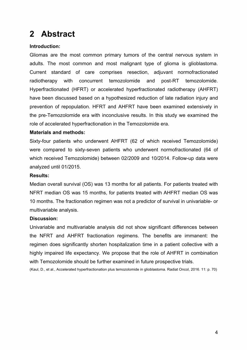
2 Abstract
Introduction:
Gliomas are the most common primary tumors of the central nervous system in
adults. The most common and most malignant type of glioma is glioblastoma.
Current standard of care comprises resection, adjuvant normofractionated
radiotherapy with concurrent temozolomide and post-RT temozolomide.
Hyperfractionated (HFRT) or accelerated hyperfractionated radiotherapy (AHFRT)
have been discussed based on a hypothesized reduction of late radiation injury and
prevention of repopulation. HFRT and AHFRT have been examined extensively in
the pre-Temozolomide era with inconclusive results. In this study we examined the
role of accelerated hyperfractionation in the Temozolomide era.
Materials and methods:
Sixty-four patients who underwent AHFRT (62 of which received Temozolomide)
were compared to sixty-seven patients who underwent normofractionated (64 of
which received Temozolomide) between 02/2009 and 10/2014. Follow-up data were
analyzed until 01/2015.
Results:
Median overall survival (OS) was 13 months for all patients. For patients treated with
NFRT median OS was 15 months, for patients treated with AHFRT median OS was
10 months. The fractionation regimen was not a predictor of survival in univariable- or
multivariable analysis.
Discussion:
Univariable and multivariable analysis did not show significant differences between
the NFRT and AHFRT fractionation regimens. The benefits are immanent: the
regimen does significantly shorten hospitalization time in a patient collective with a
highly impaired life expectancy. We propose that the role of AHFRT in combination
with Temozolomide should be further examined in future prospective trials.!(Kaul, D., et al., Accelerated hyperfractionation plus temozolomide in glioblastoma. Radiat Oncol, 2016. 11: p. 70)
5
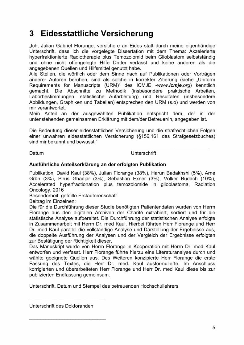
3 Eidesstattliche Versicherung „Ich, Julian Gabriel Florange, versichere an Eides statt durch meine eigenhändige Unterschrift, dass ich die vorgelegte Dissertation mit dem Thema: Akzelerierte hyperfraktionierte Radiotherapie plus Temozolomid beim Glioblastom selbstständig und ohne nicht offengelegte Hilfe Dritter verfasst und keine anderen als die angegebenen Quellen und Hilfsmittel genutzt habe. Alle Stellen, die wörtlich oder dem Sinne nach auf Publikationen oder Vorträgen anderer Autoren beruhen, sind als solche in korrekter Zitierung (siehe „Uniform Requirements for Manuscripts (URM)“ des ICMJE -www.icmje.org) kenntlich gemacht. Die Abschnitte zu Methodik (insbesondere praktische Arbeiten, Laborbestimmungen, statistische Aufarbeitung) und Resultaten (insbesondere Abbildungen, Graphiken und Tabellen) entsprechen den URM (s.o) und werden von mir verantwortet. Mein Anteil an der ausgewählten Publikation entspricht dem, der in der untenstehenden gemeinsamen Erklärung mit dem/der Betreuer/in, angegeben ist. Die Bedeutung dieser eidesstattlichen Versicherung und die strafrechtlichen Folgen einer unwahren eidesstattlichen Versicherung (§156,161 des Strafgesetzbuches) sind mir bekannt und bewusst.“ ____________________________ Datum Unterschrift Ausführliche Anteilserklärung an der erfolgten Publikation
Publikation: David Kaul (38%), Julian Florange (38%), Harun Badakhshi (5%), Arne Grün (3%), Pirus Ghadjar (3%), Sebastian Exner (3%), Volker Budach (10%), Accelerated hyperfractionation plus temozolomide in glioblastoma, Radiation Oncology, 2016 Besonderheit: geteilte Erstautorenschaft Beitrag im Einzelnen: Die für die Durchführung dieser Studie benötigten Patientendaten wurden von Herrn Florange aus den digitalen Archiven der Charité extrahiert, sortiert und für die statistische Analyse aufbereitet. Die Durchführung der statistischen Analyse erfolgte in Zusammenarbeit mit Herrn Dr. med Kaul. Hierbei führten Herr Florange und Herr Dr. med Kaul parallel die vollständige Analyse und Darstellung der Ergebnisse aus, die doppelte Ausführung der Analysen und der Vergleich der Ergebnisse erfolgten zur Bestätigung der Richtigkeit dieser. Das Manuskript wurde von Herrn Florange in Kooperation mit Herrn Dr. med Kaul entworfen und verfasst. Herr Florange führte hierzu eine Literaturanalyse durch und wählte geeignete Quellen aus. Des Weiteren konzipierte Herr Florange die erste Fassung des Textes, die Herr Dr. med. Kaul ausformulierte. Im Anschluss korrigierten und überarbeiteten Herr Florange und Herr Dr. med Kaul diese bis zur publizierten Endfassung gemeinsam. Unterschrift, Datum und Stempel des betreuenden Hochschullehrers ____________________________
Unterschrift des Doktoranden ____________________________
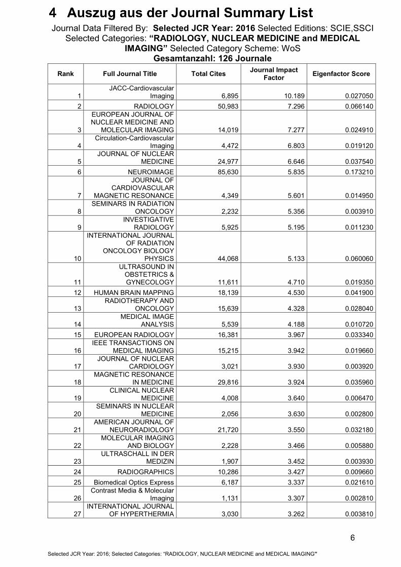
Journal Data Filtered By: Selected JCR Year: 2016 Selected Editions: SCIE,SSCI Selected Categories: “RADIOLOGY, NUCLEAR MEDICINE and MEDICAL
IMAGING” Selected Category Scheme: WoS Gesamtanzahl: 126 Journale
Rank Full Journal Title Total Cites Journal Impact Factor Eigenfactor Score
1 JACC-Cardiovascular
Imaging 6,895 10.189 0.027050 2 RADIOLOGY 50,983 7.296 0.066140
3
EUROPEAN JOURNAL OF NUCLEAR MEDICINE AND
MOLECULAR IMAGING 14,019 7.277 0.024910
4 Circulation-Cardiovascular
Imaging 4,472 6.803 0.019120
5 JOURNAL OF NUCLEAR
MEDICINE 24,977 6.646 0.037540 6 NEUROIMAGE 85,630 5.835 0.173210
7
JOURNAL OF CARDIOVASCULAR
MAGNETIC RESONANCE 4,349 5.601 0.014950
8 SEMINARS IN RADIATION
ONCOLOGY 2,232 5.356 0.003910
9 INVESTIGATIVE
RADIOLOGY 5,925 5.195 0.011230
10
INTERNATIONAL JOURNAL OF RADIATION
ONCOLOGY BIOLOGY PHYSICS 44,068 5.133 0.060060
11
ULTRASOUND IN OBSTETRICS & GYNECOLOGY 11,611 4.710 0.019350
12 HUMAN BRAIN MAPPING 18,139 4.530 0.041900
13 RADIOTHERAPY AND
ONCOLOGY 15,639 4.328 0.028040
14 MEDICAL IMAGE
ANALYSIS 5,539 4.188 0.010720 15 EUROPEAN RADIOLOGY 16,381 3.967 0.033340
16 IEEE TRANSACTIONS ON
MEDICAL IMAGING 15,215 3.942 0.019660
17 JOURNAL OF NUCLEAR
CARDIOLOGY 3,021 3.930 0.003920
18 MAGNETIC RESONANCE
IN MEDICINE 29,816 3.924 0.035960
19 CLINICAL NUCLEAR
MEDICINE 4,008 3.640 0.006470
20 SEMINARS IN NUCLEAR
MEDICINE 2,056 3.630 0.002800
21 AMERICAN JOURNAL OF
NEURORADIOLOGY 21,720 3.550 0.032180
22 MOLECULAR IMAGING
AND BIOLOGY 2,228 3.466 0.005880
23 ULTRASCHALL IN DER
MEDIZIN 1,907 3.452 0.003930 24 RADIOGRAPHICS 10,286 3.427 0.009660 25 Biomedical Optics Express 6,187 3.337 0.021610
26 Contrast Media & Molecular
Imaging 1,131 3.307 0.002810
27 INTERNATIONAL JOURNAL
OF HYPERTHERMIA 3,030 3.262 0.003810
1 Selected JCR Year: 2016; Selected Categories: “RADIOLOGY, NUCLEAR MEDICINE and MEDICAL IMAGING”
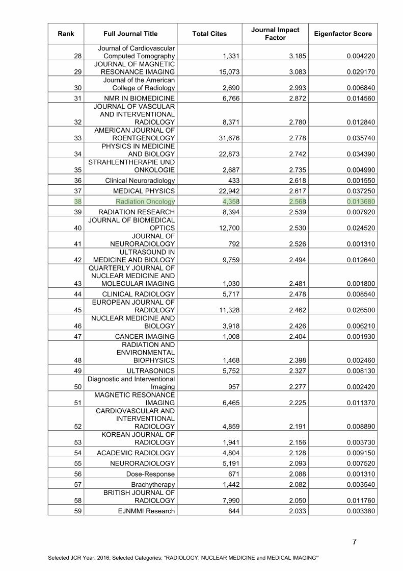
Rank Full Journal Title Total Cites Journal Impact Factor Eigenfactor Score
28 Journal of Cardiovascular
Computed Tomography 1,331 3.185 0.004220
29 JOURNAL OF MAGNETIC
RESONANCE IMAGING 15,073 3.083 0.029170
30 Journal of the American
College of Radiology 2,690 2.993 0.006840 31 NMR IN BIOMEDICINE 6,766 2.872 0.014560
32
JOURNAL OF VASCULAR AND INTERVENTIONAL
RADIOLOGY 8,371 2.780 0.012840
33 AMERICAN JOURNAL OF
ROENTGENOLOGY 31,676 2.778 0.035740
34 PHYSICS IN MEDICINE
AND BIOLOGY 22,873 2.742 0.034390
35 STRAHLENTHERAPIE UND
ONKOLOGIE 2,687 2.735 0.004990 36 Clinical Neuroradiology 433 2.618 0.001550 37 MEDICAL PHYSICS 22,942 2.617 0.037250 38 Radiation Oncology 4,358 2.568 0.013680 39 RADIATION RESEARCH 8,394 2.539 0.007920
40 JOURNAL OF BIOMEDICAL
OPTICS 12,700 2.530 0.024520
41 JOURNAL OF
NEURORADIOLOGY 792 2.526 0.001310
42 ULTRASOUND IN
MEDICINE AND BIOLOGY 9,759 2.494 0.012640
43
QUARTERLY JOURNAL OF NUCLEAR MEDICINE AND
MOLECULAR IMAGING 1,030 2.481 0.001800 44 CLINICAL RADIOLOGY 5,717 2.478 0.008540
45 EUROPEAN JOURNAL OF
RADIOLOGY 11,328 2.462 0.026500
46 NUCLEAR MEDICINE AND
BIOLOGY 3,918 2.426 0.006210 47 CANCER IMAGING 1,008 2.404 0.001930
48
RADIATION AND ENVIRONMENTAL
BIOPHYSICS 1,468 2.398 0.002460 49 ULTRASONICS 5,752 2.327 0.008130
50 Diagnostic and Interventional
Imaging 957 2.277 0.002420
51 MAGNETIC RESONANCE
IMAGING 6,465 2.225 0.011370
52
CARDIOVASCULAR AND INTERVENTIONAL
RADIOLOGY 4,859 2.191 0.008890
53 KOREAN JOURNAL OF
RADIOLOGY 1,941 2.156 0.003730 54 ACADEMIC RADIOLOGY 4,804 2.128 0.009150 55 NEURORADIOLOGY 5,191 2.093 0.007520 56 Dose-Response 671 2.088 0.001310 57 Brachytherapy 1,442 2.082 0.003540
58 BRITISH JOURNAL OF
RADIOLOGY 7,990 2.050 0.011760 59 EJNMMI Research 844 2.033 0.003380
2 Selected JCR Year: 2016; Selected Categories: “RADIOLOGY, NUCLEAR MEDICINE and MEDICAL IMAGING”
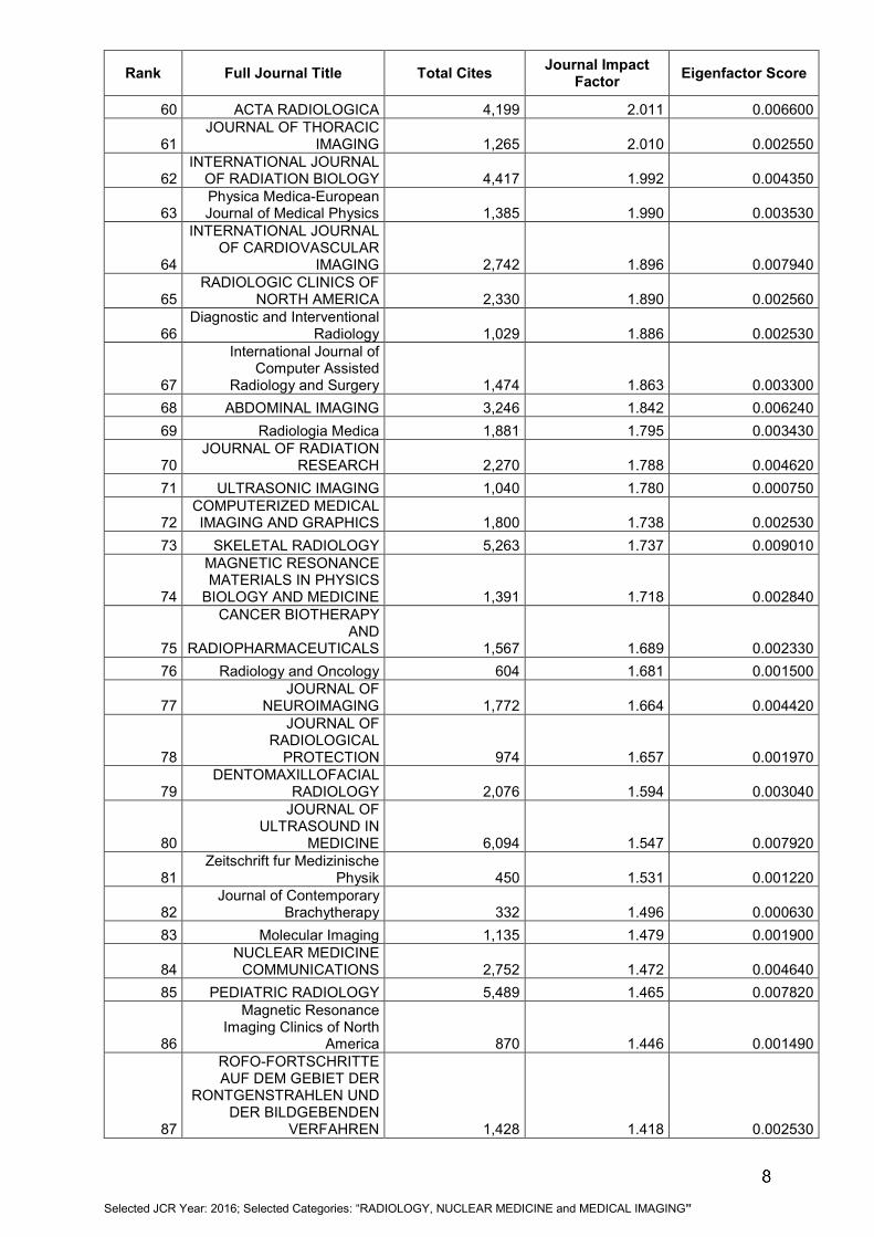
Rank Full Journal Title Total Cites Journal Impact Factor Eigenfactor Score
60 ACTA RADIOLOGICA 4,199 2.011 0.006600
61 JOURNAL OF THORACIC
IMAGING 1,265 2.010 0.002550
62 INTERNATIONAL JOURNAL
OF RADIATION BIOLOGY 4,417 1.992 0.004350
63 Physica Medica-European Journal of Medical Physics 1,385 1.990 0.003530
64
INTERNATIONAL JOURNAL OF CARDIOVASCULAR
IMAGING 2,742 1.896 0.007940
65 RADIOLOGIC CLINICS OF
NORTH AMERICA 2,330 1.890 0.002560
66 Diagnostic and Interventional
Radiology 1,029 1.886 0.002530
67
International Journal of Computer Assisted
Radiology and Surgery 1,474 1.863 0.003300 68 ABDOMINAL IMAGING 3,246 1.842 0.006240 69 Radiologia Medica 1,881 1.795 0.003430
70 JOURNAL OF RADIATION
RESEARCH 2,270 1.788 0.004620 71 ULTRASONIC IMAGING 1,040 1.780 0.000750
72 COMPUTERIZED MEDICAL IMAGING AND GRAPHICS 1,800 1.738 0.002530
73 SKELETAL RADIOLOGY 5,263 1.737 0.009010
74
MAGNETIC RESONANCE MATERIALS IN PHYSICS
BIOLOGY AND MEDICINE 1,391 1.718 0.002840
75
CANCER BIOTHERAPY AND
RADIOPHARMACEUTICALS 1,567 1.689 0.002330 76 Radiology and Oncology 604 1.681 0.001500
77 JOURNAL OF
NEUROIMAGING 1,772 1.664 0.004420
78
JOURNAL OF RADIOLOGICAL
PROTECTION 974 1.657 0.001970
79 DENTOMAXILLOFACIAL
RADIOLOGY 2,076 1.594 0.003040
80
JOURNAL OF ULTRASOUND IN
MEDICINE 6,094 1.547 0.007920
81 Zeitschrift fur Medizinische
Physik 450 1.531 0.001220
82 Journal of Contemporary
Brachytherapy 332 1.496 0.000630 83 Molecular Imaging 1,135 1.479 0.001900
84 NUCLEAR MEDICINE
COMMUNICATIONS 2,752 1.472 0.004640 85 PEDIATRIC RADIOLOGY 5,489 1.465 0.007820
86
Magnetic Resonance Imaging Clinics of North
America 870 1.446 0.001490
87
ROFO-FORTSCHRITTE AUF DEM GEBIET DER
RONTGENSTRAHLEN UND DER BILDGEBENDEN
VERFAHREN 1,428 1.418 0.002530
3 Selected JCR Year: 2016; Selected Categories: “RADIOLOGY, NUCLEAR MEDICINE and MEDICAL IMAGING”
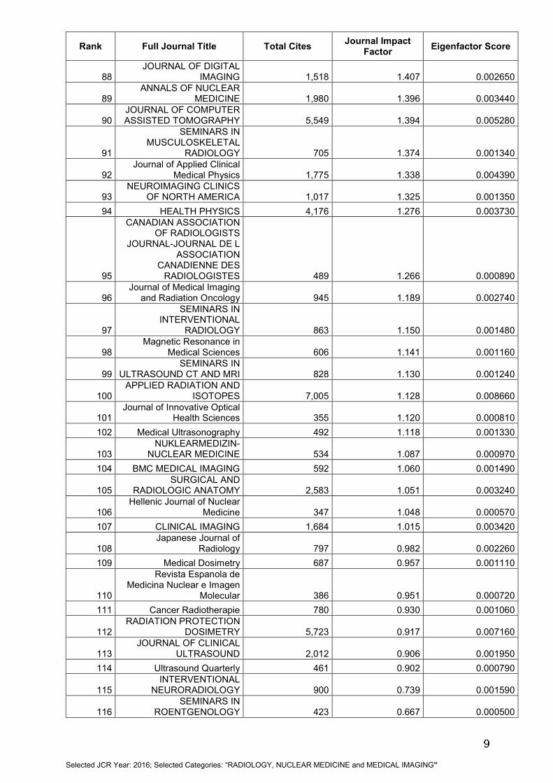
Rank Full Journal Title Total Cites Journal Impact Factor Eigenfactor Score
88 JOURNAL OF DIGITAL
IMAGING 1,518 1.407 0.002650
89 ANNALS OF NUCLEAR
MEDICINE 1,980 1.396 0.003440
90 JOURNAL OF COMPUTER ASSISTED TOMOGRAPHY 5,549 1.394 0.005280
91
SEMINARS IN MUSCULOSKELETAL
RADIOLOGY 705 1.374 0.001340
92 Journal of Applied Clinical
Medical Physics 1,775 1.338 0.004390
93 NEUROIMAGING CLINICS
OF NORTH AMERICA 1,017 1.325 0.001350 94 HEALTH PHYSICS 4,176 1.276 0.003730
95
CANADIAN ASSOCIATION OF RADIOLOGISTS
JOURNAL-JOURNAL DE L ASSOCIATION
CANADIENNE DES RADIOLOGISTES 489 1.266 0.000890
96 Journal of Medical Imaging
and Radiation Oncology 945 1.189 0.002740
97
SEMINARS IN INTERVENTIONAL
RADIOLOGY 863 1.150 0.001480
98 Magnetic Resonance in
Medical Sciences 606 1.141 0.001160
99 SEMINARS IN
ULTRASOUND CT AND MRI 828 1.130 0.001240
100 APPLIED RADIATION AND
ISOTOPES 7,005 1.128 0.008660
101 Journal of Innovative Optical
Health Sciences 355 1.120 0.000810 102 Medical Ultrasonography 492 1.118 0.001330
103 NUKLEARMEDIZIN-
NUCLEAR MEDICINE 534 1.087 0.000970 104 BMC MEDICAL IMAGING 592 1.060 0.001490
105 SURGICAL AND
RADIOLOGIC ANATOMY 2,583 1.051 0.003240
106 Hellenic Journal of Nuclear
Medicine 347 1.048 0.000570 107 CLINICAL IMAGING 1,684 1.015 0.003420
108 Japanese Journal of
Radiology 797 0.982 0.002260 109 Medical Dosimetry 687 0.957 0.001110
110
Revista Espanola de Medicina Nuclear e Imagen
Molecular 386 0.951 0.000720 111 Cancer Radiotherapie 780 0.930 0.001060
112 RADIATION PROTECTION
DOSIMETRY 5,723 0.917 0.007160
113 JOURNAL OF CLINICAL
ULTRASOUND 2,012 0.906 0.001950 114 Ultrasound Quarterly 461 0.902 0.000790
115 INTERVENTIONAL
NEURORADIOLOGY 900 0.739 0.001590
116 SEMINARS IN
ROENTGENOLOGY 423 0.667 0.000500
4 Selected JCR Year: 2016; Selected Categories: “RADIOLOGY, NUCLEAR MEDICINE and MEDICAL IMAGING”
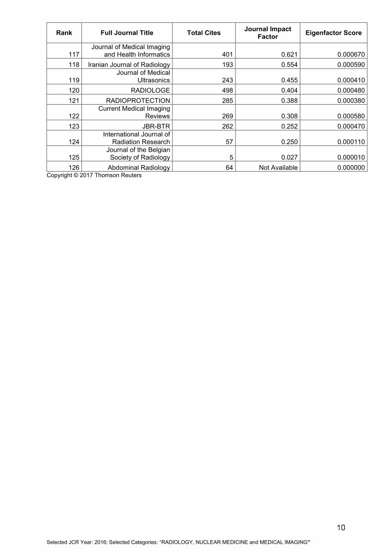
Rank Full Journal Title Total Cites Journal Impact Factor Eigenfactor Score
117 Journal of Medical Imaging
and Health Informatics 401 0.621 0.000670 118 Iranian Journal of Radiology 193 0.554 0.000590
119 Journal of Medical
Ultrasonics 243 0.455 0.000410 120 RADIOLOGE 498 0.404 0.000480 121 RADIOPROTECTION 285 0.388 0.000380
122 Current Medical Imaging
Reviews 269 0.308 0.000580 123 JBR-BTR 262 0.252 0.000470
124 International Journal of
Radiation Research 57 0.250 0.000110
125 Journal of the Belgian Society of Radiology 5 0.027 0.000010
126 Abdominal Radiology 64 Not Available 0.000000 Copyright © 2017 Thomson Reuters
5 Selected JCR Year: 2016; Selected Categories: “RADIOLOGY, NUCLEAR MEDICINE and MEDICAL IMAGING”
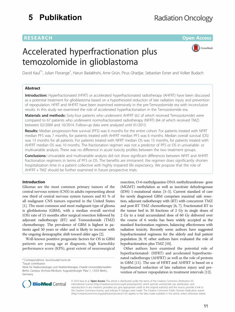
RESEARCH Open Access
Accelerated hyperfractionation plustemozolomide in glioblastomaDavid Kaul*†, Julian Florange†, Harun Badakhshi, Arne Grün, Pirus Ghadjar, Sebastian Exner and Volker Budach
Abstract
Introduction: Hyperfractionated (HFRT) or accelerated hyperfractionated radiotherapy (AHFRT) have been discussedas a potential treatment for glioblastoma based on a hypothesized reduction of late radiation injury and preventionof repopulation. HFRT and AHFRT have been examined extensively in the pre-Temozolomide era with inconclusiveresults. In this study we examined the role of accelerated hyperfractionation in the Temozolomide era.
Materials and methods: Sixty-four patients who underwent AHFRT (62 of which received Temozolomide) werecompared to 67 patients who underwent normofractionated radiotherapy (NFRT) (64 of which received TMZ)between 02/2009 and 10/2014. Follow-up data were analyzed until 01/2015.
Results: Median progression-free survival (PFS) was 6 months for the entire cohort. For patients treated with NFRTmedian PFS was 7 months, for patients treated with AHFRT median PFS was 6 months. Median overall survival (OS)was 13 months for all patients. For patients treated with NFRT median OS was 15 months, for patients treated withAHFRT median OS was 10 months. The fractionation regimen was not a predictor of PFS or OS in univariable- ormultivariable analysis. There was no difference in acute toxicity profiles between the two treatment groups.
Conclusions: Univariable and multivariable analysis did not show significant differences between NFRT and AHFRTfractionation regimens in terms of PFS or OS. The benefits are immanent: the regimen does significantly shortenhospitalization time in a patient collective with highly impaired life expectancy. We propose that the role ofAHFRT + TMZ should be further examined in future prospective trials.
IntroductionGliomas are the most common primary tumors of thecentral nervous system (CNS) in adults representing aboutone third of central nervous system tumors and 81 % ofall malignant CNS tumors reported in the United States[1]. The most common and most malignant type of gliomais glioblastoma (GBM), with a median overall survival(OS) rate of 15 months after surgical resection followed byadjuvant radiotherapy (RT) and Temozolomide (TMZ)chemotherapy. The prevalence of GBM is highest in pa-tients aged 50 years or older and is likely to increase withthe ongoing demographic shift toward older ages [2].Well-known postitive prognostic factors for OS in GBM
patients are young age at diagnosis, high Karnofskyperformance score (KPS), great extent of neurosurgical
resection, O-6-methylguanine-DNA methyltransferase- gene(MGMT) methylation as well as isocitrate dehydrogenase(IDH) 1-mutational status [3–5]. Current standard of carefor newly diagnosed GBM comprises maximal safe resec-tion, adjuvant radiotherapy with (RT) with concurrent TMZand post-RT TMZ chemotherapy [6, 7]. Fractionated RT tothe tumor bed in 30 fractions of 2 Gy in single doses of2 Gy to a total accumulated dose of 60 Gy delivered overthe course of 6 weeks has been widely accepted as thestandard fractionation regimen, balancing effectiveness withradiation toxicity. Recently some authors have suggestedhypofractionated regimens for the elderly and frail patientpopulation [8, 9] other authors have evaluated the role ofhypofractionation plus TMZ [10].Other authors have examined the potential role of
hyperfractionated- (HFRT) and accelerated hyperfractio-nated radiotherapy (AHFRT) as well as the role of protonsin GBM [11]. The use of HFRT and AHFRT is based on ahypothesized reduction of late radiation injury and pre-vention of tumor repopulation in treatment intervals [12].
* Correspondence: [email protected]†Equal contributorsKlinik für Radioonkologie und Strahlentherapie, Charité UniversitätsmedizinBerlin, Campus Virchow-Klinikum, Augustenburger Platz 1, 13353 Berlin,Germany
© 2016 Kaul et al. Open Access This article is distributed under the terms of the Creative Commons Attribution 4.0International License (http://creativecommons.org/licenses/by/4.0/), which permits unrestricted use, distribution, andreproduction in any medium, provided you give appropriate credit to the original author(s) and the source, provide a link tothe Creative Commons license, and indicate if changes were made. The Creative Commons Public Domain Dedication waiver(http://creativecommons.org/publicdomain/zero/1.0/) applies to the data made available in this article, unless otherwise stated.
Kaul et al. Radiation Oncology (2016) 11:70 DOI 10.1186/s13014-016-0645-3
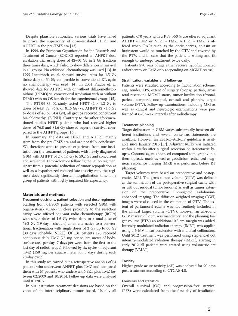
Despite plausible rationales, various trials have failedto prove the superiority of dose-escalated HFRT andAHFRT in the pre-TMZ era [13].In 1994, the European Organization for the Research and
Treatment of Cancer (EORTC) reported an AHFRT doseescalation trial using doses of 42–60 Gy in 2 Gy fractionsthree times daily, which failed to show differences in survivalin all groups. No additional chemotherapy was used [13]. In1999 Lutterbach et. al. showed survival rates for 1.5 Gythrice daily to 54 Gy comparable to conventional RT, againno chemotherapy was used [14]. In 2001 Prados et. al.showed data for AHFRT with or without difluromethylor-nithine (DFMO) vs. conventional irradiation with or withoutDFMO with no OS benefit for the experimental groups [15].The RTOG 83–02 study tested HFRT (2 × 1.2 Gy to
doses of 64.8, 72, 76.8, or 81.6 Gy) vs. AHFRT (2 ×1.6 Gyto doses of 48 or 54.4 Gy), all groups received concurrentbis-chloroethyl (BCNU). Contrary to the other aforemen-tioned studies HFRT patients who had received higherdoses of 76.8 and 81.6 Gy showed superior survival com-pared to the AHFRT groups [16].In summary, the data on HFRT and AHFRT mainly
stem from the pre-TMZ era and are not fully conclusive.We therefore want to present experience from our insti-tution on the treatment of patients with newly diagnosedGBM with AHFRT of 2 × 1.6 Gy to 59,2 Gy and concurrentand sequential Temozolomide following the Stupp regimen.Apart from a potential reduction of tumor repopulation aswell as a hypothesized reduced late toxicity rate, the regi-men does significantly shorten hospitalization time in agroup of patients with highly impaired life expectancy.
Materials and methodsTreatment decisions, patient selection and dose regimensStarting from 01/2009 patients with resected GBM withorgans-at-risk (OAR) in close proximity to the resectioncavity were offered adjuvant radio-chemotherapy (RCTx)with single doses of 1.6 Gy twice daily to a total dose of59.2 Gy (19 days schedule) as an alternative to a conven-tional fractionation with single doses of 2 Gy up to 60 Gy(30 days schedule, NFRT). Of 131 patients 126 receivedcontinuous daily TMZ (75 mg per square meter of body-surface area per day, 7 days per week from the first to thelast day of radiotherapy), followed by six cycles of adjuvantTMZ (150 mg per square meter for 5 days during each28-day cycle).In this study we carried out a retrospective analysis of 64
patients who underwent AHFRT plus TMZ and comparedthem with 67 patients who underwent NFRT plus TMZ be-tween 02/2009 and 10/2014. Follow-up data were analyzeduntil 01/2015.In our institution treatment decisions are based on the
votes of an interdisciplinary tumor board. Usually all
patients <70 years with a KPS >50 % are offered adjuvantAHFRT + TMZ or NFRT + TMZ. AHFRT + TMZ is of-fered when OARs such as the optic nerves, chiasm orbrainstem would be touched by the CTV and covered bythe PTV, and in case that the patient is willing and fitenough to undergo treatment twice daily.Patients ≥70 yeas of age either receive hypofractionated
radiotherapy or TMZ only (depending on MGMT-status).
Stratification, variables and follow-upPatients were stratified according to fractionation scheme,age, gender, KPS, extent of surgery (biopsy, partial-, grosstotal resection), MGMT-status, tumor localization (frontal,parietal, temporal, occipital, central) and planning targetvolume (PTV). Follow-up examinations, including MRI aswell as clinical and neurologic examinations were per-formed at 6–8 week intervals after radiotherapy.
Treatment planningTarget delineation in GBM varies substantially between dif-ferent institutions and several consensus statements areavailable. However, an ESTRO-ACROP guideline is avail-able since January 2016 [17]. Adjuvant RCTx was initiatedwithin 4 weeks after surgical resection or stereotactic bi-opsy. Contrast agent enhanced computed tomography in athermoplastic mask as well as gadolinium enhanced mag-netic resonance imaging (MRI) was performed before RTplanning.Target volumes were based on preoperative and postop-
erative MRI. The gross tumor volume (GTV) was definedas the summation of the postoperative surgical cavity withor without residual tumor lesion(s) as well as tumor exten-sion on the preoperative T1-weighted gadolinium-enhanced imaging. The diffusion-weighted imaging (DWI)images were also used in the estimation of GTV. The ex-tent of peritumoral edema was not routinely included inthe clinical target volume (CTV), however, an all-roundGTV margin of 2 cm was mandatory. For the planning tar-get volume (PTV) an additional 0.5 cm margin was added.Intensity-modulated radiation therapy (IMRT) was appliedusing a 6-MV linear accelerator with multileaf collimators.Until 2012 treatment was performed using step-and-shootintensity-modulated radiation therapy (IMRT), starting inearly 2012 all patients were treated using volumetric arctherapy (VMAT).
ToxicityHigher grade acute toxicity (≥3°) was analyzed for 90 dayspost treatment according to CTCAE 4.0.
Formulas and statisticsOverall survival (OS) and progression-free survival(PFS) were calculated from the first day of irradiation
Kaul et al. Radiation Oncology (2016) 11:70 Page 2 of 7
using Kaplan-Meier analysis and the log-rank test.Progression was defined retrospectively by clinicalnote assessments that included integration of imagingand clinical status. Subgroups were compared using uni-variable analysis and the Cox proportional hazard modelfor multivariable analysis. A p-value of less than 0.05 wasconsidered statistically significant. A p-value of less than 0.1was considered a trend. All variables from the univariableanalysis were included in multivariable analysis. All statis-tical analyses were performed using IBM SPSS Statistics 19(New York, USA).
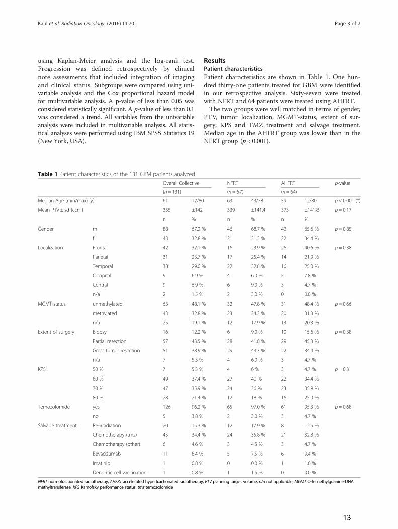
ResultsPatient characteristicsPatient characteristics are shown in Table 1. One hun-dred thirty-one patients treated for GBM were identifiedin our retrospective analysis. Sixty-seven were treatedwith NFRT and 64 patients were treated using AHFRT.The two groups were well matched in terms of gender,
PTV, tumor localization, MGMT-status, extent of sur-gery, KPS and TMZ treatment and salvage treatment.Median age in the AHFRT group was lower than in theNFRT group (p < 0.001).
Table 1 Patient characteristics of the 131 GBM patients analyzedOverall Collective NFRT AHFRT p-value
(n = 131) (n = 67) (n = 64)
Median Age (min/max) [y] 61 12/80 63 43/78 59 12/80 p < 0.001 (*)
Mean PTV ± sd [ccm] 355 ±142 339 ±141.4 373 ±141.8 p = 0.17
n % n % n %
Gender m 88 67.2 % 46 68.7 % 42 65.6 % p = 0.85
f 43 32.8 % 21 31.3 % 22 34.4 %
Localization Frontal 42 32.1 % 16 23.9 % 26 40.6 % p = 0.38
Parietal 31 23.7 % 17 25.4 % 14 21.9 %
Temporal 38 29.0 % 22 32.8 % 16 25.0 %
Occipital 9 6.9 % 4 6.0 % 5 7.8 %
Central 9 6.9 % 6 9.0 % 3 4.7 %
n/a 2 1.5 % 2 3.0 % 0 0.0 %
MGMT-status unmethylated 63 48.1 % 32 47.8 % 31 48.4 % p = 0.66
methylated 43 32.8 % 23 34.3 % 20 31.3 %
n/a 25 19.1 % 12 17.9 % 13 20.3 %
Extent of surgery Biopsy 16 12.2 % 6 9.0 % 10 15.6 % p = 0.38
Partial resection 57 43.5 % 28 41.8 % 29 45.3 %
Gross tumor resection 51 38.9 % 29 43.3 % 22 34.4 %
n/a 7 5.3 % 4 6.0 % 3 4.7 %
KPS 50 % 7 5.3 % 4 6 % 3 4.7 % p = 0.3
60 % 49 37.4 % 27 40 % 22 34.4 %
70 % 47 35.9 % 24 36 % 23 35.9 %
80 % 28 21.4 % 12 18 % 16 25.0 %
Temozolomide yes 126 96.2 % 65 97.0 % 61 95.3 % p = 0.68
no 5 3.8 % 2 3.0 % 3 4.7 %
Salvage treatment Re-irradiation 20 15.3 % 12 17.9 % 8 12.5 %
Chemotherapy (tmz) 45 34.4 % 24 35.8 % 21 32.8 %
Chemotherapy (other) 6 4.6 % 3 4.5 % 3 4.7 %
Bevacizumab 11 8.4 % 5 7.5 % 6 9.4 %
Imatinib 1 0.8 % 0 0.0 % 1 1.6 %
Dendritic cell vaccination 1 0.8 % 1 1.5 % 0 0.0 %
NFRT normofractionated radiotherapy, AHFRT accelerated hyperfractionated radiotherapy, PTV planning target volume, n/a not applicable, MGMT O-6-methylguanine-DNAmethyltransferase, KPS Karnofsky performance status, tmz temozolomide
Kaul et al. Radiation Oncology (2016) 11:70 Page 3 of 7
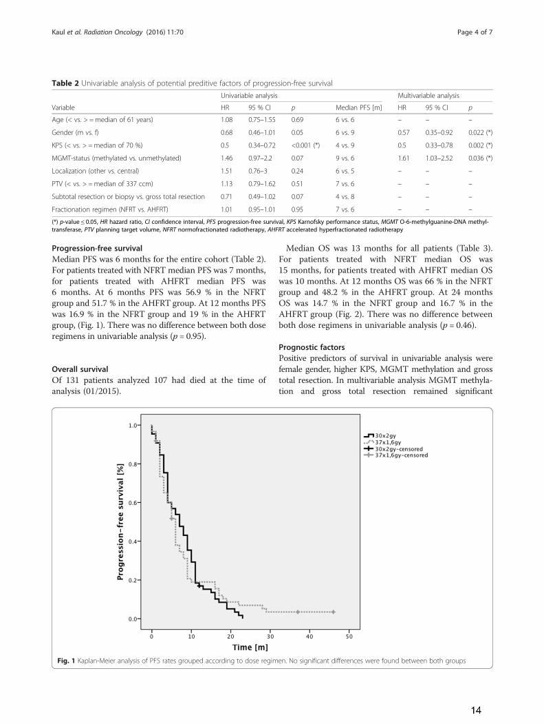
Progression-free survivalMedian PFS was 6 months for the entire cohort (Table 2).For patients treated with NFRT median PFS was 7 months,for patients treated with AHFRT median PFS was6 months. At 6 months PFS was 56.9 % in the NFRTgroup and 51.7 % in the AHFRT group. At 12 months PFSwas 16.9 % in the NFRT group and 19 % in the AHFRTgroup, (Fig. 1). There was no difference between both doseregimens in univariable analysis (p = 0.95).
Overall survivalOf 131 patients analyzed 107 had died at the time ofanalysis (01/2015).
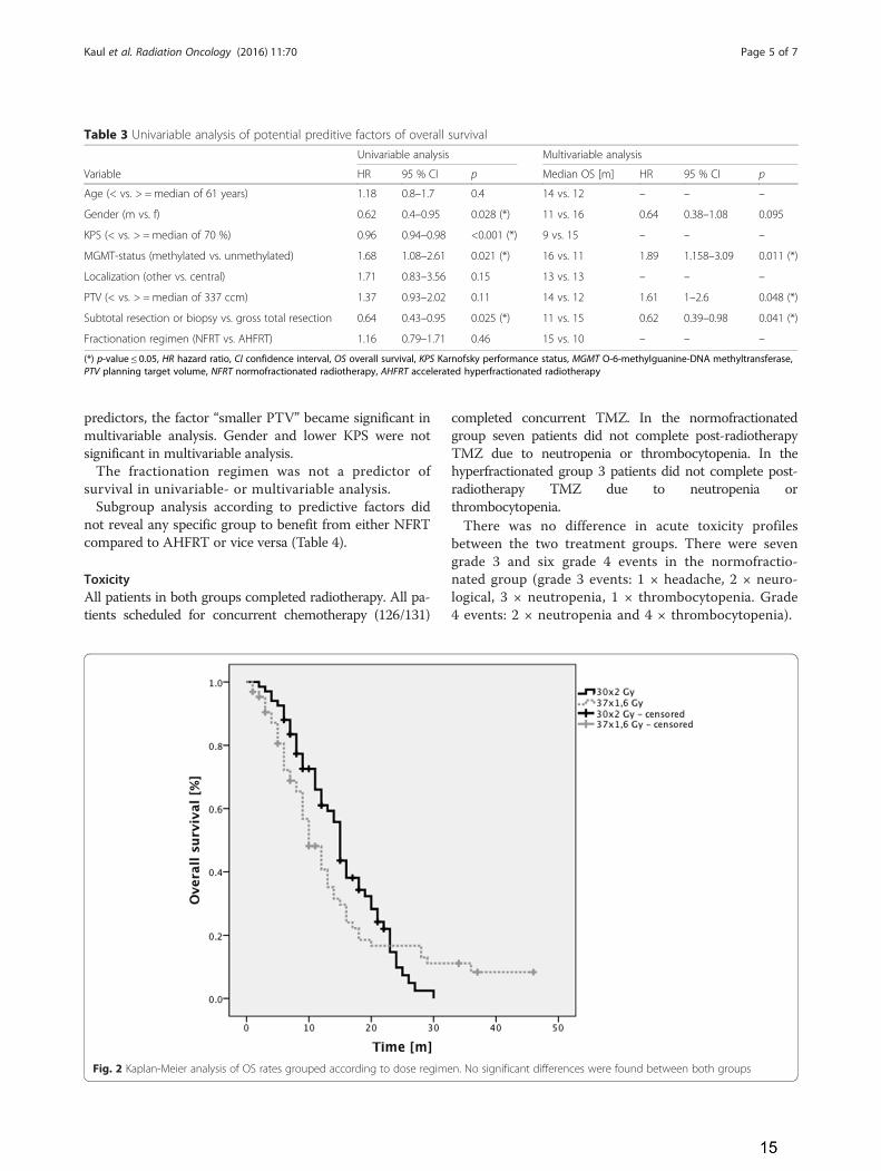
Median OS was 13 months for all patients (Table 3).For patients treated with NFRT median OS was15 months, for patients treated with AHFRT median OSwas 10 months. At 12 months OS was 66 % in the NFRTgroup and 48.2 % in the AHFRT group. At 24 monthsOS was 14.7 % in the NFRT group and 16.7 % in theAHFRT group (Fig. 2). There was no difference betweenboth dose regimens in univariable analysis (p = 0.46).
Prognostic factorsPositive predictors of survival in univariable analysis werefemale gender, higher KPS, MGMT methylation and grosstotal resection. In multivariable analysis MGMT methyla-tion and gross total resection remained significant
Table 2 Univariable analysis of potential preditive factors of progression-free survivalUnivariable analysis Multivariable analysis
Variable HR 95 % CI p Median PFS [m] HR 95 % CI p
Age (< vs. > =median of 61 years) 1.08 0.75–1.55 0.69 6 vs. 6 – – –
Gender (m vs. f) 0.68 0.46–1.01 0.05 6 vs. 9 0.57 0.35–0.92 0.022 (*)
KPS (< vs. > =median of 70 %) 0.5 0.34–0.72 <0.001 (*) 4 vs. 9 0.5 0.33–0.78 0.002 (*)
MGMT-status (methylated vs. unmethylated) 1.46 0.97–2.2 0.07 9 vs. 6 1.61 1.03–2.52 0.036 (*)
Localization (other vs. central) 1.51 0.76–3 0.24 6 vs. 5 – – –
PTV (< vs. > = median of 337 ccm) 1.13 0.79–1.62 0.51 7 vs. 6 – – –
Subtotal resection or biopsy vs. gross total resection 0.71 0.49–1.02 0.07 4 vs. 8 – – –
Fractionation regimen (NFRT vs. AHFRT) 1.01 0.95–1.01 0.95 7 vs. 6 – – –
(*) p-value ≤ 0.05, HR hazard ratio, CI confidence interval, PFS progression-free survival, KPS Karnofsky performance status, MGMT O-6-methylguanine-DNA methyl-transferase, PTV planning target volume, NFRT normofractionated radiotherapy, AHFRT accelerated hyperfractionated radiotherapy
Fig. 1 Kaplan-Meier analysis of PFS rates grouped according to dose regimen. No significant differences were found between both groups
Kaul et al. Radiation Oncology (2016) 11:70 Page 4 of 7
predictors, the factor “smaller PTV” became significant inmultivariable analysis. Gender and lower KPS were notsignificant in multivariable analysis.The fractionation regimen was not a predictor of
survival in univariable- or multivariable analysis.Subgroup analysis according to predictive factors did
not reveal any specific group to benefit from either NFRTcompared to AHFRT or vice versa (Table 4).
ToxicityAll patients in both groups completed radiotherapy. All pa-tients scheduled for concurrent chemotherapy (126/131)
completed concurrent TMZ. In the normofractionatedgroup seven patients did not complete post-radiotherapyTMZ due to neutropenia or thrombocytopenia. In thehyperfractionated group 3 patients did not complete post-radiotherapy TMZ due to neutropenia orthrombocytopenia.There was no difference in acute toxicity profiles
between the two treatment groups. There were sevengrade 3 and six grade 4 events in the normofractio-nated group (grade 3 events: 1 × headache, 2 × neuro-logical, 3 × neutropenia, 1 × thrombocytopenia. Grade4 events: 2 × neutropenia and 4 × thrombocytopenia).
Fig. 2 Kaplan-Meier analysis of OS rates grouped according to dose regimen. No significant differences were found between both groups
Table 3 Univariable analysis of potential preditive factors of overall survivalUnivariable analysis Multivariable analysis
Variable HR 95 % CI p Median OS [m] HR 95 % CI p
Age (< vs. > =median of 61 years) 1.18 0.8–1.7 0.4 14 vs. 12 – – –
Gender (m vs. f) 0.62 0.4–0.95 0.028 (*) 11 vs. 16 0.64 0.38–1.08 0.095
KPS (< vs. > =median of 70 %) 0.96 0.94–0.98 <0.001 (*) 9 vs. 15 – – –
MGMT-status (methylated vs. unmethylated) 1.68 1.08–2.61 0.021 (*) 16 vs. 11 1.89 1.158–3.09 0.011 (*)
Localization (other vs. central) 1.71 0.83–3.56 0.15 13 vs. 13 – – –
PTV (< vs. > = median of 337 ccm) 1.37 0.93–2.02 0.11 14 vs. 12 1.61 1–2.6 0.048 (*)
Subtotal resection or biopsy vs. gross total resection 0.64 0.43–0.95 0.025 (*) 11 vs. 15 0.62 0.39–0.98 0.041 (*)
Fractionation regimen (NFRT vs. AHFRT) 1.16 0.79–1.71 0.46 15 vs. 10 – – –
(*) p-value ≤ 0.05, HR hazard ratio, CI confidence interval, OS overall survival, KPS Karnofsky performance status, MGMT O-6-methylguanine-DNA methyltransferase,PTV planning target volume, NFRT normofractionated radiotherapy, AHFRT accelerated hyperfractionated radiotherapy
Kaul et al. Radiation Oncology (2016) 11:70 Page 5 of 7
In the hyperfractionated group there were two grade 3events and six grade 4 events (grade 3 events: 1 × neuro-logical, 1 × nausea/vomiting. Grade 4 events: 3 × neutro-penia, 3 × thrombocytopenia).
DiscussionSurvivalMost studies on hyperfractionation and acceleratedhyperfractionation stem from the pre-TMZ era, com-parability of PFS and OS rates is thus limited. In ourstudy median OS was 13 months for all patients, 15 monthsfor patients treated using NFRT and 10 months for patientstreated with AHFRT. Univariable and multivariable analysisdid not show significant differences between the fraction-ation regimens. This is worthwile to know, because anAHFRT-regimen with 3.5 weeks overall treatment time wascapable to equalize the OS-results of the classical 6 weekstreatment. Bearing in mind the limited prognosis of thesepatients the dose-intensified treatment is a clear benefit.One of the first studies on AHFRT in GBM was pub-
lished in 1994 by González et al. who used doses of42–60 Gy in 2 Gy fractions three times a day. Mediansurvival was 8.7 ± 0.7 months and no statistically significantdifferences were found for the four different dose-levelgroups [13].Lutterbach et. al. published median OS rates of 8.8 months
for 1.5 Gy thrice daily to 54 Gy [14].
In 2001 Prados et al. published survival rates of pa-tients treated with AHFRT ± DFMO vs. conventionalirradiation ± DFMO with no OS benefit for the experi-mental groups (8.6–9.8 months) [15].Werner et al. published the RTOG 83–02 data in
1996, patients received HFRT (2 × 1.2 Gy to doses of64.8, 72, 76.8, or 81.6 Gy) vs. AHFRT (2 ×1.6 Gy todoses of 48 or 54.4 Gy), all groups received concurrentBCNU. Contrary to the other aforementioned studiesHFRT patients who had received higher doses of 76.8and 81.6 Gy showed superior survival compared to theAHFRT groups. The authors found median OS rates be-tween 10.8 and 12.7 months [16].In 2005 Stupp et al. published data demonstrating a
survival benefit for GBM patients that received concur-rent Temozolomide with postoperative radiation, withmedian survival of 14.6 months for patients receivingconcurrent therapy versus 12.1 months for patientswho received only radiotherapy [7]. This treatment hassince become the standard of care for primary GBMand is referred to as the “Stupp regimen” in everydayclinical routine.OS rates for all patients of 13 months as shown here
are comparable to the data published by Stupp et al. andwe did not find significant differences in OS betweenAHFRT and NFRT in our patient collective.
LimitationsOur study had several limitations. Firstly, the two groupsanalyzed were not perfectly matched in terms of age.Secondly, the MGMT-status is unknown in approximately20 % of patients in both treatment groups. Thirdly, noanalysis of chronic toxicity was performed due to the in-trinsic uncertainties of retrospective analysis. Fourthly, thenumber of patients analyzed here in both groups mightsimply be too low to find significant differences in survivalbetween the both regimens. Fifthly, patients with GBM inclose proximity to the brainstem were more likely to re-ceive AHFRT, potentially biasing OS rates.
ConclusionsThe role of AHFRT in the TMZ era remains unclear.The potential benefits are a reduction of tumor repopu-lation as well as reduced late toxicity. Other benefits areimmanent; the regimen does significantly shortenhospitalization time in a patient collective with highly im-paired life expectancy. We propose that the role of AHFRT+TMZ should be further examined in future prospectivetrials.
Competing interestsThe authors declare that they have no competing interests.
Table 4 Subgroup analysis of potential preditive factors ofoverall survival did not identify any specific subgroup to benefitfrom either NFRT compared to AHFRT or vice versa
Median OS [m]
NFRT AHFRT p
Variable
Age < median of 61 years 15 12 0.66
> =median of 61 years 15 9 0.28
Gender m 14 9 0.31
f 16 14 0.98
KPS < median of 70 % 12 6 0.16
> =median of 70 % 15 13 0.67
MGMT-status methylated 16 15 0.73
unmethylated 14 9 0.09
Localization other 15 10 0.41
central 9 17 0.44
PTV < median of 337 ccm 15 12 0.82
> =median of 337 ccm 15 9 0.24
Extent of resection Subtotal resection or biopsy 13 8 0.14
gross total resection 15 13 0.6
KPS Karnofsky performance status, MGMT O-6-methylguanine-DNAmethyltransferase, PTV planning target volume, NFRT normofractionatedradiotherapy, AHFRT accelerated hyperfractionated radiotherapy
Kaul et al. Radiation Oncology (2016) 11:70 Page 6 of 7
Authors’ contributionsDK drafted the manuscript, performed statistical analysis and supervised thediscussion of the manuscript. JF helped drafting the manuscript, collecteddata and helped with statistical analysis. HB planned the study and took partin the discussion of the manuscript. AG, PG and SB took part in thediscussion of the manuscript. VB planned the study and helped drafting themanuscript. All authors approved the final version of this manuscript.
Received: 11 February 2016 Accepted: 10 May 2016
References1. Ostrom QT, Gittleman H, Farah P, Ondracek A, Chen Y, Wolinsky Y, Stroup
NE, Kruchko C, Barnholtz-Sloan JS. CBTRUS statistical report: Primary brainand central nervous system tumors diagnosed in the United States in2006–2010. Neuro Oncol. 2013;15 Suppl 2:ii1–56.
2. Paszat L, Laperriere N, Groome P, Schulze K, Mackillop W, Holowaty E. Apopulation-based study of glioblastoma multiforme. Int J Radiat Oncol BiolPhys. 2001;51:100–7.
3. Lacroix M, Abi-Said D, Fourney DR, Gokaslan ZL, Shi W, DeMonte F, Lang FF,McCutcheon IE, Hassenbusch SJ, Holland E, et al. A multivariate analysis of416 patients with glioblastoma multiforme: prognosis, extent of resection,and survival. J Neurosurg. 2001;95:190–8.
4. Bauchet L, Mathieu-Daude H, Fabbro-Peray P, Rigau V, Fabbro M, Chinot O,Pallusseau L, Carnin C, Laine K, Schlama A, et al. Oncological patterns ofcare and outcome for 952 patients with newly diagnosed glioblastoma in2004. Neuro Oncol. 2010;12:725–35.
5. Leu S, von Felten S, Frank S, Vassella E, Vajtai I, Taylor E, Schulz M, Hutter G,Hench J, Schucht P, et al. IDH/MGMT-driven molecular classification oflow-grade glioma is a strong predictor for long-term survival. Neuro Oncol.2013;15:469–79.
6. Keles GE, Anderson B, Berger MS. The effect of extent of resection on timeto tumor progression and survival in patients with glioblastoma multiformeof the cerebral hemisphere. Surg Neurol. 1999;52:371–9.
7. Stupp R, Mason WP, van den Bent MJ, Weller M, Fisher B, Taphoorn MJ,Belanger K, Brandes AA, Marosi C, Bogdahn U, et al. Radiotherapy plusconcomitant and adjuvant temozolomide for glioblastoma. N Engl J Med.2005;352:987–96.
8. Malmstrom A, Gronberg BH, Marosi C, Stupp R, Frappaz D, Schultz H,Abacioglu U, Tavelin B, Lhermitte B, Hegi ME, et al. Temozolomide versusstandard 6-week radiotherapy versus hypofractionated radiotherapy inpatients older than 60 years with glioblastoma: the Nordic randomised,phase 3 trial. Lancet Oncol. 2012;13:916–26.
9. Roa W, Kepka L, Kumar N, Sinaika V, Matiello J, Lomidze D, Hentati D,Guedes de Castro D, Dyttus-Cebulok K, Drodge S, et al. International AtomicEnergy Agency Randomized Phase III Study of Radiation Therapy in Elderlyand/or Frail Patients With Newly Diagnosed Glioblastoma Multiforme. J ClinOncol. 2015;33:4145–50.
10. Chen C, Damek D, Gaspar LE, Waziri A, Lillehei K, Kleinschmidt-DeMastersBK, Robischon M, Stuhr K, Rusthoven KE, Kavanagh BD. Phase I trial ofhypofractionated intensity-modulated radiotherapy with temozolomidechemotherapy for patients with newly diagnosed glioblastoma multiforme.Int J Radiat Oncol Biol Phys. 2011;81:1066–74.
11. Mizumoto M, Tsuboi K, Igaki H, Yamamoto T, Takano S, Oshiro Y, Hayashi Y,Hashii H, Kanemoto A, Nakayama H, et al. Phase I/II trial of hyperfractionatedconcomitant boost proton radiotherapy for supratentorial glioblastomamultiforme. Int J Radiat Oncol Biol Phys. 2010;77:98–105.
12. Withers HR, Peters LJ, Thames HD, Fletcher GH. Hyperfractionation. Int JRadiat Oncol Biol Phys. 1982;8:1807–9.
13. Gonzalez DG, Menten J, Bosch DA, van der Schueren E, Troost D, HulshofMC, Bernier J. Accelerated radiotherapy in glioblastoma multiforme: a dosesearching prospective study. Radiother Oncol. 1994;32:98–105.
14. Lutterbach J, Weigel P, Guttenberger R, Hinkelbein W. Acceleratedhyperfractionated radiotherapy in 149 patients with glioblastomamultiforme. Radiother Oncol. 1999;53:49–52.
15. Prados MD, Wara WM, Sneed PK, McDermott M, Chang SM, Rabbitt J, PageM, Malec M, Davis RL, Gutin PH, et al. Phase III trial of acceleratedhyperfractionation with or without difluromethylornithine (DFMO) versusstandard fractionated radiotherapy with or without DFMO for newlydiagnosed patients with glioblastoma multiforme. Int J Radiat Oncol BiolPhys. 2001;49:71–7.
16. Werner-Wasik M, Scott CB, Nelson DF, Gaspar LE, Murray KJ, Fischbach JA,Nelson JS, Weinstein AS, Curran WJ, Jr. Final report of a phase I/II trial ofhyperfractionated and accelerated hyperfractionated radiation therapy withcarmustine for adults with supratentorial malignant gliomas. RadiationTherapy Oncology Group Study 83–02. Cancer. 1996;77:1535–43.
17. Niyazi M, Brada M, Chalmers AJ, Combs SE, Erridge SC, Fiorentino A, GrosuAL, Lagerwaard FJ, Minniti G, Mirimanoff RO, et al. ESTRO-ACROP guideline“target delineation of glioblastomas”. Radiother Oncol. 2016;118:35–42.
• We accept pre-submission inquiries • Our selector tool helps you to find the most relevant journal• We provide round the clock customer support • Convenient online submission• Thorough peer review• Inclusion in PubMed and all major indexing services • Maximum visibility for your research
Submit your manuscript atwww.biomedcentral.com/submit
Submit your next manuscript to BioMed Central and we will help you at every step:
Kaul et al. Radiation Oncology (2016) 11:70 Page 7 of 7
18
6 Lebenslauf Allgemeines Name: Julian Gabriel Florange Adresse: Weinbergsweg 6., 10119 Berlin Telefon: + 491775576227 E-mail: [email protected] Geburtsdatum: 29.04.1991 Geburtsort: Hannover ------------------------------------------------------------------------------------------------- Bildungsweg 2003 – 2010 Gymnasium Adolfinum Bückeburg Juni 2010 Abitur Seit April 2011 Medizinstudium an der Charité- Universitätsmedizin Berlin Seit Sept. 2014 medizinische Promotionsarbeit im Bereich der Strahlentherapie ------------------------------------------------------------------------------------------------- Erfahrungen Juli 2010 – Jan. 2011 Ausbildung an einer Sportakademie, Provinz Fujian, China Feb. – März 2011 Krankenpflegepraktikum im Jüdischen Krankenhaus Berlin März und Sept. 2012 Krankenpflegepraktikum im Virchow-Klinikum Berlin März 2013 Famulatur Allgemeinmedizin in Bückeburg Aug. – Okt. 2013 Famulatur Onkologie und Psychiatrie in Calgary, Kanada März 2014 Famulatur Dermatologie in Afula, Israel Sept. 2014 Famulatur Chirurgie in Bückeburg Jan. – Juni 2015 Erasmus Semester in Tarragona, Spanien Oktober 2016 Zweites Staatsexamen der Humanmedizin, Berlin 21.11.2016 – 15.01.2017 Praktisches Jahr Chirurgie an der FNU, Fidschi

19
16.01. – 10.03.2017 Praktisches Jahr Chirurgie am DRK Westend, Berlin 13.03. – 02.07.2017 Praktisches Jahr Innere Medizin am Regionalspital
Emmental Burgdorf, Universität Bern, Schweiz 03.07. – 20.10.2017 Praktisches Jahr Psychiatrie an der Charité
Universitätsmedizin Berlin ------------------------------------------------------------------------------------------------- Sprachen Deutsch Muttersprache Englisch Fließend Spanisch Fließend Latein Großes Latinum ------------------------------------------------------------------------------------------------- Interessen Sport Shotokan-Ryu Karate seit 1997 Außer curriculare akademische Aktivitäten Teilnahme an Seminar zur englischen Literatur des Mittelalters, Humboldt Universität zu Berlin, 2014 – 2015
Berlin 03.08.2017
20
7 Publikationsliste Publikationen:
1. Kaul, D., Florange, J., Badakhshi, H., Grün, A., Ghadjar, P., Exner, S., Budach, V., Accelerated hyperfractionation plus temozolomide in glioblastoma. Radiat Oncol, 2016. 11: p. 70.
21
8 Danksagung Ich danke Herrn Prof. Dr. med. Dr. h.c. Volker Budach, dem Leiter der Klinik für
Radioonkologie und Strahlentherapie der Charité, Berlin, für die Möglichkeit, die
vorliegende Promotion an seinem Institut durchzuführen.
Mein Dank gilt weiterhin Herrn PD Dr. med. Harun Badakhshi für seine Hilfe bei der
Planung dieser Arbeit, seinen Anregungen im Verlauf des Entstehens und seiner
Geduld mit seinem Doktoranden.
Ganz besonders möchte ich Herrn Dr. med. David Kaul danken, dessen Expertise
bezüglich der technischen und formalen Aspekte zum erfolgreichen Abschluss
unserer Publikation und schließlich meiner Promotion geführt haben.
Weiterhin möchte ich meiner Familie danken, die mir mein Medizinstudium
ermöglichte, mir den nötigen Rückhalt in allen Situationen gab und mich zu jedem
Zeitpunkt ihr Vertrauen spüren ließ.
Meiner Frau Theresa danke ich für ihre unermüdliche und liebevolle Unterstützung.
!