Embed Size (px)
Citation preview

Vlaams Diergeneeskundig Tijdschrift, 2015, 84 243
INTRODUCTION
Canine angiostrongylosis is caused by the meta-strongyloid nematode Angiostrongylus vasorum or ‘French heartworm’ that resides in the right ventricle of the heart and the pulmonary arteries of dogs and other canids. Clinical signs can vary from subclinical to fatal in severe cases, but the most common clinical presentation is respiratory dysfunction, followed by
Autochthonous Angiostrongylus vasorum infection in a Border colliein Belgium
Autochtone Angiostrongylus vasorum-infectie bij een bordercolliein België
1C. Sarre, 2A. Willems, 2S. Daminet, 1E. Claerebout
1 Department of Virology, Parasitology and Immunology,2 Department of Medicine and Clinical Biology of Companion Animals,
Faculty of Veterinary Medicine, Ghent University, Salsiburylaan 133, 9820, Merelbeke, Belgium
BSTRACT
A nine-month-old, female Border collie was referred to the clinic because of an acute persis-tent cough and hemoptysis after excitation. Thoracic radiographs revealed a diffuse interstitial to alveolar pattern, compatible with pulmonary hemorrhage. To stabilize the dog and because of a suspected complex coagulopathy and on-going bleeding evidenced by worsening anemia, a fresh frozen plasma transfusion (10 ml/kg over four hours) was administered. Fecal examination confirmed the clinical suspicion of Angiostrongylus vasorum as the underlying cause of all the ob-served clinical signs. The dog was treated with fenbendazole (50 mg/kg SID) during 21 days. One week after treatment initiation, the dog was clinically healthy and thoracic radiographs showed a marked improvement of the pulmonary lesions.
This case illustrates that autochthonous canine A. vasorum infections can occur in Belgium and they should be taken into account in the diagnostic work-up of dogs with respiratory signs and bleeding disorders.
SAMENVATTING
Een negen maanden oude, vrouwelijke bordercollie werd doorgestuurd voor een acute persisterende hoest en hemoptysis na excitatie. Op het radiografisch onderzoek van de thorax werd een diffuus in-terstitieel tot alveolair longpatroon aangetoond, compatibel met pulmonaire hemorragie. Aangezien de hond verdacht was van complexe coagulopathie en persisterende bloeding gekenmerkt door progres-sieve anemie, werd een plasmatransfusie (“fresh-frozen” plasma) toegediend (10 ml/kg over 4 uur). Coprologisch onderzoek bevestigde het klinisch vermoeden van een Angiostrongylus vasorum-infectie. De hond werd behandeld met fenbendazole (50 mg/kg SID) gedurende 21 dagen. Eén week na het starten van de behandeling vertoonde de hond geen klinische tekenen meer en op de thoraxradiografieën werd een opvallende verbetering van de longletsels vastgesteld.
In deze casuïstiek wordt bevestigd dat autochtone Angiostrongylus vasorum-infecties in België kunnen voorkomen. Deze parasitaire aandoening dient daarom opgenomen te worden in de differentiaal- diagnose bij honden met ademhalingsstoornissen en bloedingsneiging.
A
Vlaams Diergeneeskundig Tijdschrift, 2015, 84 Case report 243
cardiovascular signs and coagulopathies. Occasion-ally, neurological signs and gastrointestinal disorders can be noticed. Transmission of the disease occurs by ingestion of gastropod mollusc intermediate hosts, such as slugs and snails, which contain infective L3 larvae (Elsheikha et al., 2014; Moeremans et al., 2011; Morgan et al., 2010; Rinaldi et al., 2014).
This parasite has a worldwide distribution but is mainly found in well-isolated endemic hot spots,

244 Vlaams Diergeneeskundig Tijdschrift, 2015, 84
which are often rural areas where contact between final host and intermediate host is facilitated. Many European countries have reported the disease with cases in certain areas of Denmark, France, Germany, Finland, Ireland, the United Kingdom and Scotland, Turkey, Switzerland, Spain and Italy (Elsheikha et al., 2014; Gallagher et al., 2012; Helm et al., 2009; Kirk et al., 2014; Rinaldi et al., 2014; Schnyder et al., 2013; Taylor et al., 2007; Traversa et al., 2013). Recent re-ports have demonstrated that the disease is on the rise and is not only spreading outside endemic foci, but is also appearing in previously free regions, such as Poland, the Netherlands, Hungary, Greece, Servia and Slovakia (Elsheikha et al., 2014; Hurníková et al., 2013; Miterpáková et al., 2014; Simin et al., 2014; Schnyder et al., 2013; Tolnai et al., 2015; van Doorn et al., 2009). Possible causes of this spread can be cli-mate change, increased non-controlled pet transporta-tion, the increase and urbanization of red fox popula-tions (that act as a reservoir for the disease), but also the development of more and better diagnostic tools and an increased awareness of the disease among vet-erinarians (Elsheikha et al., 2014; Moeremans et al., 2011).
Up till recently, Belgium seemed to be free of ca-nine A. vasorum infections despite several reports in neighbouring countries. Recently, a fatal autochtho-nous case in a 4.5-month-old male Border collie has been described nearby Liège in the southern part of Belgium (Jolly et al., 2015). In this case report, an autochthonous A. vasorum infection is described in a female Border collie, which responded well to treat-ment.
CASE REPORT
In April 2014 a nine-month-old, female Border collie of 14 kg from Braine-l’Alleud, close to Water- loo, developed an acute persistent cough and he-moptysis after excitation with small to substantial amounts of blood. Blood was not noticed in the stool or urine. The dog was active but calmer than usual. Contact with rodenticides was unlikely according to
the owner, and the dog had not been outside Belgium. She was presented to the local veterinarian, who per-formed a complete blood count (CBC), thoracic and abdominal radiographs and a throat inspection under sedation. The CBC revealed a mild to moderate regenerative anemia, moderate to severe thrombo-cytopenia and mild eosinophilia and lymphocytosis (Table 1). A blood smear was not performed at this time point. Thoracic radiographs revealed a diffuse, interstitial to alveolar pattern, more pronounced in the right hemithorax (Figure 1). Abdominal radiographs additionally revealed three foreign objects (bottle caps) in the stomach. No abnormalities were detected on throat inspection, but mild bilateral epistaxis was noted. The dog was referred to the small animal clinic of the Faculty of Veterinary Medicine (Ghent Univer-sity, Belgium).
During physical examination, the dog was very stressed, had a body temperature of 39.3°C and pink mucous membranes, a normal capillary refill time, a heart rate of 160 beats per minute, normal pulse qual-ity, normal cardiac auscultation, tachypnea without dyspnea and increased bronchovesicular lung sounds. No epistaxis or petechiae were noted. The heart fre-quency and temperature normalized after a short ac-climatization period.
Serum biochemistry, packed cell volume (PCV), blood smear and coagulation times were performed (Table 2). Coagulation times were within normal lim-its, but a progressive decline in red blood cells was noted when compared to the hematocrit performed four hours earlier by the referring veterinarian. Mode-rate regeneration (anisocytosis and polychromasia), eosinophilia and normal platelet number per high-power field were detected on the blood smear. The thrombocytopenia detected by the referring veterinar-ian was thus a pseudothrombocytopenia.
The thoracic radiographs of the referring veterinar-ian were reviewed and the pattern as described above was compatible with pulmonary hemorrhage, non-cardiogenic edema or atypical pneumonia. The pro-gressive decline of red blood cells and the hemoptysis increased the suspicion of pulmonary hemorrhage. Pulmonary hemorrhage can be caused by trauma,
Table 1. Hematology performed by the referring veterinarian.
Parameter Result Reference interval
Hematocrit 27.6 37 - 55 %Hemoglobin 9.5 12 - 18 g/dLRed blood cells 3.8 5.3 - 8.5 106/mm³Mean corpuscular volume 73 60 - 77 fLMean corpuscular hemoglobin concentration 34 32 - 36 g/dLReticulocytes 2.9 0.1 - 1.5 %Leukocytes 11660 6000 - 15000 /mm³Neutrophils 4431 3000 - 11500 /mm³Eosinophils 1516 < 1250 /mm³Lymphocytes 5480 1000 - 4800 /mm³Thrombocytes 48 180 - 500 10³/mm³

Vlaams Diergeneeskundig Tijdschrift, 2015, 84 245
coagulopathy, e.g. due to rodenticide poisoning, dis-seminated intravascular coagulation (DIC) or Angio-strongylus vasorum infection, or local lung pathology, such as neoplasia. According to the owners, the dog did not suffer from trauma, and no clear signs of lo-cal lung pathology were present. Therefore, a coagu-lopathy was suspected, although platelet count (blood smear), PT and aPTT were within normal limits. The dog was mildly sedated with butorphanol (0.04 mg/kg) and the buccal mucosal bleeding time (BMBT) was measured to detect possible thrombocytopathia. However, the test result was within normal limits. A complex coagulopathy without obvious laboratory ab-normalities was suspected and a fresh frozen plasma transfusion (10 ml/kg) was initiated. Packed red blood cells were not administered, since there were no signs of decompensated anemia. Oxygen supplementation was not necessary, since the dog was not dyspneic.
Forty-eight hours after the first recordings at the local veterinarian’s clinic, the thoracic radiographs were repeated and showed a marked improvement of the lung lesions, most likely because the pulmonary hemorrhage stopped after the plasma transfusion. There was a peripherally located patchy interstitial to
Table 2. Serum biochemistry, packed cell volume and coagulation times performed at the Faculty of Veterinary Medi-cine (Ghent University).
Parameter Result Reference interval
Albumin 25 23 - 40 g/LTotal protein 73 52 - 82 g/LCreatinine 59 44 - 159 µmol/LUrea 8.5 2.5 - 9.6 mmol/LAlanine aminotransferase 30 10 - 100 U/LAlkaline phosphatase 36 23 - 212 U/LGlucose 7.61 4.11 - 7.95 mmol/LPacked cell volume 20 37 - 55 %Prothrombin time 11 8 - 11 secondsActivated partial thromboplastin time < 9 13 – 30 seconds
Figure 1. Right lateral (A) and ventrodorsal (B) radio-graphs performed by the referring veterinarian. Dif-fuse interstitial to alveolar pattern, more pronounced in right hemithorax. Vasculature cannot be evaluated.
A B

246 Vlaams Diergeneeskundig Tijdschrift, 2015, 84
alveolar pattern (Figure 2), a radiographic pattern that is characteristic of A. vasorum, and potentially caused by pulmonary hemorrhage or non-cardiogenic edema. Fecal samples were collected during two consecutive days. Using the Baermann technique, multiple L1 larvae in the fecal sample could be identified as A. vasorum L1 larvae (Figures 3 and 4) and a fenbenda-zole treatment (Panacur®, Intervet Int. via MSD AH, Belgium; 50 mg/kg SID) was started and continued for 21 days. Amoxicillin-clavulanate treatment (Ke-sium®, Sogeval, France; 12.5 mg/kg BID) was started because the dog also developed fever, and there was a risk of secondary infection. After two days of hospitalization during which the packed cell volume had increased up to 25%, the dog was discharged and removal of gastric foreign bodies was advised after complete resolution of pulmonary disease and coagulo- pathy.
Clinically, the patient responded well to the fenben-dazole treatment and control radiographs of the thorax were taken approximately one week after starting the treatment. The dog showed no clinical signs and the thoracic radiographs demonstrated a significant im-provement of the pulmonary lesions (Figure 5).
DISCUSSION
A. vasorum has an indirect life cycle with dogs and other canids as definitive hosts. Wild canids, such as red foxes, wolves and even the Eurasian badger and otters can act as reservoir hosts for the infection in dogs (Elsheikha et al., 2014; Moeremans et al., 2011). Terrestrial molluscs, such as slugs and snails, act as in-termediate hosts and in exceptional cases, frogs can be paratenic hosts. The final host gets infected by directly taking up free-living L3 larvae or by ingesting an in-termediate or paratenic host. Although the prepatent period is set at 38 to 57 days, it can vary substantially and range between 28 and 108 days (Elsheikha et al., 2014; Moeremans et al., 2011; Taylor et al., 2007).
In several studies, it has been tried to identify po-
tential risk factors for A. vasorum infections, such as breed, sex, neutering status, body condition and age, but contradictory results were obtained. Purebreds could be at higher risk, with Cavalier King Charles spaniels, Staffordshire bull terriers, Beagles and hunt-ing dog breeds in general as the most frequently in-fected (Elsheikha et al., 2014; Moeremans et al., 2011; Morgan et al., 2010). Strikingly, in both described Bel-gian cases, the patients were bordercollies. According to some authors, the disease is most commonly seen in young animals as they display more scavenging and inquisitive behavior, which could cause them to in-gest more slugs, snails or frogs (Elsheikha et al., 2014; Moeremans et al., 2011; Morgan et al., 2010). In both Belgian cases, the dogs were under one year of age (4.5 months and 9 months).
Clinical signs in infected canids can vary from subclinical infections to severe or even fatal disease. Respiratory complaints are the most common clinical signs, followed by coagulation disorders and neuro-logical and cardiovascular signs. Gastrointestinal ab-normalities and failure of other organs occur in rare cases. Acute respiratory infections with dyspnea and severe coughing may occur but are rare and mostly
Figure 2. Right lateral (A) and ventrodorsal (B) control radiographs 48 hours later. Patchy, peripherally located interstitial to alveolar pattern, normal vasculature (no dilation of pulmonary artery). Three foreign objects (bottle caps) are present in the stomach.
A B

Vlaams Diergeneeskundig Tijdschrift, 2015, 84 247
seen in very young animals. In most cases, more chronic signs of tachypnea, tachycardia and coughing (often hemoptysis) can be seen. When the right ven-tricle is affected, endocarditis of the tricuspid valve and right heart dilatation by pulmonary hypertension are potential pathological changes. Less common, but more severe, are bleeding disorders or coagulopathies. They often lead to the death of the patient, although the underlying mechanism for coagulopathy caused by A. vasorum is incompletely understood (Morgan and Shaw, 2010). Proposed underlying mechanisms are DIC, immune mediated thrombocytopenia, throm-bocytopathy, e.g. acquired deficits in von Willebrand factor, coagulation inhibitors secreted by the parasite or accumulation of immune complexes stimulating the intrinsic coagulation system (Adamantos et al., 2015; Whitley et al., 2005). Possible clinical signs may be reduced blood-clotting capacity, subcutaneous hema-tomata, uncontrollable bleeding after injury, petechiae and hemorrhage in the abdominal or thoracic cavities or the lungs. However, routine laboratory tests to as-sess coagulopathy may be normal despite infection and bleeding tendency. Four percent of infected dogs show various neurological signs, which are thought to
be caused by bleeding in or around the central nervous system, but could also be caused by ectopic L1 larvae or hypoxia after severe respiratory distress. Chronic infections are characterized by reduced appetite, as-cites, anemia and possibly death as final outcome (Elsheikha et al., 2014; Jolly et al., 2015; Moeremans et al., 2011; Rinaldi et al., 2014; Taylor et al., 2007). In a recently published case of canine angiostrongylosis in Belgium, very mild transient respiratory complaints and mainly neurological signs were described, which eventually led to the euthanasia of the patient (Jolly et al., 2015). In the present case, the dog initially only displayed respiratory signs with hemoptysis, followed by moderate, regenerative anemia, although no cause of the coagulopathy could be detected with the avail-able laboratory methods as the platelet number, PT, aPTT and BMBT were within normal limits. How-
Figure 5. Right lateral (A) and ventrodorsal (B) control radiographs one week after treatment initiation. No more interstitial or alveolar pattern.
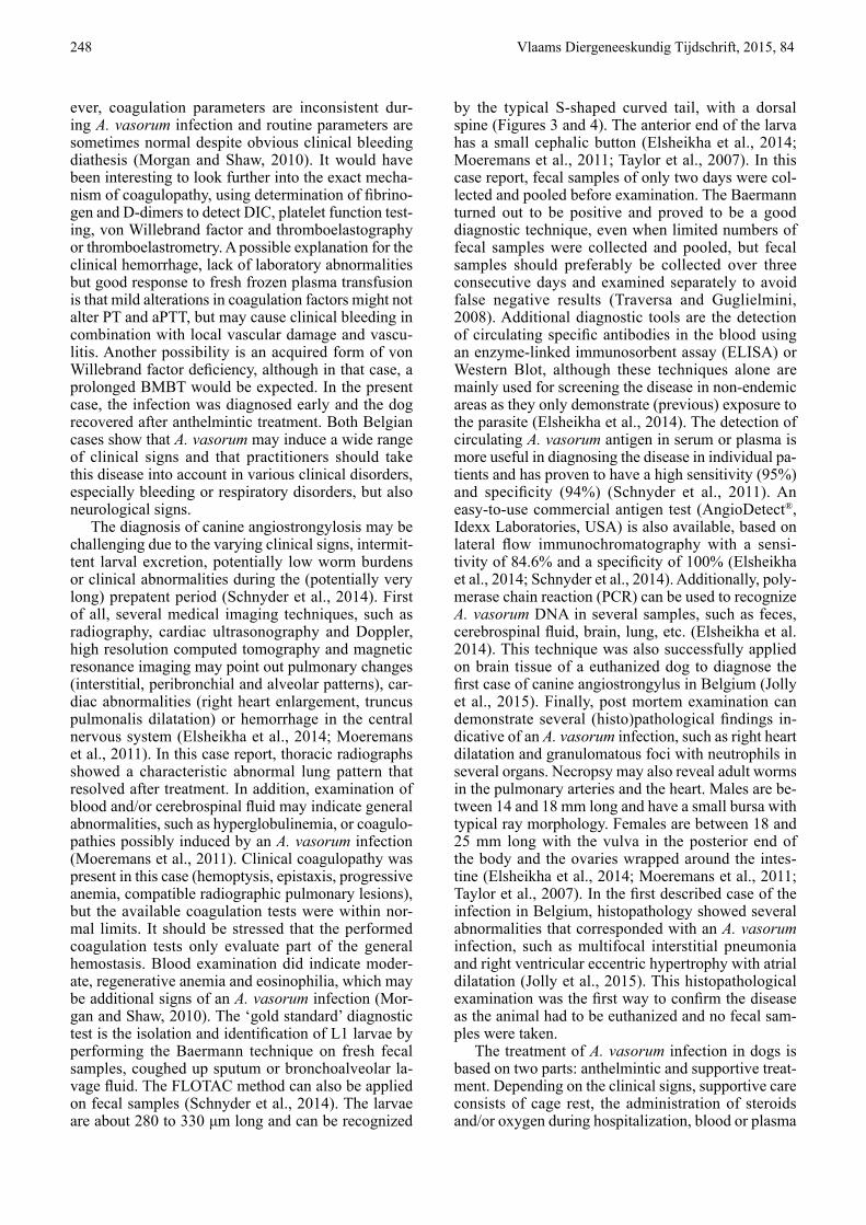
Figure 3. Iodine stained L1 larva of Angiostrongylus va-sorum (x20).
Figure 4. Close-up of the typical S-shaped tail of an iodine stained Angiostrongylus vasorum L1 larva (x40).
A B
248 Vlaams Diergeneeskundig Tijdschrift, 2015, 84
ever, coagulation parameters are inconsistent dur-ing A. vasorum infection and routine parameters are sometimes normal despite obvious clinical bleeding diathesis (Morgan and Shaw, 2010). It would have been interesting to look further into the exact mecha-nism of coagulopathy, using determination of fibrino-gen and D-dimers to detect DIC, platelet function test-ing, von Willebrand factor and thromboelastography or thromboelastrometry. A possible explanation for the clinical hemorrhage, lack of laboratory abnormalities but good response to fresh frozen plasma transfusion is that mild alterations in coagulation factors might not alter PT and aPTT, but may cause clinical bleeding in combination with local vascular damage and vascu-litis. Another possibility is an acquired form of von Willebrand factor deficiency, although in that case, a prolonged BMBT would be expected. In the present case, the infection was diagnosed early and the dog recovered after anthelmintic treatment. Both Belgian cases show that A. vasorum may induce a wide range of clinical signs and that practitioners should take this disease into account in various clinical disorders, especially bleeding or respiratory disorders, but also neurological signs.
The diagnosis of canine angiostrongylosis may be challenging due to the varying clinical signs, intermit-tent larval excretion, potentially low worm burdens or clinical abnormalities during the (potentially very long) prepatent period (Schnyder et al., 2014). First of all, several medical imaging techniques, such as radiography, cardiac ultrasonography and Doppler, high resolution computed tomography and magnetic resonance imaging may point out pulmonary changes (interstitial, peribronchial and alveolar patterns), car-diac abnormalities (right heart enlargement, truncus pulmonalis dilatation) or hemorrhage in the central nervous system (Elsheikha et al., 2014; Moeremans et al., 2011). In this case report, thoracic radiographs showed a characteristic abnormal lung pattern that resolved after treatment. In addition, examination of blood and/or cerebrospinal fluid may indicate general abnormalities, such as hyperglobulinemia, or coagulo-pathies possibly induced by an A. vasorum infection (Moeremans et al., 2011). Clinical coagulopathy was present in this case (hemoptysis, epistaxis, progressive anemia, compatible radiographic pulmonary lesions), but the available coagulation tests were within nor-mal limits. It should be stressed that the performed coagulation tests only evaluate part of the general hemostasis. Blood examination did indicate moder-ate, regenerative anemia and eosinophilia, which may be additional signs of an A. vasorum infection (Mor-gan and Shaw, 2010). The ‘gold standard’ diagnostic test is the isolation and identification of L1 larvae by performing the Baermann technique on fresh fecal samples, coughed up sputum or bronchoalveolar la-vage fluid. The FLOTAC method can also be applied on fecal samples (Schnyder et al., 2014). The larvae are about 280 to 330 μm long and can be recognized
by the typical S-shaped curved tail, with a dorsal spine (Figures 3 and 4). The anterior end of the larva has a small cephalic button (Elsheikha et al., 2014; Moeremans et al., 2011; Taylor et al., 2007). In this case report, fecal samples of only two days were col-lected and pooled before examination. The Baermann turned out to be positive and proved to be a good diagnostic technique, even when limited numbers of fecal samples were collected and pooled, but fecal samples should preferably be collected over three consecutive days and examined separately to avoid false negative results (Traversa and Guglielmini, 2008). Additional diagnostic tools are the detection of circulating specific antibodies in the blood using an enzyme-linked immunosorbent assay (ELISA) or Western Blot, although these techniques alone are mainly used for screening the disease in non-endemic areas as they only demonstrate (previous) exposure to the parasite (Elsheikha et al., 2014). The detection of circulating A. vasorum antigen in serum or plasma is more useful in diagnosing the disease in individual pa-tients and has proven to have a high sensitivity (95%) and specificity (94%) (Schnyder et al., 2011). An easy-to-use commercial antigen test (AngioDetect®, Idexx Laboratories, USA) is also available, based on lateral flow immunochromatography with a sensi- tivity of 84.6% and a specificity of 100% (Elsheikha et al., 2014; Schnyder et al., 2014). Additionally, poly-merase chain reaction (PCR) can be used to recognize A. vasorum DNA in several samples, such as feces, cerebrospinal fluid, brain, lung, etc. (Elsheikha et al. 2014). This technique was also successfully applied on brain tissue of a euthanized dog to diagnose the first case of canine angiostrongylus in Belgium (Jolly et al., 2015). Finally, post mortem examination can demonstrate several (histo)pathological findings in-dicative of an A. vasorum infection, such as right heart dilatation and granulomatous foci with neutrophils in several organs. Necropsy may also reveal adult worms in the pulmonary arteries and the heart. Males are be-tween 14 and 18 mm long and have a small bursa with typical ray morphology. Females are between 18 and 25 mm long with the vulva in the posterior end of the body and the ovaries wrapped around the intes-tine (Elsheikha et al., 2014; Moeremans et al., 2011; Taylor et al., 2007). In the first described case of the infection in Belgium, histopathology showed several abnormalities that corresponded with an A. vasorum infection, such as multifocal interstitial pneumonia and right ventricular eccentric hypertrophy with atrial dilatation (Jolly et al., 2015). This histopathological examination was the first way to confirm the disease as the animal had to be euthanized and no fecal sam-ples were taken.
The treatment of A. vasorum infection in dogs is based on two parts: anthelmintic and supportive treat-ment. Depending on the clinical signs, supportive care consists of cage rest, the administration of steroids and/or oxygen during hospitalization, blood or plasma

Vlaams Diergeneeskundig Tijdschrift, 2015, 84 249
transfusion or the administration of ACE-inhibitors, diuretics and bronchodilators (Elsheikha et al., 2014; Moeremans et al., 2011). Anthelmintics that can be used for the treatment of A. vasorum are moxidectin/imidacloprid, milbemycin oxime, fenbendazole, le-vamisole and ivermectin (Elsheikha et al., 2014; Mo-eremans et al., 2011). The last two products are not often used due to possible anaphylactic or toxic ef-fects. A single dose (0.1 ml/kg) of the moxidectin/imi-dacloprid spot-on solution (Advocate®, Bayer Animal Health, Belgium) is effective against adult and im-mature worms (Willesen et al., 2007; Schnyder et al., 2009) and so is a weekly oral administration of milbe-mycine oxime (Milbemax®, Novartis Animal Health, Belgium) at 0.5 mg/kg during one month (Conboy, 2004). Although fenbendazole (Panacur®, Intervet Int. via MSD AH, Belgium; 25-50 mg/kg SID for 5-21 days) is not registered for this disease in Europe, it is often used with good results, such as in this case re-port (Conboy, 2011; Gallagher et al., 2012; Spodsberg et al., 2013; Tebb et al., 2007; Willesen et al., 2007). Elimination of the parasite should be checked by ex-amining fecal samples after treatment and additional treatments should be performed when L1 larvae are still found (Moeremans et al., 2011). However, fecal examination after treatment was not performed in the present case. Resistance in A. vasorum worms has not been described so far (Elsheikha et al., 2014; Moer-emans et al., 2011).
Preventing infection should first of all focus on avoiding contact with the intermediate host (Taylor et al., 2007). As this is nearly impossible, prophylactic anthelmintic treatment remains the most important factor in preventing the disease. The only anthelmin-tic that is licensed in Belgium for preventive treatment against A. vasorum is moxidectin, in combination with imidacloprid (Advocate®, Bayer Animal Health, Bel-gium). In addition, milbemycine oxime in combina-tion with spinosad (Trifexis®, Elanco Animal Health, Belgium) has been proven to have a 98.8% preventive efficacy against A. vasorum adult development (Böhm et al., 2014). These products can be used in prophylac-tic strategies (monthly administration) when risk fac-tors for the disease are present, i.e. frequent contact with snails, previous exposure to the worm or located in an endemic area. In Belgium, the disease has so far only been detected in the southern part of the coun-try. Several cases have been diagnosed around Liège, Arlon and an area south of Brussels, around the town of Waterloo, which are now considered hot spots for the disease in Belgium (B. Losson, personal commu-nication). In the region around Liège, Crenosoma vul-pis infections have recently been discovered as well, so veterinarians should pay attention to distinguish both lungworm infections (Caron et al., 2014). The focus of prophylaxis should therefore also be on edu-cating and raising awareness of these emerging and potentially deadly diseases amongst veterinarians and pet owners, especially those who live in endemic foci (Elsheikha et al., 2014; Moeremans et al., 2011).
CONCLUSION
In this case report, the second confirmed case of canine angiostrongylosis in a dog in Belgium is de-scribed. The dog was presented with respiratory com-plaints, mild to moderate regenerative anemia, eosino-philia and clinical evidence of coagulopathy, without abnormalities on routine laboratory coagulation tests. An A. vasorum infection was confirmed by the iden-tification of L1 larvae after performing the Baermann technique. The dog was successfully treated with fenbendazole (Panacur®, Intervet Int. via MSD AH, Belgium). This case demonstrates that autochthonous cases of canine angiostrongylosis may occur in Bel-gium and that early diagnosis is important for the suc-cessful treatment of the infection. The disease may cause different and often vague clinical signs, diagno-sis may be challenging and the prevalence is suspected to be low. Therefore, veterinarians should be aware of the importance of this disease and they should take it into account when dealing with patients with various clinical disorders.
ACKNOWLEDGEMENTS
The authors would like to thank the veterinarians of Centre Vétérinaire du Lion in Waterloo (Belgium) for referring the patient to the Faculty of Veterinary Medicine (Ghent University) and the follow-up after treatment.
REFERENCES
Adamantos S., Waters S., Boag A. (2015). Coagulation status in dogs with naturally occurring Angiostrongylus vasorum infection. Journal of Small Animal Practice 56, 485-490.
Böhm C., Schnyder M., Thamsborg S.M., Thompson C.M., Trout C., Wolken S., Schnitzler B. (2014). Assessment of the combination of spinosad and milbemycin oxime in preventing the development of canine Angiostrongylus vasorum infections. Veterinary Parasitology 199, 272-277.
Caron Y., Merveille A.-C., Losson B., Billen F. (2014). Crenosoma vulpis infection in two young dogs in Bel-gium. Veterinary Record Case Reports 2, 1-4.
Conboy G.A. (2004). Natural infections of Crenosoma vulpis and Angiostrongylus vasorum in dogs in Atlantic Canada and their treatment with milbemycin oxime. Vet-erinary Record 155, 16-18.
Conboy G.A. (2011). Canine angiostrongylosis: The French heartworm: An emerging threat in North America. Veteri-nary Parasitology 176, 382-389.
Elsheikha H.M., Holmes S.A., Wright I., Morgan E.R., Lacher D.W. (2014). Recent advances in the epidemio-logy, clinical and diagnostic features, and control of canine cardio-pulmonary angiostrongylosis. Veterinary Research 45, 1-12.

250 Vlaams Diergeneeskundig Tijdschrift, 2015, 84
Gallagher B., Brennan S.F., Zarelli M., Mooney C.T. (2012). Geographical, clinical, clinicopathological and radiographic features of canine angiostrongylosis in Irish dogs: a retrospective study. Irish Veterinary Journal 65, 1-10.
Helm J., Gilleard J.S., Jackson M., Redman E., Bell R. (2009). A case of canine Angiostrongylus vasorum in Scotland confirmed by PCR and sequence analysis. Jour-nal of Small Animal Practice 50, 255-259.
Hurníková Z., Miterpáková M., Mandelík R. (2013). First autochtonous case of canine Angiostrongylus vasorum in Slovakia. Parasitology Research 112, 3505-3508.
Jolly S., Poncelet L., Lempereur L., Caron Y., Bayrou C., Cassart D., Grimm F., Losson B. (2015). First report of a fatal autochthonous canine Angiostrongylus vasorum in-fection in Belgium. Parasitology International 64, 97-99.
Kirk L., Limon G., Guitian F.J., Hermosilla C., Fox M.T. (2014). Angiostrongylus vasorum in Great Britain: a na-tionwide postal questionnaire survey of veterinary prac-tices. Veterinary Record 175, 1-6.
Miterpáková M., Hurníková Z., Zalewski A.P. (2014). The first clinically manifested case of angiostrongylosis in a dog in Slovakia. Acta Parasitologica 59, 661-665.
Moeremans I., Binst D., Claerebout E., Van de Maele I., Daminet S. (2011). Canine Angiostrongylus vasorum. Vlaams Diergeneeskundig Tijdschrift 80, 319-326.
Morgan E. and Shaw S., 2010. Angiostrongylus vasorum in-fection in dogs: continuining spread and developments in diagnosis and treatment. Journal of Small Animal Prac-tice 51, 616-621.
Morgan E.R., Jefferies R., van Otterdijk L., McEniry R.B., Allen F., Bakewell M., Shaw S.E. (2010). Angiostron-gylus vasorum infection in dogs: Presentation and risk factors. Veterinary Parasitology 173, 255-261.
Rinaldi L., Cortese L., Meomartino L., Pagano T.B., Pepe P., Cringoli G., Papparella S. (2014). Angiostrongylus vasorum: epidemiological, clinical and histopathological insights. BMC Veterinary Research 10, 1-7.
Schnyder M., Fahrion A., Ossent P., Kohler L., Webster P., Heine J., Deplazes P. (2009). Larvicidal effect of imida-cloprid/moxidectin spont-on solution in dogs experimen-tally inoculated with Angiostrongylus vasorum. Veteri-nary Parasitology 166, 326-332.
Schnyder M., Tanner I., Webster P., Barutzki D., Deplazes P. (2011). An ELISA for sensitive and specific detection of circulating antigen of Angiostrongylus vasorum in serum samples of naturally and experimentally infected dogs. Veterinary Parasitology 185, 152-158.
Schnyder M., Deplazes P. (2012). Cross-reactions of sera from dogs infected with Angiostrongylus vasorum in commercially available Dirofilaria immitis test kits. Para- sites and Vectors 5, 258.
Schnyder M., Schaper R., Gilbrough G., Morgan E.R., De-plazes P. (2013). Seroepidemiological survey for canine angiostrongylosis in dogs from Germany and the UK using combined detection of Angiostrongylus vasorum
antigen and specific antibodies. Parasitology 140, 442-1450.
Schnyder M., Schaper R., Pantchev N., Kowalska D., Szwedko A., Deplazes P. (2013). Serological detection of circulating Angiostrongylus vasorum antigen- and par-asite-specific antibodies in dogs from Poland. Parasito-logy Research 112, 109-117.
Schnyder M., Stebler K., Naucke T.J., Lorentz S., Deplazes P. (2014). Evaluation of a rapid device for serological in-clinic diagnosis of canine angiostrongylosis. Parasites & Vectors 7, 1-7.
Simin S., Kosić L.S., Kuruca L., Pavlović I., Savović M., Lalošević V. (2014). Moving the boundaries to the South-East: first record of autochthonous Angiostrongylus va-sorum infection in a dog in Vojvodina province, northern Serbia. Parasites & Vectors 7, 1-4.
Spodsberg E.H., Miles J.E., McEvoy F.J., Willesen J.L. (2013). Spontaneous pneumothorax secondary to granu-lomatous pneumonia caused by Angiostrongylus vasorum in a dog in Denmark. Journal of Small Animal Practice 54, 114.
Taylor M.A., Coop R.L., Wall R.L. (2007). Parasites of dogs and cats - Parasites of the circulatory system. In: Veterinary Parasitology. Third edition, Blackwell Pub-lishing, Oxford, p. 410-411.
Tebb A.I., Johnson V.S., Irwin P.J. (2007). Angiostrongylus vasorum (French heartworm) in a dog imported into Aus-tralia. Australian Veterinary Journal 85, 23-28.
Tolnai Z., Széll Z., Sréter T. (2015). Environmental deter-minants of the spatial distribution of Angiostrongylus vasorum, Crenosoma vulpis and Eucoleus aerophilus in Hungary. Veterinary Parasitology 30, 355-358.
Traversa D. And Guglielmini C. (2008). Feline aelurostron-gylosis and canine angiostrongylosis: a challenging diag-nosis for two emerging verminous pneumonia infections. Veterinary Parasitology 7, 163-174.
Traversa D., Di Cesare A., Meloni S., Frangipane di Regal-bono A., Milillo P., Pampurini F., Venco L. (2013). Ca-nine angiostrongylosis in Italy: occurrence of Angiostron-gylus vasorum in dogs with compatible clinical pictures. Parasitology Research 112, 2473-2480.
van Doorn D.C.K., van de Sande A.H., Nijsse E.R., Eysker M., Ploeger H.W. (2009). Autochthonous Angiostrongy-lus vasorum infection in dogs in The Netherlands. Veteri-nary Parasitology 162, 163-166.
Whitley N.T., Corzo-Menendez N., Carmichael N.G., Mc-Garry J.W. (2005). Cerebral and conjunctival haemor-rhages associated with von Willebrand factor deficiency and canine angiostrongylosis. Journal of Small Animal Practice 46, 75-78.
Willesen J.L., Kristensen A.T., Jensen A.L., Heine J., Koch J. (2007). Efficacy and safety of imidacloprid/moxidec-tin spot-on solution and fenbendazole in the treatment of dogs naturally infected with Angiostrongylus vasorum (Baillet, 1866). Veterinary Parasitology 147, 258-264.