Embed Size (px)
Citation preview
RESEARCH ARTICLE
Meningitis patients with Angiostrongylus
cantonensis may present without eosinophilia
in the cerebrospinal fluid in northern Vietnam
Tomoko HiraokaID1,2, Ngo Chi Cuong2,3, Sugihiro Hamaguchi4, Mihoko Kikuchi5,
Shungo KatohID1,2,6, Le Kim Anh7, Nguyen Thi Hien Anh8, Dang Duc Anh8, Chris Smith9,10,
Haruhiko Maruyama11, Lay-Myint YoshidaID2,12, Do Duy Cuong3, Pham Thanh Thuy3,13,
Koya AriyoshiID1,9*
1 Department of Clinical Medicine, Institute of Tropical Medicine (NEKKEN), Nagasaki University, Nagasaki,
Japan, 2 Department of Clinical Tropical Medicine, Nagasaki University Graduate School of Biomedical
Sciences, Nagasaki, Japan, 3 Department of Infectious Diseases, Bach Mai Hospital, Hanoi, Vietnam,
4 Department of General Internal Medicine, Fukushima Medical University, Fukushima, Japan,
5 Department of Immunogenetics, Institute of Tropical Medicine (NEKKEN), Nagasaki University, Nagasaki,
Japan, 6 Department of General Internal Medicine, Nagasaki Rosai Hospital, Nagasaki, Japan, 7 Vietnam
Research Station, Institute of Tropical Medicine (NEKKEN), Nagasaki University, Hanoi, Vietnam, 8 National
Institute of Hygiene and Epidemiology, Hanoi, Vietnam, 9 Department of Global Health, School of Tropical
Medicine and Global Health, Nagasaki University, Nagasaki, Japan, 10 Department of Clinical Research,
London School of Hygiene and Tropical Medicine (LSHTM), London, United Kingdom, 11 Department of
Infectious Diseases, Division of Parasitology, Faculty of Medicine, University of Miyazaki, Miyazaki, Japan,
12 Department of Pediatric Infectious Diseases, Institute of Tropical Medicine (NEKKEN), Nagasaki
University, Nagasaki, Japan, 13 Infection Prevention and Control, The Partnership for Health Advancement
in Vietnam (HAIVN), Hanoi, Vietnam
Abstract
Background
Eosinophilic meningitis (EM) is a rare clinical syndrome caused by both infectious and non-
infectious diseases. In tropical pacific countries, Angiostrongylus cantonensis is the most
common cause. However, the EM definition varies in the literature, and its relation to para-
sitic meningitis (PM) remains unclear.
Methodology/Principal findings
Adult and adolescent patients of 13 years old or above with suspected central nervous sys-
tem (CNS) infections with abnormal CSF findings were prospectively enrolled at a tertiary
referral hospital in Hanoi, Vietnam from June 2012 to May 2014. Patients with EM or sus-
pected PM (EM/PM) were defined by the presence of either�10% eosinophils or an abso-
lute eosinophil cell counts of�10/mm3 in the CSF or blood eosinophilia (>16% of WBCs)
without CSF eosinophils. In total 679 patients were enrolled: 7 (1.03%) had�10% CSF
eosinophilia, 20 (2.95%) had�10/mm3 CSF eosinophilia, and 7 (1.03%) had >16% blood
eosinophilia. The patients with�10% CSF eosinophilia were significantly younger (p =
0.017), had a lower body temperature (p = 0.036) than patients with�10/mm3 CSF eosino-
philia among whom bacterial pathogens were detected in 72.2% (13/18) of those who were
PLOS NEGLECTED TROPICAL DISEASES
PLOS Neglected Tropical Diseases | https://doi.org/10.1371/journal.pntd.0008937 December 22, 2020 1 / 15
a1111111111
a1111111111
a1111111111
a1111111111
a1111111111
OPEN ACCESS
Citation: Hiraoka T, Cuong NC, Hamaguchi S,
Kikuchi M, Katoh S, Anh LK, et al. (2020)
Meningitis patients with Angiostrongylus
cantonensis may present without eosinophilia in
the cerebrospinal fluid in northern Vietnam. PLoS
Negl Trop Dis 14(12): e0008937. https://doi.org/
10.1371/journal.pntd.0008937
Editor: Alessandra Morassutti, PUCRS, BRAZIL
Received: December 29, 2019
Accepted: October 30, 2020
Published: December 22, 2020
Copyright: © 2020 Hiraoka et al. This is an open
access article distributed under the terms of the
Creative Commons Attribution License, which
permits unrestricted use, distribution, and
reproduction in any medium, provided the original
author and source are credited.
Data Availability Statement: All relevant data are
within the manuscript and its Supporting
Information files.
Funding: This research is partially supported by the
Japan Initiative for Global Research Network on
Infectious Diseases (J-GRID: https://www.amed.
go.jp/program/list/01/06/001.html) (grant number
10008012) to LMY and KA from Ministry of
Education, Culture, Sport, Science & Technology in
Japan, and Japan Agency for Medical Research
and Development (AMED: https://www.amed.go.jp/
tested by culture and/or PCR. In contrast, the characteristics of the patients with >16%
blood eosinophilia resembled those of patients with�10% CSF eosinophilia. We further
conducted serological tests and real-time PCR to identify A. cantonensis. Serology or real-
time PCR was positive in 3 (42.8%) patients with�10% CSF eosinophilia and 6 (85.7%)
patients with >16% blood eosinophilia without CSF eosinophils but none of patients with
�10/mm3 CSF eosinophilia.
Conclusions
The etiology of PM in northern Vietnam is A. cantonensis. The eosinophil percentage is a
more reliable predictor of parasitic EM than absolute eosinophil count in the CSF. Patients
with PM may present with a high percentage of eosinophils in the peripheral blood but not in
the CSF.
Author summary
Eosinophilic meningitis (EM) is a rare meningitis accompanied by eosinophils in the CSF
and caused by multiple etiologies. Angiostrongylus cantonensis, which is a rat lungworm
parasite, is the most common cause in tropical Asia. Previous papers have defined EM as
CSF eosinophils�10% or CSF eosinophils�10/mm3. However, the relationship of EM to
parasitic meningitis (PM) remains unclear. This prospective study enrolled 679 patients
with suspected CNS infection who were admitted to a tertiary referral hospital in Hanoi,
Vietnam from June 2012 to May 2014. The characteristics of patients with�10% CSF
eosinophilia resembled those of patients with>16% blood eosinophilia without CSF
eosinophils, whereas those of patients with�10/mm3 CSF eosinophilia were comparable
with those of patients with typical bacterial meningitis. Serology or real-time PCR for A.
cantonensis was positive in 3 out of 7 patients with�10% CSF eosinophilia and 6 out of 7
patients with > 16% blood eosinophilia without CSF eosinophils but none of patients
with�10/mm3 CSF eosinophilia. The percentage, in contrast to the absolute eosinophil
count in CSF, is reliable for predicting parasitic EM. Patients with PM may present with
eosinophilia in the peripheral blood but not in the CSF.
Introduction
Eosinophilic meningitis (EM) is a rare clinical syndrome characterized by meningeal inflam-
mation and eosinophilic pleocytosis in the cerebrospinal fluid (CSF) [1,2]. The first case of EM
was reported in Taiwan in 1945. In this case, eosinophilia of the CSF and peripheral blood was
observed, and then Angiostrongylus cantonensis larvae were identified in the CSF [3]. Since
this report, EM cases have been recognized and reported in the Pacific Ocean islands, East
Asia, and North America [4–6]. There are various etiologies of CSF eosinophilia, including
parasitic infections of the central nervous system (CNS) and other infectious diseases, such as
tuberculous meningitis, cerebrospinal syphilis, viral and fungal meningitis, as well as noninfec-
tious causes, such as drug allergies, multiple sclerosis and neoplasms, for example, Hodgkin’s
disease or leukemia [1,2,5]. However, the most common etiologies in Southeast Asia and other
tropical countries are parasitic infectious diseases, especially A. cantonensis, Gnathostoma spi-nigerum, cysticercosis (Taenia solium) and Toxocara canis [7]. Therefore, in countries with
PLOS NEGLECTED TROPICAL DISEASES Parasitic meningitis without eosinophilia in cerebrospinal fluid
PLOS Neglected Tropical Diseases | https://doi.org/10.1371/journal.pntd.0008937 December 22, 2020 2 / 15
index.html)(grant number JP19fm0108001 and
JP20fk0108095) to LMY. The funders had no role
in study design, collection and analysis, decision to
publish, or preparation of the manuscript.
Competing interests: The authors have declared
that no competing interests exist.
tropical climates, it is important to determine whether meningitis is parasitic meningitis (PM)
because specific treatment is required [7].
The definition of EM varies. Many EM publications have followed the definition originally
suggested by Kuberski [8]; the presence of at least 10% eosinophils in the total CSF white
blood cell (WBC) count or the presence of at least 10 eosinophils/mm3 in the CSF [2]. How-
ever, this criterion was based on a limited observation of 123 CSF samples derived from 110
pediatric patients with a variety of clinical diagnoses in Hawaii [8]. Furthermore, another defi-
nition of EM was also suggested by Punyagupta et al. [9]; patients with an acute headache of
fewer than 2 months with a CSF WBC count/mm3 of 20 or more, of which 10% or more are
eosinophils [10]. The author used this criterion to select 484 patients with probable angios-
trongyliasis in Thailand. Many papers from Asia, especially Thailand, have followed this crite-
rion [11,12].
In addition, some EM papers have reported that 30–80% of patients with meningitic
angiostrongyliasis have accompanying blood eosinophilia [4,12,13]. Swanyawisuth et al. [14]
discussed the significance of peripheral eosinophilia as an indicator of meningitic angiostron-
gyliasis. They found that if patients with suspected PM had an eosinophil count of more than
798 cells in their peripheral blood, the sensitivity and specificity of meningitis due to A. canto-nensis reached 76.6% and 80.2%, respectively. This group investigated the presence of PM with
a blood serological test without lumbar puncture. Schulte et al. [15] also reported a positive
predictive value for helminth infections of 46.6% among travelers returning from tropical
countries with blood eosinophilia > 16% of the WBC count.
To our knowledge, however, few studies have systematically attempted to delineate the clin-
ical implication of various definitions of EM to date with the objective of identifying PM
[5,16]. We believe that describing the clinical characteristics of patients with EM or suspected
PM (EM/PM) classified by each definition and confirming the causative parasites will provide
useful information to clinicians to improve clinical judgment and management. To improve
the clinical management of PM in northern Vietnam, we conducted a prospective study of
CNS infection in this area. The primary objectives of this study were to investigate the epide-
miological and clinical characteristics of various definitions of EM/PM in relation to the path-
ogenic parasite. The secondary objective was to further understand the value of current
definitions of EM for predicting PM.
Methods
Ethics statement
This study was approved by the independent ethics committees of the Institute of Tropical
Medicine, Nagasaki University (approval number: 12021085–4), Nagasaki, Japan, Bach Mai
Hospital and the National Institute of Hygiene and Epidemiology as part of a “Collaborative
Study on Emerging and Re-emerging Infectious Diseases in Vietnam” (approval number:
15-IRB, 2011), Hanoi, Vietnam. Written informed consent was obtained from all patients
prior to enrollment. For those who were unconscious, a parent or guardian was asked to pro-
vide informed consent, and the data were analyzed anonymously.
Study design and setting
Between June 2012 and May 2014, we conducted a prospective observational study of undiag-
nosed febrile illness in the Infectious Disease Department of Bach Mai Hospital, which is the
largest government referral medical center in Hanoi covering patients in northern Vietnam, as
published previously [17].
PLOS NEGLECTED TROPICAL DISEASES Parasitic meningitis without eosinophilia in cerebrospinal fluid
PLOS Neglected Tropical Diseases | https://doi.org/10.1371/journal.pntd.0008937 December 22, 2020 3 / 15
Inclusion and exclusion criteria and case definition
Patients were enrolled according to the following criteria: 1) age� 13 years, 2) axillary
temperature > 37.5˚C (any time from onset to admission), and 3) lumbar puncture due to sus-
pected CNS infection by the admitting physician. The exclusion criteria were patients with a
clinically definitive diagnosis (e.g., malaria, dengue fever, mumps, food-related diarrhea, cellu-
litis, animal bite), patients with hepatitis-related disease (e.g., viral hepatitis, alcoholic liver dis-
ease, autoimmune hepatitis, cirrhosis, liver cancer), and patients with microbiologically
identified infectious diseases (e.g., already diagnosed at referral hospitals). Regarding fever cri-
teria, even if patients did not have fever at admission, they were enrolled as long as they had
had fever at any time point from onset to admission. Patients and samples to be enrolled were
determined on the following morning of each admission day.
We defined abnormal CSF as CSF protein > 0.4 g/l and CSF absolute WBC count> 5/
mm3. At Bach Mai Hospital, we defined EM/PM cases in three ways: 1) the eosinophils
accounted for� 10% of the total WBCs in the CSF and the absolute number of eosinophils
was� 10/mm3 in the CSF, 2) the percentage of eosinophils was < 10% of the total WBC count
in the CSF and the absolute number of eosinophils was� 10/mm3 in the CSF, or 3) the abso-
lute number of eosinophils was< 10/mm3 in the CSF and the percentage of eosinophils
was> 16% of the peripheral blood WBC count.
Data and sample collection
We prospectively collected epidemiological data (age, gender, place of occurrence, occupation,
medical history, duration of fever, clinical presentation), and biological blood and CSF data.
We also collected initial blood samples (plasma and buffy coat) and initial CSF samples after
admission. At the hospital, the WBC type and number of cells in the CSF sample were con-
firmed by Giemsa staining when the total WBC count was greater than 10/mm3. After identi-
fying patients with suspected EM or PM, we conducted a retrospective chart review to obtain
further clinical information using hospital records.
Biological analysis
Serological tests. We tested initial blood plasma samples of suspected EM/PM patients
and 20 control patients for anti-parasite antibodies in enzyme-linked immunosorbent assay
(ELISA). The antigen tested were those of A. cantonensis, Toxocara canis, Paragonimus spp.,
and Strongyloides stercoralis. According to internal data of Miyazaki University, the sensitivity
/ specificity of parasite ELISA was 90.0 / 99.1 for strongyloidiasis, 97.1 / 97.4 for paragonimia-
sis, 97.3 / 74.6 for larva migrant syndrome due to Toxocara or Ascaris infections. But the sensi-
tivity / specificity of A. cantonensis has not yet been fully established due to lack of positive
confirmed cases in Japan. The current ELISA for testing antibodies to A. cantonensis was
based on a previously described method [18], in which A. cantonensis antigen was prepared
from fourth-stage larvae recovered from the brains of experimentally infected rats. The sensi-
tivity and specificity of this test were reported as 100% and 66.8%, respectively [19]. The 20
control patients were randomly selected from the list of non-EM patients in the same study,
including patients with normal CSF (n = 8) and with abnormal CSF (bacterial meningitis:
n = 5, tuberculosis meningitis: n = 1, and aseptic meningitis patients: n = 6).
Real-time polymerase chain reaction (PCR) for A. cantonensis. We conducted real-time
PCR analyses for A. cantonensis, which is the most common parasite specie causing EM in
Asia, using CSF samples from EM/PM patients fulfilling either of the three criteria. First, we
prepared 200 mm3 of the CSF samples, as previously described [20], and then we extracted
DNA from the samples using a QIAmp DNA Mini kit (QIAGEN, Hilden, Germany) with 100
PLOS NEGLECTED TROPICAL DISEASES Parasitic meningitis without eosinophilia in cerebrospinal fluid
PLOS Neglected Tropical Diseases | https://doi.org/10.1371/journal.pntd.0008937 December 22, 2020 4 / 15
mm3 of elution buffer. We performed TaqMan Real-time PCR for A. cantonensis with positive
and negative controls for each assay.
For the real-time PCR for A. cantonensis, we followed the protocol of Qvarnstrom et al.
[21], using TaqMan Universal Master Mix II (Thermo Fisher Scientific, Waltham, MA, USA)
and an Applied Biosystems 7500 Real Time PCR system (Applied Biosystems, Foster City, CA,
USA). A positive control was prepared from the whole worm body of A. cantonensis. We cut 3
worms into small pieces and extracted whole DNA from the worms using a QIAmp DNA
Mini kit (QIAGEN, Hilden, Germany) with 100 mm3 of elution buffer. The upper limit of
dilution of the positive control was 100,000 times for detection by TaqMan real-time PCR for
A. cantonensis.For the real-time PCR for G. spinigerum, we designed two sets of oligonucleotide primers to
amplify a 144-bp fragment of the first internal transcribed spacer (ITS1) gene of G. spinigerumand a 115-bp fragment of the second internal transcribed spacer (ITS2) gene of G. spinigerum,
based on Primer3 <https://primer3plus.com/cgi-bin/dev/primer3plus.cgi> with GenBank
accession no. AB181155. The primers targeting ITS1 were Gspi-ITS1F (5’-CATCGGCTCTG
ATCTTCGCT-3’) and Gspi-ITS1R (5’-AGACACCAACGGATGCTGTT-3’); the primers tar-
geting ITS2 were Gspi-ITS2F (5’-CATTCATCGAGCGGCAAGTG-3’) and Gspi-ITS2R (5’-
GCGTACGCACCTCGATAAGA-3’). After confirming that the G. spinigerum positive control
showed a single band by conventional PCR with each of the two sets of primers, using GoTaq
Flexi DNA Polymerase (Promega Corporation, Madison, WI, USA), we performed SYBR
Green Real-time PCR for G. spinigerum using each of the two sets of primers with Power
SYBR Green PCR Master Mix (Thermo Fisher Scientific, Waltham, MA, USA) and a 7500
Real Time PCR System (Applied Biosystems, Foster City, CA, USA). The positive control for
G. spinigerum was its whole genome, which was kindly provided by the Department of Hel-
minthology, Faculty of Tropical Medicine, Mahidol University. The whole-genome DNA con-
centration was 3100 ng/mm3. The upper limit of the positive control dilution was 1,000,000
times for detection by SYBR Green Real-time PCR.
Statistical analysis
We showed the demographic and clinical characteristics of each EM/PM group with those of
the other patients using frequencies and percentages for categorical values and the median and
interquartile range (IQR) for continuous variables. When we were comparing CSF
eosinophils� 10% group with other definition group individually or comparing the EM/PM
criteria not fulfilled group with each definition group individually, categorical variables were
compared by Fisher’s exact test, and continuous variables were compared by the Mann-Whit-
ney nonparametric test. We calculated the odds ratio (OR) with 95% confidence intervals
using logistic regression analysis. In addition, we calculated p-values among the 4 groups by
Kruskal-Wallis test for continues variables and Chi-square test for categorical variables. Statis-
tical analysis was conducted using STATA version 15 (StataCorp LLC, College Station,
TX77845 USA). All tests were two-tailed, and p< 0.05 was considered statistically significant.
Results
During the study period, from June 2012 to May 2014, 7,505 patients were admitted to the
department, and 2,458 patients were hospitalized with undiagnosed febrile illness. Among
them, 679 patients underwent lumbar puncture and were enrolled. Abnormal CSF was found
in 431 (63.5%) patients. Blood samples of all patients (100%) were available, and CSF samples
of 473 patients (69.7%) were available for this study.
PLOS NEGLECTED TROPICAL DISEASES Parasitic meningitis without eosinophilia in cerebrospinal fluid
PLOS Neglected Tropical Diseases | https://doi.org/10.1371/journal.pntd.0008937 December 22, 2020 5 / 15
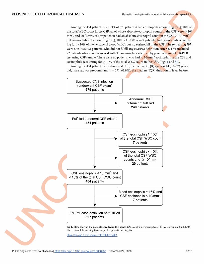
Among the 431 patients, 7 (1.03% of 679 patients) had eosinophils accounting for� 10% of
the total WBC count in the CSF, all of whose absolute eosinophil counts in the CSF were� 10/
mm3, and 20 (2.95% of 679 patients) had an absolute eosinophil count in the CSF� 10/mm3
but eosinophils not accounting for� 10%. 7 (1.03% of 679 patients) had eosinophils account-
ing for> 16% of the peripheral blood WBCs but no eosinophil in the CSF. The remaining 397
were non-EM/PM patients, who did not fulfill any EM/PM definition criteria. This included
22 patients who were diagnosed with TB meningitis as defined by positive result of TB-PCR
test using CSF sample. There were no patients who had < 10/mm3 eosinophils in the CSF and
eosinophils accounting for� 10% of the total WBC count in the CSF. (Figs 1 and S1).
Among the 431 patients with abnormal CSF, the median (IQR) age was 44 (30–57) years
old, male sex was predominant (n = 271, 62.9%), the median (IQR) duration of fever before
Fig 1. Flow chart of the patients enrolled in this study. CNS: central nervous system, CSF: cerebrospinal fluid, EM/
PM: eosinophilic meningitis or suspected parasitic meningitis.
https://doi.org/10.1371/journal.pntd.0008937.g001
PLOS NEGLECTED TROPICAL DISEASES Parasitic meningitis without eosinophilia in cerebrospinal fluid
PLOS Neglected Tropical Diseases | https://doi.org/10.1371/journal.pntd.0008937 December 22, 2020 6 / 15
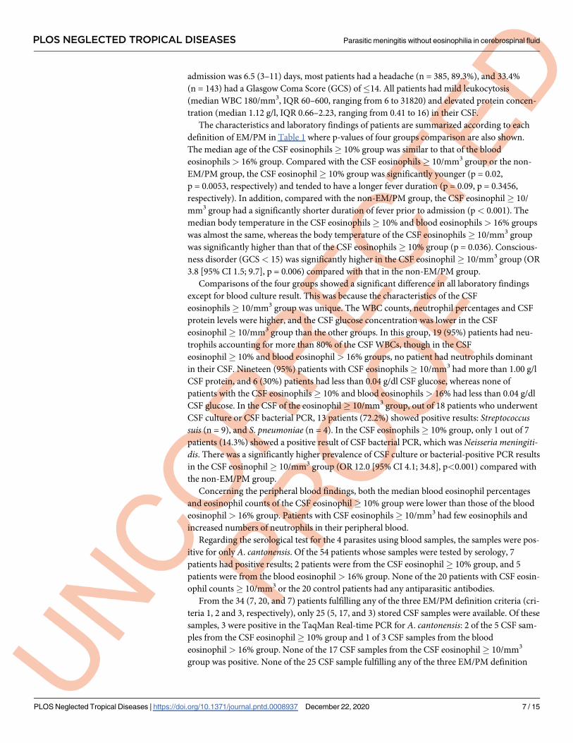
admission was 6.5 (3–11) days, most patients had a headache (n = 385, 89.3%), and 33.4%
(n = 143) had a Glasgow Coma Score (GCS) of�14. All patients had mild leukocytosis
(median WBC 180/mm3, IQR 60–600, ranging from 6 to 31820) and elevated protein concen-
tration (median 1.12 g/l, IQR 0.66–2.23, ranging from 0.41 to 16) in their CSF.
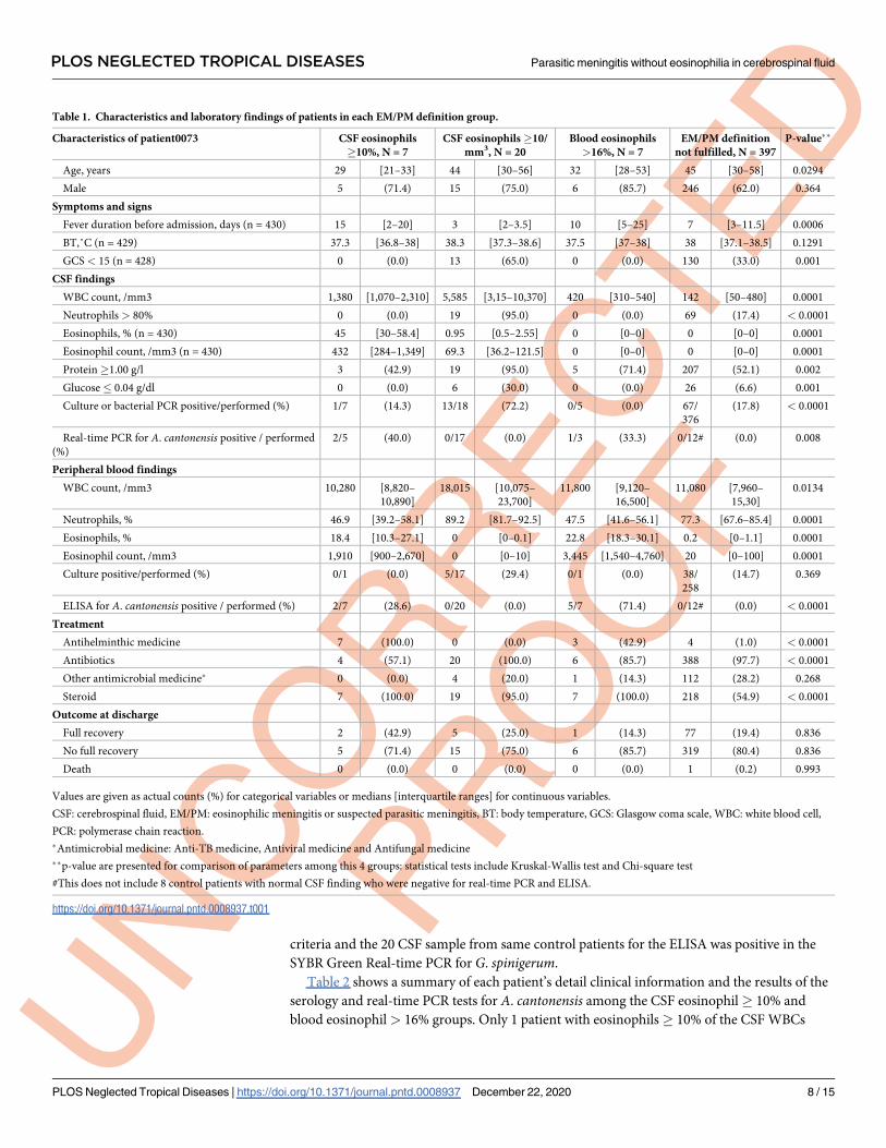
The characteristics and laboratory findings of patients are summarized according to each
definition of EM/PM in Table 1 where p-values of four groups comparison are also shown.
The median age of the CSF eosinophils� 10% group was similar to that of the blood
eosinophils > 16% group. Compared with the CSF eosinophils� 10/mm3 group or the non-
EM/PM group, the CSF eosinophil� 10% group was significantly younger (p = 0.02,
p = 0.0053, respectively) and tended to have a longer fever duration (p = 0.09, p = 0.3456,
respectively). In addition, compared with the non-EM/PM group, the CSF eosinophil� 10/
mm3 group had a significantly shorter duration of fever prior to admission (p< 0.001). The
median body temperature in the CSF eosinophils� 10% and blood eosinophils > 16% groups
was almost the same, whereas the body temperature of the CSF eosinophils� 10/mm3 group
was significantly higher than that of the CSF eosinophils� 10% group (p = 0.036). Conscious-
ness disorder (GCS < 15) was significantly higher in the CSF eosinophil� 10/mm3 group (OR
3.8 [95% CI 1.5; 9.7], p = 0.006) compared with that in the non-EM/PM group.
Comparisons of the four groups showed a significant difference in all laboratory findings
except for blood culture result. This was because the characteristics of the CSF
eosinophils� 10/mm3 group was unique. The WBC counts, neutrophil percentages and CSF
protein levels were higher, and the CSF glucose concentration was lower in the CSF
eosinophil� 10/mm3 group than the other groups. In this group, 19 (95%) patients had neu-
trophils accounting for more than 80% of the CSF WBCs, though in the CSF
eosinophil� 10% and blood eosinophil > 16% groups, no patient had neutrophils dominant
in their CSF. Nineteen (95%) patients with CSF eosinophils� 10/mm3 had more than 1.00 g/l
CSF protein, and 6 (30%) patients had less than 0.04 g/dl CSF glucose, whereas none of
patients with the CSF eosinophils� 10% and blood eosinophils > 16% had less than 0.04 g/dl
CSF glucose. In the CSF of the eosinophil� 10/mm3 group, out of 18 patients who underwent
CSF culture or CSF bacterial PCR, 13 patients (72.2%) showed positive results: Streptococcussuis (n = 9), and S. pneumoniae (n = 4). In the CSF eosinophils� 10% group, only 1 out of 7
patients (14.3%) showed a positive result of CSF bacterial PCR, which was Neisseria meningiti-dis. There was a significantly higher prevalence of CSF culture or bacterial-positive PCR results
in the CSF eosinophil� 10/mm3 group (OR 12.0 [95% CI 4.1; 34.8], p<0.001) compared with
the non-EM/PM group.
Concerning the peripheral blood findings, both the median blood eosinophil percentages
and eosinophil counts of the CSF eosinophil� 10% group were lower than those of the blood
eosinophil > 16% group. Patients with CSF eosinophils� 10/mm3 had few eosinophils and
increased numbers of neutrophils in their peripheral blood.
Regarding the serological test for the 4 parasites using blood samples, the samples were pos-
itive for only A. cantonensis. Of the 54 patients whose samples were tested by serology, 7
patients had positive results; 2 patients were from the CSF eosinophil� 10% group, and 5
patients were from the blood eosinophil > 16% group. None of the 20 patients with CSF eosin-
ophil counts� 10/mm3 or the 20 control patients had any antiparasitic antibodies.
From the 34 (7, 20, and 7) patients fulfilling any of the three EM/PM definition criteria (cri-
teria 1, 2 and 3, respectively), only 25 (5, 17, and 3) stored CSF samples were available. Of these
samples, 3 were positive in the TaqMan Real-time PCR for A. cantonensis: 2 of the 5 CSF sam-
ples from the CSF eosinophil� 10% group and 1 of 3 CSF samples from the blood
eosinophil > 16% group. None of the 17 CSF samples from the CSF eosinophil� 10/mm3
group was positive. None of the 25 CSF sample fulfilling any of the three EM/PM definition
PLOS NEGLECTED TROPICAL DISEASES Parasitic meningitis without eosinophilia in cerebrospinal fluid
PLOS Neglected Tropical Diseases | https://doi.org/10.1371/journal.pntd.0008937 December 22, 2020 7 / 15
criteria and the 20 CSF sample from same control patients for the ELISA was positive in the
SYBR Green Real-time PCR for G. spinigerum.
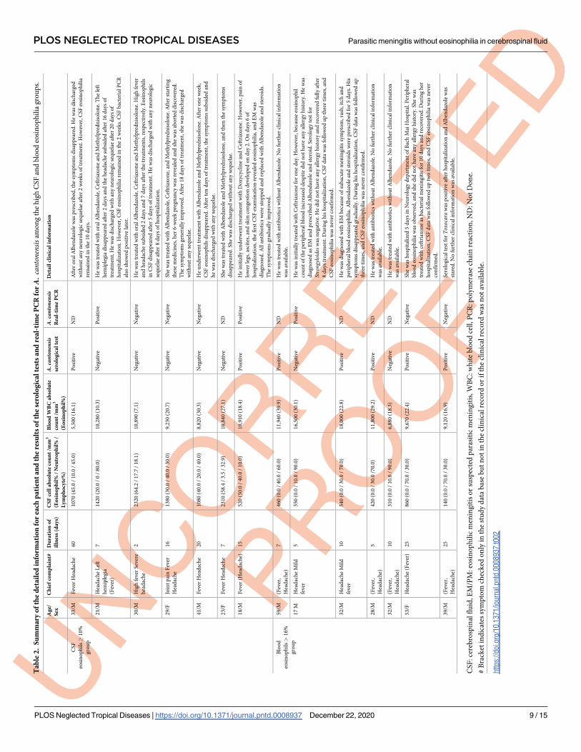
Table 2 shows a summary of each patient’s detail clinical information and the results of the
serology and real-time PCR tests for A. cantonensis among the CSF eosinophil� 10% and
blood eosinophil > 16% groups. Only 1 patient with eosinophils� 10% of the CSF WBCs
Table 1. Characteristics and laboratory findings of patients in each EM/PM definition group.
Characteristics of patient0073 CSF eosinophils
�10%, N = 7
CSF eosinophils �10/
mm3, N = 20
Blood eosinophils
>16%, N = 7
EM/PM definition
not fulfilled, N = 397
P-value��
Age, years 29 [21–33] 44 [30–56] 32 [28–53] 45 [30–58] 0.0294
Male 5 (71.4) 15 (75.0) 6 (85.7) 246 (62.0) 0.364
Symptoms and signs
Fever duration before admission, days (n = 430) 15 [2–20] 3 [2–3.5] 10 [5–25] 7 [3–11.5] 0.0006
BT,˚C (n = 429) 37.3 [36.8–38] 38.3 [37.3–38.6] 37.5 [37–38] 38 [37.1–38.5] 0.1291
GCS < 15 (n = 428) 0 (0.0) 13 (65.0) 0 (0.0) 130 (33.0) 0.001
CSF findings
WBC count, /mm3 1,380 [1,070–2,310] 5,585 [3,15–10,370] 420 [310–540] 142 [50–480] 0.0001
Neutrophils > 80% 0 (0.0) 19 (95.0) 0 (0.0) 69 (17.4) < 0.0001
Eosinophils, % (n = 430) 45 [30–58.4] 0.95 [0.5–2.55] 0 [0–0] 0 [0–0] 0.0001
Eosinophil count, /mm3 (n = 430) 432 [284–1,349] 69.3 [36.2–121.5] 0 [0–0] 0 [0–0] 0.0001
Protein�1.00 g/l 3 (42.9) 19 (95.0) 5 (71.4) 207 (52.1) 0.002
Glucose� 0.04 g/dl 0 (0.0) 6 (30.0) 0 (0.0) 26 (6.6) 0.001
Culture or bacterial PCR positive/performed (%) 1/7 (14.3) 13/18 (72.2) 0/5 (0.0) 67/
376
(17.8) < 0.0001
Real-time PCR for A. cantonensis positive / performed
(%)
2/5 (40.0) 0/17 (0.0) 1/3 (33.3) 0/12# (0.0) 0.008
Peripheral blood findings
WBC count, /mm3 10,280 [8,820–
10,890]
18,015 [10,075–
23,700]
11,800 [9,120–
16,500]
11,080 [7,960–
15,30]
0.0134
Neutrophils, % 46.9 [39.2–58.1] 89.2 [81.7–92.5] 47.5 [41.6–56.1] 77.3 [67.6–85.4] 0.0001
Eosinophils, % 18.4 [10.3–27.1] 0 [0–0.1] 22.8 [18.3–30.1] 0.2 [0–1.1] 0.0001
Eosinophil count, /mm3 1,910 [900–2,670] 0 [0–10] 3,445 [1,540–4,760] 20 [0–100] 0.0001
Culture positive/performed (%) 0/1 (0.0) 5/17 (29.4) 0/1 (0.0) 38/
258
(14.7) 0.369
ELISA for A. cantonensis positive / performed (%) 2/7 (28.6) 0/20 (0.0) 5/7 (71.4) 0/12# (0.0) < 0.0001
Treatment
Antihelminthic medicine 7 (100.0) 0 (0.0) 3 (42.9) 4 (1.0) < 0.0001
Antibiotics 4 (57.1) 20 (100.0) 6 (85.7) 388 (97.7) < 0.0001
Other antimicrobial medicine� 0 (0.0) 4 (20.0) 1 (14.3) 112 (28.2) 0.268
Steroid 7 (100.0) 19 (95.0) 7 (100.0) 218 (54.9) < 0.0001
Outcome at discharge
Full recovery 2 (42.9) 5 (25.0) 1 (14.3) 77 (19.4) 0.836
No full recovery 5 (71.4) 15 (75.0) 6 (85.7) 319 (80.4) 0.836
Death 0 (0.0) 0 (0.0) 0 (0.0) 1 (0.2) 0.993
Values are given as actual counts (%) for categorical variables or medians [interquartile ranges] for continuous variables.
CSF: cerebrospinal fluid, EM/PM: eosinophilic meningitis or suspected parasitic meningitis, BT: body temperature, GCS: Glasgow coma scale, WBC: white blood cell,
PCR: polymerase chain reaction.
�Antimicrobial medicine: Anti-TB medicine, Antiviral medicine and Antifungal medicine
��p-value are presented for comparison of parameters among this 4 groups: statistical tests include Kruskal-Wallis test and Chi-square test
#This does not include 8 control patients with normal CSF finding who were negative for real-time PCR and ELISA.
https://doi.org/10.1371/journal.pntd.0008937.t001
PLOS NEGLECTED TROPICAL DISEASES Parasitic meningitis without eosinophilia in cerebrospinal fluid
PLOS Neglected Tropical Diseases | https://doi.org/10.1371/journal.pntd.0008937 December 22, 2020 8 / 15
Ta
ble
2.
Su
mm
ary
of
the
det
ail
edin
form
ati
on
for
each
pa
tien
ta
nd
the
resu
lts
of
the
sero
log
ica
lte
sts
an
dre
al-
tim
eP
CR
forA
.ca
nton
ensis
amo
ng
the
hig
hC
SF
and
blo
od
eosi
no
ph
ilia
gro
up
s.
Ag
e/
Sex
Ch
ief
com
pla
int#
Du
rati
on
of
illn
ess
(da
ys)
CS
Fce
lla
bso
lute
cou
nt
/mm
3
(Eo
sin
op
hil
%/
Neu
tro
ph
il%
/
Ly
mp
ho
cyte
%)
Blo
od
WB
Ca
bso
lute
cou
nt
/mm
3
(Eo
sin
op
hil
%)
A.canton
ensis
sero
log
ica
lte
st
A.canton
ensis
Rea
l-ti
me
PC
R
Det
ail
clin
ica
lin
form
ati
on
CS
F
eosi
no
ph
ils�
10
%
gro
up
33
/MF
ever
Hea
dac
he
60
10
70
(45
.0/
10
.0/
45
.0)
5,5
00
(16
.1)
Po
siti
ve
ND
Aft
ero
ral
Alb
end
azo
lew
asp
resc
rib
ed,
the
sym
pto
ms
dis
app
eare
d.H
ew
asd
isch
arg
ed
wit
ho
ut
any
neu
rolo
gic
seq
uel
aeaf
ter
2w
eek
so
ftr
eatm
ent.
Ho
wev
er,C
SF
eosi
no
ph
ilia
rem
ain
edin
the
10
day
s.
21
/MH
ead
ach
eL
eft
hem
iple
gia
(Fev
er)
71
42
0(2
0.0
/0
/8
0.0
)1
0,2
80
(10
.3)
Neg
ativ
eP
osi
tive
He
was
trea
ted
wit
ho
ral
Alb
end
azo
le,C
eftr
iax
on
ean
dM
eth
ylp
red
nis
olo
ne.
Th
ele
ft
hem
iple
gia
dis
app
eare
daf
ter
2d
ays
and
the
hea
dac
he
sub
sid
edaf
ter
16
day
so
f
trea
tmen
t.H
ew
asd
isch
arg
edw
ith
any
neu
rolo
gic
seq
uel
aeaf
ter
20
day
so
f
ho
spit
aliz
atio
n.
Ho
wev
er,C
SF
eosi
no
ph
ilia
rem
ain
edin
the
2w
eek
s.C
SF
bac
teri
alP
CR
also
sho
wed
po
siti
ve
late
r.
30
/MH
igh
fev
erS
ever
e
hea
dac
he
22
32
0(6
4.2
/1
7.7
/1
8.1
)1
0,8
90
(7.1
)N
egat
ive
Neg
ativ
eH
ew
astr
eate
dw
ith
ora
lA
lben
daz
ole
,C
eftr
iax
on
ean
dM
eth
ylp
red
nis
olo
ne.
Hig
hfe
ver
and
hea
dac
he
sub
sid
ed2
day
san
d7
day
saf
ter
the
trea
tmen
ts,
resp
ecti
vel
y.E
osi
no
ph
ils
inC
SF
dis
app
eare
daf
ter
5d
ays
of
trea
tmen
t.H
ew
asd
isch
arg
edw
ith
any
neu
rolo
gic
seq
uel
aeaf
ter
8d
ays
of
ho
spit
aliz
atio
n.
29
/FJo
int
pai
nF
ever
Hea
dac
he
16
13
80
(30
.0/
40
.0/
30
.0)
9,2
30
(20
.7)
Neg
ativ
eN
egat
ive
Sh
ew
astr
eate
dw
ith
Alb
end
azo
le,C
eftr
iax
on
e,an
dM
eth
ylp
red
nis
olo
ne.
Aft
erst
arti
ng
thes
em
edic
ines
,h
er6
-wee
kp
reg
nan
cyw
asre
vea
led
and
she
was
abo
rted
dis
cov
ered
.
Th
esy
mp
tom
sg
ener
ally
imp
rov
ed.A
fter
18
day
so
ftr
eatm
ent,
she
was
dis
char
ged
wit
ho
ut
any
seq
uel
ae.
41
/MF
ever
Hea
dac
he
20
10
80
(40
.0/
20
.0/
40
.0)
8,8
20
(30
.3)
Neg
ativ
eN
egat
ive
He
un
der
wen
ttr
eatm
ent
wit
hA
lben
daz
ole
and
Met
hylp
red
nis
olo
ne.
Aft
ero
ne
wee
k,
CS
Feo
sin
op
hil
sd
isap
pea
red
.A
fter
ten
day
so
ftr
eatm
ent,
the
sym
pto
ms
sub
sid
edan
d
he
was
dis
char
ged
wit
han
yse
qu
elae
.
23
/FF
ever
Hea
dac
he
72
31
0(5
8.4
/3
.5/
32
.9)
10
,84
0(2
7.1
)N
egat
ive
ND
Sh
ew
astr
eate
dw
ith
Alb
end
azo
lean
dM
eth
ylp
red
nis
olo
ne,
and
then
the
sym
pto
ms
dis
app
eare
d.
Sh
ew
asd
isch
arg
edw
ith
ou
tan
yse
qu
elae
.
18
/MF
ever
(Hea
dac
he)
15
52
0(5
0.0
/4
0.0
/1
0.0
)1
0,9
10
(18
.4)
Po
siti
ve
Po
siti
ve
He
init
iall
yu
nd
erw
ent
trea
tmen
tw
ith
Do
xycy
clin
ean
dC
eftr
iax
on
e.H
ow
ever
,p
ain
of
low
erle
gs,
asci
tes,
and
skin
con
ges
tio
nd
evel
op
edo
nd
ay2
.O
nd
ays
6o
f
ho
spit
aliz
atio
n,
the
firs
tC
SF
exam
inat
ion
revea
led
eosi
no
ph
ilia
,an
dE
Mw
as
dia
gn
ose
d.
All
anti
bio
tics
wer
est
op
ped
and
rep
lace
dw
ith
Alb
end
azo
lean
dst
ero
ids.
Th
esy
mp
tom
sg
rad
ual
lyim
pro
ved
.
Blo
od
eosi
no
ph
ils>
16
%
gro
up
59
/M(F
ever
,
Hea
dac
he)
74
60
(0.0
/4
0.0
/6
0.0
)1
1,9
40
(39
.9)
Po
siti
ve
ND
He
was
trea
ted
wit
han
tib
ioti
csw
ith
ou
tA
lben
daz
ole
.N
ofu
rth
ercl
inic
alin
form
atio
n
was
avai
lab
le.
17
MH
ead
ach
eM
ild
fev
er
53
50
(0.0
/1
0.0
/9
0.0
)1
6,5
00
(30
.1)
Neg
ativ
eP
osi
tive
He
was
init
iall
ytr
eate
dw
ith
Cef
tria
xo
ne
for
on
ed
ay.H
ow
ever
,b
ecau
seeo
sin
op
hil
cou
nt
of
the
per
iph
eral
blo
od
incr
ease
dd
esp
ite
did
no
th
ave
any
alle
rgy
his
tory
.H
ew
as
dia
gn
ose
das
EM
and
pre
scri
bed
Alb
end
azo
lean
dst
ero
id.
Ser
olo
gy
test
for
Stro
ngyl
oide
swas
neg
ativ
e.H
ed
idn
ot
hav
ean
yal
lerg
yh
isto
ryan
dre
cover
edfu
lly
afte
r
8d
ays
trea
tmen
t.D
uri
ng
his
ho
spit
aliz
atio
n,C
SF
dat
aw
asfo
llo
wed
up
thre
eti
mes
,an
d
CS
Feo
sin
op
hil
iaw
asn
ever
con
firm
ed.
32
/MH
ead
ach
eM
ild
fev
er
10
54
0(0
.0/
30
.0/
70
.0)
18
,00
0(2
2.8
)P
osi
tive
ND
He
was
dia
gn
ose
dw
ith
EM
bec
ause
of
sub
acu
tem
enin
git
issy
mp
tom
,ra
sh,it
chan
d
per
iph
eral
blo
od
eosi
no
ph
ilia
.A
lben
daz
ole
and
ster
oid
sw
ere
pre
scri
bed
for
5d
ays.
His
sym
pto
ms
dis
app
eare
dg
rad
ual
ly.
Du
rin
gh
ish
osp
ital
izat
ion
,C
SF
dat
aw
asfo
llo
wed
up
thre
eti
mes
,an
dC
SF
eosi
no
ph
ilia
was
nev
erco
nfi
rmed
.
28
/M(F
ever
,
Hea
dac
he)
54
20
(0.0
/3
0.0
/70
.0)
11
,80
0(2
9.2
)P
osi
tiv
eN
DH
ew
astr
eate
dw
ith
anti
bio
tics
wit
ho
ut
Alb
end
azo
le.N
ofu
rth
ercl
inic
alin
form
atio
n
was
avai
lab
le.
32
/M(F
ever
,
Hea
dac
he)
10
31
0(0
.0/
10
.0/
90
.0)
6,8
80
(18
.3)
Neg
ativ
eN
DH
ew
astr
eate
dw
ith
anti
bio
tics
wit
ho
ut
Alb
end
azo
le.N
ofu
rth
ercl
inic
alin
form
atio
n
was
avai
lab
le.
53
/FH
ead
ach
e(F
ever
)2
58
60
(0.0
/7
0.0
/3
0.0
)9
,67
0(2
2.4
)P
osi
tiv
eN
egat
ive
Sh
ew
ash
osp
ital
ized
5d
ays
inN
euro
log
yd
epar
tmen
to
fB
ach
Mai
Ho
spit
al.
Per
iph
eral
blo
od
eosi
no
ph
ilia
was
ob
serv
ed,
and
she
did
no
th
ave
any
alle
rgy
his
tory
.S
he
was
trea
ted
wit
hce
ftri
axo
ne
asb
acte
rial
men
ing
itis
for
10
day
san
dre
cover
ed.D
uri
ng
her
ho
spit
aliz
atio
n,
CS
Fd
ata
was
foll
ow
edu
ptw
oti
mes
,an
dC
SF
eosi
no
ph
ilia
was
nev
er
con
firm
ed.
39
/M(F
ever
,
Hea
dac
he)
25
14
0(0
.0/
70
.0/
30
.0)
9,1
20
(16
.9)
Po
siti
ve
Neg
ativ
eS
ero
log
ical
test
for
Toxo
cara
was
po
siti
ve
afte
rh
osp
ital
izat
ion
and
Alb
end
azo
lew
as
star
ed.N
ofu
rth
ercl
inic
alin
form
atio
nw
asav
aila
ble
.
CS
F:ce
reb
rosp
inal
flu
id,E
M/P
M:eo
sin
op
hil
icm
enin
git
iso
rsu
spec
ted
par
asit
icm
enin
git
is,
WB
C:w
hit
eb
loo
dce
ll,P
CR
:p
oly
mer
ase
chai
nre
acti
on
,N
D:N
ot
Do
ne.
#B
rack
etin
dic
ates
sym
pto
mch
eck
edo
nly
inth
est
ud
yd
ata
bas
eb
ut
no
tin
the
clin
ical
reco
rdo
rif
the
clin
ical
reco
rdw
asn
ot
avai
lab
le.
htt
ps:
//doi.o
rg/1
0.1
371/jo
urn
al.p
ntd
.0008937.t002
PLOS NEGLECTED TROPICAL DISEASES Parasitic meningitis without eosinophilia in cerebrospinal fluid
PLOS Neglected Tropical Diseases | https://doi.org/10.1371/journal.pntd.0008937 December 22, 2020 9 / 15
showed both positive serology and real-time PCR results for A. cantonensis. The other 2 real-
time PCR-positive patients had negative serological tests.
Discussion
This prospective descriptive study included all patients with a suspected CNS infection at the
largest referral medical center in Hanoi, northern Vietnam. This is the first study focusing on
the implication of various EM definitions in the context of diagnosing PM. Our results indicate
that the characteristics of patients with CSF eosinophil counts� 10/mm3 but CSF eosinophil
percentages < 10% were consistent with those of bacterial meningitis patients. Further sero-
logical and real-time PCR results indicated that there might be a non-negligible number of
patients with PM without eosinophils in the CSF or fulfilling any of the previously defined EM
criteria.
We defined these EM/PM criteria because first, the criteria of CSF eosinophils� 10% was
most commonly used, followed by the absolute number of eosinophils� 10/mm3, and by
peripheral blood eosinophils >16% in previous publications [9,8,15]. Second, we followed
clinical diagnosis applied by the local clinicians at Bach Mai Hospital.
In our study, the prevalence of EM patients with CSF eosinophils accounting for� 10% of
the WBC count was 1.03% among total patients with a suspected CNS infection. This preva-
lence is higher than that previously reported in the southern and middle regions of Vietnam,
which was 0.6% among 1241 CNS-infection patients [22] and 0.69% among 1000 CNS-infec-
tion patients aged� 15 years without HIV [23], respectively. The prevalence of EM patients
with CSF eosinophils� 10% of the WBCs in our study was lower than that of the first report
from northern Vietnam, which was 1.42% among 352 CNS-infection patients [24]. However,
in this report, the definition of EM included the presence of� 10 eosinophils/mm3 in addition
to eosinophilia� 10% of the WBCs in the CSF. If the same definition was applied herein, the
prevalence would be 3.98%. These previous studies did not specifically discuss the differences
in patient characteristics according to each EM definition.
In endemic areas, the majority of patients fulfilling the EM criteria are more likely to have a
parasitic infection [1,4,5]. However, our results show that patients with eosinophils� 10 cells/
mm3 but not� 10% of the WBCs in the CSF were more likely than those in the other groups
to have bacterial meningitis because their clinical characteristics tended to present as acute,
associated with a reduced level of consciousness, an increased number of neutrophils, an
increased level of protein and a reduced level of glucose in the CSF. Therefore, the eosinophils
�10 cells/mm3 criterion should be carefully interpreted. In fact, this EM criterion was not
included in the diagnosis of EM caused by parasites in recent papers from Thailand, Vietnam
and Laos [11–13,16,22].
Intriguingly, the clinical characteristics and laboratory results of patients with blood eosino-
phils accounting for> 16% of the WBCs were similar to those of EM patients with CSF eosino-
phils accounting for� 10% of the WBCs, with the exception of CSF eosinophilia. None of the
patients in this group had any eosinophils in their CSF, though they had an abnormally high
number of cells in the CSF with a median of 420 (IQR 310–540) cells/mm3, which were pre-
dominantly neutrophils (n = 2) or lymphocytes (n = 5).
Our serological results identified 7 patients with antibodies against A. cantonensis: 2
(28.6%) patients from the CSF eosinophils� 10% group and 5 (71.4%) patients from the blood
eosinophils > 16% without CSF eosinophils group. In contrast, none of the 20 patients with
CSF eosinophil counts� 10/mm3 nor the 20 control patients had any anti-parasite antibodies.
Subsequent real-time PCR analyses identified 3 patients positive for A. cantonensis and no
patient positive for G. spinigerum, indicating that EM in these patients was due to A.
PLOS NEGLECTED TROPICAL DISEASES Parasitic meningitis without eosinophilia in cerebrospinal fluid
PLOS Neglected Tropical Diseases | https://doi.org/10.1371/journal.pntd.0008937 December 22, 2020 10 / 15
cantonensis. Interestingly, one of the PCR-positive patients had blood eosinophils > 16% of
the WBCs without CSF eosinophils. This finding, together with the highest seroprevalence
among patients with blood eosinophils > 16% and their clinical characteristics being compati-
ble with a parasitic infection, raises the hypothesis that patients in this group may have genuine
PM. To date, many papers have reported that patients with A. cantonensis-induced EM have
blood eosinophilia [2,10,20], and in a study setting where lumbar puncture was difficult to per-
form, blood eosinophilia alone was used to clinically diagnose patients with A. cantonensisinfection-induced PM [14]. However, peripheral eosinophilia and serological tests should be
cautiously used as a definitive evidence of PM. There have been no reports attempting to con-
firm A. cantonensis infection in clinically suspected patients with blood eosinophilia using
real-time PCR. According to the previously published study [10,14], exposure history to A.
cantonensis is an important clue to diagnose angiostrongyliasis. However, none of our patients
positive for serology or real-time PCR mentioned exposure history.
According to a recent study of the pharmacodynamic effects of helminth-derived molecules
using mouse models and soluble antigens of A. cantonensis larvae, an increase in blood eosino-
phil percentage was found to precede the CSF eosinophil percentage increase in mice, which
are nonpermissive hosts, with a 14-day lag [25]. Although there has been no report describing
exactly when CSF and blood eosinophil numbers begin to increase after infection with A. can-tonensis in humans, it is plausible that there might be a lag between CSF and peripheral blood
eosinophil responses among PM patients. In fact, the presence of low percentage or no eosino-
phils in the CSF during early stage of angiostrongyliasis has been previously reported [10,26].
Trevor J. Slom et al. reported that among 9 hospitalized patients with suspected EM caused by
A. cantonensis, only 5 had CSF eosinophilia in the initial lumbar puncture [26], and 8 of the 9
patients finally exhibited CSF eosinophilia after hospitalization, although detailed changes in
the patient blood eosinophil counts were not clearly reported. Furthermore, there has been a
case report about a pediatric patient with A. cantonensis-induced EM within the USA [27]. His
first CSF sample showed a WBC count of 763/mm3 with 5% eosinophils, but later his CSF
showed high eosinophilia, reaching 21% of the CSF WBCs. However, to our knowledge, no
study has performed both PCR and serology tests for diagnosing A. cantonensis on multiple
meningitis patients without CSF eosinophils. In our study patients, three, whose CSF data
were followed up, did not show CSF eosinophilia despite multiple lumbar punctures. However,
at least 3 patients were prescribed albendazole during admission and reported significant clini-
cal improvements, such as a reduction in headache severity.
The sensitivity and specificity of the current A. cantonensis ELISA and real-time PCR was
not yet established. Development of serological diagnosis of helminth infection remains diffi-
cult. Several studies attempted to establish serological tests to diagnose A. cantonensis infection
[28]. However, it is challenging to standardize parasitic meningitis with A. cantonesis because
the presence of parasite bodies cannot be demonstrated in the majority of cases thus in most
studies, positive cases were indirectly diagnosed by clinical symptoms and clinical histories
[29–31]. Similarly, none of previously published studies with PCR have shown reliable data of
sensitivity and specificity of PCR. McBride A et al. reported that 37 CSF samples (67.8%) was
positive among 57 CSF samples of patients with CSF eosinophils� 10%, using the same real-
time PCR assay [23]. But this study did not show the result of non-EM/PM patients.
The main limitation of this study is that our study population biased toward febrile patients
since the inclusion criteria included a history of fever anytime from onset to admission. Never-
theless, this was necessary in the current study as it primarily aimed to provide useful informa-
tion to clinicians working in infectious disease wards to improve clinical judgment and
management of patients suspected with meningitis. The latest literature review reported that
around 45% of adult patients with EM/PM are afebrile [32], therefore our study population
PLOS NEGLECTED TROPICAL DISEASES Parasitic meningitis without eosinophilia in cerebrospinal fluid
PLOS Neglected Tropical Diseases | https://doi.org/10.1371/journal.pntd.0008937 December 22, 2020 11 / 15
should be missing afebrile PM who did not have fever at any time point from onset to admis-
sion, and our findings be interpreted with caution and cannot be generalized to all patients
with EM/PM. A community-based study approaching mildly symptomatic but infected indi-
viduals in the highly endemic area, and/or a hospital-based study including patients in the
neurology department are warranted to reveal the whole clinical picture of PM/EM.
Our study has several other limitations. First, this was a single referral hospital-based study
with a limited number of EM/PM patients. We could not have sufficient statistical power to
demonstrate the significance of clinical features and sensitivity and specificity of the real-time
PCR. However, Bach Mai Hospital is the biggest tertiary hospital in northern Vietnam and our
study patients were identified as a consequence of screening a substantial number of patients
with suspected CNS infections and CSF results. Collaborative studies are necessary for further
investigation of such a rare clinical syndrome. A community-based study could provide a
broader perspective on the epidemiology of EM/PM and a different prevalence of EM/PM.
Since lumbar puncture was not required as a routine investigation for all patients with a head-
ache, many mild cases of EM/PM may be treated as a nonspecific headache. Second, while gen-
eral information was prospectively collected, the detailed clinical information of EM/PM
patients was collected retrospectively from medical records. In particular, the information of
three patients with high blood eosinophilia but CSF eosinophil percentage <10% was missing.
Third, we used a serological test for A. cantonensis identification, which has not been validated
in the context of northern Vietnamese population. We do not fully know the baseline sero-
prevalence of people at a various degree of high risk of exposure to these parasites thus we
need carefully interpret the positive results with this serology test. Forth, it is possible that
patients with negative results in the real-time PCR or serological tests may have had CSF eosin-
ophilia due to other infectious or noninfectious causes, which we did not aggressively investi-
gate. However, regarding noninfectious causes, among the patients with CSF
eosinophils� 10% or blood eosinophils > 16% patients, any drug allergy was not confirmed,
and neoplasms were unlikely because of their long-term clinical courses. Tuberculosis may
also cause EM or even blood eosinophilia but none of EM/PM suspected patients was diag-
nosed as tuberculous meningitis as defined by PCR test with their CSF samples. One of
patients with blood eosinophils > 16% group was positive Toxocara spp. serology in Bach Mai
Hospital, whose serological test for A. cantonensis was also positive in this study. There is also
a possibility of G. spinigerum infection because its infection in swamp eels and in human at
Vietnam were reported [33,34] and this area is included in an endemic area of this parasite.
However, none of patients with CSF eosinophil� 10% or blood eosinophil > 16% accompa-
nied its typical symptoms, such as swelling of the limbs, the trunk or the face with migration.
None of their real-time PCR for G. spinigerum was positive. Furthermore, a possibility of false
negative result of the real-time PCR test for A. cantonensis cannot be excluded due to insuffi-
cient sensitivity.
In conclusion, in northern Vietnam, the prevalence of EM was 1.03% among patients with
a history of fever, suspected of having CNS infection if the definition of EM is
eosinophils� 10% of CSF WBC. Patients with CSF eosinophils� 10/mm3 without high CSF
eosinophilia more than 10% can be bacterial meningitis. Therefore, the percentage is more
reliable than the absolute eosinophil count in the CSF for predicting PM. Despite the lack of
exposure history to the parasite, clinical features, serology and PCR suggest that the most likely
etiology of EM seems to be A. cantonensis in this area. Our results re-confirmed previously
reported findings that PM due to A. cantonensis infection may have CSF eosinophils less than
10% or even subsequent CSF eosinophils with no CSF eosinophils at the beginning. Diagnos-
ing PM is challenging.
PLOS NEGLECTED TROPICAL DISEASES Parasitic meningitis without eosinophilia in cerebrospinal fluid
PLOS Neglected Tropical Diseases | https://doi.org/10.1371/journal.pntd.0008937 December 22, 2020 12 / 15
Supporting information
S1 Strobe Checklist.
(DOC)
S1 Fig. Relationships with each definitions of EM/PM case.
(TIF)
Acknowledgments
The authors would like to thank Dr. Poom Adisakwattana, Department of Helminthology,
Mahidol University for providing DNA of G. spinigerum as a positive control of the real-time
PCR.
Author Contributions
Conceptualization: Tomoko Hiraoka, Sugihiro Hamaguchi, Shungo Katoh, Koya Ariyoshi.
Data curation: Tomoko Hiraoka, Ngo Chi Cuong, Shungo Katoh, Le Kim Anh.
Formal analysis: Tomoko Hiraoka.
Funding acquisition: Lay-Myint Yoshida, Koya Ariyoshi.
Investigation: Tomoko Hiraoka, Ngo Chi Cuong, Sugihiro Hamaguchi, Le Kim Anh, Nguyen
Thi Hien Anh.
Methodology: Tomoko Hiraoka, Mihoko Kikuchi, Haruhiko Maruyama.
Project administration: Ngo Chi Cuong, Le Kim Anh, Dang Duc Anh, Lay-Myint Yoshida,
Do Duy Cuong, Pham Thanh Thuy, Koya Ariyoshi.
Resources: Le Kim Anh, Nguyen Thi Hien Anh, Dang Duc Anh, Lay-Myint Yoshida, Do Duy
Cuong.
Supervision: Dang Duc Anh, Chris Smith, Do Duy Cuong, Pham Thanh Thuy.
Validation: Haruhiko Maruyama.
Visualization: Tomoko Hiraoka, Mihoko Kikuchi.
Writing – original draft: Tomoko Hiraoka.
Writing – review & editing: Tomoko Hiraoka, Sugihiro Hamaguchi, Mihoko Kikuchi,
Shungo Katoh, Chris Smith, Koya Ariyoshi.
References1. Weller PF. Eosinophilic meningitis. Am J Med. 1993; 95: 250–253. https://doi.org/10.1016/0002-9343
(93)90275-t PMID: 8368222
2. Lo RV, Gluckman SJ. Eosinophilic meningitis. Am J Med. 2003; 114: 217–223. https://doi.org/10.1016/
s0002-9343(02)01495-x PMID: 12637136
3. Beaver PC, Rosen L. Memorandum on the first report of angiostrongylus in man, by nomura and lin,
1945. Am J Trop Med Hyg. 1964; 13: 589–590. https://doi.org/10.4269/ajtmh.1964.13.589 PMID:
14196058
4. Wang QP, Lai DH, Zhu XQ, Chen XG, Lun ZR. Human angiostrongyliasis. Lancet Infect Dis. 2008; 8:
621–630. https://doi.org/10.1016/S1473-3099(08)70229-9 PMID: 18922484
5. Hughes PA, Magnet AD, Fishbain JT. Eosinophilic meningitis: a case series report and review of the lit-
erature. Mil Med. 2003; 168: 817–821. PMID: 14620646
PLOS NEGLECTED TROPICAL DISEASES Parasitic meningitis without eosinophilia in cerebrospinal fluid
PLOS Neglected Tropical Diseases | https://doi.org/10.1371/journal.pntd.0008937 December 22, 2020 13 / 15
6. Hochberg NS, Park SY, Blackburn BG, Sejvar JJ, Gaynor K, Chung H, et al. Distribution of eosinophilic
meningitis cases attributable to Angiostrongylus cantonensis, Hawaii. Emerg Infect Dis. 2007; 13:
1675–1680. https://doi.org/10.3201/eid1311.070367 PMID: 18217550
7. Sawanyawisuth K, Chotmongkol V. Eosinophilic meningitis. Handb Clin Neurol. 2013; 114: 207–215.
https://doi.org/10.1016/B978-0-444-53490-3.00015-7 PMID: 23829911
8. Kuberski T. Eosinophils in cerebrospinal fluid: criteria for eosinophilic meningitis. Hawaii Med J. 1981;
40: 97–98. PMID: 7251346
9. Punyagupta S, Bunnag T, Juttijudata P, Rosen L. Eosinophilic meningitis in Thailand. Epidemiologic
studies of 484 typical cases and the etiologic role of Angiostrongylus cantonensis. Am J Trop Med Hyg.
1970; 19: 950–958. PMID: 5531201
10. Punyagupta S, Juttijudata P, Bunnag T. Eosinophilic meningitis in Thailand. Clinical studies of 484 typi-
cal cases probably caused by Angiostrongylus cantonensis. Am J Trop Med Hyg. 1975; 24: 921–931.
PMID: 1200257
11. Schmutzhard E, Boongird P, Vejjajiva A. Eosinophilic meningitis and radiculomyelitis in Thailand,
caused by CNS invasion of Gnathostoma spinigerum and Angiostrongylus cantonensis. J Neurol Neu-
rosurg Psychiatry. 1988; 51: 80–87. https://doi.org/10.1136/jnnp.51.1.80 PMID: 3351533
12. Chotmongkol V, Sawanyawisuth K, Thavornpitak Y. Corticosteroid treatment of eosinophilic meningitis.
Clin Infect Dis. 2000; 31: 660–662. https://doi.org/10.1086/314036 PMID: 11017811
13. Jitpimolmard S, Sawanyawisuth K, Morakote N, Vejjajiva A, Puntumetakul M, Sanchaisuriya K, et al.
Albendazole therapy for eosinophilic meningitis caused by Angiostrongylus cantonensis. Parasitol Res.
2007; 100: 1293–1296. https://doi.org/10.1007/s00436-006-0405-7 PMID: 17177056
14. Sawanyawisuth K, Sawanyawisuth K, Senthong V, Limpawattana P, Intapan PM, Tiamkao S, et al.
Peripheral eosinophilia as an indicator of meningitic angiostrongyliasis in exposed individuals. Mem Inst
Oswaldo Cruz. 2010; 105: 942–944. https://doi.org/10.1590/s0074-02762010000700020 PMID:
21120370
15. Schulte C, Krebs B, Jelinek T, Nothdurft HD, von Sonnenburg F, Loscher T. Diagnostic significance of
blood eosinophilia in returning travelers. Clin Infect Dis. 2002; 34: 407–411. https://doi.org/10.1086/
338026 PMID: 11753824
16. Ming DKY, Rattanavong S, Bharucha T, Sengvilaipaseuth O, Dubot-Perès A, Newton PN, et al. Angios-
trongylus cantonensis DNA in cerebrospinal fluid of persons with eosinophilic meningitis, Laos. Emerg
Infect Dis. 2017; 23: 2112–2113. https://doi.org/10.3201/eid2312.171107 PMID: 29148389
17. Katoh Shungo, Ngo Chi Cuong Sugihiro Hamaguchi, Pham Thanh Thuy Do Duy Cuong, Le Kim Anh,
et al. Challenges in diagnosing svrub typhus among hospitalized patients with undifferentiated fever at
a national tertiary hospital in northern Vietnam. PLoS Negl Trop Dis. 2019 Dec 5; 13(12):e0007928.
https://doi.org/10.1371/journal.pntd.0007928 PMID: 31805053
18. Cross JH, Chi JC. ELISA for the detection of Angiostrongylus cantonensis antibodies in patients with
eosinophilic meningitis. Southeast Asian J Trop Med Public Health. 1982 Mar; 13(1):73–76 PMID:
7051339
19. Nuamtanong S. The evaluation of the 29 and 31 kDa antigens in female Angiostrongylus cantonensis
for serodiagnosis of human angiostrongyliasis. Southeast Asian J Trop Med Public Health. 1996 Jun;
27(2):291–296 PMID: 9279992
20. Qvarnstrom Y, Xayavong M, da Silva AC, Park SY, Whelen AC, Calimlim PS, et al. Real-time polymer-
ase chain reaction detection of Angiostrongylus cantonensis DNA in cerebrospinal fluid from patients
with eosinophilic meningitis. Am J Trop Med Hyg. 2016; 94: 176–181. https://doi.org/10.4269/ajtmh.15-
0146 PMID: 26526920
21. Qvarnstrom Y, da Silva AC, Teem JL, Hollingsworth R, Bishop H, Graeff-Teixeira C, et al. Improved
molecular detection of Angiostrongylus cantonensis in mollusks and other environmental samples with
a species-specific internal transcribed spacer 1-based TaqMan assay. Appl Environ Microbiol. 2010;
76: 5287–5289. https://doi.org/10.1128/AEM.00546-10 PMID: 20543049
22. Trung NHD, Phuong TLT, Wolbers M, van Minh HN, Thanh VN, Van MP, et al. Aetiologies of central
nervous system infection in Viet Nam: a prospective provincial hospital-based descriptive surveillance
study. PLoS One. 2012; 7: e37825. https://doi.org/10.1371/journal.pone.0037825 PMID: 22662232
23. McBride A, Chau TTH, Hong NTT, Mai NTH, Anh NT, Thanh TT, et al. Angiostrongylus cantonensis is
an important cause of eosinophilic meningitis in Southern Vietnam. Clin Infect Dis. 2017; 64: 1784–
1787. https://doi.org/10.1093/cid/cix118 PMID: 28158507
24. Taylor WR, Nguyen K, Nguyen D, Nguyen H, Horby P, Nguyen HL, et al. The spectrum of central ner-
vous system infections in an adult referral hospital in Hanoi, Vietnam. PLoS One. 2012; 7: e42099.
https://doi.org/10.1371/journal.pone.0042099 PMID: 22952590
PLOS NEGLECTED TROPICAL DISEASES Parasitic meningitis without eosinophilia in cerebrospinal fluid
PLOS Neglected Tropical Diseases | https://doi.org/10.1371/journal.pntd.0008937 December 22, 2020 14 / 15
25. Wan S, Sun X, Wu F, Yu Z, Wang L, Lin D, et al. Chi3l3: a potential key orchestrator of eosinophil
recruitment in meningitis induced by Angiostrongylus cantonensis. J Neuroinflammation. 2018; 15: 1–
31. https://doi.org/10.1186/s12974-017-1027-y PMID: 29301548
26. Slom TJ, Cortese MM, Gerber SI, Jones RC, Holtz TH, Lopez AS, et al. An outbreak of eosinophilic
meningitis caused by Angiostrongylus cantonensis in travelers returning from the Caribbean. N Engl J
Med. 2002; 346: 668–675. https://doi.org/10.1056/NEJMoa012462 PMID: 11870244
27. Thyssen A, Mitchell M, Qvarnstrom Y, Rao S, Benke TA, Glode MP. Eosinophilic meningitis in a previ-
ously healthy 13-year-old child. Pediatr Infect Dis J. 2013; 32: 194–198. https://doi.org/10.1097/INF.
0b013e31827c9726 PMID: 23328824
28. Barratt J, Chan D, Sandaradura I, Malik R, Spielman D, Lee R, et al. Angiostrongylus cantonensis: a
review of its distribution, molecular biology and clinical significance as a human pathogen. Parasitology.
2016 Aug; 143(9):1087–118. https://doi.org/10.1017/S0031182016000652 Epub 2016 May 26. PMID:
27225800
29. Chen JX, Chen MX, Ai L, Chen JH, Chen SH, Zhang YN, et al. A protein microarray for the rapid screen-
ing of patients suspected of infection with various food-borne helminthiases. PLoS Negl Trop Dis. 2012;
6(11):e1899. https://doi.org/10.1371/journal.pntd.0001899 Epub 2012 Nov 29. PMID: 23209851
30. Intapan PM, Maleewong W, Polsan Y, Sawanyawisuth K, Chotmongkol V. Specific IgG antibody sub-
classes to Angiostrongylus cantonensis in patients with angiostrongyliasis. Asian Pac J Allergy Immu-
nol. 2002 Dec; 20(4):235–40. PMID: 12744624
31. Chen MX, Chen JX, Chen SH, Huang DN, Ai L, Zhang RL. Development of Lateral Flow Immunoassay
for Antigen Detection in Human Angiostrongylus cantonensis Infection. Korean J Parasitol. 2016 Jun;
54(3):375–80. https://doi.org/10.3347/kjp.2016.54.3.375 Epub 2016 Jun 30. PMID: 27417097
32. McAuliffe L, Fortin Ensign S, Larson D, Bavaro M, Yetto J, Cathey M, et al. Severe CNS angiostrongy-
liasis in a young marine: a case report and literature review. Lancet Infect Dis. 2019 Apr; 19(4):e132–
e142. https://doi.org/10.1016/S1473-3099(18)30434-1 Epub 2018 Nov 16. PMID: 30454904
33. Sieu TP, Dung TT, Nga NT, Hien TV, Dalsgaard A, Waikagul J, Murrell KD. Prevalence of Gnathostoma
spinigerum infection in wild and cultured swamp eels in Vietnam. J Parasitol. 2009 Feb; 95(1):246–8.
https://doi.org/10.1645/GE-1586.1 PMID: 19245276
34. Sakamoto M, Sato F, Mizuno Y, Komatsuzaki M, Yoshikawa K, Yoshida M, et al. [Gnathostomiasis
caused by Gnathostoma spinigerum etiologically diagnosed upon extraction of the worm from the skin].
Kansenshogaku Zasshi. 2004 May; 78(5):442–5. Japanese. https://doi.org/10.11150/
kansenshogakuzasshi1970.78.442 PMID: 15211867
PLOS NEGLECTED TROPICAL DISEASES Parasitic meningitis without eosinophilia in cerebrospinal fluid
PLOS Neglected Tropical Diseases | https://doi.org/10.1371/journal.pntd.0008937 December 22, 2020 15 / 15