Embed Size (px)
Citation preview
General rights Copyright and moral rights for the publications made accessible in the public portal are retained by the authors and/or other copyright owners and it is a condition of accessing publications that users recognise and abide by the legal requirements associated with these rights.
Users may download and print one copy of any publication from the public portal for the purpose of private study or research.
You may not further distribute the material or use it for any profit-making activity or commercial gain
You may freely distribute the URL identifying the publication in the public portal If you believe that this document breaches copyright please contact us providing details, and we will remove access to the work immediately and investigate your claim.
Downloaded from orbit.dtu.dk on: Apr 04, 2019
Automated Solid-Phase Subcloning Based on Beads Brought into Proximity byMagnetic Force
Hudson, Elton P.; Nikoshkov, Andrej; Uhlén, Mathias; Rockberg, Johan
Published in:P L o S One
Link to article, DOI:10.1371/journal.pone.0037429
Publication date:2012
Document VersionPublisher's PDF, also known as Version of record
Link back to DTU Orbit
Citation (APA):Hudson, E. P., Nikoshkov, A., Uhlén, M., & Rockberg, J. (2012). Automated Solid-Phase Subcloning Based onBeads Brought into Proximity by Magnetic Force. P L o S One, 7(5), -. DOI: 10.1371/journal.pone.0037429
Automated Solid-Phase Subcloning Based on BeadsBrought into Proximity by Magnetic ForceElton P. Hudson1, Andrej Nikoshkov1, Mathias Uhlen1,2,3, Johan Rockberg1*
1 School of Biotechnology, KTH – Royal Institute of Technology, Stockholm, Sweden, 2 Science for Life Laboratory, KTH – Royal Institute of Technology, Stockholm,
Sweden, 3 The Novo Nordisk Foundation Center for Biosustainability, Technical University of Denmark, Hørsholm, Denmark
Abstract
In the fields of proteomics, metabolic engineering and synthetic biology there is a need for high-throughput and reliablecloning methods to facilitate construction of expression vectors and genetic pathways. Here, we describe a new approachfor solid-phase cloning in which both the vector and the gene are immobilized to separate paramagnetic beads andbrought into proximity by magnetic force. Ligation events were directly evaluated using fluorescent-based microscopy andflow cytometry. The highest ligation efficiencies were obtained when gene- and vector-coated beads were brought intoclose contact by application of a magnet during the ligation step. An automated procedure was developed usinga laboratory workstation to transfer genes into various expression vectors and more than 95% correct clones were obtainedin a number of various applications. The method presented here is suitable for efficient subcloning in an automated mannerto rapidly generate a large number of gene constructs in various vectors intended for high throughput applications.
Citation: Hudson EP, Nikoshkov A, Uhlen M, Rockberg J (2012) Automated Solid-Phase Subcloning Based on Beads Brought into Proximity by MagneticForce. PLoS ONE 7(5): e37429. doi:10.1371/journal.pone.0037429
Editor: Jorg D. Hoheisel, Deutsches Krebsforschungszentrum, Germany
Received February 27, 2012; Accepted April 23, 2012; Published May 18, 2012
Copyright: � 2012 Hudson et al. This is an open-access article distributed under the terms of the Creative Commons Attribution License, which permitsunrestricted use, distribution, and reproduction in any medium, provided the original author and source are credited.
Funding: This work was supported by grants from the Novo Nordisk Foundation, Knut and Alice Wallenberg Foundation and the VINNOVA ProNova center. Thefunders had no role in study design, data collection and analysis, decision to publish, or preparation of the manuscript.
Competing Interests: The authors have declared that no competing interests exist.
* E-mail: [email protected]
Introduction
Versatile, reliable, and fast DNA cloning platforms are essential
to the creation of novel synthetic biological systems, in which it is
often desirable to interchange genes and other functional elements
within a vector or between vectors. A range of subcloning
methodologies has been developed for the synthesis and manip-
ulation of genetic constructs, each with an unique dependence on
endonuclease [1,2], recombinase [3,4], or exonuclease [5,6]
treatment prior to insert-vector annealing and ligation. Several
methods eliminate the need for restriction enzymes and rely solely
on polymerase extension to create gene constructs [7,8]. Recently
the 16.5 kbp mouse mitochondrial genome was synthesized in vitro
from overlapping oligos [9]. These solution phase methods are
powerful and versatile, but can be hampered by the incompati-
bility of required enzymes, time-consuming DNA purifications, or
intermediate bacterial transformation to repair and amplify
intermediate constructs.
The immobilization and hybridization of DNA onto para-
magnetic beads or other solid-phases have shown to be invaluable
in several applications including next generation DNA sequencing
[10,11], genotype/phenotype coupling [12–14], microfluidics-
based biosensors [15] and expression-profiling chips [16]. A
solid-phase bead platform, in which one or multiple DNA
components are attached to a solid support, would provide
a physical handle on the DNA construct, allowing for quick
purification eliminating the need for gel or spin column
purification steps, but also providing easy analysis, using flow-
based sorting at any step in the cloning process [10]. Such a cloning
platform could lead to reduced side reactions, lower background
and enable high throughput automation.
Here we show a subcloning platform in which target genes are
inserted into bead-immobilized acceptor vectors via solid-phase
annealing and ligation. To demonstrate the utility of the platform,
we have cloned human gene fragments, coding for potential
cancer therapeutic targets, into bacterial, fungal, and mammalian
expression vectors. We also demonstrate scale-up and automation
of the platform and use it to transfer 95 target genes from a donor
vector into a mammalian expression vector.
Results
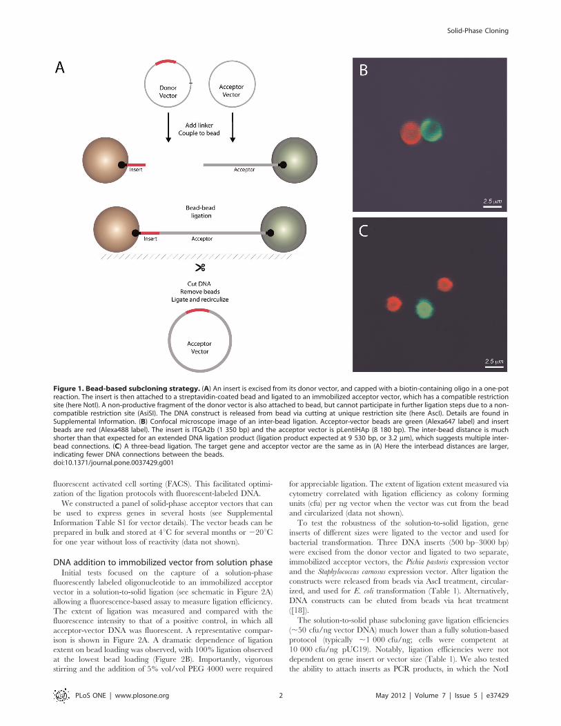
The principle of the solid phase cloning methodA schematic view of the solid-phase cloning platform is shown in
Figure 1A. The solid-phase cloning platform is centered upon an
immobilized acceptor vector, which is linearized, biotinylated at
one end and attached to streptavidin-coated paramagnetic beads
[17]. The free end is modified to allow for subsequent DNA
addition (e.g. treated with a restriction endonuclease). Near the
biotin-streptavidin linkage is a unique restriction site (here AscI),
which is used to release the vector from the bead. The target genes
are inserted into bead-immobilized acceptor vectors via solid-
phase annealing and ligation. Typical bead loadings were
approximately 1500 DNA molecules per bead (0.5 ng vector
DNA/mg bead for a 5,000 base pair vector). The target gene can
be in the solution phase, as a PCR product or excised from a donor
vector, or can itself be attached to a bead. After target-vector
ligation, the beads are collected via magnet and excess reagents are
washed away. The beads can subsequently be used in additional
reactions or the construct can be removed from the bead for
transformation. The beads are large enough to be detected by
PLoS ONE | www.plosone.org 1 May 2012 | Volume 7 | Issue 5 | e37429
fluorescent activated cell sorting (FACS). This facilitated optimi-
zation of the ligation protocols with fluorescent-labeled DNA.
We constructed a panel of solid-phase acceptor vectors that can
be used to express genes in several hosts (see Supplemental
Information Table S1 for vector details). The vector beads can be
prepared in bulk and stored at 4uC for several months or 220uCfor one year without loss of reactivity (data not shown).
DNA addition to immobilized vector from solution phaseInitial tests focused on the capture of a solution-phase
fluorescently labeled oligonucleotide to an immobilized acceptor
vector in a solution-to-solid ligation (see schematic in Figure 2A)
allowing a fluorescence-based assay to measure ligation efficiency.
The extent of ligation was measured and compared with the
fluorescence intensity to that of a positive control, in which all
acceptor-vector DNA was fluorescent. A representative compar-
ison is shown in Figure 2A. A dramatic dependence of ligation
extent on bead loading was observed, with 100% ligation observed
at the lowest bead loading (Figure 2B). Importantly, vigorous
stirring and the addition of 5% vol/vol PEG 4000 were required
for appreciable ligation. The extent of ligation extent measured via
cytometry correlated with ligation efficiency as colony forming
units (cfu) per ng vector when the vector was cut from the bead
and circularized (data not shown).
To test the robustness of the solution-to-solid ligation, gene
inserts of different sizes were ligated to the vector and used for
bacterial transformation. Three DNA inserts (500 bp–3000 bp)
were excised from the donor vector and ligated to two separate,
immobilized acceptor vectors, the Pichia pastoris expression vector
and the Staphylococcus carnosus expression vector. After ligation the
constructs were released from beads via AscI treatment, circular-
ized, and used for E. coli transformation (Table 1). Alternatively,
DNA constructs can be eluted from beads via heat treatment
([18]).
The solution-to-solid phase subcloning gave ligation efficiencies
(,50 cfu/ng vector DNA) much lower than a fully solution-based
protocol (typically ,1 000 cfu/ng; cells were competent at
10 000 cfu/ng pUC19). Notably, ligation efficiencies were not
dependent on gene insert or vector size (Table 1). We also tested
the ability to attach inserts as PCR products, in which the NotI
Figure 1. Bead-based subcloning strategy. (A) An insert is excised from its donor vector, and capped with a biotin-containing oligo in a one-potreaction. The insert is then attached to a streptavidin-coated bead and ligated to an immobilized acceptor vector, which has a compatible restrictionsite (here NotI). A non-productive fragment of the donor vector is also attached to bead, but cannot participate in further ligation steps due to a non-compatible restriction site (AsiSI). The DNA construct is released from bead via cutting at unique restriction site (here AscI). Details are found inSupplemental Information. (B) Confocal microscope image of an inter-bead ligation. Acceptor-vector beads are green (Alexa647 label) and insertbeads are red (Alexa488 label). The insert is ITGA2b (1 350 bp) and the acceptor vector is pLentiHAp (8 180 bp). The inter-bead distance is muchshorter than that expected for an extended DNA ligation product (ligation product expected at 9 530 bp, or 3.2 mm), which suggests multiple inter-bead connections. (C) A three-bead ligation. The target gene and acceptor vector are the same as in (A) Here the interbead distances are larger,indicating fewer DNA connections between the beads.doi:10.1371/journal.pone.0037429.g001
Solid-Phase Cloning
PLoS ONE | www.plosone.org 2 May 2012 | Volume 7 | Issue 5 | e37429
and AscI sites were added via primers. PCR amplicons were
treated with NotI-AscI and phosphatase before ligation to
immobilized acceptor vector. They were not purified from the
PCR reaction mixture. This approach gave lower (,20–30 cfu/
ng) ligation efficiencies.
Bead-bead mediated sub-cloningWe hypothesized that ligation efficiency at higher bead loadings
could be improved if both vector and gene DNA were
immobilized onto beads; here the paramagnetic beads could be
brought into very close contact with a magnet. This bead-bead
subcloning process is depicted in Figure 1A. The gene is excised
from its donor vector in a series of one-pot enzymatic reactions,
capped with a biotinylated oligo, and attached to bead (see
Supplemental Methods S1 and Supplemental Figures S1, S2, S3
for details). A second DNA fragment is also attached to bead, but
its non-complementary restriction site prevents it from participat-
ing in subsequent ligation steps. Excess reagents and non-
biotinylated DNA are then removed. The donor- and acceptor
beads are then co-incubated with DNA ligase in the presence of
a magnet. Alternatively, the insert DNA can be prepared via PCR
with a biotinylated primer. In this way it can be attached to the
bead, treated with NotI, and ligated to vector beads.
We sought to characterize the critical bead-bead ligation step
using both confocal microscopy and flow cytometry. We attached
red and green fluorophores to gene- and vector beads, re-
spectively. After ligation, the beads were examined qualitatively
with a confocal microscope or quantitatively using a flow
cytometer.
Representative microscopy images are shown in Figures 1B and
1C. Figure 1B shows a pairwise bead-bead construct and Figure 1C
shows a rare three-bead construct in which a single acceptor bead
is attached to two insert beads. These multi-bead constructs
migrated together on the slide surface, indicating that the beads
were attached through their DNA. The bead-bead constructs were
non-uniform with regard to inter-bead distance, which suggests
Figure 2. Ligation of solution phase DNA to a bead-immobilized vector. (A) A fluorescence-based assay for determining extent of ligation.Beads with immobilized vector are incubated with a Alexa 488 fluorescent oligo. The extent of ligation is measured via flow cytometry. Bead loadingwas at 1 ng vector DNA (pHISZ)/ug bead vector. Positive: Beads in which pHISZ vector is fully fluorescently labeled. Ligation: Beads after ligation.Negative: Beads in which pHISZ vector is not fluorescently labeled. The extent of ligation f is measured as a percentage of the Positive signal. (B)Extent of ligation is reduced at high bead loadings.doi:10.1371/journal.pone.0037429.g002
Table 1. Ligation efficiencies of bead-based subcloning strategies.*
AcceptorVector
Size(kb)
DonorInsert
Size(kb) Solution-Bead
Bead-BeadMAGNET ON
Bead-BeadMAGNET OFF
Correct cfu/ng Correct cfu/ng Correct cfu/ng
pPICZaCp 3.6 TNF 0.47 5/8 60 7/8 550 7/8 100
EGFR 1.86 7/8 30 8/8 880 4/7 300
ITGAL 3.19 7/8 30 7/8 750 4/8 350
pSCEM2 7.6 TNF 0.47 7/8 50 13/13 750 2/8 90
EGFR 1.86 7/8 70 12/13 1500 5/8 610
ITGAL 3.19 8/8 50 12/13 820 6/12 340
*Vector and donor beads were loaded at 0.5 ng DNA/mg bead.doi:10.1371/journal.pone.0037429.t001
Solid-Phase Cloning
PLoS ONE | www.plosone.org 3 May 2012 | Volume 7 | Issue 5 | e37429
multiple DNA attachments between beads. The gene ITGAL2b is
1350 bp, corresponding to approximately 0.46 mm for extended
B-form DNA [19], while the magnetic beads have a diameter of
2.8 mm. The acceptor vector pLentiHAp is 8180 bp (2.76 mm).
Successful constructs would be expected at 9 530 bp (approxi-
mately 3.22 mm inter-bead distance). Constructs showing sub-
stantially shorter inter-bead distance (e.g. Fig. 1B) may be
indicative of multiple DNA connections, while those showing the
3.22 mm theoretical distance for a ITGAL2b-pLentiHAp con-
struct (e.g. Fig. 1C) suggest fewer DNA connections.
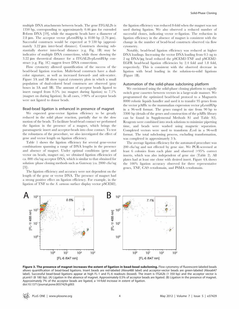
Flow cytometry allowed quantification of the success of the
bead-bead ligation reaction. Multi-bead constructs have a dual-
color signature, as well as increased forward- and side-scatter.
Figure 3A and 3B show typical cytometry plots in which a small
population of dual-colored bead constructs are observed (gray
boxes in 3A and 3B). The amount of acceptor beads ligated to
insert ranged from 0.5% (no magnet during ligation) to 7.1%
(magnet on during ligation). In all cases, .90% of acceptor beads
were not ligated to donor beads.
Bead-bead ligation is enhanced in presence of magnetWe expected gene-vector ligation efficiency to be greatly
reduced in the solid phase reaction, partially due to the slow
motion of the beads. To facilitate bead-bead contact we performed
the ligation in the presence of a magnet, which brings the
paramagnetic insert and acceptor-beads into close contact. To test
the robustness of the procedure, we also investigated the effect of
gene and vector length on ligation efficiency.
Table 1 shows the ligation efficiency for several gene-vector
combinations spanning a range of DNA lengths in the presence
and absence of magnet. Under optimal conditions (gene and
vector on beads, magnet on), we obtained ligation efficiencies of
ca. 800 cfu/ng acceptor DNA, which is similar to that obtained for
solution–phase cloning methods such as Gateway (ca. 2000 cfu/ng
[3]).
The ligation efficiency and accuracy were not dependent on the
length of the gene or vector DNA. The presence of magnet had
a strong positive effect on ligation efficiency. For example, in the
ligation of TNF to the S. carnosus surface display vector pSCEM2,
the ligation efficiency was reduced 8-fold when the magnet was not
used during ligation. We also observed a reduced number of
successful clones, indicating vector re-ligation. The reduction in
ligation efficiency in the absence of magnet is consistent with the
change in the number of bead-bead constructs observed via flow
cytometry.
Notably, bead-bead ligation efficiency was reduced at higher
DNA loadings. Increasing the vector DNA loading from 0.5 ng to
2 ng DNA/mg bead reduced the pSCEM2-TNF and pSCEM2-
EGFR bead-bead ligation efficiencies by 2.4 fold and 1.8 fold,
respectively. This is consistent with the observed decrease in
ligation with bead loading in the solution-to-solid ligations
(Figure 1B).
Automation of the solid-phase subcloning platformWe envisioned using the solid-phase cloning platform to rapidly
switch gene cassettes between vectors in a large-scale manner. We
programmed the optimized bead-bead protocol to a Magnatrix
8000 robotic liquids handler and used it to transfer 95 genes from
the vector pAff8c to the mammalian expression vector pLentiHAp
in a 96-well format. The genes ranged in size from 90 bp to
3300 bp (details of the genes and construction of the pAff8c library
can be found in Supplemental Methods S1 and Table S3).
Reagents were combined into stock solutions to minimize pipetting
time, and beads were washed using magnetic separation.
Completed vectors were used to transform E.coli in a 96-well
format. The total subcloning process, excluding transformation,
was completed in approximately 3 h.
The average ligation efficiency for the automated procedure was
200 cfu/ng and not effected by gene size. We PCR-screened at
least 6 colonies from each plate and observed .95% correct
inserts, which was also independent of gene size (Table 2). All
plates had at least one clone with desired insert. Figure 4A shows
the 100% ligation accuracy observed for three representative
genes, TNF, CA9 ectodomain, and PSMA ectodomain.
Figure 3. The presence of magnet increases the extent of ligation in bead-bead subcloning. Flow cytometry of fluorescent-labeled beadsallows quantification of bead-bead ligations. Insert beads are red-labeled (Alexa488 label) and acceptor-vector beads are green-labeled (Alexa647label). Successful bead-bead ligations appear at high FL-1 and FL-6 readouts (boxed). The insert is ITGA2b (1 350 bp) and the acceptor vector ispLenti1 (8 180 bp). (A) Ligation in the absence of magnet. Approximately 0.5% of acceptor beads are ligated. (B) Ligation in the presence of magnet.Approximately 7% of the acceptor beads are ligated, a 14-fold increase in extent of ligation.doi:10.1371/journal.pone.0037429.g003
Solid-Phase Cloning
PLoS ONE | www.plosone.org 4 May 2012 | Volume 7 | Issue 5 | e37429
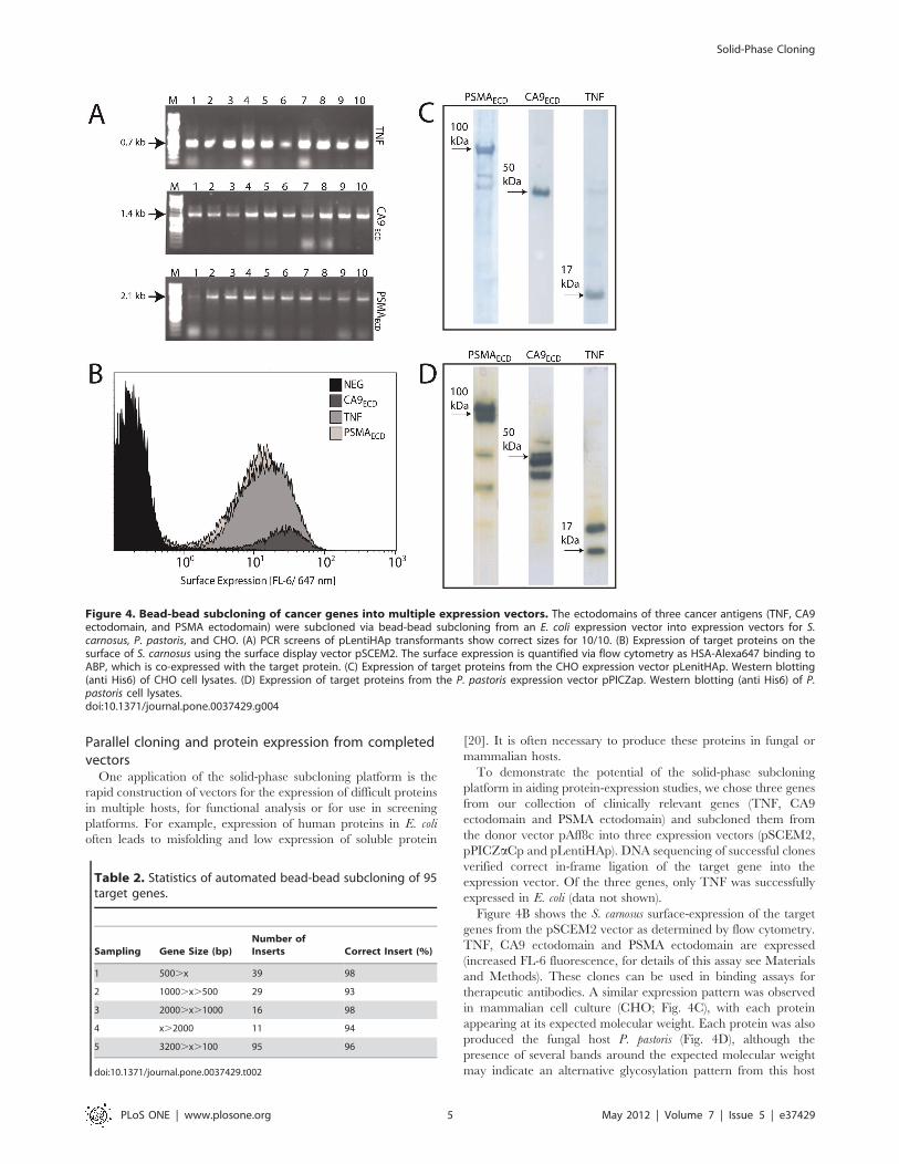
Parallel cloning and protein expression from completedvectors
One application of the solid-phase subcloning platform is the
rapid construction of vectors for the expression of difficult proteins
in multiple hosts, for functional analysis or for use in screening
platforms. For example, expression of human proteins in E. coli
often leads to misfolding and low expression of soluble protein
[20]. It is often necessary to produce these proteins in fungal or
mammalian hosts.
To demonstrate the potential of the solid-phase subcloning
platform in aiding protein-expression studies, we chose three genes
from our collection of clinically relevant genes (TNF, CA9
ectodomain and PSMA ectodomain) and subcloned them from
the donor vector pAff8c into three expression vectors (pSCEM2,
pPICZaCp and pLentiHAp). DNA sequencing of successful clones
verified correct in-frame ligation of the target gene into the
expression vector. Of the three genes, only TNF was successfully
expressed in E. coli (data not shown).
Figure 4B shows the S. carnosus surface-expression of the target
genes from the pSCEM2 vector as determined by flow cytometry.
TNF, CA9 ectodomain and PSMA ectodomain are expressed
(increased FL-6 fluorescence, for details of this assay see Materials
and Methods). These clones can be used in binding assays for
therapeutic antibodies. A similar expression pattern was observed
in mammalian cell culture (CHO; Fig. 4C), with each protein
appearing at its expected molecular weight. Each protein was also
produced the fungal host P. pastoris (Fig. 4D), although the
presence of several bands around the expected molecular weight
may indicate an alternative glycosylation pattern from this host
Figure 4. Bead-bead subcloning of cancer genes into multiple expression vectors. The ectodomains of three cancer antigens (TNF, CA9ectodomain, and PSMA ectodomain) were subcloned via bead-bead subcloning from an E. coli expression vector into expression vectors for S.carnosus, P. pastoris, and CHO. (A) PCR screens of pLentiHAp transformants show correct sizes for 10/10. (B) Expression of target proteins on thesurface of S. carnosus using the surface display vector pSCEM2. The surface expression is quantified via flow cytometry as HSA-Alexa647 binding toABP, which is co-expressed with the target protein. (C) Expression of target proteins from the CHO expression vector pLenitHAp. Western blotting(anti His6) of CHO cell lysates. (D) Expression of target proteins from the P. pastoris expression vector pPICZap. Western blotting (anti His6) of P.pastoris cell lysates.doi:10.1371/journal.pone.0037429.g004
Table 2. Statistics of automated bead-bead subcloning of 95target genes.
Sampling Gene Size (bp)Number ofInserts Correct Insert (%)
1 500.x 39 98
2 1000.x.500 29 93
3 2000.x.1000 16 98
4 x.2000 11 94
5 3200.x.100 95 96
doi:10.1371/journal.pone.0037429.t002
Solid-Phase Cloning
PLoS ONE | www.plosone.org 5 May 2012 | Volume 7 | Issue 5 | e37429
[21]. Additionally, the CA9 protein was secreted at high titers into
the CHO culture medium, while secretion was hindered in the P.
pastoris cultures (data not shown). Solid-phase vector construction
could be used in tandem with a high-throughput protein
expression and purification platform, such as one recently
developed in our laboratory [22].
Discussion
We have developed a subcloning platform based upon the
immobilization of DNA onto paramagnetic beads. The protocol
can be used with PCR products or, in cases where additional PCR
is not desirable, with inserts excised from donor vectors. We have
circularized DNA after elution from beads for transformation into
E. coli. However, linear DNA could be used directly in
transformations of hosts capable of linear-DNA uptake. Alterna-
tively, whole bead-DNA constructs could be used for transfections
[23].
The introduction of the solid phase presents unique problems
compared to solution-phase reactions, such as enzyme-to-bead
mass-transfer limitations [24] and potentially nonproductive
conformations of DNA on the solid surface [25]. We observed
higher ligation efficiencies with lower bead surface coverage,
consistent with previous studies of DNA hybridization onto flat
surfaces [26] and lowest ligation efficiencies from solution-to-bead
ligations. These efficiencies were nearly an order of magnitude
lower than those of solution-phase ligations. This discrepancy is
most likely due to mass-transfer limitations present in the
heterogeneous ligation reaction, as the ligation efficiencies were
increased dramatically when the mixture was stirred. It may also
be due to the inaccessibility of the vector DNA on the bead
surface, as the addition of PEG increased ligation efficiencies. PEG
is known to facilitate the unfolding of DNA structures [27].
Additionally, intra-bead bridging between proximal vector strands
via ligation of compatible sticky-ends may hinder addition of the
gene insert. This problem would be exacerbated at high vector
densities, which is consistent with the observed reduced ligation
efficiencies at high bead loading (Figure 2B). While we have taken
measures to prevent intra-bead bridging via extensive dephop-
sphorylation of vector ends, the problem could be eliminated
completely by adaption of other cloning methods, which do not
rely on endonucleases, such as exonuclease ‘‘chew-back and
anneal’’ methods [6]. We expect such methods to be compatible
with the solid-phase platform.
Solid-to-solid ligations showed higher efficiencies than the
solution-to-solid ligations. This may be due to a proximity effect;
once two beads are initially linked to one another through DNA
strands, the attachment of subsequent DNA strands between them
is facilitated as in an intra-molecular reaction. Such proximity
effects are not present in the solution-to-solid ligations. This
hypothesis is supported by the nearness of some bead-bead
constructs as seen via in confocal microscopy. The presence of the
magnet increased the ligation efficiency still further, due to an
increased propensity to form an initial bead-bead ligation. This
was confirmed by a large increase in the number of linked beads
observed by flow cytometry when compared to ligations without
magnet. These results suggest that it would be possible to control
the extent of ligation quite easily by judicious application of
a magnet.
The solid-phase ligation efficiencies were independent of insert
and vector sizes in the range tested (90–3300 bp). This is consistent
with previous solid-phase DNA manipulation studies, which
demonstrated both efficient enzyme cleavage and strand DNA
hybridization at sites .20 nt from the biotin immobilization site
[28]. The ease of constructing vectors .10000 bp (e.g. ITGA2B-
pLentiHAp) suggests that longer dsDNA constructs are possible
given the correct buffer conditions. We did not observe a de-
pendence of ligation efficiency on GC content of either vector or
insert. We expect that the use of dsDNA reduces the risk of
secondary structures such as hairpins, which may hinder ligation.
Secondary structure may be necessary to consider if adapting the
method to ssDNA annealing [25].
The ability to quickly pull out bead constructs via magnetic
separation eliminated the need for gel extractions and spin-
columns and simplified automation of the protocol. The sublcon-
ing process could be fully automated, with bead capture,
enzymatic digestion, and DNA washing performed by a robot.
Solid-phase cloning could be adapted to recently-developed
microfluidics devices which are designed to manipluate magnetic
microparticles with high precision [29]. Such a platform could be
used for combinatorial vector construction on a scale not possible
with solution-phase cloning.
An automated platform would also greatly aid high-throughput
expression and function studies. We envisioned a scenario in
which several expression hosts would be tested for the optimal
expression of a panel of proteins. Using our optimized protocol, in
which restriction digests were performed simultaneously and
reagents were combined and stored as stock solutions, we were
able to transfer 95 target genes from a donor vector to
a mammalian expression vector in 3 h. A preliminary test of
several of these proteins in different expression vectors showed
interesting discrepancies in terms of secreted titer and glycosyla-
tion state.
An additional direction of the bead-based cloning method is
assembly of multicomponent constructs via sequential addition of
genetic elements [30]. The method could aid synthetic biology
efforts by accelerating the construction of vectors via the BioBrick
[1], BglBrick [2], and USER [31] methods by eliminating
intermediate transformation steps. The ease of purification of
cloning intermediates could potentially ease assembly of long
constructs. The solid-phase platform could also be easily extended
and adapted for other DNA manipulation strategies, such as
ligase-independent and endonuclease-free methods.
In conclusion, the approach shown here has demonstrated the
power of magnetic beads for standardized highly automated
subcloning of large number of fragments into expression vectors
for protein expression screening and we point out a direction for
use of magnetic beads in broader terms for generation of longer
multicomponent genetic assemblies, relevant for metabolic engi-
neering.
Methods
All enzymes were from Fermentas UAB (Vilinius, Lithuania)
and were FastDigest. We found the use of these FastDigest
enzymes to be critical for simultaneous restriction digestions.
Genes for solution-to-bead and bead-to-bead ligations were from an
E. coli ‘‘donor’’ vector pAff8c [32]. We created a library of 95
pAff8c vectors encoding the full and partial ectodomains of 65
cancer antigens (for cloning procedures and antigen details see
Supplemental Methods s1).
Acceptor beads were prepared in bulk and used in solution-to-
solid and solid-to-solid ligations. Briefly, acceptor plasmids were
linearized (NotI/AscI) and a biotinylated linker containing
a complementary AscI restriction site was attached to one end.
The plasmids were then incubated with DynaBeads M270
streptavidin-coated beads (Invitrogen, Carlsbad, CA) to create
acceptor beads. Typical bead loadings were 0.5 ng plasmid DNA/
Solid-Phase Cloning
PLoS ONE | www.plosone.org 6 May 2012 | Volume 7 | Issue 5 | e37429
mg beads, corresponding to ca. 1 500 DNA/bead. Details on
acceptor vectors, including primer sequences, see Supplemental
Information Table S1, S2, S3.
Solution-to-bead ligationsVector beads were first treated with NotI (37 C 1 hr), which
produced a free NotI end, and then washed twice in Tris buffer
pH 8. Genes from the pAff8c clone library were prepared as
NotI/AscI-digested, purified fragments, or as NotI/AscI-digested
PCR products. The ligation reaction volume was 50 mL and
included 5 mg of acceptor beads (loaded at 0.5 ng DNA/mg
beads), a 10:1 molar excess of insert DNA, 5 mL of T4 DNA ligase
buffer, 5% v/v PEG4000 and 1 mL T4 DNA ligase. The ligation
reaction proceeded at room temperature for 45 min under
shaking. The beads were removed via magnetic separation and
washed. The DNA was removed from beads with AscI treatment
(30 mL, 37uC 45 min with vigorous shaking). The new vector
constructs were circularized with addition of T4 DNA ligase and
0.5 mM ATP (22uC 10 min) and 3 mL used for bacterial
transformation.
Solution-to-bead ligation of the fluorescent capping oligo to
M270-pHISZ was performed similarly but used 10 pmol of an
18 bp oligo containing a BamHI site and a 59-AlexaFluor 488
(Eurofins MWG Operon, Ebersberg, Germany). For the positive
control, the pHISZ vector was capped with the BamHI oligo
before bead loading, ensuring near 100% ligation and maximal
fluorescence at each loading.
Bead-to-bead ligationsSolid-to-solid bead ligations were performed with acceptor
beads and donor beads. Donor beads were prepared by incubating
streptavidin-coated M270 beads with biotinylated genes from the
pAff8c clone library. Biotinylated genes were either from PCR
with biotinylated primers or constructed via excision from the
pAff8c vector followed by addition of a biotinylated oligo. For
details of the preparation of donor beads see SI Methods.
We performed the solid-to-solid ligation of acceptor and donor
beads in 1.5 mL eppendorf tubes. For ligations in the presence of
magnet we used a magnetic separation stand. In each case the
ligation volume was 50 mL, included 1 mg of M270-acceptor beads
(,0.5 ng acceptor plasmid) and 10 mg of M270-donor beads
(,5 ng donor DNA), 5 mL of T4 DNA ligase buffer, 5% v/v
PEG4000 and 1 mL T4 DNA ligase. The ligation reaction
proceeded for 60 min, with pipette stirring every 10 min. DNA
was removed from beads with AscI treatment, circularized and
used for transformation as described above.
Flow Cytometry and Confocal MicroscopyFluorophores AlexaFluor 488 and AlexaFluor 647 succinimidyl
esters were from Invitrogen. Acceptor and donor-loaded beads
were modified at pH 9 with AlexaFluor 647 and AlexaFluor 488
respectively, according to the supplier’s protocol (50 mg donor
beads and 50 mg acceptor beads were incubated with 50 mg of
fluorophore for 1 h at 22uC). In this reaction the streptavidin is
covalently modified at its amine groups. The beads were washed 3
times and re-suspended in Tris buffer pH 8. The donor-beads
were loaded at 0.5 ng DNA/mg bead with ITGAL2b cut (NotI
AscI) from the vector pAff8c and the vector beads were loaded
with pLentiHAp at 0.5 ng DNA/mg bead. Donor-acceptor
ligations were performed with the labeled beads according to the
above protocols (1 mg acceptor beads, 10 mg donor beads, 30 mL
reaction) in the presence and absence of magnet.
The ligation mixture was analyzed with a Beckman-Coulter
Gallios flow cytometer using FL-6 (647 nm) and FL-1 (488 nm)
excitation and detection. Before analysis the ligation mixture was
diluted to 300 mL in Tris buffer pH 8. Fifty thousand events were
recorded for each sample.
Automation of the subcloning procedureThe subcloning protocol was programmed onto the Magnatrix
8000 robot (NordDiag A/S, Oslo, Norway) using the manufac-
turer’s software. The pAff8c-donor library was used in 96-well
format. Enzymes, M270 beads and acceptor beads were prepared
as reagents and stored at 4uC during the procedure. Details of the
protocol can be found in Supplemental Methods S1.
Protein expressionThe target proteins TNF, CA9 and PSMA were expressed in E.
coli, P. pastoris, S. carnosus and Chinese Hamster Ovary Cells (LGC-
labstandards AB, Boras, Sweden).
E. coli RR1DM15 was transformed with pAff8c-target vector by
heat shock and subjected to Km selection. Colonies were PCR
screened with primers N2 and U5 and sequenced verified. Picked
colonies were used to inoculate 10 mL of TSB medium (+Km) and
grown overnight at 37uC. A culture aliquot (100 mL) was used to
inoculate 50 mL of TSB (+Km) which grew to OD600 = 0.8 prior
to induction with 0.5 mM IPTG. The cultures were moved to
25uC. After 6 hours the cells were pelleted, lysed with 5 mL of
7 M GdHCl (37uC 3 h). Lysis supernatant (5 mL) was run on SDS-
PAGE gel.
Staphylococcus carnosus was transformed with pSCEM-target
vector via electroporation according to a published protocol [33]
and subjected to Cm selection. Colonies were PCR screened with
primers SAPA23 and SAPA24. Picked colonies were used to
inoculate 10 mL of TSB medium (+Cm) and grown overnight at
37uC. The pSCEM2 vector encodes a surface-anchoring scaffold
upstream of the MCS. This scaffold includes albumin-binding
protein (ABP). The successful expression of the scaffold+target
protein is detected by incubation of transformed cells with
fluorescent-labeled HSA. Cells were analyzed via flow cytometry
(Beckman Coultier Galios) with FL-6 (647 nm) excitation.
Pichia pastoris strain SMD1168H and the P. pastoris expression
vector pPICZa-C (Invitrogen) were a kind gift from Dr. Harry
Brumer. P. pastoris was transformed with 0.5 mg pPICZaCp-target
vector via electroporation according to the protocol of Wu [34],
which was followed exactly except that the vector DNA was
linearized with PmeI before electroporation. Transformation
mixtures were subjected to Zeomycin selection (125 mg/mL) and
grown at 30uC. Colonies appeared after ca. 3 days. Colonies were
lysed with Lyticase (Sigma) and PCR-screened [35] with both
AOX and target-specific primers and sequence verified.
Successfully transformed Pichia pastoris colonies were cultivated
in 50 mL solutions BMGY and BMMY at 25uC according to the
Invitrogen protocol, with daily addition of MeOH to 0.5% v/v.
After 96 hrs the cells were pelleted and lysed. Lysis supernatants
were analyzed via Western blot, with anti-His6 antibodies (Sigma)
and HRP detection (Sigma).
Mammalian cells CHO-S (ATCC) were transfected via
lentiviral delivery, as described in detail in Supplemental In-
formation. Transfected cells were selected via puromycin re-
sistance. Cells were cultured for one week after transfection at
a maintained 70–80% confluence then harvested and sonicated.
Lysates were filtered and analyzed via Western blot, with anti-His6
antibodies (Sigma-Aldrich, St. Louis, MO) and HRP detection
(Sigma-Aldrich, St. Louis, MO).
Solid-Phase Cloning
PLoS ONE | www.plosone.org 7 May 2012 | Volume 7 | Issue 5 | e37429
Supporting Information
Table S1 Vectors used in this study.
(DOC)
Table S2 Oligos used in this study.
(DOC)
Table S3 Antigens in the pAff8c donor library.
(DOC)
Figure S1 Detailed bead-based subcloning strategy.
(TIF)
Figure S2 Size distribution of pAff8c donor library.
(TIF)
Figure S3 Detailed schematic of donor-bead construction and
characterization.
(TIF)
Methods S1 Preparation of acceptor plasmids. Preparation of
acceptor beads from acceptor plasmids. Preparation of pAff8c
library. Preparation of donor beads from pAff8c library.
Automation of bead-to-bead protocol. Transfection of CHO-S
and mammalian cell culture.
(DOC)
Acknowledgments
We would like to thank Novo Nordisk Foundation Center for Protein
Research (CPR) for providing cDNA for cloning and the entire staff of the
Human Protein Atlas program (HPA) and the Novo Nordisk Foundation
Center for Biosustainability for their efforts.
Author Contributions
Conceived and designed the experiments: JR MU EPH. Performed the
experiments: JR EPH AN. Analyzed the data: JR EPH. Contributed
reagents/materials/analysis tools: JR EPH AN. Wrote the paper: JR EPH
AN MU.
References
1. Shetty R, Endy D, Knight T (2008) Engineering BioBrick vectors from BioBrick
parts. Journal of Biological Engineering 2: 5.
2. Anderson J, Dueber J, Leguia M, Wu G, Goler J, et al. (2010) BglBricks: A
flexible standard for biological part assembly. Journal of biological engineering
4: 1–12.
3. Hartley J (2000) DNA Cloning Using In Vitro Site-Specific Recombination.
Genome Research 10: 1788–1795.
4. Li M, Elledge S (2007) Harnessing homologous recombination in vitro to
generate recombinant DNA via SLIC. Nature Methods 4: 251–256.
5. Aslanidis C, de Jong PJ (1990) Ligation-independent cloning of PCR products
(LIC-PCR). Nucleic acids research 18: 6069–6074.
6. Gibson D, Young L, Chuang R, Venter J, Hutchison C, et al. (2009) Enzymatic
assembly of DNA molecules up to several hundred kilobases. Nature Methods 6:
343–345.
7. Quan J, Tian J (2009) Circular Polymerase Extension Cloning of Complex Gene
Libraries and Pathways. PLoS ONE 4: e6441.
8. Klock H, Lesley S (2009) The Polymerase Incomplete Primer Extension (PIPE)
method applied to high-throughput cloning and site-directed mutagenesis.
Methods Mol Biol 498: 91–103.
9. Gibson D, Smith H, Hutchison C, Venter J, Merryman C (2010) Chemical
synthesis of the mouse mitochondrial genome. Nature Methods 7: 901–903.
10. Uhlen M (1989) Magnetic separation of DNA. Nature 340: 733–734.
11. Ronaghi M, Uhlen M, Nyren P (1998) A Sequencing Method Based on Real-
Time Pyrophosphate. Science 281: 363–365. doi :10.1126/sci-
ence.281.5375.363.
12. Kojima T, Takei Y, Ohtsuka M, Kawarasaki Y, Yamane T, et al. (2005) PCR
amplification from single DNA molecules on magnetic beads in emulsion:
application for high-throughput screening of transcription factor targets. Nucleic
acids research 33: e150. doi:10.1093/nar/gni143.
13. Gan R, Yamanaka Y, Kojima T, Nakano H (2008) Microbeads display of
proteins using emulsion PCR and cell-free protein synthesis. Biotechnology
Progress 24: 1107–1114.
14. Quan J, Saaem I, Tang N, Ma S, Negre N, et al. (2011) Parallel on-chip gene
synthesis and application to optimization of protein expression. Nature
Biotechnology 29: 449–452. doi:10.1038/nbt.1847.
15. Yeung S-W, Lee TM-H, Cai H, Hsing I-M (2006) A DNA biochip for on-the-
spot multiplexed pathogen identification. Nucleic acids research 34: e118.
doi:10.1093/nar/gkl702.
16. Roth ME, Feng L, McConnell KJ, Schaffer PJ, Guerra CE, et al. (2004)
Expression profiling using a hexamer-based universal microarray. Nature
biotechnology 22: 418–426. doi:10.1038/nbt948.
17. Stahl S, Hultman T, Olsson A, Moks T, Uhlen M (1988) Solid-Phase DNA
sequencing using the biotin-avidin system. Nucleic Acids Research 16:
3025–3038.
18. Holmberg A, Blomstergren A, Nord O, Lukacs M, Lundeberg J, et al. (2005)
The biotin-streptavidin interaction can be reversibly broken using water at
elevated temperatures. Electrophoresis 26: 501–510.
19. Smith S, Cui Y, Bustamante C (1996) Overstretching B-DNA: the elasticresponse of individual double-stranded and single-stranded DNA molecules.
Science 271: 795.20. Baneyx F, Mujacic M (2004) Recombinant protein folding and misfolding in
Escherichia coli. Nature biotechnology 22: 1399–1408. doi:10.1038/nbt1029.
21. Daly R, Hearn MTW (2005) Expression of heterologous proteins in Pichiapastoris: a useful experimental tool in protein engineering and production.
Journal Of Molecular Recognition 18: 119–138.22. Steen J, Uhlen M, Hober S, Ottosson J (2006) High-throughput protein
purification using an automated set-up for high-yield affinity chromatography.Protein expression and purification 46: 173–178. doi:10.1016/
j.pep.2005.12.010.
23. Isalan M, Santori MI, Gonzalez C, Serrano L (2005) Localized transfection onarrays of magnetic beads coated with PCR products. Nature methods 2:
113–118. doi:10.1038/nmeth732.24. Castronovo M, Radovic S, Grunwald C, Casalis L, Morgante M, et al. (2008)
Control of steric hindrance on restriction enzyme reactions with surface-bound
DNA nanostructures. Nano letters 8: 4140–4145. doi:10.1021/nl802370g.25. Riccelli PV, Merante F, Leung KT, Bortolin S, Zastawny RL, et al. (2001)
Hybridization of single-stranded DNA targets to immobilized complementaryDNA probes: comparison of hairpin versus linear capture probes. Nucleic acids
research 29: 996–1004.26. Peterson AW, Heaton RJ, Georgiadis RM (2001) The effect of surface probe
density on DNA hybridization. Nucleic acids research 29: 5163–5168.
27. Naimushin A, Quach N, Fujimoto B, Schurr J (2001) Effect of polyethyleneglycol on the supercoiling free energy of DNA. Biopolymers 58: 204–217.
28. Nilsson P, Persson B, Uhlen M, Nygren P (1995) Real-time monitoring of DNAmanipulations using biosensor technology. Analytical Biochemistry 224:
400–408.
29. Lee H, Kim J, Kim H, Kim J, Kwon S (2010) Colour-barcoded magneticmicroparticles for multiplexed bioassays. Nature materials 9: 745–749.
doi:10.1038/nmat2815.30. Ellis T, Adie T, Baldwin GS (2011) DNA assembly for synthetic biology: from
parts to pathways and beyond. Integrative biology: quantitative biosciences fromnano to macro 3: 109–118. doi:10.1039/c0ib00070a.
31. Geu-Flores F, Nour-Eldin HH, Nielsen MT, Halkier B a (2007) USER fusion:
a rapid and efficient method for simultaneous fusion and cloning of multiplePCR products. Nucleic acids research 35: e55. doi:10.1093/nar/gkm106.
32. Larsson M, Graslund S, Yuan L, Brundell E, Uhlen M, et al. (2000) High-throughput protein expression of cDNA products as a tool in functional
genomics. Journal of biotechnology 80: 143–157.
33. Rockberg J, Lofblom J, Hjelm B, Uhlen M, Stahl S (2008) Epitope mapping ofantibodies using bacterial surface display. Nat Methods, 5, 1039–1045.
34. Wu SX, Letchworth GJ (2004) High efficiency transformation by electropora-tion of Pichia pastoris pretreated with lithium acetate and dithiothreitol.
Biotechniques 36: 152–154.
35. Ayra-Pardo C, Martinez CG, de la Riva GA (1998) A single-step screeningprocedure for Pichia pastoris clones by PCR. Biotecnologia Aplicada. pp
173–175.
Solid-Phase Cloning
PLoS ONE | www.plosone.org 8 May 2012 | Volume 7 | Issue 5 | e37429


![Orbit type: Sun Synchronous Orbit ] Orbit height: …...Orbit type: Sun Synchronous Orbit ] PSLV - C37 Orbit height: 505km Orbit inclination: 97.46 degree Orbit period: 94.72 min ISL](https://img.pdfslide.net/doc/110x75/5f781053e671b364921403bc/orbit-type-sun-synchronous-orbit-orbit-height-orbit-type-sun-synchronous.jpg)