Embed Size (px)
Citation preview

Review ArticleAutophagy Dysfunction, Cellular Senescence, and AbnormalImmune-Inflammatory Responses in AMD: From Mechanisms toTherapeutic Potential
Shoubi Wang ,1 Xiaoran Wang ,1 Yaqi Cheng ,1 Weijie Ouyang ,2 Xuan Sang,1
Jiahui Liu ,3 Yaru Su ,1 Ying Liu ,1 Chaoyang Li ,1 Liu Yang ,1 Lin Jin ,1
and Zhichong Wang 1
1State Key Laboratory of Ophthalmology, Zhongshan Ophthalmic Center, Sun Yat-sen University, Guangzhou 510060, China2Eye Institute of Xiamen University, Fujian Provincial Key Laboratory of Ophthalmology and Visual Science, School of Medicine,Xiamen University, Xiamen 361102, China3Department of Ophthalmology, Dongguan People’s Hospital, Dongguan 523059, China
Correspondence should be addressed to Zhichong Wang; [email protected]
Received 5 March 2019; Accepted 17 April 2019; Published 22 May 2019
Academic Editor: Antonello Lorenzini
Copyright © 2019 Shoubi Wang et al. This is an open access article distributed under the Creative Commons Attribution License,which permits unrestricted use, distribution, and reproduction in any medium, provided the original work is properly cited.
Age-related macular degeneration (AMD) is a blinding disease caused by multiple factors and is the primary cause of vision loss inthe elderly. The morbidity of AMD increases every year. Currently, there is no effective treatment option for AMD. Intravitrealinjection of antivascular endothelial growth factor (anti-VEGF) is currently the most widely used therapy, but it only aims atneovascularization, which is an intermediate pathological phenomenon of wet AMD, not at the etiological treatment. Anti-VEGF therapy can only temporarily delay the degeneration process of wet AMD, and AMD is easy to relapse after drugwithdrawal. Therefore, it is urgent to deepen our understanding of the pathophysiological processes underlying AMD and toidentify integrated or new strategies for AMD prevention and treatment. Recent studies have found that autophagy dysfunctionin retinal pigment epithelial (RPE) cells, cellular senescence, and abnormal immune-inflammatory responses play key roles inthe pathogenesis of AMD. For many age-related diseases, the main focus is currently the clearing of senescent cells (SNCs) as anantiaging treatment, thereby delaying diseases. However, in AMD, there is no relevant antiaging application. This review willdiscuss the pathogenesis of AMD and how interactions among RPE autophagy dysfunction, cellular senescence, and abnormalimmune-inflammatory responses are involved in AMD, and it will summarize the three antiaging strategies that have beendeveloped, with the aim of providing important information for the integrated prevention and treatment of AMD and laying theground work for the application of antiaging strategies in AMD treatment.
1. Introduction
AMD is the leading cause of visual impairment among theelderly in western countries. Although AMD usually doesnot lead to complete blindness, it can result in the severe lossof central vision. A study estimated that, by 2020, 196 millionpeople will be afflicted with AMD worldwide, increasing to288 million people by 2040. As a result, the cost of AMD ispredicted to increase to $59 billion over the next 20 years
[1], suggesting that AMD is becoming a major public healthissue. Currently, there is no effective treatment for 80% to85% of the 30 to 50 million AMD patients worldwide [2].AMD is a multifactorial blinding disease, and the exact causeof AMD is not yet clear. It has been previously demonstratedthat oxidative stress [3], aging [4], DNA damage [5], andultraviolet radiation [6] can lead to AMD by influencingthe autophagy function of RPE cells, cellular senescence,and the immune-inflammatory response, which are closely
HindawiOxidative Medicine and Cellular LongevityVolume 2019, Article ID 3632169, 13 pageshttps://doi.org/10.1155/2019/3632169

related to each other in their mutual causation and promo-tion (Figure 1). Autophagy dysfunction results in thedecreased clearance of cellular waste in RPE cells andincreased intracellular residual corpuscles, which interferewith cell metabolism. Senescent RPE cells lead to cell dys-function and promote the senescence of surrounding cellsby secreting the senescence-associated secretory phenotype(SASP). Moreover, SNCs are apoptosis resistant, failing toenter programmed cell death and aggregating instead, furtherpromoting the development of AMD. The blood-retinal bar-rier (BRB) has an immune privilege function. The destruc-tion of the BRB could activate the immune-inflammatoryresponse of the retina and lead to the release of pattern recog-nition receptors (PRRs) and inflammasomes, the activationof immune cells and cytokines, and abnormalities of thecomplement system, which could further amplify the localinflammatory response. The abovementioned factors interactwith each other, causing lipofuscin deposition, drusen forma-tion, RPE injury, or atrophy, which can lead to photoreceptorcell damage, choroid degeneration, and ultimately, loss ofvision. These findings suggest that autophagy dysfunctionin RPE cells, cellular senescence, and abnormal immune-inflammatory responses are involved in AMD pathogenesisand promote its progress. Here, we review the pathophysio-logical processes and interactions that are involved inAMD, with the aim of providing important information forthe molecular, biological, and clinical research of AMD inthe future.
2. Autophagy Dysfunction Leads to “ClearanceSystem” Abnormalities
There are two major proteolytic systems that are responsiblefor maintaining cellular function: the proteasomal and lyso-somal systems. Both systems remove irreversibly damagedproteins and recycle amino acids for protein synthesis [2,7]. The autophagy-lysosome system is the most importantof these two systems in RPE cells [2]. Autophagy can bedivided into macroautophagy, microautophagy, andchaperone-mediated autophagy [8]. Macroautophagy, whichis considered to be the major autophagic pathway and hasbeen the most extensively studied type of autophagy, is medi-ated by the formation of an autophagosome, a double-membrane vacuole that contains the materials targeted fordegradation (cargo). The autophagosome carries cargo toand combines with the lysosome to form the autolysosome,in which the final degradation of cargo occurs. This processrequires the participation of a series of autophagy-relatedproteins (Atgs). Although observation of the double-membraned structure by transmission electron microscopy(TEM) is the gold standard for autophagy detection, it is nec-essary to assess the expression levels of LC3 II/LC3 I,p62/SQSTM1, and Atgs to estimate the level of autophagyactivity [9]. The photoreceptor outer segments (POS) arecomposed of dense discs. Proteins are synthesized in theinner segments and transported to the outer segmentsthrough ciliary ligation structures to form new discs.
Oxidative stress, aging, DNA damage, and ultraviolet radiation
Cellular senescence
Apoptosis resistance
Autophagy dysfunction
RPE residual corpuscleincrease
Abnormal cellmetabolism
SASPRPEdysfunction
PRR and inflammasomerelease
Abnormalcomplement system
Immune cell andcytokine recruitment
Abnormal immune-inflammatory responses
BRB destruction
SNCaccumulation
Local inflammatoryresponse amplification
RPE damage/atrophy Photoreceptor damage Choroidal degeneration Lipofuscin deposition Drusen formation
Vision loss
Figure 1: The relationship of RPE cell autophagy dysfunction, cellular senescence, and abnormal immune-inflammatory response in AMD.Oxidative stress, aging, DNA damage, and ultraviolet radiation can lead to RPE cell autophagy dysfunction, cellular senescence, and BRBdestruction. Autophagy dysfunction results in the decreased clearance of RPE cells and increased intracellular residual corpuscles, whichinterferes with cell metabolism. Senescent RPE cells lead to cell dysfunction and promote the senescence of surrounding cells by secretingSASP. Moreover, SNCs are apoptosis resistant, failing to enter programmed cell death and aggregating instead. The destruction of the BRBcould activate an abnormal immune-inflammatory response of the retina and lead to the release of PRRs and inflammasomes, theactivation of immune cells and cytokines, and the activation of abnormalities of the complement system, which could further amplify thelocal inflammatory response. These factors interact with each other, causing lipofuscin deposition, drusen formation, RPE cell injury oratrophy, photoreceptor damage, choroid degeneration, and ultimately, loss of vision.
2 Oxidative Medicine and Cellular Longevity

Therefore, the POS are being continuously renewed. Oncethe discs have been internalized, autophagosomes move fromthe apical to the basal surface, where the cargo is degraded.This process can be divided into four distinct stages: recogni-tion and attachment of the POS discs, POS disc ingestion, theformation of the autophagosome and its fusion with the lyso-some, and degradation [10–13]. RPE cells are the most activeautophagic cells in the whole body. Near the retinal fovea inprimates, each RPE cell serves approximately 40 rod cells,and up to 10% of the POS are digested on a daily basis [13–15]. If autophagic dysfunction occurs in RPE cells, the accu-mulated POS cannot be degraded, which is accompanied bylipofuscin deposition and drusen formation and, subse-quently, leads to the deaths of photoreceptor cells, vision loss,and the accelerated development of AMD [8, 16].
Studies have shown that, compared with those of the nor-mal population, the RPE cells of AMD patients demonstrateincreased numbers of autophagosomes, decreased LC3 II/Iconcentrations, decreased autophagy flow, and increased vul-nerability to oxidative stress, indicating that autophagy dys-function in RPE cells is involved in AMD [17]. TheRB1CC1/FIP200 gene is involved in the induction of autoph-agy. The deletion of RB1CC1/FIP200 resulted in multipleautophagy defects, including a decreased ratio of LC3 II/LC3I concentrations, the accumulation of autophagy-targetedprecursors, and increased numbers of mitochondria. Age-related degeneration of RPE cells was also observed, accom-panied by the formation of atrophic patches, the subretinalmigration of activated microglial cells, the sub-RPE deposi-tion of inflammatory and oxidatively damaged proteins anddrusen, and occasional foci of choroidal neovascularization[18]. The RPE-specific deletion of Atg5 or Atg7 in miceinduced autophagy deficiency. Markers of oxidatively dam-aged proteins and DNA were found to accumulate in RPEcells. Retinal degeneration was also observed in 35% of theAtg5ΔRPE mice and 45% of the Atg7ΔRPE mice aged 8 to 24months old. In addition, the degeneration severity increasedwith age while the POS thickness decreased. Early AMD-like RPE defects were found in all the Atg5ΔRPE and Atg7ΔRPE
mice starting at 13 months, including uneven RPE thickness,RPE hypertrophy/hypotrophy, pigmentary irregularities,choroidal neovascularization, and necrosis [19]. The visualcycle is fundamental to vision. RPE utilizes all-trans retinol(ROL) to synthesize the chromophore 11-cis retinal (RAL),which is then shuttled across the interphotoreceptor matrixto POS by the interphotoreceptor retinoid-binding protein(IRBP). Within the POS, 11-cis RAL is bound to G protein-coupled receptors (opsins) to form a light-sensitive visual pig-ment. Under light stimulation, 11-cis RAL transforms into anall-trans configuration, altering the three-dimensional struc-ture of the opsin protein and activating the phototransductionsignaling cascade. All-trans RAL then releases from the opsinprotein, transforms intoall-transROL,and is transportedbackto the RPE to be recycled back into 11-cis RAL. The Atg5ΔRPE
mice showed abnormal POS degradation and decreased visualcycle activity [20] while the 11-cis-RAL content was normal inAtg7ΔRPE mice, and only abnormal RPE homeostasis wasobserved [16]. During this process, Atg5-dependent auto-phagy required the participation of Beclin1 [20].
Lipofuscin is a kind of photosensitizer and spontaneouslyoxidative substance, which can increase mitochondrial stressand irreversibly inhibit lysosomal protease activity followinglight irradiation, leading to RPE cell damage. Once formed,lipofuscin cannot be degraded by proteasomal or lysosomalenzymes or be transferred out of cells by extracellular secre-tion [13]. The accumulation of lipofuscin in RPE cells isone of the factors that leads to AMD [2]. A2E is the primaryspontaneous fluorophore of lipofuscin. In retinal diseases,A2E oxidation products are involved in complement activa-tion and inflammation [16, 21]. The combined use of A2Ewith the autophagy inhibitor 3-methyladenine (3-MA)resulted in the death of the RPE cells and increased reactiveoxygen species (ROS) production [22]. Research has shownthat the inhibition of autophagy increases lipofuscin-likeautofluorescence (LLAF) while the activation of autophagyreduces it [14], suggesting that improving the autophagylevels in RPE cells can reduce lipofuscin accumulation, thusdelaying the development of AMD.
Oxidative stress, one of the pathogenic factors of AMD,can mediate reactions to DNA damage, alter autophagylevels, and regulate cellular senescence [3]. Oxidative stresscan induce electron leakage from the mitochondrial electrontransport chain, followed by the formation of hydroxyl radi-cals and peroxides. The central retina is vulnerable to expo-sure to an exceptionally high burden of oxidative stress,which increases during aging. Sustained oxidative stress leadsto impaired autophagy, protein accumulation, inflammatoryresponse activation, and the formation of the AMD patho-logical phenotype [13]. The upregulation of autophagy byrapamycin decreased the oxidative stress-induced generationof ROS, whereas the inhibition of autophagy by 3-MA or bythe knockdown of either ATG7 or BECN1 increased ROSgeneration, exacerbated the oxidative stress-induced reduc-tion of mitochondrial activity, reduced cell viability, andincreased lipofuscin concentrations [7]. Glucosamine (GlcN)is a naturally occurring amino monosaccharide with immu-nosuppressive effects that can inhibit the inflammatoryresponse and the epithelial-mesenchymal transformation ofRPE cells and protect retinal glial cells from oxidative stress.GlcN can decrease the native POS-derived LLAF through theinduction of autophagy, partly through the AMPK-mTORpathway [23]. Melatonin is an antioxidant that scavenges freeradicals and has anti-inflammatory, antitumor, and antian-giogenic effects. Melatonin upregulates the expression ofLC3 II and Beclin1 and downregulates p62 to promoteautophagy [24]. The abovementioned evidence suggests thatautophagy plays a key role in protecting RPE cells from oxi-dative stress and lipofuscin deposition.
3. RPE Cellular Senescence Leads to CellDysfunction and Promotes the Senescence ofNeighboring Cells
Cellular senescence was first mentioned by Hayflick andMoorhead in 1961 [25]. Aging is characterized by the declin-ing ability to maintain homeostasis in multiple tissues andlimited somatic cell division. These inabilities can be
3Oxidative Medicine and Cellular Longevity

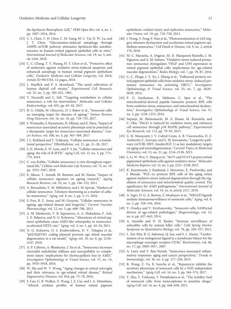
observed at the cellular level as the dysfunction of self-repairand renewal, cell cycle arrest, and the appearance of SNCs[26]. Changes in the immune system function and the apo-ptotic resistance of SNCs result in SNC accumulation [27],causing a range of age-related diseases, such as Alzheimer’sdisease, osteoarthritis, pulmonary fibrosis, and AMD [28,29]. The p16INK4A-pRB and p53-p21CIP1/WAF1 pathways areprimarily involved in the mechanism of cellular senescence(Figure 2) [26, 30]. The activation of p53 upregulatesp21CIP1/WAF1 and inhibits the cell cycle proteins cyclin A, E,and D. The activation of the pRB pathway is mediated byp16INK4A, which is independent of p53. p16INK4A inhibitsCyclin A-, Cyclin E-, and Cyclin D-dependent kinase com-plexes, which normally phosphorylate and inactivate pRB.Dephosphorylated pRB represses the G1/S transition by
sequestering E2F transcription factors, thereby inhibitingE2F-dependent gene expression [30]. Although SNCs areblocked at the G0/G1 or G2/M stages and cannot undergo celldivision, they can still exist in a long-term metabolicallyactive state, accompanied by the upregulation of inflamma-tory factors, chemokines, matrix remodeling proteases, andgrowth factors, which are collectively referred to as SASP.SASP in the tissue microenvironment promotes a series ofinflammation cascades and accelerates the senescence of sur-rounding cells [28, 31], which is related to age-related inflam-matory reactions, metabolic disorders, stem cell dysfunction,and chronic diseases [29]. The SASP components varydepending on cell type and senescence trigger factors. Theproinflammatory cytokines IL-1α, IL-1β, IL-6, and IL-8 areclassical SASP components. Multiple genes are involved inthe biological regulation of SASP, including NK-κB, p38-MAPK, mTOR, and GATA4 [28].
Cellular senescence can be divided into two types: repli-cative senescence (RS) and stress-induced premature senes-cence (SIPS) [32, 33]. Recently, scholars have proposed athird type, developmentally programmed senescence (DPS)[31]. RS is caused by telomere shortening during cell replica-tion [28]. A telomere is a type of complex composed of pro-teins and nucleotides containing TTAGGG repeats found atthe ends of eukaryotic chromosomes [33]. To protect againstgenomic instability caused by shortened telomeres, DNAdamage response (DDR) activates to induce a series of cas-cade reactions, including ATM/ATR-mediated p53-p21CIP1/-WAF1 and p16INK4A-pRB pathway activation, cell cycle arrest,and apoptosis. Precipitating factors for SIPS include oxida-tive stress, oncogenes, genotoxic damage, chemotherapy,and viral infection [26, 30, 31]. DPS can occur anywhere dur-ing the process of mammalian embryo formation. Interest-ingly, DNA damage markers and the DNA damage-dependent kinase ATM/ATR were not detected in DPS cells.Megakaryocytes and NK cells are the only adult cell typesthat appear to undergo DPS [31]. Currently, the followingmarkers are used to determine cell senescence: (1) altered cel-lular morphology (often enlarged, flat, multivacuoled, andmultinucleated); (2) increased Senescence β-Galactosidase(SA-β-GAL) activity; (3) the accumulation of DNA damagefoci; (4) the accumulation of senescence-associated hetero-chromatic foci (SAHF) and other chromatin modifications;(5) chromosomal instability; (6) the induction of SASP; and(7) the altered expression of senescence-related genes (i.e.,p53, p21CIP1/WAF1, p16INK4A, pRB, and cyclin-dependentkinases) [31, 32, 34].
Cellular senescence is one of the pathogenic factorsunderlying AMD. The senescence-accelerated OXYS rat isan animal model of AMD that can spontaneously undergoan AMD-like retinopathy, including RPE degeneration, lossof photoreceptors, and the decreased expression of vascularendothelial growth factor (VEGF) and pigment epithelial-derived factor (PEGF) [35, 36]. Chorionic capillary mem-brane attack complex (MAC) deposition can cause chorioniccapillary degeneration and RPE atrophy, leading to dryAMD. Senescent chorioretinal endothelial cells are signifi-cantly stiffer than normal cells, which correlates with highercytoskeletal Rho activity and more susceptibility to MAC
Oxidative stressUltravioletradiation DNA
damage
Telomereshortening
Causes
FOXOsignalingpathway
mTORsignalingpathway
p53-p21signalingpathway
p16-RBsignalingpathway
Calciumsignalingpathway
Mechanisms
Cellular senescence
Consequence
Characteristics
M
G1
S
G2
Apoptosis
Growth arrest Apoptosis resistance SASP
Figure 2: An overview of cellular senescence. A variety of stimuli,such as oxidative stress, DNA damage, ultraviolet radiation, andtelomere shortening can induce a series of reactions, including theactivation of the FOXO signaling pathway, the mTOR signalingpathway, the p53-p21 signaling pathway, the p16-Rb signalingpathway, and the calcium signaling pathway, ultimately leading tocellular senescence. SNCs have several primary characteristics,such as growth arrest, apoptosis resistance, and SASP secretion.
4 Oxidative Medicine and Cellular Longevity

injury [37]. Each microglial cell possesses ramified, branch-ing processes that exhibit rapid, constitutive motility, whichenables the cell to effectively survey the extracellular milieuin its vicinity. While microglial somata are evenly spacedand relatively stationary in the uninjured state, followingfocal injury, microglia promptly polarize their processesand migrate in the direction of the injury to cluster aroundthe injury site. However, a thickened glial layer, decreasedbranch numbers, shortened lengths, and slowed movementmay occur during aging, which can lead to changes inhomeostasis and promote long-term retinal neuroinflamma-tion, as reflected by increased levels of complement C3 andCFB [38], further promoting AMD progress.
Telomere shortening of RPE cells is also one of the char-acteristics of AMD. Late passage RPE cells from primary cul-ture demonstrated a reduced capacity for cellular division,which could be caused by telomere shortening [39]. A2Ecould contribute to RPE cellular senescence, accompaniedby telomere deprotection and deletion, and telomerase over-expression rescued A2E-mediated RPE cellular senescence,indicating that telomere dysfunction plays an important rolein A2E-based RPE cellular senescence [40].
Long-term and chronic oxidative stress, which are patho-genic factors of AMD, can be generated by cigarettes, hydro-gen peroxide (H2O2), tert-butyl hydroperoxide (TBHP), andlight and can result in the premature aging of RPE cells,which is characterized by increases in ROS and SA-β-GALactivities, higher expression levels of p53, p21WAF1/CIP1,p16INK4A, and SASP factors, the accumulation of p-γH2AXfoci and 8-OHdG DNA damage lesions, mitochondrial dys-function, increased VEGF, and decreased CFH [41–44].HTRA1 is closely related to AMD and can accelerate H2O2-mediated RPE senescence through the p38 pathway [44].
Many studies have shown that inhibiting oxidative stresscan reduce RPE senescence. For instance, fullerenol, an effec-tive free radical scavenger and antioxidant, can strengthenthe antioxidant reaction of RPE and alleviate DNA damageby activating SIRT1 and downregulating p53 and p21CIP1/-WAF1 levels [42]. SIRT1, a member of the SIRT family, isthe primary longevity gene that prolongs life and reducescancer-associated metabolic syndrome [45]. Humanin hasbeen shown to have anti-inflammatory and cell-protectiveeffects in a variety of cell types. Humanin alleviates RPE oxi-dative stress damage and senescence by phosphorylatingSTAT3 and inhibiting caspase-3 activation [43]. Both SIRT1and STAT3 have protective effects on RPE cells. Under oxi-dative stress, SIRT1 is downregulated, while STAT3 is upreg-ulated, and the regulation of STAT3 is independent of SIRT1[46]. PCG1α, a transcription regulator, is involved in mito-chondrial metabolism and is associated with many age-related diseases. PCG1α protects RPE cells from oxidativestress by upregulating antioxidant enzymes and DDR and isregulated by AMPK and SIRT1 during the process of post-transcriptional modification and activation [47].
Interfering with the proageing effects of SNCs, either byeliminating SNCs entirely or by shutting down their secre-tory machinery, is now being considered as a potential strat-egy for treating diseases associated with aging. The selectiveremoval of SNCs can prolong life and reduce some sideeffects of drugs, such as bone marrow suppression, cardiacdysfunction, and toxic effects. Broadly, three strategies havebeen used for the selective elimination of SNCs (Table 1):(1) immune-mediated SNC clearance, which utilizes anti-bodies targeting senescence-specific surface antigens to clearSNCs; (2) senescent cell lysis (senolysis), which leads to thedeath of SNCs by activating apoptotic pathways; and (3)
Table 1: Candidate senotherapies.
Cell/agent Target (or targets) Target SNC types Refs
Immune-mediated SNC clearance
NK cells NKG2D IMR-90 cells, cancer cells [48, 50]
Macrophages oxPCCD36 SNCs in atherosclerotic plaques [51]
MonocytesMIF-CXCR2 axis SNCs in atherosclerotic plaques [49]
CD44 HUVECs [28]
Ipilimumab CTLA-4, PD1 Cancer cells [52]
Senolysis
D p21CIP1/WAF1, tyrosine kinases Fat precursor cells [54]
KKKQ PI3K Human endothelial cells, mouse BMSCs [54]
D+Q p21CIP1/WAF1, p16INK4A BCL-Xl, PAI-2, SASP MEFs, IMR-90 cells [54–56]
ABT-263 BCL-2, BCL-W, BCL-Xl MEFs, IMR-90 cells, HUVECs [27, 28, 53]
ABT-737 BCL-W, BCL-Xl MEFs, IMR-90 cells, HUVECs [27, 28, 53]
FOXO4-related-peptide p53 IMR-90 cells [61]
AP20187 p16INK4A Mouse BMSCs [62]
SASP neutralization
Rapamycin mTOR1 IMR-90 cells, MEFs [28, 29, 53]
Metformin IκB, IKKα/β IMR-90 cells [63]
JAK1/2 inhibitors JAK1/2 Human primary preadipocytes, HUVECs [65]
UBX0101 SASP factors Chondrocytes [27, 66]
5Oxidative Medicine and Cellular Longevity

SASP neutralization, including the inhibition of SASP-related signaling cascades, interference with the SASP secre-tome, and the inhibition of individual secretion factors.Among these, senolysis holds the most therapeutic promise.Currently, no relevant strategies for SNC clearance has beenapplied to AMD treatment [27, 28].
3.1. Immune Surveillance Mediates SNC Clearance. NK cellsare a component of the innate immune system. One of thereceptors responsible for NK cell activation, the NKG2Dreceptor, has been implicated in the interaction betweenNK cells and SNCs during tumorigenesis, tumor therapy,and tissue injury. The NKG2D receptor recognizes theligands MICA/B and ULBP1-6 on the surface of SNCs torecruit NK cells for immune surveillance regulation. Forexample, NK cells mediate the clearance of SNCs during liverfibrosis [48]. Intercellular adhesion molecule 1 (ICAM-1) iscommonly present on the surface of SNCs and might cooper-ate with NKG2D ligands to amplify the cytotoxicity of NKcells [49]. P53-positive SNC accumulation mediates the gen-eration of CCL2, 3, 4, and 5 and CXCL1 and 2. These cyto-kines activate NK cells and recruit immune cells to clearsenescent tumor cells [50]. Macrophages are tissue-residentphagocytes equipped with a complete arsenal of pathogenrecognition receptors that enable them to sense potentialrisks. Upon stimulation, macrophages acquire context-dependent phenotypes by undergoing either classical M1 orM2 polarized activation. For example, senescent hepatic stel-late cells (HSCs) release SASP factors such as IFN-c and IL-6that skew macrophages toward the M1 state to attack HSCsduring liver diseases [49]. Furthermore, the macrophagescavenger receptor CD36 (oxPCCD36) is enriched in both ath-erosclerotic plaques and on SNC membranes, serving as acritical participant in macrophage recognition. This receptorproduces a surface accessible phagocytic “eat me signal” tofacilitate the recognition of SNCs and oxidized lipoproteinsas part of its immune surveillance function [51]. In additionto NK cells and macrophages, monocytes also participate inimmune-mediated SNC clearance. Senescent fibroblasts canstimulate monocyte production in the bone marrow via therobust secretion of two inflammatory SASP components,GM-CSF and G-CSF. These SASP factors can direct mono-cytes to the SNCs. Then, SNCs promote the differentiationof these monocytes into macrophages via the secretion ofM-CSF [49]. Ipilimumab, an antibody that enhances cyto-toxic T cell activation through the blockade of the cytotoxicT-lymphocyte-associated protein 4 (CTLA-4) receptor orthe antiprogrammed cell death protein 1 (PD1), can activatethe immune surveillance response that has been suppressedin cancer cells [52]. In addition, antibodies againstsenescence-specific surface antigens, such as CD44 in endo-thelial cells, could induce a direct immune response ordeliver cytotoxic drugs to senescent lesions to mediate SNCclearance [28].
3.2. Senolysis Mediates SNC Apoptosis. Dasatinib (D), a tyro-sine kinase inhibitor, can inhibit cell replication and migra-tion and induce apoptosis [53]. Quercetin (Q) is a flavonoidcomplement. D alone can downregulate p21CIP1/WAF1,
clearing senescent fat precursor cells. Q clears senescenthuman endothelial cells and mouse bone marrow mesenchy-mal stem cells (BM-MSCs). D+Q can downregulate p21CIP1/-WAF1, BCL-Xl, and PAI-2 and effectively clear senescentfibroblasts (MEFs) [54]. Bleomycin induces the age-dependent accumulation of senescent MEFs in the lungs, fur-ther leading to pulmonary fibrosis. D+Q treatment can clearSNCs mediated by bleomycin and downregulate p16INK4 andthe SASP components MCP-1, IL-6, MMP-2, and TGF-β[55]. Transplanting relatively small numbers of SNCs intoyoung mice caused persistent physical dysfunction andspread cellular senescence to host tissues. The applicationof D+Q decreased the numbers of naturally occurring senes-cent cells and decreased the secretion of the SASP compo-nents IL-6, IL-8, MCP-1, PAI-1, and GM-CSF. Moreover,the administration of D+Q to both senescent cell-transplanted young mice and naturally aged mice alleviatedphysical dysfunction, increased posttreatment survival, andreduced mortality hazard [56]. The application of SNCremoval has not been studied in AMD, but studies haveshown that Q can protect RPE cells from oxidative stressthrough its antioxidant effects [57], inhibit choroid neovas-cularization [58], and upregulate BCL-2 while downregulat-ing Bax [59, 60]. ABT-263, an activator of themitochondrial apoptosis pathway, can inhibit Bcl-2, BCL-W, and BCL-Xl. ABT-737 can inhibit BCL-W and BCL-Xl.Both ABT-263 and ABT-737 are involved in removing senes-cent MEFs from pulmonary and human umbilical vein endo-thelial cells (HUVECs) [27, 28, 53]. FOXO4 is elevated inSNCs and maintains their viability. FOXO4 exists in thePML body and combines with p53 DNA-SCARS. DRI is akind of polypeptide that has been used in phase I clinical tri-als against solid tumors. Researchers have designed and syn-thesized FOXO4-DRI to effectively and powerfully targetSNCs andmediate p53-dependent apoptosis to remove SNCsby destroying PML/DNA-SCARS in SNCs and competingwith FOXO4 to bind to P53. At the tissue level, FOXO4-DRI alleviated hepatic dysfunction induced by chemotherapyand improved the frailty properties and renal functions ofboth xpdTTD/TTD mice (an animal model of prematureaging) and naturally aged mice [61]. In another study, SNCswere marked using p16INK4A. An aging BubR1H/H mousemodel containing INK-ATTAC lines was established, whichshowed shortened lifetime, lordosis, cataracts, and the aggre-gation of p16INK4A-positive cells. AP20187, a synthetic drugthat induces apoptosis through cell membrane dimerization,was given to the BubR1H/H mice. AP20187 activated INK-ATTAC, which aided the accurate identification ofp16INK4A-positive SNCs and effectively cleared them whilenot affecting normal cells, reducing the senescent phenotypesof adipose tissue, skeletal muscle, and the eye [62].
3.3. SASP Neutralization Mediates the Weakened ProagingEffect of SNCs. SASP inhibitors include rapamycin, metfor-min, and JAK1/2 inhibitors. Rapamycin reduces the secre-tome of inflammatory factors in SNCs by inhibitingmTOR1 [28, 53], playing a role in prolonging lifespan, andreducing age-related fatty tissue loss, heart failure, and cogni-tive impairment [29]. Metformin inactivates NF-κB and
6 Oxidative Medicine and Cellular Longevity

reduces SASP component levels by inhibiting the phosphor-ylation of IκB and IKKα/β [63]. JAK is a tyrosine kinase thatis highly active in SNCs [64]. Using siRNA or JAK inhibitorsto inhibit the secretion of the SASP factors IL-6, IL-8, andMCP-1 in both senescent adipose progenitor cells andHUVECs improved the physical functions of elderly miceand alleviated insulin resistance and stem cell dysfunction[29, 65]. UBX0101, a senolytic molecule, can combine withMMP-13, IL-6, and IL-1β [27]. The intra-articular injectionof UBX0101 selectively eliminated SNCs after anterior cruci-ate ligament transection (ACLT), attenuated the develop-ment of posttraumatic OA, reduced pain, and increasedcartilage development [66].
Among the three aging-therapy strategies, senolysis holdsthe most therapeutic promise for two reasons. First, the per-manent removal of SNCs leads to the durable abolishment ofdeleterious SASP components. Second, once SNCs are elim-inated, there is no risk of tumorigenic “escape” from senes-cence, which may be possible if SNCs are permitted tolinger indefinitely [27]. However, almost all drugs have off-target and bystander effects. For example, the removal ofp16INK4A-positive cells by senolytic drugs has the followingproblems: (1) not all senescent cells necessarily haveincreased p16INK4A expression; (2) not every cell with sub-stantial p16INK4A expression is senescent; (3) targeting agingmechanisms can phenocopy the effects of genetic or pharma-cological SNC clearance without actually affecting SNCs; and(4) hypothetically, the genetic clearance of p16INK4A-positivecells could have the same effects on a particular downstreamphenotype as a drug that affects that downstream phenotypedirectly, without affecting truly senescent p16INK4A-positivecells. To determine whether senolytic drugs actually causethe alleviation of senescence-associated phenotypes due toSNC clearance requires following a modified set of Koch’spostulates, which are the following: (1) individual SNCs ortransplanted SNCs must have a senescent phenotype; (2)the clearance of SNCs genetically or pharmacologically mustbe associated with the alleviation of the phenotype; and (3)the effects on the phenotype should persist even after thedrug has been removed [29].
SNC clearance can alleviate the further senescence andtissue damage of surrounding cells, thereby delaying diseaseprogression. However, SNC clearance methods are not uni-versal and depend on the types of cells and diseases, whichcomplicates the treatment prospects [67]. At present, thescavenging of SNCs has not been applied to AMD, and alarge amount of in-depth research is needed to confirmwhether SNC clearance is feasible for AMD prevention andtreatment.
4. Abnormal Immune-Inflammatory ResponsesAre Pathogenic Factors for AMD
Inflammation is the body’s response to cell and tissue dam-age and occurs through a series of processes that are designedfor the eventual clearance of pathogens and the repair ofdamaged tissue. Acute inflammation is a short-term processthat involves leukocyte infiltration, the removal of the trigger,and tissue repair. Chronic inflammation is a prolonged
response that can result in tissue injury or destruction if theinciting trigger is not neutralized [1]. Inflammation is a com-mon cause of age-related diseases. Chronic inflammation isinvolved in AMD [68], and the “immune-inflammation”model of AMD has been broadly accepted [69]. The retinais a purported “immune privileged” site, protected by theBRB, ocular anti-inflammatory and anti-immune proteins,and the anterior chamber-associated immune deviation[70]. Once these protective mechanisms are destroyed,abnormal immune and inflammatory responses occur, accel-erating the development of AMD.
4.1. PRR and Inflammasome Release Mediates a ChronicInflammatory Response. The body recognizes exogenouspathogens and endogenous risk factors through pattern rec-ognition receptors (PRRs) that sense microbes through con-served molecular structures called pathogen-associatedmolecular patterns (PAMPs). PPRs include the Toll-likereceptors (TLRs), the nucleotide-binding oligomerization-(NOD-) like receptors (NLRs), the RIG-I-like receptors(RLRs), the C-type lectin receptors, and advanced glycosyla-tion end product (AGE) receptors (RAGE) [71]. When thesereceptors bind to their corresponding ligands, inflamma-somes in cells activate, causing the release of inflammatorymediators. NLRP3 is a member of the NLR family, whichcan assemble into a large oligomeric structure through therecruitment of an adaptor protein, ASC, and procaspase-1and can subsequently produce mature IL-1β and IL-18through a two-step process. Classically, the first step, referredto as inflammasome priming, involves the NF-κB-mediatedsynthesis of the inactive precursors pro-IL-1β and pro-IL-18 in response to the recognition of a specific ligand by itscorresponding PRR and the upregulation of inflammasomecomponents, including NLRP3. A second signal is requiredfor NLRP3 oligomerization, the recruitment of ASC and pro-caspase-1, and the subsequent cleavage of procaspase-1 intoits active form, leading to the processing of pro-IL-1β andpro-IL-18 and eventually to the release of the mature cyto-kines IL-1β and IL-18 [72]. High expression levels of NLRP3,IL-1β, and IL-18 can be detected in the photoreceptor andRPE cells of AMD patients [73]. Mitochondrial dysfunction,oxidative stress, and drusen can overly activate NLRP3 [13].Laser-induced choroidal neovascularization (CNV), a mousemodel of wet AMD, is exacerbated in NLRP3-/- mice [74].However, due to the existence of several nonspecific com-mercially available anti-NLRP3 antibodies that questionscurrent interpretation of results reporting NLRP3 expressionand upregulation in the RPE cells of AMD patients, the prob-lems with NLRP3 activation in RPE cells and the measure-ments of this process have been signalized recently [75].The study argues that RPE cells may not contain meaningfulamounts of NLRP3 to contribute to diseased states and sug-gests that if NLRP3 is implicated in AMD, it is more likelyto be related to immune cells, either resident or infiltrating.Thus, further evidence is required to characterize the pres-ence and source and activation of pro-IL-18 in AMD.
Alu is the most abundant transposable element, which istranscribed into Alu RNAs, and the accumulation of AluRNAs has been confirmed to be related to AMD [76]. Alu
7Oxidative Medicine and Cellular Longevity

RNAs, by reducing DICER1, can activate the inflammasomein RPE cells and increase IL-18 levels, leading to geographicatrophy. Additionally, DICER1 deficiency combined withAlu RNA accumulation resulted in increased IL-18 levels,which led to RPE cell death via the activation of caspase-8through a Fas ligand-dependent mechanism [1].
In addition to RAGE, some substances that are secretedby dead cells and damaged tissues are also receptors forAGEs, including amyloid β-protein (Aβ). In the central ner-vous system, the accumulation of Aβ is associated with theactivation of neurodegenerative and inflammatory pathways.In the ocular system, Aβ upregulates IL-1β, IL-18, and TNF-α in RPE cells. The intravitreal injection of Aβ can activateinflammation [77]. AGEs accumulate with aging. AGEdeposits were found in drusen, and studies have suggestedthat AGE plays a role in the promotion of oxidative stress,apoptosis, and lipofuscin accumulation. The in vitro incuba-tion of RPE cells with AGEs resulted in the upregulation ofthe anti-inflammatory cytokines IL-10, IL-1ra, and IL-9 andthe proinflammatory cytokines IL-4, IL-15, and IFN-γ, whileother proinflammatory cytokines, such as IL-8, MCP-1, andIP10, were downregulated, suggesting a that parainflamma-tion state occurred under AGE stimulation [78]. Parainflam-mation, a state between normal and inflammatory responses,is thought to be beneficial for the host. However, if tissuemalfunction is sustained over long periods, parainflamma-tion can become chronic and maladaptive. In AMD, the bal-ance between stress-induced damage and parainflammationis often disrupted due to environmental and genetic factors,resulting in a chronic inflammatory state [79]. One explana-tion for the shift from early AMD to late AMD is that triggerscan switch an aging homeostatic parainflammatory responseinto a persistent low-grade inflammatory response, leading tothe loss of RPE cells and/or pathological angiogenesis [80].All of these data suggest that PRRs and inflammasomes haveclose associations with AMD.
4.2. Abnormal Complement System Amplifies CascadeReaction. The complement system is part of the host innateimmune system that enables many essential functions,including the following: (1) the opsonization and lysis ofmicroorganisms, (2) the recruitment of inflammatory cells,(3) the removal of dead cells, (4) the regulation of antibodyproduction, and (5) the removal of immune complexes.There are three classic complement pathways: the classicalpathway, the mannan-binding lectin (MBL) pathway, andthe alternative pathway. All of these pathways ultimately leadto the formation of the cytolytic MAC [68]. The complementsystem is a double-edged sword for the retina. A low level ofcomplement activation is beneficial to immune privilege, andRPE cells can produce complement components belonging tothe classical pathway and the alternative pathway such asmembrane-binding regulators and soluble regulators to pre-vent excessive complement pathway activation [81]. How-ever, if the complement pathway is overactivated, it candamage retinal tissues and lead to the chemotactic aggrega-tion of immunocytes. Studies have shown that plasma con-centrations of the activation products C3a, C3d, Ba, Bb,C5a, and CFH are high in AMD patients. Similar alterations
were observed for C3, C3d, and C5-9 in drusen. C-reactiveprotein (CRP) and C5 were primarily found in dry AMD,while C3a and C5a were primarily found in wet AMD [82,83]. CRP is a biomarker of acute inflammation and playsan essential role in the innate immune response to tissueinjury and/or infection, inducing complement activation viathe alternative pathway [69]. Complement factor H (CFH)and HTRA1/ARMS polymorphisms contribute to more than50% of the genetic risk for AMD [84]. CRP damages cells andtissues by binding to DNA or phosphocholine that has beenexposed in injured cells and activating the classical comple-ment pathway, resulting in the formation of the C3 conver-tase, which generates C3b. By binding the inhibitor CFH,C3b promotes the complement cascade and the formationof the C5 convertase. Polymorphisms in the complementcomponents C2 and Factor B (CFB) are protective forAMD. C2 is a component of the classical complement path-way, and CFB is involved in the alternative pathway. Geneticand functional data suggest that this protective effect is morelikely to be mediated by mutations in the CFB gene than bymutations in the C2 gene. The AMD-associated CFB variantsmodulate the activation of the alternative complement path-way and, therefore, may lead to an overall deregulation of thecomplement system, which may lead to the further amplifica-tion and inflammation of the complement cascade [83]. Thecomplement system is also closely associated with inflamma-tion. The inflammasome can be activated by a number oftriggers, notably C5b-9 and C3a. The C3a-mediated ATPrelease prompts the P2X7 receptor to bind to and activateNLRP3 [1]. Thus, the complement system and the inflamma-some can synergize to promote AMD progression afterabnormal activation.
4.3. The Activation of Immune Cells and Cytokines PromotesInflammation. Immune cells in a normal retina includemicroglial cells (MCs), macrophages, and dendritic cells.MCs play roles in neuronal homeostasis and immune surveil-lance, which are normally absent from the outer retina butcan infiltrate into the subretinal space and become activatedduring aging and AMD, likely to support the RPE cells andclear age-related debris. However, MCs may also induce oxi-dative stress and promote further degeneration. Two chemo-kines, CX3CL1 and CCL2, and their respective receptors,CX3CR1 and CCR2, play important functions for the recruit-ment of macrophages/microglia to tissue lesions [70]. InCX3CR1-deficient (CX3CR1-/-) mice, MCs and drusen-likedeposits accumulated subretinally with age [85]. InCX3CR1-/-/CCL2-/- (double knockout) mice, AMD-like reti-nal lesions developed, characterized by abnormal RPE cells,drusen, photoreceptor atrophy, and choroidal neovasculari-zation [86]. Macrophages, a predominant cell type associatedwith chronic inflammation, are the most prominent inflam-matory cells observed in AMD tissue, outnumbering subret-inal MCs and lymphocytes in AMD eyes. Macrophagessecrete a wide range of cytokines, chemokines, complementfactors, and growth factors, all of which depend on the incit-ing stimuli, macrophage subtype, and location. Macrophagescan display as different subclasses, namely, the M1 and M2macrophages. M1 macrophages have been shown to be
8 Oxidative Medicine and Cellular Longevity

proinflammatory, with an IL-12high, IL-23high, and IL-10low
phenotype, while the M2 macrophages are relatively anti-inflammatory with IL-12low, IL-23low, and IL-10high pheno-type. In addition, CXCL9, CXCL10, and CXCL11 representM1 chemokines, and CCL17 and CCL22 represent M2 che-mokines [70]. CXCL11 is strongly immunoreactive and asso-ciated with drusen. The upregulation of CXCL1, along withviperin and RSAD2, may play a role in driving the inflamma-tory response via the NF-κB and JAK-STAT pathways [78].IL-17 has previously been shown to be involved in inflamma-tion and autoimmune diseases and can be produced by Tcells and innate immune cells (ILC). The IL-17 cytokine fam-ily includes six members named IL-17A-F. IL-17A, producedprimarily by Th17 cells, is the primary subfamily member.Under specific conditions, other inflammatory cells such asneutrophils and even macrophages can produce IL-17A. IL-17A homodimers bind IL-17 receptor C (IL-17RC)/IL-17RA heterodimers, which are involved in proinflammatoryresponses. IL-17 produced by γδT, and ILC promoted exper-imental intraocular neovascularization during laser-inducedCNV in mice. Additionally, there was a greater increase inthe expression levels of IL-17RC in the blood of siblings withAMD compared to that in the blood of their respective sib-lings without AMD, and increased levels of IL-17RC cancause damage to photoreceptors [70, 87]. Therefore, immunecells can secrete inflammatory cytokines, further promotingretinal inflammatory responses.
5. Autophagy Dysfunction, Cellular Senescence,and Abnormal Immune-InflammatoryResponses Can Promote or InhibitEach Other
Autophagy dysfunction, cellular senescence, and abnormalimmune-inflammatory responses interact with each other.Autophagy dysfunction accompanied by lipofuscin accumu-lation and ROS increases, can activate inflammatory reac-tions, further promoting long-term and chronic cascadeinflammation and accelerating RPE cell senescence [13].Nrf2, a basic leucine zipper transcription factor, regulates acoordinated transcriptional program that allows cellularredox homeostasis while protecting the cell from oxidativeinjury [1]. Nrf2 physically interacts with a negative regulatorKeap1, which targets the Nrf2 protein for ubiquitination andproteasomal degradation within the cytoplasm, thus limitingits activity. However, under oxidative stress, Keap1undergoes a conformational modification and releases Nrf2,allowing it to undergo translocation to the nucleus, where itbinds to antioxidant response elements (AREs), thus activat-ing the transcription of its target genes [88]. P62/SQSTM1, amultidomain protein that regulates autophagy, has beenlinked to inflammation, apoptosis, and age-related patholo-gies. In RPE cells, p62 promotes autophagy and simulta-neously enhances a Nrf2-mediated antioxidant response toprotect against acute oxidative stress and mediate anti-inflammatory effects via the inhibition of the NK-κB path-way. It appears that the role Nrf2 plays in autophagy, espe-cially through interactions with p62, is strongly dependent
on the cellular context as there are many reports suggestingthat this protein acts differently depending on the cellularstate [8]. Aging can lead to the downregulation of Nrf2 [1].The administration of a p62/SQSTM1-encoding plasmid inOXYS mice decreased the incidence and severity of retinop-athy and downregulated proinflammatory cytokines [36].All of this data suggests that autophagy, cellular senescence,and inflammation can be linked through p62/SQSTM1 andthat p62/SQSTM1 can be used as a target for the improve-ment of autophagy, the inhibition of retinal inflammation,and the antiaging of RPE cells.
SIRT6 and autophagic markers are upregulated in theRPE cells of aged mice. Intravitreal injections of Aβ activatedSIRT6, autophagy, and inflammation. Silencing SIRT6 led tothe decreased expression levels of Beclin1, ATG5, and LC3.Using 3-MA to inhibit autophagy mediated by Aβ led todecreased levels of IL-1β, IL-6, IL-8, IL-12b, NLRP3, andTNF-α [77], indicating that autophagy dysfunction resultedin the inhibition of inflammation. A2E, a major componentof toxic lipofuscin that has been implicated in AMD, isdeposited in RPE cells with age and can secreteinflammation-associated and angiogenic factors. The contin-uous incubation of RPE cells with A2E induced autophagythrough the AKT/mTOR pathway and decreased cell viabil-ity in a concentration- and time-dependent manner. Theapplication of 3-MA decreased the number of autophago-somes and LC3 puncta induced by A2E, increased theinflammation-associated expression levels of proteins,including ICAM, IL-1β, IL-2, IL-6, IL-8, IL-17A, IL-22, andSDF-1, and upregulated VEGFA expression. In contrast,rapamycin augmented A2E-mediated autophagy and attenu-ated the protein expression of inflammation-associated andangiogenic factors [21], indicating that autophagy dysfunc-tion was accompanied by the upregulation of inflammatoryresponses. In addition, intracellular protein accumulationand autophagy inhibition can mediate NLRP3 activationand Alu RNA accumulation in RPE cells [89–91], thus acti-vating inflammation [8, 18]. Therefore, changes to theautophagy and inflammatory responses are not unidirec-tional, and autophagy dysfunction can be accompanied byeither the promotion or inhibition of inflammation.
With the depletion of glutathione (GSH) from RPE cells,increased autophagy and SIPS activation were apparent, asreflected by increased LC3 expression levels, autophagic vac-uoles, and autophagic flux and an increased percentage ofSA-β-positive cells, SAHFs, and cell cycle arrest at the G1phase, indicating that SIPS and increased autophagyoccurred simultaneously. However, the inhibition of autoph-agy with 3-MA promoted SIPS whereas inducing autophagywith rapamycin attenuated SIPS [92].
In summary, autophagy dysfunction, cellular senescence,and abnormal immune-inflammatory responses interactwith each other and jointly participate in and promote AMD.
6. Conclusion
AMD is a blinding disease caused by genetic and envi-ronmental factors. The roles of autophagy dysfunction inRPE cells, cellular senescence, and abnormal immune-
9Oxidative Medicine and Cellular Longevity

inflammatory responses have been recognized in AMD. Therelationships among these three processes can be described asboth stimulating and restrictive. Autophagy dysfunction inRPE cells leads to clearance system abnormalities. Cellularsenescence leads to cell dysfunction and the promotion ofsenescence among neighboring cells. Abnormal immune-inflammatory responses lead to chronic retinal inflamma-tion. Autophagy dysfunction can accelerate the senescenceof RPE cells, while either promoting or inhibiting inflamma-tion. In conclusion, if improved autophagy, alleviated cellularsenescence, and the inhibition of abnormal retinal immune-inflammation responses can be achieved simultaneously, itmay be possible to delay the progress of AMD and to obtainbetter clinical efficacy. At present, these three antiaging strat-egies have achieved good results when applied to atheroscle-rosis, pulmonary fibrosis, and osteoarthritis. Although thereis currently no relevant application of these strategies forAMD, the use of antiaging strategies for AMD preventionand treatment is expected to achieve a new breakthrough inthe future.
Conflicts of Interest
All the authors declare that there are no financial or any otherconflicts of interest.
Acknowledgments
This work was supported by grants from the ProvincialFrontier and Key Technology Innovation Special Fund ofGuangdong Province (No. 2015B020227001) and theScience and Technology Program of Guangzhou (No.2016201604030016).
References
[1] S. Datta, M. Cano, K. Ebrahimi, L. Wang, and J. T. Handa,“The impact of oxidative stress and inflammation on RPEdegeneration in non-neovascular AMD,” Progress in Retinaland Eye Research, vol. 60, pp. 201–218, 2017.
[2] I. Johansson, V. T. Monsen, K. Pettersen et al., “The marine n-3 PUFA DHA evokes cytoprotection against oxidative stressand protein misfolding by inducing autophagy and NFE2L2in human retinal pigment epithelial cells,” Autophagy,vol. 11, no. 9, pp. 1636–1651, 2015.
[3] J. Blasiak, M. Piechota, E. Pawlowska, M. Szatkowska,E. Sikora, and K. Kaarniranta, “Cellular senescence in age-related macular degeneration: can autophagy and DNA dam-age response play a role?,” Oxidative Medicine and CellularLongevity, vol. 2017, Article ID 5293258, 15 pages, 2017.
[4] O. S. Kozhevnikova, E. E. Korbolina, N. I. Ershov, and N. G.Kolosova, “Rat retinal transcriptome: effects of aging andAMD-like retinopathy,” Cell Cycle, vol. 12, no. 11, pp. 1745–1761, 2013.
[5] J. Blasiak, S. Glowacki, A. Kauppinen, and K. Kaarniranta,“Mitochondrial and nuclear DNA damage and repair in age-related macular degeneration,” International Journal of Molec-ular Sciences, vol. 14, no. 2, pp. 2996–3010, 2013.
[6] K. V. Chalam, V. Khetpal, R. Rusovici, and S. Balaiya, “Areview: role of ultraviolet radiation in age-related macular
degeneration,” Eye & Contact Lens: Science & Clinical Practice,vol. 37, no. 4, pp. 225–232, 2011.
[7] S. K. Mitter, C. Song, X. Qi et al., “Dysregulated autophagy inthe RPE is associated with increased susceptibility to oxidativestress and AMD,” Autophagy, vol. 10, no. 11, pp. 1989–2005,2014.
[8] K. Kaarniranta, P. Tokarz, A. Koskela, J. Paterno, andJ. Blasiak, “Autophagy regulates death of retinal pigment epi-thelium cells in age-related macular degeneration,” Cell Biol-ogy and Toxicology, vol. 33, no. 2, pp. 113–128, 2017.
[9] D. J. Klionsky, K. Abdelmohsen, A. Abe, M. J. Abedin,H. Abeliovich, A. A. Arozena et al., Guidelines for the Useand Interpretation of Assays for Monitoring Autophagy,Autophagy, 3rd edition, 2016.
[10] R. W. Young, “The renewal of photoreceptor cell outer seg-ments,” Journal of Cell Biology, vol. 33, no. 1, pp. 61–72, 1967.
[11] R. W. Young and B. Droz, “The renewal of protein in retinalrods and cones,” Journal of Cell Biology, vol. 39, no. 1,pp. 169–184, 1968.
[12] R. W. Young and D. Bok, “Participation of the retinal pigmentepithelium in the rod outer segment renewal process,” Journalof Cell Biology, vol. 42, no. 2, pp. 392–403, 1969.
[13] K. Kaarniranta, D. Sinha, J. Blasiak et al., “Autophagy and het-erophagy dysregulation leads to retinal pigment epitheliumdysfunction and development of age-related macular degener-ation,” Autophagy, vol. 9, no. 7, pp. 973–984, 2013.
[14] L. Lei, R. Tzekov, H. Li et al., “Inhibition or stimulation ofautophagy affects early formation of lipofuscin-like autofluo-rescence in the retinal pigment epithelium cell,” InternationalJournal of Molecular Sciences, vol. 18, no. 4, p. 728, 2017.
[15] E. Keeling, A. J. Lotery, D. A. Tumbarello, and J. A. Ratnayaka,“Impaired cargo clearance in the retinal pigment epithelium(RPE) underlies irreversible blinding diseases,” Cells, vol. 7,no. 2, p. 16, 2018.
[16] L. Perusek, B. Sahu, T. Parmar et al., “Di-retinoid-pyridinium-ethanolamine (A2E) accumulation and the maintenance of thevisual cycle are independent of Atg7-mediated autophagy inthe retinal pigmented epithelium,” Journal of Biological Chem-istry, vol. 290, no. 48, pp. 29035–29044, 2015.
[17] N. Golestaneh, Y. Chu, Y. Y. Xiao, G. L. Stoleru, and A. C.Theos, “Dysfunctional autophagy in RPE, a contributing factorin age-related macular degeneration,” Cell Death & Disease,vol. 8, no. 1, article e2537, 2017.
[18] J. Yao, L. Jia, N. Khan et al., “Deletion of autophagy inducerRB1CC1 results in degeneration of the retinal pigment epithe-lium,” Autophagy, vol. 11, no. 6, pp. 939–953, 2015.
[19] Y. Zhang, S. D. Cross, J. B. Stanton, A. D. Marmorstein, Y. Z.Le, and L. Y. Marmorstein, “Early AMD-like defects in theRPE and retinal degeneration in aged mice with RPE-specificdeletion of Atg5 or Atg7,” Molecular Vision, vol. 23, pp. 228–241, 2017.
[20] J. Y. Kim, H. Zhao, J. Martinez et al., “Noncanonical autoph-agy promotes the visual cycle,” Cell, vol. 154, no. 2, pp. 365–376, 2013.
[21] J. Zhang, Y. Bai, L. Huang et al., “Protective effect of autophagyon human retinal pigment epithelial cells against lipofuscinfluorophore A2E: implications for age-related macular degen-eration,” Cell Death & Disease, vol. 6, no. 11, article e1972,2015.
[22] K. A. Saadat, Y. Murakami, X. Tan et al., “Inhibition ofautophagy induces retinal pigment epithelial cell damage by
10 Oxidative Medicine and Cellular Longevity
the lipofuscin fluorophore A2E,” FEBS Open Bio, vol. 4, no. 1,pp. 1007–1014, 2014.
[23] C. L. Chen, Y. H. Chen, C. M. Liang, M. C. Tai, D. W. Lu, andJ. T. Chen, “Glucosamine-induced autophagy throughAMPK–mTOR pathway attenuates lipofuscin-like autofluo-rescence in human retinal pigment epithelial cells in vitro,”International Journal of Molecular Sciences, vol. 19, no. 5, arti-cle 1416, 2018.
[24] C. C. Chang, T. Y. Huang, H. Y. Chen et al., “Protective effectof melatonin against oxidative stress-induced apoptosis andenhanced autophagy in human retinal pigment epitheliumcells,” Oxidative Medicine and Cellular Longevity, vol. 2018,Article ID 9015765, 12 pages, 2018.
[25] L. Hayflick and P. S. Moorhead, “The serial cultivation ofhuman diploid cell strains,” Experimental Cell Research,vol. 25, no. 3, pp. 585–621, 1961.
[26] T. Nacarelli and C. Sell, “Targeting metabolism in cellularsenescence, a role for intervention,” Molecular and CellularEndocrinology, vol. 455, pp. 83–92, 2017.
[27] B. G. Childs, M. Gluscevic, D. J. Baker et al., “Senescent cells:an emerging target for diseases of ageing,” Nature ReviewsDrug Discovery, vol. 16, no. 10, pp. 718–735, 2017.
[28] S. Watanabe, S. Kawamoto, N. Ohtani, and E. Hara, “Impact ofsenescence-associated secretory phenotype and its potential asa therapeutic target for senescence-associated diseases,” Can-cer Science, vol. 108, no. 4, pp. 563–569, 2017.
[29] J. L. Kirkland and T. Tchkonia, “Cellular senescence: a transla-tional perspective,” EBioMedicine, vol. 21, pp. 21–28, 2017.
[30] S. N. Mowla, E. W. Lam, and P. S. Jat, “Cellular senescence andaging: the role of B-MYB,” Aging Cell, vol. 13, no. 5, pp. 773–779, 2014.
[31] C. von Kobbe, “Cellular senescence: a view throughout organ-ismal life,” Cellular and Molecular Life Sciences, vol. 75, no. 19,pp. 3553–3567, 2018.
[32] E. Sikora, T. Arendt, M. Bennett, and M. Narita, “Impact ofcellular senescence signature on ageing research,” AgeingResearch Reviews, vol. 10, no. 1, pp. 146–152, 2011.
[33] A. Bernadotte, V. M. Mikhelson, and I. M. Spivak, “Markers ofcellular senescence. Telomere shortening as a marker of cellu-lar senescence,” Aging, vol. 8, no. 1, pp. 3–11, 2016.
[34] S. Ewa, B. Z. Anna, and M. Grazyna, “Cellular senescence inageing, age-related disease and longevity,” Current VascularPharmacology, vol. 12, no. 5, pp. 698–706, 2013.
[35] A. M. Markovets, V. B. Saprunova, A. A. Zhdankina, F. Azh,L. E. Bakeeva, and N. G. Kolosova, “Alterations of retinal pig-ment epithelium cause AMD-like retinopathy in senescence-accelerated OXYS rats,” Aging, vol. 3, no. 1, pp. 44–54, 2011.
[36] N. G. Kolosova, O. S. Kozhevnikova, D. V. Telegina et al.,“p62/SQSTM1 coding plasmid prevents age elated maculardegeneration in a rat model,” Aging, vol. 10, no. 8, pp. 2136–2147, 2018.
[37] A. P. Cabrera, A. Bhaskaran, J. Xu et al., “Senescence increaseschoroidal endothelial stiffness and susceptibility to comple-ment injury: implications for choriocapillaris loss in AMD,”Investigative Opthalmology & Visual Science, vol. 57, no. 14,pp. 5910–5918, 2016.
[38] W. Ma and W. T. Wong, “Aging changes in retinal microgliaand their relevance to age-related retinal disease,” RetinalDegenerative Diseases, vol. 854, pp. 73–78, 2016.
[39] S. Cao, G. B. Walker, X. Wang, J. Z. Cui, and J. A. Matsubara,“Altered cytokine profiles of human retinal pigment
epithelium: oxidant injury and replicative senescence,”Molec-ular Vision, vol. 19, pp. 718–728, 2013.
[40] J. Wang, Y. Feng, P. Han et al., “Photosensitization of A2E trig-gers telomere dysfunction and accelerates retinal pigment epi-thelium senescence,” Cell Death & Disease, vol. 9, no. 2, article178, 2018.
[41] M. C. Marazita, A. Dugour, M. D. Marquioni-Ramella, J. M.Figueroa, and A. M. Suburo, “Oxidative stress-induced prema-ture senescence dysregulates VEGF and CFH expression inretinal pigment epithelial cells: implications for age-relatedmacular degeneration,” Redox Biology, vol. 7, pp. 78–87, 2016.
[42] C.-C. Zhuge, J.-Y. Xu, J. Zhang et al., “Fullerenol protects ret-inal pigment epithelial cells from oxidative stress–induced pre-mature senescence via activating SIRT1,” InvestigativeOpthalmology & Visual Science, vol. 55, no. 7, pp. 4628–4638, 2014.
[43] P. G. Sreekumar, K. Ishikawa, C. Spee et al., “Themitochondrial-derived peptide humanin protects RPE cellsfrom oxidative stress, senescence, and mitochondrial dysfunc-tion,” Investigative Opthalmology & Visual Science, vol. 57,no. 3, pp. 1238–1253, 2016.
[44] Supanji, M. Shimomachi, M. Z. Hasan, M. Kawaichi, andC. Oka, “HtrA1 is induced by oxidative stress and enhancescell senescence through p38 MAPK pathway,” ExperimentalEye Research, vol. 112, pp. 79–92, 2013.
[45] C. H. Mazucanti, J. V. Cabral-Costa, A. R. Vasconcelos, D. Z.Andreotti, C. Scavone, and E. M. Kawamoto, “Longevity path-ways (mTOR, SIRT, Insulin/IGF-1) as key modulatory targetson aging and neurodegeneration,” Current Topics in MedicinalChemistry, vol. 15, no. 21, pp. 2116–2138, 2015.
[46] L. Li,W.Wei, Y. Zhang et al., “SirT1 and STAT3 protect retinalpigmented epithelium cells against oxidative stress,”MolecularMedicine Reports, vol. 12, no. 2, pp. 2231–2238, 2015.
[47] K. Kaarniranta, J. Kajdanek, J. Morawiec, E. Pawlowska, andJ. Blasiak, “PGC-1α protects RPE cells of the aging retinaagainst oxidative stress-induced degeneration through the reg-ulation of senescence and mitochondrial quality control. Thesignificance for AMD pathogenesis,” International Journal ofMolecular Sciences, vol. 19, no. 8, article 2317, 2018.
[48] A. Sagiv, D. G. A. Burton, Z. Moshayev et al., “NKG2D ligandsmediate immunosurveillance of senescent cells,” Aging, vol. 8,no. 2, pp. 328–344, 2016.
[49] Y. Ovadya and V. Krizhanovsky, “Senescent cells: SASPecteddrivers of age-related pathologies,” Biogerontology, vol. 15,no. 6, pp. 627–642, 2014.
[50] A. Iannello and D. H. Raulet, “Immune surveillance ofunhealthy cells by natural killer cells,” Cold Spring HarborSymposia on Quantitative Biology, vol. 78, pp. 249–257, 2013.
[51] L. Xin-Min, R. G. Salomon, Q. Jun, and S. L. Hazen, “Confor-mation of an endogenous ligand in a membrane bilayer for themacrophage scavenger receptor CD36,” Biochemistry, vol. 46,no. 17, pp. 5009–5017, 2007.
[52] A. Lasry and Y. Ben-Neriah, “Senescence-associated inflam-matory responses: aging and cancer perspectives,” Trends inImmunology, vol. 36, no. 4, pp. 217–228, 2015.
[53] R. Wang, Z. Yu, B. Sunchu et al., “Rapamycin inhibits thesecretory phenotype of senescent cells by a Nrf2-independentmechanism,” Aging Cell, vol. 16, no. 3, pp. 564–574, 2017.
[54] Y. Zhu, T. Tchkonia, T. Pirtskhalava et al., “The Achilles’ heelof senescent cells: from transcriptome to senolytic drugs,”Aging Cell, vol. 14, no. 4, pp. 644–658, 2015.
11Oxidative Medicine and Cellular Longevity

[55] M. J. Schafer, T. A. White, K. Iijima et al., “Cellular senescencemediates fibrotic pulmonary disease,” Nature Communica-tions, vol. 8, article 14532, 2017.
[56] M. Xu, T. Pirtskhalava, J. N. Farr et al., “Senolytics improvephysical function and increase lifespan in old age,” NatureMedicine, vol. 24, no. 8, pp. 1246–1256, 2018.
[57] X. XR, Y. HT, L. H. Y Y, Y. XW, and D. SH, “Quercetin phos-pholipid complex significantly protects against oxidative injuryin ARPE-19 cells associated with activation of Nrf2 pathway,”European Journal of Pharmacology, vol. 770, pp. 1–8, 2016.
[58] P. Zhuang, Y. Shen, B. Lin, W. Zhang, and G. C. Y. Chiou,“Effect of quercetin on formation of choroidal neovasculariza-tion (CNV) in age-related macular degeneration (AMD),” EyeScience, vol. 26, no. 1, pp. 23–29, 2011.
[59] X. Cao, M. Liu, J. Tuo, D. Shen, and C. C. Chan, “The effects ofquercetin in cultured human RPE cells under oxidative stressand in Ccl2/Cx3cr1 double deficient mice,” Experimental EyeResearch, vol. 91, no. 1, pp. 15–25, 2010.
[60] S. Weng, L. Mao, Y. Gong, T. Sun, and Q. Gu, “Role of querce-tin in protecting ARPE-19 cells against H2O2-induced injuryvia nuclear factor erythroid 2 like 2 pathway activation andendoplasmic reticulum stress inhibition,” Molecular MedicineReports, vol. 16, no. 3, pp. 3461–3468, 2017.
[61] M. P. Baar, R. M. C. Brandt, D. A. Putavet et al., “Targeted apo-ptosis of senescent cells restores tissue homeostasis in responseto chemotoxicity and aging,” Cell, vol. 169, no. 1, pp. 132–147.e16, 2017.
[62] D. J. Baker, T. Wijshake, T. Tchkonia et al., “Clearance ofp16Ink4a-positive senescent cells delays ageing-associated dis-orders,” Nature, vol. 479, no. 7372, pp. 232–236, 2011.
[63] O. Moiseeva, X. Deschenes-Simard, E. St-Germain et al., “Met-formin inhibits the senescence-associated secretory phenotypeby interfering with IKK/NF-κB activation,” Aging Cell, vol. 12,no. 3, pp. 489–498, 2013.
[64] F. Gurau, S. Baldoni, F. Prattichizzo et al., “Anti-senescencecompounds: a potential nutraceutical approach to healthyaging,” Ageing Research Reviews, vol. 46, pp. 14–31, 2018.
[65] M. Xu, T. Tchkonia, H. Ding et al., “JAK inhibition alleviatesthe cellular senescence-associated secretory phenotype andfrailty in old age,” Proceedings of the National Academy of Sci-ences of the United States of America, vol. 112, no. 46,pp. E6301–E6310, 2015.
[66] O. H. Jeon, C. Kim, R. M. Laberge et al., “Local clearance ofsenescent cells attenuates the development of post-traumaticosteoarthritis and creates a pro-regenerative environment,”Nature Medicine, vol. 23, no. 6, pp. 775–781, 2017.
[67] M. Kritsilis, S. V. Rizou, P. Koutsoudaki, K. Evangelou,V. Gorgoulis, and D. Papadopoulos, “Ageing, cellular senes-cence and neurodegenerative disease,” International Journalof Molecular Sciences, vol. 19, no. 10, article 2937, 2018.
[68] D. G. Telander, “Inflammation and age-related maculardegeneration (AMD),” Seminars in Ophthalmology, vol. 26,no. 3, pp. 192–197, 2011.
[69] F. Parmeggiani, M. R. Romano, C. Costagliola et al., “Mecha-nism of inflammation in age-related macular degeneration,”Mediators of Inflammation, vol. 2012, Article ID 546786, 16pages, 2012.
[70] J. D. Ash, C. Grimm, J. G. Hollyfield, R. E. Anderson, M. M.LaVail, and C. B. Rickman, “Retinal Degenerative Diseases,”in Advances in Experimental Medicine and Biology, vol. 801,Springer, New York, NY, USA, 2014.
[71] D. Fawkner-Corbett, A. Simmons, and K. Parikh, “Micro-biome, pattern recognition receptor function in health andinflammation,” Best Practice & Research Clinical Gastroenter-ology, vol. 31, no. 6, pp. 683–691, 2017.
[72] L. Celkova, S. L. Doyle, and M. Campbell, “NLRP3 inflamma-some and pathobiology in AMD,” Journal of Clinical Medicine,vol. 4, no. 1, pp. 172–192, 2015.
[73] Y. Wang, J. W. Hanus, M. S. Abu-Asab et al., “NLRP3 upreg-ulation in retinal pigment epithelium in age-related maculardegeneration,” International Journal of Molecular Sciences,vol. 17, no. 1, p. 73, 2016.
[74] S. L. Doyle, M. Campbell, E. Ozaki et al., “NLRP3 has a protec-tive role in age-related macular degeneration through theinduction of IL-18 by drusen components,” Nature Medicine,vol. 18, no. 5, pp. 791–798, 2012.
[75] C. Kosmidou, N. E. Efstathiou, M. V. Hoang et al., “Issues withthe specificity of immunological reagents for NLRP3: implica-tions for age-related macular degeneration,” Scientific Reports,vol. 8, no. 1, p. 461, 2018.
[76] Y. E. Hwang, Y. M. Baek, A. Baek, and D.-E. Kim, “Oxidativestress causes Alu RNA accumulation via PIWIL4 sequestrationinto stress granules,” BMB Reports, vol. 52, no. 3, pp. 196–201,2018.
[77] Y. Feng, J. Liang, Y. Zhai et al., “Autophagy activated by SIRT6regulates Aβ induced inflammatory response in RPEs,” Bio-chemical and Biophysical Research Communications, vol. 496,no. 4, pp. 1148–1154, 2018.
[78] T. Lin, G. B. Walker, K. Kurji et al., “Parainflammation associ-ated with advanced glycation endproduct stimulation of RPEin vitro: implications for age-related degenerative diseases ofthe eye,” Cytokine, vol. 62, no. 3, pp. 369–381, 2013.
[79] E. Ozaki, M. Campbell, A. S. Kiang, M. Humphries, S. L.Doyle, and P. Humphries, “Inflammation in age-relatedmacular degeneration,” in Retinal Degenerative Diseases, J.Ash, C. Grimm, J. Hollyfield, R. Anderson, M. LaVail, andC. Bowes Rickman, Eds., vol. 801 of Advances in Experi-mental Medicine Biology, p. 229, Springer, New York, NY,USA, 2014.
[80] J. Liu, D. A. Copland, S. Theodoropoulou et al., “Impairingautophagy in retinal pigment epithelium leads to inflamma-some activation and enhanced macrophage-mediated angio-genesis,” Scientific Reports, vol. 6, no. 1, article 20639, 2016.
[81] V. L. Perez and R. R. Caspi, “Immune mechanisms in inflam-matory and degenerative eye disease,” Trends in Immunology,vol. 36, no. 6, pp. 354–363, 2015.
[82] H. P. Scholl, P. Charbel Issa, M. Walier et al., “Systemic com-plement activation in age-related macular degeneration,” PLoSOne, vol. 3, no. 7, article e2593, 2008.
[83] J. D. Lambris and A. P. Adamis, “Inflammation and RetinalDisease: Complement Biology and Pathology,” in Advancesin Experimental Medicine and Biology, Springer, New York,NY, USA, 2010.
[84] H. Du, X. Xiao, T. Stiles, C. Douglas, D. Ho, and P. X. Shaw,“Novel mechanistic interplay between products of oxidativestress and components of the complement system in AMDpathogenesis,” Open Journal of Ophthalmology, vol. 6, no. 1,pp. 43–50, 2016.
[85] C. Combadière, C. Feumi, W. Raoul et al., “CX3CR1-dependentsubretinal microglia cell accumulation is associated with cardinalfeatures of age-related macular degeneration,” The Journal ofClinical Investigation, vol. 117, no. 10, pp. 2920–2928, 2007.
12 Oxidative Medicine and Cellular Longevity

[86] C. C. Chan, R. J. Ross, D. Shen et al., “Ccl2/Cx3cr1-deficientmice: an animal model for age-related macular degeneration,”Ophthalmic Research, vol. 40, no. 3-4, pp. 124–128, 2008.
[87] S. Camelo, “Potential sources and roles of adaptive immunityin age-related macular degeneration: shall we rename AMDinto autoimmune macular disease?,” Autoimmune Diseases,vol. 2014, Article ID 532487, 11 pages, 2014.
[88] M. M. Sachdeva, M. Cano, and J. T. Handa, “Nrf2 signaling isimpaired in the aging RPE given an oxidative insult,” Experi-mental Eye Research, vol. 119, pp. 111–114, 2014.
[89] H. Shi, Z. Zhen, X. Wang et al., “Inhibition of autophagyinduces IL-1β release from ARPE-19 cells via ROS mediatedNLRP3 inflammasome activation under high glucose stress,”Biochemical and Biophysical Research Communications,vol. 463, no. 4, pp. 1071–1076, 2015.
[90] N. Piippo, A. Korkmaz, M. Hytti et al., “Decline in cellularclearance systems induces inflammasome signaling in humanARPE-19 cells,” Biochimica et Biophysica Acta (BBA) - Molec-ular Cell Research, vol. 1843, no. 12, pp. 3038–3046, 2014.
[91] K. Kaarniranta, A. Koskela, S. Felszeghy, N. Kivinen,A. Salminen, and A. Kauppinen, “Fatty acids and oxidizedlipoproteins contribute to autophagy and innate immunityresponses upon the degeneration of retinal pigment epithe-lium and development of age-related macular degeneration,”Biochimie, vol. 159, pp. 49–54, 2018.
[92] Y. Sun, Y. Zheng, C. Wang, and Y. Liu, “Glutathione depletioninduces ferroptosis, autophagy, and premature cell senescencein retinal pigment epithelial cells,” Cell Death & Disease, vol. 9,no. 7, p. 753, 2018.
13Oxidative Medicine and Cellular Longevity

Stem Cells International
Hindawiwww.hindawi.com Volume 2018
Hindawiwww.hindawi.com Volume 2018
MEDIATORSINFLAMMATION
of
EndocrinologyInternational Journal of
Hindawiwww.hindawi.com Volume 2018
Hindawiwww.hindawi.com Volume 2018
Disease Markers
Hindawiwww.hindawi.com Volume 2018
BioMed Research International
OncologyJournal of
Hindawiwww.hindawi.com Volume 2013
Hindawiwww.hindawi.com Volume 2018
Oxidative Medicine and Cellular Longevity
Hindawiwww.hindawi.com Volume 2018
PPAR Research
Hindawi Publishing Corporation http://www.hindawi.com Volume 2013Hindawiwww.hindawi.com
The Scientific World Journal
Volume 2018
Immunology ResearchHindawiwww.hindawi.com Volume 2018
Journal of
ObesityJournal of
Hindawiwww.hindawi.com Volume 2018
Hindawiwww.hindawi.com Volume 2018
Computational and Mathematical Methods in Medicine
Hindawiwww.hindawi.com Volume 2018
Behavioural Neurology
OphthalmologyJournal of
Hindawiwww.hindawi.com Volume 2018
Diabetes ResearchJournal of
Hindawiwww.hindawi.com Volume 2018
Hindawiwww.hindawi.com Volume 2018
Research and TreatmentAIDS
Hindawiwww.hindawi.com Volume 2018
Gastroenterology Research and Practice
Hindawiwww.hindawi.com Volume 2018
Parkinson’s Disease
Evidence-Based Complementary andAlternative Medicine
Volume 2018Hindawiwww.hindawi.com
Submit your manuscripts atwww.hindawi.com