Embed Size (px)
Citation preview

CELLULAR & MOLECULAR BIOLOGY LETTERS http://www.cmbl.org.pl
Received: 20 July 2011 Volume 17 (2012) pp 249-257 Final form accepted: 16 February 2012 DOI: 10.2478/s11658-012-0009-4 Published online: 25 February 2012 © 2012 by the University of Wrocław, Poland
§ Both authors contributed equally to the work
* Author for correspondence. e-mail: [email protected], tel.: +86-0993-2058002, fax: +86-0993-2058612
Abbreviations used: 3-MA – 3-methyladenine; BCV – Brucella-containing vacuole; CFU – colony forming units; DMEM – Dulbecco’s Modified Eagle’s Medium; ER – endoplasmic reticulum; FBS – fetal bovine serum; GFP – green fluorescent protein; MOI, multiplicity of infection; MTT – 3-(4,5-dimethylthiazol-2-yl)-2,5-diphenyl tetrazolium bromide; PBS – phosphate buffered saline; TBST – Tris buffered saline with Tween
Research article
AUTOPHAGY FAVORS Brucella melitensis SURVIVAL IN INFECTED MACROPHAGES
FEI GUO1§, HUI ZHANG 2§, CHUANGFU CHEN1*, SHENGWEI HU2, YUANZHI WANG3, JUN QIAO1, YAN REN3, KE ZHANG1, YONG WANG1
and GUOQING DU1 1College of Animal Science and Technology, Shihezi University, Shihezi 832003, China, 2Key Laboratory of Xijiang Endemic and Ethnic Disease, Shihezi University, Shihezi 832003, China, 3College of Medicine, Shihezi
University, Shihezi, 832003, China
Abstract: This study investigated the role of autophagy in the survival of the invasive bacterium Brucella melitensis strain 16M in murine macrophages. Here, Brucella melitensis 16M was found to trigger autophagosome formation, enhance autophagy flux and increase the expression level of the autophagy marker protein LC3-II. When autophagy was pharmacologically inhibited by 3-methyladenine (3-MA), Brucella replication efficiency was significantly decreased (p < 0.05). These results suggest that autophagy favors Brucella melitensis 16M survival in murine macrophages.
Key words: Autophagy, Brucella melitensis 16M, LC3-II, LC3-I, Autophagosome, Autophagy flux, 3-Methyladenine, Intracellular pathogens, Murine macrophage RAW264.7, Autophagic vesicle INTRODUCTION
Brucella spp. are gram-negative facultative intracellular pathogens that cause brucellosis, which is one of the five most common bacterial zoonoses in the world and has accompanied human civilization for more than 2000 years [1].
Vol. 17. No. 2. 2012 CELL. MOL. BIOL. LETT.
250
In humans, brucellosis causes endocarditis, arthritis, meningitis, osteoarticular complications and neurological disorders. In domestic species, Brucella spp. mainly cause abortion and sterility. Transmission of brucellosis to humans mainly occurs by consuming unpasteurized dairy products. People may also be infected by inhalation of contaminated dust or aerosols. Therefore, Brucella spp. have been classified as potential bioterror threat agents, and generated significant interest in the biosecurity and world health communities [2-4]. Autophagy is an important cell survival process during nitrogen starvation conditions, and it also plays a housekeeping role, removing superfluous or aged organelles. Autophagy has also been linked to host cell control of several intracellular microorganisms, such as Legionella pneumophila [5] and Coxiella burnetii [6]. However, since it is an important host defense mechanism, some pathogens have also evolved strategies to exploit or subvert autophagy, such as Mycobacterium tuberculosis [7] and Listeria monocytogenes [8]. Thus, certain pathogens harness autophagy, leading to persistent infection and pathogenesis. These pathogens, by intersecting the autophagy pathway, either avoid or delay the interaction with the degradative compartment or use this pathway to escape into the cytoplasm [7-8]. For Brucella spp., within macrophages and other professional phagocytes, as well as nonprofessional phagocytes such as HeLa or Vero cells, the bacterium survives and efficiently replicates [9-11]. Upon entry into mammalian cells, the intracellular pathogen Brucella abortus resides within a membrane-bound compartment, the Brucella-containing vacuole (BCV), maturation of which is controlled by the bacterium to generate a replicative organelle derived from the endoplasmic reticulum (ER) [12]. Jean Celli has reported that VirB deficient and attenuated strains are found in lysosomes [13]. At the ultrastructural level, one of the first lines of evidence suggesting that Brucella is diverted towards the autophagic pathway in HeLa cells was the observation of multiple membranes surrounding the bacteria [14]. Pizarro-Cerda and Cossart reported that the virulent Brucella abortus distributed in the perinuclear region within compartments resembling autophagosomes and then the same author showed that virulent Brucella abortus was able to associate with autophagosomes bypassing late but not early endosomal compartments [15]. However, whether Brucella melitensis 16M can induce autophagy in murine macrophages and the role of autophagy in this infection remain unknown. In this work, we show that autophagy favors Brucella melitensis 16M survival in infected macrophages. MATERIALS AND METHODS
Bacteria and cell cultures Brucella melitensis strain 16M (WT) was grown under aerobic conditions in tryptose soy broth (Difco Laboratories, Detroit, Mich.) for 72 h at 37°C. For incubation, the bacterial suspensions were adjusted spectrophotometrically to an optical density at 600 nm (OD600) corresponding to 104 CFU of B. melitensis
CELLULAR & MOLECULAR BIOLOGY LETTERS
251
strain 16M. Murine macrophage RAW264.7 cells were routinely cultured at 37°C in a 5% CO2 atmosphere in Dulbecco’s Modified Eagle’s Medium (DMEM) supplemented with 10% fetal bovine serum (FBS).
Reagents, antibodies and plasmids 3-methyladenine (3-MA) and rapamycin (rapa) were purchased from Sigma Aldrich (St. Louis, Missouri). Polyclonal antibodies for β-actin and LC3 were purchased from Cell Signaling Technology (CST, USA). The plasmid pGFP-LC3 was kindly provided by Dr. Yin Han (Department of College of Medicine, Lan Zhou University, China).
Brucella infection Macrophages were infected with Brucella melitensis strain 16M at a multiplicity of infection (MOI) of 100:1. Bacteria were centrifuged onto macrophages at 400g for 5 min at 4°C and incubated for 30 min at 37°C with 5% CO2 atmosphere. After washes, the cells were incubated for 1 h in medium supplemented with 100 µg/ml gentamicin. Assay for monitoring autophagy Autophagosome formation was measured by LC3 punctate staining, as described previously [17]. RAW264.7 cells were transfected with a plasmid that expresses GFP-LC3 fusion protein. After 24 h, cells were incubated for 1 h at 37°C in DMEM medium with 1/10,000 DMSO (Ctrl), 16M (MOI=100), or 100 nM rapamycin (Rapa). Then, the 16M infected group was incubated with 10 mM 3-methyladenine (3-MA) for 2 h. Following fixation, cells were immediately visualized by laser scanning confocal microscopy (ZEISS, Germany). We counted the percentages of GFP-LC3-positive autophagic vacuoles in transfected cells. The data were from three independent experiments and were statistically analyzed using the Student-Newman-Keuls test. To further measure autophagosome formation, electron microscopy was performed as described [16].
Western blotting analysis For the western blotting, 1x10⁶ cells were collected and washed with phosphate buffered saline at room temperature. The cells were lysed in 1x SDS sample buffer, and the total cell lysate was sonicated for 15 s and centrifuged at 10,000 g for 5 min at 4°C The supernatant was collected and boiled at 95°C for 5 min. The lysates were subjected to 15% SDS–PAGE and transferred onto a nitrocellulose membrane according to the standard protocols. The membrane was incubated in blocking buffer [5% nonfat dry milk in Tris buffered saline with Tween (TBST)] for 1 h at room temperature, washed three times with TBST, and incubated with the specific primary antibody (1:750) overnight at 4°C with gentle agitation. Immunoreactive bands were detected by incubating the membrane for 1 h at room temperature with a horseradish peroxidase-conjugated secondary antibody diluted 1:1000 in the blocking buffer. The membrane was washed and developed using LumiGLO chemiluminescent
Vol. 17. No. 2. 2012 CELL. MOL. BIOL. LETT.
252
substrate. The band intensity was quantified using GS-800 Calibrated Transparency and Quantity One Software.
Drug treatments Cells were co-incubated in 24-well plates for 2 h with 3-MA, which is an autophagy inhibitor, at the indicated concentrations. After infection, the treated cells were incubated at 37°C with 5% CO2. To evaluate Brucella internalization, after 30 min of infection, fresh media, supplemented with the same concentration of the drugs and 80 µg/ml gentamicin, were added to kill extracellular bacteria. After 45 min the cells were lysed and finally CFU were counted after three days.
MTT assay To test cytotoxicity of reagents and cell viability, cells were seeded in 96-well plates. After 24 h, the medium was changed, and 3-MA (10 mM) was added with continued incubation for up to 48 h. The incubation medium in the test wells was replaced with 50 μl MTT [3-(4,5- dimethylthiazol- 2-yl)-2,5-diphenyl tetrazolium bromide] solution, and the cells were incubated for 4 h at 37°C. After incubation, the MTT solution was discarded, and 150 μl DMSO was added to dissolve the precipitate completely at room temperature. The optical density was then measured at 570 nm using a spectrophotometer. RESULTS
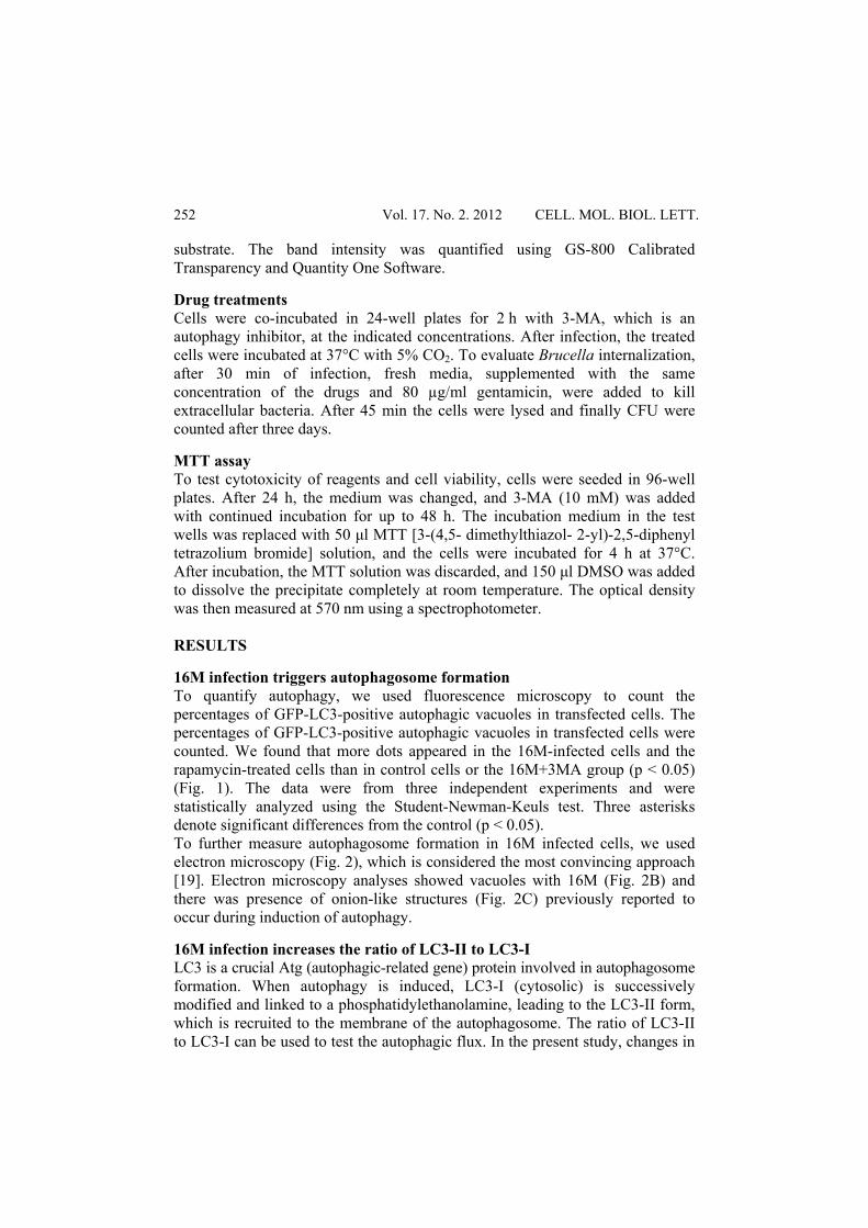
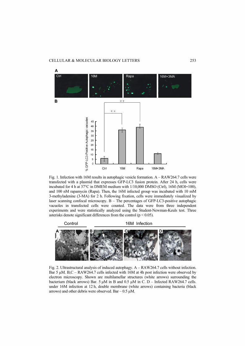
16M infection triggers autophagosome formation To quantify autophagy, we used fluorescence microscopy to count the percentages of GFP-LC3-positive autophagic vacuoles in transfected cells. The percentages of GFP-LC3-positive autophagic vacuoles in transfected cells were counted. We found that more dots appeared in the 16M-infected cells and the rapamycin-treated cells than in control cells or the 16M+3MA group (p < 0.05) (Fig. 1). The data were from three independent experiments and were statistically analyzed using the Student-Newman-Keuls test. Three asterisks denote significant differences from the control (p < 0.05). To further measure autophagosome formation in 16M infected cells, we used electron microscopy (Fig. 2), which is considered the most convincing approach [19]. Electron microscopy analyses showed vacuoles with 16M (Fig. 2B) and there was presence of onion-like structures (Fig. 2C) previously reported to occur during induction of autophagy.
16M infection increases the ratio of LC3-II to LC3-I LC3 is a crucial Atg (autophagic-related gene) protein involved in autophagosome formation. When autophagy is induced, LC3-I (cytosolic) is successively modified and linked to a phosphatidylethanolamine, leading to the LC3-II form, which is recruited to the membrane of the autophagosome. The ratio of LC3-II to LC3-I can be used to test the autophagic flux. In the present study, changes in
CELLULAR & MOLECULAR BIOLOGY LETTERS
253
Fig. 1. Infection with 16M results in autophagic vesicle formation. A – RAW264.7 cells were transfected with a plasmid that expresses GFP-LC3 fusion protein. After 24 h, cells were incubated for 4 h at 37°C in DMEM medium with 1/10,000 DMSO (Ctrl), 16M (MOI=100), and 100 nM rapamycin (Rapa). Then, the 16M infected group was incubated with 10 mM 3-methyladenine (3-MA) for 2 h. Following fixation, cells were immediately visualized by laser scanning confocal microscopy. B – The percentages of GFP-LC3-positive autophagic vacuoles in transfected cells were counted. The data were from three independent experiments and were statistically analyzed using the Student-Newman-Keuls test. Three asterisks denote significant differences from the control (p < 0.05).
Fig. 2. Ultrastructural analysis of induced autophagy. A – RAW264.7 cells without infection. Bar 5 μM. B,C – RAW264.7 cells infected with 16M at 4h post infection were observed by electron microscopy. Shown are multilamellar structures (white arrows) surrounding the bacterium (black arrows) Bar. 5 μM in B and 0.5 μM in C. D – Infected RAW264.7 cells. under 16M infection at 12 h, double membrane (white arrows) containing bacteria (black arrows) and other debris were observed. Bar – 0.5 μM.
Vol. 17. No. 2. 2012 CELL. MOL. BIOL. LETT.
254
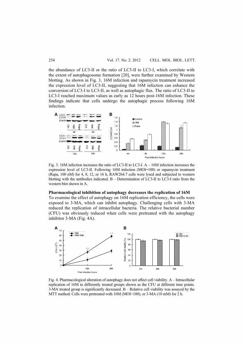
the abundance of LC3-II or the ratio of LC3-II to LC3-I, which correlate with the extent of autophagosome formation [20], were further examined by Western blotting. As shown in Fig. 3, 16M infection and rapamycin treatment increased the expression level of LC3-II, suggesting that 16M infection can enhance the conversion of LC3-I to LC3-II, as well as autophagic flux. The ratio of LC3-II to LC3-I reached maximum values as early as 12 hours post-16M infection. These findings indicate that cells undergo the autophagic process following 16M infection.
Fig. 3. 16M infection increases the ratio of LC3-II to LC3-I. A – 16M infection increases the expression level of LC3-II. Following 16M infection (MOI=100) or rapamycin treatment (Rapa, 100 nM) for 4, 8, 12, or 16 h, RAW264.7 cells were lysed and subjected to western blotting with the antibodies indicated. B – Determination of LC3-II to LC3-I ratio from the western blot shown in A.
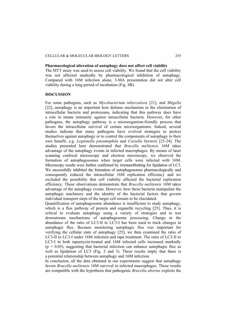
Pharmacological inhibition of autophagy decreases the replication of 16M To examine the effect of autophagy on 16M replication efficiency, the cells were exposed to 3-MA, which can inhibit autophagy. Challenging cells with 3-MA reduced the replication of intracellular bacteria. The relative bacterial number (CFU) was obviously reduced when cells were pretreated with the autophagy inhibitor 3-MA (Fig. 4A).
Fig. 4. Pharmacological alteration of autophagy does not affect cell viability. A – Intracellular replication of 16M in differently treated groups shown as the CFU at different time points. 3-MA treated group is significantly decreased. B – Relative cell viability was assayed by the MTT method. Cells were pretreated with 16M (MOI=100), or 3-MA (10 mM) for 2 h.
CELLULAR & MOLECULAR BIOLOGY LETTERS
255
Pharmacological alteration of autophagy does not affect cell viability The MTT assay was used to assess cell viability. We found that the cell viability was not affected markedly by pharmacological inhibition of autophagy. Compared with 16M infection alone, 3-MA presentation did not alter cell viability during a long period of incubation (Fig. 4B). DISCUSSION
For some pathogens, such as Mycobacterium tuberculosis [21], and Shigella [22], autophagy is an important host defense mechanism in the elimination of intracellular bacteria and protozoans, indicating that this pathway does have a role in innate immunity against intracellular bacteria. However, for other pathogens, the autophagy pathway is a microorganism-friendly process that favors the intracellular survival of certain microorganisms. Indeed, several studies indicate that many pathogens have evolved strategies to protect themselves against autophagy or to control the components of autophagy to their own benefit, e.g. Legionella pneumophila and Coxiella burnetii [23-24]. The studies presented here demonstrated that Brucella melitensis 16M takes advantage of the autophagy events in infected macrophages. By means of laser scanning confocal microscopy and electron microscopy, we observed the formation of autophagosomes when target cells were infected with 16M. Microscopy results were further confirmed by immunoblotting for lipidation of LC3. We successfully inhibited the formation of autophagosomes pharmacologically and consequently reduced the intracellular 16M replication efficiency and we excluded the possibility that cell viability affected the bacterial replication efficiency. These observations demonstrate that Brucella melitensis 16M takes advantage of the autophagy events. However, how these bacteria manipulate the autophagic machinery and the identity of the bacterial factors that govern individual transport steps of the target cell remain to be elucidated. Quantification of autophagosome abundance is insufficient to study autophagy, which is a flux pathway of protein and organelle recycling [25]. Thus, it is critical to evaluate autophagy using a variety of strategies and to test downstream mechanisms of autophagosome processing. Change in the abundance of the ratio of LC3-II to LC3-I has been used to track changes in autophagic flux. Because monitoring autophagic flux was important for verifying the cellular state of autophagy [25], we then examined the ratio of LC3-II to LC3-I under 16M infection and rapa treatment. The ratio of LC3-II to LC3-I in both rapamycin-treated and 16M infected cells increased markedly (p < 0.05), suggesting that bacterial infection can enhance autophagic flux as well as lipidation of LC3 (Fig. 2 and 3). These results imply that there is a potential relationship between autophagy and 16M infection. In conclusion, all the data obtained in our experiments suggest that autophagy favors Brucella melitensis 16M survival in infected macrophages. These results are compatible with the hypothesis that pathogenic Brucella abortus exploits the
Vol. 17. No. 2. 2012 CELL. MOL. BIOL. LETT.
256
autophagic machinery of HeLa cells to establish an intracellular niche favorable for its replication within the ER [11, 26]. Developing means of selectively inhibiting autophagy in infected cells should be viewed as a new window of opportunity in dealing with hard-to-eliminate intracellular pathogens.
Acknowledgements. This work was supported by grants from the National Basic Research Program of China (973 Program) (2010CB530203). REFERENCES
1. Capasso, L. Bacteria in two-millennia-old cheese, and related epizoonoses in Rome populations. J. Infect. 45 (2002) 122-127.
2. Aroian, R. and Goot, F.G. Pore-forming toxins and cellular non-immune defenses (CNIDs). Curr. Opin. Microbiol. 10 (2007) 57-61.
3. Sarinas, P.S. and Chitkara, R.K. Brucellosis. Semin. Respir. Infect. 18 (2003) 168-182.
4. Gibbs, E.P. Emerging zoonotic epidemics in the interconnected global community. Vet. Rec. 157 (2005) 673-679.
5. Otto, G.P., Wu, M.Y., Clarke, M., Lu, H., Anderson, O.R., Hilbi, H., Shuman, H.A. and Kessin, R.H. Macroautophagy is dispensable for intracellular replication of Legionella pneumophila in Dictyostelium discoideum. Mol. Microbiol. 51 (2004) 63-71.
6. Romano, P.S., Gutierrez, M.G., Beron, W., Rabinovitch, M. and Colombo, M.I. The autophagic pathway is actively modulated by phase II Coxiella burnetii to efficiently replicate in the host cell. Cell. Microbiol. 9 (2007) 891-897.
7. Gutierrez, M.G., Master, S.S., Singh, S.B., Taylor, G.A., Colombo, M.I. and Deretic, V. Autophagy is a defense mechanism inhibiting BCG and Mycobacterium tuberculosis survival in infected macrophages. Cell 119 (2004) 753-765.
8. Py, B.F., Lipinski, M.M. and Yuan, J. Autophagy limits Listeria monocytogenes intracellular growth in the early phase of primary infection. Autophagy 3 (2007) 117-126.
9. Jones, S.M. and Winter, A.J. Survival of virulent and attenuated strains of Brucella abortus in normal and gamma interferon-activated murine peritoneal macrophages. Infect. Immun. 60 (1992) 3011-3014.
10. Smith, R., Adams, L.G., Sowa, B.A. and Ficht, T.A. Induction of lymphocyte responsiveness by the outer membrane-peptidoglycan complex of rough strains of Brucella abortus. Vet. Immunol. Immunopathol. 26 (1990) 31-48.
11. Gorvel, J.P. and Moreno, E. Brucella intracellular life: from invasion to intracellular replication. Vet. Microbiol. 90 (2002) 281-297.
12. Starr, T., Ng, T.W., Wehrly, T.D., Knodler, L.A. and Celli, J. Brucella intracellular replication requires trafficking through the late endosomal/lysosomal compartment. Traffic 9 (2008) 678-694.
CELLULAR & MOLECULAR BIOLOGY LETTERS
257
13. Celli, J., de Chastellier, C., Franchini, D.M., Javier P.C., Moreno, E. and Gorvel, J.P. Brucella evades macrophage killing via VirB-dependent sustained interactions with the endoplasmic reticulum. J. Exp. Med. 198 (2003) 545-556.
14. Pizarro-Cerda, J., Meresse, S., Parton, R.G., van der, G.G., Sola-Landa, A., Lopez-Goni, I., Moreno, E. and Orvel, J.P. Brucella abortus transits through the autophagic pathway and replicates in the endoplasmic reticulum of nonprofessional phagocytes. Infect. Immun. 66 (1998) 5711-5724.
15. Pizarro-Cerda, J. and Cossart, P. Bacterial adhesion and entry into host cells. Cell. 124 (2006) 715-727.
16. Li, M., Jiang, X., Liu, D., Na, Y., Gao, G.F. and Xi, Z. Autophagy protects LNCaP cells under androgen deprivation conditions. Autophagy 4 ( 2008) 54-60.
17. Yuk, J.M., Shin, D.M., Lee, H.M., Yang, C.S. and Jin, H.S. Vitamin D3 induces autophagy in human monocytes/macrophages via cathelicidin. Cell Host Microbe 6 (2009) 231-243.
18. Espert, L., Denizot, M., Grimaldi, M., Robert, H.V., Gay, B. and Varbanov, M. Autophagy is involved in T cell death after binding of HIV-1 envelope proteins to CXCR4. J. Clin. Invest. 116 (2006) 2161-2172.
19. Mizushima, N. Methods for monitoring autophagy. Int. J. Biochem. Cell Biol. 36 (2004) 2491-2502.
20. Kabeya, Y., Mizushima, N., Ueno, T., Yamamoto, A. and Kirisako, T. LC3 a mammalian homologue of yeast Apg8p is localized in autophagosome membranes after processing. EMBO J. 19 (2000) 5720-5728.
21. Gutierrez, M.G., Master, S.S., Singh, S.B., Taylor, G.A., Colombo, M.I. and Deretic, V. Autophagy is a defense mechanism inhibiting BCG and Mycobacterium tuberculosis survival in infected macrophages. Cell 119 (2004) 753-766.
22. Ogawa, M., Yoshimori, T., Suzuki, T., Sagara, H., Mizushima, N. and Sasakawa, C. Escape of intracellular Shigella from autophagy. Science 307 (2005) 727-737.
23. Horwitz, M.A. The Legionnaires' disease bacterium (Legionella pneumophila) inhibits phagosome–lysosome fusion in human monocytes. J. Exp. Med. 158 (1983) 2108-2126.
24. Swanson, M.S. and Isberg, R.R. Association of Legionella pneumophila with the macrophage endoplasmic reticulum. Infect. Immun. 63 (1995) 3609-3620.
25. Zhu, C., Wang, X., Xu, F., Bahr, B.A., Shibata, M., Uchiyama, Y., Hagberg, H. and Blomgren, K. The influence of age on apoptotic and other mechanisms of cell death after cerebral hypoxia-ischemia. Cell Death Diff. 12 (2005) 162-176.
26. Arenas, G.N., Staskevich, A.S., Aballay, A. and Mayorga, L.S. Intracellular trafficking of Brucella abortus in J774 macrophages. Infect. Immun. 68 (2000) 4255-4263.