Embed Size (px)
Citation preview
358
http://journals.tubitak.gov.tr/veterinary/
Turkish Journal of Veterinary and Animal Sciences Turk J Vet Anim Sci(2013) 37: 358-361© TÜBİTAKdoi:10.3906/vet-1201-25
Epididymitis and orchitis caused by Brucella melitensis biovar 3 in a Merino ram
Esra BÜYÜKCANGAZ1,*, Aylin ALASONYALILAR DEMİRER2, Sevil ERDENLİĞ3, Sabire Deniz MISIRLIOĞLU2
1 Department of Microbiology, College of Veterinary Medicine, Uludağ University, 16059, Bursa, Turkey2 Department of Pathology, College of Veterinary Medicine, Uludağ University, 16059, Bursa, Turkey
3 Pendik Veterinary Control and Research Institute, 34890 İstanbul, Turkey
* Correspondence: [email protected]
1. IntroductionBrucellosis is a leading cause of abortion, sterility, orchitis, arthritis, and hygromas in animals (1–3). The disease is considered to be highly endemic and causes zoonoses worldwide as reported by the World Organisation for Animal Health (4).
Orchitis in rams is generally associated with multiple bacterial agents (5,6) while epididymitis is mainly identified with Brucella ovis, which produces a non-zoonotic infection in sheep. On the other hand, B. melitensis is the main etiologic agent of small ruminant brucellosis and, as a highly virulent bacterium, plays a significant role in human brucellosis (1,3,4).
2. Case historyThis report describes the pathological and microbiological characteristics of epididymitis and orchitis in a ram caused by B. melitensis biovar 3. A 1-year-old Merino ram with a history of chronic weight loss was referred for necropsy. A preliminary diagnosis of paratuberculosis was made based on the history and clinical signs.
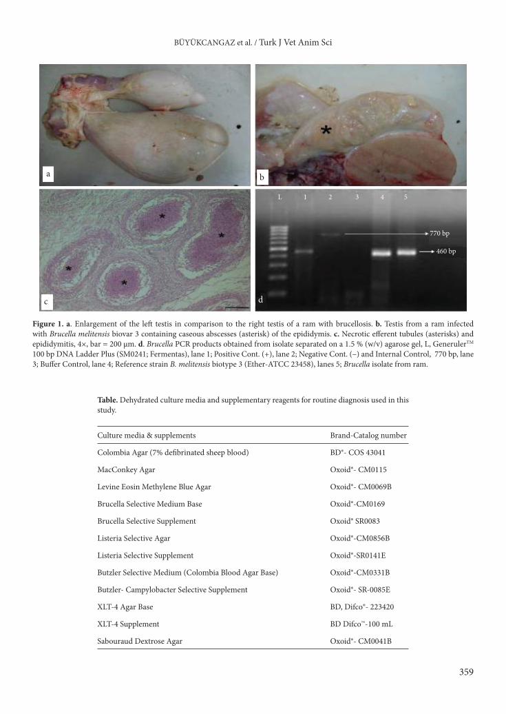
The animal was euthanized and a necropsy was performed. On gross examination, the left testis was greatly enlarged (Figure 1a). The tunica vaginalis was thickened and adhesions were seen between the parietal and visceral layers. The tunic cavity was distended with approximately
360 mL of fibrinopurulent exudate. The left testis had a soft, pasty consistency. On cut surface the epididymis was characterized by foci of necrosis, multiple yellow fibrinopurulent foci (Figure 1b), and diffuse thickening and fibrinous adhesions of the testicular vaginal tunics. The right testis was atrophic, but normal in texture. Gross lesions of paratuberculosis were not observed in the animal.
Impression smears of tissues and fibrinous content taken from the right testis were stained with Gram and modified Ziehl-Neelsen methods, and organ specimens were collected for histopathological and microbiological examinations. A loop of exudate and tissue taken from aseptic cut surfaces of the organs (liver, lung, kidneys, and brain) were inoculated onto agar plates shown in the Table for routine microbiological diagnosis.
A Bru-Com Vet® (Secace Biotechnologies, Italy) commercial kit was used for molecular detection of the Brucella suspected isolate. All reagents were prepared at 22 °C and DNA extraction and the PCR conditions were carried out according to Buyukcangaz et al. (7). Ten microliters of amplicons and DNA molecular weight standards (Fermentas®, USA) were loaded on 2% agarose gel in TAE using 0.6 µL/mL of ethidium bromide at 100 W for 75 min. The amplicons were visualized under UV light and photographed by MiniBIS Pro® (DNR’s Bioimaging Systems, Israel).
Abstract: This paper describes a case of fibrinopurulent epididymitis and orchitis in a Merino ram owing to Brucella melitensis biovar 3, detected by histopathology, bacteriology, and PCR. Gross lesions included enlargement of the scrotum by abundant fibrinous exudate and adhesions between testicular tunics. Histopathologic lesions were characterized by epididymal abscesses and testicular atrophy. B. melitensis was cultured from the exudate as well as the internal organs of the ram. After conventional biotyping procedures were applied, the isolate was identified as B. melitensis biovar 3. The isolate was also confirmed as Brucella spp. by genus-specific PCR.
Key words: Brucella melitensis biovar 3, epididymitis, orchitis, ram
Received: 19.01.2012 Accepted: 25.07.2012 Published Online: 03.06.2013 Printed: 27.06.2013
Case Report
359
BÜYÜKCANGAZ et al. / Turk J Vet Anim Sci
a b
c d
770 bp
460 bp
L 1 2 3 4 5
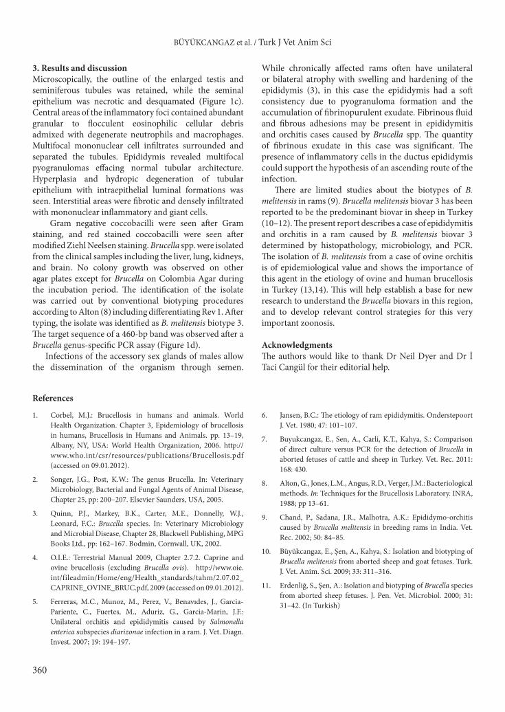
Figure 1. a. Enlargement of the left testis in comparison to the right testis of a ram with brucellosis. b. Testis from a ram infected with Brucella melitensis biovar 3 containing caseous abscesses (asterisk) of the epididymis. c. Necrotic efferent tubules (asterisks) and epididymitis, 4×, bar = 200 µm. d. Brucella PCR products obtained from isolate separated on a 1.5 % (w/v) agarose gel, L, GenerulerTM 100 bp DNA Ladder Plus (SM0241; Fermentas), lane 1; Positive Cont. (+), lane 2; Negative Cont. (–) and Internal Control, 770 bp, lane 3; Buffer Control, lane 4; Reference strain B. melitensis biotype 3 (Ether-ATCC 23458), lanes 5; Brucella isolate from ram.
Table. Dehydrated culture media and supplementary reagents for routine diagnosis used in this study.
Culture media & supplements Brand-Catalog number
Colombia Agar (7% defibrinated sheep blood) BD®- COS 43041
MacConkey Agar Oxoid®- CM0115
Levine Eosin Methylene Blue Agar Oxoid®- CM0069B
Brucella Selective Medium Base Oxoid®-CM0169
Brucella Selective Supplement Oxoid® SR0083
Listeria Selective Agar Oxoid®-CM0856B
Listeria Selective Supplement Oxoid®-SR0141E
Butzler Selective Medium (Colombia Blood Agar Base) Oxoid®-CM0331B
Butzler- Campylobacter Selective Supplement Oxoid®- SR-0085E
XLT-4 Agar Base BD, Difco®- 223420
XLT-4 Supplement BD Difco™-100 mL
Sabouraud Dextrose Agar Oxoid®- CM0041B
360
BÜYÜKCANGAZ et al. / Turk J Vet Anim Sci
3. Results and discussionMicroscopically, the outline of the enlarged testis and seminiferous tubules was retained, while the seminal epithelium was necrotic and desquamated (Figure 1c). Central areas of the inflammatory foci contained abundant granular to flocculent eosinophilic cellular debris admixed with degenerate neutrophils and macrophages. Multifocal mononuclear cell infiltrates surrounded and separated the tubules. Epididymis revealed multifocal pyogranulomas effacing normal tubular architecture. Hyperplasia and hydropic degeneration of tubular epithelium with intraepithelial luminal formations was seen. Interstitial areas were fibrotic and densely infiltrated with mononuclear inflammatory and giant cells.
Gram negative coccobacilli were seen after Gram staining, and red stained coccobacilli were seen after modified Ziehl Neelsen staining. Brucella spp. were isolated from the clinical samples including the liver, lung, kidneys, and brain. No colony growth was observed on other agar plates except for Brucella on Colombia Agar during the incubation period. The identification of the isolate was carried out by conventional biotyping procedures according to Alton (8) including differentiating Rev 1. After typing, the isolate was identified as B. melitensis biotype 3. The target sequence of a 460-bp band was observed after a Brucella genus-specific PCR assay (Figure 1d).
Infections of the accessory sex glands of males allow the dissemination of the organism through semen.
While chronically affected rams often have unilateral or bilateral atrophy with swelling and hardening of the epididymis (3), in this case the epididymis had a soft consistency due to pyogranuloma formation and the accumulation of fibrinopurulent exudate. Fibrinous fluid and fibrous adhesions may be present in epididymitis and orchitis cases caused by Brucella spp. The quantity of fibrinous exudate in this case was significant. The presence of inflammatory cells in the ductus epididymis could support the hypothesis of an ascending route of the infection.
There are limited studies about the biotypes of B. melitensis in rams (9). Brucella melitensis biovar 3 has been reported to be the predominant biovar in sheep in Turkey (10–12). The present report describes a case of epididymitis and orchitis in a ram caused by B. melitensis biovar 3 determined by histopathology, microbiology, and PCR. The isolation of B. melitensis from a case of ovine orchitis is of epidemiological value and shows the importance of this agent in the etiology of ovine and human brucellosis in Turkey (13,14). This will help establish a base for new research to understand the Brucella biovars in this region, and to develop relevant control strategies for this very important zoonosis.
AcknowledgmentsThe authors would like to thank Dr Neil Dyer and Dr İ Taci Cangül for their editorial help.
References
1. Corbel, M.J.: Brucellosis in humans and animals. World Health Organization. Chapter 3, Epidemiology of brucellosis in humans, Brucellosis in Humans and Animals. pp. 13–19, Albany, NY, USA: World Health Organization, 2006. http://www.who.int/csr/resources/publications/Brucellosis.pdf (accessed on 09.01.2012).
2. Songer, J.G., Post, K.W.: The genus Brucella. In: Veterinary Microbiology, Bacterial and Fungal Agents of Animal Disease, Chapter 25, pp: 200–207. Elsevier Saunders, USA, 2005.
3. Quinn, P.J., Markey, B.K., Carter, M.E., Donnelly, W.J., Leonard, F.C.: Brucella species. In: Veterinary Microbiology and Microbial Disease, Chapter 28, Blackwell Publishing, MPG Books Ltd., pp: 162–167. Bodmin, Cornwall, UK, 2002.
4. O.I.E.: Terrestrial Manual 2009, Chapter 2.7.2. Caprine and ovine brucellosis (excluding Brucella ovis). http://www.oie.int/fileadmin/Home/eng/Health_standards/tahm/2.07.02_CAPRINE_OVINE_BRUC.pdf, 2009 (accessed on 09.01.2012).
5. Ferreras, M.C., Munoz, M., Perez, V., Benavıdes, J., Garcia-Pariente, C., Fuertes, M., Aduriz, G., Garcia-Marin, J.F.: Unilateral orchitis and epididymitis caused by Salmonella enterica subspecies diarizonae infection in a ram. J. Vet. Diagn. Invest. 2007; 19: 194–197.
6. Jansen, B.C.: The etiology of ram epididymitis. Onderstepoort J. Vet. 1980; 47: 101–107.
7. Buyukcangaz, E., Sen, A., Carli, K.T., Kahya, S.: Comparison of direct culture versus PCR for the detection of Brucella in aborted fetuses of cattle and sheep in Turkey. Vet. Rec. 2011: 168: 430.
8. Alton, G., Jones, L.M., Angus, R.D., Verger, J.M.: Bacteriological methods. In: Techniques for the Brucellosis Laboratory. INRA, 1988; pp 13–61.
9. Chand, P., Sadana, J.R., Malhotra, A.K.: Epididymo-orchitis caused by Brucella melitensis in breeding rams in India. Vet. Rec. 2002; 50: 84–85.
10. Büyükcangaz, E., Şen, A., Kahya, S.: Isolation and biotyping of Brucella melitensis from aborted sheep and goat fetuses. Turk. J. Vet. Anim. Sci. 2009; 33: 311–316.
11. Erdenliğ, S., Şen, A.: Isolation and biotyping of Brucella species from aborted sheep fetuses. J. Pen. Vet. Microbiol. 2000; 31: 31–42. (In Turkish)

361
BÜYÜKCANGAZ et al. / Turk J Vet Anim Sci
12. Erdenlig, S., Baklan E.A., Aksoy H.Y.: The identification, characterization and distribution of Brucella isolates in Turkey in the last two years, 2007 to 2008. VLA International Conference - Animal Diseases, 2-4 September, Royal Holloway, University of London UK, 2009.
13. Scientific Committee on Animal Health and Animal Welfare: 2001, European Commission, Brucellosis in sheep and goats (Brucella melitensis). http://ec.europa.eu/food/fs/sc/scah/out59_en.pdf (accessed on 09.01.2012).
14. Şimşek, H., Erdenliğ, S., Oral, B., Tülek, N.: Typing and biotyping of Brucella isolates of human origin and their epidemiologic evaluation Klimik Derg. 2004; 17: 103-106. (In Turkish)