Embed Size (px)
Citation preview

AVIAN GUT FUNCTION IN HEALTH AND DISEASE

Poultry Science Symposium SeriesVolumes 1–28
1 Physiology of the Domestic Fowl*2 Protein Utilization by Poultry*3 Environmental Control in Poultry Production*4 Egg Quality – a Study of the Hen’s Egg*5 The Fertility and Hatchability of the Hen’s Egg*6 i. Factors Affecting Egg Grading*
ii. Aspects of Poultry Behaviour*7 Poultry Disease and World Economy8 Egg Formation and Production9 Energy Requirements of Poultry*
10 Economic Factors Affecting Egg Production*11 Digestion in the Fowl*12 Growth and Poultry Meat Production*13 Avian Coccidiosis*14 Food Intake Regulation in Poultry*15 Meat Quality in Poultry and Game Birds16 Avian Immunology17 Reproductive Biology of Poultry18 Poultry Genetics and Breeding19 Nutrient Requirements of Poultry and Nutritional Research*20 Egg Quality – Current Problems and Recent Advances*21 Recent Advances in Turkey Science22 Avian Incubation23 Bone Biology and Skeletal Disorders24 Poultry Immunology*25 Poultry Meat Science26 Poultry Feedstuffs27 Welfare of the Laying Hen28 Avian Gut Function in Health and Disease
*Out of printVolumes 1–24 were not published by CABI. Those still in print maybe ordered from:
Carfax Publishing CompanyPO Box 25, Abingdon, Oxfordshire OX14 3UE, UK

Avian Gut Function in Health and Disease
Poultry Science Symposium SeriesVolume Twenty-eight
Edited by
G.C. PerryDepartment of Clinical Veterinary Science, University of Bristol, UK

CABI is a trading name of CAB International
CABI Head Office CABI North American Office Nosworthy Way 875 Massachusetts AvenueWallingford 7th FloorOxon OX10 8DE Cambridge, MA 02139UK USA
Tel: +44 (0)1491 832111 Tel: +1 617 395 4056Fax: +44 (0)1491 833508 Fax: +1 617 354 6875E-mail: [email protected] E-mail: [email protected]: www.cabi.org
© CAB International 2006. All rights reserved. No part of this publication may be reproduced in any form or by any means, electronically, mechanically, by photocopying, recording or otherwise, without the prior permission of the copyright owners.
A catalogue record for this book is available from the British Library, London, UK.
A catalogue record for this book is available from the Library of Congress,Washington DC, USA.
ISBN-10: 1-84593-1807ISBN-13: 978-1-84593-1803
Produced and Typeset in 10/12pt Souvenir Light by Columns Design Ltd, ReadingPrinted and bound in the UK by Biddles, Kings Lynn
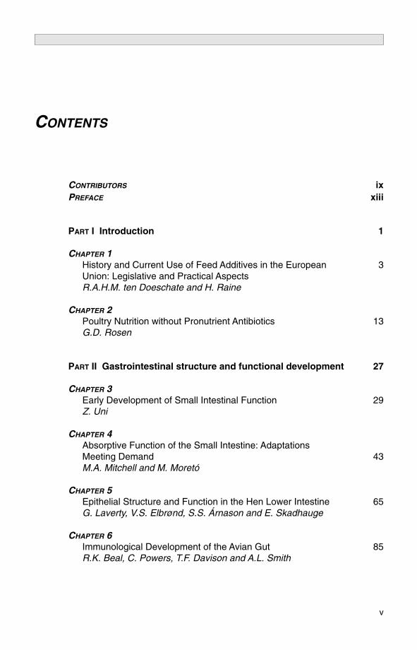
CONTENTS
CONTRIBUTORS ixPREFACE xiii
PART I Introduction 1
CHAPTER 1History and Current Use of Feed Additives in the European 3Union: Legislative and Practical AspectsR.A.H.M. ten Doeschate and H. Raine
CHAPTER 2Poultry Nutrition without Pronutrient Antibiotics 13G.D. Rosen
PART II Gastrointestinal structure and functional development 27
CHAPTER 3Early Development of Small Intestinal Function 29Z. Uni
CHAPTER 4Absorptive Function of the Small Intestine: AdaptationsMeeting Demand 43M.A. Mitchell and M. Moretó
CHAPTER 5Epithelial Structure and Function in the Hen Lower Intestine 65G. Laverty, V.S. Elbrønd, S.S. Árnason and E. Skadhauge
CHAPTER 6Immunological Development of the Avian Gut 85R.K. Beal, C. Powers, T.F. Davison and A.L. Smith
v

PART III Gastrointestinal flora 105
CHAPTER 7Molecular Approaches to the Analysis of Gastrointestinal Microbial Ecosystems 107H.J. Flint, E.C.M. Leitch, S.H. Duncan, A.W. Walker, A.J. Patterson, M.T. Rincon, K.P. Scott and P. Louis
CHAPTER 8Microbes of the Chicken Gastrointestinal Tract 124J. Apajalahti and A. Kettunen
CHAPTER 9Mechanisms of Pathogen Control in the Avian Gastrointestinal Tract 138A.M. Donoghue, M.B. Farnell, K. Cole and D.J. Donoghue
PART IV Nutritional effects 157
CHAPTER 10Effect of Non-starch Polysaccharidases on Avian Gastrointestinal Function 159M.R. Bedford
CHAPTER 11Effects of Amino Acid and Protein Supply on Nutrition and Health 171M.T. Kidd and A. Corzo
CHAPTER 12The Role of Feed Processing on Gastrointestinal Function and Health in Poultry 183B. Svihus
CHAPTER 13Wet Litter: its Causes and Prevention and the Role of Nutrition 195S.R. Collett
CHAPTER 14Micronutrient Supply: Influence on Gut Health and Immunity 210K.C. Klasing
PART V Pathology 225
CHAPTER 15Virally Induced Gastrointestinal Diseases of Chickens and Turkeys 227J.S. Guy
vi Contents

CHAPTER 16The Gastrointestinal Tract as a Port of Entry for Bacterial Infections in Poultry 244J.P. Christensen, M.S. Chadfield, J.E. Olsen and M. Bisgaard
CHAPTER 17Parasite Genetics, Protection and Antigen Identification 259D.P. Blake, M.W. Shirley and A.L. Smith
PART VI Immunological and pathogen control 273
CHAPTER 18Developments and Pitfalls of Feed Acidification in ControllingGut Pathogens in Poultry, with Emphasis on Salmonella 275F. van Immerseel, I. Gantois, L. Bohez, L. Timbermont, F. Boyen, I. Hautefort, J.C.D. Hinton, F. Pasmans, F. Haesebrouck and R. Ducatelle
CHAPTER 19Competitive Exclusion in Poultry Production 294C. Schneitz
CHAPTER 20Campylobacters and their Bacteriophage in Poultry 311P.L. Connerton and I.F. Connerton
CHAPTER 21Breeding for Disease Resistance 322S.C. Bishop
PART VII Monitoring and practical experience 339
CHAPTER 22The EU Perspective on the Monitoring of Zoonoses and Zoonotic Agents 341S. Idei
CHAPTER 23Gut Problems: the Field Experience and What it Means to thePoultry Farmer 350S.A. Lister
Poster abstracts 361
Index 409
Contents vii

This page intentionally left blank

T. Acamovic, Avian Science Research Centre, Scottish Agricultural College,West Mains Road, Edinburgh, EH9 3JG, UK; (address for communication)SAC, Ayr Campus, Ayr, KA6 5HW, UK; e-mail: [email protected]
J. Apajalahti, Alimetrics Ltd, Höyläämötie 1, FIN-00380, Helsinki, Finland; e-mail: [email protected]
S.S. Árnason, Department of Physiology, University of Iceland, IS-101Reykjavik, Iceland
R.K. Beal, Division of Immunology and Immunopathology, Institute forAnimal Health, Compton, Newbury, Berkshire, RG20 7NN, UK
M.R. Bedford, Zymetrics, Chestnut House, Beckhampton, Marlborough,Wiltshire, SN8 1QJ, UK; e-mail: [email protected]
M. Bisgaard, Department of Veterinary Pathobiology, The Royal Veterinaryand Agricultural University Stigbojlen 4, DK-1870, Frederiksberg C.,Denmark
S.C. Bishop, Roslin Institute, Midlothian, EH25 9PS, UK; tel: +44(0)131 527 4200; fax 44 (0)131 440 0434 ; e-mail: [email protected]
D.P. Blake, Institute for Animal Health, Compton, Newbury, Berkshire,RG20 7NN, UK; tel: 44 (0)1635 577293; fax: 44 (0)1635 57726; e-mail:[email protected]
L. Bohez, Department of Pathology, Bacteriology and Avian Disease,Research Centre on Veterinary Food Safety and Zoonoses, Faculty ofVeterinary Medicine, Ghent University, Salisburylaan, 133, B-9820,Merelbeke, Belgium
F. Boyen, Department of Pathology, Bacteriology and Avian Disease,Research Centre on Veterinary Food Safety and Zoonoses, Faculty ofVeterinary Medicine, Ghent University, Salisburylaan, 133, B-9820,Merelbeke, Belgium
M.S. Chadfield, Department of Veterinary Pathobiology, The RoyalVeterinary and Agricultural University, Stigbojlen 4, DK-1870,Frederiksberg C., Denmark
J.P. Christensen, Department of Veterinary Pathobiology, The RoyalVeterinary and Agricultural University, Stigbojlen 4, DK-1870,Frederiksberg C, Denmark; e-mail: [email protected]
K. Cole, Department of Poultry Science, University of Arkansas,Fayetteville, Arkansas 72701, USA
ix
CONTRIBUTORS

S.R. Collett, The University of Georgia, College of Veterinary Medicine,Poultry Diagnostic and Research Center, 953 College Station Road,Athens, Georgia, 30602-4875, USA; e-mail: [email protected]
I.F. Connerton, Division of Food Sciences, School of Biosciences,University of Nottingham, Sutton Bonington Campus, Loughborough,LE12 5RD, UK; tel: +44 (0)115 9516119; fax: +44 (0)115 9516162; e-mail: [email protected]
P.L. Connerton, Division of Food Sciences, School of Biosciences,University of Nottingham, Sutton Bonington Campus, Loughborough,LE12 5RD, UK; tel: +44 (0)115 9516119; fax: +44 (0)115 9516162
R.G. Cooper, Department of Physiology, University of Zimbabwe, MountPleasant Drive, Harare, Zimbabwe
A. Corzo, Department of Poultry Science, Mississippi State University, Box9665, Mississippi 39762,USA; fax: 662 325 8292
T.F. Davison, Formerly Division of Immunology and Immunopathology,Institute for Animal Health, Compton, Newbury, Berkshire, RG20 7NN,UK; e-mail: [email protected]
R.A.H.M. ten Doeschate, ABNA Ltd, ABNA House, Oundle Road,Peterborough, PE2 9PW, UK; e-mail: [email protected]
A.M. Donoghue, Poultry Production and Product Safety Research Unit,ARS, USDA, Fayetteville, Arkansas, USA
D.J. Donoghue, Department of Poultry Science, University of Arkansas,Fayetteville, Arkansas 72701, USA
R. Ducatelle, Department of Pathology, Bacteriology and Avian Disease,Research Centre on Veterinary Food Safety and Zoonoses, Faculty ofVeterinary Medicine, Ghent University, Salisburylaan, 133, B-9820,Merelbeke, Belgium
S.H. Duncan, Microbial Ecology Group, Gut Health Division, RowettResearch Institute, Greenburn Road, Bucksburn, Aberdeen, AB21 9SB,UK
V.S. Elbrønd, Department of Animal and Veterinary Basic Science, TheRoyal Veterinary and Agricultural University, DK-1870, Frederiksberg C,Denmark
M.B. Farnell, Poultry Production and Product Safety Research Unit, ARS,USDA, Fayetteville, Arkansas, USA
H.J. Flint, Microbial Ecology Group, Gut Health Division, Rowett ResearchInstitute, Greenburn Road, Bucksburn, Aberdeen, AB21 9SB, UK;email: [email protected]
I. Gantois, Department of Pathology, Bacteriology and Avian Disease,Research Centre on Veterinary Food Safety and Zoonoses, Faculty ofVeterinary Medicine, Ghent University, Salisburylaan, 133, B-9820,Merelbeke, Belgium
J.S. Guy, College of Veterinary Medicine, North Carolina State University,4700 Hillsborough Street, Raleigh, North Carolina 27606, USA; fax: 919-513-6464; e-mail: [email protected]
F. Haesebrouck, Department of Pathology, Bacteriology and AvianDisease, Research Centre on Veterinary Food Safety and Zoonoses,
x Contributors

Faculty of Veterinary Medicine, Ghent University, Salisburylaan, 133, B-9820, Merelbeke, Belgium
I. Hautefort, Molecular Microbiology Group, Institute of Food Research,Norwich Research Park, Norwich, NR4 7UA, UK
J. Hinton, Molecular Microbiology Group, Institute of Food Research,Norwich Research Park, Norwich, NR4 7UA, UK
S. Idei, European Commission, Health and Consumer ProtectionDirectorate-General, Directorate D – Food Safety: Production andDistribution Chain; D2 – Biological Risks,Commission Européenne ; B-1049 Bruxelles/ Europese Commissie, B-1049 Brussels,Belgium ; tel:(32–2) 299 11 11; e-mail: [email protected]
A. Kettunen, Alimetrics Ltd, Höyläämötie 14, FIN-00380, Helsinki, FinlandM.T. Kidd, Department of Poultry Science, Mississippi State University, Box
9665, Mississippi 39762, USA; fax: 662 325 8292; e-mail:[email protected]
K.C. Klasing, Department of Animal Science, University of California Davis,Davis, California 95616, USA; tel: 530-752-1901; fax: 530-752-0175; e-mail: [email protected]
G. Laverty, Department of Biological Sciences, University of Delaware,Newark, Delaware 19716, USA; e-mail: [email protected]
E.C.M. Leitch, Microbial Ecology Group, Gut Health Division, RowettResearch Institute, Greenburn Road, Bucksburn, Aberdeen, AB21 9SB,UK
S.A. Lister, Crowshall Veterinary Services, Attleborough, Norfolk, UK;(address for communication: SAC, Ayr Campus, Ayr, KA6 5HW); tel:+44 (0)1953 455454; fax: +44 (0)1953 455661; e-mail: [email protected]
P. Louis, Microbial Ecology Group, Gut Health Division, Rowett ResearchInstitute, Greenburn Road, Bucksburn, Aberdeen, AB21 9SB, UK
M.A. Mitchell, Scottish Agricultural College, Bush Estate, Midlothian, EH16AA, UK; e-mail: [email protected]
M. Moretó, Departament de Fisiologia, Facultat de Farmàcia, Av. de JoanXXIII s/n, 08028 Barcelona, Spain
J.E. Olsen, Department of Veterinary Pathobiology, The Royal Veterinaryand Agricultural University, Stigbojlen 4, DK-1870, Frederiksberg C,Denmark
F. Pasmans, Department of Pathology, Bacteriology and Avian Disease,Research Centre on Veterinary Food Safety and Zoonoses, Faculty ofVeterinary Medicine, Ghent University, Salisburylaan, 133, B-9820,Merelbeke, Belgium
A.J. Patterson, Microbial Ecology Group, Gut Health Division, RowettResearch Institute, Greenburn Road, Bucksburn, Aberdeen, AB21 9SB,UK
C. Powers, Division of Immunology and Immunopathology, Institute forAnimal Health, Compton, Newbury, Berkshire, RG20 7NN, UK
H. Raine, ABNA Ltd, ABN House, Oundle Road, Peterborough, PE2 9PW,UK
Contributors xi

M.T. Rincon, Microbial Ecology Group, Gut Health Division, RowettResearch Institute, Greenburn Road, Bucksburn, Aberdeen, AB21 9SB,UK
G.D. Rosen, Holo-Analysis Services Ltd., 66 Bathgate Road, Wimbledon,London, SW19 5PH, UK; tel./fax 020 8946 9575; e-mail: [email protected]
C. Schneitz, Orion Corporation Orion Pharma, Animal Health, PO Box 425,FI-20101 Turku, Finland; fax: 2358-9-4521764; e-mail: [email protected]
K.P. Scott, Microbial Ecology Group, Gut Health Division, Rowell ResearchInstitute, Greenburn Road, Bucksburn, Aberdeen, AB21 9SB, UK
M.W. Shirley, Eimerian Genomics Group, Institute for Animal Health,Compton, Newbury, Berkshire, RG20 7NN, UK; tel: 01635 577293; fax:01635 577263
E. Skadhauge, Department of Animal and Veterinary Basic Science, TheRoyal Veterinary and Agricultural University, DK-1870, Frederiksberg C,Denmark
A.L. Smith, Enteric Immunology Group, Institute for Animal Health,Compton, Newbury, Berkshire, RG20 7NN, UK; tel: 44 (0)1635577293; fax: 44 (0)1635 577263; e-mail: [email protected]
B. Svihus, Department of Animal and Aquacultural Sciences, NorwegianUniversity of Life Sciences, PO Box 5003, N-1432 Ås, Norway; e-mail:[email protected]
L. Timbermont, Department of Pathology, Bacteriology and Avian Disease,Research Centre on Veterinary Food Safety and Zoonoses, Faculty ofVeterinary Medicine, Ghent University, Salisburylaan, 133, B-9820,Merelbeke, Belgium
Z. Uni, Department of Animal Sciences, Faculty of Agricultural, Food andEnvironmental Quality Sciences, The Hebrew University of Jerusalem,PO Box 12, Rehovot 76-100, Israel; e-mail: [email protected]
F. Van Immerseel, Department of Pathology, Bacteriology and AvianDiseases, Research Centre on Veterinary Food Safety and Zoonoses,Faculty of Veterinary Medicine, Ghent University, Salisburylaan, 133, B-9820, Merelbeke, Belgium; e-mail: [email protected]
A.W. Walker, Microbial Ecology Group, Gut Health Division, RowettResearch Institute, Greenburn Road, Bucksburn, Aberdeen, AB21 9SB,UK
xii Contributors

The proceedings which comprise this volume are taken from the 28thSymposium in a series organized by the UK branch of the World’s PoultryScience Association. The chosen topic attracted a large audience, members ofwhich came from all continents except South America. This probablydemonstrates the importance that poultry scientists and industries worldwideattach to avian gut function.
Since the late 1960s, when the Swann Committee in the UK investigatedthe use of antibiotics in animal husbandry and veterinary medicine, pressureon their use as growth promoters has grown. Swann recommended that onlyantibiotics which ‘have little or no application as therapeutic agents in man oranimals and will not impair the efficacy of a prescribed therapeutic drug ordrugs through the development of resistant strains of organisms’ should beused for growth promotion. The UK government accepted these recommenda-tions and, subsequently, the European Union adopted this principle, although ithad to resist pressure from Sweden for a complete ban on antibiotic growthpromoters.
Political pressure continued and, since the mid-1990s, several antibioticshave been banned and withdrawn from the market. Concerns have also beenexpressed over the use of certain coccidiostats, with the result that some of theearlier products have now also been withdrawn.
These restrictions have taken place because of concerns over drugresistance and possible carry-over to consumers of poultry products. However,little consideration has been given to the consequences of their withdrawal topoultry health and performance. The worldwide interest in this symposium,therefore, demonstrates the concern of poultry scientists about thesechallenges, but also highlights the dearth of sound scientific knowledge aboutavian gut function in healthy birds.
Scientists of international standing were invited to present their views at thesymposium. The starting point was a description of the history of feedadditives. This was followed by a paper describing the likely consequences ofthe withdrawal of prescription-free use of antimicrobials, which concluded witha brief consideration of likely alternatives.
Four papers dealt with the development of the gastrointestinal tract, itsfunction related to growth, uptake of nutrients and immune development.
This was followed by three papers concerned with gut microbiology, includingthe analysis of the microbial ecosystems, a description of the more commonlyencountered microbes and a discussion of mechanisms of pathogen control.
xiii
PREFACE
Six papers covered nutritional effects. These included the effect of nutrients,anti-nutrients, non-nutrients or toxins and microflora on gut health and birdgrowth. Unfortunately, due to health reasons, the main author was unable tocomplete his review paper so it has not been included in this volume. The effect ofnon-starch polysaccharides on gut function were covered, as well as amino acidand protein supply on nutrition and health. The last three papers in this sectiondealt with the role of feed processing on gut function, micronutrient supply and itseffect on immunity and, lastly, the association between nutrition and wet litter.
Three papers dealt with pathology. A description of virally inducedgastrointestinal diseases of chickens and turkeys was followed by a paperhighlighting the intestinal tract as a portal of entry of bacteria. The final paperin this section described a new approach to the investigation of thehost–parasite relationship in which parasite genetics, DNA fingerprinting andselection by immunity can be used.
The immunological and pathogen control session contained papers dealingwith feed acidification, competitive exclusion, the influence of vaccines andcampylobacters and their bacteriophage. The author of the paper describingthe influence of some specific vaccines on the intestinal health of broilers wasunable to provide a review chapter for these proceedings. This is anunfortunate omission, since the use of vaccines is clearly an important practicalapproach to the control of gut pathogens. The concluding paper in this sessiondiscussed breeding for disease resistance and the role of gene mapping andmicroarray studies.
The concluding session included a description of the legislative role of the EUin monitoring zoonoses and zoonotic infections, and the final paper brought thediscussions back to the farm level by asking: what does it mean to the farmer?
In addition to the formal presentations, 47 poster abstracts were offered byscientists working in the field of avian gut function.
I am indebted to the Organizing Committee for their contributions andsupport during the preparation and conduct of the symposium. I hope thatreaders of these proceedings will feel that the subject was adequately coveredand brought up to date by the distinguished panel of speakers.
The efficiency with which the Symposium was run was due entirely to theefforts of Rita and Christine, who worked tirelessly to ensure that the interests ofthe sponsors and the needs of speakers and delegates were well catered for. I owethem my gratitude for making my responsibilities so much easier to handle.
Organizing Committee
G.C. Perry (Chairman), J.A. Parsons (Secretary), K.J. McCracken (ex-officio,Treasurer), T. Acamovic, A. Ball, P. Barrow, F. Davison, P. Hocking (ex-officio,President, WPSA UK Branch), T. Humphrey, G. Mead, M. Mitchell and G. Rosen.
Administrative secretaries
Rita Hinton and Christine Rowlings.
xiv Preface

PART I
Introduction

This page intentionally left blank

CHAPTER 1History and current use of feed additives in the European Union: legislative and practical aspects
R.A.H.M. ten Doeschate* and H. RaineABNA Ltd., Peterborough, UK; *e-mail: [email protected]
ABSTRACT
Whilst the term feed additives encompasses a variety of products, this chapterwill concentrate on product groups such as antibiotic growth promoters,coccidiostats and enzymes. These products have been, and are, subject toscrutiny and licensing at European Union (EU) and national level.
The history of antibiotic growth promoters serves as a case study for thedevelopment of a product group, ensuing legislation, pressure group activityand the subsequent demise of the product group.
EU legislation has evolved over the years from a situation of limitedscope and relatively relaxed rules to the current system where the scope isbeing extended to more products and with stricter rules. The current EUregulation (1831/2003) is based on the precautionary principle related tohuman health, animal health and the environment. At present there are signsthat legislation has tightened to such a level that certain products are notavailable on the EU market, maybe as a result of high costs or delays, due tothe registration process. A balance needs to be found between effectivelegislation regulating the safe use of additives and allowing new product andconcept development.
From a practical nutritionist’s point of view the whole area is verycomplicated, with several products on the market, and it is unclear whetherthey are additives in the legal sense or not, but there is a responsibility on thenutritionist to ensure compliance with the law. With antibiotic growth promotersdisappearing, several ‘replacement’ products need to be evaluated, which isanother big challenge for nutritionists.
© CAB International 2006. Avian Gut Function in Health and Disease (ed. G.C. Perry) 3

INTRODUCTION
Feed additives encompass a variety of products. According to the currentlyapplicable legislation (EC 1831/2003, Art. 2 (2a)), ‘feed additives’ meanssubstances, microorganisms or preparations – other than feed materials andpremixtures – which are intentionally added to feed or water to perform, inparticular, one or more of the functions mentioned in Article 5 (3). Article 5 (3)can be summarized as follows: a feed additive should favourably affect one ofthe following:
● characteristics of feed or animal products;● colour of ornamental fish or birds;● the environment;● animal production, performance or welfare through positive effects at gut
level,
or satisfy the nutritional needs of animals or have a coccidiostatic or histo-monostatic effect.
This, potentially, gives a huge range of possible additives but, for the scopeof this chapter only product groups will be covered such as antibiotic growthpromoters, coccidiostats and enzymes. The reason for this is that these productshave been, and are, subject to a fair degree of scrutiny and approval/authorization at EU and national level.
This chapter is intended to give an overview of the history of both feedadditives and applicable EU legislation, which will inevitably be incomplete butwill hopefully serve to give an understanding of the subject. Further, it will showhow current use of additives is impacted by legislation, and how this andcommercial practice interact.
HISTORY
For antibiotic growth promoters the story began in the 1940s, with a 1946 (Mooreet al.) and/or a 1949 (Stokestad et al.) paper being widely cited as the first scientificpaper(s) to note a growth-promoting effect of feeding antibiotics. The interest inantibiotics actually stemmed from a quest to find substitutes for the animal proteingrowth factor. Vitamin B12 was partly identified as a factor promoting growth in theabsence of animal protein, but it was found that either antibiotics or antibioticgrowth medium improved growth over and above the animal protein effect.
Subsequently, antibiotics quickly found a place in poultry nutrition, whichcan be illustrated by the following quote from Heuser (1955):
One of the most spectacular recent developments in nutrition science is thediscovery that antibiotics have growth-stimulating properties when fed to chickens,turkeys and swine. Few nutritional discoveries have been so quickly and universallyapplied to feeding practice.
The nutritional community at that time was easily convinced of the benefits,and subsequently the principle has been used for as long as it was allowed.
4 R.A.H.M. ten Doeschate and H. Raine
Initially, several antibiotics were used for growth-promoting purposes but,by the late 1960s, the Swann Committee investigated their use in the light ofthe potential for the development of antibiotic resistance in bacteria. Theyconcluded that: ‘The administration of antibiotics to farm livestock, particularlyat sub-therapeutic levels, poses certain hazards to human and animal health.’In particular, it had led to resistance in enteric bacteria of animal origin. Thisresistance was transmissible to other bacteria (it had been the discovery thatthis might be so, following an epidemic of resistant Salmonella typhimurium in1963–1965, which prompted the UK government minister to appoint theSwann Committee). It had also been shown that enteric bacteria weretransferable from animals to man (House of Lords, 1998). The Swann Committeerecommended that only antibiotics which ‘have little or no application astherapeutic agents in man or animals and will not impair the efficacy of aprescribed therapeutic drug or drugs through the development of resistant strainsof organisms’ (Swann, 1969 as quoted by the House of Lords, 1998) should beusable for growth promotion. Their report named the following antibiotics, whichwere then in use for growth promotion, as unsuitable for such use:chlortetracycline, oxytetracycline, penicillin, tylosin (a macrolide related toerythromycin) and the sulphonamides. The UK Government largely acceptedthese recommendations (House of Lords, 1998).
Over the following years pressure on the use of antibiotics as growthpromoters has increased steadily. Sweden was the first country in Europe tocompletely ban the use of antibiotic growth promoters in 1985. This ban wasactually requested by the organization of Swedish farmers, and wassubsequently enforced by the Swedish Government. When Sweden joined theEU it was granted a derogation allowing the ban to continue in Sweden whilstother EU countries continued the use of antibiotic growth promoters (SOU,1997). Sweden lobbied for an EU-wide ban, and during the late 1990s inEurope the political pressure was at such a level that since 1996 severalantibiotics have disappeared completely.
Table 1.1 shows the full list of active ingredients banned or taken off themarket, with their year of disappearance. Avoparcin is the only one of thesethat has disappeared globally. Whilst all other products have been banned inthe EU, they can still be used in other parts of the world.
Before the loss of four products in 1999, it was common practice in somecompanies to switch between different ingredients on a regular basis, say every6–9 months. It was commonly thought that this helped maintain the efficacy ofthe various antibiotic growth promoters. However, after 1999, when only twoactive ingredients remained, there was no real opportunity to do this and as aresult most companies tended to use the same product all the time.
As a result of the public pressure on the use of antibiotic growth promotersin 1999, one UK integrator decided to satisfy the perceived demand for poultryproduced without an antibiotic growth promoter, and stopped their use. Otherproducers quickly followed and at present chickens destined for the retailchannel are fed on Non-growth Promoter (NGP) feed. The only chickens still fedantibiotic growth promoters in the UK are those destined for the not insignificantremaining part of the broiler industry that supplies birds to non-retail markets.
Feed additives in the EU 5

For coccidiostats, the story started later than that for antibiotic growthpromoters but on the other hand the story has not finished yet. In the 1950sthe sulpha-drugs started to be used to control coccidiosis as a disease, butdevelopment really started with the chemical coccidiostats in the 1960s. Theavailability of coccidiostats is generally considered to be one of the main factorsthat allowed the development of the intensive poultry industry. Ionophorecoccidiostats became available in the 1970s, after which a typical programmein the UK was to use a chemical anticoccidiostat (ACS) in the starter feeds,followed by an ionophore later on.
Pure chemical products had a habit of breaking down, i.e. resistance to themdeveloped and caused significant problems. Ionophores and chemical/ionophoremixtures seemed to enable more stable production levels, yet coccidiosis remainsever present and any lapse in hygiene or management may result in mini-outbreaks. As a consequence of the development of resistance to the purechemical products, some of these are no longer on the market whereas others,whilst available, are used very sporadically and then only in a tactical manner.Some products have been banned from use in Europe on other than strictlytechnical grounds. Nicarbazin as a pure product, for example, had its licencewithdrawn as a result of an incomplete renewal dossier, whereas some of thegeneric versions of other active ingredients have been caught in the brand-specificapprovals process and are subsequently no longer available to the industry.
Table 1.2 shows some of the products that have disappeared recently(since 2002).
The reduction of available products and the high costs involved inmaintaining registration no doubt mean that the cost to the industry ofcoccidiosis control is higher than it could have been. In particular, the loss ofgeneric versions of active ingredients means that the price paid may increasedue to lack of competition.
Currently the use of coccidiostats is controlled under the EU additivelegislation, which allows feed suppliers to decide which products to use andwhen. There is a proposal to change this situation in that the control of productchoice would move from nutritionists to veterinarians in 2012. Given that thistransfer of control means that individual farms may have individual coccidiostatprogrammes, feed manufacturers are worried about this proposal as it couldlead to huge inefficiencies and additional costs.
6 R.A.H.M. ten Doeschate and H. Raine
Table 1.1. Antibiotic growth promoters for use in poultryfeeds withdrawn from the EU market since 1996.
Active ingredient Year of withdrawal
Avoparcin 1996Spiramycin 1999Tylosin 1999Virginiamycin 1999Zinc bacitracin 1999Avilamycin 2006Flavomycin 2006

Product cycles
The history of products described earlier illustrates a common cycle ofdevelopment. The cycle starts with the scientific discovery, for example, that alow level of antibiotic improves growth. To enable the marketing of this idea,one would expect to pass legislative hurdles, but if the idea is novel thelegislator may not have thought of it and, as such, there would be no hurdles toclear initially. The product would then be marketed and, if successful, would beaccepted on the market. As the market is using a new product or concept, theregulatory framework catches up and controls the products via an authorizationsystem, resulting in an increase in legislative hurdles.
During use of a product either real problems turn up or pressure groupscampaign against products based on perceived problems, resulting in ever-increasing legislative hurdles. Eventually these may/will become so difficult toovercome that the product is either banned outright or the company decides notto defend the dossier, resulting in the withdrawal of that product from the market.One by one, products thus disappear until finally the whole product group willhave gone. A new group of products then appears on the market to fill the nowvacant niche or solve the problem, which starts the whole cycle again.
Currently, everything evolves faster and there is a belief that these productcycles are also speeding up. As an example, one could compare the cycles ofantibiotic growth promoters (with bacitracin as an example) with those of non-starch polysaccharide (NSP) enzymes, as shown in Table 1.3.
Based on this, it could be predicted that some of the products we nowconsider to be normal may, in the not too distant future, be no longer available,unless action is taken to defend them. Of course this is only a hypothesis, butnevertheless it is an interesting one to consider.
History of legislation
Not only is the history of products or product groups of interest but the history oflegislation is also of interest, especially if one looks at the shift in its emphasis. Atthe EU level, Directive 70/524 was really the first one that tried to regulate theuse of feed additives across the EU Member States. The aim was expressed thus:
Feed additives in the EU 7
Table 1.2. Coccidiostats that have disappeared from themarket since 2002.
Active ingredient Brand name
Amprolium AmprolAmprolium/ethopabate Amprol PlusClopidol CoydenClopidol/methylbenzoquate LerbekNicarbazin VariousSalinomycin Generic versionsMonensin Generic versions

Whereas the provisions laid down by law, regulation or administrative actionconcerning additives in feedingstuffs, insofar as they exist, differ as regards theirbasic principles; whereas it follows that they directly affect the establishment andfunctioning of the common market and should therefore be harmonized.
However:
Whereas because of special situations of certain Member States, and in particularbecause of their different systems of animal feeding, it is necessary in certain cases toallow derogations and Member States should also retain the power to suspend theuse of certain additives or to lower the maximum levels if animal or human health isendangered; whereas Member States should not, however, be able to have recourseto that power in order to hinder the free movement of the various products.
This in effect meant that even though the directive was intended to lead toconsistent legislation across the EU, it did not achieve this. As seen earlier, thefact that the Scandinavian countries banned anti-growth promoters ahead ofthe rest of Europe resulted in a campaign to get these products banned in thewhole of the EU.
According to Directive 70/524, authorization for an additive shall only begiven if it is efficacious and does not adversely affect human or animal healthor the environment, nor harm the consumer by impairing the characteristics ofanimal products (CONSLEG, 2003). So, both efficacy and protection arementioned, but protection was based more on known negatives rather than onthe precautionary principle seen now in Regulation 1831/2003.
Directive 70/524 appears complicated, with many annexes and differentauthorization periods for different products.
The industry worked with the Directive and the country-specificimplementation of the Directive into law as well as possible. Regulation1831/2003 states, however:
Experience with the application of Council Directive 70/524/EES of 23 November1970 concerning additives in feedingstuffs has shown that it is necessary to reviewall the rules on additives in order to take into account the need to ensure a greaterdegree of protection of animal and human health and of the environment. It is alsonecessary to take into account the fact that technological progress and scientificdevelopments have made available new types of additives, such as those used onsilage or in water.
(OJ L 268, 2003)
8 R.A.H.M. ten Doeschate and H. Raine
Table 1.3. Product cycles of Bacitracin and NSP enzymes.
Bacitracin NSP enzymes
Scientific development 1950s 1980sInitial legislative hurdles None NoneMarket product 1958 1980sAcceptance in market 1960s 1990sIncrease in legislative hurdles 1990s Late 1990sEU ban 1998 1831/2003: some products for
some species

Regulation 1831/2003 thus sets out to do this, with a change of emphasistowards the protection of human health, animal health and the environment,based on the precautionary principle.
The replacement of Directive 70/524 with regulation 1831/2003 has had anumber of consequences, one of which is that as it is a Regulation it appliesdirectly in each Member State, and there should thus be less opportunity forcountry-specific rules and derogations. This should make life easier for anadditive supplier once the product is approved but may create difficulties for asupplier with a product that may be considered not to be an additive in onecountry but could be considered an illegal additive in another.
Regulation 1831/2003 was published on 18 October 2003 and appliedfrom 18 October 2004. There was a transitional period during which anyadditive previously authorized under Directive 70/524 and already on themarket needed to be notified for evaluation to prevent its removal from themarket. This period was 1 year and started from the date the Regulation cameinto force, which was 20 days after the publication in the Official Journal of theEuropean Union, i.e. 7 November.
Any additive that has missed this deadline has to be considered as a newapplication and cannot thus be marketed until the application process has beencompleted successfully. Any additives that did meet the deadline and wereauthorized under 70/524 now require a full application to be submitted as well,either 1 year before expiration of the time-limited authorization based on70/524 or, in the case of old, permanently approved additives, within 7 yearsof the entry into force of Regulation 1831/2003. All additives authorized underregulation 1831/2003 will be given time-limited authorizations to allowtechnological progress and scientific developments to be taken into account inthe review of the product authorizations.
CURRENT LEGISLATIVE PRINCIPLES
The basic principle of Regulation 1831/2003 is that only those additivesapproved may be placed on the market. Approval is based on the presence ofpositive effects within one of the categories of additives and the absence ofnegative effects on human health, animal health and welfare, environment andusers’ and consumers’ interests.
Positive effects need to be proved in appropriate trial work showingstatistically significant benefits. Absence of negative effects is, of course, almostimpossible to prove, but toxicity studies and residue studies need to be part ofthe application. For the evaluation of the data one needs to remember that it isclearly stated that action by the Community relating to human health, animalhealth and the environment should be based on the precautionary principleand, as such, an additive will not receive the benefit of the doubt.
The categories of additives identified in 1831/2003 are:
1. Technological additives: any substance added to feed for a technologicalpurpose.
Feed additives in the EU 9

2. Sensory additives: any substance, the addition of which improves orchanges the organoleptic properties of the feed, or the visual characteristics ofthe food derived from animals.3. Nutritional additives (such as amino acids).4. Zootechnical additives: any additive used to affect favourably theperformance of animals in good health or used to affect favourably theenvironment.5. Coccidiostats and histomonostats.
Within these categories there is then a subdivision into functional groups,and the applicant has to suggest into which category and functional group anadditive should be classified. This classification is important, as some of theother rules depend on which category the additive is in. For example, post-marketing monitoring is required for additives in categories 3, 4 and 5, and forthose falling under the Genetically Modified Organisms (GMO) regulations, butnot for additives in categories 1 and 2 (OJ L268, 2003).
Given all this, and the presence of all sorts of products on the marketwhich look like additives but are not (yet) authorized under regulation1831/2003, one could ask the question: when is an additive not an additive?
Some ‘additives’ are classified as feed materials, and as long as no additiveclaims are made they can be used as feed materials. Merely making aperformance claim does not turn a feed material into an additive but it doesincrease the risk that an ingredient may be considered an additive by anauthority. However, some of the products being marketed as replacementproducts for antibiotic growth promoters (identified as ‘magic potions’ by somepeople in the industry) appear more like additives than feed materials, yet arenot authorized as such.
Also, some additives might be promoted for a use other than the one forwhich they are authorized, e.g. when zootechnical claims are made for atechnological additive. It is not necessarily so that such a claim should mean itshould be authorized as a zootechnical additive, but some authorities may sayso. If one were to make any medicinal claims however, then a new set of ruleswould apply, and one would probably prefer to avoid the medicinal additives.
Currently this is a ‘grey’ area, and it will be necessary to find out how thelegislators and enforcement agencies will deal with the issue in the future.
PUTTING LEGISLATION INTO PRACTICE
Understanding the principles of the current regulations is, of course, necessaryand useful, but it is also important to apply the legislation correctly. A nutritionistin the compound feed business has many things to consider in evaluating any ofthe available additives. The first question to ask is whether the product is legalfor the purpose under consideration. This is not as easy as it should be, assuppliers are not always totally sure themselves. Secondly, one needs to considerwhether the product would be allowed to be used under Health and Safetyrules, the interpretation of which is company specific. Some of the additives
10 R.A.H.M. ten Doeschate and H. Raine

which would be legal to use from the point of view of Regulation 1831/2003have given some concern with respect to Health and Safety and thus couldeither not be used or would need feedmill modifications to allow use.
After answering these questions, the nutritionist then needs to make thecommercial decisions about whether to use a particular product. The firstquestions in this respect would be: does the product work under the particularconditions in which it will be used, and are any interactions with other dietcomponents likely? If a product works it is necessary to consider whether it isthe best possible choice and whether its use would be cost-effective.
Most nutritionists faced with these questions would prefer to conduct theirown comparative studies. To evaluate products to replace antibiotic growthpromoters, some companies tend to run pen trials comparing a range of productswith both a positive and a negative control. As the removal of antibiotic growthpromoters has taken place, our company has conducted several trials of this typeand it is interesting to note that in nearly all of them the positive control (withantibiotic growth promoter) outperformed the negative control. These studieswere run in a pen trial facility, but to replicate a commercial environment thebirds were started on a mixture of new shavings and some used litter from aprevious crop. The trials are statistically evaluated and if products show a positiveresponse and all the other questions can be answered positively, thenconsideration is given to the use of those products in the field.
Complications of legislation in practice
With the demise of antibiotic growth promoters there is, naturally, huge interestin products that could potentially fill the now vacant niche in the market.However, some of these ‘magic potion’ products look like they may be in the‘grey’ area mentioned earlier, where it is not clear whether their use is legal.One could consider that some of the feed materials, technological products orsensory additives actually would require authorization as zootechnicaladditives. If not, then maybe some of the claims made are difficult to justify andit might leave a nutritionist exposed if he or she decided to use the productswith the aim of improving zootechnical performance.
Another issue to consider is that the EU regulations are quite demanding andinvolved, both in terms of the cost of getting a product authorized and in theduration of the authorization process. As a result, international additive suppliersmay either ignore the EU altogether or, alternatively, a novel product may notbecome available in the EU for quite some time after it has been launched inother markets. This would result in a lower level of competitiveness of animalproduction in the EU, which is a bad outcome if one is trying to do businesswithin the EU, but a good outcome if one is trying to compete with the EU.
Based on the (perceived) lack of a level playing field for animal productionbetween the EU and elsewhere, producers often request that if they cannot useantibiotic growth promoters then imported poultry should also not have beenfed antibiotic growth promoters. Whereas that makes sense in terms of trade, itdoes not make sense if one considers the original reasoning behind the ban of
Feed additives in the EU 11
AGPs. The hypothesis on which the ban is based is that the use of AGPs couldinduce the development of antibiotic-resistant pathogenic microbes. If thishypothesis is correct, then AGPs should be globally banned. It does not matterwhere in the world AGPs are used if, as a consequence, resistant pathogensoccur. Obviously, contaminated poultry meat could be a major vector in thetransfer of resistant pathogens, but it is not the only possible route.
If the EU thesis is that there is a real danger from the use of AGPs, then theEU should have no physical contact whatsoever with countries that are stillusing them, as there is a risk that resistant pathogens could enter the EU. Asthis is clearly impossible, there are only two logical conclusions: (i) if the use ofAGPs poses a threat to human and animal health and the environment, theiruse should stop across the globe; or (ii) if there is no threat from their useproducers in the EU are simply being forced down a route that leads to theirbecoming less competitive.
CONCLUSIONS
The use of additives and the legislation associated with them has evolved overthe years and, no doubt, will continue to evolve in the future. Regulation1831/2003 is still relatively new and, as a result, it is not always clear how tointerpret the rules, but the future will show how the interaction of authority andindustry will work out. The legislation needs to regulate the use of additives insuch a way that safety is ensured, whilst still allowing development of newproducts and concepts. Practical nutritionists have to make decisions on theuse of additives and need to understand and work with legislation.
REFERENCES
CONSLEG (2003) Consolidated text of: Council Directive of 23 November 1970 concerningadditives in feeding-stuffs (70/524/EEC). Office for Official Publications of the EuropeanCommunities (accessed via Internet).
Heuser, G.F. (1955) Feeding Poultry. J. Wiley and Sons, Inc., New York; Chapman and Hall Ltd.,London.
House of Lords (1998) Select Committee on Science and Technology – Seventh Report. HMSO,London.
Moore, P.R., Evenson, A., Luckey, T.D., McCoy, C., Elvehjem, C.A. and Hart, E.B. (1946) Use ofsulfasuxidine, streptothricin, and streptomycin in nutritional studies with the chick. Journal ofBiological Chemistry 165, 437–441.
OJ L 268 (2003) Regulation (EC) No 1831/2003 of the European Parliament and of the Councilof 22 September 2003 on additives for use in animal production. OJ L 268, 18 October2003, p. 29.
SOU (Swedish Ministry of Agriculture) (1997) Antimicrobial feed additives. Report from theCommission on Antimicrobial Feed Additives. SOU 1997:132, Stockholm.
Stokstad, E.L.R., Jukes, T.H., Pierce, J., Page, A.C. Jun. and Franklin, A.L. (1949) The multiplenature of the animal protein factor. Journal of Biological Chemistry 180, 647–654.
Swann, M.M. (1969) Report: Joint Committee on the Use of Antibiotics in Animal Husbandry andVeterinary Medicine. HMSO, London.
12 R.A.H.M. ten Doeschate and H. Raine

CHAPTER 2Poultry nutrition without pronutrientantibiotics
G.D. RosenHolo-Analysis Services Ltd., London, UK; e-mail: [email protected]
ABSTRACT
Ongoing bans around the world on the veterinary prescription-free use ofantimicrobials in poultry production evoke several questions. What are theresults of a ban? Are there effective replacements? Which are the candidates?How is efficacy best assessed? What are the specific effects of chronological,diet ingredient content, environmental, genetic, geographical, managementaland nutrient content independent variables? Are admixtures useful?
Current nomenclature in feed additives can be off-putting and non-transparent for consumers. The scientific use of terms such as additives, non-nutrients, probiotics and prebiotics as descriptors is inapt. Better terminologyincludes pronutrients, microbials and saccharides. The routine use of duplexgeneric descriptors is recommended, jointly specifying function and nature of aproduct with or without any relevant brand name.
The net result of a ban for a producer is reduced productivity and profit. Asyet there are no proven, fully effective replacement products amongst theplethora of diverse candidates on offer, as well as nutrients per se. Largevariations in nutritional responses pose major problems, evidenced bycoefficients of variation of feed intake, liveweight gain, egg production, feedconversion and mortality effects, respectively, of 203–1604, 101–240,141–301, 109–261 and 304–1783%.
Cogent efficacy assessments relate to the setting and the meeting ofstandards for efficient replacement. Comprehensive holo-analytical mathematicalmodels can be elaborated to provide predictive algebraic equations and software– based on all available negatively controlled published test data – for quantifyingfeed intake, liveweight, egg production, feed conversion, mortality andeconomically dependent variable responses with confidence limits in terms of allavailable independent variables. Basic terms studied are: (i) level of controlperformance; (ii) duration; (iii) dosage; (iv) test year; and (v) country. Some ofthe other independent variables required to relate research conditions to praxis
© CAB International 2006. Avian Gut Function in Health and Disease (ed. G.C. Perry) 13

include: (i) sex; (ii) housing; (iii) feed processing; (iv) disease; (v) part-purifieddiet; (vi) mode of action/metabolic test; (vii) individual product brands; and (viii)several dietary ingredient and nutrient contents.
Currently, exogenous enzymes appear to be leading contenders, withsaccharides and microbials in pursuit. The potentials of nutrients, acids andbotanicals in poultry nutrition are topics for future holo-analyses.
INTRODUCTION
Advancing worldwide progressive withdrawals of the use of pronutrientantibiotics in poultry feeds pose a challenge to food animal producers and theirassociated supply industries of how best to compensate for consequent loss ofproductivity (Rosen, 2006). Several questions arise, with most immediateurgency in the European Union, which will have finalized its progressive banson the use of veterinary prescription-free feed antibiotics with effect from1 January 2006. This poses the following questions:
● What are the results of a ban?● Are there effective replacements?● Which are the candidates?● How is efficacy assessed?● What are the specific effects of genetic, managemental, chronological,
geographical and dietary variables?● Are admixtures useful?
There is already widespread concern that the net result of a ban forproducers will be reduced productivity and/or profitability, comporting astrong likelihood of consumer price inflation. As yet there is little or noagreement on whether or not any fully-effective replacement products areavailable amongst the plethora of diverse candidates on offer. This stems inpart from inadequate research as yet in this field and also from problemsposed by the manifest substantial variations in nutritional responses to feedantibiotics. The latter have coefficients of variation in their respective effectson feed intake, liveweight gain, egg production, feed conversion and mortalityof 203–1604, 101–240, 140–301, 109–261 and 304–1783% (Rosen, 2005,unpublished results).
In essence, we are faced by the need firstly to set standards whereby theefficiency of replacements can be assessed and, thereafter, with the task of howbest to meet such standards, in part at least at the outset and wholly as soon aspossible thereafter.1
NOMENCLATURE
Substandard, ill-chosen and inaccurate nomenclature has long been anundesirable feature in this field. The very term additive has little or no
1 Symbols used in the text and tables are defined in the Appendix.
14 G.D. Rosen
consumer appeal in the sense that it may be vaguely accurate but it carriesauras of minority, subsidiarity, afterthought and optional extra (Rosen, 1996).Garland (1995) considered that the term ‘additive’ had unhealthy connotationsfor current products from the poultry industry. The well-known use of additivesin locomotive fuels also precipitates an unhealthy consumer reaction abouttheir use in both foods and feeds. Such a descriptor of invaluable feedcomponents is ill-conceived, non-transparent and relatively non-descriptive.
As yet there is no legal definition of a feed additive in the USA. However,its definition in the European Union is somewhat abstruse and over-complicated:
● ‘feed additives’ refers to substances, microorganisms or preparations –other than feed material and premixtures – which are intentionally addedto feed or water in order to perform, in particular, one or more of thefunctions mentioned in Article 5 (3);
● ‘feed materials’ refers to various products of vegetable or animal origin – intheir natural state, fresh or preserved, and to products derived from theindustrial processing thereof and to organic or inorganic substances,whether or not containing additives, which are intended for use in oralanimal feeding either directly as such or, after processing, in thepreparation of compound feedingstuffs or as carriers of premixtures;
● ‘premixtures’ refers to mixtures of feed additives or mixtures of one ormore feed additives with feed materials or water used as carriers, notintended for direct feeding to animals.
Article 5(3) states that:
The feed additive shall:
● favourably affect the characteristics of feed;● favourably affect the characteristics of animal products;● favourably affect the characteristics of the colour of ornamental fish and
birds;● satisfy the nutritional needs of animals;● favourably affect the environmental consequences of animal production;● favourably affect animal production, performance or welfare, particularly
by affecting the gastrointestinal flora or digestibility of feedingstuffs; or ● have a coccidiostatic or histomonostatic effect.
In stark contrast, the European Commission in 2002 rejected, withoutexplanation, adoption of the following proposed simplified definition that: ‘Afeed additive is a product to be placed on the market for use in animalnutrition, excluding feed materials, feedingstuffs, premixtures, processing aidsand veterinary medicinal products.’
Whilst we are stuck legislatively in Europe with ‘feed additive’, in nutritionscience it is more meaningful and precise to term these valuable dietarycomponents ‘pronutrients’. A pronutrient is simply defined as a substancewhich improves the value of nutrients (Rosen, 1997). In a random test with100 consumers worldwide, none of whom had ever heard the termpronutrient, 86% responded more or less with ‘something good for nutrition’
Pronutrient antibiotics 15

when asked what they thought a pronutrient might be. Pronutrient is a logicalsemantic counterpart to the traditional, widely accepted and equallymeaningful term ‘antinutrient’, as follows:
�↑
Better performance|
PRONUTRIENT|
NUTRIENT MIX|
ANTINUTRIENT|
Worse performance↓�
Three other malapropisms in this field are non-nutrient feed additives,probiotics and prebiotics. The descriptor ‘non-nutrient’ used by the US NationalResearch Council may be apposite as to nature, but it is incorrect andmisleading as to nutritive value.
The term ‘probiotic’ was originally and correctly introduced by Winter(1955) in his studies on the significance for therapy and diet of antibioticsubstances from flowering plants (with special reference to nasturtiums, cressand horseradish). He correctly averred that: ‘We can possibly therefore callthese substances, due to their local function, probiotics; they influence lifewithout having the character of vitamins: they are antibiotic against pathogenicmicrobes and they are therefore probiotic for the infected organism.’ In thisway Winter also corrected the unfortunate anomaly introduced in 1941 byWaksman (1954), when he referred to actinomycin and other life-saving drugsas antibiotics, true though this appellation may be in microbiology. In fact, itwas a marine geographer (Maury, 1869) who first correctly used the termantibiotic with reference to his inclination to an ‘antibiotic hypothesis’,concerning his disbelief in the presence or possibility of life existing in theearth’s as-yet-unexplored deep ocean beds.
Ten years later the word probiotic was used by Lilley and Stillwell (1965)for a stimulatory protozoal growth factor produced by another protozoan.Parker (1974) used the term probiotic to describe ‘organisms which contributeto the intestinal balance’, thereby covering up the identity of a microbial agent.The use of the term probiotic was continued when Fuller (1989) defined it as ‘alive microbial feed supplement which beneficially affects the host animal byimproving its intestinal balance’. Neither the FDA nor the EuropeanCommission have concurred in the use of this flexible and loose terminology.The FDA (together with AAFCO) specifies (direct-fed) microbials. The EUCommission specifies microorganisms. One may conclude therefore that anypronutrient which improves animal health and/or performance could bedubbed as probiotic.
A ‘prebiotic’ is correctly defined, with a historical and traditional usage in
16 G.D. Rosen

chemistry, as a substance believed to have been involved in the origin of life,i.e. before life. Gibson and Roberfroid (1995) used this descriptor, defining it asa ‘nondigestible food ingredient that beneficially affects the host by selectivelystimulating the growth and/or activity of one or a limited number of bacteria inthe colon and thus improves host health’. Such substances are, however,synthesized by living organisms. They act on live microorganisms that inhabitlive host animals. In this context they might more appropriately and correctlyhave been termed as postbiotic. In nutrition such substances should bedescribed as nutrient sources for gut microbiota, intestinal immuno-modulators,pathogen binders, etc., according to their modes of action.
The routine use of duplex descriptors in nutrition would be moretransparent, jointly defining both nature and function as, for example, inpronutrient xylanase, diformate, oregano, B. cereus, etc.; prophylactic narasin,L. salivarius, B. acidilactici, etc.; therapeutic penicillin, tylosin phosphate,lincomycin hydrochloride, etc.; and pro-environmental phytase, protease, etc.Whenever relevant in this field, individual brand names should be used withtheir duplex descriptor.
HOLO-ANALYSIS IN ANIMAL NUTRITION
The setting of standards for the efficient replacement of antibiotics in poultrynutrition can best be achieved by the use in praxis of holo-analytical empiricalantibiotic models, which quantify their nutritional responses for individualcircumstances of use (Rosen, 2004a). The meeting of such standards is theneffected by the deployment of analogous models for proposed replacements,such as acids, botanicals (including essential oils), enzymes, microbials,saccharides, etc. These pronutrient types with their various modes of actionhave a common function sparing the limiting nutrient in a diet. There is a needto also include nutrients per se as potential antibiotic replacements in ourresearch and development programmes.
In praxis, holo-analytical models are utilized in software designed todetermine the specific nutritive and economic values in each and everysituation. For some pronutrients, it may be necessary to provide softwaretargeted on statistically significant subgroups. For example, regarding copper inpigs – models derived from 628 publications (1928–1995) using 789 start-to-finish tests on 32,000 pigs (Rosen and Roberts, 1996) – separate models wereused for six subclasses within and outside North America, for weaners,slaughter pigs to target weight and slaughter pigs to target date, as exemplifiedin Table 2.1.
The nutritional responses with confidence limits are used to assess theequivalence or significance of any difference between a pair of candidates.Different dosages can also be assessed in order to optimize net profit for anyuser’s situation.
A holo-analysis integrates all available test results in a multiple regressionempirical model, quantifying a dependent variable nutritional response interms of all available genetic, chronological, environmental, geographical,
Pronutrient antibiotics 17
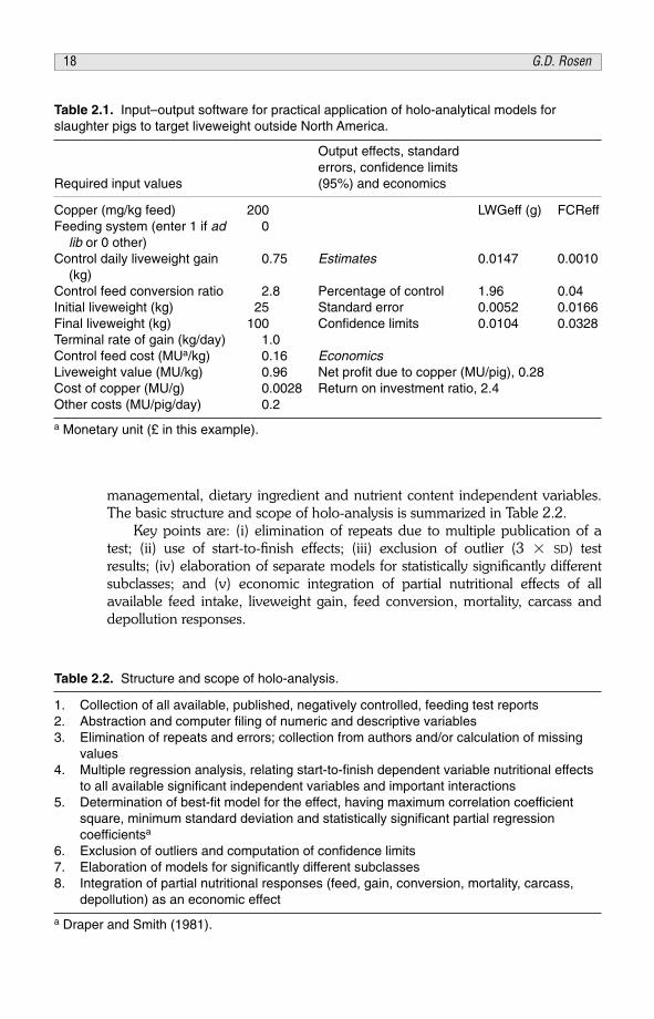
managemental, dietary ingredient and nutrient content independent variables.The basic structure and scope of holo-analysis is summarized in Table 2.2.
Key points are: (i) elimination of repeats due to multiple publication of atest; (ii) use of start-to-finish effects; (iii) exclusion of outlier (3 � SD) testresults; (iv) elaboration of separate models for statistically significantly differentsubclasses; and (v) economic integration of partial nutritional effects of allavailable feed intake, liveweight gain, feed conversion, mortality, carcass anddepollution responses.
Table 2.1. Input–output software for practical application of holo-analytical models forslaughter pigs to target liveweight outside North America.
Output effects, standarderrors, confidence limits
Required input values (95%) and economics
Copper (mg/kg feed) 200 LWGeff (g) FCReffFeeding system (enter 1 if ad 0
lib or 0 other)Control daily liveweight gain 0.75 Estimates 0.0147 0.0010
(kg)Control feed conversion ratio 2.8 Percentage of control 1.96 0.04Initial liveweight (kg) 25 Standard error 0.0052 0.0166Final liveweight (kg) 100 Confidence limits 0.0104 0.0328Terminal rate of gain (kg/day) 1.0Control feed cost (MUa/kg) 0.16 EconomicsLiveweight value (MU/kg) 0.96 Net profit due to copper (MU/pig), 0.28Cost of copper (MU/g) 0.0028 Return on investment ratio, 2.4Other costs (MU/pig/day) 0.2
a Monetary unit (£ in this example).
Table 2.2. Structure and scope of holo-analysis.
1. Collection of all available, published, negatively controlled, feeding test reports2. Abstraction and computer filing of numeric and descriptive variables3. Elimination of repeats and errors; collection from authors and/or calculation of missing
values4. Multiple regression analysis, relating start-to-finish dependent variable nutritional effects
to all available significant independent variables and important interactions5. Determination of best-fit model for the effect, having maximum correlation coefficient
square, minimum standard deviation and statistically significant partial regression coefficientsa
6. Exclusion of outliers and computation of confidence limits7. Elaboration of models for significantly different subclasses8. Integration of partial nutritional responses (feed, gain, conversion, mortality, carcass,
depollution) as an economic effect
a Draper and Smith (1981).
18 G.D. Rosen

Table 2.3 contains a guideline to the number of tests used in theprogression of holo-analysis (Rosen, 2004b).
Exploratory and preliminary models based on 20–100 tests could beinvaluable in guiding the course of future research and development. Workingmodels normally require 100–300 tests. Successive increments of 30–60 teststhereafter are used to update models. Expansion beyond 300 tests normallyhas only marginal effects on partial regression coefficients.
Model structures
The basic nature and structure of holo-analytical models is illustrated forpresent purposes with a large test resource of the five most utilized antibiotics inbroiler feeds (Table 2.4). The four models for ‘Bromycin’ shown in Table 2.5incorporate key variables, level of control performance, duration, year of test,logarithmic dosage, anticoccidial feed and disease. This set of models accountsonly for 5–69% of the variations in the four nutritional responses.
Partial regression coefficients for valid models must have logical algebraicsigns. In Table 2.5 the control performance terms are all negative showing, aswould be expected, that the magnitude of response at a given dosage is reducedwith superior negative control performances. Within the dosage range 0–100 ppmin this data set, the control performance and dosage partial regression coefficientsare critical for optimal posology. As expected, gain, conversion and mortalityeffects are greater in the presence of diagnosed or endemic disease.
Table 2.3. Numbers of negatively controlled tests used in holo-analysis.
Number of tests Applications
3 EU efficacy minimum for first registration10–20 Guide to the use of an average dosage for an average response20–50 Exploratory models50–100 Preliminary models100–300 Working models>300 Updated models
Table 2.4. ‘Bromycin’ resource for broiler feed pronutrientantibiotics.
Number of tests
Antibiotic FDIeff/LGWeff/FCReff MORTeff
All 1709 708Bacitracin MD 194 82Bacitracin Zn 640 346Chlortetracycline 299 88Oxytetracycline 247 76Virginiamycin 329 116
Pronutrient antibiotics 19

A large variety of other independent variables must, however, beinvestigated for the elaboration of comprehensive working models for use inpraxis. Many of these are listed in Table 2.6.
Inclusion of all available significant variables raises R2 and SD reduces inworking models. Normally, chronological and geographical variables disappearwith the inclusion of additional significant variables such as those in Table 2.6or, alternatively, they regress to minor contributors to response variations.Examples of two working models are detailed in Table 2.7 for phytase inbroilers.
These larger models account for 64 and 72%, respectively, of the variationin feed intake and liveweight gain responses. Further model development,however, beyond 75% is largely restricted by the unavailability in publishedreports of key variables such as temperature, altitude and/or the absence of keydietary factors. Nevertheless, models such as those in Table 2.7 are alreadyuseful in the appraisal of effective antibiotic replacements.
Table 2.5. ‘Bromycin’ holo-analytical models.
FDIeff = – 79.5 – 0.0380 FDIC + 2.98 DUR + 1.02 EXD R2 0.052 – 24.3 COC SD 126
LWGeff = – 72.4 – 0.0114 LWGC + 1.02 DUR + 0.871 EXD + 19.5 log(ABP+1) R2 0.171 – 16.8 COC + 76.1 VETSD 47.4
FCReff = 0.301 – 0.161 FCRC + 0.00306 DUR – 0.00159 EXD – 0.0286 log(ABP+1)R2 0.287 – 0.116 VETSD 0.100
MORTeff = – 1.13 – 0.647 MORTC + 0.0363 DUR + 1.081 log(ABP+1)R2 0.685 – 2.12 VETSD 3.12
Table 2.6. Independent variables used in holo-analytical nutritional models.
control performance feed process maizeb gross energyc
duration antibiotic sorghum net energyc
year of test anticoccidial wheat crude proteindose antihistomonial barley crude fatinitial age metabolic test oats crude fibrenot day-old diet marker rye calciumsex part-purified diet animal fat phosphorusphased dose disease challenge vegetable oil lysinefactor 2 dosea supplier test animal protein methionineselected weight birds institute test vegetable protein methionine + cystinehousing country wheat offal threoninestocking density brand rice bran tryptophan
a Second antibiotic/enzyme/acid/microbial/other pronutrient/nutrient.b As dietary concentrations (columns 3 and 4).c Digestible or metabolizable energy are alternatives.
20 G.D. Rosen

Model applications
Holo-analytical models can be used to compare classes of pronutrients (e.g.antibiotics versus enzymes) and also specific generic or branded individualproducts (e.g. zinc bacitracin versus phytase as generics or Albac zinc bacitracinversus Natuphos phytase as brands).
The mean characteristics of the class resources in Table 2.8 manifeststriking differences between the conditions used in their test programmesduring the course of research and development, e.g. mean enzyme testduration is 17 days less than antibiotic using 59 versus 39% caged birds, withonly 15 versus 40% anticoccidial in 31 versus 17% mash feeds. Models forthese two classes have been used to compare gain and conversion responses inpraxis conditions at a current broiler performance level (Rosen, 2001). Atrespective mean dosages of 18 ppm and 7.5 units/g feed there is no significantdifference in antibiotic and enzyme responses. The enzyme model also predictsthat the mean one-point difference in feed conversion effect could beeliminated by the use of a higher enzyme dosage of 9.2 u/g feed, i.e. +1.7 u/g.
In Table 2.9, zinc bacitracin and phytase are compared for equalinvestment and for equal feed conversion improvement. At equal investmentzinc bacitracin (80 ppm) is five conversion points better than phytase, which isineffective at 500 units/kg feed, a dosage commonly deployed for depollutionand phosphorus economy. These models also show that a 3.2-point conversionimprovement by 42 ppm zinc bacitracin would require a 20-fold higher phytasedosage (10,000 u/kg feed).
Table 2.7. Phytase broiler holo-analytical feed intake and liveweight gain models.
FDIeff = 232 – 0.136FDIC + 20.0 DUR + 226 log(PHY+1) – 514 log(P+1) – 78.9 CAG + 93.0 NDOR2 0.641 SE 56.0 0.014 1.83 46.3 54.2 13.2 18.2SD 62.1 p 0.000 0.000 0.000 0.000 0.000 0.000 0.000
+ 65.2 COC + 7.02 Ca – 150 NAT – 222 NOV – 207 FIN – 0.573 MZP + 71.0 AOFSE 12.0 2.38 14.5 23.7 21.3 0.018 19.9p 0.000 0.004 0.000 0.000 0.000 0.002 0.000
+ 12.1 ROP – 14.7 PFP – 9.13 AFP – 1.66 VOPSE 0.264 0.311 0.269 0.352p 0.000 0.000 0.000 0.001
LWGeff = 118 – 0.231 LWGC + 16.4 DUR + 168 log(PHY+1) – 339 log(P+1) – 49.6 CAG + 54.2 NDO R2 0.717 SE 33.3 0.017 1.08 26.0 29.0 7.67 8.85SD 35.4 p 0.000 0.000 0.000 0.002 0.000 0.000 0.000
+ 48.1 COC – 86.3 NAT – 142 NOV – 122 FIN – 0.716 MZP – 0.662 SOP – 1.97 BAPSE 6.72 7.71 13.1 12.5 0.012 0.021 0.055p 0.000 0.000 0.006 0.000 0.000 0.000 0.000
+ 105 AOF + 5.58 ROP – 7.41 PFPSE 13.0 0.156 0.174 p 0.000 0.000 0.000
Pronutrient antibiotics 21

Table 2.8. Pronutrient class comparison of antibiotics and exogenous enzymes in broilers.
DURn LWGC (g) LWGeff (g) FCRC FCReff MORTC MORTeff (days) CAG MAL COC MAS
Antibiotics 1709/708 1235 38.0 2.274 –0.0650 4.32 –0.41 47.7 0.39 0.30 0.40 0.17Enzymes 1869/365 1133 57.0 1.946 –0.100 6.83 –1.80 30.5 0.59 0.51 0.15 0.31
For 42-day-old, as-hatched on the floor, pellet feeds, with anticoccidial LWGC 2326 g; FCRC 1.777
LWGeff (g) FCReff
Antibiotics (17.9 ppm) 33.7 –0.041Enzymes (7.5 u/g) 33.8 –0.030
22G.D. Rosen
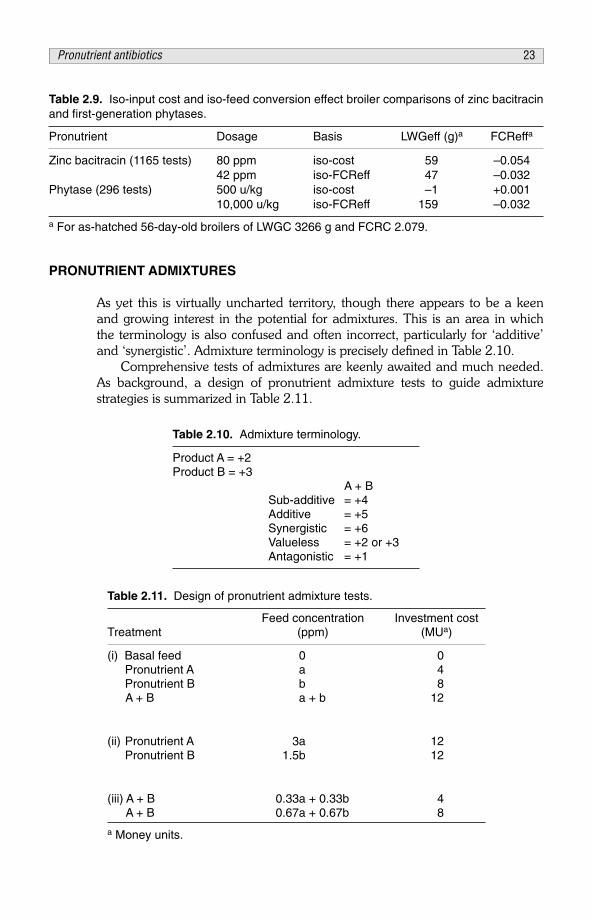
PRONUTRIENT ADMIXTURES
As yet this is virtually uncharted territory, though there appears to be a keenand growing interest in the potential for admixtures. This is an area in whichthe terminology is also confused and often incorrect, particularly for ‘additive’and ‘synergistic’. Admixture terminology is precisely defined in Table 2.10.
Comprehensive tests of admixtures are keenly awaited and much needed.As background, a design of pronutrient admixture tests to guide admixturestrategies is summarized in Table 2.11.
Table 2.9. Iso-input cost and iso-feed conversion effect broiler comparisons of zinc bacitracinand first-generation phytases.
Pronutrient Dosage Basis LWGeff (g)a FCReffa
Zinc bacitracin (1165 tests) 80 ppm iso-cost 59 –0.05442 ppm iso-FCReff 47 –0.032
Phytase (296 tests) 500 u/kg iso-cost –1 +0.00110,000 u/kg iso-FCReff 159 –0.032
a For as-hatched 56-day-old broilers of LWGC 3266 g and FCRC 2.079.
Table 2.10. Admixture terminology.
Product A = +2Product B = +3
A + BSub-additive = +4Additive = +5Synergistic = +6Valueless = +2 or +3Antagonistic = +1
Table 2.11. Design of pronutrient admixture tests.
Feed concentration Investment costTreatment (ppm) (MUa)
(i) Basal feed 0 0Pronutrient A a 4Pronutrient B b 8A + B a + b 12
(ii) Pronutrient A 3a 12Pronutrient B 1.5b 12
(iii) A + B 0.33a + 0.33b 4A + B 0.67a + 0.67b 8
a Money units.
Pronutrient antibiotics 23

Table 2.11 encompasses the following: (i) basal 2 � 2 factorial tests; (ii) iso-mixture cost tests for higher single dosages; and (iii) iso-mixture costs for eachfactorial component.
Concerning pronutrient admixtures the literature contains several examplesof diverse interactions in broiler nutrition. In the total number of 2 � 2 factorialtests (17) on phytases and xylanases reported up to 2004, mainly (15) forwheat-based diets, interactions ranged from antagonistic to synergistic, related,at least in part, to the presence or absence of substantial declared or undeclaredenzyme side activities in phytase and xylanase products (Rosen, 2004c).
DISCUSSION AND CONCLUSION
The task of setting standards for the efficient replacement of pronutrientantimicrobials in poultry nutrition is greatly facilitated by the availability in theliterature of thousands of negatively-controlled test results, which provide largedata resources for the elaboration of holo-analytical models (Rosen, 1995). Thesetting of specific standards using such models for the assessment of efficacy ineach and every individual circumstance of use is well advanced in the case ofenzymes.
Preliminary models are also available for a pronutrient saccharide product(Rosen, 2005). The studies to date have indicated the likelihood that enzymesrepresent the current best antibiotic replacement prospect. In the use ofenzymes, however, care is needed in distinguishing between nutritional anddepollution responses and their economic implications.
A simple Seven Question Test has been devised as a starting point in theappraisal of the efficacy and modelling status of the many hundreds of candidateson offer as antibiotic replacements. Illustrative answers are given in Table 2.12.
The answers shown to Questions 1–3 counsel a cautious approach toProduct X with its need to expand its data base as soon as possible. In suchcases where own tests are preferred the best policy is to investigate Product X at
Table 2.12. Seven Question Test for pronutrient antimicrobial replacement candidates(illustrative answer).
Question Reply
1. How many properly controlled feeding tests do you have on the efficacy of Product X? 20
2. How many of these have no negative controls? 23. Can you supply a bibliography for questions 1 and 2? yes4. How many times out of ten does Product X improve liveweight gain
and feed conversion? 7/105. What are the coefficients of variation in the gain and conversion
responses? 100–200%6. What dosage of Product X will maximize return on my investment? x ppm because…7. Can you supply me with a model to predict responses to Product X
under my specific conditions? yes
24 G.D. Rosen

the average dosage used in tests to date with half and double doses, at least ina dose-response trial. The answers to Questions 4–6 are very satisfactory,particularly if supporting data justify the dosage advocated in reply to Question6. A positive answer to Question 7 with provision of software to utilize themodel for Product X is optimal.
The next priorities in modelling programmes are for the modelling of organicacids used individually or in admixtures, followed by microbials and botanicals.The role of nutrients as antibiotic replacements also needs to be defined.
REFERENCES
Draper, N.R. and Smith, H. (1981) Applied Regression Analysis, 2nd edn. John Wiley and SonsInc., New York, 709 pp.
Fuller, R. (1989) Probiotics in man and animals. Journal of Applied Bacteriology 66, 365�378.Garland, P.W. (1995) Range of substances currently available and problems to be addressed for
the future. In: Senkoylu, N. (ed.) Proceedings of the 10th European Symposium on PoultryNutrition, 15–19 October, Antalya, Turkey, pp. 203–207.
Gibson, G.R. and Roberfroid, M.B. (1995) Dietary modulation of the human colonic microbiota:introducing the concept of prebiotics. Journal of Nutrition 125, 1401–1412.
Lilley, D.M. and Stillwell, R.H. (1965) Probiotics: growth-promoting factors produced by micro-organisms. Journal of Bacteriology 89, 747–48.
Maury, N.F. (1869) The Physical Geography of the Sea and its Meteorology, 14th edn. SampsonLow, Son and Marston, London, 324 pp.
Parker, R.B. (1974) Probiotics, the other half of the antibiotic story. Animal Nutrition and Health29, 4�8.
Rosen, G.D. (1995) Antibacterials in poultry and pig nutrition. In: Wallace, R.J. and Chesson, A.(eds) Biotechnology in Animal Feeds and Animal Feeding, VCH Verlagsgesellschaft mbH,D69451 Weinheim, Germany, pp. 143–172.
Rosen, G.D. (1996) Feed additive nomenclature. World’s Poultry Science Journal 54, 53�57.Rosen, G.D. (1997) Future prospects for pronutrients in poultry production. In: Proceedings of the
16th Scientific Day, 16 October, University of Pretoria, Pretoria, Republic of South Africa,pp. 60–79.
Rosen, G.D. (2001) Multi-factorial efficacy evaluation of alternatives to antimicrobials in pronutri-tion. British Poultry Science 42S, S104–S105.
Rosen, G.D. (2004a) Holo-analysis in animal nutrition. Poultry International 25 (12), 17�18, 21.Rosen, G.D. (2004b) How many broiler feeding tests are required to elaborate working models
for the prediction of nutritional effects of pronutrients in praxis? British Poultry Science 45S1,S8–S9.
Rosen, G.D. (2004c) Admixture of exogenous phytases and xylanases in broiler nutrition. In:Abstracts and Proceedings, XXII World’s Poultry Congress, 13–17 June, Istanbul (CD-ROM).
Rosen, G.D. (2005) Holo-analysis of the effects of genetic, managemental, chronological anddietary variables on the efficacy of a pronutrient mannanoligosaccharide in broilers. BritishPoultry Abstracts 1 (1), 27–29.
Rosen, G.D. (2006) Setting and meeting standards for the efficient replacement of pronutrientantibiotics in poultry and pig nutrition. In: Barung, D., de Jong, J., Kies, A.K. and Verstegan,M.W.A. (eds) Antimicrobial Growth Promoters: Where do We Go From Here? WageningenAcademic Publishers, Wageningen, Netherlands, pp. 381–397.
Rosen, G.D. and Roberts, P. (1996) A Comprehensive Survey of the Response of Growing Pigs toSupplementary Copper in Feed. Field Investigations and Nutrition Services Ltd., London, p. 94.
Pronutrient antibiotics 25

Waksman, S.A. (1954) In: My life with the Microbes. Robert Hale Ltd., London, p. 210.Winter, A.G. (1955) The significance for therapy and diet of antibiotic substances from flowering
plants (with special reference to nasturtiums, cress and horseradish). Die Medizinische 2,73–80.
APPENDIX. SYMBOLS, VARIABLES AND UNITS
Symbol Variable (unit) Symbol Variable (unit)
ABP antibiotic (ppm) MORTC control mortality (%)AFP animal fat (%) MORTeff effect on MORTC (%)AOF added oil/fat (0 or 1) MU money unitBAP barley (%) MZP maize (%)Ca calcium (g/kg) n number of testsCAG cage housing (0 or 1) NAT Natuphos (0 or 1)COC anticoccidial feed (0 or 1) NDO not day-old (0 or 1)CV coefficient of variation (%) NOV Novo phytase (0 or 1)DUR test duration (days) p probabilityEXD test year – 1900 P phosphorus (g/kg)FCRC control feed conversion ratio PFP poultry fat (%)FCReff effect on FCRC PHY phytase (u/g feed)FDIC control feed intakea R2 multiple correlation coefficient squareFDIeff effect on FDICa ROP rape oil (%)FIN Finase (0 or 1) SD standard deviationlog log10 SE standard errorLWGC control liveweight gaina SOP sorghum (%)LWGeff effect on LWGCa u unitsMAL male (0 or 1) VET presence of disease (0 or 1)MAS mash feed (0 or 1) VOP vegetable oil (%)MD methylene disalicylate Zn zinc
a broiler and turkey (g), pig (kg/day).
26 G.D. Rosen

PART II
Gastrointestinal structure andfunctional development

This page intentionally left blank

CHAPTER 3Early development of small intestinal function
Z. UniDepartment of Animal Sciences, Faculty of Agricultural, Food andEnvironmental Quality Sciences, The Hebrew University of Jerusalem,Israel; e-mail: [email protected]
Dedicated to the memory of David Sklan. A unique individual and scientist –my partner in exploring the chicken intestine.
ABSTRACT
To accommodate the rapid transition to external nutrient sources, the chickensmall intestine goes through morphological, cellular and molecular changestowards the end of incubation. The weight of the intestine, as a proportion ofembryonic weight, increases from approximately 1% at 17 days of incubation(17E) to 3.5% at hatch. At this time, the embryonic small intestinal villi aredivided into two main developmental stages, which differ in both length andshape. Mucin-producing cells can be observed from 17E and at that timecontain only acidic mucin. The activity and RNA expression of brush-borderenzymes, which digest disaccharides and small peptides, and of majortransporters (sodium–glucose transporter and ATPase) which are found at 15E,begin to increase at 19E (2 days prior to hatch), and increase further on theday of hatch. Although the digestive capacity begins to develop a few daysbefore hatch, most of the development occurs post-hatch when the neonatalchick begins consuming feed. During the post-hatch period, the weight of thesmall intestine increases at a faster rate than body mass. Rapid enterocyteproliferation and differentiation occur. In addition the intestinal crypts, whichbegin to form at hatch, are clearly defined several days post-hatch, increasingin both cell number and size. Goblet cells produce acidic and neutral mucins insimilar proportions. Studies have shown that feeding immediately post-hatch oreven pre-hatch accelerates the functional development of the small intestine,while delayed access to external feed arrests the development of the smallintestine’s mucosal layer and changes mucin dynamics.
© CAB International 2006. Avian Gut Function in Health and Disease (ed. G.C. Perry) 29

INTRODUCTION
The investigation of intestinal cell dynamics is crucial to understanding bothdigestive physiology and the efficiency of animal production. The epitheliallayer of the intestine, the site of nutrient digestion and absorption, is composedof a continuously renewable population of cells in which stem cells, located inthe crypt region, give rise to enterocyte and goblet cells. These cells migrate upthe villi where they exhibit changes in functional characteristics.
The developing intestine undergoes morphogenesis and cyto-differentiation. Enzymes, transporters and glycosidation patterns changedramatically during the transition from embryo to independent chick,accompanying the crucial transition that occurs in the nutrient source of thehatchling. The yolk sac consists primarily of lipids and proteins, while theexternal feed consists of carbohydrates, lipids and proteins. This major changedemands suitable redeployment of the cellular mechanisms that catalyse thedigestion or hydrolysis and absorption of food components.
As the chicks make the metabolic and physiological transition from eggnutrition (i.e. the yolk) to exogenous feed, the yolk lipids – such as oleic acid –are quickly absorbed (this occurs close to hatch), whereas dietarycarbohydrates and amino acids are not well utilized because of limitations indigestive and absorptive capacity. Therefore, the digestive system plays a majorrole in determining the development potential of the hatchling. Nutrientdigestion, absorption, assimilation and incorporation into developing andgrowing tissues, including skeletal muscle, depend directly on the functionalcapabilities of the intestinal epithelial layer.
To achieve optimal muscle growth rates – and ultimately muscle and meatyields in poultry – it is essential to maximize enterocyte development, functionand performance. Early functions of the gastrointestinal tract (GIT) are vital forthe chicken’s growth performance. Hence, it is crucial to achieve optimalintestinal development and functional capacity. This chapter examines themorphological and functional development of the small intestine in chickens.
GROSS AND STRUCTURAL ASPECTS OF THE DEVELOPING SMALL INTESTINE
As incubation progresses, the body weight of the embryo increases, as does theweight of the small intestine. However, small intestinal weight increases at amuch greater rate than does body weight, the latter showing only a smallincrease close to hatch. The enhanced growth rate of the small intestine canclearly be seen by calculating the weight of the intestine as a percentage ofembryo weight. During the last 3 days of incubation this increases fromapproximately 1 on day 17 of embryonic age (17E) to 3.5 at hatch.
The morphology of the small intestine also changes rapidly. Histologyindicates that the intestine – including the external muscular layers and the villi– are growing rapidly. Villi at 15E are rudimentary; however, at 17E, villi atdifferent stages of development are observed.
30 Z. Uni

Two main developmental stages can be seen, differing in length and shape:the larger villi are often pear-shaped and the smaller ones (approximately 65%of the size of the large ones) are narrower, with a rocket-like shape. Adjacent toeach larger villus there appears to be one smaller one. A similar pattern wasobserved at 18E and 19E, with villi at both developmental stages growing,mainly in length, while maintaining similar shapes and distribution ratios. At19E there was some ‘budding’ at the base of existing villi. These budsdeveloped considerably and, by 20E, an additional ‘wave’ of small villi wasfound, comprising some 30% of the total.
At this embryonic stage, one day before hatch, the earlier waves of villicontinue to grow and the third series of developing villi are approximately 65%of the size of the next largest ones. Villus growth rate between 19E and 20Ewas 30–40%; between 20E and day of hatch, however, growth was twofold forvilli from all three developmental stages (Uni et al., 2003b).
Further rapid changes are observed post-hatch, with significantmorphological development of the small intestine occurring immediately afterhatch. The intestines of hatchlings increase in weight more rapidly than doesbody mass. Growth rate of the intestines relative to body weight was greatest at5–7 days of age in chicks (Uni, 1999). A two- to fourfold increase in intestinallength was observed up to 12 days of age, while the weight of the threeintestinal segments (duodenum, jejunum and ileum) increased seven- totenfold.
Villi increase in size and number, giving them a greater absorptive surfaceper unit of intestine. This rapid morphological development immediately afterhatch involves differing rates of increase in villus volume in the duodenum,jejunum and ileum. Although duodenal villus growth is almost complete by day7, jejunum and ileum development continues beyond 14 days of age (Uni etal., 1995, 1999).
Fig. 3.1. Representative light microscopy (� 200) of intestinal villi from the jejunum of thebroiler embryo at 17 days (17E) of incubation (a) and at 20 days (20E) of incubation (b).Sections show the pear-shaped and rocket-shaped villi. Villi in different stages of develop-ment can be observed. Staining was with haematoxylin-eosin. Bar = 10 �m.
Gut development 31
Enterocyte density ranges from 200,000–280,000 cells/cm2 in all threesegments of the small intestine and changes little with age. Nevertheless, anincrease in the total number of enterocytes per villus is observed with age,resulting from the dramatic increase in villus length (Uni et al., 1998a).
Crypt formation and enterocyte proliferation and migration in the developingsmall intestine
A continuous renewal process occurs in the intestinal mucosa as proliferatingcells in the mucosal crypts differentiate, predominantly to enterocytes, whichmigrate up the villus and are sloughed into the lumen from the villus tip. Duringthis migration process, the enterocytes acquire differentiated functions in termsof digestion, absorption and mucin secretion (Traber et al., 1991, 1992; Ferrariset al., 1992).
The site of enterocyte proliferation and the rate of their migration varyamong vertebrates. In most mammals, proliferation is restricted to the crypts atthe base of the villi, and their progeny migrate up the villus where theyspecialize and lose their capacity to divide (Cheng and Leblond, 1974a,b).However, in lower vertebrates such as amphibians (McAvoy et al., 1975;McAvoy and Dixon, 1978a,b) and turtles (Wurth and Musacchia, 1964), theproliferating zones lie in troughs between mucosal folds and not in clearlydefined zones of proliferation as in the higher orders of vertebrates.
In most mammals, the small intestinal crypts develop in the prenatal periodfrom the flat intervillus epithelium (Marshall and Dixon, 1978; Quaroni,1985a). DNA synthesis and cell proliferation are common over the entire lengthof the villus until birth, after which proliferation becomes restricted to the cryptarea (Quaroni, 1985a, b).
In poultry, at hatch, all small intestinal enterocytes are proliferating; withtime, the proportion of proliferating cells decreases rapidly, reachingapproximately 50% in the crypt 2–3 days post-hatch. Interestingly, by day 10post-hatch, proliferating enterocytes are not restricted to the crypt region butalso exist along the villi. Their proportion along the villi decreases quickly in thefirst days post-hatch and then more slowly, reaching approximately 10% (Uniet al., 1998b). Lack of access to feed increases the proportion of proliferatingenterocytes, both in the crypt and along the villus (Geyra et al., 2001a, b).
In hatching birds, a single crypt is observed per villus with a relatively smallnumber of cells per crypt. During the initial days post-hatch, crypt numbersincrease by branching and, at the same time, a process of hyperplasia occurs inthe crypt region. After 4–5 days, the rate of growth declines and by the end ofthe second week post-hatch, three to four crypts are observed per villus.
Enterocyte migration from crypt to villus tip takes approximately 72 h in 4-day-old chicks and 96 h in older birds (Uni et al., 1998b; Geyra et al., 2001b).At hatch, small intestinal enterocytes are round and non-polar; from 24–48 hpost-hatch, these cells increase rapidly in length and develop pronouncedpolarity and a defined brush border.
32 Z. Uni

Thus, the small intestine of a newly hatched chick is immature andundergoes dramatic changes during the first few days post-hatch. This isdemonstrated by the immaturity of the crypts and the absence of a definedzone of enterocyte proliferation. During the initial 4–5 days post-hatch, thecrypt–villus axis is established with a distinct area of proliferation and constantenterocyte migration. The extensive changes in the morphological developmentof the small intestine close to and immediately post-hatch include thedifferentiation of enterocytes and the definition of crypts, as well as a many-foldenlargement of the intestine’s absorptive surface.
DEVELOPMENT OF DIGESTIVE AND ABSORPTIVE FUNCTIONS OF THEINTESTINAL MUCOSA
In addition to luminal digestion of feed-derived macromolecules, the finalstages of digestion are carried out by membrane-anchored enzymes at thebrush-border region of the enterocyte cells. These include sucrase-isomaltase(SI), peptidases and phosphatases (Semenza, 1986).
Fig. 3.2. Light microscope section of jejunal sections at hatching (a); round and non-polarenterocytes can be observed. At day 7 post-hatch (b) enterocytes are polar. Bar = 10 �m.
Gut development 33

Chickens have a considerable capacity to hydrolyse disaccharides via thebrush-border enzyme SI immediately post-hatch and even pre-hatch (Uni,1999; Uni et al., 2003b). SI expression increases rapidly just before hatch andcontinues to increase post-hatch (Sklan et al., 2003). The pre-hatch expressionis intriguing because intestinal enterocytes have not yet been exposed tocarbohydrates. This was explained by the homeobox gene CDX which isinvolved in inducing SI expression in mammals (Ferraris, 2001) and in chickens(Sklan et al., 2003).
The major transporter for glucose is the sodium–glucose transporter(SGLT-1), which is expressed during the prenatal period. Expression of SGLT-1increases 2 days pre-hatch and continues to increase post-hatch (Sklan et al.,2003; Uni et al., 2003b).
Another enzyme which plays a major role in protein digestion, via thehydrolysis of peptides, is the brush-border enzyme aminopeptidase. Thisenzyme is expressed at a relatively high constant level (compared to the SGLTand SI genes) from 15E until hatch (Uni et al., 2003b).
For absorption to occur, sodium is required in the lumen and, onceabsorbed, must be removed from enterocytes to maintain ionic equilibrium.Sodium transport occurs via the enterocyte’s basolateral Na+/K+-ATPase. Theexpression of this enzyme is detected 2 days prior to hatching (Uni et al., 2003b).
An examination of the activities of the jejunal brush-border membraneenzymes maltase, aminopeptidase, SGLT-1 and ATPase during the last periodof embryonic development revealed low activity of both maltase andaminopeptidase at 15E and 17E. Activities of all the enzymes and transportersexamined began to increase at 19E and further increased on the day of hatch.
The relative mRNA expressions of the different enzymes and transporterswere correlated, as were their activities (r = 0.75–0.96); however, expressionwas not correlated with the enzymatic activities.
In general, at 19E, the mRNA levels of the apical and basolateral digestiveenzymes and nutrient transporters have significantly increased relative to theirlevels at 15E and 17E. This elevation in gene expression is followed by anincrease in the biochemical activities of the enterocyte enzymes andtransporters on the day of hatch.
The intestinal mucosal layer appears to change rapidly during the late pre-hatch period, and the late-term embryonic intestine seems to have the capacityto digest and absorb disaccharides and short peptides 2 days prior to hatch.These major changes in the expression and localization of the functional brush-border proteins prepare the framework for ingestion of carbohydrate- andprotein-rich exogenous feed post-hatch.
Calculation of the total mucosal enzyme activity on a regional basis yieldsactivity curves for disaccharides, gamma glutamyl transferase and alkalinephosphatase that increase curvilinearly with age. Regional activity of mucosalenzymes is related to the digestive capacity in the three different regions of theintestine: the jejunum has the highest regional capacity to digest disaccharides,the duodenum has the lowest capacity and the ileum is intermediate (Uni et al.,1998a). From the end of the first week post-hatch, mucosal enzyme activity permass of intestine is closely correlated to the number of enterocytes per villus,
34 Z. Uni

suggesting that the amount of enzyme activity expressed per enterocyte doesnot change greatly with age.
Development and function of mucous-secreting goblet cells
The epithelium of the GIT is covered by a mucous layer that acts as a mediumfor protection, lubrication and transport between the luminal contents and theepithelial cells. The mucous layer is composed predominantly of mucinglycoproteins produced by mucous-secreting goblet cells. Other components ofthe mucous layer are water, various serum and cellular macromolecules,electrolytes, microorganisms and sloughed cells.
Mucin-type glycoprotein is believed to be capable of aggregating severalbacterial species and preventing the attachment of pathogenic bacteria bymodulating their adherence to the intestinal epithelium. The mucous layer is acomponent of the innate host response which is regulated in response toinflammation and infection. A number of protective mucous layer-associatedproteins are either co-secreted with mucins or interact with mucousenvironments to perform their protective action (Allen and Kent, 1968; Allen,1981, 1989; Allen et al., 1993).
Mucin production for maintenance of the mucous layer is the responsibilityof the goblet cells. The goblet cells arise by mitosis from pluripotential stem cellsat the base of the crypt or from poorly differentiated cells in the lower cryptregion – referred to as oligomucous cells.
Mucins are categorized into three distinct families: gel-forming, soluble andmembrane-bound. Secreted gel-forming mucins are characterized by their largesize, viscoelastic behaviour and high content of carbohydrates. Secretory gel-forming mucin genes have a large central region made up of multiple tandemrepeats which vary in length and sequence, but all have been found to containthreonine and/or serine residues. The amino- and carboxy-terminal regions (D-domains) are very lightly glycosylated, have a high cysteine content and areimportant to the mucin multimerization process. Mucin glycoproteins exhibit ahigh level of heterogeneity resulting from the diversity in length, composition,branching and degree of sulphation and acetylation of the oligosaccharidechains attached to the peptide backbone.
Mucins are classified into neutral and acidic subtypes, and are alsodistinguished by sulphated or non-sulphated groups. Expression of differentmucins, defined by differences in either their protein backbones or theirglycosylation patterns, has been shown to vary both between and withintissues, and during intestinal ontogenesis. Mucin molecules are assembled andstored in large, membrane-bound granules before secretion from the gobletcells. Secretory mucins are secreted from the apical surface of the goblet cellsby either baseline secretion or compound exocytosis. Degradation of themucous layer is part of the balance between synthesis secretion and breakdownof the mucous layer (Forstner, 1978; Forstner and Forstner, 1985, 1994;Forstner et al., 1995).
An investigation of the ontogenesis and development of mucin-producing
Gut development 35

cells in the chicken small intestine (Uni et al., 2003a) showed that mucin-producing cells can be detected from 17E, containing only acidic mucin. Afterhatch and until day 7 post-hatch, the small intestine contains similarproportions of goblet cells producing acidic and neutral mucins. An increasinggradient of goblet cell density was observed along the duodenal–ileal axis.
Thus, the development of small intestinal mucous-secreting cells in chicksoccurs in the late embryonic and immediate post-hatch period.
An analysis of the expression of the chicken intestinal mucin gene(Smirnov et al., 2004) revealed a sustained increase in mucin mRNAexpression from 17E to day 3 post-hatch, at which point expression stabilized.
This increase in mucin mRNA levels parallels the increase in the number ofgoblet cells occurring from the late incubation stage through the first week post-hatch. Western-blot analysis showed the lowest amount of mucin glycoproteinto be in the duodenum, and an increase in content distally along the smallintestine.
Once mucin is synthesized in the goblet cells and secreted to the intestinalsurface, it forms a layer which undergoes continuous degradation and renewal.In 3-week-old chickens, the adhering mucous is of similar thickness throughoutall segments of the mature small intestine.
Studies in chickens and turkeys have confirmed that feeding conditions,dietary composition and microbial flora influence the different parameters ofmucin dynamics in the avian small intestine (Smirnov et al., 2004, 2005).However, the role of mucin in absorption and protection against pathogens,and the age at which the mucin layer acts as a functional barrier, are not yetfully understood.
EARLY FEEDING AND GUT DEVELOPMENT
At hatch, the immature small intestine is physiologically active and primed forthe activities of food assimilation (Uni et al., 2003a). Delayed access to feedafter hatch creates an abnormal physiological situation and leads to thenegative consequences of a lack of enteral nutrition. Nutrient supplyimmediately after hatch is a critical factor for small intestinal development inchicks. The common practice in which feed is first made available to chicksmore than 48 h post-hatch may depress subsequent development (Nunez et al.,1996; Noy et al., 2001).
Feeding immediately post-hatch led to an acceleration in intestinalmorphological development, whereas late access to external feed resulted indelayed development of the small intestine’s mucosal layer (Uni et al., 1998a;Geyra et al., 2001a; Uni and Ferket, 2003). Furthermore, birds denied accessto first feed for 24 to 48 h exhibited decreased villus length (Yamauchi et al.,1996), decreased crypt size and fewer crypts per villi, as well as decreasedenterocytic migration rate (Geyra et al., 2001b). This finding was accompaniedby a decrease in the number of enterocytes and an increase in the density ofgoblet cells producing acid and neutral mucins in the jejunal and ileal villi at theend of the fasting period (Uni et al., 2003a).
36 Z. Uni

Fig. 3.3. Representative light micrographs of jejunum stained with Alcian blue (a, b) orperiodic acid-Schiff (c, d). Goblet cells producing only acid mucin can be observed at 18 days(18E) of incubation (a, c). On the day of hatch (b, d) goblet cells producing both acidic andneutral mucin are observed. Crypts are not yet developed and goblet cells are distributed allalong the villi. (Magnification � 200; bar = 50 �m at both stages).
Furthermore, the size of the cross-sectional area of an individual goblet cellin the small intestinal segments was significantly greater in fasting chicks than incontrols (Uni et al., 2003a). This accumulation of mucin in the goblet cells
Gut development 37

Fig. 3.4. a–c: number of enterocytes (�) and goblet cells (●) (right axis) and the percentage of cells staining positive for Alcian blue (AB+ve, �) orperiodic acid-Schiff (PAS+ve, o) (left axis) per villus column in various regions of the small intestine of broiler embryos and chicks; d–f: villus surfacearea (�) (right axis) and density of goblet cell populations (AB+ve, �; PAS+ve, o) (right axis) in various regions of small intestine of broilersembryos and chicks. Values are means with standard errors represented by vertical bars (when they do not fall within the symbols). Differencesbetween the percentage of AB+ve and PAS+ve were significant (p < 0.05) at 3 days before hatch in the duodenum, jejunum and ileum.
38Z. Uni

might be due to its increased synthesis and/or decreased secretion. Mucinbiosynthesis changes with fasting-associated alterations in the migration rate ofepithelial cells from the proliferating crypt zones (Shea-Donohue et al., 1985)and with perturbations in the differentiation rates of precursor cells into maturegoblet cells (De Ritis et al., 1975; Wattel et al., 1979; Shub et al., 1983).
Since the mucous layer has both protective and transport functions, time ofaccess to feed in the first hours post-hatch is important to the normaldevelopment of the mucous cover in the chicken small intestine. Thus, sinceearly access to feed is critical to intestinal development, nutrient supply to theintestine by early feeding (at the hatchery, in the delivery boxes or to theembryo before hatch by in ovo feeding methods) (Uni and Ferket, 2003) isexpected to enhance development of the small intestine.
Tako et al. (2004, 2005) demonstrated that administration of exogenousnutrients and minerals into the amnion enhances intestinal development byincreasing villus size and by generating early activation of the enterocyte brush-border membrane.
The intra-amniotic administration of carbohydrates, �-hydroxy-�-methylbutyrate (HMB), Na+Cl– ions and zinc-methionine led to elevatedmRNA levels of the genes encoding nutrient enzymes and transporters,followed by their increased biochemical activities in the embryo. Researchperformed with early-feeding methods indicates that the small intestines of inovo-fed hatchlings are at a functional stage of development equivalent to thatof conventionally fed 2-day-old chicks.
Chicken intestinalmucin gene
Control
Control
17E 19E Hatch 3d
17E 19E Hatch 3d
18S
2.0
1.5
1.0
0.5
0.017E 19E Hatch 3 d 7 d
Age (days)
d
cb
a a
Muc
in g
ene
mR
NA
exp
ress
ion
(AU
)
Fig. 3.5. Mucin gene expression in the chicken small intestine. Amplification of chickenintestinal mucin mRNA and 18 S RNA. Changes with age in mucin mRNA expression weremeasured by semi-quantitative RT-PCR and expressed relative to the expression of 18 SrRNA. Values are means ± SEM, n = 5. Means without a common letter are significantlydifferent (p < 0.05).
Gut development 39

SUMMARY
All aspects of the early development of the small intestine have importantinfluences on later growth and development of the whole bird. The effects ofearly feeding and the immediate environment of the chick upon the initiationand development of GIT morphology, digestive and absorptive function andimmunological and bioprotective barrier activities must all be considered inrelation to husbandry and nutritional regimes and in the context of optimizationof commercial production strategies and methods.
REFERENCES
Allen, A. and Kent, P.W. (1968) Biosynthesis of intestinal mucins. Biochemical Journal 106,301–309.
Allen, A. (1981) Structure and function of gastrointestinal mucus. In: Johnson, L.R. (ed.)Physiology of the Gastrointestinal Tract. Raven Press, New York, pp. 617–639.
Allen, A. (1989) Gastrointestinal mucus. In: Handbook of Physiology. The GastrointestinalSystem. Salivary, Gastric, Pancreatic, and Hepatobiliary Secretion. American PhysiologySociety, Bethesda, Maryland, pp. 359–382.
Allen, A., Flemstrom, G., Garner, A. and Kivilaakso, E. (1993) Gastroduodenal mucosal protec-tion. Physiological Reviews 73, 823–857.
Cheng, H. and Leblond, C.P. (1974a) Origin, differentiation and renewal of the four main epithe-lial cell types in the mouse small intestine. I: columnar cell. American Journal of Anatomy141, 461–479.
0
100
200
300
400
500
a a a a a aa
b
a
b
ZnMet Control
17E 18E 19E Hatch 7
Age (days)
Vill
us s
urfa
ce a
rea
(mm
2)
Fig. 3.6. Effect of intra-amniotic administration of Zinc-Methionine (ZnMet) on days 17E, 18Eand 19E, day of hatch and 7 days post-hatch on the jejunal small intestinal villus surfacearea. Values are means ± SEM, n = 4. Means with different letters differ (p < 0.05).
40 Z. Uni

Cheng, H. and Leblond, C.P. (1974b) Origin, differentiation and renewal of the four main epithe-lial cells in the mouse small intestine. IV: unitarian theory of the origin of the four epithelialcell types. American Journal of Anatomy 141, 537–561.
De Ritis, G., Falchuk, Z.M. and Trier, J.S. (1975) Differentiation and maturation of cultured fetalrat jejunum. Developmental Biology 45, 304–317.
Ferraris, R.P. (2001) Dietary and developmental regulation of intestinal glucose transport.Physiology Review 77, 257–302.
Ferraris, R.P., Villenas, S.A., Hirayama, B.A. and Diamond, J. (1992) Effect of diet on glucosetransporter site density along the intestinal crypt–villus axis. American Journal of Physiology262, G1060–G1068.
Forstner, G.G. and Forstner, J.F. (1985) Structure and function of gastrointestinal mucus. In:Desnuelle, P., Sjostrom, H. and Noren, O. (eds) Molecular and Cellular Biology of Digestion.Vol. II, Elsevier Science Publishers B.V. (Biomedical Division), Amsterdam.
Forstner, J.F. (1978) Intestinal mucins in health and disease. Digestion 17, 234–263. Forstner, J.F. and Forstner, G.G. (1994) Gastrointestinal mucus. In: Johnson, L.R. (ed.)
Physiology of the Gastrointestinal Tract, 3rd edn. Raven Press, New York, pp. 1255–1284.Forstner, J.F., Oliver, M.G. and Sylvester, F.A. (1995) Production, structure and biologic relevance
of gastrointestinal mucins. In: Blaser, M.J., Smith, P.D., Ravdin, J.I., Greenberg, H.B. andGuerrant, R.L. (eds) Infections of the Gastrointestinal Tract, Raven Press, New York, pp. 7– 88.
Geyra, A., Uni, Z. and Sklan, D. (2001a) The effect of fasting at different ages on growth andtissue dynamics in the small intestine of the young chick. British Journal of Nutrition 86,53–61.
Geyra, A., Uni, Z. and Sklan, D. (2001b) Enterocyte dynamics and mucosal development in theposthatch chick. Poultry Science 80, 776–782.
Marshall, J.A. and Dixon, K.E. (1978) Cell specialization in the epithelium of the small intestine offeeding Xenopus laevis tadpoles. Journal of Anatomy 126, 133–144.
McAvoy, J.W. and Dixon, K.E. (1978a) Cell specialization in the small intestinal epithelium ofadult Xenopus laevis: functional aspects. Journal of Anatomy. 125, 237–245.
McAvoy, J.W. and Dixon, K.E. (1978b) Cell specialization in the small intestinal epithelium ofadult Xenopus laevis: structural aspects. Journal of Anatomy 125, 155–169.
McAvoy, J.W., Dixon, K.E. and Marshall, J.A. (1975) Effects of differences in mitotic activity, stageof cell cycle, and degree of specialization of donor cells on nuclear transplantation inXenopus laevis. Developmental Biology 45, 330–339.
Noy, Y., Geyra, A. and Sklan, D. (2001) The effect of early feeding on growth and small intestinaldevelopment in the posthatch poult. Poultry Science 80, 912–919.
Nunez, M.C., Bueno, J.D., Ayudarte, M.V., Almendros, A., Suarez, M.D. and Gol, A. (1996)Dietary restriction-induced biochemical and morphometric changes in the small intestine ofnursing piglets. Journal of Nutrition 126, 933–944.
Quaroni, A. (1985a) Crypt cell development in newborn rat small intestine. Journal of CellularBiology 100, 1601–1610.
Quaroni, A. (1985b) Pre- and postnatal development of differentiated functions in rat intestinalepithelial cells. Developmental Biology 111, 280–292.
Semenza, G. (1986) Anchoring and biosynthesis of stalked brush border membrane proteins: gly-cosidases and peptidases of enterocytes and renal tubuli. Annual Review of Cell Biology 2,2255–2313.
Shea-Donohue, T., Dorval, T.E.D., Montcalm, E., El-Bayer, H., Durakovich, A., Conklin, J.J. andDubois, A. (1985) Alterations in gastric mucus secretion in rhesus monkeys after exposure toionizing radiation. Gastroenterology 88, 685–690.
Shub, M.D., Pang, K.Y., Swann, D. and Walker, W.A. (1983) Age-related changes in chemicalcomposition and physical properties of mucus glycoprotein from rat small intestine.Biochemical Journal 215, 405–411.
Gut development 41

Sklan, D., Geyra, A., Tako, E., Gal-Gerber, O. and Uni, Z. (2003) Ontogeny of brush bordercarbohydrate digestion and uptake in the chick. British Journal of Nutrition 89, 747–753.
Smirnov, A., Sklan, D. and Uni, Z. (2004) Mucin dynamics in the chick small intestine are alteredby starvation. Journal of Nutrition 134, 736–742.
Smirnov, A., Perez, R., Amit-Romach, E., Sklan, D. and Uni, Z. (2005) Mucin dynamics andmicrobial populations in chicken small intestine are changed by dietary probiotic andantibiotic growth promoter supplementation. Journal of Nutrition 135, 187–192.
Tako, E., Ferket, P.R. and Uni, Z. (2004) The effects of in ovo feeding of carbohydrates and beta-hydroxy-beta-methylbutyrate on the development of chicken intestine. Poultry Science 83,2023–2028.
Tako, E., Ferket, P.R. and Uni, Z. (2005) Changes in chicken intestinal Zinc exporter (ZnT1)mRNA expression and small intestine functionality following an intra-amniotic Zinc-Methionine (ZnMet) administration. Journal of Nutritional Biochemistry 16, 339–346.
Traber, P.G., Gumucio, D.L. and Wang, W. (1991) Isolation of intestinal epithelial cells for thestudy of differential gene expression along the crypt–villus axis. American Journal ofPhysiology 260, 895–903.
Traber, P.G., Yu, L., We, G. and Judge, J. (1992) Sucrase-Isomaltase gene expression along thecrypt-villus axis of human small intestine is regulated at the level of mRNA abundance.American Journal of Physiology 262, G123–G130.
Uni, Z. (1999) Functional development of the small intestine in domestic birds: cellular and mole-cular aspects. Poultry and Avian Biology Reviews 10, 167–179.
Uni, Z. and Ferket, P.R. (2003) Enhancement of development of oviparous species by in ovofeeding. Patent number US 6,592,878.
Uni, Z., Noy, Y. and Sklan, D. (1995) Posthatch changes in morphology and function of the smallintestines in heavy- and light-strain chicks. Poultry Science 74, 1622–1629.
Uni, Z., Ganot, S. and Sklan, D. (1998a) Posthatch development of mucosal function in thebroiler small intestine. Poultry Science 77, 75–82.
Uni, Z., Platin, R. and Sklan, D. (1998b) Cell proliferation in chicken intestinal epithelium occursboth in the crypt and along the villus. Journal of Comparative Physiology (B) 168, 24– 247.
Uni, Z., Noy, Y. and Sklan, D. (1999) Posthatch development of small intestinal function in thepoult. Poultry Science 78, 215–222.
Uni, Z., Smirnov, A. and Sklan, D. (2003a) Pre- and posthatch development of goblet cells in thebroiler small intestine: effect of delayed access to feed. Poultry Science 82, 320–327.
Uni, Z., Tako, E., Gal-Garber, O. and Sklan, D. (2003b) Morphological, molecular, and functionalchanges in the chicken small intestine of the late-term embryo. Poultry Science 82,1747–1754.
Wattel, W., Van Huis, G.A., Kramer, M.F. and Geuze, J.J. (1979) Glycoprotein synthesis in themucous cell of the vascularly perfused rat stomach. American Journal of Anatomy 156,313–320.
Wurth, M.A. and Musacchia, X.J. (1964) Renewal of intestinal epithelium in the freshwater turtle,Chrysemys picta. Anatomical Record 148, 427–439.
Yamauchi, K., Kamisoyama, H. and Isshiki, Y. (1996) Effects of fasting and re-feeding on struc-tures of the intestinal villi and epithelial cells in White Leghorn hens. British Poultry Science37, 909–921.
42 Z. Uni

CHAPTER 4Absorptive function of the smallintestine: adaptations meeting demand
M.A. Mitchell1* and M. Moretó2
1Scottish Agricultural College, Bush Estate, Midlothian, UK; 2Departament de Fisiologia, Facultat de Farmàcia, Barcelona, Spain; *e-mail: [email protected]
ABSTRACT
The mucosal epithelium of the small intestine constitutes a highly dynamicinterface with the external environment through the delivery, digestive processingand absorption of nutrients. The intestinal mucosa is capable of rapid andextensive morphological and functional adaptation in response to evolutionarychanges, genetic selection, altered dietary composition or food availability andpathological processes. The function of the intestine and adaptations therein maybe studied at the molecular, cellular, organic or whole animal level, employing avariety of in vitro and in vivo techniques. This review addresses the mechanismsmediating adaptations in mucosal structure and function in chickens in response toselection for production traits and in the face of thermal stress and of changes indietary sodium intake. The data presented draw upon the outputs of studies usingmembrane vesicles, isolated enterocytes, in vitro sheets and in vivo perfusionpreparations. The findings of the different methodologies and approaches arecompared and contrasted and their contribution to characterization of digestiveand absorptive function in the whole animal is considered. These studies helpelucidate the basic mechanisms involved in nutrient absorption from the small andlarge intestine in the fowl and an understanding of the adaptive potential of themucosa and the underlying mechanisms may provide the sound scientific basis fornutritional strategies that exploit the physiological adaptations induced by therequirement for improved growth rates or efficiency of production under a widerange of climatic conditions.
INTRODUCTION
The mucosa of the gastrointestinal tract is a functional interface between theenvironment and the internal physiological compartments of the organism. Assuch, the mucosal and associated cells constitute a dynamic and metabolically
© CAB International 2006. Avian Gut Function in Health and Disease (ed. G.C. Perry) 43

active barrier possessing selective permeability (Baumgart and Dignass, 2002).This barrier has multiple functions that involve the digestion, transport anduptake of specific substances and nutrients and exclusion of microorganismsand toxins. The processes of translocation (motility or peristalsis), digestion andabsorption occur in a micro-environment modified by the intestinal mucosa(e.g. secretion) and the ancillary organs (pancreas, liver). The importance of‘the intestinal barrier’ in understanding gut function and gut health in poultryhas been recently reviewed (Hughes, 2005).
Optimal digestive and absorptive functions are essential for growth,development and health of the animal. In addition, the intestine must act as aphysical barrier to pathogenic organisms and toxins and play a role in bothinnate and acquired immunity. The integration of the digestive, absorptive andimmune function of the gastrointestinal tract and the genetic regulation of theseprocesses are central to animal production and health.
This chapter focuses upon absorptive function of the small intestine and onits capacity for adaptive responses in the face of varying demands andchallenges. Areas of interest that have not been addressed herein but whichhave been discussed recently elsewhere include the effects of specificpathologies upon absorptive function, e.g. ‘malabsorption syndrome’ (Rebel etal., 2006) and the effects of antibiotics and growth promoters on intestinalstructure and function (Miles et al., 2006).
Whilst information relating to fundamental mechanisms of absorption andadaptation may be drawn from research upon many species, particularattention is paid to examples in avian species and, where possible, specificfindings in poultry. This is pertinent because of the central role of nutrientassimilation and processing in the full expression of the genetic potential inmodern, highly selected lines of poultry. Thus optimum production in bothmeat birds and egg layers is dependent upon the efficient digestion andabsorption of nutrients and their provision in the required amounts and formsto the various biochemical pathways underpinning the desirable productiontraits. Whilst laying hens have a high requirement for absorption of nutrientsincluding carbohydrate, protein, lipid and calcium in sustaining high levels ofegg production, the demands of very rapid growth rate in meat-type birdspresent unique challenges for the development and function of the intestinalabsorptive epithelium.
Artificial genetic selection has proved extremely successful in theimprovement of growth rate and food conversion efficiency in the commercialbroiler chicken. Many of the physiological mechanisms mediating theseresponses and their consequences have yet to be fully characterized (Scanes,1987; Siegel and Dunnington, 1987). It is clear, however, that in order tosupport the increased growth rate of the ‘demand organs’ such as muscle,bone, fat, skin and feathers, appropriate adaptations must occur in the ‘supplyorgans’ including the intestine, liver, cardiovascular and respiratory systems(Lilja, 1983; Lilja et al., 1985; McNabb et al., 1989).
It has been postulated that failure of the supply systems to meet thedemands of a disproportionately large growth rate may underlie many of thecurrent pathologies and welfare problems encountered in commercial broiler
44 M.A. Mitchell and M. Moreto

production (Savory, 1995). It has also been suggested that the apparentincreased susceptibility to ‘stress’ of the modern broiler fowl may also beattributable to the exploitation of the genetic potential for growth in these birdsin the absence of compensatory development of the correspondinghomeostatic and regulatory mechanisms (Teeter, 1994).
The mucosal epithelium of the small intestine constitutes a highly dynamicinterface with the external environment through the delivery, processing andabsorption of nutrients. The intestinal mucosa is capable of rapid and extensivemorphological and functional adaptation in response to evolutionary, geneticand ontogenetic development demands (Ferraris, 2001), as well as toenvironmental and nutritional challenges (Julian, 2005).
In the domestic fowl, intensive artificial genetic selection has resulted incommercial meat breeds (broiler chickens) whose growth rates and foodconversion efficiencies greatly exceed those of their genetic predecessors(Griffin and Goddard, 1994). Central to the balance of physiological supplyand demand in the rapidly growing broiler chicken is the availability anduptake of essential nutrients. There is evidence that genetic selection for growthrate has resulted in both anatomical and physiological adaptations in the smallintestine of the broiler which are consistent with improved efficiency ofdigestion and absorption of dietary substrates under normal productionconditions (Mitchell and Smith, 1990, 1991; Smith et al., 1990).
As it has been proposed, however, that the capacity for the intestine toabsorb and assimilate nutrients may pose a proximal constraint upon the rateof growth in precocial species – including the domestic fowl (Obst andDiamond, 1992) – it is essential that feeding regimes and dietary compositionare optimized to take full advantage of the available absorptive mechanisms.Changes in dietary formulation may be necessary if nutrient requirements, foroptimum performance, are modified in the face of environmental stressors but,in turn, may be further influenced by any concomitant adaptive response inintestinal function.
It may be proposed that the increased growth of ‘demand’ tissues such asskeletal muscle should be accompanied by appropriate adaptations in structureand function of ‘support tissues’ such as the gastrointestinal tract (Rance et al.,2002). Indeed, previous studies have demonstrated such adaptations in thesmall intestine of the highly selected broiler chicken in terms of crypt celldynamics and enterocyte migration rates (Smith et al., 1990). Such birds,however, appear to have a small intestinal mucosal compartment (relativelysmaller, compared to body size, than that in genetically unselected lines(Mitchell and Smith, 1991)), and it is proposed that functional adaptations interms of nutrient absorption at the enterocyte level have supported theincreased demands of elevated growth rate (Mitchell and Smith, 1990). It isnow considered that nutrient absorption and adaptations therein may representthe rate-limiting step in further genetic improvements in broiler chicken growthrates (Croom et al., 1998, 1999).
Gut function 45

ADAPTATIONS IN INTESTINAL ABSORPTIVE FUNCTION
Adaptive regulation of nutrient (glucose and amino acid) transportation in thevertebrate intestine has been reviewed in the past (Karasov and Diamond,1983, 1987; Jenkins and Thomson, 1994; Ferraris and Diamond, 1997). It isproposed that many mechanisms have been involved in adaptation, includingmucosal mass, specific transport systems and the sodium gradient, and thesignals for adaptive responses included the effects of dietary solutes, starvation,hyperphagia, dietary bulk, diabetes, intestinal position, intestinal resection,hibernation, lactation and ageing, and also differences between species.
Major conclusions (e.g. Karasov and Diamond, 1983) were that increasedmetabolic requirements should be met by increased absorption involvingincreased mucosal mass, whereas nutritionally essential and non-essentialsolutes used as caloric sources should, respectively, repress and induce theirown transport. A later review addressed the nature of the mechanisms ofadaptation in intestinal sugar transport (Ferraris and Diamond, 1997).
Non-specific adaptations are described that lead to parallel changes intransport of different nutrients involving alterations in mucosal surface area andthe ratio of absorbing to non-absorbing cells. Specific adaptations, for singlesubstrates, considered include changes in transporter site densities and affinityconstants. The effects upon sugar transport of changes in diet, energy budgets andenvironmental salinity, as well as intestinal resection, starvation, age and stress, arediscussed. This work also emphasizes the importance of different routes ofsubstrate uptake, including the transporter-mediated pathway and uptake by thenon-specific paracellular route. Clearly many, but not all, of the physiologicalconditions and challenges described above are pertinent to avians and specificallyto poultry, and where information is available these have been addressed.
Evolutionary adaptation
The function of the intestinal epithelial cells or enterocytes may change oradapt in response to a wide number of demands or challenges. These includethe following: (i) evolutionary forces; (ii) genetic selection (natural or artificial);(iii) responses to altered physiological status; (iv) environmental challenges orconstraints upon nutrient availability; (v) dietary composition; (vi) exposure toanti-nutrients or toxins; (vii) disease states; and (viii) external human factorssuch as surgical interventions.
The most essential and basic nutrients whose absorptive processes aremost likely to exhibit adaptive regulation are sugars and amino acids (Karasovand Hume, 1997) and, in particular, the transport of hexose sugars hasreceived a great deal of attention as glucose absorption is a fundamental majorpathway in the intestines of all species. Glucose absorption is the most studiedtopic in this field and many major insights into the understanding of transportmechanisms have evolved from these studies (Reuss, 2000). Glucose transportmechanisms represent the key model for understanding nutrient absorptionsystems. Adaptations in this process have been studied widely (Karasov, 1988).
46 M.A. Mitchell and M. Moreto

The specific nutrient requirements of the different classes and species ofvertebrates have resulted in the development of matched digestive andabsorptive functions. Higher vertebrates may be divided into two groups: thosewhich eat diets with a high proportion of carbohydrates (herbivores andomnivores) and those that eat relatively carbohydrate-free diets (carnivores).Vertebrates differ dramatically in the amounts of food they eat daily, and dailymetabolic rates and food requirements of endotherms (e.g. mammals andbirds) are generally of an order of magnitude higher than those of ectothermicfish, amphibians and reptiles (Karasov, 1988).
Thus the uptake of glucose and amino acids in the small intestine ofmammals and birds is around 13-fold greater than in reptiles and fish. This isachieved by changes in intestinal morphology and functional capacity of theabsorptive mechanisms (Karasov, 1987, 1988). Differences in morphologicalfeatures such as villus size and microvillus length have been demonstratedbetween mammals and reptiles and correlated with turnover numbers forglucose transporter sites and transporter density, representing a majorcontribution to regional and species differences in glucose uptake (Ferraris etal., 1989). The intestinal absorptive epithelium exhibits an unusual featurerelated to this evolutionary development of physiological function (Diamondand Hammond, 1992).
Generally, natural selection eliminates unutilized capacities because of theircosts. In the case of intestinal absorptive capacity this often exceeds naturalnutrient loads by an order of two- to threefold. This apparent uneconomicimbalance may well underpin the adaptive capacity of the small intestine andthus acts as a buffer against changing demand, availability and requirement.Such spare capacities may play a very important role in the responses ofanimals to their changing environments and physiological status (Karasov andMcWilliams, 2005). It may be argued that in the development of flight, birdsmay have required a highly efficient gut to maximize absorption for theminimum organ weight (Karasov, 1987, 1988). In the selection of modernpoultry for high production efficiency these evolutionary processes may havebeen exploited to optimize digestive absorptive efficiency (Mitchell and Smith,1990, 1991).
Whilst the mechanisms of carbohydrate and amino acid absorption arefundamentally similar in mammals and avians and have been wellcharacterized (Miller et al., 1973; Boorman, 1976; Levin, 1976, 1984; Lerner,1984; Austic, 1985; Vinardell, 1992), adaptive responses in avian absorptionhave received relatively little attention (Levin and Mitchell, 1982). It may besafely assumed, however, that many adaptations exhibited by mammals willalso be observed in birds. In the domestic fowl the capacity to absorb nutrientsdepends mainly on the mucosal surface area of the small and large intestine, aswell as on the functional properties of the specific nutrient transporters presentin the brush border and the basolateral membranes.
There are specific transport systems for the major dietary hexoses. Glucoseis absorbed across the apical sodium-dependent SGLT-1 system, expressedalong the small and large intestine (Ferrer et al., 1994; Garriga et al., 1999b;Barfull et al., 2002); fructose is taken up by the apical-facilitated GLUT5-type
Gut function 47

system (Garriga et al., 2004) and both sugars are transported to the interstitialcompartment through the basolateral GLUT2 transporter (Garriga et al., 1997,2000). Detailed studies, however, of the responses by these systems duringadaptations in poultry intestinal function are in their infancy.
Ontogenetic adaptation
The efficient development of the small intestine, its structure and digestive andabsorptive functions immediately prior to and around birth or hatch and duringthe early stages of growth is vital to the further development of the organism. Inthe case of commercial poultry it is recognized that early intestinal function isrelated to later full expression of growth potential and other production traits.This topic has been reviewed by Sklan (2001) and is described elsewhere inthis volume (Uni, Chapter 3, this volume).
Ontogenetic development of absorptive function has been examined inturkeys (Sklan and Noy, 2003; Applegate et al., 2005) and in ducks (King et al.,2000; Watkins et al., 2004). It is concluded that absorptive capacity increases inproportion to growth rate and body weight in order to sustain these functions,but that absorption rate (depending upon the parameter upon which this iscalculated) may decrease after the first day or few days following hatch. In thechicken, monosaccharide absorption measured in vivo and expressed per unitbody weight decreases with age (Gonzalez et al., 1996). Sodium-dependentglucose uptake of brush border membrane vesicles prepared from jejunal tissueof the chicken is greatest during the first week post-hatch and then declines, asdoes the density of the SGLT-1 brush border membrane glucose transporter(Vasquez et al., 1997; Barfull et al., 2002).
Interestingly, similar developmental changes have been observed in the redjungle fowl, a wild genetic precursor of the domesticated chicken, whichexhibited a decreased specific absorption of glucose with age post-hatch but anincreased absorptive capacity primarily related to body weight, which wasmainly attributed to a corresponding increase in gut mass and the size of theabsorptive compartment (Jackson and Diamond, 1995).
Of particular interest to commercial broiler production is the fact that thedigestive/absorptive mechanisms of the chick may be stimulated towards morerapid maturation by early feeding (see Uni, Chapter 3, this volume). Delayedaccess to feed and water can impede development of the absorptive functionsof turkey poults (Corless and Sell, 1999), whereas early provision of substratespromotes development of absorptive function (Noy and Pinchasov, 1993;Sklan, 2001). Early food deprivation, which is common in commercial practice,reduces crypt depth, crypt cell proliferation, crypts per villus, villus height andcrypt cell migration rates in comparison to fed chicks, and clearly wouldtherefore depress subsequent development and absorptive function (Geyra etal., 2001).
The important transporter systems are present in the pre-hatch embryo.Expression of the mRNA for SGLT-1 can be demonstrated from days 15 or 17of incubation and the activity of the transporter and other brush border
48 M.A. Mitchell and M. Moreto

membrane enzymes has been shown to increase at day 19 (Sklan et al., 2003;Uni et al., 2003).
It is possible that manipulation of the transporter expression and functionin ovo may facilitate increased absorptive activity from hatch in rapidly growingbirds, thus improving nutrient assimilation and early growth and efficiency. Ingeneral it may be concluded that the elevated requirements for nutrientabsorption required in the rapidly growing broiler chicken are met by anelevated specific transporter activity (which declines marginally with age),coupled with a major increase in the size of the transport compartment in termsof active mature enterocytes and total absorptive surface area.
Fasting and food restriction
Fasting, restricted feeding and re-alimentation possibly represent the moststudied stimuli for intestinal adaptation. These treatments have been employedto elucidate the mechanisms involved in whole gut and mucosal adaptations inmany species (Ferraris and Carey, 2000). Early studies demonstrated thatfasting may increase absorption per unit of active mucosa or gut weight,although total absorption by the animal might be reduced (Levin, 1974). Inmammals it is postulated that apparently increased glucose absorption duringfasting might represent an adaptation supporting cerebral function in the faceof inanition, but the associated more generalized reductions in glucoseutilization (Das et al., 2001). Caloric restriction also stimulates sugar absorptionin a manner similar to that of short-term fasting (Casirola et al., 1996).
Most recent studies have developed an integrated model of themechanisms mediating the cellular adaptations occurring during fasting and re-feeding (Habold et al., 2005). These studies have examined the effects of thesetreatments upon gene and protein abundance and cellular localization ofglucose transporters (SGLT-1, GLUT-2, GLUT-5) as well as those of keygluconeogenic enzymes in enterocytes from rats. It was concluded thatincreased expression of the SGLT-1 during fasting may allow efficientabsorption of glucose from low concentrations as soon as food becameavailable. Reductions in basolateral GLUT-2 abundance may be compensatedfor by transfer of glucose to the blood through membrane trafficking, involvingenhanced glucose-6-phosphatase activity. Fasting-induced increased passivepermeability of the mucosa also increases the potential for glucose uptake viathe paracellular route. Finally, re-feeding triggered synthesis of GLUT-2 andGLUT-5 and apical (brush border) recruitment of GLUT-2 in order to facilitategreater glucose absorption as the substrate became available.
Responses in hexose absorption in the domestic fowl during fasting havealso been studied in detail (Levin and Mitchell, 1976, 1979a, b, 1980, 1982).These studies used transport kinetic analysis of in vivo absorption datacorrected for the presence of an unstirred layer of water and reported increasedtransport function at the cellular level, as indicated by increased maximumtransport capacities (Vmax or Jmax) and/or increased transporter affinity orreduced Kms. These studies did not quantify the transporter numbers or
Gut function 49

measure the expression of the associated genes but it may be proposed thatsimilar effects to those observed in mammals may responsible for theadaptations in transport.
More recent studies indicate that the morphological changes in the intestineand mucosa in response to fasting for 1, 2 or 3 days are similar to thoseobserved in mammals in terms of villus height, surface cell area and mitoticnumbers (Yamamauchi and Tarachai, 2000). In vivo studies have confirmedthe increase in duodenal absorption of glucose in acutely or intermittentlystarved chicks (Rayo et al., 1992). Indeed, these procedures were able todemonstrate the effect of age in young birds, which is generally the reduction inuptake rates (vide infra). Gal-Garber et al. (2000) described a lower transportaffinity but higher transporter activity (SGLT-1) in brush border membranevesicles from food-deprived chicks compared to that in fed controls. Increasedexpression of transporter mRNA was thought to reflect up-regulation by lowsubstrate concentrations. These findings are consistent with the correspondingknowledge in mammalian systems.
Most recently, Maneewan and Yamauchi (2005) have described therestoration of histological appearance of villi and apparent absorptive functionby the re-feeding of single nutrients (protein, fat or carbohydrate) in previouslyfasted chickens compared to birds receiving ‘formula diets’. It was concludedthat a balanced or complete formulation was required to restore all aspects ofstructure and function to the absorptive compartment.
Dietary composition
The concept of dietary modulation of absorptive function has been exploredextensively in mammalian systems and a number of hypotheses relating to theunderlying mechanisms have been proposed (Karasov, 1992; Ferraris, 2001).In wild birds seasonal changes in diet result in adaptive responses in theabsorption rates of individual substrates (Afik et al., 1997). The effects ofdifferent protein/carbohydrate dietary compositions upon intestinal functionhave been described in wild and domestic turkeys and the findings suggest thatthe commercially produced strains are able to regulate the digestive andabsorptive functions of protein in response to altered composition (McCann etal., 2000; Foye and Black, 2003).
The effects of dietary fibre have also been demonstrated in domestic fowl.The absorption of both hexose and pentose sugars has been shown to respondto the inclusion of grass to increase the fibre content of standard diets, anddifferential responses were observed in different regions of the intestine (Savoryand Mitchell, 1991; Savory, 1992). In turkey poults early feeding of highdietary carbohydrate up-regulates the glucose transport system in the smallintestine and stimulates mucosal carbohydrate digestion enzyme activity, e.g.maltase (Suvarna et al., 2005). This, of course, is an important finding inrelation to the design of dietary formulations for early feeding and enhancedmaturation of the digestive/absorptive systems. Glucose absorption in the fowlis also known to respond to altered dietary sodium (Barfull et al., 2002;
50 M.A. Mitchell and M. Moreto

Gal-Garber et al., 2003) and the mechanisms mediating these adaptiveresponses have been studied in some detail, as described below.
Adaptations to dietary sodium intake
In chickens, glucose is absorbed in the small intestine as well as in some regionsof the large intestine. Indeed, the proximal caecum (Ferrer et al., 1994) and therectum or colon (Amat et al., 1996) can also take up sugars against aconcentration gradient. Although the total sugar uptake capacity of the distalregions is low, due to its relatively small surface compared to that of the smallintestine, it role is not negligible as it may account for up to 5% of totalintestinal absorption capacity.
The uptake of sugars by distal regions may serve two functions: (i) toabsorb hexose not absorbed in the small intestine or present in the contentsemptied by the caecum; and (ii) to reabsorb glucose excreted in the urine andappearing the rectum and caecum by retro-peristalsis from the coprodeum. Inthe chicken intestinal distal regions, glucose is transported by an SGLT-1isoform that is very similar (if not identical) to the protein present in the smallintestine (Garriga et al., 1999b). SGLT-1 is located in the apical or brush bordermembrane, and transports glucose and galactose along with Na+ from thelumen to the cytosol.
Sugar absorption is a very well-regulated process in most vertebrates. Inmammals, SGLT-1 responds to changes in luminal glucose concentration, itfollows a circadian rhythm and can adapt to several physiological andpathological conditions (Ferraris, 2001). However, in some wild birds, dietarycarbohydrate does not affect its uptake and this has been attributed to the highpredominance of the passive paracellular absorption compared to mediateduptake in these species (Chediak et al., 2001).
Ferraris (2001) suggested that SGLT-1 can be regulated by luminal Na+, asdietary Na+ can affect the expression of SGLT-1. However, the current resultsstrongly indicate that these effects are mediated by the renin–angiotensin–aldosterone (RAA) system, rather than by direct effects of Na+ at the sugarabsorption site. It is interesting to point out that a low-Na+ diet reduces theamount of SGLT-1 in the apical membrane of the proximal caecum andrectum, as well as of the the distal ileum. This is therefore an effect that isrestricted to distal regions of the chicken intestine, while the absorptive functionin the duodenum and jejunum is unaltered. This means that the basic needs ofglucose are still fulfilled, even in strictly long-term low-Na+ conditions.
The role of the RAA system in the regulation of glucose transport capacityof the chicken intestinal distal regions is well established (see Fig. 4.1).
First, there is a close correlation between Na+ intake, plasma aldosteroneconcentration and SGLT-1 expression (Garriga et al., 2000); and, secondly, thelow-Na+ effects can be mimicked by the administration of subcutaneousaldosterone to chickens fed a high-Na+ diet (Garriga et al., 2001). Although analdosterone-mediated mechanism can apparently explain all or most of theeffects of low-Na+ dietary intake on glucose uptake, other hormones may be
Gut function 51
involved (De la Horra et al., 2001). The response affects both apical andbasolateral glucose transporters of distal intestine, the effect being moredramatic in the rectum that in the distal ileum (Garriga et al., 1999a) and, onceestablished, it is reversed within 4 h after resalination (Garriga et al., 2000),
100
75
50
25
0
100
75
50
25
0
200
150
100
50
0
200
150
100
50
0
J I R J I R J I R J I R J I R J I R J I R
HS LS HS+
Cpt
LS+
Cpt
HS+
Spi
LS+
Spi
HS+
Aldo
Ald
oste
rone
(pg
/ml)
Ren
in (
ng/m
l)αG
lc1M
e V
max
(%
)D
-Glc
Vm
ax (
%)
GLUT-2
SGLT-1
Fig. 4.1. Effects of low- and high-sodium diets on plasma renin and angiotensin concentra-tions and relative uptakes of hexoses by the SGLT-1 brush border and GLUT-2 basolateralmembrane system. HS, high sodium intake; LS, low sodium intake; Cpt, Captopril,angiotensin-converting enzyme inhibitor; Spi, Spironolactone, mineralcorticoid receptorinhibitor; Aldo, aldosterone administered subcutaneously by osmotic mini-pump; SGLT-1,apical glucose transporter; GLUT-2, basolateral glucose transporter; D-Glc, D-glucose;�Glc1Me, glucose analogue, specific for SGLT-1; Vmax, maximal transport capacity; J,jejunum; I, ileum; R, rectum (Garriga et al., 2001).
52 M.A. Mitchell and M. Moreto

suggesting that regulation involves the control of vesicle trafficking to theplasma membrane of mature enterocytes.
Further studies demonstrated that the reduction in the number of Na+-dependent D-glucose co-transporters by aldosterone does not affect theamount of SGLT-1 mRNA, supporting the view that the effects of dietary Na+
on intestinal hexose transport involve post-transcriptional regulation of SGLT-1(Barfull et al., 2002).
The reduction of SGLT-1 expression is paralleled by up-regulation of theNa+, H+ exchanger and the amiloride-sensitive Na+ channel in the distalintestine (Donowitz et al., 1998; Laverty et al., 2006). This indicates that theadaptive mechanisms in response to low-Na+ intakes give priority to thehomeostasis of Na+, probably because the variable plasma Na concentrationoccupies a relevant position in homeostatic hierarchy ranking. Suppression ofNa+–glucose uptake at the distal intestine suggests that this mechanism is notefficient enough to fulfil the supply of Na+ during low-Na+ intakes and thatother systems with higher capacity to scavenge luminal Na+ are required.
Heat stress
It is well established that chronic heat stress reduces growth rate and the feedconversion efficiency in broiler chickens (Geraert et al., 1996a; Temim et al.,2000). Whilst these effects are in part attributable to the hyperthermia-induceddecrease in food intake, growth depression may also be mediated directly bythe associated metabolic and endocrine responses (Geraert et al., 1996b; Lin etal., 2004), as indicated by paired feeding studies. Broiler chickens are moresusceptible to heat stress than are slower growing domestic fowl, and both theiradaptive and pathological responses to extended thermal challenge involvemultiple organs and systems (Altan et al., 2003). It is therefore possible thatboth inanition and changes in metabolic and endocrine status will induceadaptive responses in intestinal absorptive function.
The ability of the vertebrate intestine to adapt during altered nutrientavailability and thermal challenges is well recognized (Karasov and Diamond,1983; Diamond and Karasov, 1987; Mitchell and Carlisle, 1992; Levin, 1994),and characterization of the mechanisms mediating the responses in theabsorption of individual nutrients during exposure to specific stressors willunderpin the design of appropriate nutritional strategies.
Adaptations in absorption of substrates from the small intestine in responseto chronic heat stress have received detailed examination. One series of studieshas characterized the effects of elevated environmental temperatures upon invivo absorption of a hexose sugar and an amino acid in vivo, which wasfollowed by elucidation of the cellular mechanisms using isolated enterocytepreparations and, finally, the full characterization of the alterations intransporter functions employing preparations of membrane vesicles. Thesestudies are described in detail below.
In chickens, elevated environmental temperature reduces food intake butapparently increases the capacity of the intestinal epithelium to take up sugars.
Gut function 53

Thus moderate chronic heat stress may induce an increased potential forhexose and amino acid absorption per unit of tissue mass (e.g. dry weight) inthe small intestine of the broiler chicken, and this response may be mediated bychanges in both active uptake of substrate by the enterocytes and an elevatedpassive transfer through the absorptive epithelium (Mitchell and Carlisle, 1992).
This apparently enhanced absorption capacity was confirmed in in vitrostudies where isolated enterocytes from chronically heat-adapted birds showeda 50% increase in galactose accumulation ratio when compared to cells fromcontrol chickens (Mitchell et al., 1995). However, the precise mechanisms ofthis adaptation and the contributions of reduced food intake associated withheat stress and hyperthermia per se were not clear. It was suggested that theenhanced uptake of galactose and methionine observed during heat stressreflected an adaptation to optimize nutrient absorption in reponse to reducedluminal nutrition and decreases in the size of the absorptive compartment.
Further mechanistic studies elucidated the systems mediating the alteredhexose absorption and established whether the effects were attributable tolower food intake or to heat stress per se (Garriga et al., 2006). Glucosetransport kinetics were examined in preparations of apical and basolateralmembranes of the jejunum of broiler chickens maintained in climatic chambersfor 2 weeks. Morphological analysis of the intestinal tissue was also undertaken.Experimental groups were: (i) control ad libitum group (CAL), fed ad libitumand in thermoneutral conditions (20°C, 50% RH); (ii) heat stress ad libitumgroup (HSAL), kept in a heated environment (30°C, 70% RH); and (iii) controlpair-fed group (CPF), maintained in thermoneutral conditions and fed thesame amount of food as consumed by the HSAL group.
This approach facilitated discrimination between adaptive responsesinduced by decreased food intakes and those caused by heat stress orhyperthermia. CPF and HSAL groups showed reduced body weight gain; onlyHSAL birds exhibited marked hyperthermia. Morphometric studies showedthat the fresh weight and length of the jejunum were reduced in the HSALgroup and that this effect is independent of food restriction. Distal jejunumshowed shorter villi in CPF and HSAL groups while microvilli in both groupswere significantly longer than in CAL birds. The kinetics of hexose absorptionmeasured in membrane vesicles are presented in Fig. 4.2.
It is clear that whilst no major differences between the CAL and CPFgroups exist, there are substantial changes in the absorptive process in theHSAL group. The kinetic constants for these transport processes and theabundance of the SGLT-1 (measured as phloridzin binding) and GLUT-2(measured as cytochlasin-B binding) in the brush border and basolateralmembranes, respectively, are shown in Table 4.1.
The activity and expression of apical SGLT-1 were increased by about 50% inHSAL chickens, without any effects in the CPF group. No changes in basolateralGLUT-2 were induced by heat stress or food restriction. During prolongedexposure to elevated thermal loads the chicken jejunum increased its apicalmicrovillous surface to compensate for the reduction in villous size due to foodrestriction. The increased apical hexose transport capacity is entirely dependent onadaptations of SGLT-1 expression to heat stress and not on reduced food intake.
54 M.A. Mitchell and M. Moreto

This series of studies are the first to fully characterize the mechanisms ofhexose absorption in birds and adaptations in the process at the in vivo level,in cells using in vitro preparations and then finally at the membrane level usingvesicles. The work has demonstrated that the mechanisms underlying theapparent increase in substrate uptake in perfused intestine from heat-stressedchickens may be explained, at least in part, by an increase in transport at thebrush border membrane through the SGLT-1 system caused by heat stress andnot by the associated reduced food intake. The gene regulation and endocrineprocesses mediating the adaptations are now under investigation.
Luminal nutrient concentrations
Whilst the major focus of studies upon adaptations in nutrient transport hasinvolved characterization of uptake of individual substrates using experimentalpreparations, few of these have considered the actual concentrations of thesesubstrates in the intestinal lumen in vivo. The intestinal epithelium acts as theinterface between the ingesta, the intercellular fluids and the circulation.
Absorption of individual nutrients is mediated by a number of systems(Alpers, 1987; Hopfer, 1987). These include both active transporter-mediatedand passive mediated and non-mediated components (Pappenheimer, 1990;Buddington, 1992; Stevens, 1992; Sadowski and Medding, 1993; Karasov andCork, 1994). Experimental determinations of nutrient transport or transfer ratesand capacities are based upon exposure of intestinal tissue, cells or membrane
0
250
500
750
1000
1250
0 25 50 75
αGlc1Me (mM)
pmol
αGlc
1Me/
mg
prot
·s
Fig. 4.2. Kinetics of �Glc1Me uptake by BBMV of jejunum. Total �Glc1Me influx (pmol/mgprot�s) was determined in vesicles from CAL (●), CPF (�) and HSAL (�) chickens. Values arethe means ± SEM of five separate experiments. Total influx can be broken down into a non-saturable linear component and a saturable component. The saturable components of controlgroups CAL and CPF are grouped thus ( ), which is different to that of the HSALgroup ( ). The non-saturable components of all groups are grouped thus (--------). The calculated kinetic constants are shown in Table 4.1.
Gut function 55

preparations either in vivo or in vitro to a given range of substrateconcentrations in the perfusates or incubation media.
The basis for the selection of these concentrations is often unspecified andthey are frequently matched to the characteristics of the transport componentto be studied. Whilst this approach provides a sound mechanisticunderstanding of the individual transport systems and adaptations therein tophysiological state, environmental challenge and altered dietary composition, itis difficult to relate the findings to absorption in the whole animal duringnormal feeding. Indeed, it has been proposed that ‘uncertainty about luminalconcentration makes it difficult to calculate the relative contributions ofmediated versus non-mediated transport from measures made in vivo or invitro’ (Karasov and Cork, 1994).
A first step towards achieving a more integrated model of whole animalabsorption is to measure the composition of the luminal fluid in the smallintestine. This fluid not only contains the bulk phase concentrations of thenutrients to which the digestive/absorptive surface is exposed, but represents amajor determinant of the micro-environment in which digestion and absorptiontake place. More specifically, the luminal fluid determines the ionic, pH andosmolarity micro-environment of the unstirred layer at the brush bordermembrane of active enterocytes.
Surprisingly few attempts have been made to measure comprehensivelythe luminal concentrations of substrates and electrolytes in the luminal bulkphase, and those studies which do exist report either limited data or conflictingresults, the latter being partly attributable to the different species studied andto different locations of measurement within the small intestine (Cole, 1961;Nasset, 1964; Steiner and Gray, 1969; Nixon and Mawer, 1970; Mongin,1976; Murakami et al., 1977; Ferraris et al., 1990; Savory and Mitchell, 1991;Karasov and Cork, 1994; Barfull et al., 2002). Specific questions may
Table 4.1. Kinetic parameters of: (i) �Glc1Me uptake in BBMV (brush border membranevesicles) and specific phlorizin binding; and (ii) D-Glc uptake in BLMV (basolateral membranevesicles) and specific cytochalasin B binding. Values are the means ± SEM of five separateexperiments. No differences were found between experimental groups (Garriga et al., 2006).
CAL CPF HSAL
BBMVVmax (pmol �Glc1Me/mg prot·s) 392 ± 12 402 ± 67 604 ± 57*Km (mM) 2.4 ± 0.1 2.2 ± 0.3 2.4 ± 0.2Kd (nL/mg prot·s) 8.8 ± 0.8 8.8 ± 1.1 8.2 ± 1.7B50 Phz (pmol/mg prot) 153 ± 5.0 143 ± 3.0 238 ± 7.0*
BLMVVmax (pmol D-Glc/mg prot·s) 1210 ± 76 1125 ± 101 1177 ± 84Km (mM) 11.6 ± 1.2 11.7 ± 0.5 12.0 ± 2.0Kd (nL/mg protcs) 20.0 ± 1.8 21.3 ± 1.0 18.9 ± 1.3B1 cyto B (pmol/mg prot) 40.4 ± 0.8 42.1 ± 1.7 39.7 ± 1.3
CAL, control ad libitum group; CPF, control pair-fed group; HSAL, heat stress ad libitum group.
56 M.A. Mitchell and M. Moreto
therefore be posed with regard to luminal substrates and intestinal function ina range of species – including the domestic fowl, which has been the subjectof numerous mechanistic studies relating to intestinal substrate absorption andadaptation (e.g. Mitchell and Levin, 1981; Levin et al., 1983; Mitchell andSmith, 1990; Mitchell and Carlisle, 1992; Mitchell and Hunter, 1996; Hunteret al., 2000).
Thus, it may be asked: ‘what substrate and electrolyte concentrations areavailable to the absorptive compartment throughout the small intestine and dothese conditions change with dietary composition, food intake, environmentand physiological status?’ Intestinal luminal fluid contains the bulk phaseconcentrations of the nutrients to which the digestive/absorptive surface isexposed and represents a major determinant of the micro-environment inwhich digestion and absorption take place.
A recent study (Mitchell et al., 2006) has determined the concentrations ofglucose, methionine, sodium and potassium concentrations and the pH andosmolality of luminal fluid from different regions of the small intestine of broilerchickens. Comparisons of these parameters were made in birds maintained inthermoneutral conditions and groups exposed to chronic heat stress; this lattercondition is reported to alter absorption of nutrients as well as food intake andgrowth (Mitchell and Carlisle, 1992; Mitchell et al., 1999). It was demonstratedthat luminal glucose and methionine concentrations exhibited a proximal–distalgradient (Table 4.1) and that for most of the small intestine theseconcentrations were considerably higher than the concentrations associatedwith the kinetic characteristics of the known transport systems.
This suggests that passive paracellular absorption must play a significantpart in nutrient uptake under normal conditions of food intake and even duringreduced intake such as in periods of heat stress. It also supports the hypothesisthat adaptations in absorptive mechanisms may be mediated by changes inboth the active transporter-dependent component and the passive paracellularroute. Luminal fluid also contains less sodium and more potassium thanplasma and interstitial fluid and has a higher osmolarity (Table 4.2).
Luminal fluid also exhibits a proximal–distal pH gradient, increasing fromaround 6.4 in the upper small intestine to almost 8.0 in the distal ileum. Heatstress reduced the proximal–distal gradient for luminal glucose and reducedabsolute concentrations, possibly as a result of reduced food intake. Methionineconcentrations were also decreased, whilst luminal sodium increased. Luminalosmolarity also increased during heat stress.
It is clear that the composition of the luminal fluid presented to theabsorptive compartment or mucosa in control and heat-stressed birds differssubstantially and this must influence interpretation of the measurements ofsubstrate absorption in the context of whole-animal nutrient uptake underdifferent environmental conditions. All these features described above willaffect the absorptive process for specific nutrients and must be taken intoaccount in the design of experimental techniques for the characterization ofthe absorptive process and adaptations therein. Also, these considerationsmust be included in the interpretation of current and previously publishedfindings.
Gut function 57

It was concluded that the luminal fluid of the small intestine of the domesticfowl may be described as a hyperosmolar, low-sodium, high-potassium solutionof non-physiological pH. The associated concentrations of major nutrients varysignificantly with anatomical location and may differ substantially from thoseassumed from known characteristics of transport systems or used in manyexperimental protocols. It is apparent that the composition of luminal fluid in thesmall intestine has important implications for the design of physiologicallymeaningful nutrient absorption studies and their interpretation in relation toabsorptive mechanisms, capacities and adaptations.
SUMMARY
The small intestine – and in particular the digestive/absorptive compartment orepithelial mucosa – is capable of extensive adaptation in the face of a numberof nutritional and environmental challenges. The responses in digestive andabsorptive function are often accompanied by alterations in mucosalmorphology, villus and microvillus surface area and structure and changes incrypt cell dynamics, migration and maturation. The adaptations usually favourmore efficient absorption of nutrients in the face of the imposed stress orchallenge and the intestine appears to regulate a reserve capacity in absorptivefunction to optimize nutrient uptake and assimilation both during and followingany dietary, nutritional or environmental stress.
The control or regulation of these processes is important in the context ofpoultry production, as optimization of nutrient digestion and absorptionunderpins efficient growth and development and the more complete expressionof genetic potential. Understanding of the mechanisms mediating adaptiveresponses in absorptive function and how they relate to bird growth and
Table 4.2. Luminal concentrations of glucose, methionine, sodium, potassium and super-natant pH and osmolarity in the duodenum, jejunum and ileum of control (C, thermoneutral,20°C) and heat-stressed (HS, 30°C) broiler chickens (values are presented as the mean ±one SEM, n = 12) (Mitchell et al., 2006).
Temperature regime Duodenum Jejunum Ileum
Glucose C 68.1 ± 6.4 58.9 ± 4.0 7.6 ± 1.6(mmol/l) HS 56.1 ± 6.8 44.4 ± 3.3 12.4 ± 2.4
Methionine C 11.0 ± 3.5 6.8 ± 1.9 3.0 ± 1.6(mmol/l) HS 1.9 ± 1.2 1.3 ± 0.5 1.3 ± 0.3
Sodium C 67.3 ± 5.0 68.5 ± 4.0 83.5 ± 6.5(mmol/l) HS 118.4 ± 2.8 83.2 ± 7.6 71.3 ± 4.0
Potassium C 19.8 ± 1.3 13.3 ± 1.0 26.8 ± 2.4(mmol/l) HS 12.2 ± 0.5 14.5 ± 1.4 37.1 ± 3.0
pH C 6.4 ± 0.06 6.6 ± 0.16 7.7 ± 0.09HS 6.6 ± 0.06 6.8 ± 0.16 8.2 ± 0.26
Osmolarity C 389 ± 16.7 430 ± 7.8 342 ± 4.9(mOsm/l) HS 463 ± 6.9 452 ± 22.2 413 ± 13.6
58 M.A. Mitchell and M. Moreto
development may facilitate the design of more effective nutritional strategiesand formulations for use in commercial chicken production, particularly inconditions where environmental stressors may otherwise compromise efficientproduction.
REFERENCES
Afik, D., Darken, B.W. and Karasov, W.H. (1997) Is diet shifting facilitated by modulation ofintestinal nutrient uptake? Test of an adaptational hypothesis in yellow-rumped warblers.Physiological Zoology 70, 213–221.
Alpers, D.H. (1987) Digestion and absorption of carbohydrates and proteins. In: Johnson, L.R.(ed.) Physiology of the Gastrointestinal Tract 2nd edn. Raven Press, New York,pp. 1469–1488.
Altan, O., Pabuccuoglu, A., Altan, A., Konyalioglu, S. and Bayraktar, H. (2003) Effect of heatstress on oxidative stress, lipid peroxidation and some stress parameters in broilers. BritishPoultry Science 44, 545–550.
Amat, C., Planas, J.M. and Moretó, M. (1996) Kinetics of hexose uptake by the small and largeintestine of the chicken. American Journal of Physiology 271, R1085–R1089.
Applegate, T.J., Karcher, D.M. and Lilburn, M.S. (2005) Comparative development of the smallintestine in the turkey poult and Pekin duckling. Poultry Science 84, 426–431.
Austic, R.E. (1985) Development and adaptation of protein digestion. Journal of Nutrition 115,686–697.
Barfull, A., Garriga, C., Tauler, A. and Planas, J.M. (2002) Regulation of SGLT-1 expression inresponse to Na(+) intake. American Journal of Physiology 282, R738–R743.
Baumgart, D.C. and Dignass, A.U. (2002) Intestinal barrier function. Current Opinions in ClinicalNutrition and Metabolic Care 5, 685–694.
Boorman, K.N. (1976) Digestion and absorption of protein. In: Boorman, K.N. and Freeman,B.M. (eds) Digestion in the Fowl. British Poultry Science Ltd., Edinburgh, UK, pp. 27–61.
Buddington, R.K. (1992) Intestinal nutrient transport during ontogeny of vertebrates. AmericanJournal of Physiology 263, R503–R509.
Casirola, D.M., Rifkin, B., Tsai, W. and Ferraris, R.P. (1996) Adaptations of intestinal nutrienttransport to chronic caloric restriction in mice. American Journal of Phsyiology 271,G192–G200.
Chediak, J.G., Caviedes-Vidal, E. and Karasov, W.H. (2001) Passive absorption of hydrophiliccarbohydrate probes by the house sparrow Passer domesticus. Journal of ExperimentalBiology 204, 723–731.
Cole, A.S. (1961) Soluble material in the gastrointestinal tract of rats under normal feeding condi-tions. Nature 191, 502–503.
Corless, A.B. and Sell, J.L. (1999) The effects of delayed access to feed and water on the physicaland functional development of the digestive system of young turkeys. Poultry Science 78,1158–1169.
Croom, W.J., McBride, B., Bird, A.R., Fan, K.-Y., Odle, J., Froetschel, M. and Taylor, I.L. (1998)Regulation of intestinal glucose absorption: a new issue in animal science. Canadian Journalof Animal Science 78, 1–13.
Croom, W.J., Brake, J., Havenstein, G.B., Christensen, V.L., McBride, B., Peebles, E.D. andTaylor, I.L. (1999) Is intestinal absorption capacity rate-limiting for performance in poultry?Journal of Applied Poultry Research 8, 242–252.
Das, S., Yadav, R.K. and Nagchoudhuri, J. (2001) Effect of fasting on the intestinal absorption ofD-glucose and D-xylose in rats in vivo. Indian Journal of Physiology and Pharmacology 45,451–456.
Gut function 59

De la Horra, M.C., Cano, M., Peral, M.J., Calonge, M.L. and Ilundáin, A.A. (2001) Hormonalregulation of chicken intestinal NHE and SGLT-1 activities. American Journal of Physiology280, R655–R660.
Diamond, J. and Hammond, K. (1992) The matches achieved by natural selection betweenbiological capacities and their natural loads. Experientia 15, 551–557.
Diamond, J.M. and Karasov, W.H. (1987) Adaptive regulation of intestinal nutrient transporters.Proceedings of the National Academy of Sciences 84, 2242–2245.
Donowitz, M., De la Horra, C., Calonge, M.L., Wood, I.S., Dyer, J., Grible, S.M., Sanchedz deMedina, F., Tse, C.M., Shirazi-Beechey, S.P. and Ilundáin, A.A. (1998) In birds, NHE2 ismajor brush border Na/H exchanger in colon and is increased by a low-NaCl diet. AmericanJournal of Physiology 274, R1659–R1669.
Ferraris, R.P. (2001) Dietary and developmental regulation of intestinal sugar transport.Biochemical Journal 360, 265–276.
Ferraris, R.P. and Carey, H.V. (2000) Intestinal transport during fasting and malnutrition. AnnualReview of Nutrition 20, 195–219.
Ferraris, R.P. and Diamond, J. (1997) Regulation of intestinal sugar transport. PhysiologicalReviews 77, 257–302.
Ferraris, R.P., Lee, P.P. and Diamond, J.M. (1989) Origin of regional and species differences inintestinal glucose uptake. American Journal of Physiology 257, G689–G697.
Ferraris, R.P., Sasan Yasharpour, K.C., Lloyd, K., Mirzayan, R. and Diamond, J.M. (1990)Luminal glucose concentrations in the gut under normal conditions. American Journal ofPhysiology 259, G822–G837.
Ferrer, R., Gil, M., Moretó, M., Oliveras, M. and Planas, J.M. (1994) Hexose transport across theapical and basolateral membrane of enterocytes from different regions of the chickenintestine. Pflügers Archive 426, 83–88.
Foye, O.T. and Black, B.L. (2006) Intestinal adaptation to diet in the young domestic and wildturkey (Melleagris gallopavo). Comparative Biochemistry and Physiology A143, 184–192.
Gal-Garber, O., Mabjeesh, S.J., Sklan, D. and Uni, Z. (2000) Partial sequence and expression ofthe gene for and activity of the sodium-glucose transporter in the small intestine of fed,starved and refed chickens. Journal of Nutrition 130, 2174–2179.
Gal-Garber, O., Mabjeesh, S.J., Sklan, D. and Uni, Z. (2003) Nutrient transport in the smallintestine: Na+,K+-ATPase expression and activity in the small intestine of the chicken asinfluenced by dietary sodium. Poultry Science 82, 1127–1133.
Garriga, C., Moretó, M. and Planas, J.M. (1997) Hexose transport across the basolateralmembrane of the chicken jejunum. American Journal of Physiology 272, R1330–R1335.
Garriga, C., Moretó, M. and Planas, J.M. (1999a) Hexose transport in the apical and basolateralmembranes of enterocytes in chickens adapted to high and low NaCl intakes. Journal ofPhysiology 514 (1), 189–199.
Garriga, C., Rovira, N., Moretó, M. and Planas, J.M. (1999b) Expression of Na+-glucose co-transporter in brush border membrane of the chicken intestine. American Journal ofPhysiology 276, R627–R631.
Garriga, C., Moretó, M. and Planas, J.M. (2000) Effect of resalination on intestinal glucose trans-port in chickens adapted to low Na+ intakes. Experimental Physiology 85, 371–378.
Garriga, C., Planas, J.M. and Moretó, M. (2001) Aldosterone mediates the changes in hexosetransport induced by low sodium intake in chicken distal intestine. Journal of Physiology 535(1), 197–205.
Garriga, C., Barfull, A. and Planas, J.M. (2004) Kinetic characterization of apical D-fructose trans-port in chicken jejunum. Journal of Membrane Biology 197, 71–76.
Garriga, C., Hunter, R.R., Amat, C., Planas, J.M., Mitchell, M.A. and Moretó, M. (2006) Heatstress increases apical glucose transport in the chicken jejunum. American Journal ofPhysiology 290, R195–R201.
60 M.A. Mitchell and M. Moreto

Geraert, P.A., Padilha, J.C.F. and Guillaumin, S. (1996a) Metabolic and endocrine changesinduced by chronic heat exposure in broiler chickens: growth performance, body composi-tion and energy retention. British Journal of Nutrition 75, 195–204.
Geraert, P.A., Padilha, J.C.F. and Guillaumin, S. (1996b) Metabolic and endocrine changesinduced by chronic heat exposure in broiler chickens: biological and endocrine variables.British Journal of Nutrition 75, 205–216.
Geyra, A., Uni, Z. and Sklan, D. (2001)The effect of fasting at different ages on growth and tissuedynamics in the small intestine of the young chick. British Journal of Nutrition 86, 53–61.
Gonzalez, E., Marti, T. and Vinardell, M.P. (1996) Comparative study of the small intestinal mono-saccharide and amino acid transport as a function of age. Nutrition Research 16, 865–868.
Griffin, H.D. and Goddard, C. (1994) Rapidly growing broiler (meat type) chickens: their originand use for comparative studies of the regulation of growth. International Journal ofBiochemistry 26, 19–28.
Habold, C., Foltzer-Jourdainne, C., Le Maho, Y., Ligno, J.H. and Oudart, H. (2005) Intestinalgluconeogenesis and glucose transport according to body fuel availability in rats. Journal ofPhsyiology 566, 575–586.
Hopfer, U. (1987). Membrane transport mechanisms for hexoses and amino acids in the smallintestine. In: Johnson, L.R. (ed.) Physiology of the Gastrointestinal Tract, 2nd edn. RavenPress, New York, pp. 1499–1527.
Hughes, R.J. (2005) An integrated approach to understanding gut function and gut health ofchickens. Asia Pacific Journal of Clinical Nutrition 14, S27.
Hunter, R.R., Mitchell, M.A., Garriga, C., Mitjans, M., Amat, C., Planas, J.M. and Moreto, M.(2000) Chronic heat stress induces an adaptive increase of SGLT-1 expression in broilerchickens. Poultry Science 79 (1), 100.
Jackson, S. and Diamond, J. (1995) Ontogenic development of gut function, growth andmetabolism in a wild bird, the red jungle fowl. American Journal of Physiology 269,R1163–R1173.
Jenkins, A.P. and Thompson, R.P.H. (1994) Mechanisms of small intestinal adaptation. DigestiveDisease 12, 15–27.
Julian, R.J. (2005) Production and growth related disorders and other metabolic diseases of poul-try: a review. The Veterinary Journal 3, 350–369.
Karasov, W.H. (1987) Nutrient requirements and design and function of guts in fish, reptiles andmammals. In: Dejours, P., Bolis, L., Taylor, C.R. and Weible, E.R. (eds) ComparativePhysiology: Life in Water and on Land. Fidia Research Series IX, Liviana Press, Padua, Italy,pp. 181–199.
Karasov, W.H. (1988) Nutrient transport across vertebrate intestine. In: Advances in Comparativeand Environmental Physiology, Vol. 2, Chapter 4. Springer Verlag, Berlin and Heidelberg,pp. 131–171.
Karasov, W.H. (1992) Tests of the adaptive modulation hypothesis for dietary control of intestinalnutrient transport. American Journal of Phsyiology 263, R496–R502.
Karasov, W.H. and Cork, S.J. (1994) Glucose absorption by a nectarivorous bird: the passivepathway is paramount. American Journal of Physiology 267, G18–G26.
Karasov, W.H. and Diamond, J.M. (1983) Adaptive regulation of sugar and amino acid transportby vertebrate intestine. American Journal of Physiology 245, G443–G462.
Karasov, W.H. and Diamond, J. (1987) Adaptation of intestinal nutrient transport. In: Johnson,L.R. (ed.) Physiology of the Gastrointestinal Tract, 2nd edn. Raven Press, New York,pp. 1489–1497.
Karasov, W.H. and Hune, I.D. (1997) Vertebrate gastrointestinal system. In: Dantzler, W. (ed.)Handbook of Physiology, Vol. 1, Section 13: Comparative Physiology, pp. 409–480.
Karasov, W.H. and McWilliam, S.R. (2005) Digestive constraints in mammalian and avianecology. In: Starck, J.M. and Wang, T. (eds) Physiological and Ecological Adaptations toFeeding in Vertebrates. Science Publishers, Enfield, New Hampshire, pp. 246–267.
Gut function 61

King, D.E., Asem, E.K. and Adeola, O. (2000) Ontogenetic development of intestinal digestivefunctions in White Pekin ducks. Journal of Nutrition 130, 57–62.
Laverty, G., Elbrond, V.S., Árnason, S.S. and Skadhauge, E. (2006) Endocrine regulation of iontransport in the avian lower intestine. General and Comparative Endocrinology,http://www.sciencedirect.com
Lerner, J. (1984) Cell membrane amino acid transport processes in the domestic fowl (Gallusdomesticus). Comparative Biochemistry and Physiology 78A, 205–215.
Levin, R.J. (1974) A unified theory for the action of partial or complete reduction of food intakeon jejunal hexose absorption in vivo. In: Dowling, R.H. and Riecken, E.O. (eds) IntestinalAdaptation. FK-Schattauer Verlag, Stuttgart, Germany, pp. 125–133.
Levin, R.J. (1976) Digestion and absorption of carbohydrate: from embryo to adult. In:Boorman, K.N. and Freeman, B.M. (eds) Digestion in the Fowl. British Poultry Science Ltd.,Edinburgh, UK, pp. 63–116.
Levin, R.J. (1984) Absorption from the alimentary tract. In: Freeman, B.M. (ed.) Physiology andBiochemistry of the Domestic Fowl, Vol. 5. Academic Press, London, pp. 1–37.
Levin, R.J. (1994) Digestion and absorption of carbohydrates – from molecules and membranesto humans. American Journal of Clinical Nutrition 59, 690S–698S.
Levin, R.J. and Mitchell, M.A. (1976) The differential effects of starvation on the kinetics ofelectrogenic glucose absorption in the jejunum and ileum of the domestic fowl. Journal ofPhysiology 257, 31P–32P.
Levin, R.J. and Mitchell, M.A. (1979) The effects of fasting on real kinetic parameters for L-valineabsorption measured in jejunum and ileum in vivo in fowls. Journal of Physiology 295,20P–21P.
Levin, R.J. and Mitchell, M.A. (1979) Adaptation of neutral amino acid transfer systems in theavian small intestine during starvation. Journal of Physiology 298, 47P.
Levin, R.J. and Mitchell, M.A. (1980) Three types of kinetic response in amino acid intestinalabsorption in vivo to fasting. Journal of Physiology 306, 42P.
Levin, R.J. and Mitchell, M.A. (1982) Intestinal adaptations to fasting – use of corrected parametersto assess responses of jejunal and ileal absorption in vivo. In: Robinson, J.W.L., Dowling, R.H. and Riecken, E.O. (eds) Mechanisms of Intestinal Adaptation. MTP Press Ltd., Lancaster,UK.
Levin, R.J., Mitchell, M.A. and Barber, D.C. (1983) Comparison of jejunal and ileal absorptivefunctions for glucose and valine in vivo – a technique for estimating real Km and Jmax in thedomestic fowl. Comparative Biochemistry and Physiology A, 74 (4), 961–966.
Lilja, C. (1983) A comparative study of postnatal growth and organ development in some speciesof bird. Growth 47, 317–339.
Lilja, C., Sperber, I. and Marks, H.L. (1985) Postnatal growth and organ development inJapanese Quail selected for high growth rate. Growth 49, 51–62.
Lin, H., Malheiros, R.D., Moraes, V.M.B., Careghi, C., Decuypere, E. and Buyse, J. (2004)Acclimation of broiler chickens to chronic high environmental temperatures. Archives furGeflugelkunde 68, 39–46.
Maneewan, B. and Yamauchi, K. (2005) Recovery of duodenal villi and cells in chickens refedprotein, carbohydrate and fat. British Poultry Science 46, 415–423.
McCann, R., Foye, O. and Black, B. (2000) Effect of diet on intestinal glucose and alaninetransport rates in wild vs domestic turkey. FASEB Journal 14, A364.
McNabb, F.M.A., Dunnington, E.A., Freeman, T.B. and Siegel, P.B. (1989) Thyroid hormones andgrowth patterns of embryonic and post-hatch chickens from lines selected for high and lowjuvenile body weight. Growth, Development and Ageing 53, 87–92.
Miles, R.D., Butcher, G.D., Henry, P.R. and Littell, R.C. (2006) Effect of antibiotic growth pro-moters on broiler performance, intestinal growth parameters, and quantitative morphology.Poultry Science, 85, 476–485.
62 M.A. Mitchell and M. Moreto

Miller, D.S., Houghten, D., Burrill, P., Herzberg, G.R. and Lerner, J. (1973) Specificity characteristicsof the intestinal absorption of model amino acids in domestic fowl. Comparative Biochemistryand Physiology 44A, 17–34.
Mitchell, M.A. and Carlisle, A.J. (1992) The effects of chronic exposure to elevated environmentaltemperature on intestinal morphology and nutrient absorption in the domestic fowl (Gallusdomesticus). Comparative Biochemistry and Physiology A 101A, 137–142.
Mitchell, M.A. and Hunter, R.R. (1996) Effects of chronic heat stress upon intestinal absorption ofDL-methonine and methionine hydroxy analogue in vivo in the broiler chicken. BritishPoultry Science 37 (supplement), S88–S89.
Mitchell, M.A. and Levin, R.J. (1981) Amino acid-absorption in jejunum and ileum in vivo – akinetic comparison of function on surface area and regional bases. Experientia 37 (3),265–266.
Mitchell, M.A. and Smith, M.W. (1990) Jejunal alanine uptake and structural adaptation inresponse to genetic selection for growth rate in the domestic fowl (Gallus domesticus) in vitro.Journal of Physiology 424, 7P.
Mitchell, M.A. and Smith, M.W. (1991) The effects of genetic selection for increased growth rateon mucosal and muscle weights in the different regions of the small intestine of the domesticfowl (Gallus domesticus). Comparative Biochemistry and Physiology A 99A, 251–258.
Mitchell, M.A., Carlisle, A.J. and Hunter, R. (1995) Increased enterocyte hexose accumulation inresponse to chronic heat stress. Italian Journal of Gastroenterology 27, 162.
Mitchell, M.A., Carlisle, A.J. and Hunter, R.R. (1999) Cellular mechanisms of adaptation inintestinal hexose absorption in the broiler chicken during chronic heat stress. Poultry Science78 (supplement 1), 56 (248).
Mitchell, M.A., Hunter, R.R., Sandercock, D.A. and Lemme, A. (2006) Composition of theluminal fluid in the small intestine of the domestic fowl. British Poultry Science
Mongin, P. (1976) Ionic constituents and osmolality of the small intestinal fluids of the laying hen.British Poultry Science 17, 383–392.
Murakami, E., Saito, M. and Suda, M. (1977) Contribution of diffusive pathway in intestinalabsorption of glucose in rat under normal feeding condition. Experientia 33, 1469.
Nasset, E.S. (1964) The role of the digestive tract in protein metabolism. American Journal ofDigestive Diseases (new series) 9, 175–190.
Nixon, S.E. and Mawer, G.E. (1970) The digestion and absorption of protein in man. BritishJournal of Nutrition 24, 241–258.
Noy, Y. and Pinchasov, Y. (1993) Effect of a single post-hatch intubation of nutrients on sub-sequent early performance of broiler chickens and turkey poults. Poultry Science 72,1861–1866.
Obst, B.S. and Diamond, J. (1992) Ontogenesis of intestinal nutrient transport in domesticchickens (Gallus gallus) and its relation to growth. The Auk 109, 451–464.
Pappenheimer, J.R. (1990) Paracellular intestinal absorption of glucose, creatinine and mannitolin normal animals: relation to body size. American Journal of Physiology 259, G290–G299.
Rance, K.A., McEntee, G.M. and McDevitt, R.M. (2002) Genetic and phenotypic relationshipsbetween and within support and demand tissues in a single line of broiler chickens. BritishPoultry Science 43, 518–527.
Rayo, J.M., Esteban, S. and Tur, J.A. (1992) Effect of starvation on the in vivo intestinal absorptionof sugars and amino acids in young chickens (Gallus domesticus). Archive Internationales dePhysiologie de Biochemie et de Biophysique 100, 155–158.
Rebel, J.M.J., Balk, F.R.M., Post, J., Van Hemert, S., Zekarias, B. and Stockhofe, N. (2006)Malabsorption syndrome in broilers. World’s Poultry Science Journal 62, 17–29.
Reuss, L. (2000) One hundred years of inquiry: the mechanism of glucose absorption int theintestine. Annual Review of Physiology 62, 939–946.
Gut function 63

Sadowski, D.C. and Medding, J.B. (1993) Luminal nutrients alter tight-junction permeability inthe rat jejunum: an in vivo perfusion model. Canadian Journal of Physiology andPharmacology 71, 835–839.
Savory, C.J. (1992) Gastrointestinal morphology and absorption of monosaccharides in fowlsconditioned to different types and levels of dietary fibre. British Journal of Nutrition 67,77–89.
Savory, C.J. (1995) Broiler welfare: problems and prospects. Archives fur Geflugelkunde,Sonerheft 1, 48–52.
Savory, C.J. and Mitchell, M.A. (1991) Absorption of hexose and pentose sugars in vivo inperfused intestinal segments in the fowl. Comparative Biochemistry and Physiology 101A(4), 969–974.
Scanes, C.G. (1987) The physiology of growth, growth hormone and other growth factors inpoultry. In: Dietert, R.R. (ed.) Critical Reviews in Poultry Biology 1. CRC Press, Florida,pp. 51–105.
Siegel, P.B. and Dunnington, E.A. (1987) Selection for growth in chickens. In: Dietert, R.R. (ed.)Critical Reviews in Poultry Biology 1. CRC Press, Florida, pp. 1–24.
Sklan, D. (2001) Development of the digestive tract of poultry. World’s Poultry Science Journal57, 415–428.
Sklan, D. and Noy, Y. (2003) Functional development and intestinal absorption in the youngpoult. British Poultry Science 44, 651–658.
Sklan, D., Geyra, A., Tako, E., Gal-Gerber, O. and Uni, Z. (2003) Ontogeny of brush bordercarbohydrate digestion and uptake in the chick. British Journal of Nutrition 89, 747–753.
Smith, M.W., Mitchell, M.A. and Peacock, M.A. (1990) Effects of genetic selection on growth rateand intestinal structure in the domestic fowl (Gallus domesticus). Comparative Biochemistryand Physiology A 97, 57–63.
Steiner, M. and Gray, S.J. (1969) Effect of starvation on intestinal amino acid absorption.American Journal of Physiology 217, 747–752.
Stevens, B.R. (1992) Vertebrate intestine apical membrane mechanisms of organic nutrienttransport. American Journal of Physiology 263, R458–R463.
Suvarna, S., Christensen, V.L., Ort, D.T. and Croom, W.J. (2005) High levels of dietary carbo-hydrate increase glucose transport in poult intestine. Comparative Biochemistry andPhysiology A – Molecular and Integrative Physiology 141, 257–263.
Teeter, R.G. (1994) Optimising production of heat-stressed broilers. Poultry Digest, May, 10–16.Temim, S., Chagneau, A.M., Peresson, R. and Tesseraud, S. (2000) Chronic heat exposure alters
protein turnover of three different skeletal muscles in finishing broiler chickens fed 20 or 25%protein diets. Journal of Nutrition 130, 813–819.
Uni, Z., Tako, E., Gal-Garber, O. and Sklan, D. (2003) Morphological, molecular and functionalchanges in the chicken small intestine of the late-term embryo. Poultry Science 82,1747–1754.
Vasquez, C., Rovira, N., Ruiz-Gutierrez, V. and Planas, J.M. (1997) Developmental changes inglucose transport, lipid composition and fluidity of jejunal BBM. American Journal ofPhysiology 273, R1086–R1093.
Vinardell, M.P. (1992) Age influences on amino acid intestinal transport. ComparativeBiochemistry and Physiology 103A, 169–171.
Watkins, E.J., Butler, P.J. and Kenyon, B.P. (2004) Post-hatch growth of the digestive system inwild and domesticated ducks. British Poultry Science 45, 331–341.
Yamamauchi, K. and Tarachai, P. (2000) Changes in intestinal villi, cell area and intracellularauotphagic vacuoles realted to intestinal function in chickens. British Poultry Science 41,416–423.
64 M.A. Mitchell and M. Moreto

CHAPTER 5Epithelial structure and function in the henlower intestine
G. Laverty,1* V.S. Elbrønd,2 S.S. Árnason3 andE. Skadhauge2
1Department of Biological Sciences, University of Delaware, Newark,Delaware, USA; 2Department of Basic Animal and Veterinary Sciences, The Royal Veterinary and Agricultural University, Frederiksberg, Denmark; 3Department of Physiology, University of Iceland, Reykjavik, Iceland; *e-mail: [email protected]
ABSTRACT
In birds, transport processes in the lower intestine mediate absorption of ions,water and a variety of organic substrates, including significant amounts ofglucose, amino acids derived from protein associated with urate spheres, andshort-chain fatty acids derived from fermentation processes. These transportpathways contribute to both osmoregulation and energy homeostasis. Althoughbirds lack a urinary bladder, evidence has shown that ureteral urine, entering thedistal lower intestine, is forced into the colon, caecae and even distal portions ofthe small intestine. Further, substrates also enter the lower intestine from thesmall intestine. Thus, the lower intestine serves as an ‘integrating segment’ formixing and modification of both urinary and intestinal inputs. Of particularinterest is that much of the transport activity of the lower intestine is regulated bythe hormone aldosterone, which in turn varies inversely with dietary saltcontent. Numerous studies have shown that following acclimation to low-saltdiets, sodium-linked organic substrate co-transporters are largely suppressed,while electrogenic sodium channels (ENaCs) are induced. These changes can bemanipulated, at least in part, by resalination of low-salt hens, or by aldosteroneadministration to high-salt-acclimated birds. In the coprodeum, the changes intransport are paralleled by extensive remodelling of the mucosal surface, withlow-salt acclimation increasing cell numbers, microvillus density and length andthe proportion of ‘mitochondrial-rich’ (MR) cells. The latter may be the sites ofproton secretion in the lower intestine. A cAMP-activated chloride secretionpathway is also present in both colon and coprodeum, and may be mediated bya CFTR-like Cl– channel. There are still a number of unresolved issues, includingwhether other hormones contribute to the regulation of transport activity.
© CAB International 2006. Avian Gut Function in Health and Disease (ed. G.C. Perry) 65

INTRODUCTION
The lower intestine of the domestic fowl includes the cloaca (proctodeum,urodeum and coprodeum), the colon and the paired caeca. The colon,coprodeum and caeca are known to be highly active, transporting epithelia thatcontribute to both energy homeostasis and osmoregulation. Birds lack aurinary bladder and ureteral urine enters the cloaca, where it is moved byretroperistaltic muscle contractions into the more proximal regions of the colonand caeca where it mixes with chyme entering from the small intestine.
It is well established that the lower intestine controls mixing, retrogradeperistalsis and transport activities in order to regulate the conservation ofnutrient substrates, ions and water. The retrograde movement of urineanteriorly (Skadhauge, 1968; Goldstein and Braun, 1986), for example, hasbeen shown to be influenced by dietary nitrogen levels (Waldenstedt andBjörnjag, 1995), and urine osmolarity and hydration state modulate colonicretroperistaltic muscle contractions (Brummermann and Braun, 1995). Thus,colon and caeca are the final regulators of nutrient uptake and post-renaladjustments to water and electrolyte excretion.
Microbial fermentation, particularly in the caeca, produces large amountsof volatile fatty acids (Annison et al., 1968; Titus and Ahearn, 1992), anddegradation of urinary uric acid spheres results in the production of peptidesand amino acids, derived from protein associated with these spheres (Boykinand Braun, 1996; Casotti and Braun, 2004). Transport of hexoses, peptides,amino acids and volatile fatty acids – as well as of ions and water by thesetissues – has been established and demonstrates the marked transport capacityof the lower intestine (see Thomas, 1982; Skadhauge, 1993; Laverty andSkadhauge, 1999). Furthermore, the production of ammonia from uratedegradation may provide a mechanism for nitrogen recycling via microbialincorporation into amino acids and subsequent epithelial absorption of theseamino acids (Karasawa, 1999).
This chapter will focus on the morphology and transport processes foundin the lower intestine, with an emphasis on dietary and hormonal regulation ofthese systems.
MORPHOLOGY AND TRANSPORT PROCESSES IN THE LOWER INTESTINE
The basic morphology of the transporting segments of the lower intestineconsists of a mucosal surface amplified by either villi, as in the colon and basiscaecum (closest to the ileocaecal junction) (Dantzer, 1989; Clauss et al., 1991;Laverty et al., 2001), or by flatter mucosal folds or rugae, as in the coprodeumand corpus caecum (Clauss et al., 1988; Dantzer, 1989; Elbrønd et al., 1991,1998; Mayhew et al., 1992) (see Fig. 5.1). Crypts are found in all threestructures. The innermost layer is a simple columnar epithelium, which in bothcolon and caeca has an extensively developed microvillus brush border. Thecoprodeum has relatively sparse and short microvilli on normal or high-saltdiets, but undergoes a marked development during acclimation to low-salt
66 G. Laverty et al.

diets, including increases in microvillus density and height (see below). Thelamina epithelialis is composed of three major cell types that have been welldescribed in the colon and coprodeum, but less so in the caecum (see Figs5.2a, b).
The major transporting cell has been designated the ‘absorptive epithelialcell’ (AEC). In addition there are goblet cells and mitochondria-rich (MR) cells,which have been particularly well described in the coprodeum (Elbrønd et al.,1998, 1999, 2004). The MR cells – sometimes in the past referred to as ‘brushcells’ and perhaps identical to ‘chloride cells’ (in reference to their reaction withAgNO3, Clauss et al., 1988) – have been the subject of recent investigation(Elbrønd et al., 2004). In the coprodeum these cells are localized to the upper
Fig. 5.1. Scanning electron micrographs (SEMs) of hen lower intestine showing how differentregions amplify the absorptive mucosal surface area. The upper left panel (a) presents thecoprodeum with foldings and openings to crypts in between. The lower left panel (b) is fromthe colon and illustrates a mucosa with densely packed and leaf-shaped villi with cryptopenings in between. The right-hand panels (c and d) present, respectively, high and lowmagnification views of the basis caeci in which densely packed villi and crypts are seen(similar to those of the colon). Figures 5.1c and 5.1d were kindly provided by Dr V. Dantzer,The Royal Veterinary and Agricultural University, Frederiksberg, Denmark.
Gut epithelium 67

lateral margins of mucosal folds, and express both carbonic anhydrase (CA)and the epithelial isoform of the sodium hydrogen exchanger, NHE-2,suggesting a possible role in proton secretion. Both colon and coprodeum havean acidic microclimate on the mucosal surface, presumably arising from protonsecretion and unstirred layer effects (Holtug et al., 1992; Laverty et al., 1994).Thus, MR cells may be important in proton secretion by these epithelia, whichin turn may provide a mechanism for absorption of volatile fatty acids (Holtug,1989).
There have been numerous studies on the transport properties of the henlower intestine, both in vivo and in vitro. The epithelia of these segmentsdisplay a high capacity for NaCl absorption and solute-linked water absorptionagainst osmotic gradients (Bindslev and Skadhauge, 1971a, b; Choshniak etal., 1977; Lind et al., 1980 a, b; Árnason and Skadhauge, 1991). In addition,there is secretion of both H+ and K+ ions (Skadhauge and Thomas, 1979; Lindet al., 1980a; Rice and Skadhauge, 1982a; Grubb and Bentley, 1990; Laverty
Fig. 5.2. Light micrograph (LM) of epon-embedded, toluidine blue-stained tissue from thehen coprodeum. The three major epithelial cell types: absorptive intestinal epithelial cells(AEC), goblet cells (GC) and mitochondria-rich cells (MR) are present in the epithelium onthe mucosal folds (a). Figure 5.2b is a transmission electron micrograph of colonic epitheliumwhich, with respect to the cell types, is similar to that of the coprodeum. To increase thecellular surface area the cells are amplified apically with microvilli. The figure also illustratesmore clearly the ultrastructural differences between the three cell types, such as: (i) electrondensity of the cytoplasm and nucleus; (ii) number of mitochondria; (iii) length and packingdensity of microvilli; and (iv) cytoplasmic granules.
68 G. Laverty et al.

et al., 1994) and a cAMP-stimulated chloride secretion pathway (Clauss et al.,1988; Árnason and Skadhauge, 1991; Árnason, 1997).
In general, these properties are not unlike those of the mammalian colon.However, unlike the mammalian colon, the hen colon also expresses Na+-linked co-transporters for hexoses and amino acids, transport characteristicsmore akin to those of the small intestine (Lind et al., 1980a, b; Clauss et al.,1984; Árnason and Skadhauge, 1991). Phloridzin-sensitive hexose transporthas also been demonstrated in isolated cells from the proximal caecum (Ferreret al., 1986). However, another study found that, while both glucose andacetate stimulated transport metabolically, there did not appear to be a hexose-stimulated co-transporter in the caecal bulb (Grubb, 1991).
Most of the studies of hen lower intestinal transport have focused on theregulation of transport activity by dietary salt and aldosterone. Numerousstudies have documented the need to optimize dietary Na+ and Cl– intakelevels in fowl, as these affect growth rates, bone development and excretamoisture content, among other variables (Smith et al., 2000; Murakami et al.,2001). Additionally, there has been some work related to the effects of waterdeprivation and osmoregulatory hormones, such as arginine vasotocin (AVT)and prolactin. These areas will be the subject of the following sections. Whathas become very clear from both morphological and physiological studies isthat the transporting segments of the lower intestine represent a high-capacitysystem for both nutrient and electrolyte absorption as well as waterconservation.
Effects of dietary salt and aldosterone on the structure and function of thelower intestine
The most widely studied aspect of lower intestinal function is the strikingchange in transport activity that accompanies acclimation of hens to varyinglevels of dietary salt. Much of this work has been done in vitro, using the Ussingchamber and voltage clamping to measure currents associated with net ionflux. Under voltage clamp conditions, these short circuit currents (ISC) arenearly equal to active net Na+ absorption, as shown by isotopic fluxmeasurements (Choshniak et al., 1977; Lind et al., 1980a, b).
When hens are maintained on high-salt (HS) or medium-salt diets(including commercial feeds), Na+ transport and ISC in the coprodeum are closeto zero, with a high epithelial resistance. However, when acclimated to low-salt(LS) diets (< 1 mmol Na+/kg BW/d intake), this tissue markedly increases Na+
transport, with net fluxes and ISC values as high as 14 �Eq/cm2/h (380 �A/cm2)(Choshniak et al., 1977; Clauss et al., 1988; Árnason and Skadhauge, 1991).Nearly all of this current can be inhibited by the diuretic amiloride, indicatingthat the net Na+ transport conforms to a model of electrogenic entry of Na+ atthe apical membrane via amiloride-sensitive sodium channels (ENaCs) andactive basolateral extrusion via Na+/K+ ATPase pumps. This change in transportalone represents one of the largest ranges in transport capacity for any tissue,and is clearly an adaptation for maximal salt conservation.
Gut epithelium 69

In the colon, the changes that accompany dietary salt acclimation are morecomplex. Again, with LS acclimation, transport and ISC are characterized byelectrogenic, ENaC-mediated sodium absorption. However, on HS, ENaCchannel activity, as measured by amiloride-sensitive ISC, almost completelydisappears. Sodium transport continues in this tissue, but apical Na+ entry nowoccurs through a variety of Na+-linked substrate co-transporters. These includeNa+-hexose co-transport (Lind et al., 1980 a, b; Árnason and Skadhauge,1991; Clauss et al., 1991), attributed to: (i) sodium glucose luminal transporter(SGLT) family symporter (Bindslev et al., 1997; Laverty et al., 2001); (ii) Na+
amino acid co-transport (Lind et al., 1980 a, b; Árnason and Skadhauge,1991); and (iii) Na+/H+ exchange, mediated primarily by a sodium hydrogenexchanger-2 (NHE-2) isoform (Donowitz et al., 1998; De la Horra et al., 2001).Thus, under HS conditions, maximal Na+ transport requires the presence ofhexoses and amino acids to stimulate these transporters.
This transition in colonic transport pattern has been confirmed by uptakestudies with isolated brush border membrane vesicles (BBMV). Looking at ratesof Na+ gradient-dependent glucose uptake, Garriga and co-workers (1999a)have shown markedly reduced Vmax values for glucose uptake in BBMVprepared from LS-acclimated hens (see Mitchell and Moretó, Chapter 4, thisvolume). It is considered that this co-transporter pattern seen with HSacclimation not only helps to conserve nutrient substrates derived from bothurinary and intestinal inputs, but also functions in water conservation.
Indeed, there is recent evidence that Na+-linked symporters, such as SGLT,can drive large fluxes of water (Loo et al., 2002), and a number of in vivoperfusion studies have shown that the hen lower intestine and that of otherspecies can generate solute-linked water absorption sufficient to offset imposedosmotic pressure gradients (lumen > blood) of almost 200 mOsm/kg (Bindslevand Skadhauge, 1971b; Skadhauge and Thomas, 1979; Rice and Skadhauge,1982a; Goldstein and Braun, 1986; Goldstein et al., 1986). Thus, one functionof HS colonic co-transporter activity may be to compensate for osmotic waterfluxes into the hyperosmotic lumen. We therefore propose that both colon andcoprodeum of the hen can regulate transport activities to either optimize saltconservation or to minimize osmotic water loss.
There have been some in vivo and in vitro studies of caecal transport. Themain segment (caecal bulb, or corpus caeci) also displays high rates of mainlyamiloride-sensitive sodium absorption (Rice and Skadhauge, 1982b; Thomasand Skadhauge, 1989a, b; Grubb, 1991; Grubb and Bentley, 1992). In vivoperfusion studies show that this transport is accompanied by high rates ofsolute-linked water transport which, taken together with the retrograde flow ofurine into the caecal lumen, indicates that the caeca may be major contributorsto overall water balance (Rice and Skadhauge, 1982b; Thomas andSkadhauge, 1989a, b). Ion and water fluxes were shown to be enhanced byinclusion of glucose or acetate in the perfusate (Rice and Skadhauge, 1982b),but this effect is probably metabolic rather than co-transporter mediated(Grubb, 1991).
Both LS acclimation and aldosterone (ALDO) treatment enhanced netsodium transport (Thomas and Skadhauge, 1989a, b; Grubb and Bentley,
70 G. Laverty et al.
1992), as did dehydration (Rice and Skadhauge, 1982b; Thomas andSkadhauge, 1989b). As mentioned above, phloridzin-sensitive glucose uptake,presumably reflecting co-transporter activity, has been demonstrated in isolatedcells from the proximal caecum (basis caeci), the region closest to the ileocaecaljunction, while other regions apparently lose this transport capacity during thefirst 2 weeks of age (Ferrer et al., 1986).
It is interesting to look in more detail at the dietary salt levels at which theLS to HS transitions in the colon and coprodeum take place. Figure 5.3summarizes data, derived from logistical regression analysis, of hexose andamino acid-stimulated ISC, amiloride-sensitive ISC, and circulating ALDO levels,for colons of hens chronically acclimated to one of six different salt intake levels(Árnason and Skadhauge, 1991). As stated earlier, with increasing dietary salthexose and amino acid-stimulated ISC increase whereas amiloride-sensitive ISCand ALDO levels decline but, interestingly, all the major changes take placeover a relatively small range of Na+ intake, between 1 and 10 mmol Na+/kgBW/d. Plasma ALDO levels show a particularly steep decline, with a halfmaximal intake level of 0.9 mmol Na+/kg BW/d and a detection threshold atabout 2 mmol Na+/kg BW/day.
In contrast, the half maximal dietary levels for the transport activities ofENaCs and co-transporters lie between 3.8 and 5.9 mmol Na+/kg BW/day.Thus, while there is a strong correlation between plasma ALDO levels andtransport pattern at the extremes of dietary intake (very low and very high), thisrelation breaks down somewhat at intermediate levels. While it has generallybeen assumed that ALDO accounts for most or all of the changes induced bydietary salt manipulation, these studies raise the possibility that other hormonesat factors may also be involved (see below).
When hens are initially placed on LS diets, it takes 5–10 days for fullexpression of the chronic, steady-state LS transport pattern (complete amiloridesensitivity) (Thomas and Skadhauge, 1982; Skadhauge, 1983). The response isfaster in the colon than in the coprodeum (Fig. 5.4). There seem to be two ormore distinct phases to the long-term acclimation. In the first few days there ispresumably a recruitment or activation of ENaC channels, as well assuppression of co-transporter activity in the colon, as plasma ALDO levels rise.
As noted earlier (Fig. 5.3), the highest levels of plasma ALDO require verylow levels of salt intake, but this probably takes a period of days to develop, aswhole animal electrolyte balances shift. This early phase may involve amechanism for relatively rapid genomic responses, as recently described forother aldosterone-sensitive tissues such as the intercalated cells of the renal distaltubule. Briefly, elevated ALDO has been shown to transcriptionally activateserum and glucocorticoid-activated kinase (SGK), which then phosphorylatesand inactivates the ubiquitin ligase Nedd4–2 (Bhargava et al., 2004; McCormicket al., 2005). Nedd4–2 normally targets membrane ENaC units for proteolyticdestruction. Thus, by inhibiting this ubiquitination system, ALDO indirectlyincreases surface expression of ENaCs. No experiments have yet beenperformed to investigate this signalling pathway in hen lower intestine.
Over a longer time course in the coprodeum, there is additionally a majorremodelling of the mucosal surface. This includes a near doubling of total cell
Gut epithelium 71

number and increases in microvillus length and density (Fig. 5–5a) (Elbrønd etal., 1991, 1998; Mayhew et al., 1992). Together, these changes more thandouble the total apical surface area in the fully LS-acclimated coprodeum, thusproviding for increased transport capacity. There does not appear to be anysuch tissue remodelling in the colon (unpublished observations). Measurementsof cell turnover rate in the coprodeum, using bromodeoxyuridine labelling,showed that 14–16 d were required for newly labelled cells to move from thecrypts to the tops of the mucosal folds (Elbrønd et al., 1998). This low turnoverrate supports the idea of a slower component to the LS acclimation, ascompared to the colon.
LS acclimation also caused an increase in the relative proportion of MRcells in the coprodeum, from about 2–5% of total cell number in HS hens to ashigh as 26% in LS hens (Elbrønd et al., 1999). This may correlate with anincreased H+ ion secretion rate in LS versus HS coprodeum (Laverty et al.,1994).
Fig. 5.3. Logistic regression curves showing changes in plasma aldosterone and in short-circuit current (ISC) responses to administration of amiloride, hexose (galactose) or aminoacids (leucine + lysine), as a function of the log of steady-state dietary salt (Na+) intake (datafrom Árnason and Skadhauge, 1991). Hens were acclimated to one of six different dietsresulting in Na+ intake that ranged from 0.2 to 24.7 mmol/kg BW/day. Data shown are forcolonic tissues; there is no hexose- or amino acid-stimulated current in the coprodeum. Notethat most of the changes take place within the short range of 1–10 mmol/kg BW/day of dietaryNa+ intake, emphasized by the shaded area. Cross-hatches indicate the dietary Na+ intakelevels at which each parameter is half maximal. Delta ISC amiloride (AMI) and delta ISC aminoacids (AA) had identical half-maximal values of 3.8 mmol/kg BW/day. For delta ISC galactose(GAL) the half-maximal value was at 5.9 and for plasma aldosterone (ALDO) it was at0.9 mmol/kg BW/day.
72 G. Laverty et al.

When LS-acclimated hens are acutely resalinated, the transport patterns inboth colon and coprodeum revert back to those seen with HS over a muchshorter period of 24–72 h (Fig. 5.4), corresponding to a rapid reduction inplasma ALDO (Thomas and Skadhauge, 1982; Clauss et al., 1988, 1991;Elbrønd et al., 1998). There is a slower decrease in both microvillus length anddensity in the coprodeum, as seen in Fig. 5–5b (Elbrønd et al., 1998), but noapparent changes in the colon (Laverty et al., 2001).
If aldosterone is administered to HS hens by injection (three doses/day),ENaC activity in the colon (amiloride-sensitive ISC) rises to high levels, similarto those of LS, after only 24 h, although amino acid-stimulated ISC is notsuppressed at this time (Thomas et al., 1980; Clauss et al., 1991). However, the24-h response in the coprodeum represents only a small fraction of the LSactivity and even 6 d of ALDO treatment induced only 40% of the chronic LSamiloride-sensitive ISC (Thomas et al., 1980; Clauss et al., 1988; Elbrønd et al.,1998). During this period of stimulation, new microvilli appear and increases inboth mean microvillus height and MR cell proporition are observed (Elbrønd etal., 1998). These results again point to tissue remodelling in the coprodeum asa major part of the acclimation to low dietary salt.
On the other hand, if ALDO is administered to LS hens in combinationwith acute resalination, the hormone is able to maintain LS transport patternsin both colon and coprodeum during a period when resalination wouldotherwise cause transport to revert to the HS patterns. In one study a 24-hALDO regimen – given at different times after the beginning of resalination –showed a gradual reduction in the ability of the coprodeum to sustain the highrates of amiloride-sensitive ISC, with almost complete insensitivity to ALDO by5 days post-resalination (Clauss et al., 1984). The same protocol, however,
(a) (b)500
450
400
350
300
250
200
150
100
50
0
–50
Salt depletion (days)
0 2 4 6 80 8 16 24
Time following resalination (h)
Chronic HS
I SC (
µA/c
m2 )
1.2 days
4 days
10–20 h
Coprodeum
Colon
Fig. 5.4. Time courses of changes in initial short-circuit current (ISC) during NaCl depletion(a) and acute resalination (b) in the hen colon and coprodeum. Note different abscissa scales(days versus hours) for the two panels. HS, high-salt diet. Ordinate axis scales are the samefor both panels. Numbers in boxes refer to the estimated half-times for depletion and resali-nation for the two tissues. Resalination half-times are the same for both colon andcoprodeum. The zero value (a) and chronic HS value (b) are derived from fully acclimated HShens. Means + SEM. Data are derived and replotted from Thomas and Skadhauge, 1982 andfrom Skadhauge, 1983.
Gut epithelium 73

resulted in full maintenance of colonic amiloride sensitivity for 5 days (Clauss etal., 1984). If ALDO treatment is started at the same time as resalination andcontinued over a period of days, the high amiloride-sensitive ISC and low co-transporter activity levels are maintained for up to 7 days (Árnason, 1997;Laverty et al., 2001).
The data from these various studies indicate that high levels of ALDOalone can both induce ENaC channel activity and suppress co-transporteractivity. On the other hand, it is not known whether other hormones contributeto the induction of co-transporters when ALDO levels drop. Thus, theregression data in Fig. 5.3 show that co-transporter activity is only half maximalat steady-state dietary salt levels that result in undetectable ALDOconcentrations (Árnason and Skadhauge, 1991). Similarly, Fig. 5.3 also showsthat the half maximal dietary level for amiloride-sensitive ISC is close to thedetection threshold for ALDO and considerably higher than its half maximallevel. Thus, there may be either changes in the ALDO sensitivity of tissues orother factors that interact with ALDO at medium salt intakes.
The BBMV methods mentioned earlier have confirmed and supported themajor role of ALDO in suppressing co-transporter activity. Thus, LS diets andALDO administration result in low Vmax values for sodium gradient-driven
Fig. 5.5. Scanning electron-micrographs (SEMs) of the epithelial surface of hen coprodeumwith insets of transmission electron-micrographs (TEMs) from birds on, respectively, a low-salt (LS, a) and a high-salt (HS, b) diet. Figure 5.5a presents the three major cell types andtheir localization. The cell surfaces are amplified with long and well-developed microvilli(inset), and the mitochondria-rich cells (MR) are the longest and most densely packed whencompared to the absorptive intestinal epithelial cells (AEC). In Fig. 5.5b the epithelial cellshave only a few and very short microvilli. The number of MR cells is very limited and onlyAEC and goblet cells (GC) are presented in the figures.
74 G. Laverty et al.

glucose uptake, whereas resalination of LS hens increases Vmax (Garriga et al.,1999a, 2000, 2001; Barfull et al., 2002). Even more convincing was thefinding that both captopril – a blocker of angiotensin converting enzyme – andspironolactone – a blocker of mineralocorticoid receptors – prevented thereduction in Vmax seen with LS acclimation (Garriga et al., 2001). These datasupport a role for the renin–angiotensin–aldosterone axis in control of colonicco-transporter activity.
Several studies have also looked at co-transporter suppression at theprotein expression level. These studies have focused on a colonic isoform ofSGLT, which appears to be different from the hen small intestinal transporter,based on antibody specificity (Donowitz et al., 1998; Garriga et al., 1999b). Inboth whole cell extracts (Bindslev et al., 1997) and BBMV preparations (Barfullet al., 2002) Western blotting results show a decrease in colonic SGLT proteinexpression with LS acclimation. At the same time, Northern blotting failed todetect any changes in SGLT mRNA between LS and HS, suggesting post-transcriptional regulation (Barfull et al., 2002). We have further shown thatSGLT protein expression can be maintained at the suppressed LS level withALDO administered along with resalination, while resalination with vehicleinduces the activity and expression of the co-transporter (Laverty et al., 2001).
Interestingly, while glucose-stimulated ISC was significantly elevated afteronly 1 day of resalination (above both LS controls and 1 day ALDO plusresalination), the SGLT protein expression did not increase until after 7 days ofHS. This suggests either the presence of a pool of available SGLT that can beshuttled to the plasma membrane, or control of the transport activity ofmembrane-expressed proteins during acute changes in salt intake.
Recent studies have begun to look at a possible role for the actincytoskeleton in controlling ENaC activity in the hen lower intestine. In othersystems, studies have provided evidence for multiple functions of actin inENaC regulation. In renal epithelial cells, for example, submembranous actin-binding proteins such as fodrin and ankyrin bind to the alpha subunit of ENaCchannels and stabilize them within the apical membrane domain, thusmaintaining cell polarity (Smith et al., 1991; Rotin et al., 1994).
Aside from this structural role, studies in the A6 kidney cell line (Cantielloet al., 1991) and in artificial membranes (Berdiev et al., 1996) have shown thatshort actin filaments increase ENaC activity, possibly mediated by actin-bindingproteins. Furthermore, protein kinase A (PKA) was shown to stimulate ENaCactivity by phosphorylating actin and thereby inhibiting elongation of actinfilaments. The stimulatory effects of arginine vasopressin may also be mediatedby PKA effects on actin (Prat et al., 1993a, b).
Finally, actin also plays a role in the aldosterone-dependent trafficking ofENaC-containing membrane vesicles to and from the apical membrane (Verreyet al., 1995; Rehn et al., 1998). Preliminary studies in the hen lower intestinehave compared Na+ transport and plasma ALDO with the amount anddistribution of globular (G) actin monomers and filamentous actin, underdifferent dietary conditions. These findings provide support for both structuraland regulatory roles of actin in ENaC-mediated Na+ transport in these tissues(V.S. Elbrønd, personal observations).
Gut epithelium 75

Transport of other electrolytes
In vivo perfusion studies of the hen lower intestine revealed a net secretory fluxfor K+ ions and absorptive fluxes of NH4
+ and PO42� (Skadhauge and
Thomas, 1979; Rice and Skadhauge, 1982a). Of these, phosphate flux wasconsidered to be passive, while those for ammonium and potassium ionsappeared to be coupled to Na+ ion fluxes (Skadhauge and Thomas, 1979).There have been additional in vitro studies of K+ secretion; under voltageclamp conditions, net secretion is consistently demonstrated, indicating anactive transport pathway for this ion, although accounting for only 5–10% oftotal ion current (Lind et al., 1980a; Grubb and Bentley, 1990; Munck andMunck, 1990). Net secretion was higher in colons from HS- than LS-acclimatedhens (Lind et al., 1980a; Munck and Munck, 1990) and could be enhanced bymanipulations that increased Na+ absorptive flux, probably due to increasedactivity of the Na+/K+ ATPase (Munck and Munck, 1990).
Under open-circuit conditions and in the presence of physiologically highmucosal concentrations of K+ (50 mM), net secretion was abolished, althoughboth unidirectional fluxes remained high (Grubb and Bentley, 1990). Thisstudy also demonstrated a significant enhancement of K+ unidirectionalabsorptive flux in colons from hens acclimated to a low K+ dietary intake,which effectively abolished net secretion (voltage clamp conditions). Thus, thecolon appears to have a highly regulated and adaptive transport pathway forK+. The components of K+ transport in the hen colon have not been wellstudied, although one report demonstrated the presence of an ouabain-sensitive K+-activated ATPase on apical membranes of both colon and caecum(Calonge et al., 1997). This activity was sodium independent. In themammalian colon a H+/K+-ATPase activity is thought to drive proton secretion,but inhibitors of this enzyme failed to block activity of the hen colonic K+-ATPase activity.
As noted earlier, proton secretion is also seen in the hen lower intestine(Lind et al., 1980a; Laverty et al., 1994). This activity may be a function, atleast in part, of the carbonic anhydrase-positive MR cells (Elbrønd et al., 2004)and may play a role in the absorption of volatile fatty acids (Holtug et al.,1992). HS acclimation completely eliminated H+ secretion in the coprodeum,but increased secretion rates in colonic tissues (Laverty et al., 1994). Thecolonic H+ secretion was insensitive to both Na+ ion replacement and ouabain.This study and others (Holtug et al., 1992) also demonstrated an acidicmicroclimate, a near-surface extracellular pH lower than that of the bulk phase,in both colon and coprodeum. This microclimate probably develops inassociation with an unstirred mucous layer on the mucosal surface, and mayhelp to neutralize the charge on volatile fatty acids, thus making themmembrane permeant.
A well-documented transport activity of the hen lower intestine is that ofchloride secretion. In some cases a small flux into the lumen is seen underbasal conditions (Lind et al., 1980a), but this secretory transport can bemarkedly stimulated by agents that raise intracellular cAMP. Thus theophylline,a phosphodiesterase inhibitor, has been extensively used to activate this
76 G. Laverty et al.

transport pathway (Clauss et al., 1988, 1991; Árnason and Skadhauge, 1991;Árnason, 1997). Chloride secretion in the coprodeum is consistently increasedwith LS acclimation, whereas responses to LS acclimation in the colon havebeen mixed. The electrogenic Cl– secretion pathway appears to be similar tothat of the mammalian intestine, and may drive net water flow into the lumenunder stimulated conditions. In this regard, the Cl– secretion pathway isprobably relevant in the pathogenesis of secretory diarrhoea in the fowl.
Besides theophylline, Cl– secretion can be activated by the adenylatecyclase agonist forskolin (unpublished observations) and by vasoactiveintestinal peptide (VIP, Munck et al., 1984) and serotonin (Hansen andBindslev, 1989). These latter responses imply physiological regulation. It hasalso been demonstrated that the avian intestine, including the colon, expressesreceptors for guanylin-like endogenous compounds (Krause et al., 1995). Thesecompounds are known to activate Cl– secretion via cyclic GMP activation(Forte, 2004). To our knowledge, however, there have not been any functionalstudies of these compounds in the hen lower intestine.
Secretion of chloride in the hen lower intestine is sensitive to basolateralbumetanide, a diuretic that inhibits Na+-2Cl–-K+ symport (Clauss et al., 1988,1991). Thus, as in the mammalian intestine, this probably represents the majoruptake step for net Cl– secretion. Apical exit in the mammalian colon is thoughtto be mediated by cAMP/PKA-activated cystic fibrosis transmembrane regulator(CFTR) chloride channels (Greger, 2000), although other chlorideconductances may also play a role. In isolated cells from the hen colon onestudy (Fischer et al., 1991) identified, by patch clamp methods, two chloridechannel types, both of which were blocked by NPPB, a commonly used butnon-specific blocker of CFTR channels (Schultz et al., 1999). It would be ofinterest to determine whether CFTR-like activity accounts for the cAMP-activated Cl– secretion in the hen lower intestine.
Effects of other hormones
The logistical regression analysis of Fig. 5.3, and other findings, indirectlyimplicate other hormones in the regulation of hen lower intestinal transport.The most likely candidates are corticosterone, prolactin and arginine vasotocin(AVT). Non-stressed plasma corticosterone, like aldosterone, decreases withincreasing dietary salt, although only at the highest levels of intake (Árnasonand Skadhauge, 1991). It is also elevated with water deprivation (Árnason etal., 1986). Prolactin and AVT are also elevated by water deprivation (Árnasonet al., 1986), and are significantly increased at the highest levels of dietary saltintake (Árnason and Skadhauge, 1991). The changes in plasma hormonelevels with dietary salt intake take place at levels well above those at which themajor changes in transport take place, thus calling into question their role inthe control of these transport systems. Nevertheless, several studies providesome evidence for effects on lower intestinal transport under certain conditions.
Both corticosterone and the synthetic analogue dexamethasone enhancedamiloride-sensitive ISC in the colon when administered by injection as little as
Gut epithelium 77

4–5 h prior to tissue removal (Grubb and Bentley, 1989). With regard toprolactin, although this hormone is generally regarded as a vertebrateosmoregulatory hormone (Loretz and Bern, 1982), there have been very fewstudies of its effects in birds. The increases in plasma levels with dehydrationand HS acclimation (Árnason et al., 1986; Árnason and Skadhauge, 1991)suggest that this hormone might function both in the minimization of waterexcretion and in increased Na+ excretion.
On the other hand, prolactin was shown to inhibit water absorption in therat colon (Krishnamra et al., 2001), thus making any interpretation morecomplicated. In hens, one study found decreased Na+ and water absorption byeverted jejunal and colonic sacs after 7 days of prolactin injections (Morley etal., 1981). Prolactin infusions were also shown to increase renal excretion ofNa+ and Cl– in feral chickens (Roberts, 1998). Thus, these studies support thehypothesis of prolactin decreasing Na+ transport under HS conditions, perhapsby decreasing ENaC activity. However, the inhibitory effect on waterabsorption does not fit well with elevated prolactin levels associated with waterdeprivation.
There is more information regarding the possible role of AVT in lowerintestinal transport. Most of this comes from a series of studies on colonicBBMV preparations from hens acclimated to different conditions. Looking attwo transport systems, the SGLT glucose transporter and the NHE-2 sodiumhydrogen exchanger, these studies confirmed the reduction in Na+ gradient-driven glucose transport activity (decreased Vmax for glucose uptake; see alsoGarriga et al., 1999a) and also found increased NHE-2 activity with LSacclimation (Donowitz et al., 1998; De la Horra et al., 2001). When hens werewater deprived for 4 days, BBMV glucose transport and NHE-2 activity wereboth significantly increased in the colon, as well as in both ileum and jejunum(De la Horra et al., 1998). Under these conditions both AVT and ALDO areelevated above baseline (Árnason et al., 1986).
When tissues were removed and pre-incubated directly with hormones for3 h prior to BBMV preparation, AVT exposure resulted in increased BBMVglucose uptake in the colon, ileum and jejunum (De la Horra et al., 2001). Pre-incubation with ALDO, on the other hand, had no effect on glucose uptake,although it did increase colonic NHE-2 activity. The authors suggest, based onthese findings, that colonic SGLT transport activity is primarily regulated byAVT rather than by aldosterone.
However, there are several factors that should also be considered in thisinterpretation. First, direct exposure of lower intestinal tissues to aldosterone, invitro, has previously been shown to be without effect – even for up to 8 h – onamiloride-sensitive ISC (Elbrønd and Skadhauge, 1992). Secondly, whileelevated aldosterone in water-deprived hens was associated with increased –rather than decreased – SGLT activity (De la Horra et al., 1998) , the actuallevel of plasma ALDO under these conditions was not nearly as high as thatseen with LS acclimation (Árnason et al., 1986; Árnason and Skadhauge,1991).
Moreover, this interpretation does not explain the suppression of SGLTactivity under LS acclimation. On the other hand, these are interesting studies,
78 G. Laverty et al.

because they might suggest that water deprivation – with increases in both AVTand ALDO – ultimately activates SGLT co-transporter activity and, asmentioned earlier, associated solute-linked water absorption. In this case,elevated AVT would override the suppressive effects of modest increases inALDO and provide a physiologically appropriate response to aid in waterconservation. The possible opposing effects of ALDO and AVT on glucose (andpossibly other) co-transporter activity needs to be confirmed.
SUMMARY
The lower intestine of the domestic fowl has been the subject of extensiveinvestigation for several decades. These studies have provided a wealth ofinformation about the transport properties and morphology of these tissues(coprodeum, colon and caeca). It is now clear that the epithelia of these tissuesmaintain a diverse array of substrate transporters that contribute to bothosmotic and nutritional homeostasis. The lower intestine is positioned in such away as to allow for final recovery and adjustment to both urine and chyme.Moreover, retroperistaltic contractions force these materials into the highlydeveloped caeca, where both fermentative breakdown and further absorptionof nutrients, ions and water can take place.
Particularly intriguing are the changes in transport capacity and patternsthat are associated with variations in the dietary Na+ intake. Thus, on low-saltdiets, the lower intestine provides a high-capacity transport system for saltconservation. On the other hand, with high salt intake levels, and possibly inresponse to water deprivation, the colon and coprodeum transport systemsswitch to a pattern that seems adapted to minimizing osmotic water loss intothe intestinal lumen. In this regard, the hen lower intestine has also become avaluable model for the study of actions of aldosterone, particularly thesuppression of co-transporter (e.g. SGLT) activity and the extensive tissueremodelling seen in the coprodeum with LS diets.
Ongoing studies are continuing to look at these aspects, as well as at thecontributions of other hormones in transport regulation, the characteristics ofchloride secretion by these tissues and the role of the actin cytoskeleton inregulating sodium channel activity. These tissues may also provide novelsystems to study the signal transduction mechanisms and cellular responses ofaldosterone and other hormones.
ACKNOWLEDGEMENTS
Some of the work reported here was supported by the National ScienceFoundation, NSF IBN0343478 (GL), by the Icelandic Research Council, byThe Research Fund and Sattmalasjodur of The University of Iceland (SSA) andby SJVF (VSE). Technical assistance was also provided by Inge Bjerring-Nielsen, Hanne Holm and Gunnel Holden.
Gut epithelium 79

REFERENCES
Annison, E.F., Hill, K.J. and Kenworthy, R. (1968) Volatile fatty acids in the digestive tract of thefowl. British Journal of Nutrition 22, 207–216.
Árnason, S.S. (1997) Aldosterone and the control of lower intestinal Na+ absorption and Cl–
secretion in chickens. Comparative Biochemistry and Physiology 118A, 257–259.Árnason, S.S. and Skadhauge, E. (1991) Steady-state sodium absorption and chloride secretion
of colon and coprodeum, and plasma levels of osmoregulatory hormones in hens in relationto sodium intake. Journal of Comparative Physiology B 161, 1–14.
Árnason, S.S., Rice, G.E., Chadwick, A. and Skadhauge, E. (1986) Plasma levels of arginine,vasotocin, prolactin, aldosterone and corticosterone during prolonged dehydration in thedomestic fowl: effects of dietary NaCl. Journal of Comparative Physiology B 156, 383–397.
Barfull, A., Garriga, C., Tauler, A. and Planas, J.M. (2002) Regulation of SGLT1 expression inresponse to Na+ intake. American Journal of Physiology. Regulatory, IntegrativeComparative Physiology 282, R738–R743.
Berdiev, B.K., Prat, A.G., Cantiello, H.F., Ausiello, D.A., Fuller, C.M., Jovov, B., Benos, D.J. andIsmailov, I.I. (1996) Regulation of epithelial sodium channels by short actin filaments.Journal of Biological Chemistry 271, 17704–17710.
Bhargava, A., Wang, J. and Pearce, D. (2004) Regulation of epithelial ion transport by aldosteronethrough changes in gene expression. Molecular Cellular Endocrinology 217, 189–196.
Bindslev, N. and Skadhauge, E. (1971a) Salt and water permeability of the epithelium of thecoprodeum and large intestine in the normal and dehydrated fowl (Gallus domesticus). Invivo perfusion studies. Journal of Physiology 216, 735–751.
Bindslev, N. and Skadhauge, E. (1971b) Sodium chloride absorption and solute-linked water flowacross the epithelium of the coprodeum and large intestine in the normal and dehydratedfowl (Gallus domesticus). In vivo perfusion studies. Journal of Physiology 216, 753–768.
Bindslev, N., Hirayama, B.A. and Wright, E.M. (1997) Na/D-glucose co-transport and SGLT1expression in hen colon correlates with dietary Na+. Comparative Biochemistry andPhysiology 118A, 219–227.
Boykin, S.L.B. and Braun, E.J. (1996) A role for serum albumin in the excretion of uric acid.Federation of American Socities for Experimental Biology Journal 10, A389.
Brummermann, M. and Braun, E.J. (1995) Effect of salt and water balance on colonic motility ofwhite leghorn roosters. American Journal of Physiology 268, R690–R698.
Calonge, M.L., De la Horra, C., Cano, M., Sanchez-Aguayo, I. and Ilundain, A.A. (1997) Apicalouabain-sensitive K+-activated ATPase activity in colon and caecum of the chick. PflügersArchiv European Journal of Physiology 433, 330–335.
Cantiello, H.F., Stow, J.L., Prat, A.G. and Ausiello, D.A. (1991) Actin filaments regulate epithelialNa channel activity. American Journal of Physiology: Cell Physiology 261, C882–C888.
Casotti, G. and Braun, E.J. (2004) Protein location and elemental composition of urine spheresin different avian species. Journal of Experimental Zoology 301A, 579–587.
Choshniak, I., Munck, B.G. and Skadhauge, E. (1977) Sodium chloride transport across thechicken coprodeum. Basic characteristics and dependence on sodium chloride intake.Journal of Physiology 271, 489–503.
Clauss, W., Árnason, S.S., Munck, G.B. and Skadhauge, E. (1984) Aldosterone-induced sodiumtransport in lower intestine: effects of varying NaCl intake. Pflügers Archiv European Journalof Physiology 401, 354–360.
Clauss, W., Dantzer, V. and Skadhauge, E. (1988) A low-salt diet facilitates Cl secretion in henlower intestine. Journal of Membrane Biology 102, 83–96.
Clauss, W., Dantzer, V. and Skadhauge, E. (1991) Aldosterone modulates electrogenic Cl secre-tion in the colon of the hen (Gallus domesticus). American Journal of Physiology Regulatoryand Integrative Comparative Physiology 261, R1533–R1541.
80 G. Laverty et al.

Dantzer, V. (1989) Ultrastructural differences between the two major components of chickencaeca. Journal of Experimental Zoology Supplement 3, 21–31.
De la Horra, M.C., Calonge, M.L. and Ilundáin, A.A. (1998) Effect of dehydration on apicalNa+–H+ exchange activity and Na+-dependent sugar transport in brush-border membranevesicles isolated from chick intestine. Pflügers Archiv European Journal of Physiology 436,112–116.
De la Horra, M.C., Cano, M., Peral, M.J., Calonge, M.L. and Ilundain, A.A. (2001) Hormonalregulation of chicken intestinal NHE and SGLT-1 activities. American Journal of Physiology.Regulatory, Integrative Comparative Physiology 280, R655–R660.
Donowitz, M., De la Horra, C., Calonge, M.L., Wood, I.S., Dyer, J., Gribble, S.M., Sanchez deMedina, F., Tse, C.M., Shirazi-Beechey, S.P. and Ilundáin, A.A. (1998) In birds, NHE2 is majorbrush-border Na+/H+ exchanger in colon and is increased by a low-NaCl diet. AmericanJournal of Physiology. Regulatory, Integrative Comparative Physiology 274, R1659–R1669.
Elbrønd, V.S. and Skadhauge, E. (1992) Na transport during long-term incubation of the henlower intestine: no aldosterone effect. Comparative Biochemistry Physiology 101A, 203–208.
Elbrønd, V.S., Dantzer, V., Mayhew, T.M. and Skadhauge, E. (1991) Avian lower intestine adaptsto dietary salt (NaCl) depletion by increasing transepithelial sodium transport and micro-villous membrane surface area. Experimental Physiology 76, 733–744.
Elbrønd, V.S., Skadhauge, E., Thomsen, L. and Dantzer, V. (1998) Morphological adaptations toinduced changes in transepithelial sodium transport in chicken lower intestine (coprodeum):a study of resalination, aldosterone stimulation, and epithelial turnover. Cell and TissueRessearch 292, 543–552.
Elbrønd, V.S., Dantzer, V. and Skadhauge, E. (1999) Differences in epithelial morphology corre-late to Na+ transport: a study of the proximal, mid-, and distal regions of the coprodeumfrom hens on high and low NaCl diet. Journal of Morphology 239, 75–86.
Elbrønd, V.S., Jones, C.J.P. and Skadhauge, E. (2004) Localization, morphology and function ofthe mitochondria-rich cells in relation to transepithelial Na+ transport in chicken lowerintestine (coprodeum). Comparative Biochemistry and Physiology 137A, 683–696.
Ferrer, R., Planas, J.M. and Moretó, M. (1986) Characteristics of the chicken proximal caecumhexose transport system. Pflügers Archiv European Journal of Physiology 407, 100–104.
Fischer, H., Kromer, W. and Clauss, W. (1991) Two types of chloride channels in hen colonepithelial cells identified by patch-clamp experiments. Journal of Comparative Physiology B161, 333–338.
Forte, L.R. Jr. (2004) Uroguanylin and guanylin peptides: pharmacology and experimentaltherapeutics. Pharmacology and Therapeutics 104, 137–162.
Garriga, C., Moretó, M. and Planas, J.M. (1999a) Hexose transport in the apical and basolateralmembranes of enterocytes in chickens adapted to high and low NaCl intakes. Journal ofPhysiology (Lond.) 514 (1), 189–199.
Garriga, C., Rovira, N., Moretó, M. and Planas, J.M. (1999b) Expression of Na+-D-glucose co-transporter in brush-border membrane of the chicken intestine. American Journal ofPhysiology. Regulatory, Integrative Comparative Physiology 276, R627–R631.
Garriga, C., Moretó, M. and Planas, J.M. (2000) Effects of resalination on intestinal glucosetransport in chickens adapted to low Na+ intakes. Experimental Physiology 85, 371–378.
Garriga, C., Planas, J.M. and Moretó, M. (2001) Aldosterone mediates the changes in hexosetransport induced by low sodium intake in chicken distal intestine. Journal of Physiology 535(1), 197–205.
Goldstein, D.L. and Braun, E.J. (1986) Lower intestinal modification of ureteral urine in hydratedhouse sparrows. American Journal of Physiology 250, R89–R95.
Goldstein, D.L. Hughes, M.R. and Braun, E.J. (1986) Role of the lower intestine in the adapta-tion of gulls (Larus glaucescens) to seawater. Journal of Experimental Biology 123, 345–357.
Greger, R. (2000) Role of CFTR in the colon. Annual Review of Physiology 62, 467–491.
Gut epithelium 81

Grubb, B.R. (1991) Avian caecum: role of glucose and volatile fatty acids in transepithelial ion trans-port. American Journal of Physiology 260, Gastrointestinal and Liver Physiology G703–G710.
Grubb, B.R. and Bentley, P.J. (1989) Avian colonic ion transport: effects of corticosterone anddexamethasone. Journal of Comparative Physiology B 159, 131–138.
Grubb, B.R. and Bentley, P.J. (1990) Potassium transport across the intestines of the fowl, Gallusdomesticus. Journal of Comparative Physiology B 160, 17–22.
Grubb, B.R. and Bentley, P.J. (1992) Effects of corticosteroids on short-circuit current across thecaecum of the domestic fowl, Gallus domesticus. Journal of Comparative Physiology B 162,690–695.
Hansen, M.B. and Bindslev, N. (1989) Serotonin-induced chloride secretion in hen colon.Possible second messengers. Comparative Biochemistry and Physiology 94A, 315–321.
Holtug, K. (1989) Mechanisms of absorption of short-chain fatty acids coupling to intracellular pHregulation. Acta Veterinaria Scandinavica (Supplement) 86, 126–133.
Holtug, K., McEwan, G.T.A. and Skadhauge, E. (1992) Effects of propionate on the acid microcli-mate of hen (Gallus domesticus) colonic mucosa. Comparative Biochemistry and Physiology103A, 649–652.
Karasawa, Y. (1999) Significant role of the nitrogen recycling system through the caeca occurs inprotein-depleted chickens. Journal of Experimental Zoology 283, 418–425.
Krause, W.J., Freeman, R.H., Eber, S.L., Hamra, F.K., Fok, K.F., Currie, M.G. and Forte, L.R.(1995) Distribution of Escherichia coli heat-stable enterotoxin/guanylin/uroguanylin receptorsin the avian intestinal tract. Acta Anatomica 153, 210–219.
Krishnamra, N., Ousingsawat, J. and Limiomwongse, L. (2001) Study of acute pharmacologicaleffects of prolactin on calcium and water transport in the rat colon by an in vivo perfusiontechnique. Canadian Journal of Physiology and Pharmacology 79, 415–421.
Laverty, G. and Skadhauge, E. (1999) Physiological roles and regulation of transport activities inthe avian lower intestine. Journal of Experimental Zoology 283, 480–494.
Laverty, G., Holtug, K., Elbrønd, V.S., Ridderstråle, Y. and Skadhauge, E. (1994) Mucosal acidifi-cation and an acid microclimate in the hen colon in vitro. Journal of ComparativePhysiology B 163, 633–641.
Laverty, G., Bjarnadóttir, S., Elbrønd, V.S. and Árnason, S.S. (2001) Aldosterone suppressesexpression of an avian colonic sodium–glucose co-transporter. American Journal ofPhysiology. Regulatory, Integrative and Comparative Physiology 281, R1041–R1050.
Lind, J., Munck, B.G., Olsen, O. and Skadhauge, E. (1980a) Effects of sugars, amino acids andinhibitors on electrolyte transport across hen colon at different sodium chloride intakes.Journal of Physiology 305, 315–325.
Lind, J., Munck, B.G. and Olsen, O. (1980b) Effects of dietary intake of sodium chloride on sugarand amino acid transport across isolated hen colon. Journal of Physiology 305, 327–336.
Loo, D.D.F., Wright, E.M. and Zeuthen, T. (2002) Water pumps. Journal of Physiology 542 (1),53–60.
Loretz, C.A. and Bern, H.A. (1982) Prolactin and osmoregulation in vertebrates.Neuroendocrinology 35, 292–304.
Mayhew, T.M., Elbrønd, V.S., Dantzer, V. and Skadhauge, E. (1992) Quantitative analysis of fac-tors contributing to expansion of microvillous surface area in the coprodeum of hens trans-ferred to a low-NaCl diet. Journal of Anatomy 181, 73–77.
McCormick, J.A., Bhalla, V., Pao, A.C. and Pearce, D. (2005) SGK1: a rapid aldosterone-inducedregulator of renal sodium reabsorption. Physiology 20, 134–139.
Morley, M., Scanes, C.G. and Chadwick, A. (1981) The effect of ovine prolactin on sodium andwater transport across the intestine of the fowl (Gallus domesticus). ComparativeBiochemistry and Physiology 68A, 61–66.
Munck, B.G., Andersen, V. and Voldsgård, P. (1984) Chloride transport across the isolated hencolon. In: Skadhauge, E. and Heintze, K. (eds) Intestinal Absorption and Secretion. MTPPress, Lancaster, UK, pp. 373–386.
82 G. Laverty et al.

Munck, L.K. and Munck, B.G. (1990) Intestinal transport of potassium. Effects of changing apicaland basolateral influx of sodium in the isolated mucosa of the hen (Gallus domesticus)colon. Comparative Biochemistry and Physiology 96A, 181–186.
Murakami, A.E., Oviedo-Rondón, E.O., Martins, E.N., Pereira, M.S. and Scapinello, C. (2001)Sodium and chloride requirements of growing broiler chickens (twenty-one to forty-two daysof age) fed corn–soybean diets. Poultry Science 80, 289–294.
Prat, A.G., Ausielleo, D.A. and Cantiello, H.F. (1993a) Vassopressin and proteinkinase A activateG Protein-sensitive epithelial Na channels. American Journal of Physiology: Cell Physiology265, C218–C223.
Prat, A.G., Bertorello, A.M., Ausielleo, D.A. and Cantiello, H.F. (1993b) Activation of theepithelial Na channels by protein kinase A requires actin filaments. American Journal ofPhysiology: Cell Physiology 265, C224–C233.
Rehn, M., Weber, W.M. and Clauss, W. (1998) Role of the cytoskeleton in stimulation of Na chan-nels in A6 cells by changes in osmolality. European Journal of Physiology 146, 270–279.
Rice, G.E. and Skadhauge, E. (1982a) The in vivo dissociation of colonic and coprodeal trans-epithelial transport in NaCl-depleted domestic fowl. Journal of Comparative Physiology B146, 51–56.
Rice, G.E. and Skadhauge, E. (1982b) Caecal water and electrolyte absorption and the effects ofacetate and glucose in dehydrated, low-NaCl diet hens. Journal of Comparative PhysiologyB 147, 61–64.
Roberts, J.R. (1998) The effect of acute or chronic administration of prolactin on renal function inferal chickens. Journal of Comparative Physiology B 168, 25–31.
Rotin, D., Bar-Sagi, D., O’Brodovich, H., Merilainen, J., Lehto, V.P., Canessa, C.M., Rossier, B.C.and Downey, G.P. (1994) An SH3 binding region in the epithelial Na channel (�rENaC)mediates its localization at the apical membrane. The European Molecular BiologyOrganization Journal 13, 4440–4450.
Schultz, B.D., Singh, A.K., Devor, D.C. and Bridges, R.J. (1999) Pharmacology of CFTR chloridechannel activity. Physiological Reviews 79 (1), S109–S144.
Skadhauge, E. (1968) The cloacal storage of urine in the rooster. Comparative Biochemistry andPhysiology 24, 7–18.
Skadhauge, E. (1983) Temporal adaptation and hormonal regulation of sodium transport in theavian intestine. In: Gilles-Baillien, M. and Gilles, R. (eds) Intestinal Transport. Springer-Verlag, Berlin, pp. 284–294.
Skadhauge, E. (1993) Basic characteristics and hormonal regulation of ion transport in avianhindguts. In: Clauss, W. (ed.) Advances in Comparative and Environmental Physiology, Vol.16. Springer-Verlag, Berlin, pp. 67–93.
Skadhauge, E. and Thomas, D.H. (1979) Transepithelial transport of K+, NH4+, inorganic
phosphate and water by hen (Gallus domesticus) lower intestine (colon and coprodeum)perfused luminally in vivo. Pflügers Archiv European Journal of Physiology 379, 237–243.
Smith, A., Rose, S.P., Wells, R.G. and Pirgozliev, V. (2000) Effect of excess dietary sodium,potassium, calcium and phosphorus on excreta moisture of laying hens. British PoultryScience 41, 598–607.
Smith, P.R., Saccomani, E.H., Joe, E.H., Angelides, K.J. and Benos, D.J. (1991) Amiloride-sensitive sodium channel is linked to the cytoskeleton in the renal epithelial cells. Proceedingsof National Academy of Natural Sciences of the USA 88, 6971–6975.
Thomas, D.H. (1982) Salt and water excretion in birds: the lower intestine as an integrator ofrenal and intestinal excretion. Comparative Biochemistry and Physiology 71A, 527–535.
Thomas, D.H. and Skadhauge, E. (1982) Time course of adaptation to low- and high-NaCl dietsin the domestic fowl. Effects on electrical behaviour of isolated epithelia from the lowerintestine. Pflügers Archiv European Journal of Physiology 395, 165–170.
Thomas, D.H. and Skadhauge, E. (1989a) Water and electrolyte transport by the avian caeca.Journal of Experimental Zoology Supplement 3, 95–102.
Gut epithelium 83

Thomas, D.H. and Skadhauge, E. (1989b) Function and regulation of the avian caecal bulb:influence of dietary NaCl and aldosterone on water and electrolyte fluxes in the hen (Gallusdomesticus) perfused in vivo. Journal of Comparative Physiology B 159, 51–60.
Thomas, D.H., Jallageas, M., Munck, B.G. and Skadhauge, E. (1980) Aldosterone effects onelectrolyte transport of the lower intestine (coprodeum and colon) of the fowl (Gallus domes-ticus) in vitro. General and Comparative Endocrinology 40, 44–51.
Titus, E. and Ahearn, G.A. (1992) Vertebrate gastrointestinal fermentation: transport mechanismsfor volatile fatty acids. American Journal of Physiology. Regulatory, Integrative andComparative Physiology 262, R547–R553.
Verrey, F., Groscurth, P. and Bolliger, U. (1995) Cytoskeletal disruption in A6 kidney cells: impacton endo/exocytosis and NaCl transporter regulation by antidiuretic hormone. Journal ofMembrane Biology 145, 193–204.
Waldenstedt, L. and Björnhag, G. (1995) Retrograde flow of urine from cloaca to caeca in layinghens in relation to different levels of nitrogen intake. Deutsche Tierärtzliche Wochenschrift102, 168–169.
84 G. Laverty et al.

CHAPTER 6Immunological development of the avian gut
R.K. Beal,1 C. Powers,1 T.F. Davison2* and A.L. Smith1
1Division of Immunology, Institute for Animal Health, Newbury, UK;2Formerly of the above; *e-mail: [email protected]
ABSTRACT
The gut is a highly specialized site for absorption of nutrients but alsorepresents an important barrier between the external and internalenvironments. It is the site of replication and/or portal of entry for severalimportant pathogens and requires an array of different immune effector cellsthat are organized in specialized tissues such as the bursa of Fabricius, caecaltonsils, Meckel’s diverticulum and Peyers’ patches, or more broadly distributedin the gut epithelium or the sub-epithelial lamina propria.
Both the innate and adaptive immune effector mechanisms play importantroles in protection from gut pathogens.
The period after hatching is especially crucial for the development of thegut immune system. With the onset of food intake and microbial colonizationthe immune system must differentiate between innocuous food componentsand potentially harmful pathogens. At this stage the immune system isimmature and the chick must rely on innate effector mechanisms and maternalantibodies, mainly IgY transmitted from the hen via the yolk. This makes thechick especially vulnerable to a number of pathogens, until the adaptiveimmune system has developed sufficiently to produce effective immuneresponses. Development of the adaptive immune system depends on thearrival and replication of specialized leukocytes at key locations in the gut,especially the B and T lymphocytes.
Investigations on the influence of age at primary infection with Salmonellaenterica have shown that clearance from the gut is associated with the ability toproduce strong adaptive immune responses. Salmonella persists in the gutbeyond 8–9 weeks of age, irrespective of the age at first exposure, indicatingthe importance of a fully mature gut immune system to control infection.
© CAB International 2006. Avian Gut Function in Health and Disease (ed. G.C. Perry) 85

INTRODUCTION
The gut, being a specialized organ concerned with digestion, requires a largemucosal surface that allows intimate contact for the absorption of nutrientmolecules. This is achieved through millions of villi lined with columnarepithelial cells which undergo continual renewal and are produced from stemcells lining the crypts. The gut is a crucial barrier between the internal milieuand the external environment. In addition, it provides a residence and site forthe replication of many commensal organisms, some of which break downcellulose and uric acid in the caecum and make important contributions to thedigestive process. The gut is also an established site of infection for viruses,bacteria and parasites. Although some pathogens remain within the gut lumenothers, such as Salmonella and Eimeria, invade the lining to gain access tointernal tissues where their replication can cause disease. To protect the hostagainst disease it is essential that defence mechanisms be employed along themucosal surfaces. These defences include physical, chemical and cellularbarriers to pathogen entry, as well as a highly specialized gut immune system.The gut-associated lymphoid tissue (GALT) comprises lymphoid cells residingin the epithelial lining and distributed in the underlying lamina propria, as wellas specialized lymphoid structures located at strategic sites along the gut.
Gut immune responses include both chemical and cellular responses thatare either non-specific or broadly specific and, lacking memory, are referred toas innate responses. In addition the adaptive immune system which ismediated by both B and T lymphocytes has both specificity and memory andmakes important contributions to pathogen control through its populations ofspecialized inducer, effector and immuno-regulatory cells. The gut B and T cellsshare much in common with counterparts in the systemic immune system, butalso have some very distinct differences.
An important component of the immune system is a capacity for differentcell populations to interact, which is achieved by co-stimulatory molecules(direct cell to cell contact) and by the release of soluble mediators such ascytokines. A broad range of cell types can produce different cytokines andthese are involved in recruiting cells to infected sites and in inducing cellproliferation, activation and suppression (for a review of avian cytokines seeKaiser et al., 2005).
The GALT is a key immunological system estimated to have morelymphocytes than any of the other lymphoid tissues (Kasahara et al., 1993b).Like the spleen, the GALT provides a major system involved in thedevelopment of immune responses (Bar-Shira and Friedman, 2005). The gutimmune system, in contrast to the other luminal immune systems such as thoseprotecting the respiratory and reproductive tracts, must be capable ofdifferentiating between two types of foreign materials. Harmless nutrientmolecules in the gut need to be tolerated, whereas antigens derived frompathogens must evoke appropriate protective immune responses.
A fine balance between tolerance and response is essential for gut healthand to provide optimal conditions for the absorption of nutrients, and also toact as an efficient barrier to the entry of pathogens. Moreover, normal gut
86 R.K. Beal et al.

structure is very sensitive to perturbation and the level of a local immuneresponse can often result in gross pathology. Classical signs of an ongoingimmune response in the gut include: villus atrophy and hyperplasia ofenterocytes in the crypts.
ORGANIZATION OF THE AVIAN GUT IMMUNE SYSTEM
Lymphocyte populations in the epithelial lining, and also in the lamina propria,are markedly affected by age, genotype, pathogen status and diet. However, inthe mature healthy chicken, the epithelial cells lining the apical regions of thevilli are heavily interspersed with lymphocytes (intra-epithelial lymphocytes,IEL; see Fig. 6.1).
This IEL population principally comprises the following: (i) natural killer(NK) cells (Gobel et al., 2001); and (ii) ��-T cells and ��-T cells, the latterprincipally CD8+ with a minor population of CD4+ cells (Lillehoj et al., 2004).More than 50% of the TCR-positive IEL population have been found to expressCD8�� homodimers, rather than the �� heterodimer observed elsewhere(Imhof et al., 2000) and, in addition, small numbers of B cells (Vervelde andJeurissen, 1993) and heterophils (Bar-Shira and Friedman, 2005) have beenidentified. The cell populations distributed in the gut will be discussed in greaterdetail later in this review.
Lymphocytes are also distributed throughout the lamina propria underlyingthe epithelial lining. Although NK, B and T lymphocytes are present, thecomposition of this population is considerably different from the IELpopulation. In terms of lymphocyte numbers, B and T cells predominate(~90%), with the remainder comprising the NK cell phenotype. In contrast tothe IEL population, the T cell population of the lamina propria contains only asmall proportion of ��-T cells (~ 10%), with the much larger ��-T cellpopulation dominated by CD4+ T cells and only a minor population of CD8+
cells.Chickens lack highly structured lymph nodes such as those found in
mammals, but have a number of distinct lymphoid aggregates that line thelength of the gut. In their simplest form these consist of: (i) a specializedlymphoepithelium containing M cells which are phagocytic; and (ii) sampleantigens from the gut lumen which transport these cells to underlyingmacrophages and dendritic cells (Burns and Maxwell, 1986; Gallego et al.,1995; Jeurissen et al., 1999). In close proximity to these cells, and with varyingdegrees of organization, are follicles rich in B and T lymphocytes, usuallyundergoing cell division.
The chicken foregut is considered to have relatively little organizedlymphoid tissue compared with the hindgut (Befus et al., 1980), althoughoesophageal tonsils have been described, located at the junction of theoesophagus and the proventriculus (Olah et al., 2003). Up to 8 tonsillar unitshave been described; each consisting of a crypt lined with lymphoepitheliumand surrounded by dense lymphoid tissue organized into B- and T-cell
Gut immunity 87

Fig. 6.1. Schematic drawing of a section through the wall of the avian intestine showing the villus structure, epithelial layer and underlyinglamina propria, with the different types of cells found in these regions.
88R.K. Beal et al.

regions. Organized lymphoid tissue is also located throughout the entirelamina propria of the proventriculus above the glandular units (Jeurissenet al., 1994).
A specialized lymphoid structure, Meckel’s diverticulum, develops at thejunction of the duodenum and jejunum in the young chick (Olah and Glick,1984). The importance of this lymphoid aggregate is not fully understood but,since it is formed from the lining of the vitelline duct, in the newly hatchedchick it may help prevent pathogens gaining entry to or from the yolk sac whilstit is being re-adsorbed in the abdomen. Several lymphoid structures with thecharacteristics of mammalian Peyer’s patches (PP) can be found scatteredthroughout the intestine (Befus et al., 1980).
Although not evident at hatching, chicken PP become visible to the nakedeye by the second week (Befus et al., 1980) and can increase in number toabout five by 16 weeks of age. The PP anterior to the ileocaecal junction is themost obvious and this becomes especially prominent after infection. Caecaltonsils (CT) at the mouth of each of the caeca are prominent, and the mostwell-studied structures of the GALT; however, numerous lymphoid follicles arepresent throughout the caecal wall, especially towards the apical region of thecaecum (del Cacho, et al. 1993; Kitagawa et al., 1996, 1998).
The colon is generally lacking in lymphoid follicles but a few solitary oneshave been observed in the proctodeal and urodeal regions of the cloaca (Befuset al., 1980), and they are particularly abundant around the bursal duct(Friedman et al., 2003).
The bursa of Fabricius, a lymphoepithelial outgrowth from theproctodeum, is one of the primary lymphoid organs of birds where Blymphocyte development occurs during the late embryonic stage and in theyoung chick. The role of the bursa as a primary lymphoid organ essential forthe development of B cells is discussed in a later section. However, it has beensuggested that the bursa could have a role as a secondary lymphoid organ(Friedman et al., 2003).
There is good evidence that antigens from the gut are transported from thecloacal region up the bursal duct and into the bursal lumen (Schaffner et al.,1974; Sorvari et al., 1975). These antigens cross the lining of the bursal folliclesthrough specialized phagocytic epithelial cells (similar to M cells) and then enterthe bursal medulla, where they are taken up by resident macrophages – andprobably by dendritic cells.
The process of retrograde peristalsis, usually considered as a means ofincreasing water conservation from kidney secretions entering the cloaca, couldalso be the means by which bursal follicles sample hindgut bacteria (Friedmanet al., 2003). It has been reported that the introduction of antigens into thebursa results in increased B-cell responses in the peripheral tissues aftersubsequent systemic challenge (Ekino et al., 1979), but these introducedantigens can only induce B-cell priming and do not appear to induce antibodyresponses directly (Ratcliffe et al., 1996).
Bramburger and Friedman (see Abstracts, this volume) have suggested thatthe bursa functions as a secondary lymphoid tissue as it has the capacity toexpress immunoglobulin (Ig) transporter genes and actively to secrete
Gut immunity 89

antibodies. Whilst antibody production by follicles within, or in the vicinity of,the bursa could be valuable in the young chick it is not clear that this function ismaintained in older birds after the bursa has regressed.
IMMUNE EFFECTOR CELLS
Innate immune cell populations
Macrophages
In avian species, as in mammals, the principal roles of macrophages are inphagocytosis and in antigen presentation. These cells express a number ofpattern recognition molecules on their surface in order to detect invadingpathogens, including mannose receptors and scavenger receptors.
They also express a range of Toll-like receptors (TLR) that recognize avariety of compounds derived from bacteria, fungi, viruses and parasites.Expression in the chicken of TLR1/6/10, TLR 2 (type 2), TLR4, TLR5 andTLR7 have been demonstrated (Dil and Qureshi, 2002; Iqbal et al., 2005a, b;Philbin et al., 2005). Macrophages also express Fc receptors that bindantibody and complement receptors that bind opsonized pathogens (reviewedin Qureshi et al., 2000). Activated macrophages increase in size, produce avariety of cytokines and up-regulate phagocytic and cytotoxic activity(Qureshi, 2003).
NK cells
The function and characterization of mammalian NK cells is relatively welldefined by comparison with avian equivalents. These cells are large andlymphoid, with prominent intracellular granules containing perforin andgranzymes, but do not express the T cell receptor (TCR) or B cell receptor(BCR). In mammals NK cells can recognize infecting pathogens coated withantibody via Fc receptors that bind IgG1 and IgG3, thus allowing antibody-dependent cell cytotoxicity to take place.
Recognition of carbohydrate molecules on the surface of infected cells byC-type lectins also enables NK cells to recognize and kill intracellularpathogens. Additionally, these cells recognize the levels of MHC Class Imolecules on the surface of cells and prevent the killing of healthy self cellsexpressing normal levels of MHC Class I molecules (Janeway et al., 1999).Although NK cell biology is still not fully understood, the properties of NK cellshave led some to suggest that they provide a first line of defence and areparticularly important in responses to viruses and tumours.
In the chicken early studies were limited to assaying NK cell-like activity(Sharma and Okazaki, 1981; Chai and Lillehoj, 1988; Lillehoj and Chai,1988). More recently, NK cell populations have been characterized in thechicken on the basis of the classic NK cell phenotype; these cells were CD8+
but lacked specific markers for the T- or B-cell lineage (Gobel et al., 2001).Within the spleen and peripheral blood the proportions of these cells are very
90 R.K. Beal et al.

low (< 1%), but the proportion present in the IEL poplulation is much greater(30%).
Heterophils
These are the most abundant polymorphonuclear leukocytes in the chickenand play an important role in the phagocytosis and destruction of invadingpathogens. Chicken heterophils lack myeloperoxidase and do not produceoxygen radicals; their killing mechanisms mainly rely on the production ofdefensins (Brune et al., 1972; Penniall and Spitznagel, 1975; Evans et al.,1994). Heterophils are associated with inflammation and are recruited earlyafter infection; for example, large numbers of heterophils infiltrate the laminapropria following Salmonella infections (Henderson et al., 1999; VanImmerseel et al., 2002).
Chicken heterophils express a range of pattern recognition molecules similarto those expressed by macrophages, including Fc and complement receptors.Heterophils express a range of TLR molecules including: TLR1/6/10, both formsof TLR2, TLR3, TLR4, TLR5 and TLR7 (Iqbal et al., 2005a; Kogut et al., 2005).Moreover, heterophils have the capacity to degranulate and to produce a rangeof pro-inflammatory cytokines in response to specific TLR agonists.
Paneth cells
These are specialized epithelial cells located in the crypts of the small intestineand have been described in detail in mammals (reviewed by Ganz, 2000).Paneth cells are rich in secretory granules that contain lysozyme, secretoryphospholipase A2 and �- and �-defensins, which all have anti-microbial activityin vitro against bacteria (Selsted et al., 1984), fungi (Selsted et al., 1985; Lehreret al., 1988) and some enveloped viruses (Lehrer et al., 1985; Daher et al.,1986). It is considered that these cells help maintain the gastrointestinal barrierin the crypt lumen (Ayabe et al., 2000) and contribute to the host’s defenceagainst pathogenic microorganisms.
Paneth cells have not yet been described and may be scarce in birds(Bezuidenhout and Van Aswegen, 1990). �-defensins have been identified inthe chicken (Zhao et al., 2001; Higgs et al., 2005) but have been associatedwith heterophils and macrophages (Evans et al., 1994; Zhao et al., 2001;Sugiarto and Yu, 2004), leading some to speculate that these cells could havean important antimicrobial role in the avian gut.
Adaptive immunity
T cells
As in mammals, chicken T cells are classified on the basis of their T-cellreceptors either as TCR��+ (��-T cells) or TCR��+ (��-T cells). Chickens haveonly two families of TCR��+ cells expressing either V�1 (recognized by themonoclonal antibody TCR2) – referred to as the TCR��1 subfamily – andthose expressing V�2 (recognized by the monoclonal antibody TCR3) –
Gut immunity 91
referred to as the TCR��2 subfamily. The tissue distributions of TCR2+ andTCR3+ cells differ. Within the IEL population very few TCR3+ cells are presentin the intestine and TCR2+ cells are relatively abundant in the intestinal laminapropria (in contrast, both populations are abundant in the spleen).
In a series of elegant experiments Chen et al. (1989) and Cihak et al.(1991) investigated functional differences between TCR2+ and TCR3+ cells.Injection of anti-TCR2 monoclonal antibody into the embryo followed by post-hatch thymectomy resulted in depletion of TCR2+ cells and elevatedproportions of TCR3+ T cells. The resulting chickens responded normally to arange of different antigens (both T cell-dependent and -independent) and hadnormal levels of circulating IgG and IgM, but were severely compromised intheir capacity to produce IgA.
In the chicken, ��-T cells are present in higher proportions in the peripheralblood and spleen than in mice and humans (Cooper et al., 1991). In thethymus and peripheral blood the majority of ��-T cells express neither the CD8nor the CD4 marker on their cell surface, whereas those in the gut arepredominantly CD8+ (Sanchez-Garcia and McCormack, 1996). Whilst thebiological functions of ��-T cells are still unclear, in vitro studies havedemonstrated that they are capable of cytotoxicity (Chen et al., 1994) and mayalso have immunoregulatory functions (Quere et al., 1990). Chicken ��-T cellsrespond poorly to stimulation by either mitogens or to TCR crosslinking(Sowder et al., 1988; Cooper et al., 1991); however, the addition of ��-T cellsto cultures restored the capacity of ��-T cells to proliferate in vitro (Arstila et al.,1993).
Kasahara et al. (1993a) demonstrated that these ��-T cells requireexogenous sources of the cytokine interleukin (IL)-2, to potentiate stimulationby mitogen or TCR cross-linking. Studies using mice have also demonstratedthat ��-T cells are involved in the regulation of ��-T cells. When antibodieswere used to modulate ��-T cells in vivo, both an increase in IL-2 productionby CD4+ T cells and increased cytotoxicity by CD8+ T cells were observed(Kaufmann et al., 1993), although this has not yet been tested in the chicken.
B cells
The chicken provided the first evidence for the differentiation of lymphocytepopulations into those derived from the bursa of Fabricius (B cells) and thosederived from the thymus (T cells) (Glick et al., 1956; Cooper et al., 1965). Themajor function of B cells is the production of antibodies. Classically, activationof B cells is dependent on the specific antigen and to assistance from CD4+ Tcells (T cell help).
T cell-dependent antigens bind to surface antibody on the B cell, areinternalized, processed and then re-expressed as peptides in association withMHC Class II molecules before recognition by helper T cells; these helper cellsthen deliver activating signals to the B cell via cytokines and co-stimulatorymolecules. Activation of B cells can occur independently of T cell help, usuallyby highly repetitive antigenic structures that cross-link many BCRs (Toivanen etal., 1972; Toivanen and Toivanen, 1973; Dil and Qureshi, 2002).
92 R.K. Beal et al.

Once activated, B cells differentiate to form plasma cells that produce andsecrete antibodies into the local environment. Repeated exposure to an antigenresults in class switching from IgM to IgG and IgA (see below). It has beensuggested that peripheral B cells are pre-programmed for switching to aparticular class before they leave the bursa (Kincade and Cooper, 1973; Ivanyi,1975).
Antibodies
Only three classes of chicken antibodies have been described andcharacterized: (i) IgM; (ii) IgY (the avian homologue of mammalian IgG); and(iii) IgA. IgM is the first antibody produced in response to primary challengeand is expressed on the surface of all B cells prior to class switching. Thestructure and properties of chicken IgM are very similar to those of mammalianIgM (reviewed in Higgins, 1975). IgM is present in the serum predominantly ineither pentameric (Leslie and Clem, 1969) or tetrameric configurations;however, a small amount can also be present as a monomer (Lebacq-Verheyden et al., 1974; Higgins, 1976). The role of IgM in immune responsesin the gut is limited; it is present in bile and intestinal washes only in very smallquantities (Leslie et al., 1971; Bienenstock et al., 1973b).
IgY is the predominant antibody active against systemic infection, and italso plays a major role as the vehicle of maternal immunity, passing to thechick via the yolk sac (Van Meter et al., 1969).
The predominant secretory antibody in the chicken, as in mammals, is IgA(Leslie et al., 1971). It is present at high concentrations in intestinal fluids andbile at 3.5–12 mg/ml, compared with IgM at < 100�g/ml (Lebacq-Verheydenet al., 1972; Bienenstock et al., 1973b; Mockett, 1986). In avian serum IgA ispredominantly present as a monomer, whereas in secretions it is polymeric(Bienenstock et al., 1973b). The majority of antibody-forming plasma cells inthe intestine produce IgA, which is transported across the gut epithelium andinto the gut lumen by a process similar to that described in mammals (Peppardet al., 1986). Polymeric IgA binds to epithelial cells or hepatocytes using thesecretory component (SC); it is then internalized and transported across thecells and into the lumen (Bienenstock et al., 1973a; Parry and Porter, 1978;Rose et al., 1981; Peppard et al., 1986).
DEVELOPMENT OF THE GUT IMMUNE SYSTEM
After hatching, considerable changes take place in the structure andorganization of the GALT. During the first 5 days epithelial cells in the cryptsand villi are rapidly proliferating; however, after this stage, proliferation islimited to defined proliferative zones in the crypts (Uni et al., 2000). Epithelialcells themselves also undergo rapid change: from round, non-polar enterocytesto elongated cells with typical enterocyte morphology by 24 h post-hatch(Geyra et al., 2001).
Staining for proliferating cell nuclear antigen (PCNA) has revealed thatmost enterocytes in the crypts and villi are proliferating at hatch, but that only
Gut immunity 93

those enterocytes in the crypts are still proliferating by 3 days post-hatch(Geyra et al., 2001).
Development of goblet cells in the chicken in the immediate post-hatchperiod has been studied by Uni et al. (2003); these are apparent in theintestinal epithelium as early as 3 days before hatching. Goblet cells presentbefore hatching stained mainly for acidic mucins, but after hatching there wasan increase in neutral mucins. Chicks whose feed was withheld for 48 h hadhigher levels of intracellular mucin, which could be due to either impairedmucin secretion or to enhanced mucin production (Uni et al., 2003).
Change in the structure of the intestinal epithelium could have profoundimplications for the ability of the newly hatched chick to resist enteric infection:low mucous production, enterocyte immaturity and a general lack of villus andcrypt development reduces the physical barriers within the intestine forpreventing invasion of enteric pathogens.
Pre-/post-hatch population of the gut with immune cells
Increases in the numbers and diversity of different cell types in the intestinalepithelium and lamina propria and development of the specialized lymphoidtissues such as the PP and CT (Befus et al., 1980; Vervelde and Jeurissen,1993; Gomez Del Moral et al., 1998) are a consequence of the export ofthymic T cells and bursal B cells.
Dunon et al. (1997) monitored T cell export from the thymus andproposed that the process occurred in a series of three waves, each wavebeginning with the export of TCR1+ cells, followed 2–3 days later by the exportof TCR2+ and TCR3+ cells. Thus TCR1+ cells are exported to the periphery at15–17 E, around the time of hatching and at 6–8 days post-hatch. TCR2+ andTCR3+ cells migrate to the periphery at 18–20 E, at 2–4 days post-hatch and at9–11 days. Studies tracking bursal cells using 3H-thymidine demonstrated thatB cells begin to be exported from the bursa around day 18 of embryonicdevelopment (Linna et al., 1969; Hemmingsson and Linna, 1972).
At hatching, histological studies have indicated that the intestinal tract isvery low in lymphoid cell content (Bar-Shira and Friedman, 2005). In theepithelium the number of lymphocytes increased with age as cells wereexported from the thymus, with peak numbers present by approximately 8weeks post-hatch (Vervelde and Jeurissen, 1993).
In recent studies by Bar-Shira et al. (2003), measurements of theexpression of specific mRNA produced by different cell subsets was used tomonitor age-related changes in lymphocyte populations in different parts ofthe gut. These workers observed a large increase in CD3+ cells at 4 days post-hatch in all parts of the gut. The number of B cells also increased significantly,initially in the small intestine (4 days post-hatch) and then in the large intestineand caecal tonsils (6 days post-hatch). However, at hatching, the level of Bcell-specific mRNA expression was greater in the CT than any of the otherparts of the gut. These authors suggested that GALT lymphocytes arefunctionally immature at hatching and measurements of mRNA for cytokines,
94 R.K. Beal et al.

such as IL-2 and interferon-� (IFN- �), indicated that functional maturation ofboth B and T cells was biphasic, with the first stage occurring during the firstweek after hatching and the second stage during the second week (Bar-Shiraet al., 2003).
During the first 4 weeks post-hatch there are also important changes in theproportions of different lymphocyte subsets within the different gutcompartments. For example, there is a shift from TCR2+ to TCR1+ cells in theintestinal epithelium, and also an increase in the percentage of IEL expressingCD8. In the lamina propria there are increases in the percentage oflymphocytes expressing TCR2 (Lillehoj and Chung, 1992). The lamina propriaof 1-day-old chicks is poorly developed, containing little stroma (capillaries,lacteals, reticular and muscle fibres) and few lymphocytes (Yason et al., 1987).By 17 days post-hatch the lamina propria has changed significantly, withincreases in stromal tissue, lymphocytes, mononuclear cells and eosinophils.
Cells of the innate immune system develop earlier than T and B cells, withlarge numbers of heterophils and monocytes being present in the blood athatching (Burton and Harrison, 1969; Wells et al., 1998). During the first 2weeks post-hatch there are further increases in polymorphonuclear cells in allparts of the intestine (Bar-Shira and Friedman, 2005).
DEVELOPMENT OF IMMUNE RESPONSES TO MODEL ANTIGENS
Systemic responses
There have been few reports in the literature of changes in immune responsesto model antigens. Mast and Goddeeris (1999) reported changes in antibodyresponses following in ovo (E16 or E18) or subcutaneous immunizations (1-, 7-or 12-day-old chicks) of bovine serum albumin (BSA) administered with anadjuvant. Antigen-specific IgM and IgY were produced only in those chicksimmunized at 12 days, peaking at 7 and 10 days post-immunization for IgMand IgY, respectively.
The chickens immunized at 7 and 10 days were capable of an anamnesticresponse after a second immunization; however, those immunized at 1 day oldwere not. These data suggest that young chicks (< 1 week old) are not capableof mounting an effective antibody response to antigen.
Responses to oral antigens
In a similar series of experiments, Bar-Shira et al. (2003) investigated thecapacity of gut antigens to evoke antibody responses in differently aged birds.When chickens were immunized orally with BSA, or cloacally withhaemocyanin, only those aged 8 days or older at the time of immunizationwere capable of mounting specific antibody responses. Other studies by Klipperet al. (2001, 2004) demonstrated that oral antigens induced tolerance in chicksless than 3 days of age.
Gut immunity 95

Whilst this appears to be a good mechanism for preventing inappropriateimmune responses to food antigens later in life, it could also risk thedevelopment of tolerance to antigens derived from pathogens. However, suchchickens become capable of developing immune responses to these entericpathogens as their GALT develops.
Klipper et al. (2004) hypothesized that the presence of maternal antibodiesat hatching may help prevent tolerance against antigens derived frompathogens. They suggested that maternal antibodies block interactions betweenthe immune cells involved in generating tolerance and pathogen-derivedantigens. The progeny of hens immunized with BSA were compared with thosefrom non-immunized hens and their capacity to produce BSA-specificantibodies measured after oral immunisation at days 1–6 post-hatch and anoral booster at 2 weeks of age.
Chicks exhibiting high levels of maternal anti-BSA antibodies producedhigh levels of their own anti-BSA antibodies after this booster immunization. Incomparison, oral immunization at 1–6 days induced tolerance in the progenyof non-immunized hens. Due to the paucity of information on oral tolerance inthe chicken, this phenomenon is still poorly understood and would benefit fromfurther investigation.
DEVELOPMENT OF RESPONSES TO PATHOGENS
Importance of maternal antibodies for preventing infection
The young chick is usually endowed with high levels of maternal antibodies,mainly IgY derived from the yolk (see earlier). These antibodies have atransient, but essential, role in protecting chicks from pathogens such as:Eimeria, Cryptosporidium, Salmonella enterica and Campylobacter (Rose,1972; Smith et al., 1994; Hassan and Curtiss, 1996; Hornok et al., 1998;Sahin et al., 2003).
In challenge experiments, the progeny of immunized – or infected – hens were compared with the progeny of control hens. This represents asomewhat contrived system, for levels of specific maternal antibodies againstthe infecting pathogen were elevated due to the recently acquired infection/immunization (Wallach et al., 1995), but it demonstrates the capacity ofmaternal antibodies to confer protection to the progeny. The transitory natureof maternal protection has been demonstrated with E. maxima, wheresignificant protection occurred only in chicks from the eggs laid between 17and 30 days post-infection (Rose, 1972).
Development of protection against infection with age
Studies into the development of the immune response – and subsequentprotection against enteric pathogens – with age have not been undertaken inmany experimental systems.
96 R.K. Beal et al.

Chickens infected with the broad host strains of Salmonella enterica (suchas Typhimurium and Enteritidis) show marked age-related differences in theirdisease biology. Those infected soon after hatch (< 3 days) develop a severesystemic disease that results in high mortality, whilst those infected when olderdevelop very few clinical symptoms, with infection predominantly limited to theintestine (Barrow et al., 1988; Gast and Beard, 1989). The differences ininfection biology at these two stages may simply be due to more Salmonellacrossing the gut epithelium and gaining access to systemic sites, as aconsequence of the immaturity of the gut structure (Uni et al., 2000; Geyra etal., 2001). Studies in our laboratory have examined more subtle differences inthe capacity of chicks infected at 1, 3 and 6 weeks to clear an oral S.Typhimurium infection (Beal et al., 2004).
Regardless of age at infection, none of the chicks exhibited clinical signs ofdisease and all cleared the infection at a similar age (approximately 9–11weeks). The chicks infected at 1 week old also had reduced antigen specificantibody and T cell proliferation compared with those infected at 3 and 6weeks. Furthermore, chickens infected at 1 week of age were less protectedagainst re-challenge than those initially infected at 3 and 6 weeks.
These results have important implications to the producers of broilerchickens. First, at whatever age chicks become infected they are still likely toremain infected at the age of slaughter (6–8 weeks). Secondly, vaccination isunlikely to be successful in protecting these chicks from infection, as protectiveimmune responses to enteric Salmonella infection do not appear to developuntil chickens reach 9–11 weeks of age.
Interestingly, the infection biology of the intestinal parasite E. tenella doesnot appear to alter with age (Lillehoj, 1988). Regardless of the age at infection(1, 7, 14 and 21 days of age), all chickens produced similar numbers ofoocysts; unfortunately, immune responses were not measured in this study.Clearly, age-related effects on immunity are dependent on both the nature ofthe invading pathogen and the mechanisms employed by the host.
The age of infection has a major impact on the capacity of chickens torespond to enteric viruses, such as reoviruses which infect predominantly viathe faecal/oral route. Studies carried out recently by Mukiibi-Muka and Jones(1999) demonstrated an increased resistance of chickens to reovirus infectionwith increasing age, and an associated increase in their capacity to producevirus specific antibodies. Chicks infected on the day of hatch were incapable ofproducing significant virus specific IgA, whilst those infected at 3 weeksexhibited a significant increase in intestinal IgA.
Implications for vaccination
In recent years there has been pressure from many quarters to reduce, or evento eliminate, the use of antibiotics and other drug-based feed additives inpoultry diets. As a consequence the poultry industry has placed increasingreliance on vaccination as protection against pathogens. The gut immunesystem is poorly developed in the neonate; this has important implications for
Gut immunity 97

developing vaccine strategies against enteric pathogens. A better understandingof the development of gut immune responses in the neonate is a prerequisitefor the development of new vaccination strategies against enteric pathogens.
REFERENCES
Arstila, T.P., Toivanen, P. and Lassila, O. (1993) Helper activity of CD4+ alpha beta T cells isrequired for the avian gamma delta T cell response. European Journal of Immunology 23,2034–2037.
Ayabe, T., Satchell, D.P., Wilson, C.L., Parks, W.C., Selsted, M.E. and Ouellette, A.J. (2000)Secretion of microbicidal alpha-defensins by intestinal Paneth cells in response to bacteria.Nature Immunology 1, 113–118.
Bar-Shira, E. and Friedman, A. (2005) Ontogeny of gut associated immune competence in thechick. Israel Journal of Veterinary Medicine 60, 42–50.
Bar-Shira, E., Sklan, D. and Friedman, A. (2003) Establishment of immune competence in theavian GALT during the immediate post-hatch period. Developmental and ComparativeImmunology 27, 147–157.
Barrow, P.A., Simpson, J.M. and Lovell, M.A. (1988) Intestinal colonization in the chicken offood-poisoning Salmonella serotypes; microbial charactreistics associated with faecalexcretion Avian Pathology 17, 571–588.
Beal, R.K., Wigley, P., Powers, C., Hulme, S.D., Barrow, P.A. and Smith, A.L. (2004) Age atprimary infection with Salmonella enterica serovar Typhimurium in the chicken influencespersistence of infection and subsequent immunity to re-challenge. Veterinary Immunologyand Immunopathology 100, 151–164.
Befus, A.D., Johnston, N., Leslie, G.A. and Bienenstock, J. (1980) Gut-associated lymphoidtissue in the chicken. I. Morphology, ontogeny, and some functional characteristics of Peyer’spatches. Journal of Immunology 125, 2626–2632.
Bezuidenhout, A.J. and Van Aswegen, G. (1990) A light microscopic and immunocytochemicalstudy of the gastrointestinal tract of the ostrich (Struthio camelus L.). Onderstepoort Journalof Veterinary Research 57, 37–48.
Bienenstock, J., Gauldie, J. and Perey, D.Y. (1973a) Synthesis of IgG, IgA, IgM by chicken tissues:immunofluorescent and 14C amino acid incorporation studies. Journal of Immunology 111,1112–1118.
Bienenstock, J., Perey, D.Y., Gauldie, J. and Underdown, B.J. (1973b) Chicken A: physico-chemical and immunochemical characteristics. Journal of Immunology 110, 524–533.
Brune, K., Leffell, M.S. and Spitznagel, J.K. (1972) Microbicidal activity of peroxidaseless chickenheterophile leukocytes. Infection and Immunity 5, 283–287.
Burns, R.B. and Maxwell, M.H. (1986) Ultrastructure of Peyer’s patches in the domestic fowl andturkey. Journal of Anatomy 147, 235–243.
Burton, R.R. and Harrison, J.S. (1969) The relative differential leucocyte count of the newlyhatched chick. Poultry Science 48, 451–453.
Chai, J.Y. and Lillehoj, H.S. (1988) Isolation and functional characterization of chicken intestinalintra-epithelial lymphocytes showing natural killer cell activity against tumour target cells.Immunology 63, 111–117.
Chen, C.H., Sowder, J.T., Lahti, J.M., Cihak, J., Losch, U. and Cooper, M.D. (1989) TCR3: athird T-cell receptor in the chicken. Proceedings of the National Academy of Sciences of theUSA of America 86, 2351–2355.
Chen, C.H., Gobel, T.W., Kubota, T. and Cooper, M.D. (1994) T cell development in the chicken.Poultry Science 73, 1012–1018.
98 R.K. Beal et al.

Cihak, J., Ziegler-Heitbrock, H.W., Stein, H. and Losch, U. (1991) Effect of perinatal anti-TCR2treatment and thymectomy on serum immunoglobulin levels in the chicken. Journal ofVeterinary Medicine, Series B, 38, 28–34.
Cooper, M.D., Peterson, R.D. and Good, R.A. (1965) Delineation of the thymic and bursallymphoid systems in the chicken. Nature 205, 143–146.
Cooper, M.D., Chen, C.L., Bucy, R.P. and Thompson, C.B. (1991) Avian T cell ontogeny.Advances in Immunology 50, 87–117.
Daher, K.A., Selsted, M.E. and Lehrer, R.I. (1986) Direct inactivation of viruses by humangranulocyte defensins. Journal of Virology 60, 1068–1074.
del Cacho, E., Gallego, M., Sanz, A. and Zapata, A. (1993) Characterization of distal lymphoidnodules in the chicken caecum. The Anatomical Record 237, 512–517.
Dil, N. and Qureshi, M.A. (2002) Differential expression of inducible nitric oxide synthase isassociated with differential Toll-like receptor-4 expression in chicken macrophages fromdifferent genetic backgrounds. Veterinary Immunology and Immunopathology 84, 191–207.
Dunon, D., Courtois, D., Vainio, O., Six, A., Chen, C.H., Cooper, M.D., Dangy, J.P. and Imhof,B.A. (1997) Ontogeny of the immune system: gamma/delta and alpha/beta T cells migratefrom thymus to the periphery in alternating waves. Journal of Experimental Medicine 186,977–88.
Ekino, S., Matsuno, K., Harada, S., Fujii, H., Nawa, Y. and Kotani, M. (1979) Amplification ofplaque-forming cells in the spleen after intracloacal antigen stimulation in neonatal chicken.Immunology 37, 811–815.
Evans, E.W., Beach, G.G., Wunderlich, J. and Harmon, B.G. (1994) Isolation of antimicrobialpeptides from avian heterophils. Journal of Leukocyte Biology 56, 661–665.
Friedman, A., Bar-Shira, E. and Sklan, D. (2003) Ontogeny of gut-associated immune com-petence in the chick. World’s Poultry Science Journal 59, 209–219.
Gallego, M., del Cacho, E. and Bascuas, J.A. (1995) Antigen-binding cells in the cecal tonsil andPeyer’s patches of the chicken after bovine serum albumin administration. Poultry Science74, 472–479.
Ganz, T. (2000) Paneth cells – guardians of the gut cell hatchery. Nature Immunology 1, 99–100.Gast, R.K. and Beard, C.W. (1989) Age-related changes in the persistence and pathogenicity of
Salmonella typhimurium in chicks. Poultry Science 68, 1454–1460.Geyra, A., Uni, Z. and Sklan, D. (2001) Enterocyte dynamics and mucosal development in the
posthatch chick. Poultry Science 80, 776–782.Glick, B., Chang, T.S. and Jaap, R.G. (1956) The bursa of Fabricius and antibody production.
Poultry Science 35, 224–225.Gobel, T.W., Kaspers, B. and Stangassinger, M. (2001) NK and T cells constitute two major,
functionally distinct intestinal epithelial lymphocyte subsets in the chicken. InternationalImmunology 13, 757–762.
Gomez Del Moral, M., Fonfria, J., Varas, A., Jimenez, E., Moreno, J. and Zapata, A.G. (1998)Appearance and development of lymphoid cells in the chicken (Gallus gallus) caecal tonsil.The Anatomical Record 250, 182–189.
Hassan, J.O. and Curtiss, R., 3rd (1996) Effect of vaccination of hens with an avirulent strain ofSalmonella typhimurium on immunity of progeny challenged with wild-Type Salmonellastrains. Infection and Immunity 64, 938–944.
Hemmingsson, E.J. and Linna, T.J. (1972) Ontogenetic studies on lymphoid cell traffic in thechicken. I. Cell migration from the bursa of Fabricius. International Archives of Allergy andApplied Immunology 42, 693–710.
Henderson, S.C., Bounous, D.I. and Lee, M.D. (1999) Early events in the pathogenesis of aviansalmonellosis. Infection and Immunity 67, 3580–3586.
Higgins, D.A. (1976) Fractionation of fowl immunoglobulins. Research in Veterinary Science 21,94–99.
Gut immunity 99

Higgs, R., Lynn, D.J., Gaines, S., McMahon, J., Tierney, J., James, T., Lloyd, A.T., Mulcahy, G.and O’Farrelly, C. (2005) The synthetic form of a novel chicken beta-defensin identified insilico is predominantly active against intestinal pathogens. Immunogenetics 57, 90–98.
Hornok, S., Bitay, Z., Szell, Z. and Varga, I. (1998) Assessment of maternal immunity toCryptosporidium baileyi in chickens. Veterinary Parasitology 79, 203–212.
Imhof, B.A., Dunon, D., Courtois, D., Luhtala, M. and Vainio, O. (2000) Intestinal CD8 alphaalpha and CD8 alpha beta intraepithelial lymphocytes are thymus derived and exhibit subtledifferences in TCR beta repertoires. Journal of Immunology 165, 6716–6722.
Iqbal, M., Philbin, V.J. and Smith, A.L. (2005a) Expression patterns of chicken Toll-like receptormRNA in tissues, immune cell subsets and cell lines. Veterinary Immunology andImmunopathology 104, 117–127.
Iqbal, M., Philbin, V.J., Withanage, G.S., Wigley, P., Beal, R.K., Goodchild, M.J., Barrow, P.,McConnell, I., Maskell, D.J., Young, J., Bumstead, N., Boyd, Y. and Smith, A.L. (2005b)Identification and functional characterization of chicken toll-like receptor 5 reveals afundamental role in the biology of infection with Salmonella enterica serovar Typhimurium.Infection and Immunity 73, 2344–2350.
Ivanyi, J. (1975) Immunodeficiency in the chicken. I. Disparity in suppression of antibodyresponses to various antigens following surgical bursectomy. Immunology 28, 1007–1013.
Janeway, C.A., Travers, P., Walport, M. and Capra, J.D. (1999) Immunobiology: the ImmuneSystem in Health and Disease, 4. Elsevier, London.
Jeurissen, S.H.M., Vervelde, L. and Janse, M. (1994) Structure and function of lymphoid tissuesin the chicken. Poultry Science Reviews 5, 183–207.
Jeurissen, S.H., Wagenaar, F. and Janse, E.M. (1999) Further characterization of M cells in gut-associated lymphoid tissues of the chicken. Poultry Science 78, 965–972.
Kaiser, P., Poh, T.Y., Rothwell, L., Avery, S., Balu, S., Pathania, U.S., Hughes, S., Goodchild, M.,Morrell, S., Watson, M., Bumstead, N., Kaufman, J. and Young, J.R. (2005) A genomicanalysis of chicken cytokines and chemokines. Journal of Interferon and Cytokine Research25, 467–484.
Kasahara, Y., Chen, C.H. and Cooper, M.D. (1993a) Growth requirements for avian gamma deltaT cells include exogenous cytokines, receptor ligation and in vivo priming. European Journalof Immunology 23, 2230–2236.
Kasahara, Y., Chen, C.L., Gobel, T.W.F., Bucy, R.P. and Cooper, M.D. (1993b) Intraepithelial lym-phocytes in birds. In: Kiyono, H. and McGhee, J.R. (eds) Mucosal Immunology:Intraepithelial Lymphocytes. Raven Press, New York.
Kaufmann, S.H., Blum, C. and Yamamoto, S. (1993) Crosstalk between alpha/beta T cells andgamma/delta T cells in vivo: activation of alpha/beta T-cell responses after gamma/delta T-cellmodulation with the monoclonal antibody GL3. Proceedings of the National Academy ofSciences of the USA of America 90, 9620–9624.
Kincade, P.W. and Cooper, M.D. (1973) Immunoglobulin A: site and sequence of expression indeveloping chicks. Science 179, 398–400.
Kitagawa, H., Imagawa, T. and Uehara, M. (1996) The apical caecal diverticulum of the chickenidentified as a lymphoid organ. Journal of Anatomy 189 (3), 667–672.
Kitagawa, H., Hiratsuka, Y., Imagawa, T. and Uehara, M. (1998) Distribution of lymphoid tissuein the caecal mucosa of chickens. Journal of Anatomy 192 (2), 293–298.
Klipper, E., Sklan, D. and Friedman, A. (2001) Response, tolerance and ignorance following oralexposure to a single dietary protein antigen in Gallus domesticus. Vaccine 19, 2890–2897.
Klipper, E., Sklan, D. and Friedman, A. (2004) Maternal antibodies block induction of oraltolerance in newly hatched chicks. Vaccine 22, 493–502.
Kogut, M.H., Iqbal, M., He, H., Philbin, V., Kaiser, P. and Smith, A. (2005) Expression andfunction of Toll-like receptors in chicken heterophils. Developmental and ComparativeImmunology 29, 791–807.
100 R.K. Beal et al.

Lebacq-Verheyden, A.M., Vaerman, J.P. and Heremans, J.F. (1972) A possible homologue ofmammalian IgA in chicken serum and secretions. Immunology 22, 165–175.
Lebacq-Verheyden, A.M., Vaerman, J.P. and Heremans, J.F. (1974) Quantification and distri-bution of chicken immunoglobulins IgA, IgM and IgG in serum and secretions. Immunology27, 683–692.
Lehrer, R.I., Daher, K., Ganz, T. and Selsted, M.E. (1985) Direct inactivation of viruses by MCP-1and MCP-2, natural peptide antibiotics from rabbit leukocytes. Journal of Virology 54,467–472.
Lehrer, R.I., Ganz, T., Szklarek, D. and Selsted, M.E. (1988) Modulation of the in vitro candi-dacidal activity of human neutrophil defensins by target cell metabolism and divalent cations.The Journal of Clinical Investigation 81, 1829–1835.
Leslie, G.A. and Clem, L.W. (1969) Phylogen of immunoglobulin structure and function. 3.Immunoglobulins of the chicken. Journal of Experimental Medicine 130, 1337–1352.
Leslie, G.A., Wilson, H.R. and Clem, L.W. (1971) Studies on the secretory immunologic systemof fowl. I. Presence of immunoglobulins in chicken secretions. Journal of Immunology 106,1441–1446.
Lillehoj, H.S. (1988) Influence of inoculation dose, inoculation schedule, chicken age, and hostgenetics on disease susceptibility and development of resistance to Eimeria tenella infection.Avian Diseases 32, 437–444.
Lillehoj, H.S. and Chai, J.Y. (1988) Comparative natural killer cell activities of thymic, bursal,splenic and intestinal intraepithelial lymphocytes of chickens. Developmental andComparative Immunology 12, 629–643.
Lillehoj, H.S. and Chung, K.S. (1992) Postnatal development of T-lymphocyte subpopulations inthe intestinal intraepithelium and lamina propria in chickens. Veterinary Immunology andImmunopathology 31, 347–360.
Lillehoj, H.S., Min, W. and Dalloul, R.A. (2004) Recent progress on the cytokine regulation ofintestinal immune responses to Eimeria. Poultry Science 83, 611–623.
Linna, T.J., Hemmingsson, E. and Back, R. (1969) A method for local labelling of the thymus andof the bursa of Fabricius of the chicken with tritiated thymidine. Acta Pathologica etMicrobiologica Scandinavica 77, 557–558.
Mast, J. and Goddeeris, B.M. (1999) Development of immunocompetence of broiler chickens.Veterinary Immunology and Immunopathology 70, 245–256.
Mockett, A.P.A. (1986) Monoclonal antibodies used to isolate IgM from chicken bile and aviansera and to detect specific IgM in chicken sera. Avian Pathology 15, 337.
Mukiibi-Muka, G. and Jones, R.C. (1999) Local and systemic IgA and IgG responses of chicks toavian reoviruses: effects of age of chick, route of infection and virus strain. Avian Pathology28, 54–60.
Olah, I. and Glick, B. (1984) Meckel’s diverticulum. I. Extramedullary myelopoiesis in the yolksac of hatched chickens (Gallus domesticus). The Anatomical Record 208, 243–252.
Olah, I., Nagy, N., Magyar, A. and Palya, V. (2003) Esophageal tonsil: a novel gut-associatedlymphoid organ. Poultry Science 82, 767–770.
Parry, S.H. and Porter, P. (1978) Characterization and localization of secretory component in thechicken. Immunology 34, 471–478.
Penniall, R. and Spitznagel, J.K. (1975) Chicken neutrophils: oxidative metabolism in phagocyticcells devoid of myeloperoxidase. Proceedings of the National Academy of Sciences of theUSA of America 72, 5012–5015.
Peppard, J.V., Hobbs, S.M., Jackson, L.E., Rose, M.E. and Mockett, A.P. (1986) Biochemicalcharacterization of chicken secretory component. European Journal of Immunology 16,225–229.
Philbin, V.J., Iqbal, M., Boyd, Y., Goodchild, M.J., Beal, R.K., Bumstead, N., Young, J. andSmith, A.L. (2005) Identification and characterization of a functional, alternatively spliced
Gut immunity 101

Toll-like receptor 7 (TLR7) and genomic disruption of TLR8 in chickens. Immunology 114,507–521.
Quere, P., Bhogal, B.S. and Thorbecke, G.J. (1990) Characterization of suppressor T cells forantibody production by chicken spleen cells. II. Comparison of CT8+ cells from con-canavalin A-injected normal and bursa cell-injected agammaglobulinaemic chickens.Immunology 71, 523–529.
Qureshi, M.A. (2003) Avian macrophage and immune response: an overview. Poultry Science 82,691–698.
Qureshi, M.A., Heggen, C.L. and Hussain, I. (2000) Avian macrophage: effector functions inhealth and disease. Developmental and Comparative Immunology 24, 103–119.
Rose, M.E. (1972) Immunity to coccidiosis: maternal transfer in Eimeria maxima infections.Parasitology 65, 273–282.
Rose, M.E., Orlans, E., Payne, A.W. and Hesketh, P. (1981) The origin of IgA in chicken bile: itsrapid active transport from blood. European Journal of Immunology 11, 561–564.
Sahin, O., Luo, N., Huang, S. and Zhang, Q. (2003) Effect of Campylobacter-specific maternalantibodies on Campylobacter jejuni colonization in young chickens. Applied andEnvironmental Microbiology 69, 5372–5379.
Sanchez-Garcia, F.J. and McCormack, W.T. (1996) Chicken gamma delta T cells. Current Topicsin Microbiology and Immunology 212, 55–69.
Schaffner, T., Mueller, J., Hess, M.W., Cottier, H., Sordat, B. and Ropke, C. (1974) The bursa ofFabricius: a central organ providing for contact between the lymphoid system and intestinalcontent. Cellular Immunology 13, 304–312.
Selsted, M.E., Szklarek, D. and Lehrer, R.I. (1984) Purification and antibacterial activity of anti-microbial peptides of rabbit granulocytes. Infection and Immunity 45, 150–154.
Selsted, M.E., Szklarek, D., Ganz, T. and Lehrer, R.I. (1985) Activity of rabbit leukocyte peptidesagainst Candida albicans. Infection and Immunity 49, 202–206.
Sharma, J.M. and Okazaki, W. (1981) Natural killer cell activity in chickens: target cell analysisand effect of antithymocyte serum on effector cells. Infection and Immunity 31, 1078–1085.
Smith, N.C., Wallach, M., Miller, C.M., Braun, R. and Eckert, J. (1994) Maternal transmission ofimmunity to Eimeria maxima: Western blot analysis of protective antibodies induced byinfection. Infection and Immunity 62, 4811–4817.
Sorvari, T., Sorvari, R., Ruotsalainen, P., Toivanen, A. and Toivanen, P. (1975) Uptake of environ-mental antigens by the bursa of Fabricius. Nature 253, 217–219.
Sowder, J.T., Chen, C.H., Ager, L.L., Chan, M.M. and Cooper, M.D. (1988) A large sub-population of avian T cells express a homologue of the mammalian T gamma/delta receptor.Journal of Experimental Medicine 167, 315–322.
Sugiarto, H. and Yu, P.L. (2004) Avian antimicrobial peptides: the defence role of beta-defensins.Biochemical and Biophysical Research Communications 323, 721–727.
Toivanen, P. and Toivanen, A. (1973) Bursal and postbursal stem cells in chicken. Functionalcharacteristics. European Journal of Immunology 3, 585–595.
Toivanen, P., Toivanen, A. and Good, R.A. (1972) Ontogeny of bursal function in chicken. 3.Immunocompetent cell for humoral immunity. Journal of Experimental Medicine 136,816–831.
Uni, Z., Geyra, A., Ben-Hur, H. and Sklan, D. (2000) Small intestinal development in the youngchick: crypt formation and enterocyte proliferation and migration. British Poultry Science 41,544–551.
Uni, Z., Smirnov, A. and Sklan, D. (2003) Pre- and posthatch development of goblet cells in thebroiler small intestine: effect of delayed access to feed. Poultry Science 82, 320–327.
Van Immerseel, F., De Buck, J., De Smet, I., Mast, J., Haesebrouck, F. and Ducatelle, R. (2002)Dynamics of immune cell infiltration in the caecal lamina propria of chickens after neonatalinfection with a Salmonella Enteritidis strain. Developmental and Comparative Immunology26, 355–364.
102 R.K. Beal et al.

Van Meter, R., Good, R.A. and Cooper, M.D. (1969) Ontogeny of circulating immunoglobulin innormal, bursectomized and irradiated chickens. Journal of Immunology 102, 370–374.
Vervelde, L. and Jeurissen, S.H. (1993) Postnatal development of intra-epithelial leukocytes inthe chicken digestive tract: phenotypical characterization in situ. Cell and Tissue Research274, 295–301.
Wallach, M., Smith, N.C., Petracca, M., Miller, C.M., Eckert, J. and Braun, R. (1955) Elmeriamaxima gametocyte antigens: potential use in a subunit maternal vaccine against coccidiosisin chickens. Vaccine 13, 347–354.
Wells, L.L., Lowry, V.K., DeLoach, J.R. and Kogut, M.H. (1998) Age-dependent phagocytosisand bactericidal activities of the chicken heterophil. Developmental and ComparativeImmunology 22, 103–109.
Yason, C.V., Summers, B.A. and Schat, K.A. (1987) Pathogenesis of rotavirus infection in variousage groups of chickens and turkeys: pathology. American Journal of Veterinary Research 48,927–938.
Zhao, C., Nguyen, T., Liu, L., Sacco, R.E., Brogden, K.A. and Lehrer, R.I. (2001) Gallinacin-3, aninducible epithelial beta-defensin in the chicken. Infection and Immunity 69, 2684–2691.
Gut immunity 103

This page intentionally left blank

PART III
Gastrointestinal flora

This page intentionally left blank

CHAPTER 7Molecular approaches to the analysis ofgastrointestinal microbial ecosystems
H.J. Flint,* E.C.M. Leitch, S.H. Duncan, A.W. Walker, A.J. Patterson, M.T. Rincon, K.P. Scott and P. LouisMicrobial Ecology Group, Gut Health Division, Rowett Research Institute,Aberdeen, UK; *e-mail: [email protected]
ABSTRACT
Molecular analyses of ribosomal sequence diversity in mammalian and aviangut ecosystems reveal that a high proportion of gut bacteria do not correspondto known, cultured species. Many of the dominant microbial colonizers of thelarge intestine and rumen, of which the majority are related to the Bacteroidesor Firmicutes groups, are highly sensitive to oxygen or are fastidious in theirgrowth requirements. A wide range of ‘culture-independent’ approaches is nowavailable for tracking the microbial composition of gut samples, ranging fromspecific probes to profiling techniques that exploit ribosomal sequenceinformation. It remains crucially important, however, to be able to relateparticular phylogenetic groups to their roles in the gut, and to their impactupon the host. This can be achieved through the isolation of abundant, butpreviously little-studied, groups of gut microbes, through molecular analysis ofshifts in microbial communities in vivo and in vitro and, in some cases, throughthe targeting of functionally significant genes. Examples of these approaches, asapplied to the breakdown of dietary carbohydrates, the metabolism of short-chain fatty acids (SCFAs) and the spread of antibiotic resistance genes, will beused to illustrate their potential for advancing understanding of complex gutecosystems.
INTRODUCTION
The microorganisms that colonize the gut of mammals and birds have a majorinfluence on the nutrition and health status of their hosts. Some species,notably many pathogens, have the ability to survive in the externalenvironment and in alternative host species. For this reason, and because themicrobial ecosystems represented by different host species share many
© CAB International 2006. Avian Gut Function in Health and Disease (ed. G.C. Perry) 107
common features, it is important to consider gut microbial ecology as adiscipline that crosses host–species boundaries, including comparison of themammalian and avian gut.
In both types of host, the large intestine and caecum develop a dense,predominantly anaerobic, microbial community, while the small intestinesupports a less dense community that is dominated by lactic acid bacteria. Theroles of these communities in supplying energy and nutrients to the host, inharbouring or resisting colonization by pathogens and in the development ofthe host’s immune system continue to be major topics for research.
Molecular approaches offer new prospects for answering some of the mostbasic questions in gut microbial ecology. These cover the following: (i) the trueextent of microbial diversity, including detection of uncultivated organisms; (ii)the influence of diet and inter-host variation upon species composition of themicrobiota; (iii) the sequence of early colonization; and (iv) the existence ofspecialized communities within particular micro-niches and in different regionsof the gut.
INFORMATION FROM 16S RRNA SEQUENCES
Ribosomes are universally present in living cellular organisms, and the genesthat encode ribosomal rRNA molecules have provided a vast amount ofvaluable information on microbial phylogeny, diversity and evolution (Woese,1987). The small subunit rRNA molecule (16S in bacteria), in particular, hasproved valuable, as it has been possible to identify regions that are highlyconserved in sequence, e.g. identical in all bacteria, as well as regions thatvary in sequence between different phylogenetic groups (Weisburg et al.,1991).
PCR amplification, using primers that target conserved regions from DNAthat is extracted from a gut or faecal sample, yields products that shouldcomprise all the 16S rRNA coding sequences present in the extracted DNA.Simple library construction, followed by sequencing of the PCR products, hasbeen widely used to analyse the complexity of the bacterial and, in some cases,also archaean and eukaryotic, microbial colonizers of the gut in mammals,birds and insects.
Diversity in gut microbial communities
A number of clone library analyses of 16S TRNA gene sequence diversity areavailable for human faeces and large intestine (Wilson and Blitchington,1996; Suau et al., 1999; Hayashi et al., 2002; Hold et al., 2002; Eckburg etal., 2005). There is general agreement that around 75% of the eubacterialsequences detected do not correspond to known cultured organisms andrepresent species that are yet to be described (cut-offs used vary from97–99% for the degree of sequence identity corresponding to a knownspecies).
108 H.J. Flint et al.

In all cases, the dominant groups found are: (i) the CFB (Cytophaga,Flavobacterium, Bacteroides) group of Gram-negative bacteria; (ii) theClostridium cluster XIVa group of Gram-positive bacteria; and (iii) theClostridium cluster IV group of Gram-positive bacteria. Interestingly, the samethree groups are also found to be predominant in the rumen of cattle (Whitfordet al., 1998; Tajima et al., 1999), in the large intestines of pigs (Pryde et al.,1999; Leser et al., 2002) and in horses (Daly et al., 2001).
In the case of pigs, Streptococcus/Lactobacillus are also numericallydominant. Several studies have used 16S clone libraries to investigate thedistribution of microorganisms in different regions of the gastrointestiinal tract(GIT) (e.g. Wang et al., 2004). These have confirmed earlier information fromcultural studies, indicating a major shift between the stomach, small intestineand large intestine in monogastric mammals and man, with the more denseand complex anaerobically dominated communities occurring in the largeintestine.
Studies in poultry (Apajalahti et al., 2001; Gong et al., 2002; Zhu et al.,2002; Lu et al., 2003) have detected a high proportion of sequencescorresponding to Clostridium-related anaerobes (Firmicutes) in the caecum.In particular, relatives of the Cl. leptum (cluster IV) and Cl. coccoides (clusterXIVa) groups were found, as in the mammalian large intestine (Table 7.1)and rumen. Zhu et al. (2002) also reported a high frequency of theClostridium cluster IX (Sporomusa) group and of Gram-negative, enteric,coliform bacteria. The Sporomusa group, which includes major propionateproducers, has also been detected in the pig and human intestine (Pryde etal., 1999; Walker et al., 2005). None of these studies reported highproportions of CFB (Bacteroides) or Bifidobacterium in the chicken gut,although these groups were detected in previous cultural studies. A cleardifference was reported between the small intestine – which is dominated byrelatives of Lactobacillus, Streptococcus and Enterococcus (up to 80%) – and
Table 7.1. Estimates of the eubacterial composition of gut microbial communities based onsequencing of 16S rRNA clone libraries.
Chicken Human Pig
Caecum Colon Stool Intestine
(Percentage sequences or OTUs recovered)
Clostridium cluster XIVa (Cl. coccoides 27a 46b 44c 25d
group)Clostridium cluster IV (Cl. leptum group) 20 15 20 19Other Gram-positives 25 5 2 33CFB (Bacteroides) 2 26 31 11Other (inc. proteobacteria) 21 7 3 8
a Zhu et al. (2002).b Hold et al. (2002).c Suau et al. (1999).d Leser et al. (2002).
Gut microbial ecosystems 109
the caecum, in agreement with earlier cultural studies (Barnes 1972;Salanitro et al., 1974).
It should be stressed that 16S clone library analyses are by no means freefrom potential biases and pitfalls. Large numbers of PCR cycles can lead toover-representation of certain groups, making it desirable to keep the cyclenumber low (Wilson and Blitchington, 1996). The method used for nucleic acidextraction is also important in equalizing release of DNA from different groupsof microbes, and in minimizing degradation. Certain widely used ‘universal’primer combinations may fail to amplify some eubacterial groups. In particular,bifidobacteria (which are high G+C content, Gram-positive bacteria) amplifypoorly with certain ‘universal’ primer pairs, accounting for their apparentabsence from many clone libraries. Extensive discussions of 16S methodologiesare available elsewhere (von Wintzingerode et al., 1997; Zoetendal et al., 2004)and their relative merits are summarized in Table 7.2.
Exploiting ribosomal gene sequences: specific probes and primers
The ribosomal database now includes more than 100,000 16S sequences(Zoetendal et al., 2004; Ribosomal Database Project II at http://rdp.cme.msu.
Table 7.2. 16S rRNA-based approaches for studying diversity in microbial communities.
SampleIdentification capacity Quantification Sensitivity Comment
Sequence analysis of PCR species/ limited gooda lowb costly, laboriousamplicons strain
Oligonucleotide probingQuantitative hybridization targeted high good low laborious FISH (quantitative) groups limited good lowc inArrays high semi-quant. low development
Specific PCR amplificationCompetitive PCR targeted limited good high little used Real-time PCR groups limited good high costly
Profiling approachesDGGE, TGGE limitedd good poor low/highe rapid,T-RFLP limited good poor low/high comparativeSSCP limited good poor low/high
Localization studiesFISH targeted limited qualitative moderatec direct
groups visualization
a Subject to possible biases in DNA extraction, PCR amplification.b Limited by numbers of clones that can be sequenced.c FISH methods detect single cells, but microscopic methods limited by number of fields thatcan be searched; sensitivity can be improved, e.g. by FACS sorting.d Amplification of targeted group; definitive identification of bands within that group requirescloning and sequencing.e Depending on specificity of the primer combinations used.
110 H.J. Flint et al.

edu/index.jsp), and variable regions are widely used to design oligonucleotideprobes that specifically recognize particular phylogenetic groups. Probes can belabelled radioactively or with fluorescent tags. Binding of radio-labelled probesto extracted ribosomal RNA has been used extensively to quantify theabundance of particular groups in gut samples (Stahl et al., 1988).
An increasingly attractive option, however, is hybridization of fluorescentprobes to whole cells (FISH) (Langendijk et al., 1995; Franks et al., 1998),accompanied by automated microscopic counting or recovery andenumeration by fluorescence-activated cell sorting (FACS) (Amann et al., 1990;Zwirglmaier 2005).
FISH-based approaches have the advantage of providing information onmorphology and localization, but enumeration is essentially limited to cells inthe liquid phase and to relatively abundant groups (> 0.1% of total cells).Nevertheless, panels of FISH probes that cover the major microbial groups canprovide valuable information on individual variation and on shifts occurringwithin the gut microbial community (e.g. Franks et al., 1998; Duncan et al.,2003; Walker et al., 2005).
A logical extension of the use of probes is to construct array-baseddetection using a wide range of specific oligonucleotides. Although technicallychallenging, this may provide at least semi-quantitative descriptions ofmicrobial communities (Small et al., 2001; Wilson et al., 2002). A high-throughput checkerboard DNA–DNA hybridization approach based on totalgenomic DNA has also been reported for microbial community analysis(Socransky et al., 2004).
As an alternative approach, specific primer pairs can be used inconjunction with real-time PCR to quantify specific microbial groupings (Tajimaet al., 2001). This provides greater sensitivity of detection. As with probingapproaches, great care must be taken to validate specificity of detection underthe conditions used. The DNA extraction method used for real-time-basedapproaches is also a crucial factor in obtaining reliable data.
Rapid profiling approaches
Rapid profiling techniques in microbial ecology have been developed to studyspecies diversity and bacterial community dynamics where other methods, suchas cloning, are unsuitable due to their time-consuming and costly nature. Awide range of methods has been explored for ‘fingerprinting’ microbialcommunities based on chemical, e.g. lipid, analysis, but nucleic acid-basedmethods that combine PCR amplification of ribosomal sequences with asequence-sensitive separation technique are now dominant.
In denaturing gradient gel electrophoresis (DGGE) (Fig. 7.1) or variantsthat employ temperature gradients (TGGE), one of the primers used foramplification carries a GC-rich ‘clamp’ to prevent complete denaturation of theamplified, double-stranded DNA (Muyzer and Smalla, 1998; Zoeteandal et al.,1998). Amplicons disassociate in accordance with their sequence-dependentmelting properties, decreasing their electrophoretic mobility through an
Gut microbial ecosystems 111

Fig. 7.1. DGGE analysis of bacteria attached to starch particles (S) or present in the liquid medium (L) of an anaerobic fermentor inoculatedwith a human faecal sample. Samples were analysed at different times after inoculation (4–6 h), as indicated. Consistent differences inbanding pattern are evident between the attached and non-attached communities (E.C. Leitch, unpublished results).
112H.J. Flint et al.

acrylamide gel containing a linearly increasing chemical (usually urea orformamide) or thermal denaturant.
Although, in theory, a single base change can be detected, in practice,amplicons produced from gut communities using universal eubacterial primersyield highly complex patterns, which make it difficult to identify all bandspresent, even when they are recovered for sequencing (Kocherginskaya et al.,2001). Primers targeted at narrower groups provide patterns that are moreeasily interpreted (e.g. Knarreborg et al., 2002).
DGGE-based amplification from DNA broadly reflects genomic diversity(although the rRNA gene copy number can vary widely between bacteria:Coenye and Vandamme, 2003). It is also possible to amplify extractedribosomal RNA, however, and comparison with the DNA-based DGGE profilegives some indication of the physiological state of bacteria in situ, sincebacterial rRNA/DNA content is known to be related to growth rate (Zoetendalet al., 1998).
An alternative profiling approach is to cleave PCR-amplified 16Ssequences with particular restriction enzymes, and perform size separation ofcleavage products by capillary electrophoresis. In the T-RFLP method, oneprimer is end-labelled either radioactively or, more usually, fluorescently (Woodet al., 1998; Nagashima et al., 2003; Wang et al., 2004) and only the terminalfragment is detected. In principle, the sizes of cleavage products for any givenbacterium are predictable from the sequence database, and the abundance ofdifferent fragments can be quantified using an automated DNA sequencer. Afurther approach is provided by single-strand, conformational polymorphism(SSCP), which is based on variations in the migration of single-stranded DNAdue to secondary structure.
Variability in the intergenic regions within ribosomal operons has also beenused for microbial community analysis (e.g. Larue et al., 2005).
RELATING STRUCTURE TO FUNCTION
The rationale behind many fingerprinting studies is that the observed responsesof individual phylotypes, e.g. as defined by DGGE bands, to diet or growthstage will help to define their function, and this can be followed by sequence-based identification. Likewise, FISH probes can provide information onlocalization and morphology of organisms ahead of their identification andisolation. Other approaches are available, however, that address the issue offunctionality more directly.
Stable isotope probing
Certain substrates are utilized only by specialized microorganisms within acomplex community. If these substrates can be labelled isotopically in the intactsystem, and the label becomes incorporated into microbial DNA, it is possibleto recover this DNA by density gradient ultra-centrifigation. Subsequent
Gut microbial ecosystems 113

amplification and sequencing of 16S rRNA sequences can then reveal thosespecies that had incorporated the labelled substrate (Radajewski et al., 2000).
This technique, and variations of it – e.g. using RNA – is referred tocollectively as stable isotope probing (SIP). While, in principle, providing apowerful means of relating sequence to function, the rapid dissemination oflabel from many substrates, e.g. due to metabolic cross-feeding, maycomplicate analysis in situations as complex as those found in the gut.
Targeting of functionally relevant genes
Molecular studies have also targeted microbial genes that are of key interest inrelation to disease and host nutrition, e.g. those encoding toxins, antibioticresistance and adhesion factors. Generic PCR primers and probes can bedesigned in some cases where sequence conservation is high. Examplesinclude antibiotic resistance genes (Aminov et al., 2001; Lu et al., 2002) andamplification of the APS reductase gene to track sulphate-reducing bacteria(Deplancke et al., 2000).
In general, however, the degree of sequence divergence betweenfunctionally similar genes from different bacteria precludes the design of trulygeneric, gene-specific oligonucleotides.
Metagenomics
The ability to prepare gene libraries from mixed microbial communities, ratherthan from cultured organisms, means that genes can be studied from hithertouncultured microorganisms, either by selecting inserts that encode functions ofinterest or by random sequencing. Thus, the approach can identify novel genesand provide information on members of the gut community that may beunder-represented or absent in culture collections (Steele and Streit, 2005).
If metagenome libraries are constructed so as to maximize insert sizes,using BAC vectors, this increases the chance of recovering whole operons andof identifying the origin of the insert from the presence of a 16S rRNA gene.Smaller insert libraries are, however, technically easier to construct. Thepossibility that vast sequencing projects can lead to resolution of wholechromosomes is suggested by recent work (Tyson et al., 2004) but, for gutcommunities, the extent of the diversity would make this an enormous task.
Cultivation
In many environments, e.g. soils, oceans, a high proportion (> 99%) ofmicrobes are thought to be very slow growing and these are seldom, if ever,recovered in culture. This is not the case in the gut, where a certain minimumgrowth rate is a likely prerequisite for survival. In practice, careful anaerobicprocedures appear capable of revealing most of the gut bacterial diversity,
114 H.J. Flint et al.

but work of this type usually pre-dated the introduction of 16S rRNAmethodologies.
Thus, it seems likely that the majority of gut bacteria in humans and farmanimals have been cultured at some time or other, e.g. Barnes (1972) andFinegold et al. (1983), but many cultures have been lost and, unfortunately,cannot be related to the wealth of ribosomal sequence data that is nowemerging.
The value of cultured isolates in providing functional information isobvious, and these still offer the best opportunities for exploiting the progress inmolecular methodologies. It is possible for isolated organisms to be subjected togenetic analysis, e.g. through gene knockouts or transposon mutagenesis, thusallowing genes to be related to their functions (Handfield and Levesque, 1999).Gene transfer methodologies still require development for most gut anaerobes,but some progress is being made (Accetto et al., 2005).
SOME APPLICATIONS OF MOLECULAR MICROBIAL ECOLOGY
Regulation of SCFA metabolism in the human colon
Anaerobic fermentation of diet- and host-derived substrates provides most ofthe energy for microbial growth and metabolism in the large intestine ofmammals and birds. The products of microbial fermentation influence the hostin many different ways, acting as energy sources, regulators of gene expressionand cell differentiation, inhibitors of pathogens or, in some cases, as toxins andpro-carcinogens.
In the human large intestine, there has been much interest in the proposedrole of butyrate, one of the three main SCFA products of microbial fermentation,in maintaining gut health and in preventing colitis and colorectal cancer(McIntyre et al., 1993; Mortensen and Clausen, 1996). The microorganismsresponsible for many of the key metabolic transformations, including butyrateformation (Pryde et al., 2002), have, however, remained little understood untilrecently. In fact, butyrate-producing bacteria were found to be readily isolated,although most are strictly anaerobic and their 16S rRNA sequences reveal thatthe majority are Firmicutes, related to Eubacterium rectale/Roseburia spp. or toFaecalibacterium prausnitzii (Barcenilla et al., 2000; Duncan et al., 2002a).Related butyrate-producing bacteria are reported to form a significant fraction ofthe microbiota of the chicken caecum (Gong et al., 2002).
In view of their phylogenetic diversity, the possibility was considered ofbasing the enumeration of butyrate-producing bacteria on genes involved inbutyrate synthesis. Degenerate primers were designed that recognize butyratekinase/phosphotransbutyrylase genes from a wide range of Clostridium-relatedanaerobes. Butyrate kinase sequences, however, proved to be undetectable in34 out of 38 human colonic butyrate producers, correlating with a lack ofbutyrate kinase activity (Louis et al., 2004).
It appears that the alternative CoA transferase enzyme (Diez-Gonzalez etal., 1999; Duncan et al., 2002b) provides the major route for butyrate synthesis
Gut microbial ecosystems 115

in the human colon (Louis et al., 2004). The physiological significance of thisfinding is not yet established, but may be connected to the high acetateconcentrations found in the colonic environment. The genes for the centralpathway of butyrate synthesis, although present in butyrate-producing strains,have proved less amenable as targets for the design of consensus primers.
Specific 16S rRNA-based FISH probes have now been designed for thetwo most dominant groups of butyrate producers, enabling their enumerationin faecal or colonic samples (Suau et al., 2001; Hold et al., 2003; Walker et al.,2005). Meanwhile, additional probes are available for other phylogeneticgroups that correspond to particular metabolic outputs (Harmsen et al., 2002;Loy et al., 2003). Thus the CFB- (Bacteroides) and Selenomonas-relatedgroups should account for most of the major producers of propionate, whileBifidobacterium and Lactobacillus probes account for many lactic acid-producing bacteria.
A panel of FISH probes was used to investigate the influence of pH uponin vitro fermentor communities established from human faecal inocula andsupplied with a mixture of polysaccharide substrates (Walker et al., 2005)Fig. 7.2. A pH value of 6.5 was shown to select strongly for the CFB group(Bac 303), while pH 5.5 favoured butyrate-producing bacteria (RTec, FpTan).As a result, butyrate concentrations were some fourfold higher at pH 5.5compared with those at 6.5, while propionate and acetate concentrations roseat pH 6.5. Since fermentable dietary carbohydrates that escape digestion in thesmall intestine tend to reduce the pH of the proximal colon, they may beexpected to boost butyrate formation.
Such a butyrogenic effect has been widely reported, for example, forresistant starches (Topping and Clifton, 2001; Pryde et al., 2002). Thecorrelation that is evident from Fig. 7.2 between system SCFA outputs and keyfunctional groups of bacteria targeted by FISH probes goes some way towardsestablishing ecological roles for several of the major phylogenetic groups ofhuman gut bacteria.
The ability to target and accurately enumerate significant, functionallydefined groups with probes or PCR primers provides an important approachthat has also been applied, for example, to quercitin-degrading bacteria fromthe human colon (Simmering et al., 2002) and to lactic acid bacteria.
Defining the bacteria that colonize insoluble substrates in the gut
Much of the dietary carbohydrate that survives digestion in the small intestine isinsoluble, in the form of plant cell wall fragments. Such substrate particles,along with host-produced mucous and mucosal surfaces, provide potential sitesfor the development of specialized microbial biofilms, although these willnecessarily be limited by rapid gut transit and cell turnover. It seems likely thatthe microbiota within these biofilms include the key primary metabolizers of theunderlying substrate.
Molecular methodologies allow the identification of such biofilm-associatedpopulations. Using a combination of PCR (cloning and DGGE) and non-PCR
116 H.J. Flint et al.

0
5
10
15
20
25
30
35
d1, 5.5 d2, 5.5 d1, 6.5 d2, 6.5
Donor/pH
Con
cent
ratio
n (m
M)
butyrate
acetate
propionate
0
10
20
30
40
50
60
70
80
90
d1, 5.5 d2, 5.5 d1, 6.5 d2, 6.5
Donor/pH
Tota
l eub
acte
rial c
ount
(%
)
XIVaRrecBac303Prop853Fprau
(a)
(b)
Fig. 7.2. pH-dependent changes in short-chain fatty acid concentration (a) and bacterialnumbers detected by FISH probes (see text) (b) in an anaerobic fermentor system inoculatedwith human faecal bacteria. Data are from Walker et al. (2005). Separate experiments areshown for two faecal communities (d1, d2) in which pH was shifted from 5.5 to 6.5. Samplesare for time points taken after extensive periods (around 150 h) of equilibration of the com-munity at each pH value.
Gut microbial ecosystems 117

(FISH)-based technologies, Leitch et al. (2004) found preliminary evidence tosuggest that, under conditions found in the human gut, a specialized biofilmcommunity can form on insoluble substrates (Fig. 7.1). Furthermore, biofilmcomposition is greatly influenced by the nature of the substrate. Similarly, thesubstrate-associated bacterial fractions in the ovine rumen differ between sheepfed a diet of grass or starch, as determined by cloning and DGGE (Larue et al.,2005).
Tracking and detecting new classes of antibiotic resistance genes in gutbacteria
The increase in microbial resistance to clinically important antibiotics and thepresumed impact that veterinary or agricultural use of antibiotics has on thespread of resistance genes is of widespread concern. Past research has focusedlargely on pathogenic microorganisms, but it is clear that non-pathogenic gutmicrobes are equally exposed to antibiotic selection and represent potentialreservoirs of antibiotic resistance genes.
Indeed, new resistance genes have been identified in commensal gutmicrobes, including tet(W) (Barbosa et al., 1999), which has proved subsequentlyto be among the most abundant of all resistance genes, and is also present inpathogens such as Arcanobacterium pyogenes (Billington et al., 2002).
Taking advantage of the generally high degree of sequence conservationfound within classes of resistance genes, the distribution of known resistancegene sequences in environmental samples can be tracked by PCR approaches(Aminov et al., 2001; Lu et al., 2002; Villedieu et al., 2003) or by macro-array-based methods that use a range of gene-targeted probes (Patterson et al.,2004) (Fig. 7.3). These studies have shown that the distribution of bacterialresistance genes between gut samples from different hosts is more similar thanbetween oral and gut samples from the same host species.
It remains possible that many types of resistance gene are still unknown,and it has become apparent recently that selective pressure from antibioticsdrives the formation of mosaic resistance genes that arise by homologousrecombination between existing types of resistance genes (Stanton et al., 2005).This has to be taken into account when interpreting data obtained fromtargeted probes and primers. An important approach to searching for these andother novel genes is to screen metagenome libraries, selecting for inserts thatconfer resistance to a given antibiotic.
CONCLUSIONS
Molecular techniques have revolutionized the study of microbial ecology inrecent years by revealing previously suspected, but often undescribed,microbial diversity, while also providing powerful culture-independent methodsfor studying the composition of microbial communities. These approaches areproving informative when applied to avian gut communities. To be of practical
118 H.J. Flint et al.

value, however, molecular descriptions of the microbial community must berelated to function. Techniques such as stable isotope probing andmetagenomics offer new avenues, but research on isolated gut microorganismsand their interactions continues to be of key importance.
ACKNOWLEDGEMENTS
The Rowett Institute is supported by SEERAD (Scottish Executive Environmentand Rural Affairs Department). MTR is supported by the EU framework projectGEMINI (QLK3-2002-02056). AJP is supported by the EU framework Vproject ARTRADI (QLK2-CT-2002-00843). CL and AWW are supported by aBBSRC-SEERAD research grant.
REFERENCES
Accetto, T., Peterka, M. and Avgustin, G. (2005) Type II restriction modification systems ofPrevotella bryantii TC1–1 and Prevotella ruminicola 23 strains and their effect on theefficiency of DNA introduction via electroporation. FEMS Microbiology Letters 247, 177–183.
Fig. 7.3. Macro-array detection of antibiotic resistance genes in a human faecal sample. Themacro-array was spotted (in quadruplicate) with amplified DNA sequences that recognized23 different classes of tetracycline resistance genes and ten different classes of erythromycinresistance genes, and hybridized against DNA extracted from a faecal sample, labelledwith 32P. 16S rRNA sequences were spotted on to the four corners as controls (Pattersonet al., 2004).
Gut microbial ecosystems 119

Aminov, R.I., Garrigues-Jeanjean, N. and Mackie, R.I. (2001) Molecular ecology of tetracyclineresistance: Development and validation of primers for detection of tetracycline resistance genesencoding ribosomal protection proteins. Applied and Environmental Microbiology 67, 22–32.
Amann, R.I., Binder, B.J., Olson, R.J., Chisholm, S.W., Devereux, A. and Stahl, D.A. (1990)Combination of 16S rRNA-targeted oligonucleotide probes with flow cytometry for analyzingmixed microbial populations. Applied and Environmental Microbiology 56, 1919–1925.
Apajalahti, J.H.A., Kettunen, A., Bedford, M.R. and Holben, W.E. (2001) Percent G+C profilingaccurately reveals diet-related differences in the gastrointestinal microbial community ofbroiler chickens. Applied and Environmental Microbiology 67, 5656–5667.
Barbosa, T.M., Scott, K.P. and Flint, H.J. (1999) Evidence for recent intergeneric transfer of a newtetracycline resistance gene, tetW, isolated from Butyrivibrio fibrisolvens and the occurrenceof tetO, in ruminal bacteria. Environmental Microbiology 1, 53–64.
Barcenilla, A., Pryde, S.E., Martin, J.C., Duncan, S.H., Stewart, C.S. and Flint, H.J. (2000)Phylogenetic relationships of dominant butyrate producing bacteria from the human gut.Applied and Environmental Microbiology 66, 1654–1661.
Barnes, E.M. (1972) The avian intestinal flora with particular reference to the ecological signifi-cance of the cecal anaerobic bacteria. American Journal of Clinical Nutrition 25, 1475–1497.
Billington, S.J., Songer, J.G. and Jost, B.H. (2002) Widespread distribution of a tet(W) deter-minant among tetracycline-resistant isolates of the animal pathogen Arcanobacteriumpyogenes. Antimicrobial Agents and Chemotherapy 46, 1281–1287.
Coenye, T. and Vandamme, P. (2003) Intragenomic heterogeneity between multiple 16S riboso-mal RNA operons in sequenced bacterial genomes. FEMS Microbiology Letters 228, 45–49.
Daly, K., Stewart, C.S., Flint, H.J. and Shirazi-Beechey, S.P. (2001) Bacterial diversity within theequine large intestine as revealed by molecular analysis of cloned 16S rRNA genes. FEMSMicrobiology Ecology 38, 141–151.
Deplancke, B., Hristova, K.R., Oakley, H.A., McCracken, V.J., Aminov, R., Mackie, R.I. andGaskins, H.R. (2000). Molecular ecological analysis of the succession and diversity ofsulphate-reducing bacteria in the mouse gastrointestinal tract. Applied and EnvironmentalMicrobiology 66, 2166–2174.
Diez-Gonzalez, F., Bond, D.R., Jennings, E. and Russell, J.R. (1999) Alternative schemes ofbutyrate production in Butyrivibrio fibrisolvens and their relationship to acetate utilization,lactate production, and phylogeny. Archives of Microbiology 171, 324–330.
Duncan, S.H., Hold, G.L., Barcenilla, A., Stewart, C.S. and Flint, H.J. (2002a) Roseburia intesti-nalis sp. nov., a novel saccharolytic, butyrate-producing bacterium from human faeces.International Journal of Systematic Evolutionary Microbiology 52, 1615–1620.
Duncan, S.H., Barcenilla, A., Stewart, C.S., Pryde, S.E. and Flint, H.J. (2002b) Acetate utilizationand butyryl CoA:acetate CoA transferase in human colonic bacteria. Applied andEnvironmental Microbiology 68, 5186–5190.
Duncan, S.H., Scott, K.P., Ramsay, A.G., Harmsen, H.J.M., Welling, G.W., Stewart, C.S. andFlint, H.J. (2003) Effects of alternative dietary substrates on competition between humancolonic bacteria in an anaerobic fermentor. Applied and Environmental Microbiology 69,1136–1142.
Eckburg, P.B., Bik, E.M., Bernstein, C.N., Purdom, E., Dethlefsen, L., Sargent, M., Gill, S.R.,Nelson, K.E. and Relman, D.A. (2005) Diversity of the human intestinal microbial flora.Science 308, 1635–1638.
Finegold, S.M., Sutter, V.L. and Mathison, G.E. (1983) Normal indigenous flora. In: Hentges, D.J.(ed.) Human Intestinal Microflora in Health and Disease. Academic Press, New York, pp. 3–31.
Franks, A.H., Harmsen, H.J.M., Raangs, G.C., Jansen, G.J., Schut, F. and Welling, G.W. (1998)Variations of bacterial populations in human faeces quantified by fluorescent in situhybridization with group-specific 16S rRNA-targeted oligonucleotide probes. Applied andEnvironmental Microbiology 64, 3336–3345.
120 H.J. Flint et al.

Gong, J., Forster, R.J., Yu, H., Chambers, J.R., Sabour, P.M., Wheatcroft, R. and Chen, S. (2002)Diversity and phylogenetic analysis of bacteria in the mucosa of chicken caeca and compari-son with bacteria in the cecal lumen. FEMS Microbiology Letters 208, 1–7.
Handfield, M. and Levesque, R.C. (1999) Strategies for isolation of in vivo expressed genes frombacteria. FEMS Microbiology Reviews 23, 69–91.
Harmsen, H.J.M., Raangs, G.C., He, T., Degener, J.E. and Welling, G.W. (2002) Extensive set of16S rRNA-based probes for detection of bacteria in human feces. Applied andEnvironmental Microbiology 68, 2982–2990.
Hayashi, H., Sakamoto, M. and Benno, Y. (2002) Phylogenetic analysis of the human gut micro-biota using 16S rDNA clone libraries and strictly anaerobic culture-based methods.Microbiology and Immunology 46, 535–548.
Hold, G.L., Pryde, S.E., Russell, V.J., Furrie, E. and Flint, H.J. (2002) Assessment of microbialdiversity in human colonic samples by 16S rDNA sequence analysis. FEMS MicrobiologyEcology 39, 33–39.
Hold, G.L., Schwiertz, A., Blaut, M. and Flint, H.J. (2003) Development of oligonucleotideprobes to quantify specific groups of butyrate-producing bacteria within the human fecalflora. Applied and Environmental Microbiology 69, 4320–4324.
Kocherginskaya, S.A., Aminov, R.I. and White, B.A. (2001) Analysis of the rumen bacterialdiversity under two different diet conditions using denaturing gradient gel electrophoresis,random sequencing, and statistical ecology approaches. Anaerobe 7, 119–134.
Knarreborg, A., Simon, M.A., Engberg, R.M., Jensen, B.B. and Tannock, G.W. (2002) Effects ofdietary fat source and subtherapeutic levels of antibiotic on the bacterial community in theileum of broiler chickens at various ages. Applied and Environmental Microbiology 68,5918–5924.
Langendijk, P.S., Schut, F., Jansen, G.J., Raangs, G.C., Kamphuis, G.R., Wilkinson, M.H.F. andWelling, G.W. (1995) Quantitative fluorescence in situ hybridization of Bifidobacterium spp.with genus-specific 16S rRNA-targeted probes and its application in faecal samples. Appliedand Environmental Microbiology 61, 3069–3075.
Larue, R., Yu, Z., Parisi, A., Egan, A.R. and Morrison, M. (2005) Novel microbial diversityadherent to plant biomass in the herbivore gastrointestinal tract, as revealed by ribosomalintergenic spacer analysis and rrs gene sequencing. Environmental Microbiology 7, 530–543.
Leitch, E.C., Walker, A., Duncan, S.H. and Flint, H.J. (2004) Comparison of the microbiotaattached to insoluble food substrates with the planktonic population from in vitro humancolon simulations. Reproduction Nutrition and Development 44 (1), S16.
Leser, T.D., Amenuvor, J.Z., Jensen, T.K., Lindecrona, R.H., Boye, M. and Moller, K. (2002)Culture-independent analysis of gut bacteria: the pig gastrointestinal tract microbiotarevisited. Applied and Environmental Microbiology 68, 673–690.
Louis, P., Duncan, S.H., McCrae, S.I., Millar, J., Jackson, M.S. and Flint, H.J. (2004) Limiteddistribution of butyrate kinase among human colonic bacteria. Journal of Bacteriology 186,2099–2106.
Loy, A., Hom, M. and Wagner, M. (2003) ProbeBase: an online resource for rRNA-targettedoligonucloetide probes. Nucleic Acids Research 331, 514–516.
Lu, J., Sanchez, S., Hofacre, C., Maurer, J.J., Harmon, B.G. and Lee, M.D. (2002) Evaluation ofbroiler litter with reference to the microbial composition as assessed by using 16S rRNA andfunctional gene markers. Applied and Environmental Microbiology 69, 901–908.
Lu, J., Idris, U., Harmon, B., Hofacre, C., Maurer, J.J. and Lee, M.D. (2003) Diversity andsuccession of the intestinal bacterial community of the maturing broiler chicken. Applied andEnvironmental Microbiology 69, 6816–6824.
McIntyre, A., Gibson, P.R. and Young, G.P. (1993) Butyrate production from dietary fiber andprotection against large bowel cancer in a rat model. Gut 34, 386–391.
Mortensen, P.B. and Clausen, M.R. (1996) Short chain fatty acids in the human colon: relation to
Gut microbial ecosystems 121

gastrointestinal health and disease. Scandinavian Journal of Gastroenterology 31 (216),132–148.
Muyzer, G. and Smalla, K. (1998) Application of denaturing gradient gel electrophoresis (DGGE)and temperature gradient gel electrophoresis (TGGE) in microbial ecology. Antonie vonLeeuwenhoek 73, 127–141.
Nagashima, K., Hisada, T., Sato, M. and Mochizuki, J. (2003) Application of new primer–enzymecombinations to terminal restriction fragment length polymorphism profiling of bacterial pop-ulations in human feces. Applied and Environmental Microbiology 69, 1935–1939.
Patterson, A.J., Flint, H.J. and Scott, K.P. (2004) Investigating the distribution of antibioticresistance across Europe by the use of a macroarray system. International Journal ofAntimicrobial Agents 24, S150–S150.
Pryde, S.E., Richardson, A.J., Stewart, C.S. and Flint, H.J. (1999) Molecular analysis of themicrobial diversity present in the colonic wall, colonic lumen, and cecal lumen of a pig.Applied and Environmental Microbiology 65, 5372–5377.
Pryde, S.E., Duncan, S.H., Hold, G.L., Stewart, C.S. and Flint, H.J. (2002) The microbiology ofbutyrate formation in the human colon. FEMS Microbiology Letters 217, 133–139.
Radajewski, S., Ineson, P., Parekh, N.T.R. and Murrell, J.C. (2000) Stable isotope probing as atool in microbial ecology. Nature 403, 646–649.
Salanitro, J.P., Fairchilds, I.G. and Zgornicki, Y.D. (1974) Isolation, characteristics and identifica-tion of anaerobic bacteria from the chicken caecum. Applied Microbiology 27, 678–687.
Simmering, R., Pforte, H., Jacobasch, G. and Blaut, M. (2002) The growth of the flavonoid-degrading intestinal bacterium Eubacterium ramulus is stimulated by dietary flavonoids invivo. FEMS Microbiology Ecology 40, 243–248.
Small, J., Call, D.R., Brockman, F.J., Straub, T.M. and Chandler, D.P. (2001) Direct detection of16S rRNA in soil extracts by using oligonucleotide microarrays. Applied and EnvironmentalMicrobiology 67, 4708–4716.
Socransky, S.S., Haffajee, A.D., Smith, C., Martin, L., Haffajee, J.A., Uzel, N.G. and Goodson,J.M. (2004) Use of checkerboard DNA–DNA hybridization to study complex microbialecosystems. Oral Microbiology and Immunology 19, 352–362.
Stahl, D.A., Flesher, B., Mansfield, H.R. and Montgomery, L. (1988) Use of phylogeneticallybased hybridization probes for studies of ruminal microbial ecology. Applied andEnvironmental Microbiology 54, 1079–1084.
Stanton, T.B., Humphrey, S.B., Scott, K.P. and Flint, H.J. (2005) Hybrid tet genes and tet genenomenclature: request for opinion. Antimicrobial Agents and Chemotherapy 49, 1265–1266.
Steele, H.L. and Streit, W.R. (2005) Metagenomics: advances in ecology and biotechnology.FEMS Microbiology Letters 247, 105–111.
Suau, A., Bonnet, R., Sutren, M., Godon, J.J., Gibson, G.R., Collins, M.D. and Dore, J. (1999)Direct analysis of genes encoding 16S rRNA from complex communities reveals many novelmolecular species within the human gut. Applied and Environmental Microbiology 24,4799–4807.
Suau, A., Rochet, V., Sghir, A., Gramet, G., Brewaeys, S., Sutren, M., Rigottier-Gois, L. and Dore,J. (2001) Fusobacterium prausnitzii and related species represent a dominant group withinthe human fecal flora. Systematic and Applied Microbiology 24, 139–145.
Tajima, K., Aminov, R.I., Nagamine, T., Ogata, K., Nakamura, M., Matsui, H. and Benno, Y.(1999) Rumen bacterial diversity as determined by sequence analysis of 16S rDNA libraries.FEMS Microbiology Ecology 29, 159–169.
Tajima, K., Aminov, R.I., Nagamine, T., Matsui, H., Nakamura, M. and Benno, Y. (2001) Diet-dependent shifts in the bacterial population of the rumen revealed with real-time PCR.Applied and Environmental Microbiology 67, 2766–2774.
Topping, D.L. and Clifton, P.M. (2001) Short chain fatty acids and human colonic function: rolesof resistant starch and nonstarch polysaccharides. Physiological Reviews 81, 1031–1064.
122 H.J. Flint et al.

Tyson, G.W., Chapman, J., Hugenholtz, P., Allen, E.E., Ram, R.J., Richardson, P.M., Solovyov, V.V.,Rubin, E.M., Rokhsar, D.S. and Banfield, J.F. (2004) Community structure and metabolismthrough reconstruction of microbial genomes from the environment. Nature 428, 37–43.
Villedieu, A., Diaz-Torres, M.L., Hunt, N., McNab, R., Spratt, D.A., Wilson, M. and Mullany, P.(2003) Prevalence of tetracycline resistance genes in oral bacteria. Antimicrobial Agents andChemotherapy 47, 878–882.
von Wintzingerode, F., Gobel, U.B. and Stackebrandt, E. (1997) Determination of microbialdiversity in environmental samples: pitfalls of PCR-based methods. FEMS MicrobiologyReviews 21, 213–229.
Walker, A.W., Duncan, S.H., McWilliam Leitch, E.C., Child, M.W. and Flint, H.J. (2005) pH andpeptide supply can radically alter bacterial populations and short chain fatty acid ratioswithin microbial communities from the human colon. Applied and EnvironmentalMicrobiology 71, 3692–3700.
Wang, M., Ahrne, S., Antonsson, M. and Molin, G. (2004) T-RFLP combined with principal com-ponent analysis and 16S rRNA gene sequencing: an effective strategy for comparison of fecalmicrobiota in infants of different ages. Journal of Microbiological Methods 59, 53–69.
Weisburg, W.G., Barns, S.M., Pelletier, D.A. and Lane, D.J. (1991) 16S ribosomal DNA ampli-fication for phylogenetic study. Journal of Bacteriology 173, 697–703.
Whitford, M.F., Forster, R.J., Beard, C.E., Gong, J. and Teather, R.M. (1998) Phylogenetic analy-sis of rumen bacteria by comparative sequence analysis of cloned 16S rDNA libraries.Anaerobe 4, 153–163.
Wilson, K.H. and Blitchington, R.B. (1996) Human colonic biota studied by ribosomal DNAsequence analysis. Applied and Environmental Microbiology 62, 2273–2278.
Wilson, K.H., Wilson, W.J., Rodasevitch, J.L., DeSantis, T.Z., Viswanathan, V.S., Kuczmarski, T.A.and Anderson, G.L. (2002) High density microarray of small subunit ribosomal RNA probes.Applied and Environmental Microbiology 68, 2535–2541.
Woese, C. (1987) Bacterial evolution. Microbiological Reviews 51, 221–271.Wood, J., Scott, K.P., Avgustin, G., Newbold, C.J. and Flint, H.J. (1998) Estimation of the relative
abundance of different Bacteroides and Prevotella ribotypes in gut samples by restrictionenzyme profiling of PCR-amplified 16S rRNA gene sequence. Applied and EnvironmentalMicrobiology 64, 3683–3689.
Zhu, X.Y., Zhong, T., Pandya, Y. and Joerger, R.D. (2002) 16S rRNA-based analysis of microbiotafrom the caecum of broiler chickens. Applied and Environmental Microbiology 68, 124–137.
Zoetendal, E.G., Akkermans, A.D.L. and DeVos, W.M. (1998) Temperature gradient gel elec-trophoresis analysis of 16S rRNA from human faecal samples reveals stable and host-specificcommunities of active bacteria. Applied and Environmental Microbiology 64, 3854–3859.
Zoetendal, E.G., Collier, C.T., Koike, S., Mackie, R.I. and Gaskins, H.R. (2004) Molecular eco-logical analysis of the gastrointestinal microbiota: a review. Journal of Nutrition 134,465–472.
Zwirglmaier, K. (2005) Fluorescence in situ hybridisation (FISH) – the next generation. FEMSMicrobiology Letters 246, 151–158.
Gut microbial ecosystems 123

CHAPTER 8Microbes of the chicken gastrointestinaltract
J. Apajalahti* and A. KettunenAlimetrics Ltd., Helsinki, Finland; *e-mail: [email protected]
ABSTRACT
The gastrointestinal tract of an adult chicken is inhabited by up to 1013
bacteria. This microbiota has a wide metabolic potential and it affects both thenutrition and health of the host. The microbial populations in the chicken gutgrow rapidly, the density plateauing within 3–4 days of hatching. The structuralevolution, however, is likely to continue for the entire life of a broiler chicken.
The effect of microbes on the commercial aspects of a poultry enterprise isapparent to the farmer only in extreme cases. Known pathogens, such asClostridium perfringens and Escherichia coli, may reduce bird growth andliveability and human pathogens, such as Salmonella enterica, may lead tocarcass condemnation and reduced income. Most of the time, however, theeffect of bacteria is less obvious to the grower, and one or many of theunidentified bacterial species present may cause non-specific enteritis.
The composition of the intestinal bacterial community has been studied bymany researchers. The methods used here varied markedly and, therefore, it isoften impossible to know whether the varying observations are due togeography, feeding regimen, bird management or to the methods used formicroflora analysis. The microbial community of the caecum is far more diverseand less well characterized than that of the crop and small intestine. Theabundance of bacteria is significantly affected by feed composition andingredients. We have numerous means of modulating the microflora and thesecan be applied as soon as knowledge from well-designed studies unveils thecorrect bacterial targets.
INTRODUCTION
The gastrointestinal tract (GIT) of warm-blooded animals, whose diet containscomplex carbohydrates, is densely populated by bacteria. The exact densityand composition of the microflora is markedly affected by the bacterial
124 © CAB International 2006. Avian Gut Function in Health and Disease (ed. G.C. Perry)

composition of the inoculum received at birth or hatch, the structure of thehost’s intestinal epithelium and the diet. Gastrointestinal microfloradifferentially affect the nutrition and health of different animal species. Inruminants, the resident microflora are essential in converting the diet into theform available for the animal, whereas for the monogastric animals thenutrients in the diet are already in the form available for the host withoutmicrofloral involvement.
However, microbes in the GIT of monogastric animals also greatly affectperformance of the host animal. Bacteria compete with the host for dietarycompounds but, on the other hand, produce metabolites that provide energyfor the host. The relative intensity of such bacterial activities determines theoverall effect of bacteria on the feed conversion efficiency. The effect ofmicroflora on energy balance is difficult to quantify and, therefore, it is one ofthe hidden factors that can explain occasional unexpected growth performance.Bacteria affecting the health of animals are more obvious since they causemortality or clear growth suppression. There are few bacterial diseases inbroiler chickens where the causative agents are indisputably known but thereare many syndromes in which the aetiology and causative agents remain to beidentified. Examples of names used for such syndromes are dysbacteriosis,unspecific enteritis and wet litter problems.
The above-mentioned effects of bacteria on animal production havesignificant economic consequences; impaired feed conversion efficiency,reduced liveability, growth suppression and the presence of foodbornepathogens all cause financial losses for the animal producer. These are themajor reasons for the recent increased interest in understanding thedevelopment of microbial community in the GIT, its composition, the role ofdifferent bacterial species and possibilities for manipulation of the compositionthrough dietary modulation. During the last 10 years tools available formicrofloral analysis have developed and these have opened new avenues forthis field of research.
Particularly important developments have opened up possibilities fordetermining the overall abundance of bacteria and also for tracking the types ofbacteria that cannot be cultured under laboratory conditions. The researchdone with such tools has revealed the presence of an earlier undiscoveredbacterial world which first created confusion and the feeling of ignorance but,after the first reaction, has led to research projects aimed at understanding therole of the entire bacterial community on the health and performance ofproduction animals. This chapter provides an introduction to the incomingnovel data and points out specific data items that have been widelymisunderstood by the general audience.
GROWTH OF GUT MICROBES
Newborn wild birds become exposed to bacteria from the very moment ofhatching. Their GIT receives the first inoculum of bacteria from the surface ofthe eggshells which is heavily populated by bacteria from the intestine of the
Gut microbes 125

mother bird and the natural environment. In a modern hatchery, however,eggshells and the hatching rooms are kept as bacteria-free as possible. This islikely to affect the development of the microflora, immune system and theintestinal physiology of the broiler chickens.
If the birds are not intentionally inoculated with, e.g. competitive exclusioncultures at hatch, the first inoculum received by the newly hatched chicks maybe random and, perhaps, represent species foreign to the GIT of the chickens.The effect of the first inoculum may last over the entire life of a broiler chickenby directing the development of the immune system (immunological tolerance)and the normal intestinal microflora.
Bacteria first entering the GIT of chickens have been found to grow veryrapidly. The initially sterile habitats of the ileum and caecum were inhabited by108 and 1010 bacteria per gram of digesta, respectively, in 1 day (Apajalahti etal., 2004). The maximum bacterial density was found to be reached in lessthan 1 week; by that time the ileum had >109 and the caecum >1011
bacteria/g of digesta (Apajalahti et al., 2004). The shared data are from thestudy that used a culture independent method, flow cytometry, for bacterialenumeration to ensure that unculturable bacteria were also included(Apajalahti et al., 2002).
If we assumed that 100 bacteria initially inoculated the caecum, and thegrowth proceeded unlimited with no acceleration or retardation phase, thedoubling time of 40 min would lead to a density of 1010 bacteria/g in 18 h.Correspondingly, with 80 min doubling time it would take 36 h to reach thesame bacterial density (Fig. 8.1). In reality, exponential growth is soon followedby a retardation phase caused by nutrient depletion and accumulation ofinhibitory metabolites. The rapid initial growth detected in the GIT of chicks ispossible only if the growth medium is highly rich in nutrients. In newly hatchedchicks the most likely initial source of growth substrate for bacteria is theresidue of yolk sack contents.
Once the maximum bacterial density has been reached the subsequentgrowth of bacteria in the GIT occurs in synchrony with the digesta passage rate,keeping the bacterial density in each part of the GIT constant. Accordingly, theturnover time of digesta in an intestinal compartment would be equal to thebacterial doubling time in the same part of the digestive tract. Since digestapassage rate in the small intestine is higher than in the caecum or cloacae, thebacteria inhabiting the small intestine have to be able to grow faster than thoseresiding in the lower GIT to resist washout. It is worth noting that bacteriacapable of adhering to intestinal linings can resist washout even when theirgrowth rate is lower than the rate of digesta passage.
Taking into account that small intestinal digesta are very rich in nutrients itis striking how efficiently birds can keep bacterial numbers low in this criticalpart of the digestive tract. Chickens have several mechanisms that restrictbacterial growth in the proximal GIT, including the following: (i) chemicalinhibition (e.g. acids and bile); (ii) highly competitive rates of nutrientabsorption (large absorptive surface and active transport); (iii) high passagerates of digesta (washout of slowly growing bacteria); (iv) continuous sloughingof the epithelial cells and mucous (washout of adhered bacteria); and (v) the
126 J. Apajalahti and A. Kettunen

immunological defence mechanisms (IgA and cell mediated). Failure to preventbacterial growth in the proximal small intestine would, no doubt, lead to rapidstarvation of the host.
16S RIBOSOMAL RNA-BASED BACTERIAL SURVEYS
Even in the richest culture media only a minority of bacteria can be capturedand grown as pure cultures. As different bacteria may require different,mutually exclusive growth conditions it is hardly possible to find conditions thatare suitable for all bacteria. Therefore, culture-based studies tend significantly tounderestimate bacterial numbers and diversity in complex bacterial habitats.Furthermore, growth requirements for many bacteria remain unknown andsuch bacteria cannot be isolated under any known culture conditions.
Today, several culture independent methods are available for exploringtotal microbial communities. However, few of the DNA-based methods aretotally independent of prior knowledge of the microbial community. Per centguanine + cytosine (%G+C) profiling of the microbial community DNA is sucha method, but it only pictures the total community and bacterial genera withoutrevealing the identity of individual microbial species (Apajalahti et al., 1998,2001).
Fig 8.1. Theoretical bacterial densities reached at various doubling times. The grey linesshow the time it takes for bacteria to exceed the density of 1011/g of digesta assumingunlimited bacterial growth at indicated doubling times and an initial inoculum of 100 bacteria.The black line is a realistic bacterial growth curve which is in line with the bacterial densitiesmeasured in the caecum of broiler chickens (Apajalahti et al., 2004).
Gut microbes 127

In order to acquire more detailed information on the composition of themicrobial community, methods based on polymerase chain reaction (PCR) arefrequently used alone and in combination with %G+C profiling (Apajalahti etal., 2002; Holben et al., 2002, 2004). The major principle of PCR is to havetwo DNA oligomers called primers, which are complementary to the DNAsequence of the target DNA and, therefore, selectively bind to the specific DNA.With matching primers present, an enzyme called DNA polymerase amplifiesthe piece of DNA between the two primers so many times that its concentrationreaches a detectable level. As PCR reaction is dependent on the binding of theprimers, their precise base sequence largely determines the results obtained.
PCR reaction is a crucial element when studying the composition ofmicrobial communities by a method called 16S ribosomal RNA (16S rRNA)sequencing. In this method the exact nucleotide sequence of the PCR productsamplified from the 16S rRNA gene is determined. At least one copy of 16SrRNA gene is present in the chromosome of all prokaryotic organisms and itsnucleotide sequence has, therefore, been chosen as the basis for modernmicrobial taxonomy. The identity and phylogenetic position of a microbe isdetermined by its 16S rRNA sequence and the relationship to other microbial16S rRNA sequences.
The first step in the 16S rRNA sequencing-based survey is theamplification of a variable region from the 16S rRNA genes of all bacteriapresent. This is achieved by using primers targeted at non-variable (conserved)regions of the gene. It would be ideal for the 16S rRNA-based surveys ifidentical or highly conserved primer sequences were present in all bacteria(universal primers), whereas the DNA sequence surrounded by the primerswould be highly variable. If this was the case and there was no bias inamplification, the relative abundance of the 16S rRNA genes originating fromdifferent bacteria would be exactly the same before and after the PCR reaction.However, even though there are published lists of universal primers, no singleprimer pair truly captures all microbes, but inevitably leads to a bias of varyingdegree. It is worth noting that no PCR-based method locates all bacteria in anundefined bacterial community.
Table 8.1 shows the bacterial coverage of primer pairs that have been usedin 16S rRNA-based microbial community surveys in broiler chickens. Wecompared each primer sequence to a public 16S rRNA sequence database,Ribosomal Database Project (RDP II; Cole et al., 2005). First, the percentage ofRDP-II 16S sequences with 100% similarity to primers (no mismatches) wasrecorded, and then the proportion of sequences with one mismatch.
The rationale behind the approach was that PCR with primers carrying onemismatch were still likely to produce an amplification product but less efficientlythan with primers identical to the target DNA. For the primer pair 8F/1492R, thecoverage with no mismatches was 64% for the forward primer 8F and 84% forthe reverse primer 1492R. Assuming that the more universal primer, 1492R,captured all the microbes that were captured by the less universal forwardprimer 8F the coverage would be the 64%, as shown in Table 8.1. Based on theanalysis presented in Table 8.1, the primer pair that achieved the highestcoverage, and hence likely to lead to the smallest bias, would be 518F/928R.
128 J. Apajalahti and A. Kettunen

Table 8.2a shows the results of two 16S-based microbial communitysurveys of the chicken ileum. Although one of the surveys was carried out inUSA and the other one in Finland, the proportions of the microbial clusters aresurprisingly similar. Based on these studies, the genus Lactobacillus clearlydominates ileal microbial community of the chicken (Lu et al., 2003; Apajalahtiand Kettunen, 2006). The presence of clostridial cluster XIVa in the ileum mayreflect the presence of a caecal microbial community resulting from reverseperistalsis (Table 8.2a).
Table 8.2b shows the results of four studies that investigated the compositionof the microbial community in the caecum of chickens. Geographically theresearch covered Georgia and Delaware in the USA, Finland in Northern Europeand a global area comprising 500 chickens from Australia, the UK, Finland,France and the USA (Zhu et al., 2002; Lu et al., 2003; Holben et al., 2004;Apajalahti and Kettunen, 2006).
The results of the study carried out in Delaware differ significantly fromthose of the other studies, apparently due to low 16S sequence coverage of theprimers 63F/1387R which were used in the relevant part of the study (Tables8.1 and 8.2b). With these primers 89% of bacteria in the caecum appeared tobelong to clostridial cluster IX, the abundance of which was negligible in thestudies using primers with a better coverage (Table 8.2b).
Therefore, it seems likely that the results are strongly biased due to the oddprimer selection (Zhu et al., 2002). The surveys also referred to otherintroduced methodological approaches and primers. However, in the Delawaresurvey only the random cloning part of the study was included since it was theonly one that could be considered quantitative and, therefore, comparable toother studies. The other three surveys’ findings are in line with each other,indicating that the majority of the caecal microbes belonged to clostridialclusters IV and XIVa, and to the genera Bacteroides, Lactobacillus andBifidobacterium.
Significant abundance of Bifidobacterium was found in only one of thestudies. This can partly be explained by the fact that none of the primer pairs
Table 8.1. Coverage of primer pairs used in 16S rRNA sequencing of chicken gut microbes.
Maximum coverage (%)
Primer pairs No mismatches allowed One mismatch allowed References
518F/928R 74 94 Apajalahti and Kettunen, 2006518F/928R 74 94 Holben et al., 20048F/926R 39 83 Lu et al., 20038F/1494R 62 89 Lu et al., 20038F/1522R 44 69 Lu et al., 20038F/1492R 64 91 Zhu et al., 200263F/1387R 13 30 Zhu et al., 2002
a Coverage percentages for primer pairs are based on the assumption that the primer with the highercoverage would also cover all the sequences captured by the lower coverage primer. Thus, the percent-ages are referred to as maximum possible coverage for a given primer pair.
Gut microbes 129

used in the three studies had a 100% match to the 16S sequences ofbifidobacteria. The 16S rRNA gene of bifidobacteria shows a low degree ofsimilarity to the genes of the other microbes and, therefore, it is hardly possibleto find primers with wide general coverage among all bacteria that would alsoefficiently capture bifidobacteria. Consequently, the proportion ofbifidobacteria is likely to be underestimated in all of the microbial surveys.
The forward primer 8F used in the Georgia study shows so muchdissimilarity to bifidobacteria that it is not surprising that bifidobacteriaremained undetected in that study. The primers 518F and 928R used in theglobal and Finnish studies show only one mismatch to bifidobacteria andshould thus also capture representatives of this genus, though with reducedefficiency. In the global study, bifidobacteria comprised 8% of the caecalmicrobial community, whereas in the Finnish study that used identical primersthe abundance of bifidobacteria remained below detection limit.
Samples from these two studies have also been analysed by %G+C
Table 8.2. Relative abundance of bacterial groups found in the ileum(a) and caecum (b) of broiler chickens in surveys based on 16S rRNAsequencing.a
(a) Geographical location of study
1Finland 2Georgia, USA
Number of birds included 20 5Proportion of bacterial groups (%)
Lactobacillus 80 86Clostridial cluster XIVa 10 6Bacteroides < 5 < 5Enterococcus < 5 < 5Escherichia < 5 < 5Streptococcus < 5 < 5Others < 5 < 5
(b) Geographical location of study
1Finland 3Global 2Georgia, USA 4Delaware, USA
Number of birds included 20 500 5 1Proportion of bacterial groups (%)
Clostridial cluster XIVa 42 42 45 3Clostridial cluster IV < 3 13 35 5Clostridial cluster IX < 3 < 3 < 3 89Bacteroides 29 9 5 < 3Lactobacillus 12 11 < 3 < 3Bifidobacterium < 3 8 < 3 < 3Eubacterium desmolans < 3 < 3 9 < 3Escherichia < 3 < 3 < 3 < 3Others 13 14 5 < 3
a The source of data is indicated by superscripts: 1Apajalahti and Kettunen, 2006; 2Lu et al.,2003; 3Holben et al., 2004; 4Zhu et al., 2002.
130 J. Apajalahti and A. Kettunen

profiling to capture the overall diversity of caecal bacterial communities. Resultsof this analysis, shown in Fig. 8.2, indicate that in the caecum of the chickensfrom Finland there were no microbes in the high %G+C area wherebifidobacteria should show up. In contrast, the %G+C profile representing theaverage caecal community of the 500 birds included in the global studyrevealed significant abundance of bacteria in the high %G+C range consistentwith the %G+C of the bifidobacteria (~ 65%G+C).
Hence, it appears that there was a true difference in the levels ofbifidobacteria in the global and Finnish studies. While the level of bifidobacteriawas truly low in the Finnish study, the actual levels of bifidobacteria in the USstudies remain unknown.
20 30 40 50 60 70 80
% G+C content of chromosomal DNA
20 30 40 50 60 70 80
% G+C content of chromosomal DNA
Finnish study
Global study
Fig. 8.2. Per cent guanine + cytosine profiles of the caecal microbial communities in thechickens included in two 16S rRNA sequencing-based surveys. The profiles presented arerelated to the microbial analysis presented in Table 8.2b for the Finnish and global studies.
Gut microbes 131

MISINTERPRETATIONS IN BACTERIAL NOMENCLATURE
In the early days of microbiology, bacteria were classified according to theirmorphology, e.g. cocci (spheres) and bacilli (rods). Later on, rod-shapedbacteria were divided into those carrying endospores and those that did not.Aerobic rods with endospores formed the genus Bacillus, whereas the genuscontaining their anaerobic counterparts was named Clostridium. Later,chemical structure and physiological characteristics were taken into accountand bacterial nomenclature advanced; bacterial species were divided intoseveral phyla, classes, orders, families and genera.
The genus Clostridium is well known to people in the poultry industry,mainly because of one pathogenic species, Cl. perfringens. Unfortunately thisspecies has affected the reputation of the entire bacterial class Clostridia, whichis the most abundant bacterial class in the intestine of both broiler chickens andother homeothermic animals (Fig. 8.3). It is worth noting that the genusClostridium is only one out of 15 genera in the family Clostridiaceae, which isonly one of eight families in the order Clostridiales which, again, is only one ofthree orders in the class Clostridia (Cole et al., 2005). Furthermore, Cl.perfringens is only distantly related to the majority of the more than 100species of the genus Clostridium.
Recently, studies on evolutionary relationships of bacteria by 16S rRNAsequencing have led to a new nomenclature that rearranges the grouping ofbacteria within the order Clostridiales. This classification is by no means in linewith the classification presented in Fig. 8.3, but divides the bacteria in the orderClostridiales into clusters based on evolutionary relatedness (Collins et al.,1994). Cl. perfringens and many other pathogenic clostridia belong toclostridial cluster I, whereas the bulk of bacteria in the chicken (and human)GIT belong to clostridial cluster XIVa (Table 8.2). Many bacterial speciesbelonging to clostridial cluster XIVa have positive traits such as production ofbutyric acid, which promotes the health of the intestinal epithelium. It isobvious that a change in the mindset of laymen and preciseness in thestatements of professionals is needed when dealing with the presence and roleof clostridia in the gastrointestinal tract.
The use of the 16S rRNA sequencing approach for bacterial surveys hasrevealed the entire diversity of bacterial communities in the GIT of animals.One outcome has been the discovery of many bacterial genera and species thathave never been isolated or known to exist before. This is most likely due tothe fact that there are no methods of growing them under laboratoryconditions. Since new bacterial taxa can be described only when theirrepresentatives have been isolated and their physiological characteristicsdetermined in the laboratory, the unculturable bacteria discovered in 16S rRNAsurveys can only be called phylotypes or operational taxonomic units (OTUs),as defined by their 16S rRNA sequence and evolutionary relationships toknown bacterial species.
In our own discovery work we have found about 2000 different 16Ssequences in chickens. These form, altogether, 140 genus-level (Sab < 95%)clusters (Cole et al., 2005). The role of phylotypes in the health and
132 J. Apajalahti and A. Kettunen

performance of the host can be revealed only through epidemiological studiesand studies transferring suspect microbial communities from one host toanother.
EFFECT OF ANTIMICROBIALS ON GUT MICROFLORA
The routine use of feed antibiotics during recent decades has most probablygradually shifted the microbial community of the gastrointestinal tract from
domain: Bacteriaphylum: Firmicutes
class: Clostridiaorder: Thermoanaerobacterialesorder: Haloanaerobialesorder: Clostridiales
family: Lachnospiraceaefamily: Peptostreptococcaceaefamily: Eubacteriaceaefamily: Peptococcaceaefamily: Heliobacteriaceaefamily: Acidaminococcaceaefamily: Syntrophomonadaceaefamily: Clostridiaceae
genus: Acetivibriogenus: Acidaminobactergenus: Alkaliphilusgenus: Anaerobactergenus: Caloramatorgenus: Caloranaerobactergenus: Caminicellagenus: Doreagenus: Natronincolagenus: Sarcinagenus: Sporobactergenus: Tepidibactergenus: Thermobrachiumgenus: Tindalliagenus: Clostridium
Clostridial
clusters I to XIX
Fig. 8.3. Family tree showing taxonomic position of the bacterial genus Clostridium withinthe class Clostridia. Text in bold highlights taxonomic entities comprising the genusClostridium. Based on evolutionary relationships bacteria belonging to the order Clostridialeshave also been divided into clostridial clusters I to XIX. This clustering does not follow theclassification shown by the family tree.
Gut microbes 133

microbes that are sensitive to antibiotics towards those that tolerate antibiotics.Consequently, the microflora that are currently considered normal actuallyrepresent antibiotic-tolerant microflora. This being the case, most of ourknowledge and experience of poultry management is based on birds withantibiotic-tolerant GIT microflora.
Furthermore, veterinary science has grown accustomed to recognizingdiseases developing from those microflora that actually represent antibiotic-tolerant microbes. The ban of routine use of growth-promoting antibiotics haschanged the situation and removed the selection pressure against antibiotic-sensitive microbes. In the current situation, the selection pressure is solelybased on the efficiency of microbes to grow on feed residues under theconditions of the GIT. This may lead to the outgrowth and establishment ofunforeseeable microbes from the largely unknown intestinal microbialcommunity of the animals.
Studies on the effects of antibiotics have traditionally been based oncultivation of the most commonly recognized pathogens (Butaye et al., 2003).Cl. perfringens has been shown to be sensitive to a large variety of antibiotics,while the effects of antibiotics on coliforms, campylobacteria, enterococci andspecies of Salmonella have been more variable (Butaye et al., 2003). In fact,there are few studies that have studied the effects of antibiotics on chicken GItract microbes using modern microbial techniques.
Apajalahti and Kettunen (2006) found that monensin decreased the totalnumber of ileal microbes, resulting in reduction of competition for nutrients.Hence, the positive effect of monensin on the body weight gain of broilerchickens may not be solely based on the prevention of coccidiosis, but also onreduction of overall bacterial growth in the small intestine. However, thecomposition of the microflora was unaffected by monensin in both the ileumand caecum of chickens.
When bacitracin methylene disalicylate and phenoxy methyl penicillin weretested in combination with monensin, microbial numbers in the small intestinewere further reduced and body weight gain was even higher than withmonensin alone (Apajalahti and Kettunen, 2006). Unlike with monensin alone,significant changes were also observed in the composition of gut microflora,including a remarkable decrease in the abundance of lactobacilli and anincrease in E. coli.
Lee (2006) observed similar changes in the numbers of lactobacilli and E.coli with antibiotics. Lactobacilli appear to react to a variety of antibiotics, astheir abundance was shown to decrease also with sub-therapeutic levels ofavilamycin and salinomycin in a study using both DGGE gel band sequencingand lactic acid bacteria-specific PCR primers (Knarreborg et al., 2002). Incontrast, lactobacilli were favoured in the upper GIT of chickens whenvirginiamycin was added to feed (Van Kessel et al., Abstracts, this volume).When an antibiotic is included in feed the overall dynamics of gastrointestinalmicrobial community change and, depending on the relative tolerance ofdifferent bacterial species to the antibiotic, their relative abundance maychange unexpectedly.
134 J. Apajalahti and A. Kettunen

PRACTICAL IMPLICATIONS OF THE EMERGING INFORMATION
The growth of bacteria in the GIT of chickens can be divided into at least twodistinct colonization phases. First, bacteria entering the untouched environmentof the chick intestine grow exponentially. During the first colonization phase,bacteria that happened to be present in the hatching environment increaserapidly, leading to a fully colonized gut in just a few days. The secondmaturation phase starts from the fully colonized gut and is characterized by: (i)a low growth rate equal to that of the digesta passage; and (ii) gradual selectionof bacteria that most efficiently adapt to the prevailing conditions.
Kinetics of natural bacterial growth in the GIT has major implications onthe use of direct-fed microbes (DFM) – also known as probiotics or competitiveexclusion cultures. When given to newly hatched chicks before or at the earlycolonization phase, commercial cultures can be applied in numbers exceedingthose of natural colonizers by several orders of magnitude, at acceptable cost.This is because of the fact that natural bacterial level in the hatcheries is lowand can easily be outnumbered by DFMs. If the initial growth rate of acommercial culture was equal to that of the endogenous microflora it wouldcomprise the bulk of the plateaued microbial community present in the ileumand caecum of birds before the bacterial maturation phase.
If the commercial DFM preparation was given to birds only after the firstcolonization phase (days from hatching), the cell count of the commercialculture provided would inevitably fall several orders of magnitude below that ofthe competing endogenous microflora and thus would meet hard competitionfor nutrients and the ecological niche during the bacterial maturation phase.Indeed, a commercially successful DFM culture should be provided to birds at,or very soon after, hatching and should grow at a rate comparable to or higherthan authentic microflora.
At first sight, the ongoing revolution in basic GIT microbiology may appear tocomplicate the application of the most recent knowledge to animal farming, sincenew, previously unknown bacterial species are emerging and the classification ofbacteria into good and bad ones may be unclear. However, it is obvious that nowthat we are capable of detecting and quantifying all intestinal bacteria instead ofonly a small fraction of them, comprehensive understanding is closer than ever.
Surveys carried out so far using the DNA sequencing approach haveshown that bacteria in the gut of healthy chickens may not vary drastically indifferent parts of the world. The major bacterial groups in the ileum andcaecum of birds in Finland and the USA appeared to be the same, even thoughsome differences were observed in the relative abundance of the bacteria.Perhaps the common breeds of broiler chickens accommodate similar bacterialflora, but differences in feeding regimens in different countries affect theproportions of different bacteria.
Additional studies are invaluable in providing information on the effects offeed composition and environmental conditions on the microfloralcomposition. Well-directed bacterial surveys, even in the short term, may revealbacterial indicators linked to performance parameters, feed conversionefficiency and apparent/true metabolizable energy.
Gut microbes 135

Many new bacterial species will be found in the near future with theadvanced molecular techniques available. It is most unlikely that all the newspecies will be neutral or beneficial for the health of the host. In fact, it ispossible that some of the disease syndromes we are witnessing today on broilerfarms are caused by unknown bacterial species that may have benefited fromthe removal of growth-promoting antibiotics. Indeed, we are getting used tomicrobial problems such as unspecific enteritis, dysbacteriosis and wet litter toan extent that most of us are not even expecting their aetiology to be unveiled.It is difficult to fight against undiscovered bacteria the existence of which cannotbe positively proven. If the causative agents of such disorders can bediscovered by these novel tools, regular monitoring of pathogen abundancewould be straightforward and it would be possible to start screening forproducts to suppress such harmful organisms.
Relatively few bacterial poultry pathogens are known today. This may beone of the reasons for the bad reputation of clostridia. Cl. perfringens is a truepathogen when the conditions in the GIT support its toxin production.However, it is often found in the intestine in relatively high numbers with nosymptoms of necrotic enteritis or growth suppression.
Nearly all bacterial species belonging to the bacterial class Clostridia arenon-pathogenic and even beneficial. Recently, many novel species belonging tothis class have been given fresh genus and species names with no reference toclostridia. Still, scientifically they are members of the class Clostridia and sharesignificant 16S rRNA sequence homology. Strangely enough, most of thepathogenic clostridia belong to the clostridial cluster I, and are only distantlyrelated to the bulk of clostridia. In general, one should consider the abundanceof clostridia as a positive health indicator and worry only when the abundanceof the cluster I exceeds the detection limit in sequencing surveys.
REFERENCES
Apajalahti, J. and Kettunen, A. (2006) Rational development of novel microbial modulators. In:Barug, D., de Jong, J., Kies, A.K. and Verstegen, M.W.A. (eds) Antimicrobial Growth Promoters.Where do We Go From Here? Wageningen Academic Publishers, Wageningen, Netherlands.
Apajalahti, J., Sarkilahti, L.K., Maki, B.R., Heikkinen, J.P., Nurminen, P.H. and Holben, W.E.(1998) Effective recovery of bacterial DNA and percent-guanine-plus-cytosine- based analy-sis of community structure in the gastrointestinal tract of broiler chickens. Applied andEnvironmental Microbiology 64, 4084–4088.
Apajalahti, J., Kettunen, A., Bedford, M.R. and Holben, W.E. (2001) Percent G+C profiling accu-rately reveals diet-related differences in the gastrointestinal microbial community of broilerchickens. Applied and Environmental Microbiology 67, 5656–5667.
Apajalahti, J., Kettunen, H., Kettunen, A., Holben, W.E., Nurminen, P.H., Rautonen, N. andMutanen, M. (2002) Culture-independent microbial community analysis reveals that insulinin the diet primarily affects previously unknown bacteria in the mouse caecum. Appied andEnvironmental Microbiology 68, 4986–4995.
Apajalahti, J., Kettunen, A. and Graham, H. (2004) Characteristics of the gastrointestinal micro-bial communities, with special reference to the chicken. World’s Poultry Science Journal 60,223–232.
136 J. Apajalahti and A. Kettunen

Butaye, P., Devriese, L.A. and Haesebrouck, F. (2003). Antimicrobial growth promoters used inanimal feed: effects of less well known antibiotics on gram-positive bacteria. ClinicalMicrobiology Reviews 16, 175–188.
Cole, J.R., Chai, B., Farris, R.J., Wang, Q., Kulam, S.A., McGarrell, D.M., Garrity, G.M. andTiedje, J.M. (2005) The Ribosomal Database Project (RDP-II): sequences and tools for high-throughput rRNA analysis. Nucleic Acids Research 33, D294–D296.
Collins, M.D., Lawson, P.A., Willems, A., Cordoba, J.J., Fernandez-Garayzabal, J., Garcia, P.,Cai, J., Hippe, H. and Farrow, J.A. (1994) The phylogeny of the genus Clostridium: pro-posal of five new genera and eleven new species combinations. International Journal ofSystematic Bacteriology 44, 812–826.
Holben, W.E., Sarkilahti, L.K., Williams, P., Saarinen, M. and Apajalahti, J. (2002) Phylogeneticanalysis of intestinal microflora indicates a novel Mycoplasma phylotype in farmed and wildsalmon. Microbiological Ecology 44, 175–185.
Holben, W.E., Feris, K.P., Kettunen, A. and Apajalahti, J. (2004) GC fractionation enhancesmicrobial community diversity assessment and detection of minority populations of bacteriaby denaturing gradient gel electrophoresis. Applied and Environmental Microbiology 70,2263–2270.
Knarreborg, A., Simon, M.A., Engberg, R.M., Jensen, B.B. and Tannock, G.W. (2002) Effects ofdietary fat source and subtherapeutic levels of antibiotic on the bacterial community in theileum of broiler chickens at various ages. Applied and Environmental Microbiology 68,5918–5924.
Lee, M.D. (2006) Molecular basis for AGP effects in animals. In: Barug, D., de Jong, J., Kies, A.K.and Verstegen, M.W.A. (eds) Antimicrobial Growth Promoters. Where do we Go From Here?Wageningen Academic Publishers, Wageningen, Netherlands.
Lu, J., Idris, U., Harmon, B., Hofacre, C., Maurer, J.J. and Lee, M.D. (2003) Diversity and suc-cession of the intestinal bacterial community of the maturing broiler chicken. Applied andEnvironmental Microbiology 69, 6816–6824.
Zhu, X.Y., Zhong, T., Pandya, Y. and Joerger, R.D. (2002) 16S rRNA-based analysis of microbiotafrom the caecum of broiler chickens. Applied and Environmental Microbiology 68, 124–137.
Gut microbes 137

CHAPTER 9Mechanisms of pathogen control in theavian gastrointestinal tract
A.M. Donoghue,1* M.B. Farnell,1 K. Cole2 andD.J. Donoghue2
1Poultry Production and Product Safety Research Unit, ARS, USDA,Fayetteville, Arkansas, USA; 2Department of Poultry Science, University ofArkansas, Fayetteville, Arkansas, USA; *e-mail: [email protected]
ABSTRACT
The environment of the avian enteric system is complex and dynamic. Withmany different types of bacteria and up to 1012 viable bacteria/g of digesta,how does the gut maintain a commensal microflora while protecting itself fromharmful organisms? Mechanisms by which pathogens are thought to becontrolled in the gut include: (i) physiological barriers such as mucin and pH;(ii) competition for enteric attachment sites; (iii) competition with the host andother microbes for nutrients, such as short-chain fatty acids; (iv) production ofantimicrobial compounds or agents, including bacteriocins and bacteriophage;and (v) stimulation of the immune system.
The intestinal mucosa functions as a primary host defence against theconstant presence of antigens from food and the wide variety of beneficial andharmful microorganisms in the gut lumen. There is emerging evidence of asophisticated ‘cross-talk’ between the enteric microflora and the host, as well assimilar communication between the microbial inhabitants in the intestinallumen. Understanding the interactions between commensal and pathogenicbacteria and the avian gastrointestinal tract will enhance the discovery ofnew ways to exploit the potential benefits of this complex and dynamicenvironment.
INTRODUCTION
The environment of the avian enteric system is complex and dynamic. With thelikelihood of more than 500 different types of bacteria and the presence of upto 1012 viable bacteria/g of digesta, how does the gut maintain a commensalmicroflora while protecting itself from harmful organisms?
138 © CAB International 2006. Avian Gut Function in Health and Disease (ed. G.C. Perry)

Mechanisms controlling pathogens in the gut involve: (i) the structure,physiology and environment of the enteric system; (ii) competition for entericattachment sites; (iii) competition with the host and other microbes fornutrients; (iv) production of antimicrobial compounds or agents such asbacteriocins and bacteriophage; and (v) stimulation of the immune system.However, some pathogenic bacteria can adapt to this hostile environment tocolonize and invade the gut mucosa. Because of the dynamic state of theenvironment and the complexity of the interactions involved, it has beendifficult to understand how different factors influence the physiology of the gutand limit pathogenic influence.
Furthermore, the characteristics that determine whether a microbe is acommensal or a pathogen are unclear; for example, certain strains ofSalmonella can be highly infectious in poultry, whereas others act ascommensals. Campylobacter, another foodborne pathogen, can cause illness inhumans from an infective dose as low as 500 cells, whereas no sign of diseaseis observed in poultry colonized with 1010 cfu/g of caecal content. This chapterwill explore the mechanisms that operate in the gut to protect the bird frompathogenic bacteria and consider briefly how some pathogens have developedstrategies to survive in such a dynamic environment.
GUT MUCOSAL BARRIER
Key physico-chemical properties
The enteric system has evolved in order to balance the digestion of nutrientswith protection of the host from undesirable organisms by both physical andchemical means. The mucosal layer provides a physical and a physiologicalbarrier. Epithelial cells have an apical membrane covered by a glycocalyx andtight junctions between adjacent cells that restrict penetration by pathogens(Hecht, 1995). Segments of the gastrointestinal tract (GIT) provide anunfavourable physico-chemical environment for bacterial pathogens.
For example, the pH of the crop, proventriculus and gizzard can be highlyacidic (pH range 2.5–4.8; Denbow, 2000), which is inhibitory to thecolonization of acid-sensitive bacteria like Salmonella. The lower gut is lessacidic and more favourable to many organisms, with the largest populations ofanaerobic bacteria residing in the caeca. Even in these regions, however, lacticacid-producing bacteria (e.g. Lactobacillus, Bifidobacterium spp.) can reducegut pH, thereby inhibiting growth of various pathogens (Rolf, 1991).
The role of mucins in pathogen protection
Gastrointestinal mucin is a component of the luminal barrier and an integral lineof defence against invading pathogens (Moncada et al., 2003). The integrity ofthe mucosal layer is critical in host resistance to enteric disease (Mantle andAllen-Vercoe, 1989). The epithelium of the intestinal tract is covered by a layer
Gut pathogen control 139
of mucous that is composed predominantly of mucin glycoproteins, which aresynthesized and secreted by goblet cells, forming a gel that adheres to themucosal surface (Forstner et al., 1995). This layer acts as a barrier between theluminal contents and the absorptive system of the intestine, and protects themucosal surface from exogenous or endogenous luminal irritants.
Expression of various mucins, defined by differences in either their proteinbackbones or glycosylation patterns, varies both between and within tissues(Gendler and Spicer, 1995). Mucin genes are regulated at the transcriptionallevel by cytokines (IL-4), bacterial metabolic products and growth factors(TGF�; Temann et al., 1997). Mucin biosynthesis is also influenced byconditions or agents that affect the differentiation of precursor cells into maturegoblet cells, as well as by those that uncouple the processes of glycosylationand protein synthesis or which influence protein synthesis generally, such asfasting or malnutrition (Sharma et al., 1997; Langhout et al., 1999).
Presence of this mucous layer may prevent bacterial translocation, sincepathogens must first pass through the mucous layer in order to adhere to andinvade the epithelial cells. Reducing mucin production in rats by starvationallowed a rapid translocation of S. typhimurium through the Peyer’s patches(Sakamoto et al., 2004). The significant atrophy of villi and reduction inmucous production that were observed provide evidence of the importance ofthe mucosal barrier in inhibiting bacterial translocation.
Mucin can also serve as a substrate for fermentation by commensalbacteria. Gusils et al. (2003) demonstrated that Lactobacillus spp. could adhereto purified intestinal mucin from chicken and Jonsson et al. (2001) found thatthe addition of mucin to the growth medium initiated mucin-binding propertiesin several strains of Lactobacillus. Some studies have described mucin as a sitefor bacterial adhesion (Vimal et al., 2000), with subsequent competitionbetween pathogenic and other bacteria (Craven and Williams, 1998; Pascualet al., 1999).
The native microflora and certain pathogenic bacteria are also involved inmucin turnover by stimulating mucin gene expression (Dohrman et al., 1998)and also by producing mucin-degrading enzymes (Deplancke et al., 2002). Forexample, the human pathogen Helicobacter pylori uses mucous from thestomach to protect itself from the acidic environment, attaching to the mucosalcell surface by producing phospholipases, which dissolve the phospholipidlayer (Bengmark and Jeppsson, 1995). In addition, this organism utilizesmucin-degrading glyocosidases and glucosulphatases to destroy glycoproteinsin the mucous, leading to inflammation and ulceration (Slomiany et al., 1987).
Bismuth compounds, an effective treatment for H. pylori-induced ulcers inhumans, inhibit bacterial enzymes that H. pylori utilizes to obtain nutrients andto penetrate the mucosal layer. We have also used these compounds, withsome success, to reduce caecal colonization of broilers with C. jejuni (Farnell etal., 2006a), suggesting that manipulation of enzymes in the mucosal layer maybe an effective strategy for reducing pathogen colonization of the GIT.
Mucin dynamics can also be altered by dietary supplements. Fernandez etal. (2000) fed chickens a diet containing xylanase, which reduces C. jejunicolonization by decreasing the viscosity of caecal mucous, altering gut transit
140 A.M. Donoghue et al.
time and possibly by ‘flushing’ C. jejuni from the GIT. Smirnov et al. (2004)observed that goblet cell density was greater in the ileum and jejunum andmucin glycoprotein levels were lower in the duodenum of chicks fed antibioticgrowth promoters. In the same study, use of probiotics increased the goblet cellcup in the lower intestines of chicks.
In vitro studies utilizing L. plantarum 299v demonstrated the ability ofprobiotics to inhibit enteropathogenic organisms by inducing the expression ofintestinal mucin genes (Mack et al., 1999). Thus, mucin plays an important rolein protecting the gut from pathogens.
THE ROLE OF MICROFLORA IN PATHOGENIC PROTECTION
Maturation of the enteric system and the influence of microflora on thedevelopment of pathogen resistance
The indigenous microflora is a key component in protecting the gut frominvasion by pathogens. This is one of the best-known phenomena in relation tothe control of enteric pathogens. The GIT of the newly hatched neonate is sterileand highly susceptible to pathogen colonization, whereas the mature bird ismuch more resistant to colonization (Nurmi and Rantala, 1973; Mead, 1998).
Several studies have shown that a newly hatched chick can be infectedwith only a few cells of Salmonella, whereas the older bird has a maturemicroflora capable of resisting pathogens (see review by Barrow, 2000). Theprotective influence of maternal transfer of enteric microflora is known forvarious warm-blooded species, including humans. Unfortunately, in manypoultry operations, transfer of microflora from the hen to her offspring nolonger occurs, because chicks are raised separately from parent flocks.
The concept of accelerating development of the normal enteric microflora,thereby increasing the resistance of young poultry to infection, was firstdescribed by Nurmi and Rantala (1973). These researchers collected microflorafrom the alimentary tract of mature chickens and used it to inoculate newlyhatched chicks, thereby reducing considerably Salmonella colonization. Thisstrategy has been called ‘competitive exclusion’, ‘the Nurmi effect’ or ‘probioticsupplementation’ and, subsequently, numerous studies have demonstratedreductions in Salmonella colonization of poultry using mixed, undefined entericcultures (Pivnick and Nurmi, 1982; Mead and Impey, 1986; Bailey, 1987;Stavric and D’Aoust, 1993; Mead, 2000, 2002; Nisbet, 2002).
Several mechanisms have been proposed for the protection provided bythe enteric microflora and therefore by effective competitive exclusion cultures(Nurmi et al., 1992; Corrier and Nisbet, 1999), including competition forbinding sites, competition for nutrients, production of antibacterial substancesand immuno-stimulation. Native microflora may competitively excludepathogenic bacteria by blocking potential adherence sites on gastrointestinalepithelia, thus increasing resistance to Salmonella. Protection by thismechanism is thought to be primarily physical, rather than involving anyimmediate change in bacterial metabolism, because the effect is so rapid.
Gut pathogen control 141

Evidence of competition for unspecified receptors is demonstrated by themat of microbial cells and interconnecting fibres of the glycocalyx that form aphysical barrier in the GIT of older birds. This has also been observed in chicksafter the oral administration of microbial preparations (Mead, 2000).
Several investigators have attempted to exploit and improve thecompetitive exclusion phenomenon by mimicking properties of efficaciousbacteria, using defined cultures or by measuring beneficial effects within theGIT. For example, Schoeni and Doyle (1992) isolated caecum-colonizingbacteria that produced anti-Campylobacter metabolites from C. jejuni-free hensand demonstrated that these isolates could protect chicks against a subsequentchallenge with C. jejuni. Attempts were made to focus on mechanisms ofCampylobacter colonization in birds, especially the chemo-attraction ofCampylobacter to mucin and the ability of the organism to use mucin as agrowth substrate. Bacteria were isolated that could grow on mucin as a solesubstrate and were presumed to occupy the same niche in the caecum asCampylobacter.
In other studies, bacterial strains isolated from washed caeca were shownto possess hydrophobic properties and their use improved the efficacy ofcompetitive exclusion cultures in chickens (Stavric and D’Aoust, 1993).Hydrophobicity of the outer bacterial surface has also been implicated inattachment of the organisms to host tissue (Kiely and Olson, 2000).
A competitive exclusion culture was developed from the microfloraoccurring in the same niche as that occupied by Campylobacter, usingscrapings of intestinal mucosa (Stern, 1994). Koenen et al. (2004b) developeda method for in vitro selection of lactic acid bacteria with immuno-modulatingproperties in chickens.
Together with others, we have developed methods of selecting candidatemicrobes on their ability to out-compete foodborne pathogens grown in vitro(Hargis et al., 2003; Donoghue et al., 2004a, b). We identified 137 isolatescapable of competing successfully in vitro against S. enteritidis, from a pool ofmore than 8 million isolates. These cultures have demonstrated prophylacticand therapeutic activity against Salmonella (Bielke et al., 2003; Tellez et al.,2005) and, to some extent, Campylobacter (Donoghue et al., 2004b) inchickens and turkeys.
Competition for nutrients
Competition for essential nutrients between native microflora and pathogenicmicrobes may be a limiting factor in colonization of the gut by invadingpathogens (Lan et al., 2005). Iron is an essential nutrient for all livingorganisms and the acquisition of iron is an important aspect of bacterialcolonization (Ratledge and Dover, 2000; van Vliet et al., 2002; Ho et al.,2004). Limited availability of iron delays the growth of bacteria and triggerstheir expression of virulence factors (Litwin and Calderwood, 1993). Incontrast, excessive iron uptake leads to iron toxicity and oxidative stress, whichhalts bacterial growth (Touati, 2000).
142 A.M. Donoghue et al.

In host tissues, most of the iron is stored intracellularly, bound bytransferrin in serum or bound at mucosal surfaces by lactoferrin (Woolridgeand Williams, 1993). Therefore, the resulting availability of free iron in the hostis extremely limited. In response to this limited availability of iron, many Gram-negative bacteria – such as Salmonella – produce small, iron-chelatingmolecules called siderophores that have a high affinity for ferric iron (Kingsleyet al., 1995; Neilands, 1995). Although Campylobacter spp. possess transportsystems for siderophores, these organisms are unable to synthesize ironchelators (Field et al., 1986). It has been reported, however, thatCampylobacter spp. can acquire iron by using siderophores produced by otherbacteria (Pickett et al., 1992).
Many in vitro studies have shown that limiting the availability of iron inculture media can inhibit growth of certain strains of E. coli, Salmonella spp.and Campylobacter spp. (Field et al., 1986; Palyada et al., 2004; Holmes et al.,2005). The iron content can be limited by adding natural or synthetic ironchelators, such as ovotransferrin, lactoferrin or EDTA, to the medium (Chartand Rowe, 1993; Lisiecki et al., 2000; Ho et al., 2004).
Competition for nutrients other than iron – such as the amino acids serine,threonine, aspartic acid and arginine – has also been suggested as a means bywhich the intestinal microflora could inhibit invading pathogens (Ushijima andSeto, 1991). For example, limiting the serine concentration in vitro slowed thegrowth of S. typhimurium, but not that of selected organisms from the chickencaecal microflora (Ha et al., 1995).
ANTIBACTERIAL PROPERTIES OF SHORT-CHAIN FATTY ACIDS
Short-chain fatty acids (SCFA) are antibacterial organic acids produced byorganisms such as Lactobacillus and Bifidobacterium spp. during fermentationof indigestible carbohydrates in the GIT (Topping and Clifton, 2001; Ricke,2003; Miller, 2004). The predominant SCFA present in the GIT are acetic,propionic and butyric acids, which are all weak acids with pKa values ofapproximately 4.8 (Topping and Clifton, 2001).
SCFA increase from undetectable levels in the caeca of day-old broilers tothe highest concentrations at 15 days of age (van der Wielen et al., 2000). Inthe early stages of chick growth, concentrations of SCFA in the caeca are toolow and the pH too high to prevent multiplication of Salmonella. As themicroflora becomes more complex, concentrations of SCFA increase andconditions are then less favourable for Salmonella (Barnes et al., 1979; Nisbetet al., 1994, 1996; Corrier et al., 1995).
SCFA have been reported to inhibit growth or reduce levels of S. enteritidis,S. typhimurium, S. pullorum, E. coli, C. jejuni and C. coli (van der Wielen et al.,2000; Topping and Clifton, 2001; Chaveerach et al., 2002; Ricke, 2003).Although not fully understood, the antibacterial mechanisms includebacteriostatic and bactericidal properties, depending on the physiological statusof the bacteria and the physico-chemical characteristics of the externalenvironment (see review, Ricke, 2003). Undissociated SCFA diffuse across
Gut pathogen control 143
bacterial lipid membranes, decreasing intracellular pH and causing cellulardamage or death of those microbes that are sensitive to such conditions (vander Wielen et al., 2000).
Furthermore, SCFA can also depress bacterial growth, since additionalenergy is required to return the internal pH of the cells to homeostatic levels(van Immerseel et al., 2004a). Organic acids are also thought to interfere withcytoplasmic membrane structure and proteins, uncoupling electron transportsystems and reducing subsequent ATP production (see review, Ricke, 2003).Other mechanisms attributed to organic acids include interference with nutrienttransport, cytoplasmic leakage due to membrane damage, disruption of outermembrane permeability and anion accumulation (Ricke, 2003).
Many factors affect the efficacy of SCFA against pathogens, including diet,gut transit time, microflora and gut pH. SCFA are reported to be morebactericidal at a lower pH, probably due to the increased availability ofundissociated acid in a low-pH environment (Corrier et al., 1990; vanImmerseel et al., 2004a).
Although the addition of SCFA has been shown to decrease bacterialgrowth in vitro, researchers have had limited success in administering SCFA invivo. The antibacterial effects of SCFA dissipate after reaching the crop(Thompson and Hinton, 1997), due in part to the fact that some organic acidsare absorbed and never reach all locations in the GIT. For example, radio-labelled propionate added to feed did not reach the intestines or caeca in highenough concentrations to be effective, and was largely absorbed in the crop,gizzard and proventriculus (Hume et al., 1993).
Van Immerseel et al. (2004b) hypothesized that the encapsulation of SCFAwould allow these compounds to remain undissociated and reach the lowerGIT. They found that chicks challenged with S. enteritidis and treated orallywith encapsulated butyric acid had significantly less caecal colonization thanhad positive control birds. Al-Tarazi and Alshawabkeh (2003) reported that theadministration of formic and propionic acids via feed reduced S. pullorum-related mortality in broilers by 58% and caecal colonization by 75%.
While the direct, oral administration of SCFA has had limited success,utilizing probiotics and/or prebiotics (non-digestible food ingredients that feedthe enteric microflora, e.g. oligosaccharides) to increase SCFA is more effective.The effectiveness of butyric acid, the optimal acid for preventing bacterialinvasion/colonization (van Immerseel et al., 2004a), was increased bysupplementing the diet with fructo-oligosaccharide and Bifidobacterium spp.(Le Blay et al., 1999; Kaplan and Hutkins, 2000).
Corrier et al. (1990) fed dietary lactose to chickens in combination with acompetitive exclusion preparation and demonstrated a significant increase inlactic acid, a decrease in caecal pH and a 3.5–4.0 log reduction in S.Typhimurium. The combined treatment also increased concentrations of acetic,propionic and butyric acids in the caeca of these birds. Kubena et al. (2001a,b) treated chicks with a competitive exclusion preparation and found thatconcentrations of propionic acid were increased and that treated birds hadgreater protection from caecal colonization by S. typhimurium.
144 A.M. Donoghue et al.

BACTERIOCINS
Bacteriocins are peptides or proteins produced by bacteria that kill or inhibitthe growth of other bacteria (Cleveland et al., 2001). Bacteriocins have activityagainst a number of pathogenic, Gram-negative bacteria (Mota-Meira et al.,2000; Arques et al., 2004) and are one of the proposed mechanisms of actionof competitive exclusion preparations (Nurmi and Rantala, 1973; Mead, 2000;Patterson and Burkholder, 2003). However, evidence for the role ofbacteriocins in the gastrointestinal tract has been limited (Rolf, 1991).
Nevertheless, the administration of bacteriocins isolated from L. salivariusand Paenibacillus polymyxa reduced Campylobacter colonization toundetectable levels in the caeca of chickens (Stern et al., 2005) and turkeys(Cole et al., 2006), whereas 106 cfu/g of Campylobacter were detected in thecaeca of positive control birds. In addition, we observed that administration ofthese bacteriocins significantly reduced crypt depth and goblet cell density inthe duodenum of turkey poults (Cole et al., 2006). Since Campylobacterpreferentially colonizes mucin in the caecal crypts of poultry (Beery et al.,1988; Meinersmann et al., 1991), this may provide clues as to how bacteriocinsalter the colonization sites and render them less suitable for Campylobacter.
INTERACTION OF GASTROINTESTINAL MICROFLORA WITH THE MUCOSALIMMUNE SYSTEM
The intestine is a major part of the immune system, the impact of which on theavian GIT is discussed in detail by Davison et al. and Klasing (Chapters 6 and14, respectively, this volume). The GIT can prevent enteric infections bysecreting defensins and secretory IgA, while phagocytes and M cells continuallysurvey the microflora of the lumen and lamina propria for potential pathogens(Sansonetti, 2004; Sugiarto and Yu, 2004). Microbial components of the nativemicroflora and probiotics have both been shown to stimulate the avianimmune system (Farnell et al., 2003; Galdeano and Perdigon, 2004; Koenen etal., 2004a) and to reduce organ invasion by Salmonella in vivo (Lowry et al.,2005).
Heterophils, the avian equivalent of mammalian neutrophils, kill pathogensby the release of toxic oxygen metabolites (oxidative burst), lytic enzymes andantimicrobial peptides (degranulation). We have observed increases inoxidative burst and degranulation in vitro when neonatal chicken heterophilswere stimulated with killed probiotic bacteria, suggesting that the avianimmune response may be potentiated by appropriate manipulation of the gutmicroflora (Farnell et al., 2006b).
VIRUSES AS ENTERIC DEFENCE AGENTS: BACTERIOPHAGES
Bacteriophages are small viruses that are found in the enteric microflora andare highly specific for target bacterial species (Gorski and Weber-Dabrowska,
Gut pathogen control 145

2005). When lytic phages encounter an appropriate host bacterium, theyattach to and penetrate the cell wall, replicate inside the cell and kill it.Although the Western world has largely ignored bacteriophages, humanbacteriophage therapy is said to have been practised in Eastern Bloc countriesfor more than 50 years, with considerable success (Kutter, 1997; Alisky et al.,1998).
The potential benefits of bacteriophage use in the poultry industry werefirst explored in 1925, when phages specific for S. pullorum were isolated fromthe faeces of hens that had recovered from a pullorum infection.Bacteriophages that were active against S. enteritidis and C. jejuni significantlyreduced contamination of chicken and turkey skin (Goode et al., 2003) andcarcasses (Higgins et al., 2005). Bacteriophages lytic for S. typhimurium andS. enteritidis and administered orally became established in the caeca, controllingcolonization and reducing bird mortality (Berchieri et al., 1991; Sklar andJoerger, 2001).
Similarly, caeca colonized following a natural challenge by Campylobacterhad reduced levels of colonization in the presence of endemic bacteriophages(Connerton et al., 2004; Attebury et al., 2005). Bacteriophages may beimportant in the population dynamics of enteric bacteria and there is recentevidence of immuno-suppressive activity (Gorski and Weber-Dabrowska,2005).
DYNAMICS OF HUMAN PATHOGENIC BACTERIA IN THE AVIAN GIT
Understanding the strategies by which zoonotic bacteria survive and adapt inthe avian gut is important, because a major mode of carcass contaminationoccurs during processing, when edible meat is exposed to intestinal contents.Campylobacter and Salmonella are the most prevalent pathogens derived frompoultry that infect humans through foodborne illness (CDC, 2004). Thesepathogens present an interesting challenge to intervention strategies, becauseonly infrequently does colonization cause clinical disease in poultry (Hargis etal., 2001).
Other foodborne pathogens, including Listeria monocytogenes andClostridium perfringens, can also colonize the avian gut and are potentiallypathogenic to humans. Enteric E. coli isolates from avian species tend to benon-pathogenic to humans; however, there is some evidence that chickens canbe colonized by E. coli 0157:H7, a highly pathogenic organism (Stavric et al.,1993; Best et al., 2003).
Enteric bacteria must cross the adherent mucin layer to attach properly tothe gut mucosa (Sharma et al., 1997). The ability of bacteria to bind to themucosal epithelium is influenced by flagellar function and other bacterialadherence factors. Bacteria have developed several strategies to penetrate thedefensive barriers of the gut. Modifications of the fimbriae and flagella areassociated with pathogenicity in certain strains of Salmonella. Type 1 fimbriaemediate mannose-sensitive haemagglutination and are thought to play a role inthe initiation of enteric colonization (Duguid and Campbell, 1969). Flagella
146 A.M. Donoghue et al.
also play a key role in S. typhimurium pathogenesis in chickens, since reducedmotility diminishes the pathogenicity of this organism (Allen-Vercoe andWoodward, 1999).
In addition to adherence factors, S. typhimurium secretes exopolysaccharideto form a biofilm that facilitates attachment to chicken gut epithelium (Ledeboerand Jones, 2005). Mutations in the wcaM gene (partly responsible for colonicacid biosynthesis) and the yhjN gene (possibly involved in cellulose biosynthesis)were found to disrupt biofilm formation (Ledeboer and Jones, 2005) and mayprovide clues for reducing colonization.
Campylobacter colonization occurs primarily in the lower intestines, wherethe organism does not adhere directly to intestinal epithelial cells but locatesmainly in the mucous layer of the caecal crypts (Beery et al., 1988;Meinersmann et al., 1991). Adherence of bacteria to mucosal surfaces ismediated by chemotactic factors (Freter and O’Brien, 1981) and bacterialadhesins, which can include fimbriae, flagella, capsules, glycocalyces,lipopolysaccharides and cell-associated lectins (Beachey, 1981). A polarflagellum confers motility on Campylobacter and the spiral shape of the cellallows the organism to move about in the viscous mucous layer (Newell et al.,1985; Lee et al., 1986).
Ziprin et al. (2001) demonstrated that the genes involved in adherence ofCampylobacter to mucin in the GIT: (i) cadF (mutations prevent the productionof fibronectin-binding protein); (ii) dnaJ (mutations reduce the organism’sability to respond well to thermal stressors); (iii) ciaB (mutants do not invadecells in vitro); and (iv) pldA (mutants do not produce proteins withphospholipase activity, thereby reducing their ability to break down the mucinlayer) are important to caecal colonization in chickens.
These mutants were avirulent in chickens, even at large challenge doses.Recent evidence supports the pathogenicity of Campylobacter for humans, butnot for poultry. Campylobacter-infected humans produce antibodies againstcytolethal distending toxin, the best-characterized Campylobacter toxin, whereasno immunoglobulins were detected in chicks colonized asymptomatically(AbuOun et al., 2005).
CONCLUSIONS
The mechanisms involved in excluding pathogens are diverse, complex anddynamic in the intestinal environment (Table 9.1). Synergistic interactionsbetween the microflora and the avian host are likely to be important incontrolling colonization by pathogenic bacteria. Future research to elucidatethese complex interactions may lead to the discovery of new strategies inpromoting intestinal integrity and the enhancement of avian health.
Gut pathogen control 147
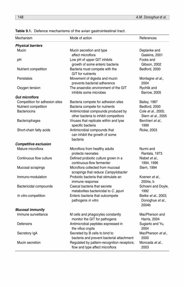
Table 9.1. Defence mechanisms of the avian gastrointestinal tract.
Mechanism Mode of action References
Physical barriersMucin Mucin secretion and type Deplanke and
affect microflora Gaskins, 2001pH Low pH of upper GIT inhibits Fooks and
growth of some enteric bacteria Gibson, 2002Nutrient competition Bacteria must compete with the Bedford, 2000
GIT for nutrientsPeristalsis Movement of digesta and mucin Montagne et al.,
prevents bacterial adherence 2004Oxygen tension The anaerobic environment of the GIT Rychlik and
inhibits some microbes Barrow, 2005Gut microfloraCompetition for adhesion sites Bacteria compete for adhesion sites Bailey, 1987Nutrient competition Bacteria compete for nutrients Bedford, 2000Bacteriocins Antimicrobial compounds produced by Cole et al., 2005;
other bacteria to inhibit competitors Stern et al., 2005Bacteriophages Viruses that replicate within and lyse Berchieri et al.,
specific bacteria 1999Short-chain fatty acids Antimicrobial compounds that Ricke, 2003
can inhibit the growth of some bacteria
Competitive exclusion Mature microflora Microflora from healthy adults Nurmi and
protects neonates Rantala, 1973Continuous flow culture Defined probiotic culture grown in a Nisbet et al.,
continuous-flow fermenter 1994, 1996 Mucosal scrapings Microflora collected from mucosal Stern, 1994
scrapings that reduce CampylobacterImmuno-modulation Probiotic bacteria that stimulate an Koenen et al.,
immune response 2004a, b Bactericidal compounds Caecal bacteria that secrete Schoeni and Doyle,
metabolites bactericidal to C. jejuni 1992In vitro competition Enteric bacteria that outcompete Bielke et al., 2003;
pathogens in vitro Donoghue et al.,2004b
Mucosal immunityImmune surveillance M cells and phagocytes constantly MacPherson and
monitor the GIT for pathogens Harris, 2004Defensins Antimicrobial peptides expressed in Sugiarto and Yu,
the villus crypts 2004Secretory IgA Secreted by B cells to bind to MacPherson et al.,
bacteria and prevent bacterial attachment 2000Mucin secretion Regulated by pattern-recognition receptors; Moncada et al.,
flow and type affect microflora 2003
148 A.M. Donoghue et al.
REFERENCES
AbuOun, M., Manning, G. Cathraw, S.A., Ridley, A., Ahmed, I.H., Wassenaar, T.M. and Newell,D.G. (2005) Cytolethal distending toxin (CDT)-negative Campylobacter jejuni strains andanti-CDT neutralizing antibodies are induced during human infection but not duringcolonization in chickens. Infection and Immunity 73, 3053–3062.
Alisky, J., Iczkowski, K., Rapoport, A. and Troitsky, N. (1998) Bacteriophages show promise asantimicrobial agents. Journal of Infection 36, 5–15.
Allen-Vercoe, E. and Woodward, M.J. (1999) The role of flagella, but not fimbriae, in theadherence of Salmonella enterica serotype Enteritidis to chick gut explant. Journal ofMedical Microbiology 48, 771–780.
Al-Tarazi, Y.H. and Alshawabkeh, K. (2003) Effect of dietary formic and propionic acids onSalmonella Pullorum shedding and mortality in layer chicks after experimental infection.Journal of Veterinary Medicine 50, 112–117.
Arques, J.L., Fernandez, J., Gaya, P., Nunez, M., Rodriguez, E. and Medina, M. (2004)Antimicrobial activity of reuterin in combination with nisin against foodborne pathogens.International Journal of Food Microbiology 95, 225–229.
Attebury, R.J., Dillon, E., Swift, C., Connerton, P.L., Frost, J.A., Dodd, C.E.R., Rees, C.E.D. andConnerton, I.F. (2005) Correlation of Campylobacter bacteriophage with reduced presenceof hosts in broiler chicken caeca. Applied and Environmental Microbiology 71, 4885–4887.
Bailey, J.S. (1987) Factors affecting microbial competitive exclusion in poultry. Food Technology41, 88–92.
Barnes, E.M., Impey, C.S. and Stevens, B.J. (1979) Factors affecting the incidence and anti-salmonella activity of the anaerobic caecal flora of the young chick. Journal of Hygiene 82,263–283.
Barrow, P.A. (2000) The paratyphoid salmonellae. Revue Scientifique et Technique 19, 351–375.Beachey, E.H. (1981) Bacterial adherence: adhesin–receptor interactions mediating the attach-
ment of bacteria to mucosal surfaces. Journal of Infectious Diseases 143, 325–345.Bedford, M.R. (2000) Exogenous enzymes in monogastric nutrition: their current value and future
benefits. Animal Feed Science and Technology 86, 1–13.Beery, J.T., Hughdahl, M.B. and Doyle, M.P. (1988) Colonization of the gastrointestinal tracts of
chicks by Campylobacter jejuni. Applied and Environmental Microbiology 54, 2365–2370.Bengmark, S. and Jeppsson, B. (1995) Gastrointestinal surface protection and mucosa recon-
ditioning. Journal of Parenteral and Enteric Nutrition 19, 410–415.Berchieri, A., Lovell, M.A. and Barrow, P.A. (1991) The activity in the chicken alimentary tract of
bacteriophages lytic for Salmonella Typhimurium. Research in Microbiology 142, 541–549.Best, A., La Ragione, R.M., Cooley, W.A., O’Connor, C.D., Velge, P. and Woodward, M.J. (2003)
Interaction with avian cells and colonization of specific pathogen-free chicks by Shiga-toxinnegative Escherichia coli O157:H7 (NCTC 12900). Veterinary Microbiology 93, 207–222.
Bielke, L.R., Elwood, A.L., Donoghue, D.J., Donoghue, A.M., Newberry, L.A., Neighbor, N.K.and Hargis, B.M. (2003) Approach for selection of individual enteric bacteria for competitiveexclusion in turkey poults. Poultry Science 82, 1378–1382.
Centers for Disease Control (CDC) (2004) Preliminary FoodNet data on the incidence of infectionwith pathogens commonly transmitted through food – selected sites, USA, 2003. Morbidityand Mortality Weekly Report, 30 April, 53, 338–343.
Chart, H. and Rowe, B. (1993) Iron restriction and the growth of Salmonella Enteritidis.Epidemiology and Infection 110, 41–47.
Chaveerach, P., Keuzenkamp, D.A., Urlings, H.A.P., Lipman, L.J.A. and van Knapen, F. (2002) Invitro study on the effects of organic acids on Campylobacter jejuni/coli populations inmixtures of water and feed. Poultry Science 81, 621–628.
Gut pathogen control 149

Cleveland, J., Montville, T.J., Nes, I.F. and Chikindas, M.L. (2001) Bacteriocins: safe, naturalantimicrobials for food preservation. International Journal of Food Microbiology 71, 1–20.
Cole, K., Farnell, M.B., Donoghue, A.M., Stern, N.J., Svetoch, E.A., Eruslanov, B.N., Kovalev,Y.N., Perelygin, V.V., Levchuck, V.P., Reyes-Herrera, I., Blore, P.J. and Donoghue, D.J. (2006)Bacteriocins reduce Campylobacter colonization and alter gut architecture in turkey poults.Poultry Science 85, 1570–1575.
Connerton, P.L., Loc Carrillo, C.M., Swift, C., Dillon, E., Scott, A., Rees, C.E., Dodd, C.E.R.,Frost, J. and Connerton, I.F. (2004) Longitudinal study of Campylobacter jejuni bacterio-phages and their hosts from broiler chickens. Applied and Environmental Microbiology 70,3877–3883.
Corrier, D.E. and Nisbet, D.J. (1999) Competitive exclusion in control of Salmonella entericaserovar Enteritidis in laying poultry in: Salmonella enterica Enteritidis. In: Saeed, A.M., Gast,R.K., Potter, M.E. and Wall, P.G. (eds) Humans and Animals: Epidemiology, Pathogenesis,and Control. Iowa State University Press, Ames, Iowa, pp. 391–396.
Corrier, D.E., Hinton, A., Ziprin, R.L., Beier, R.C. and DeLoach, J.R. (1990) Effect of dietarylactose on caecal pH, bacteriostatic volatile fatty acids, and Salmonella Typhimurium colo-nization of broiler chicks. Avian Diseases 34, 617–625.
Corrier, D.E., Nisbet, D.J., Scanlan, C.M., Hollister, A.G. and DeLoach, J.R. (1995) Control ofSalmonella Typhimurium colonization in broiler chicks with a continuous-flow characterizedmixed culture of bacteria. Poultry Science 74, 916–924.
Craven, S.E. and Williams, D.D. (1998) In vitro attachment of Salmonella Typhimurium tochicken caecal mucus: effect of cations and pretreatment with Lactobacillus spp. isolatedfrom the intestinal tracts of chickens. Journal of Food of Protection 61, 265–271.
Denbow, D.M. (2000) Gastrointestinal anatomy and physiology. In: Whittow G.C. (ed.) SturkiesAvian Physiology. Academic Press, San Diego, California, pp. 299–326.
Deplancke, B. and Gaskins, H.R. (2001) Microbial modulation of innate defense: goblet cells andthe intestinal mucus layer. American Journal of Clinical Nutrition 73, 1131S–1141S.
Deplancke, B., Vidal, O., Ganessunker, D., Donovan, S.M., Mackie, R.I. and Gaskins, H.R.(2002) Selective growth of mucolytic bacteria including Clostridium perfringens in a neonatalpiglet model of total parenteral nutrition. American Journal of Clinical Nutrition 76,1117–1125.
Dohrman, A., Miyata, S., Gallup, M., Li, J.D., Chapelin, C.A., Coste, A.E., Escudier, E., Nadel, J.and Basbaum, C. (1998) Mucin gene (MUC 2 and MUC 5AC) up-regulation by Gram-positive and Gram-negative bacteria. Biochimica et Biophysica Acta 1406, 251–259.
Donoghue, A.M., Huff, W.E., Hargis, B.M., Tellez, G. and Donoghue, D.J. (2004a).Bacteriophage and probiotics: their role in the control of Salmonella in poultry. Fifth AsianPacific Poultry Health Proceedings, Gold Coast, Australia, pp. 10–15.
Donoghue, D.J., Hargis, B.M., Tellez, G. and Donoghue, A.M. (2004b). Competitive exclusion asa means of controlling Campylobacter in poultry. Fifth Asian Pacific Poultry HealthProceedings, Gold Coast Australia, pp. 16–20.
Duguid, J.P. and Campbell, I. (1969) Antigens of the type-1 fimbriae of Salmonellae and otherenterobacteria. Journal of Medical Microbiology 2, 535–553.
Farnell, M.B., He, H. and Kogut, M.H. (2003) Differential activation of signal transduction path-ways mediating oxidative burst by chicken heterophils in response to stimulation withlipopolysaccharide and lipoteichoic acid. Inflammation 27, 225–231.
Farnell, M.B., Donoghue, A.M., Cole, K., Reyes-Herrera, I., Solis de los Santos, F., Dirain, M.L.S.,Blore, P.J., Pandya, K. and Donoghue, D.J. (2006a) Effect of oral administration of bismuthcompounds on Campylobacter colonization in broilers. Poultry Science 85, 2009–2011.
Farnell, M.B., Donoghue, A.M., de los Santos, F.S., Blore, P.J., Tellez, G., Hargis, B.M. andDonoghue, D.J. (2006b) Upregulation of oxidative burst and degranulation in chickenheterophils stimulated with probiotic bacteria. Poultry Science 85, 1900–1906.
150 A.M. Donoghue et al.

Fernandez, F., Sharma, R., Hinton, M. and Bedford, M.R. (2000) Diet influences the colonizationof Campylobacter jejuni and distribution of mucin carbohydrates in the chick intestinal tract.Cellular and Molecular Life Sciences 57, 1793–1801.
Field, L.H., Headley, V.L., Payne, S.M. and Berry, L.J. (1986) Influence of iron on growth,morphology, outer membrane protein composition, and synthesis of siderophores inCampylobacter jejuni. Infection and Immunity 54, 126–132.
Fooks, L.J. and Gibson, G.R. (2002) Probiotics as modulators of the gut flora. British Journal ofNutrition 88, S39-S49.
Forstner, J.F., Oliver, M.G. and Sylvester, F.A. (1995) Production, structure and biologic relevanceof gastrointestinal mucins. In: Blaser, J., Smith, P.D., Ravdin, J.I., Greenberg, H.B. andGuerrant, R.L. (eds) Infections of the Gastrointestinal Tract. Raven Press, New York,pp. 71–88.
Freter, R. and O’Brien, P.C.M. (1981) Role of chemotaxis in the association of motile bacteria withintestinal mucosa: fitness and virulence of nonchemotactic Vibrio cholerae mutants in infantmice. Infection and Immunity 34, 222–233.
Galdeano, C.M. and Perdigon, G. (2004) Role of viability of probiotic strains in their persistencein the gut and in mucosal immune stimulation. Journal of Applied Microbiology 97,673–681.
Gendler, S.J. and Spicer, A.P. (1995) Epithelial mucin genes. Annual Review of Physiology 57,607–634.
Goode, D., Allen, V.M. and Barrow, P.A. (2003) Reduction of experimental Salmonella andCampylobacter contamination of chicken skin by application of lytic bacteriophages. Appliedand Environmental Microbiology 69, 5032–5036.
Gorski, A. and Weber-Dabrowska, B. (2005) The potential role of endogenous bacteriophages incontrolling invading pathogens. Cellular and Molecular Life Sciences 62, 511–519.
Gusils, C., Oppezzo, O., Pizarro, R. and Gonzalez, S. (2003) Adhesion of probiotic Lactobacilli tochick intestinal mucus. Canadian Journal of Microbiology 49, 472–478.
Ha, S.A., Ricke, S.C., Nisbet, D.J., Corrier, D.E. and DeLoach, J.R. (1995) Serine utilization as apotential competition mechanism between Salmonella Typhimurium and a chicken caecalbacterium. Journal of Food Protection 57, 1074–1079.
Hargis, B.M., Caldwell, D.J. and Byrd, J.A. (2001) Microbiological pathogens: live poultry con-siderations. In: Sams, A.R. (ed.) Poultry Meat Processing. CRC Press, Boca Raton, Florida,pp. 121–135.
Hargis, B.M., Tellez, G.I., Nava, G., Donoghue, A.M., Vicente, J.L., Higgins, S.E., Donoghue,D.J. and Wolfenden, A.D. (2003) The role of beneficial microflora in controlling entericbacterial diseases: probiotics, prebiotics, and competitive exclusion. Cornell NutritionConference for Feed Manufactures, East Syracuse, New York, 21–23 October, pp. 109–118.
Hecht, G. (1995) Bugs and barriers: enteric pathogens exploit yet another epithelial function.Journal American Physiology 10, 160–166.
Higgins, J.P., Higgins, S.E., Guenther, K.L., Huff, W., Donoghue, A.M., Donoghue, D.J. andHargis, B.M. (2005) Use of a specific bacteriophage treatment to reduce Salmonella inpoultry products. Poultry Science 84, 1141–1145.
Ho, W.L., Yu, R.C. and Chou, C.C. (2004) Effect of iron limitation on the growth and cytotoxinproduction of Salmonella choleraesuis SC-5. International Journal of Food Microbiology 90,295–302.
Holmes, K., Mulholland, F., Pearson, B.M., Pin, C., McNicholl-Kennedy, J., Ketley, J.M. and Wells,J.M. (2005) Campylobacter jejuni gene expression in response to iron limitation and the roleof fur. Microbiology 151, 243–257.
Hume, M.E., Corrier, D.E., Ambrus, S., Hinton, A. and DeLoach, J.R. (1993) Effectiveness ofdietary propionic acid in controlling Salmonella Typhimurium colonization in broiler chicks.Avian Diseases 37, 1051–1056.
Gut pathogen control 151

Jonsson, H., Strom, E. and Roos, S. (2001) Addition of mucin to the growth medium triggersmucus-binding activity in different strains of Lactobacillus reuteri in vitro. FEMSMicrobiology Letters 204, 19–22.
Kaplan, H. and Hutkins, R.W. (2000) Fermentation of fructooligosaccharides by lactic acidbacteria and bifidobacteria. Applied and Environmental Microbiology 66, 2682–2684.
Kiely, L.J. and Olson, N.F. (2000) The physiochemical surface characteristics of Lactobacilluscasei. Food Microbiology 17, 277–291.
Kingsley, R., Rabsch, W., Stephens, P., Roberts, M., Reissbrodt, R. and Williams, P.H. (1995) Iron-supplying systems of Salmonella in diagnostics, epidemiology and infection. FEMSImmunology and Medical Microbiology 11, 257–267.
Koenen, M., van der Hulst, E., Leering, R.M., Jeurissen, S.H.M. and Boersma, W.J.A. (2004a)Development and validation of a new in vitro assay for selection of probiotic bacteria thatexpress immune-stimulating properties in chickens in vivo. FEMS Immunology and MedicalMicrobiology 40, 119–127.
Koenen, M.E., Kramer, J., van der Hulst, R., Heres, L., Jeurissen, S.H.M. and Boersma, W.J.A.(2004b) Immunomodulation by probiotic lactobacilli in layer- and meat-type chickens.British Poultry Science 45, 355–366.
Kubena, L.F., Bailey, R.H., Byrd, J.A., Young, C.R., Corrier, D.E., Stanker, L.H. and Rottinghaus,G.E. (2001a) Caecal volatile fatty acids and broiler chick susceptibility to SalmonellaTyphimurium colonization as affected by aflatoxins and T-2 toxin. Poultry Science 80,411–417.
Kubena, L.F., Byrd, J.A., Young, C.R. and Corrier, D.E. (2001b) Effects of tannic acid on caecalvolatile fatty acids and susceptibility to Salmonella Typhimurium colonization in broilerchicks. Poultry Science 80, 1293–1298.
Kutter, E. (1997) Phage therapy: bacteriophages as antibiotics. http://www.evergreen.edu/phage/phagetherapy/phagetherapy.htm (accessed 12 August 2005).
Lan, Y., Verstegen, M.W.A., Tamminga, S. and Williams, B.A. (2005) The role of the commensalgut microbial community in broiler chickens. World’s Poultry Science Journal 61, 95–104.
Langhout, D.J., Schutte, J.B., Van Leeuwen, P.V., Wiebenga, J. and Tamminga, S. (1999) Effectof dietary high- and low-methylated citrus pectin on the activity of the ileal microflora andmorphology of the small intestinal wall of broiler chicks. British Poultry Science 40, 340–347.
Le Blay, G., Michel, C., Blottiere, H.M. and Cherbut, C. (1999) Prolonged intake of fructo-oligosaccharides induces a short-term elevation of lactic acid-producing bacteria andpersistent increase in caecal butyrate in rats. Journal of Nutrition 129, 2231–2235.
Ledeboer, N.A. and Jones, B.D. (2005) Exopolysaccharide sugars contribute to biofilm formationby Salmonella enterica serovar Typhimurium on HEp-2 cells and chicken intestinal epithe-lium. Journal of Bacteriology 187, 3214–3226.
Lee, A., O’Rourke, J.L., Barrington, P.J. and Trust, T.J. (1986) Mucus colonization as a determi-nant of pathogenicity in intestinal infection by Campylobacter jejuni: a mouse caecal model.Infection and Immunity 51, 536–546.
Lisiecki, P., Wysocki, P. and Mikucki, J. (2000) Susceptibility of Staphylococci to natural andsynthetic iron chelators. Medycyna Doswiadczalna i Mikrobiologia 52, 103–110.
Litwin, C.M. and Calderwood, S.B. (1993) Role of iron in regulation of virulence genes. ClinicalMicrobiology Reviews 6, 137–149.
Lowry, V.K., Farnell, M.B., Ferro, P.J., Swaggerty, C.L., Bahl, A. and Kogut, M.H. (2005) Purifiedbeta-glucan as an abiotic feed additive up-regulates the innate immune response inimmature chickens against Salmonella enterica serovar Enteritidis. International Journal ofFood Microbiology 98, 309–318.
Mack, D.R., Michail, S., Wei, S., McDougall, L. and Hollingsworth, M.S. (1999) Probiotics inhibitenteropathogenic E. coli adherence in vitro by inducing intestinal mucin gene expression.American Journal of Physiology 274, G941–G950.
152 A.M. Donoghue et al.
MacPherson, A.J. and Harris, N.L. (2004) Interactions between commensal intestinal bacteriaand the immune system. Nature Reviews in Immunology 4, 478–485.
MacPherson, A.J., Dominique, G., Sainsbury, E., Harriman, G.R., Hensgartner, H. andZinkernagel, R.M. (2000) A primitive T cell-independent mechanism of intestinal mucosalIgA responses to commensal bacteria. Science 288, 2222–2226.
Mantle, M. and Allen-Vercoe, A. (1989) Gastrointestinal mucus. In: Davison, T.F. (ed.)Gastrointestinal Secretion. J.S. University Press, London, pp. 202–229.
Mead, G.C. (1998) Critical control of major pathogens in poultry. In: Hakkinen, M., Nuotio, L.,Nurmi, E. and Mead, G.C. (eds) Pathogenic Micro-organisms in Poultry and Eggs: SafeChicken for the Next Century. COST Action 97, Luxembourg, pp. 6–12.
Mead, G.C. (2000) Prospects for ‘competitive exclusion’ treatment to control salmonellas andother foodborne pathogens in poultry. Veterinary Journal 159, 111–123.
Mead, G.C. (2002) Factors affecting intestinal colonization of poultry by Campylobacter and roleof microflora in control. World’s Poultry Science Journal 58, 169–178.
Mead, G.C. and Impey, C.S. (1986) Current progress in reducing Salmonella colonization ofpoultry by ‘competitive exclusion’. Society for Applied Bacteriology Symposium Series 15,67S–75S.
Meinersmann, R.J., Rigsby, W.E., Stern, N.J., Kelley, L.C., Hill, J.E. and Doyle, M.P. (1991)Comparative study of colonizing and noncolonizing Campylobacter jejuni. American Journalof Veterinary Research 52, 1518–1522.
Miller, S.J. (2004) Cellular and physiological effects of short-chain fatty acids. Mini Reviews inMedicinal Chemistry 4, 839–845.
Moncada, D.M., Kammanadiminti, S.J. and Chadee, K. (2003) Mucin and Toll-like receptors inhost defence against intestinal parasites. Trends in Parasitology 19, 305–311.
Montagne, L., Piel, M.S. and Lallès, J.P. (2004) Effect of diet on mucin kinetics and composition:nutrition and health implications. Nutrition Reviews 62, 105–144.
Mota-Meira, M., LaPointe, G., Lacroix, C. and Lavoie, M.C. (2000) MICs of mutacin B-Ny266,nisin A, vancomycin, and oxacillin against bacterial pathogens. Antimicrobial Agents andChemotherapy 44, 24–29.
Neilands, J.B. (1995) Siderophores: structure and function of microbial iron transport com-pounds. Journal of Biological Chemistry 270, 26723–26726.
Newell, D.G., McBride, H. and Dolby, J.M. (1985) Investigations on the role of flagella in thecolonization of infant mice with Campylobacter jejuni and attachment of Campylobacterjejuni to human epithelial cell lines. Journal of Hygiene 95, 217–227.
Nisbet, D. (2002) Defined competitive exclusion cultures in the prevention of enteropathogencolonization in poultry and swine. Antonie van Leeuwenhoek 81, 481–486.
Nisbet, D.J., Corrier, D.E., Scanlan, C.M., Hollister, A.G., Beier, R.C. and DeLoach, J.R. (1994)Effect of dietary lactose and cell concentration on the ability of a continuous flow-derivedbacterial culture to control Salmonella caecal colonization in broiler chickens. Poultry Science73, 56–62.
Nisbet, D.J., Corrier, D.E., Ricke, S., Hume, M.E., Byrd, J.A. and DeLoach, J.R. (1996)Maintenance of the biological efficacy in chicks of a cecal competitive-exclusion cultureagainst Salmonella by continuous-flow fermentation. Journal of Food Protection 59,1279–1283.
Nurmi, E. and Rantala, M. (1973) New aspects of Salmonella infection in broiler production.Nature 241, 210–211.
Nurmi, E., Nuotio, L. and Schneitz, C. (1992) The competitive exclusion concept: developmentand future. International Journal of Food Microbiology 15, 237–240.
Palyada, K., Threadgill, D. and Stintzi, A. (2004) Iron acquisition and regulation inCampylobacter jejuni. Journal of Bacteriology 186, 4714–4729.
Gut pathogen control 153
Pascual, M., Hugas, M., Badiola, J.I., Monfort, J.M. and Garriga, M. (1999) Lactobacillussalivarius CTC2197 prevents Salmonella Enteritidis colonization in chickens. Applied andEnvironmental Microbiology 65, 4981–4986.
Patterson, J.A. and Burkholder, K.M. (2003) Application of prebiotics and probiotics in poultryproduction. Poultry Science 82, 627–631.
Pickett, C.J., Auffenberg, T., Pesci, E.C., Sheen, V.L. and Jusuf, S.S. (1992) Iron acquisition andhemolysin production by Campylobacter jejuni. Infectious Immunity 60, 3872–3877.
Pivnick H. and Nurmi, E. (1982) The Nurmi concept and its role in the control of Salmonellae inpoultry. In: Davies R. (ed.) Developments in Food Microbiology 1. Applied SciencesPublishers Ltd., Essex, England, pp. 41–70.
Ratledge, C. and Dover, L.G. (2000) Iron metabolism in pathogenic bacteria. Annual Review ofMicrobiology 54, 881–941.
Ricke, S.C. (2003) Perspectives on the use of organic acids and short chain fatty acids asantimicrobials. Poultry Science 82, 632–639.
Rolf, R.D. (1991) Population dynamics of the intestinal tract. In: Blankenship, L.C. (ed.)Colonization Control of Human Enteropathogens in Poultry. Academic Press, San Diego,California, pp. 59–76.
Rychlik, I. and Barrow, P.A. (2005) Salmonella stress management and its relevance to behaviourduring intestinal colonization and infection. FEMS Microbiology Reviews 29, 1021–1040.
Sakamoto, K., Mori, Y., Takagi, H., Iwata, H., Yamada, T., Futamura, N., Sago, T., Ezaki, T.,Kawamura, Y. and Hirose, H. (2004) Translocation of Salmonella Typhimurium in rats ontotal parenteral nutrition correlates with changes in intestinal morphology and mucus gel.Nutrition 20, 372–376.
Sansonetti, P.J. (2004) War and peace at mucosal surfaces. Nature Reviews in Immunology 4,953–964.
Schoeni, J.L. and Doyle, M.P. (1992) Reduction of Campylobacter jejuni colonization of chicks bycaecum-colonizing bacteria producing anti-C. jejuni metabolites. Applied and EnvironmentalMicrobiology 58, 664–670.
Sharma, R., Fernandez, F., Hinton, M. and Schumacher, U. (1997) The influence of diet on themucin carbohydrates in the chick intestinal tract. Cellular and Molecular Life Sciences 53,935–942.
Sklar, I.B. and Joerger, R.D. (2001) Attempts to utilize bacteriophage to combat Salmonellaenterica serovar Enteritidis infection in chickens. Journal of Food Safety 21, 15–29.
Slomiany, B., Bilski, J., Sarosiek, J., Murty, V.L., Dworkin, B., Van Horn, K., Zielenski, J. andSlomiany, A. (1987) Campylobacter pylordis degrades mucin and undermines gastricmucosal integrity. Biochemical and Biophysical Research Communications 144, 307–314.
Smirnov, A., Sklan, D. and Uni, Z. (2004) Mucin dynamics in the chick small intestine are alteredby starvation. Journal of Nutrition 134, 736–742.
Stavric, S., Buchanan, B. and Gleeson, T.M. (1993) Intestinal colonization of young chicks withEscherichia coli O157:H7 and other verotoxin-producing serotypes. Journal of AppliedBacteriology 74, 557–563.
Stavric, S. and D’Aoust, J.Y. (1993) Undefined and defined bacterial preparations for thecompetitive exclusion of Salmonella in poultry – a review. Journal of Food Protection 56,173–180.
Stern, N.J. (1994) Mucosal competitive exclusion to diminish colonization of chickens byCampylobacter jejuni. Poultry Science 73, 402–407.
Stern, N.J., Svetoch, E.A., Eruslanov, B.V., Kovalev, Y.N., Volodina, L.I., Perelygin, V.V., Mitsevich,E.V., Mitsevich, I.P. and Levchuk, V.P. (2005) Paenibacillus polyxma purified bacteriocin tocontrol Campylobacter jejuni in chickens. Journal of Food Protection 68, 1450–1453.
Sugiarto, H. and Yu, P.-L. (2004) Avian antimicrobial peptides: the defence role of beta defensins.Biochemical and Biophysical Research Communications 323, 721–727.
154 A.M. Donoghue et al.

Tellez, G., Higgins, S.E., Donoghue, A.M. and Hargis, H.A. (2005) Digestive physiology and therole of the micro-organism. Journal of Applied Poultry Research 15, 136–144.
Temann, U.A., Prasad, B., Gallup, M.W., Basbaum, C., Ho, S.B., Flavell, R.A. and Rankin, J.A.(1997) A novel role for murine IL-4 in vivo: induction of MUC5AC gene expression andmucin hypersecretion. American Journal of Respiratory Cell and Molecular Biology 16,471–478.
Thompson, J.L. and Hinton, M. (1997) Antibacterial activity of formic and propionic acids in thediet of hens on Salmonellas in the crop. British Poultry Science 38, 59–65.
Topping, D.L. and Clifton, P.M. (2001) Short-chain fatty acids and human colonic function: rolesof resistant starch and nonstarch polysaccharides. Physiological Reviews 81, 1031–1064.
Touati, D. (2000) Iron and oxidative stress in bacteria. Archives of Biochemistry and Biophysics373, 1–6.
Ushijima, T. and Seto, A. (1991) Selected faecal bacteria and nutrients essential for antagonism ofSalmonella Typhimurium in anaerobic continuous flow cultures. Journal of MedicalMicrobiology 35, 111–117.
van der Wielen, P.W.J.J., Biesterveld, S., Notermans, S., Hofstra, H., Urlings, B.A.P. and vanKnapen, F. (2000) Role of volatile fatty acids in development of the caecal microflora inbroiler chickens during growth. Applied and Environmental Microbiology 66, 2536–2540.
van Immerseel, F., De Buck, J., De Smet, I., Pasmans, F., Haesebrouck, F. and Ducatelle, R.(2004a) Interactions of butyric acid- and acetic acid-treated Salmonella with chicken primarycaecal epithelial cells in vitro. Avian Diseases 48, 384–391.
van Immerseel, F., Fievez, V., De Buck, J., Pasmans, F., Martel, A., Haesebrouck, F. and Ducatelle,R. (2004b) Microencapsulated short-chain fatty acids in feed modify colonization and inva-sion early after infection with Salmonella Enteritidis in young chickens. Poultry Science 83,69–74.
van Vliet, A.H.M., Ketley, J.M., Park, S.F. and Penn, C.W. (2002) The role of iron inCampylobacter gene regulation, metabolism, and oxidative stress defense. FEMSMicrobiology Reviews 26, 173–186.
Vimal, D.B., Khullar, M., Gupta, S. and Ganguly, N.K. (2000) Intestinal mucins: the binding sitesfor Salmonella Typhimurium. Molecular and Cellular Biochemistry 204, 107–117.
Woolridge, K.G. and Williams, P.H. (1993) Iron uptake mechanisms of pathogenic bacteria.FEMS Microbiology Reviews 12, 325–348.
Ziprin, R.L., Young, C.R., Byrd, J.A., Stanker, L.H., Hume, M.E., Gray, S.A., Kim, B.J. andKonkel, M.E. (2001) Role of Campylobacter jejuni potential virulence genes in caecal colo-nization. Avian Diseases 45, 549–557.
Gut pathogen control 155

This page intentionally left blank

PART IVNutritional effects

This page intentionally left blank

CHAPTER 10Effect of non-starch polysaccharidases onavian gastrointestinal function
M.R. BedfordZymetrics, Marlborough, UK; e-mail: [email protected]
ABSTRACT
Non-starch polysaccharidases, which include cellulases and xylanases, areroutinely used in poultry diets containing wheat, barley, oats, triticale and ryeand, more recently, maize. The beneficial responses observed may be based onone or more of three proposed mechanisms of action, namely: (i) cerealendosperm cell wall hydrolysis; (ii) reduction in intestinal viscosity; and (iii)provision of fermentable oligomeric substrates as a result of cell wall hydrolysis.Since each mechanism elicits a specific series of responses at the intestinallevel, and the extent of involvement of each will vary depending uponcircumstances, there are a variety of potential responses observed. Changes inintestinal function may be driven by changes in nutrient supply and/or changesin the physico-chemical conditions (the viscosity of the intestinal contents, thenutrient-binding ability of the fibre fraction and the thickness of the unstirredwater layer) of the intestine.
Nutrient supply encompasses not only improved nutrient extraction by thehost, and thus reduced availability for the intestinal flora, but also the provisionof oligomeric substrates through cell wall hydrolysis. As a result of such changesthere may be subsequent, interrelated changes in the structure and size of theintestinal tract, in pancreatic enzyme secretion, in the quantity and chemistry ofmucin secreted and in the quantity and species distribution of resident intestinalflora. The qualitative and quantitative changes in intestinal function observedare very much dependent upon the status in the absence of enzyme, whichhighlights the fact that responses upon use of enzymes are variable, beinggreatest in poor-quality diets.
© CAB International 2006. Avian Gut Function in Health and Disease (ed. G.C. Perry) 159
INTRODUCTION
Enzymes which target the non-starch polysaccharides (NSP) of the cerealscommonly used in poultry diets, be it wheat, barley, oats, triticale, rye or evenmaize, have traditionally been viewed as tools for improving the digestibility ofa ration. The use of cellulase or xylanase-rich preparations in oats/barley- orwheat/rye/triticale-based diets, respectively, has been shown to improve growthrate and feed conversion ratio of birds on a relatively consistent basis (Elwingerand Saterby, 1987; Pettersson et al., 1990; Friesen et al., 1991; Campbell andBedford, 1992).
Such a response is principally a result of the fact that NSPs seem to be anti-nutritive and poultry do not express enzymes capable of degrading them.Significant debate exists with regards to why, exactly, the NSPs are anti-nutritiveand, moreover, how these enzymes elicit such a response. The most commonlydiscussed mechanisms include cereal cell wall dissolution and subsequentexposure of the contents to more efficient digestion, and reduction in intestinalaqueous phase viscosity by depolymerization of high-molecular weight,soluble, viscous cell wall polymers (Choct, 1992; Cowan, 1992; Bedford,1993). A third mechanism is based on the knowledge that the activity of NSPenzymes in the intestine produces growth-promoting prebiotics (Morgan et al.,1992; Choct et al., 1996).
In truth it is likely that all three mechanisms play a role in any responseobserved, the contribution of each mechanism to the final result varying witheach particular situation. Since each mechanism results in quite differentinteractions with gut function, the impact that the NSPs have and thus thebenefit of use of a non-starch polysaccharidase (Nsp’ase) on avian gut functionwill vary from test to test. Nevertheless, Nsp’ases tend to reduce gut size as aproportion of body weight (although in absolute terms the intestine may belarger since enzyme-fed birds are often larger than equivalent controls) andalter function due to their facilitation of digestion, although some data witholder birds suggest the opposite (Jaroni et al., 1999; Brenes et al., 2002, 2003).
In general, if digestion is consistently suboptimal, whether due to ingredientquality, microbial interaction or anti-nutritive factors, the gastrointestinal tract(GIT) responds by increasing in both size (surface area) and digestive enzymeoutput. Even though diet digestibility may be maintained as a result of suchadaptation, the energy cost of digestion is greater and thus feed efficiency falls.Use of a Nsp’ase may improve animal performance through improved dietdigestibility, reduced endogenous inputs or, most probably, through bothmechanisms.
This chapter will review some of these interactions and attempt to provideguidance on what should be expected in different sections of the GIT under avariety of conditions, although it should be noted that such arbitrarysegmentation of the GIT does not take sufficient note of the interactive andintegrative processes between such segments which bind the whole process ofdigestion together.
160 M.R. Bedford

THE ROLE OF NSP’ASES IN AVIAN NUTRITION: RELEVANCE IN DIFFERENTSEGMENTS OF THE GASTROINTESTINAL TRACT
Crop
The crop is a diverticulum of the oesophagus into which a proportion of thefeed enters for storage if the proventriculus–gizzard complex is alreadyoccupied with feed (Moran, 1982). In the modern day broiler, which is fed adlibitum, it would be easy to assume that the use of the crop would be limited,with the bird simply topping up its proventriculus–gizzard as and whenrequired. In actual fact it is probable that, under commercial conditions, thecrop is extensively used by many broilers. Individual bird behaviour willinfluence the use of the crop.
Marini (2003) reported that 18–21-week-old turkey toms variedconsiderably in their feeding patterns, with some eating a little and often, yetothers tending to have fewer and more intensive visits to the feeder. This latterstrategy would probably result in extensive use of the crop compared with theformer, and suggests that the flock behaves as a group of individuals ratherthan as a homogenous population, with some clearly making more use of thecrop than others. In addition, the introduction of lighting programmesthroughout much of the commercial world has resulted in periods of darknessduring which the bird does not feed. As a result the flock as a whole adapts itsfeeding behaviour so that it can remain fully fed during the dark period.
Birds tend to fill their crop before the lights go out and ‘gorge’ when thelights come on (Moran, 1982). As a result, it is likely that the crop plays asignificant role in food storage in commercially produced broilers, a practicewhich may not be replicated in much of the reported literature due to thepreponderance of 24- or 23-h lighting in such work. Use of the crop issignificant since the conditions therein are favourable for the activity of manyNsp’ases – and phytase for that matter.
It is a well-hydrated environment, and depending upon the population ofresident lactobacilli and thus the concentration of lactic acid, the pH (rangesfrom 4 to 7.8; Moran, 1982) can be quite favourable for many of the fungalcellulases and xylanases in commercial use today (i.e. pH 3.5–5.5). Since feedcan reside in the crop for several hours (Moran, 1982), this organ may play asignificant role in determining the extent to which diets will respond to the useof Nsp’ases.
Not only will these enzymes initiate attack on the fibre of the grains beingfed – thereby reducing viscosity and potentially increasing access tofermentable nutrient contents – but they will also produce fermentableoligomeric carbohydrates that may influence bacterial populations and theirmetabolic processes. They may also effectively increase the surface areaavailable for bacterial attachment by ‘roughening’ the surface of endospermcell walls, a mechanism that has been proposed in ruminants (Nsereko et al.,2000).
Evidence that such events occur is provided by Jozefiak et al., whoobserved significant increments in lactic acid content of the crop when the
Polysaccharidases 161

relevant enzymes were added to barley- or wheat-based diets (Jozefiak et al.,Abstract, this volume). Furthermore, the use of Nsp’ases has been shown toinfluence crop microfloral populations (Danicke et al., 1999c; Murphy et al.,2004), although differences were noted between enzyme sources, suggestingthat not all enzymes bring about the same benefits. As a result, it is likely thatthe species and quantities of organisms seeding the proventriculus–gizzard andpoints lower down the digestive tract will be influenced by activity in the crop.Thus, although the effects may appear regional and minimal, it is possible thatthey are more far-reaching than may be envisaged at first sight.
From a structural viewpoint, Nsp’ase use clearly results in reduced crop sizewhen highly viscous diets (Lazaro et al., 2004) or diets rich in lupins (Brenes etal., 2002) are used, but effects in wheat-based diets were found to be minimal(Brenes et al., 2002; Wu and Ravindran, 2004). It is clear also that effects vary.Differences in microbial populations were observed in the crop on feedingdifferent xylanases, suggesting that there would have been effects on nutrientand NSP digestibility, but in related work no such effects were observed(Murphy et al., 2005a, b). Perhaps the differences were due to the endpointsbeing measured or to the conditions of the study in question. Regardless, it isimportant that the effects of Nsp’ase enzymes on crop physiology/microbiologyare viewed in the context of the management conditions of the trial concerned.Conditions of use should mimic commercial conditions as closely as possible ifthe interpretation desired is for commercial production.
Proventriculus–gizzard
The fact that Nsp’ases elicit a response in poultry diets may seem surprising,since the chicken is an omnivore and has evolved to digest diets in which cerealgrains play a significant part. However, under commercial conditions the bird isexposed to a diet which is dominated by one or two ingredients, thusincreasing ‘exposure’ to NSPs and, moreover, the form in which the diet is fedmay exacerbate their detrimental effects. Fine grinding of diets increasessurface area and subsequent pelleting increases the dissolution rate of solubleNSPs (Silversides and Bedford, 1999), leading to increased intestinal viscosity(Teitge et al., 1991).
Although many argue that fine grinding improves access of the contents todigestion (Carrte, 2004), it seems that fine grinding can limit development ofthe gizzard and, as a result, ironically, lead to larger average particle sizes beingfound in the small intestine than would be the case if the ingredient wascoarsely ground (Svihus et al., 1997; Hetland et al., 2004). Activation of thegizzard through the use of whole grains or cellulose has been shown todramatically improve starch digestion in wheat-rich diets, further suggestingthat the capacity of the digestive tract to deal with incoming starch is reduced ifgizzard activity is compromised.
Thus the practice of feeding of cereal-rich, finely ground, pelleted diets – asis the norm in commercial operations – has elevated the problems associatedwith cereal NSPs beyond that which would be experienced in the wild. Use of
162 M.R. Bedford

Nsp’ases seems to enhance the ability of the gizzard to grind and break opencereal endosperm cell walls (Morgan et al., 1995; Bedford and Autio, 1996),and in fact the benefits of xylanase use on NSP and DM digestibility have beenobserved to be most significant at the level of the gizzard (Murphy et al.,2005a, b), suggesting this is a major site of Nsp’ase activity. Whether this is dueto softening of the cell walls directly or to increased gizzard activity is not clear.
Gizzard activity is controlled at several levels and may, for example, beresponding to changes in fermentation patterns further down the digestive tractwhich will be discussed later. Regardless, feed form and enzyme use doesinteract, with some authors noting that the use of whole grains to stimulate theactivity of the proventriculus–gizzard can reduce the need for added exogenousenzymes (Jones and Taylor, 2001). Use of Nsp’ases tends to reduce the size ofmost digestive organs, including the gizzard, probably due to their facilitation ofdigestion resulting in feedback mechanisms down-regulating intestinal growth(Brenes et al., 2002, 2003; Lazaro et al., 2004). As will be discussed later, someof the benefits of NSP’ase use will inevitably be due to savings in endogenousinputs into the digestive process.
Small intestine
The small intestine is where most of the benefits from use of Nsp’ases and thusmost of the changes in intestinal function have been observed. Many factorsinterrelate to determine the scale of response observed, with some of thesefactors being poorly understood. Increased viscosity can independently reducedigestibility of all nutrients through direct effects on diffusion of solutes in theluminal aqueous phase (the effects being magnified for the larger solutes suchas micelles, more so for the long-chain fatty acid micelles (Danicke et al., 1997,2000)) and through increasing the effective thickness of the unstirred waterlayer.
Also, with increased intestinal viscosity, the bird must adapt in order tomaintain thorough mixing of digesta, which is particularly important foremulsification of fats. Fat-soluble vitamins are particularly prone to theinteraction between fat type and intestinal viscosity since they co-migrate withthe larger, less digestible micelles. This is clearly underlined by the fact thatmarked effects on digestibility and liver storage of Vitamin E have beenobserved in rye-based diets where tallow was the principal source of fat(Danicke et al., 1999a).
The scale and even the direction of response observed depends upon,amongst other variables, the viscosity produced at the level of the smallintestine. Viscosity of the aqueous phase of the digesta increases dramaticallyas material leaves the gizzard and enters the small intestine (Bedford andClassen, 1993; Choct et al., 1999; Lazaro et al., 2004). Re-analysis of the datafrom Bedford and Classen (1992) indicates that intake, and thus intestinaltransit rate, tends to increase with increasing small intestinal viscosity to amaximum of approximately 20–30 mPas. This seems to be an attempt tomaintain nutrient intake in response to recognition of reduced ration density. As
Polysaccharidases 163

a result, gain tends to be maintained and thus FCR deteriorates as intestinalviscosity increases to approximately 20–30 mPas.
Therefore, when an Nsp’ase is employed in diets which are < 30 mPas(which constitutes the vast majority of those featured in the literature on wheat,and much of the literature on barley), the observed response tends to be areduction in feed intake, no improvement in gain but a benefit with respect toFCR. Intestinal changes tend to be muted but often are seen as smallreductions in intestinal size and/or endogenous enzyme output (Maisonnier etal., 2003; Engberg et al., 2004). Feeding an Nsp’ase in such situations will notonly result in reduced intestinal size – as discussed previously (Simon, 1998;Brenes et al., 2002; Wu et al., 2004) – but also result in improved villusstructure, even in mature laying hens (Jaroni et al., 1999).
As intestinal viscosity increases above this threshold, the intestines struggleto maintain flow rate by peristalsis, particularly in the younger bird (Almirall etal., 1995), with the result that the bird is no longer able to move the materialthrough the intestine rapidly enough to maintain daily nutrient requirements.Gain, intake and FCR deteriorate in unison and intestinal size and enzymeoutput continue to increase. Use of enzymes in such diets (mostly relating tostudies where rye is the dominant grain, with some barley studies and a smallnumber of those on wheat) often leads to dramatic improvements in allperformance parameters: digestibility of nutrients as well as a marked reductionin digestive inputs as measured by size of digestive tract and input ofendogenous enzymes (Friesen et al., 1991; Silva and Smithard, 1997; Silva etal., 1997).
The rate at which enterocytes are replaced in the crypts is also significantlyreduced, and individual villus height increases (with concomitant increase invillus height/crypt depth ratio) when Nsp’ases are used in particularlychallenging diets, which results in considerable savings in endogenous losses(Silva and Smithard, 1996; Mathlouthi et al., 2002).
Addition of Nsp’ases clearly increases the availability of nutrients for thehost at the expense of the microflora in the small intestine and this, coupledwith the fact that their use also increases the rate at which digesta pass throughthe small intestine (i.e. residence time is reduced) as a result of more effectiveperistalsis (Danicke et al., 1999c), means that the luminal microfloralpopulations often decrease. Since the signals that encourage incrementalintestinal outputs are either indirectly related to the presence of undigestednutrients or to the presence of certain microfloral metabolites (Moran, 1982), itis not surprising that Nsp’ase use will often lead to savings in endogenouslosses. Such savings can be very significant when highly viscous diets areemployed, which partially explains why the performance enhancementsobserved are often not fully described or valued in digestibility studies(Danicke, 2001).
Where microbial populations have been investigated, the effects of addedNsp’ases have been to reduce fermentation in the small intestine (Danicke etal., 1999b), whilst concomitantly increasing caecal fermentation (Choct et al.,1999). Others have noted that xylanases can reduce total CFU in the smallintestine of young broilers fed wheat-based diets (Vahjen et al., 1998). It is also
164 M.R. Bedford

clear that the microflora is intimately involved in the small intestinal response toNsp’ases, since its absence mutes or even removes the problems associatedwith feeding viscous diets (Langhout et al., 2000; Maisonnier et al., 2003).
Bacteria are particularly involved in the effects on fat digestion since theyproduce bile acid hydrolases to protect themselves from this anti-microbial and,in doing so, compromise fat micelle formation. Feeding of highly viscous dietshas been shown to reduce the content and/or conjugation status of bile acidsand thus addition of the relevant enzyme (or additional bile salts) leads toimproved fat digestion (Smits et al., 1998; Hubener et al., 2002; Mathlouthi etal., 2002), which confirms the interrelationship between microbial status,viscosity and fat emulsification and digestion.
Caecum
The caecum is a source of Nsp’ase activity in its own right as a result of theresident microflora (Hubener et al., 2002). When feeding very poor-qualitycereals it is clear that performance is poor but mostly due to extreme individualbird variability (Choct et al., 1999). The ‘better performers’ on barley- and rye-based diets are probably those individuals supporting caecal flora whichproduce Nsp’ases that have some benefit if refluxed (Choct et al., 2001). Notonly does the addition of Nsp’ases reduce the bird-to-bird variability, but it hasalso been suggested that in extremely viscous diets they can remove plugs ofsoluble material that block the entrance to the caecum, with concomitantdramatic effects on fermentation profiles (Choct et al., 1995, 1999). In general,the effects of Nsp’ases on caecal fermentation are dependent upon the age ofthe bird, the enzyme used, its dosage and the dietary quality into which it isadministered.
The older the bird, the greater its ability to deal with poorly digested dietsand thus the smaller the response to added enzymes.
The type and dose of enzyme influences the identity and size of themolecules concerned, some of which will stimulate caecal fermentation in bothrats and chickens, with others having no effect (Choct et al., 1999). If a largeexcess of enzyme is utilized it is possible that significant quantities of free sugarswill be generated, which may lead to osmotic problems and wet litter (Schutte,1990).
The response in caecal fermentation patterns have been shown to bedependent upon the principal ingredients in the ration (Jozefiak et al., 2004)and on the enzyme type used (Murphy et al., 2004). In some cases addition ofthe exogenous enzyme has resulted in either: (i) elevated fermentation patterns(Choct et al., 1999; Jozefiak et al., 2004); (ii) no effect at all (Jozefiak et al.,2004); or (iii) in some cases a reduction in microbial populations (Murphy etal., 2004).
It is likely that the variation in response is a result of the balance between:(i) the effect the enzyme has on removing fermentable starch and protein fromthe caeca (which would probably be the case in very poorly digested diets);and (ii) its ability to deliver fermentable oligomers from NSP degradation into
Polysaccharidases 165

the caecum, both of which processes depend upon diet quality, enzymetype/dose and bird age. Such age-related changes in mucosally attachedbacteria populations in response to xylanase use in wheat/rye-based diets havebeen reported.
At 7 days of age, addition of a xylanase to a wheat/rye diet tended toincrease populations of enterobacteria and decrease those of lactic acidbacteria, whereas at 28 days of age enterobacterial numbers fell, there was noeffect on lactic acid bacteria and Gram-positive cocci increased (Hubener et al.,2002). The authors of this study suggested that the effect of adding an Nsp’asewas to shift bacterial fermentation to more proximal sites of the intestinal tract,and it is likely that in some circumstances the oligosaccharides released byNSP’ase activity are utilized almost completely in the small intestine and thushave no influence on caecal fermentation, particularly in older birds where theileum is a sizeable fermentation vessel in its own right.
It has been suggested that the microfloral responses to xylanase and tofermentable oligosaccharides may be partially explained by changes in mucinchemistry (Fernandez et al., 2000, 2002). The mechanism by which thesechanges are brought about is likely to be a result of interactions between themicroflora and the intestines. On the use of a xylanase in a wheat-based diet itwas noted that there was an increase in glycosylation of crypt-surface sialic acidresidues and, as a result, colonization by Campylobacter jejuni was reduced.
Microbial populations and fermentation patterns also react to the relativebalance between fermentable carbohydrate and nitrogen entering the caecum. Arecent review showed that feeding higher-protein diets where the protein was notrapidly digested led to delivery of protein to the hindgut where putrefaction (whichyields ammonia and amines in addition to VFAs) takes precedence over CHOfermentation (Apajalahti, 2005) and, as a result, pH increased and intestinal healthcould suffer. Since Nsp’ases produce fermentable oligosaccharides from largelyunfermentable substrates its seems that enzymes may play a pivotal role indescribing the structure of the populations which reside in the small and largeintestine through provision of carbohydrates to the caecum.
CONCLUSIONS
Clearly, cereal NSPs differ between sources and, with this, so does theirinfluence on intestinal function and microbial populations. It is clear, forexample, that in extreme circumstances highly viscous rye-based diets are farmore likely to create the environment for colonization by Clostridiumperfringens (Riddell and Kong, 1992), and that such colonization is seen in allparts of the intestinal tract with the exception of the gizzard (Craven, 2000).
Most commercial avian production is not exposed to such extremeshowever, and as a result the responses to added Nsp’ases in scientific reportsmust be considered in the context of the conditions described. Cereal type,quality and quantity in the ration, the age of bird, feed processing conditions,fat type and quantity, presence of antibiotics, cleanliness and many otherparameters influence the response observed. Such interactions have been
166 M.R. Bedford

demonstrated through mechanistic, but also through empirical, analysis ofanimal performances (Rosen, 2000). The changes in intestinal function thatcan be expected are probably the result of a multifactorial suite of interactingintestinal, dietary and microbial factors that underlie the complexity of theresponse observed.
REFERENCES
Almirall, M., Francesch, M., Perez-Vendrell, A.M., Brufau, J. and Esteve-Garcia, E. (1995) Thedifferences in intestinal viscosity produced by barley and beta-glucanase alter digesta enzymeactivities and ileal nutrient digestibilities more in broiler chicks than in cocks. Journal ofNutrition 125, 947–955.
Apajalahti, J. (2005) Comparative gut microflora, metabolic challenges, and potential opportuni-ties. Journal of Applied Poultry Research 14, 1–10.
Bedford, M.R. (1993) Mode of action of feed enzymes. Journal of Applied Poultry Research 2, 85–92.Bedford, M.R. and Autio, K. (1996) Microscopic Examination of feed and digesta from wheat-fed
broiler chickens and its relation to bird performance. Poultry Science 75, 14.Bedford, M.R. and Classen, H.L. (1992) Reduction of intestinal viscosity through manipulation of
dietary rye and pentosanase concentration is effected through changes in the carbohydratecomposition of the intestinal aqueous phase and results in improved growth rate and foodconversion efficiency of broiler chicks. Journal of Nutrition 122, 560–569.
Bedford, M.R. and Classen, H.L. (1993) An in vitro assay for prediction of broiler intestinalviscosity and growth when fed rye-based diets in the presence of exogenous enzymes.Poultry Science 72, 137–143.
Brenes, A., Marquardt, R.R., Guenter, W. and Viveros, A. (2002) Effect of enzyme addition on theperformance and gastrointestinal tract size of chicks fed lupin seed and their fractions. PoultryScience 81, 670–678.
Brenes, A., Slominski, B.A., Marquardt, R.R., Guenter, W. and Viveros, A. (2003) Effect of enzymeaddition on the digestibilities of cell wall polysaccharides and oligosaccharides from whole,dehulled, and ethanol-extracted white lupins in chickens. Poultry Science 82 1716–1725.
Campbell, G.L. and Bedford, M.R. (1992) Enzyme applications for monogastric feeds: a review.Canadian Journal of Animal Science 72, 449–466.
Carre, B. (2004) Causes for variation in digestibility of starch among feedstuffs. World’s PoultryScience Journal 60, 76–89.
Choct, M. (1992) Relationship between soluble arabinoxylan and the nutritive value of wheat forbroiler chickens. Proceedings of the 13th Western Nutrition Conference, 16–17 September,Saskatoon, Canada, pp. 41–54.
Choct, M., Hughes, R.J., Wang, J., Bedford, M.R., Morgan, A.J. and Annison, G. (1995) Feedenzymes eliminate the anti-nutritive effect of non-starch polysaccharides and modify fermen-tation in broilers. Australian Poultry Science Symposium Proceedings 7, 121–125.
Choct, M., Hughes, R.J., Wang, J., Bedford, M.R., Morgan, A.J. and Annison, G. (1996)Increased small intestinal fermentation is partly responsible for the anti-nutritive activity ofnon-starch polysaccharides in chickens. British Poultry Science 37, 609–621.
Choct, M., Hughes, R.J. and Bedford, M.R. (1999) Effects of a xylanase on individual birdvariation, starch digestion throughout the intestine, and ileal and caecal volatile fatty acidproduction in chickens fed wheat. British Poultry Science 40, 419–422.
Choct, M., Hughes, R.J., Perez-Maldonado, R. and van Barneveld, R.J. (2001) The metabolisableenergy value of sorghum and barley for broilers and layers. Australian Poultry ScienceSymposium Proceedings 13, 39–42.
Polysaccharidases 167
Cowan, W.D. (1992) Advances in feed enzyme technology. Agri-Food Industry Hi-Tech,May/June, 34–40..
Craven, S.E. (2000) Colonization of the intestinal tract by Clostridium perfringens and fecal shed-ding in diet-stressed and unstressed broiler chickens. Poultry Science 79, 843–849.
Danicke, S. (2001) Estimation of endogenous N-losses from the digestive tract of broilers by N-balance technique and linear regression analysis: effects of xylanase addition to a rye-baseddiet. Landbauforschung Volkenrode 51, 33–40.
Danicke, S., Simon, O., Jeroch, H. and Bedford, M.R. (1997) Interactions between dietary fattype and xylanase supplementation when rye-based diets are fed to broiler chickens. 2: per-formance, nutrient digestibility and the fat-soluble vitamin status of livers. British PoultryScience 38, 546–556.
Danicke, S., Jeroch, H., Bottcher, W., Bedford, M.R. and Simon, O. (1999a) Effect of dietary fattype, pentosan level and xylanases on digestibility of fatty acids, liver lipids and vitamin E inbroilers. Fett/Lipid 101, S90–S100.
Danicke, S., Vahjen, W., Simon, O. and Jeroch, H. (1999b) Effects of dietary fat type andxylanase supplementation to rye-based broiler diets on selected bacterial groups adhering tothe intestinal epithelium, on transit time of feed, and on nutrient digestibility. Poultry Science,78, 1292–1299.
Danicke, S., Vahjen, W., Simon, O. and Jeroch, H. (1999c) Effects of dietary fat type andxylanase supplementation to rye-based broiler diets on selected bacterial groups adhering tothe intestinal epithelium, on transit time of feed, and on nutrient digestibility. Poultry Science,78, 1292–1299.
Danicke, S., Jeroch, H., Bottcher, W. and Simon, O. (2000). Interactions between dietary fat typeand enzyme supplementation in broiler diets with high pentosan contents: effects on precae-cal and total tract digestibility of fatty acids, metabolizability of gross energy, digesta viscosityand weights of small intestine. Animal Feed Science Technology 84, 279–294.
Elwinger, K. and Saterby, B. (1987) The use of B-glucanase in practical broiler diets containingbarley or oats. Effect of enzyme level, type and quality of grain. Swedish Journal ofAgricultural Research 17, 133–140.
Engberg, R.M., Hedemann, M.S., Steenfeldt, S. and Jensen, B.B. (2004) Influence of wholewheat and xylanase on broiler performance and microbial composition and activity in thedigestive tract. Poultry Science 83, 925–938.
Fernandez, F., Sharma, R., Hinton, M. and Bedford, M.R. (2000) Diet influences the colonizationof Campylobacter jejuni and distribution of mucin carbohydrates in the chick intestinal tract.Cellular and Molecular Life Sciences 57, 1793–1801.
Fernandez, F., Hinton, M. and Van Gils, B. (2002) Dietary mannan-oligosaccharides and theireffect on chicken caecal microflora in relation to Salmonella Enteritidis colonization. AvianPathology 31, 49–58.
Friesen, O.D., Guenter, W., Rotter, B.A. and Marquardt, R.R. (1991) The effects of enzyme sup-plementation on the nutritive value of rye grain (Secale cereale) for the young broiler chick.Poultry Science 70, 2501–2508.
Hetland, H., Choct, M. and Svihus, B. (2004) Role of insoluble non-starch polysaccarides in poul-try nutrition. World’s Poultry Science Journal 60, 413–420.
Hubener, K., Vahjen, W. and Simon, O. (2002) Bacterial responses to different dietary cerealtypes and xylanase supplementation in the intestine of broiler chicken. Archives of AnimalNutrition 56, 167–187.
Jaroni, D., Scheideler, S.E., Beck, M.M. and Wyatt, C. (1999) The effect of dietary wheatmiddlings and enzyme supplementation – II: apparent nutrient digestibility, digestive tractsize, gut viscosity, and gut morphology in two strains of leghorn hens. Poultry Science 78,1664–1674.
168 M.R. Bedford

Jones, G.P.D. and Taylor, R.D. (2001) The incorporation of whole grain into pelleted broilerchicken diets: production and physiological responses. British Poultry Science 42, 477–483.
Jozefiak, D., Rutkowski, A., Fratczak, M. and Boros, D. (2004) The effect of dietary fibre fractionsfrom different cereals and microbial enzyme supplementation on performance, ileal viscosityand short-chain fatty acid concentrations in the caeca of broiler chickens. Journal of AnimalFeed Science 13, 487–496.
Langhout, D.J., Schutte, J.B., de Jong, J., Sloetjes, H., Verstegen, M.W.A. and Tamminga, S.(2000) Effect of viscosity on digestion of nutrients in conventional and germ-free chicks.British Journal of Nutrition 83, 533–540.
Lazaro, R., Latorre, M.A., Medel, P., Gracia, M. and Mateos, G.G. (2004) Feeding regimen andenzyme supplementation to rye-based diets for broilers. Poultry Science 83, 152–160.
Maisonnier, S., Gomez, J., Bree, A., Berri, C., Baeza, E. and Carre, B. (2003) Effects ofmicroflora status, dietary bile salts and guar gum on lipid digestibility, intestinal bile salts, andhistomorphology in broiler chickens. Poultry Science 82, 805–814.
Marini, P. (2003) Feeding behaviour of large turkey toms from 18–21 weeks of age. Proceedingsof the 26th Technical Turkeys Conference, Shrigley Hall Hotel, Macclesfield, UK, 23–25 April,51, 9–10.
Mathlouthi, N., Lalles, J.P., Lepercq, P., Juste, C. and Larbier, M. (2002) Xylanase and beta-glucanase supplementation improve conjugated bile acid fraction in intestinal contents andincrease villus size of small intestine wall in broiler chickens fed a rye-based diet. Journal ofAnimal Science 80, 2773–2779.
Moran, E.T. (1982) In: Comparative Nutrition of Fowl and Swine: the Gastrointestinal Systems.University of Guelph, Ontario, Canada.
Morgan, A.J., Mul, A.J., Beldman, G. and Voragen, A.G.J. (1992) Dietary oligosaccharides – newinsights. Agro-Food Industry Hi-Tech November/December, 35–38.
Morgan, A.J., Bedford, M.R., Tervila-Wilo, A., Autio, K., Hopeakoski-Nurminen, M., Poutanen, K.and Parkkonen, T. (1995) How enzymes improve the nutritional value of wheat. ZootecnicaInternational April, 44–48.
Murphy, T.C., Bedford, M.R. and McCracken, K.J. (2004) The effect of a range of xylanasesadded to wheat-based diet on broiler performance and microfloral populations. BritishPoultry Science 45, S61–S62.
Murphy, T.C., Bedford, M.R. and McCracken, K.J. (2005a) Effect of a range of xylanases onapparent nutrient digestibility along the digestive tract of broilers upon supplementation of awheat-based diet. British Poultry Abstracts 1, 40.
Murphy, T.C., Bedford, M.R. and McCracken, K.J. (2005b) Xylanase action on non-starch poly-saccharides along the digestive tract of broilers. British Poultry Abstracts 1, 38.
Nsereko, V.L., Morgavi, D.P., Rode, L.L.M., Beauchemin, K.A. and McAllister, T.A. (2000) Effectsof fungal enzyme preparations on hydrolysis and subsequent degradation of alfalfa hay fiberby mixed rumen microorganisms in vitro. Animal Feed Science Technology 88, 153–170.
Pettersson, D., Graham, H. and Aman, P. (1990) Enzyme supplementation of low or high crudeprotein diets for broiler chickens. Animal Production 51, 399–404.
Riddell, C. and Kong, X.-M. (1992) The influence of diet on necrotic enteritis in broiler chickens.Avian Diseases 36, 499–503.
Rosen, G.D. (2000) Multifactorial assessment of exogenous enzymes in broiler pronutrition.Targets and problems. Initial results. European Symposium on Feed Enzymes, 3. (In press)
Schutte, J.B. (1990) Nutritional implications and metabolizable energy value of D-Xylose and L-Arabinose in chicks. Poultry Science 69, 1724–1730.
Silva, S.S.P. and Smithard, R.R. (1996) Exogenous enzymes in broiler diets: crypt cell pro-liferation, digesta viscosity, short chain fatty acids and xylanase in the jejunum. BritishPoultry Science 37 (Suppl.), S77–S79.
Polysaccharidases 169

Silva, S.S.P. and Smithard, R.R. (1997) Digestion of protein, fat and energy in rye-based broilerdiets is improved by the addition of exogenous xylanase and protease. British PoultryScience 38 (Suppl.), S38–S39.
Silva, S.S.P., Gilbert, H.J. and Smithard, R.R. (1997) Exogenous polysaccharides do not improvedigestion of fat and protein by increasing trypsin or lipase activities in the small intestine.British Poultry Science 38 (Suppl.), S39–S40.
Silversides, F.G. and Bedford, M.R. (1999) Effect of pelleting temperature on the recovery andefficacy of a xylanase enzyme in wheat-based diets. Poultry Science 78, 1184–1190.
Simon, O. (1998) The mode of action of NSP hydrolysing enzymes in the gastrointestinal tract.Journal of Animal Feed Science 7, 115–123.
Smits, C.H.M., Veldman, A., Verkade, H.J. and Beynen, A.C. (1998) The inhibitory effect ofcarboxymethylcellulose with high viscosity on lipid absorption in broiler chickens coincideswith reduced bile salt concentration and raised microbial numbers in the small intestine.Poultry Science 77, 1534–1539.
Svihus, B., Herstad, O., Newman, C.W. and Newman, R.K. (1997) Comparison of performanceand intestinal characteristics of broiler chickens fed on diets containing whole, rolled orground barley. British Poultry Science 38, 524–529.
Teitge, D.A., Campbell, G.L., Classen, H.L. and Thacker, P.A. (1991) Heat pretreatment as ameans of improving the response to dietary pentosanase in chicks fed rye. Canadian Journalof Animal Science 71, 507–513.
Vahjen, W., Glaser, K., Schafer, K. and Simon, O. (1998) Influence of xylanase-supplementedfeed on the development of selected bacterial groups in the intestinal tract of broiler chicks.Journal of Agricultural Science (Cambridge) 130, 489–500.
Wu, Y.B. and Ravindran, V. (2004) Influence of whole wheat inclusion and xylanase supple-mentation on the performance, digestive tract measurements and carcass characteristics ofbroiler chickens. Animal Feed Science Technology 116, 129–139.
Wu, Y.B., Ravindran, V., Thomas, D.G., Birtles, M.J. and Hendriks, W.H. (2004) Influence ofphytase and xylanase, individually or in combination, on performance, apparent metabo-lisable energy, digestive tract measurements and gut morphology in broilers fed wheat-baseddiets containing adequate level of phosphorus. British Poultry Science 45, 76–84.
170 M.R. Bedford
CHAPTER 11Effects of amino acid and protein supplyon nutrition and health
M.T. Kidd* and A. CorzoDepartment of Poultry Science, Mississippi State University, Mississippi, USA;*e-mail: [email protected]
ABSTRACT
Modern commercial broiler strains accrete more breast meat and are marketedearlier than were previous strains, in addition to typically having different feedintake patterns. Much recent research has addressed increasing dietary aminoacid density to optimize growth and tissue accretion rates of moderncommercial broilers. Increasing amino acid density, especially early in the bird’slife, results in improved live production and carcass traits, resulting in increasedprofitability. However, supplying amino acids in excess can be costly andincrease nitrogen excretion, whereas limiting their supply can result insuboptimal yields and economic returns.
Essential amino acid (i.e. threonine, isoleucine, valine, arginine andtryptophan) experimentation with respect to their effect on broiler liveperformance, carcass traits and health is discussed. Although feeding deficientlevels of isoleucine, valine and arginine suppressed some aspects of theimmune system, fortifying these amino acids to levels above surfeit was withouteffect on immune system measurements.
Our findings suggest that the threonine, valine and isoleucine requirementsfor functionality of a normal immune system do not exceed needs for growth.Arginine, however, has vast metabolic functions related to immunity andfurther testing is warranted to determine if its need for health is higher than thatfor growth in commercial broilers. Although the dietary supply of amino acids iscritical for good growth and tissue accretion, marginal limitations or excesses donot seem to impact bird health. Future amino acid research via early feedingand in ovo may result in early growth and health benefits.
© CAB International 2006. Avian Gut Function in Health and Disease (ed. G.C. Perry) 171

INTRODUCTION
Supplying broilers diets that are adequate in all essential nutrients via least-costformulation is of utmost importance in optimizing productive efficiency.Consumer-driven trends (i.e. animal welfare concerns, the removal ofantimicrobials from diets, feeding environmentally friendly diets andheightened meat quality of broiler chicken meat) are changing our perspectivesin dietary formulation. Hence, in addition to least-cost diets that increaseefficiency and carcass yields, improved broiler health and disease resistance arealso targets achievable by diet.
Literature reviews have shown that some ingredients and nutrients inbroiler diets impact immunity (Koutsos and Klasing, 2001; Humphrey andKlasing, 2004; Kidd, 2004) and animal welfare (Whitehead, 2002). Recentfindings in the UK indicate that the welfare of broilers is impacted more fromhousing conditions than from stocking density (Dawkins et al., 2004). Theseauthors concluded that nutrition should be one of the key researched areas forimproving animal welfare regulations.
Therefore, findings point to the importance of nutrition on health andimmunity of broilers and should be further researched so that least-cost dietscan be formulated for health as well as for productive efficiency. Althoughbeyond the scope of this review, future research should further elucidatespecific mechanisms of innate and adaptive immunity so that optimizing healthefficiency does not compromise productive efficiency.
Genetic selection for increased growth rate has been shown to be negativelycorrelated with immunity (Siegel and Gross, 1980). Much of this chapter isconcerned with optimizing amino acid nutrition of modern, high-yielding broilerstrains. Unfortunately, experimentation directed at evaluating amino acid densitydoes not typically address immunity. As such, health implications of broilers inthe former studies are addressed through mortality data. However, recentresearch concerning the typical amino acids limiting after threonine (i.e.isoleucine and valine) in least-cost broiler diets on immune system functions willbe addressed, in addition to threonine needs for intestinal function.
AMINO ACID DENSITY
When attempting to reduce production costs, increasing some dietary essentialamino acids over levels that would be considered sufficient is sometimesviewed with a certain degree of scepticism. There are, however, recent studiesthat address the question of whether reducing dietary cost by decreasing aminoacid density would necessarily translate into increased profitability. Anotherpoint of concern for nutritionists centres on whether the increase in amino aciddensity results in any deleterious effects on bird health or an incidence ofmortality. While all of the aforementioned items are valid points of concern,they may not necessarily have a common answer if one takes intoconsideration the different responses observed across broiler strains, genderand market type.
172 M.T. Kidd and A. Corzo

The first parameters that can be used to evaluate the response of broilers tovarious dietary amino acid density regimes would be those related to bodyweight and feed conversion. Illustrated are the responses observed by threedifferent broiler strain crosses (two multi-purpose versus one high-yield) tobody weight gain and feed conversion (see Table 11.1).
It will be observed how amino acid density impacted live performance ofbroilers regardless of the broiler strain type used. However, the magnitude ofthe amino acid density effect may vary among strain crosses. Evidently, it haspreviously been reported how dietary amino acid density may have interactiveeffects within strain crosses. While the Ross � Ross 508 broiler has shown animprovement in various parameters, including breast meat yield when fedhigher-amino acid-density diets (Kidd et al., 2004), the Arbor Acres Plus hasshown diminishing improvements in these same parameters with increasedamino acid density (Corzo et al., 2004).
Other studies have also reported differences in dietary amino acid densityimpact on different broiler strain crosses (Smith et al., 1998; Corzo et al.,2005a). Thus, it becomes necessary to make a distinction between the nutrientneeds for optimum growth between different broiler strain crosses. Recently, ahigh-yield strain cross (Ross � Ross 708) broiler was evaluated for the effect ofamino acid density on performance, carcass traits and economic returns (Kiddet al., 2005). It was clear how feeding high-amino acid-density regimesthroughout life resulted in broilers with improved live performance and carcassand breast meat yields.
Furthermore, an increase in profitability was noted in birds fed on high-amino acid-density diets through the grower period, in contradiction to the oldbelief of ‘lower diet cost represents higher economic returns’, based on thecontribution of diet to the total scheme of production costs. However, futureresearch is necessary to validate any increase in profitability associated with
Table 11.1. Strain cross body weight and feed conversion in response to various dietaryamino acid density levels.a
0–28 days 0–42 days 0–56 days
Parameterb A B C A B C A B C
Body weight (kg)H 1.20c 1.23c 1.14d 2.19ab 2.24c 2.07e 3.11c 3.11c 2.93d
L 1.16d 1.16d 1.04e 2.14d 2.15d 1.94f 3.06cd 3.06cd 2.79e
Feed conversion(g/g)
H 1.43e 1.44de 1.43e 1.68de 1.66e 1.69de 1.86d 1.85d 1.88d
L 1.47cd 1.47cd 1.49c 1.72d 1.71d 1.78c 1.89d 1.88d 1.96c
a Strain A = multipurpose; strain B = multipurpose; strain C = high-yield; adapted from Corzoet al., 2005a.b H = high amino acid density; L = low amino acid density. Diets were isocaloric. The averagepercentage of digestible amino acid increase for TSAA, Lys, Thr and Ile for the H versus the Ldiets was 7, 8.5, 7.4 and 6%, respectively.c–f Values within comparisons with different superscripts differ (p < 0.05).
Amino acids and protein 173

dietary amino acid density, especially if strain cross and highly fluctuating feedingredient costs are taken into consideration.
Currently, human obesity is a topic of rising concern. There is a demandfor leaner foods and, with it, a reduction in animal meat fat composition.Various nutritional strategies have been reported over the years as the meansto reduce compositional fat of poultry meat. Among these is the increase inamino acid density of the diets fed to broilers. Abdominal fat is typically a goodindicator of overall body fat of broilers. Illustrated is the cumulative or phase-feeding effect that differing dietary amino acid-density regimes had onabdominal fat percentage of broilers.
Figure 11.1a represents the values of all three strain crosses (two multi-purpose and one high-yield) averaged for amino acid density, considering noobserved interactive effects. A 10 and 8% reduction in relative abdominal fatvalues of broilers was observed when feeding high-amino acid-density dietsthroughout life when compared to low-density diets, at 42 and 56 days,respectively, regardless of broiler strain.
Figure 11.1b represents the effect of amino acid phase feeding onabdominal fat percentage values. For example, the HMML abbreviationrepresents the abdominal fat percentage of broilers fed an amino acid dietaryregime consisting of high, medium, medium and low diets during the starter,grower, finisher and withdrawal feeding phases, respectively.
A progressive increase in abdominal fat percentage was observed asbroilers were chronologically exposed to diets with lower amino acid densityvalues throughout growth. In this particular case, a 24% increase in relativeabdominal fat was observed when comparing the highest with the lowestamino acid-density regimes (HHHH versus LLLL). In agreement with thepreviously discussed results, Wijtten et al. (2004) reported a decrease in bothabsolute and relative values of lipid content of the carcasses of broilers fedincreasing levels of amino acid density. It could be suggested that bothexamples illustrate the potential for the reduction in lipid tissue deposition inthe broiler carcass.
b
b
a
a
1.8
2.0
2.2
2.4
2.6HL
42 days 56 days
Abd
omin
al fa
t (%
) aab
bcc
d
2.0
2.2
2.4
2.6
2.8
3.0
HHHH HHML HMML HLLL LLLL
Abd
omin
al fa
t (%
)
(a) (b)
Fig. 11.1. Dietary amino acid density effects (high density versus low density (a) and varyingamino acid phase density (b)) on abdominal fat relative values (g fat/g carcass � 100).H, high amino acid density; M, medium amino acid density; L, low amino acid density. Valueswithin comparisons with different letters differ (p > 0.05). Adapted from Corzo et al., 2005 (a)and Kidd et al., 2004 (b).
174 M.T. Kidd and A. Corzo

In addition to the previously discussed benefits, there was a report ofincreased bird uniformity – or rather a reduction in flock body weight variability– with the use of high-amino acid-density diets (see Fig. 11.2; Corzo et al.,2004).
A previously discussed effect on body weight as mediated by amino aciddensity is illustrated in Fig. 11.2a, with a concomitant reduction in bird-to-birdbody weight variability (coefficient of variation, %). This increase in flockuniformity, while somewhat unexpected, is perhaps the result of satisfying theessential amino acid needs of the majority of a population of birds, consideringvariations in their cumulative nutrient consumption. While this represents anincrease in the similarity of the whole weights of the different carcass cut-upparts, it may also be seen favourably by processing plants as an aid inobtaining an end product with more consistent mass and yield characteristics.
There have been reports of an increase in the incidence of mortality withfurther supplementation of amino acids, particularly lysine (Latshaw, 1993;Kidd et al., 1998). However, there currently appears to be general agreementthat moderate increases in amino acid density above recommended levels(NRC, 1994) have no implications in the incidence of mortality (Bartov andPlavnik, 1998; Smith et al., 1998; Corzo et al., 2004, 2005a; Wijtten et al.,2004; Kidd et al., 2005). Furthermore, under heat stress conditions, anyincrease in dietary amino acid density has not resulted in associated increasesin mortality (Temim et al., 2000; Dozier and Moran, 2001).
Apart from the potential for satisfying the quantitative needs for aminoacids in a broiler population, there may be some additional growth stimulationmechanisms associated with an increase in dietary amino acid density. Onepossible explanation may be an increase in IGF-I concentrations, leading toprotein anabolism (Rosebrough et al., 1996; Tesseraud et al., 2003). Butperhaps an unrecognized plethora of explanations may rely on up- or down-regulating effects that individual or combined amino acids may have onmetabolic actions regarding tissue synthesis and deposition (Fafournoux et al.,2000; German et al., 2003). However, we are just beginning to understand thepossible role amino acids may play in these regulatory activities.
a
a
b
b
b
c
1200
1850
2500
3150
35 days 49 days
L
bb
bb
a
a
10
12
14
16
18
35 days 49 days
MH
LMH
Bod
y w
eigh
t (g)
Uni
form
ity (
CV,
%)
(a) (b)
Fig. 11.2. Dietary amino acid density effects on flock body weight (a) and uniformity (b).H, high amino acid density; M, medium amino acid density; L, low amino acid density. Valueswithin comparisons with different letters differ (p > 0.05). Adapted from Corzo et al., 2004.
Amino acids and protein 175

AMMONIA
Broiler companies are taking measures to reduce dietary ammonia. The goalfor ammonia concentrations in broiler houses in the USA is to be < 25 ppm.Hence, decreased productive efficiency (Miles et al., 2004) and health(Kristensen and Wathes, 2000) are observed in broilers exposed to ammonia.As the former section denotes improvements in broiler growth and carcass traitswith increased amino acid density, it must be pointed out that surplus proteinincreased nitrogen excretion and ammonia (Ferguson et al., 1998).
Furthermore, each 1% decrease in surplus protein yields around 8%decrease in nitrogen excretion (Ferguson et al., 1998; Kidd et al., 2001a).Measures such as amino acid balance and minimizing amino acid excesses,especially towards the end of flock life, will allow for increased productiveefficiency without increasing surplus protein – and hence ammonia. Ammoniaregulations in company animal welfare guidelines are tied to footpad and eyelesions. Further, means to reduce ammonia are warranted because levels aslow as 25–50 ppm have been shown to increase the incidence of respiratorydisease in chickens (Kling and Quarles, 1974).
SPECIFIC EFFECTS FOR CRITICAL AMINO ACIDS IN DIETARY FORMULATION
As interest mounts concerning nutritional modulation that boosts immunesystems, knowledge of amino acid minima that could be adjusted in diets tooptimize immunity is warranted. Can some amino acids in least-costformulation be increased over levels thought to optimize growth performanceresulting in benefits in immunity? A recent review on nutrition and immunity(Kidd, 2004) demonstrated that numerous amino acids (i.e. betaine as anamino acid derivative, and arginine) have the potential to offer immunerequirements that differ from those associated with growth performance.Additionally, will diets formulated on a digestible amino acid basis, that usesupplemental amino acids in order to minimize surplus protein, limit thefunctional capacity of the immune system? The former question will beaddressed from recent research in our laboratory on isoleucine and valine onimmunity, and tryptophan on physiological stress.
Arginine
Arginine’s role in immune system function has been studied extensively inpoultry (as reviewed by Kidd, 2004). Although numerous effects of arginine onimmunity and disease have been observed, published reports do not makemention of practical levels of arginine that could be employed in dietaryformulation to boost immunity. Thus, most experimentation with arginine inpoultry elucidates effects and mechanisms on immunity, but the levels testedare typically too deficient or excessive to be considered in practice. In anattempt to determine a practical level of arginine that could be used in dietary
176 M.T. Kidd and A. Corzo

formulation, Kidd et al. (2001b) fed varying levels of arginine near publishedguidelines (NRC, 1994) and noted increased plasma arginine with increaseddietary arginine, but no effects on immunity.
However, research evaluating dietary arginine near levels observed inpractice has resulted in reduced mortality during an Eimeria challenge (Kidd etal., 2002) and reduced skin-infected scratches at slaughter (Corzo et al., 2003).Increasing levels of dietary arginine in dietary formulation are usually achievedvia increasing soybean meal. Arginine requirements for optimal immunity andhealth in dietary formulation remain elusive.
Isoleucine
Low-protein diets containing commercially available amino acid supplements(i.e. methionine, lysine and threonine) can be limiting in isoleucine as it istypically the fourth most limiting amino acid. Hence, care must be taken inestablishing an isoleucine minimum in dietary formulation so that it does notbecome limiting.
Konashi et al. (2000) studied groups of amino acids and their effect onimmunity in chickens and found that immunodepression for some parameterswas more pronounced with the branch-chain amino acids than with othergroups of amino acids.
Hale et al. (2004) conducted dietary dose-response research withisoleucine in anticipation of predicting quadratic trends for various immuneparameters and, thus, an isoleucine recommendation for immunity. Publisheddose–response research to predict immunity needs, as opposed to only growthperformance needs, is sparse. It is warranted as it can provide definitiveestimations of differences in nutrient needs for immunity versus more typicallymeasured parameters. However, parameters tested by Hale et al. (2004) in adietary isoleucine dose–response test failed to yield recommendations (seeTable 11.2).
Hence, quadratic trends for immune organ weights, T cell receptorexpression, cutaneous basophil hypersensitivity and primary titres to sheeperythrocytes were not found. Although an acute isoleucine deficiency (58% ofthe recommended level) depressed some aspects of immunity, feeding dietsmarginally deficient in isoleucine did not appear to alter broiler chickenimmune responses.
Valine
Depending on the type of broiler diet and ingredients used, valine may be co-limiting or more limiting than isoleucine. Preliminary research with valine onimmunity of broilers was conducted by Thornton et al. (2004). Although adeficiency of valine (88% of the recommended level) depressed spleendevelopment, quadratic trends for valine and immune parameters were notfound. Taken as a whole with the data on isoleucine, above, it appears that the
Amino acids and protein 177
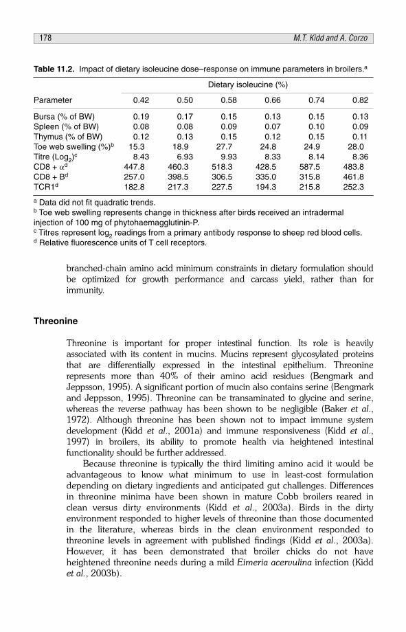
branched-chain amino acid minimum constraints in dietary formulation shouldbe optimized for growth performance and carcass yield, rather than forimmunity.
Threonine
Threonine is important for proper intestinal function. Its role is heavilyassociated with its content in mucins. Mucins represent glycosylated proteinsthat are differentially expressed in the intestinal epithelium. Threoninerepresents more than 40% of their amino acid residues (Bengmark andJeppsson, 1995). A significant portion of mucin also contains serine (Bengmarkand Jeppsson, 1995). Threonine can be transaminated to glycine and serine,whereas the reverse pathway has been shown to be negligible (Baker et al.,1972). Although threonine has been shown not to impact immune systemdevelopment (Kidd et al., 2001a) and immune responsiveness (Kidd et al.,1997) in broilers, its ability to promote health via heightened intestinalfunctionality should be further addressed.
Because threonine is typically the third limiting amino acid it would beadvantageous to know what minimum to use in least-cost formulationdepending on dietary ingredients and anticipated gut challenges. Differencesin threonine minima have been shown in mature Cobb broilers reared inclean versus dirty environments (Kidd et al., 2003a). Birds in the dirtyenvironment responded to higher levels of threonine than those documentedin the literature, whereas birds in the clean environment responded tothreonine levels in agreement with published findings (Kidd et al., 2003a).However, it has been demonstrated that broiler chicks do not haveheightened threonine needs during a mild Eimeria acervulina infection (Kiddet al., 2003b).
Table 11.2. Impact of dietary isoleucine dose–response on immune parameters in broilers.a
Dietary isoleucine (%)
Parameter 0.42 0.50 0.58 0.66 0.74 0.82
Bursa (% of BW) 0.19 0.17 0.15 0.13 0.15 0.13Spleen (% of BW) 0.08 0.08 0.09 0.07 0.10 0.09Thymus (% of BW) 0.12 0.13 0.15 0.12 0.15 0.11Toe web swelling (%)b 15.3 18.9 27.7 24.8 24.9 28.0Titre (Log2)
c 8.43 6.93 9.93 8.33 8.14 8.36CD8 + �d 447.8 460.3 518.3 428.5 587.5 483.8CD8 + Bd 257.0 398.5 306.5 335.0 315.8 461.8TCR1d 182.8 217.3 227.5 194.3 215.8 252.3
a Data did not fit quadratic trends.b Toe web swelling represents change in thickness after birds received an intradermalinjection of 100 mg of phytohaemagglutinin-P.c Titres represent log2 readings from a primary antibody response to sheep red blood cells.d Relative fluorescence units of T cell receptors.
178 M.T. Kidd and A. Corzo

Although nutritional experimentation conducted in differing environmentsrequires somewhat large and time-consuming experiments, more researchshould be conducted with threonine in this manner to better understand if itsminimum need varies as environment changes. Further, threonine requirementshould be measured in diets containing marginally deficient lysine so that ratiosto lysine can be predicted rather than optimal threonine levels.
Tryptophan
Diets fed to commercial broiler chickens are seldom limiting in dietarytryptophan. However, the inclusion of certain grains in feed formulation mayresult in this amino acid becoming the fourth limiting one. Consequently,dietary deficiency of this amino acid could have deleterious effects consideringthis amino acid’s functionalities. Apart from being an obvious constituent ofbody proteins, it has precursor roles for neurotransmitters such as melatoninand serotonin, as well as for the vitamin niacin.
Additionally, this amino acid has shown to have ketogenic activity in thechicken. It’s deficiency in diets fed to broilers has led to increasing nervousness(Corzo et al., 2005b). However, no degree of physiological stress was foundwhen tryptophan was fed in deficient amounts to broilers at 20 days of age, asobserved in the lack of alteration in those blood chemistry parameterscommonly associated with the presence of stress (see Table 11.3).
CONCLUSIONS
Although manipulation of the broiler growth curve via environmental andnutritional means has been shown to produce advantages (i.e. bone quality),feeding higher amino acid levels to optimize growth and meat yield in nutrientdensity studies does not seem to have any adverse effects on health. Someamino acids have been shown to heighten immune system parameters at
Table 11.3. Blood plasma cholesterol, heterophil/lymphocyte ratio (H/L), glucose, high/low-density lipoprotein ratio (HD/LD) and corticosterone level of broiler chicks fed diets containinggraduations of tryptophan (adapted from Corzo et al., 2005c).
Dietary Cholesterol Glucose Corticosteronetryptophan (%) (mg/dl) H/La (mg/dl) HD/LDb (ng/ml)
0.13 138.1 36.6 238.5 2.85 1.010.15 125.4 35.8 239.4 3.09 1.200.17 123.9 38.7 244.3 2.47 1.090.19 130.3 23.0 249.0 2.78 1.030.21 130.4 29.9 248.7 4.39 1.110.25 125.4 27.1 249.4 2.76 1.170.25 128.7 32.1 258.1 2.48 1.12
a Values are expressed as heterophil/lymphocyte cell count � 100.b Values are expressed as high/low-density lipoprotein.
Amino acids and protein 179

dietary levels in excess of growth needs. However, until further information isgained regarding amino acid needs for both innate and adaptive immunity,dietary formulation of amino acids should be concerned with optimizinggrowth and carcass responses. Amino acid minima in least-cost formulatedbroiler diets that support growth seem to support good health as measured bymortality and immune response data.
ACKNOWLEDGEMENT
This is Journal Article Number BC10788 from the Mississippi Agricultural andForestry Experiment Station supported by MIS-322140. Use of trade names inthis publication does not imply endorsement by the Mississippi Agricultural andForestry Experiment Station of the products, nor similar ones not mentioned.
REFERENCES
Baker, D.H., Hill, T.M. and Kleiss, A.J. (1972) Nutritional evidence concerning formation ofglycine from threonine in the chick. Journal of Animal Science 34, 582–586.
Bartov, I. and Plavnik, I. (1998) Moderate excess of dietary protein increases breast meat yield ofbroiler chicks. Poultry Science 77, 680–688.
Bengmark, S. and Jeppsson, B. (1995) Gastrointestinal surface protection and mucosa recondi-tioning. Journal of Parenteral and Enteral Nutrition 19, 410–415.
Corzo, A., Moran, Jr., E.T. and Hoehler, D. (2003) Arginine need of heavy broiler males:Applying the ideal protein concept. Poultry Science 82, 402–407.
Corzo, A., McDaniel, C.D., Kidd, M.T., Miller, E.R., Boren, B.B. and Fancher, B.I. (2004) Impactof dietary amino acid concentration on growth, carcass yield, and uniformity of broilers.Australian Journal of Agricultural Research 55, 1133–1138.
Corzo, A., Kidd, M.T., Burnham, D.J., Miller, E.R., Branton, S.L. and Gonzalez-Esquerra, R.(2005a) Dietary amino acid density effects on growth and carcass of broilers differing instrain cross and sex. Journal of Applied Poultry Research 14, 1–9.
Corzo, A., Moran, Jr. E.T., Hoehler, D. and Lemme, A. (2005b) Tryptophan need of broiler malesfrom forty-two to fifty-six days of age. Poultry Science 84, 226–231.
Corzo, A., Kidd, M.T., Thaxton, J.P. and Kerr, B.J. (2005c) Dietary tryptophan effects on growthand stress of male broiler chicks. British Poultry Science 46, 478–484.
Dawkins, M.S., Donnelly, C.A. and Jones, T.A. (2004) Chicken welfare is influenced more byhousing conditions than by stocking density. Nature 427, 342–344.
Dozier, W.A., III and Moran, Jr. E.T. (2001) Response of early- and late-developing broilers tonutritionally adequate and restrictive feeding regimens during the summer. Journal ofApplied Poultry Research 10, 92–98.
Fafournoux, P., Bruhat, A. and Jousse, C. (2000) Amino acid regulation of gene expression.Biochemical Journal 351, 1–12.
Ferguson, N.S., Gates, R.S., Taraba, J.L., Cantor, A.H., Pescatore, A.J., Ford, M.J. and Burnham,D.J. (1998) The effect of dietary crude protein on growth, ammonia concentration, and littercomposition in broiler. Poultry Science 77, 1481–1487.
German, J.B., Roberts, M.A. and Watkins, S.M. (2003) Genomics and metabolomics as markersfor the interaction of diet and health: lessons from lipids. Journal of Nutrition 133,2078S–2083S.
180 M.T. Kidd and A. Corzo

Hale, L.L., Pharr, G.T., Burgess, S.C., Corzo, A. and Kidd, M.T. (2004) Isoleucine needs of thirty-to-forty-day-old female chickens: immunity. Poultry Science 83, 1979–1985.
Humphrey, B.D. and Klasing, K.C. (2004) Modulation of nutrient metabolism and homeostasis bythe immune system. World’s Poultry Science Journal 60, 90–100.
Kidd, M.T. (2004) Nutritional modulation of immune function in broilers. Poultry Science 83,650–657.
Kidd, M.T., Kerr, B.J. and Anthony, N.B. (1997) Dietary interactions between lysine and threo-nine in broilers. Poultry Science 76, 608–614.
Kidd, M.T., Kerr, B.J., Halpin, K.M., McWard, G.W. and Quarles, C.L. (1998) Lysine levels instarter and grower-finisher diets affect broiler performance and carcass traits. Journal ofApplied Poultry Research 7, 351–358.
Kidd, M.T., Gerard, P.D., Heger, J., Kerr, B.J., Rowe, D., Sistani, K. and Burnham, D.J. (2001a)Threonine and crude protein responses in broiler chicks. Animal Feed Science andTechnology 94, 57–64.
Kidd, M.T., Peebles, E.D., Whitmarsh, S.K., Yeatman, J.B. and Wideman, Jr, R.F. (2001b) Growthand immunity of broiler chicks as affected by dietary arginine. Poultry Science 80, 1535–1542.
Kidd, M.T., Thaxton, J.P., Yeatman, J.B., Barber, S.J. and Virden, W.S. (2002) Arginine responsesin broilers: live performance. Poultry Science 80 (1), 114.
Kidd, M.T., Barber, S.J., Virden, W.S., Dozier, W.A. III, Chamblee, D.W. and Wiernusz, C.(2003a) Threonine responses of Cobb male finishing broilers in differing environmental con-ditions. Journal of Applied Poultry Research 12, 115–123.
Kidd, M.T., Pote, L.M. and Keirs, R.W. (2003b) Lack of interaction between dietary threonine andEimeria acervulina in chicks. Journal of Applied Poultry Research 12, 124–129.
Kidd, M.T., McDaniel, C.D., Branton, S.L., Miller, E.R., Boren, B.B. and Fancher, B.I. (2004)Increasing amino acid density improves live performance and carcass yields of commercialbroilers. Journal of Applied Poultry Research 13, 593–604.
Kidd, M.T., Corzo, A., Hoehler, D., Miller, E.R. and Dozier, W.A. III (2005) Broiler responsiveness(Ross x 708) to diets varying in amino acid density. Poultry Science 84, 1389–1396.
Kling, H.F. and Quarles, C.L. (1974) Effect of atmospheric ammonia and the stress of infectiousbronchitis vaccination on leghorn males. Poultry Science 53, 1161–1167.
Konashi, S., Takahashi, K. and Akiba, Y. (2000) Effects of dietary essential amino acid deficien-cies on immunological variables in broiler chickens. British Journal of Nutrition 83, 449–456.
Koutsos, E.A. and Klasing, K.C. (2001) Interactions between the immune system, nutrition, andproductivity of animals. In: Garnsworthy, P.C. and Wiseman, J. (eds) Recent Advances inAnimal Nutrition. Nottingham University Press, Nottingham, UK, pp. 173–190.
Kristensen, H.H. and Wathes, C.M. (2000) Ammonia and poultry welfare: a review. World’sPoultry Science Journal 56, 235–245.
Latshaw, J.D. (1993) Dietary lysine concentrations from deficient to excessive and the effects onbroiler chicks. British Poultry Science 34, 951–958.
Miles, D.M., Branton, S.L. and Lott, B.D. (2004) Atmospheric ammonia is detrimental to the per-formance of modern commercial broilers. Poultry Science 83, 1650–1654.
National Research Council (NRC) (1994) Nutrient Requirements of Poultry, 9th revision edition.National Academy Press, Washington DC.
Rosebrough, R.W., Mitchell, A.D. and McMurty, P.J. (1996) Dietary crude protein changes rapidyafter metabolism and plasma insulin-like growth factor I concentrations in broiler chickens.Journal of Nutrition 126, 2888–2898.
Siegel, P.B. and Gross, W.B. (1980) Production and persistency of antibodies in chickens to sheeperythrocytes. 1: directional selection. Poultry Science 59, 1–5.
Smith, E.R., Pesti, G.M., Bakalli, R.I., Ware, G.O. and Menten, J.F.M. (1998) Further studies onthe influence of genotype and dietary protein on the performance of broilers. Poultry Science77, 1678–1687.
Amino acids and protein 181

Temim, S., Chagneau, M., Guillaumin, S., Michel, J., Peresson, R. and Tesseraud, S. (2000) Doesexcess dietary protein improve growth performance and carcass characteristics in heat-exposed chickens? Poultry Science 79, 312–317.
Tesseraud, S., Pym, R.A., Bihan-Duval, E.L. and Duclos, M. (2003) Response of broilers selectedon carcass quality to dietary protein supply: live performance, muscle development, and cir-culating insulin-like growth factors (IGF-I and -II). Poultry Science 82, 1011–1016.
Thornton, S.A., Pharr, G.T., Corzo, A., Branton, S.L. and Kidd, M.T. (2004) Valine needs forimmune responses in male broilers from day 21 to 42. Poultry Science 83 (1), 186.
Whitehead, C.C. (2002) Nutrition and poultry welfare. World’s Poultry Science Journal 58,349–356.
Wijtten, P.J.A., Prak, R., Lemme, A. and Langhout, D.J. (2004) Effect of different dietary idealprotein concentrations on broiler performance. British Poultry Science 45, 504–511.
182 M.T. Kidd and A. Corzo

CHAPTER 12The role of feed processing ongastrointestinal function and health inpoultry
B. SvihusDepartment of Animal and Aquacultural Sciences, Norwegian University ofLife Sciences, Ås, Norway; e-mail: [email protected]
ABSTRACT
Nearly all feeds used in commercial poultry production are subjected to someform of feed processing. A majority of the feed is ground through a hammer milland formed into pellets by pressing the heated feed through a pellet press. Thetwo major effects of processing are thus: (i) changes in the micro- andmacrostructure of the feed; and (ii) heat-induced chemical changes of somecomponents in the feed. It has been shown that the microstructure of the feed,i.e. the particle distribution of the dissolved feed particles, has a significantimpact on gut function. Gizzard activity increases significantly, resulting in a veryfinely ground feed material entering the small intestine. An increased feedvolume in the gizzard and a decrease in gizzard pH when coarse material is fedresults in a lower jejunal microflora number and changes in microfloralcomposition. In addition, secretion of bile acids and pancreatic enzymes isenhanced.
The macrostructure determines to a large extent the feed intake pattern. Ithas been shown that a feed structure that allows for a high feed intake, such aspellets, may affect starch digestibility negatively. This may be associated withoverconsumption of feed, and may be linked to a suboptimal feed flowregulation due to reduced gizzard function. Although poultry feeds aresubjected to only moderate heat treatment, heat-induced chemical changesmay take place. A small fraction of the starch will gelatinize, solubility of fibresmay increase and proteins may form indigestible new bonds. In addition,vitamins and enzymes may lose their effect.
INTRODUCTION
More than 600 million t of processed concentrates are produced annually on aworldwide basis (Gill, 2003). Poultry feeds are the largest product group, with
© CAB International 2006. Avian Gut Function in Health and Disease (ed. G.C. Perry) 183

37% of the total feed production. A typical poultry feed production processconsists of grinding the feed ingredients in a hammer mill through a 3–4 mmsieve. Feed for layers is commonly fed as a mash feed, while other types offeed are passed through a conditioner followed by pelleting. The temperaturewill normally reach a maximum of 80–90°C during the pelleting process.
Although there has been surprisingly little research on the interactionsbetween feed processing and nutrition, it is evident that the physical andchemical changes that take place during processing may have a large impacton the performance of birds, both directly through effects on feed digestibilityand indirectly through effects on feed intake pattern and gut function. In thischapter, some effects of processing will be covered. Since any effect on nutrientabsorption efficiency will potentially affect gut function and gut health,nutritional aspects will also be addressed.
EFFECTS OF STRUCTURE
One of the functions of feed processing is to affect the structure of the feed.Grinding is performed to reduce the particle size of feed ingredients, while thepelleting process is performed to increase the particle size of the feed. Thus, ina pelleted feed, structure concerns both the particle distribution of theingredients that the pellet consists of and the structure of the pellets themselves.It is thus pertinent to describe the structure of the particles inside the pellets asthe ‘microstructure’ and the structure of the pellets themselves as the‘macrostructure’. As pellets normally disintegrate quite rapidly once they aremoistened in the upper digestive tract, the macrostructure will affect only feedintake pattern and total feed intake. The microstructure, on the other hand, willaffect gut function through the effects of particle size and distribution.
Effects of microstructure
Although grinding represents a considerable cost in terms of energyconsumption and feed mill capacity, cereals used for poultry are usually finelyground in a hammer mill fitted with a screen between 3–4.5 mm in size. Onemajor reason for this fine grinding is to ensure a good pellet quality. Finegrinding of cereals has also been considered to be beneficial for a properutilization of the feed, but this view is not supported by most scientific literature(Farrell et al., 1983; Reece et al., 1985; Reece et al., 1986a, b; Deaton et al.,1989; Proudfoot and Hulan, 1989; Lott et al., 1992; Nir et al., 1994a, b;Hamilton and Proudfoot, 1995; Nir et al., 1995; Hamilton and Kennie, 1997;Svihus et al., 2004b).
Some studies have even shown an improved performance with the use ofcoarsely ground cereals in mash form (Reece et al. 1985; Nir et al., 1990,1995). Birds prefer coarse particles and will eat more of a coarsely ground thanof a finely ground mash diet (Reece et al., 1985; Portella et al., 1988 a, b; Niret al., 1990, 1994b).
184 B.Svihus

It is thus necessary to use pelleted diets to separate the effect of feedstructure on feed intake stimulation from the effect of feed structure on feedutilization. Experiments conducted with pelleted diets containing cereals groundto different sizes usually demonstrate that coarse grinding gives similar weightgain and feed utilization to those of fine grinding (Reece et al., 1985, 1986a, b;Proudfoot and Hulan, 1989; Nir et al., 1995; Hamilton and Kennie, 1997;Svihus et al., 2004b). Considering the numerous recent results showing that theyoung broiler chicken is able efficiently to utilize diets with up to 400 gunground wheat/kg (see Svihus et al., 2004a, for references), this is notsurprising.
It has been shown that coarse particles are very finely ground in the gizzard(Hetland et al., 2002) and that this grinding does not affect passage ratethrough the intestinal tract (Svihus et al., 2002). This indicates that from anutritional point of view the cereals used for poultry feed can be ground morecoarsely than is current practice. A coarser grinding would save considerableenergy and time in the grinding process. Reece et al. (1986a) found that theenergy cost for hammer grinding of maize could be reduced from 2.9 kwh/t to2.2 kwh/t by increasing the sieve size from 4.76 to 6.35 mm. In addition,coarser grinding will reduce the amount of fines in the pelleted product andthus, potentially, reduce transport loss and dust problems in the feed mill andon the farm.
One concern with using coarsely ground cereals in the feed has been thepellet quality. As the proportion of coarse particles increases in the pellet, thismay result in more fractures due to reduced particle surface area and weakspots in the pellet. Although Svihus et al. (2004b) observed a moderatereduction in pellet durability with a coarser grinding, other data have shownimproved pellet quality with a coarser grinding (Reece et al., 1986a, b).
Roller mills are not commonly used in feed production, despite the fact thatthey grind with a lower energy consumption per ton of feed (Nir et al., 1990),give less dust during grinding and are less noisy (Heimann, 2002). It has alsobeen shown that roller mills grind cereals more uniformly and produce a loweramount of fines than do hammer mills (Nir et al., 1990, 1995). As roller millsare considered to be particularly effective for coarse grinding, the advantages ofusing a roller mill will increase when a coarse grinding is used. A majordisadvantage of roller mills is the higher cost for purchase and maintenance(Heimann, 2002).
Birds eat and swallow food without prior chewing. Although secretion ofsaliva does occur, in the chicken and turkey the saliva does not containamylase and the time exposed to saliva in the mouth is short (Duke, 1986).Thus food enters the crop or, if the gizzard is empty, passes directly to theproventriculus and gizzard largely as intact food particles. The crop may storelarge amounts of food for a considerable amount of time. Svihus et al. (2002)found up to 30 g feed in the crop 30 min after feeding starved broiler chickensweighing 1100–1400 g, with half this feed still remaining in the crop after 3 h.
Although no digestive enzymes are secreted and no grinding occurs in thecrop, feed may be softened and endogenous and microbial enzymes may beactivated. The main body of the gizzard comprises two thick, opposed lateral
The role of food processing 185

muscles and two thin anterior and posterior muscles. A thick layer ofglycoprotein that is hardened by the low pH covers the inside of the gizzard.This koilin layer is composed of a combination of rod-like and granularsecretions from the gizzard’s glands, and is continuously renewed by theseglands as it is utilized. Some birds, however, are known to shed the wholekoilin layer at intervals (McLelland, 1979). None of the domesticated birdspecies have, as yet, been reported to exhibit such behaviour, but unpublishedobservations of gizzards lacking the koilin layer in our laboratory indicate thatthis may happen in turkeys. The rod-like, hardened glycoprotein will stand outfrom the remaining glycoprotein matrix and will give the surface itscharacteristic sandpaper-like appearance (Hill, 1971). The thick muscles grindmaterial by contraction that rubs the material against the koilin layer on theinside of the gizzard. The small muscles move material between contractions ofthe large muscles.
A prerequisite for utilizing coarsely ground or un-ground cereals is a well-functioning gizzard. Feeding studies with broilers have shown that the gizzardbecomes stimulated when the feed’s microstructure is improved, e.g. whenwhole cereals replace ground cereals. It has also been shown that replacing50% ground wheat with the same quantity of ground oats gives the sameincrease in gizzard weight as that achieved by replacing 50% ground wheatwith whole wheat (Hetland et al., 2002). This indicates that a relatively smallamount of insoluble fibre (e.g. 10% oat hulls coming from 50% oats in the diet)stimulates the gizzard to the same extent as 50% whole wheat.
Current data indicate that the gizzard weight is less affected by cerealstructure in layers. However, insoluble fibre seems to play an even moreprofound role for gizzard activity in the more mature digestive tract of the layer.This corresponds with an earlier experiment with layers, where consumption of4% of feed as wood shavings resulted in a 50% heavier gizzard, whereasincluding 40% whole wheat in wheat-based diets increased the gizzard weightby only 10% (Hetland et al., 2003a).
Coarse feed particles need to be ground to a certain critical size before theycan leave the gizzard (Clemens et al., 1975; Moore, 1999), causing the volumeof gizzard contents to increase when diets with whole cereals or insoluble fibreare fed (Hetland et al., 2003a, b). Hetland et al. (2002) thus found that80–90% of particles in the duodenum of broilers were smaller than 300 �mand that this was independent of dietary particle size. This suggests that coarseparticles accumulate in the gizzard and are retained longer than are othernutrients. Hetland et al. (2005) showed that finely ground oat hulls passedthrough the gizzard much faster than coarse oat hulls. Thus, an increasedvolume of gizzard contents in birds fed diets with whole grains or high levels ofinsoluble fibre has been observed (Hetland et al., 2003a, b). Indeed, thegizzard has a remarkable ability to grind all organic constituents of feed to avery consistent particle size range regardless of the original particle size of thefeed, with some ground as finely as 0.05 mm.
It has also been shown that stimulation of the gizzard function by structuralcomponents results in a reduction in gizzard pH (Engberg et al., 2004; Bjerrumet al., 2005). This, combined with an increased retention time, improves the
186 B.Svihus

potential for the gizzard to function as a barrier to foodborne microorganisms.The same authors also observed a signifcant reduction in pathogenic microflorawhen ground wheat was replaced by whole wheat. Field evidence that moremicrostructure in the feed is beneficial to gut health has also been provided byBranton et al. (1987), where mortality due to necrotic enteritis was much lowerin birds given coarsely ground diets than in broilers given finely ground diets.
A significant increase in amylase activity and bile acid concentration hasbeen observed when diets contain more structural components (Svihus et al.,2004a). This may indicate that an increased secretory activity is the cause ofthe improvements in nutritive value sometimes observed with whole wheat orother structural components (Kiiskinen, 1996; Plavnik et al., 2002; Hetland etal., 2003a; Svihus et al., 2004a). This hypothesis is also supported by the factthat amylase activity in the jejunum and starch digestibility in the anterior,median and posterior ileum for individual birds had a correlation of 0.56, 0.54and 0.47, respectively (Svihus et al., 2004a).
The cause of this increased secretory activity remains unclear, but may beassociated with a stimulation of pancreatic secretion caused by an increase ingizzard activity. Hetland et al. (2003a) found a significant increase in amylaseactivity and bile acid secretion when gizzard activity was stimulated by oathulls. The same tendency, although not significant for amylase activity, wasseen when gizzard activity was stimulated by whole wheat.
The major stimuli causing increased exogenous enzyme secretion by thepancreas are the vagus nerve and cholecystokinin (CCK). Cholecystokinin ismainly produced in the pyloric region of birds (Denbow, 2000) and acts via thevagus nerve to stimulate pancreatic enzyme secretion (Li and Owyang, 1993).Thus, a stimulating effect of increased gizzard activity on enzyme secretionsmay be mediated through increased CCK release in the pyloric region of thegizzard. Gabriel et al. (2003) found tendencies for reduced levels of brushborder enzyme activity by the inclusion of whole wheat. It is thus possible thata stimulatory effect of coarse feed structure is limited to pancreatic enzymesecretions.
Cholecystokinin has also been shown to stimulate gastrointestinal refluxes(Duke, 1992). Svihus et al. (2004a) observed a significant increase in thecontent of bile acids in the gizzard when large wheat particles were fed. Thisindicates increased gastroduodenal refluxes, which may be caused by increasedCCK secretion. As indicated by the performance data, the increasedgastroduodenal reflux does not reduce feed consumption. The results discussedabove clearly suggest that from a nutritional point of view the microstructure offeeds for poultry can be very coarse, for example by using coarsely ground oreven un-ground cereals. The results also indicate that a coarse microstructuremay be beneficial from a gastrointestinal health point of view.
Effects of macrostructure
It is well known that feed intake will improve with pelleted diets compared tothat with mash diets. In broilers an increase in feed intake of 25% is often
The role of food processing 187

observed (Svihus et al., 2004b). As opposed to the microstructure,improvements in macrostructure do not result in increased gizzard weight orgizzard volume. In fact, a reduction in relative gizzard weight and gizzard feedcontent is observed (Engberg et al., 2002; Svihus et al., 2004b). This is due tothe fact that pellets disintegrate quite rapidly when they are moistened in theupper digestive tract.
It has been shown that the pelleting process results in a significantly finermicrostructure (Svihus et al., 2004b). This is caused by the grinding action ofthe rolls in the pellet press, and may be one contributing factor to the smallerrelative size of the gizzard. As relative size of the gizzard is reduced withincreasing weight of the bird (Hetland et al., 2002), this may also contribute toa smaller relative size of the gizzard in birds fed pellets. Pelleting results insignificant improvements in the feed/gain ratio (Engberg et al., 2002; Svihus etal., 2004b), but this is not often associated with improvements in digestibility ormetabolizable energy content.
In fact, a reduction in starch digestibility has been observed when pelleteddiets were compared to mash diets (Svihus and Hetland, 2001), and based onthese observations it has been hypothesized that some birds are overeaterswhen presented with pelleted diets. Engberg et al. (2002) also showed asignificant reduction in enzyme activities when pelleted diets were used insteadof mash diets.
The hypothesis of some birds being overconsumers is supported by furtherstudies in our laboratory indicating a slight negative correlation between feedintake of individual birds on identical diets and the apparent metabolizableenergy (AME) value determined (see Figs 12.1 and 12.2). In these studies,male Ross 208 broilers were raised in individual cages from 13 days of age andpelleted diets were provided ad libitum. AME content and correlation between
y = –0.0055x + 15.681
r2 = 0.215
13.0
13.5
14.0
14.5
15.0
200 250 300 350
Feed consumption (g)
AM
E (
MJ/
kg)
Fig. 12.1. Relationship between feed consumption and apparent metabolizable energy(AME) determined on individual birds fed on a pelleted, maize-based diet.
188 B.Svihus

feed intake and AME was determined by monitoring feed and faecesquantitatively from days 19–22, combined with determination of energycontent using a calorimeter.
These data also show that feed intake varies between birds. It is thuspossible that overconsumption among individual birds in a flock results inimpaired starch digestion due to an increased passage rate. This is consistentwith many observations of reduced variation among individuals concurrentwith an increased starch digestibility (Svihus and Hetland, 2001). The questionmay then arise on the causes of overconsumption among individual birds.
Peter Siegel was among the first to indicate that intensive breeding forweight gain had resulted in broilers that were capable of overconsuming feeddue to disturbance of the appetite control centre in the brain (Siegel andDunnington, 1987). Lacy et al. (1985) showed that broilers, as opposed tolayers, did not respond to intra-hepatic glucose infusions with reduced feedintake. This indicates that peripheral appetite control centres have also beendisturbed in broilers.
It can thus be postulated that modern breeds of broilers may overconsumefeeds, with a resulting impairment in digestibility. Our results indicate that theremay be an interaction between gizzard function and over-consumption offeeds. When feeds have more structure, either through the use of whole cerealsor through the addition of large fibre particles (oat hulls or wood shavings) intothe diet, an improvement in starch digestibility and a reduced variationbetween birds is commonly observed (Rogel et al., 1987; Svihus and Hetland,2001; Hetland et al., 2002).
Our hypothesis is that an active and well-developed gizzard will function asa food intake regulator that will ensure that the bird does not overconsumefeeds. This is due to both: (i) the active retention of large particles until they are
y = –0.0054x + 15.317r2 = 0.3145
12.0
12.5
13.0
13.5
14.0
14.5
250 300 350 400 450 500
Feed consumption (g)
AM
E (
MJ/
kg)
Fig. 12.2. Relationship between feed consumption and apparent metabolizable energy(AME) determined on individual birds fed on a pelleted, wheat-based diet.
The role of food processing 189

broken down, which results in a larger filling of the gizzard and thus less roomfor more feed to be consumed; and (ii) a stimulative effect of gizzard activity ongastroduodenal refluxes.
Effects of heat on nutritional value of pelleted feeds
For poultry, the majority of heat treatment occurs during conditioning andpelleting. During conditoning, the temperature is usually raised to 70–80°C bythe addition of steam. This temperature may increase to 85–90°C during thepelleting process. Although these temperatures are moderate compared tothose achieved during extrusion and expansion of feeds, they may result in thedestruction of the three-dimensional structure of proteins and starch, and alsoin new covalent bond formations such as disulphide bridges, iso-peptide bondsand Maillard-reactions.
There is a scarcity of published results on changes in protein availabilityduring pelleting. Carré et al. (1991) did not find any increase in proteindigestibiliy in broiler chickens as a result of pelleting, while Vande Ginste andDe Schrijver (1998) found a small but significant increase in protein digestibilityin finishing pigs when pelleting was compared to mash feeding. In extrudeddiets, a small decrease in protein digestibility is often observed (Ljøkjel et al.,2004), but since extrusion is a much more severe heating process – withtemperatures usually exceeding 110°C – it is logical to conclude that proteindigestibility is probably not affected negatively by the pelleting process.
Starch is gelatinized to some extent during the pelleting process. Theextent of starch gelatinization is usually found to vary between 5 and 20%(Svihus et al., 2004b), but no improvements in starch digestibility have beenestablished as a consequence of the pelleting process, except for a significantimprovement from pelleting of peas (Carré et al., 1991). This latterimprovement has been associated with large, starch-containing particles in thenon-pelleted diets.
The antinutritive effect of soluble fibres in the diet is well known, the majormechanism for which is an increased viscosity of the gut contents, and thus areduced digestibility and nutrient absorption. These changes may also affectgut development and chyme mobility, and they have been shown to have animpact on gut microflora composition. Thus, the viscosity effect of soluble fibresis important for gut health and function. It has been shown that processing mayincrease the solubility of fibres, thus resulting in an increased viscosity of gutcontents and reduced nutrient digestibility. Graham et al. (1989) observedincreased solubility of barley �-glucans after pelleting, and Cowieson et al.(2005) observed an increased viscosity of wheat diets with increasingconditioning temperature.
In an experiment carried out at the Norwegian University of Life Sciences,complete diets containing 77% barley, oats or wheat were fed to broilerchickens from 7 days of age. The cereal used in each diet was either addeduntreated, or it was heat-treated by conditioning at 90°C followed by pelletingand regrinding. At 21 days of age the birds were killed and digesta contents
190 B.Svihus

collected. The results presented in Table 12.1 show a tremendous increase inthe jejunal viscosity of broilers when the cereals were heat-treated, followed bya typical increase in water content in the chyme and higher stickiness of thefaeces.
It is well known that many vitamins and carotenoids are susceptible to heatdenaturation. Andersen and Sunderland (2002) found that 67% of vitamin Ewas recovered after an extrusion process, and that recovery decreased withincreasing water content. This indicates that vitamin E is heat-sensitive,although it is reasonable to conclude that much less would be destroyed duringa pelleting process, due to less water and lower temperatures. Marchetti et al.(1999) observed significant reductions in the recovery of crystalline vitaminsafter pelleting. For vitamins K and C recovery was 50%, while the B-vitaminsgenerally had recoveries between 82 and 96%. Exceptions were pyridoxineand folic acid, with recoveries of 75 and 65%, respectively. Thus, pelleting canhave a large impact on the content of some vitamins and this must be takeninto account during feed processing.
Enzymes are now routinely used in most poultry feeds. Since enzymes areproteins which act as catalysts due to their specific three-dimensional structures,they are susceptible to heat destruction through denaturation. Inborr andBedford (1994) demonstrated that �-glucanase activity was reduced to 66% ofits initial value after conditioning at 75°C followed by pelleting. Afterconditioning at 95°C, this was reduced to 16% of the initial activity. Eventhough more heat-stable enzyme products have been developed using coatingtechnologies or by selection of less heat-sensitive proteins, reduced activity dueto processing can still be expected.
Based on the above it can be concluded that the temperature should bekept as low as possible during feed processing to avoid reduction in thenutritional value of the feed. This also calls for caution in using processes suchas expansion and extrusion for the production of poultry feeds, as theseprocesses involve high temperatures and high pressure. McCracken (2002),while reviewing the literature on feed processing, reported data showing areduced performance by expander treatment of poultry feeds.
Table 12.1. Properties of gut contents in broilers fed complete cold-pelleted diets based on77% cereals either non-heated or pelleted at 90°C.
Cereal source
Oats Barley Wheat
Non-heated Heat-treated Non-heated Heat-treated Non-heated Heat-treated
Jejunal viscosity (cP) 25.9b 111.4a 26.6b 80.8a 15.4b 30.2a
DM in chyme (%) 15.7a 12.7b 18.0a 16.6b 25.0a 19.8b
Dirtiness scorec 2.9b 4.2a 2.9b 3.8a 2.9b 3.2a
ab Within each cereal category, numbers within a row which do not share common superscriptsare significantly different (p < 0.05) in a Ryan-Einot-Gabriel-Welsh test.c Subjective score of cage dirtiness, with the highest score of 5 for the dirtiest cages.
The role of food processing 191

CONCLUSION
Micro- and macrostructure of the feed as determined by processing has a largeimpact on gut function and digestive processes. Gut health may also be affectedthrough effects on available substrate in the lower digestive tract, and throughchanges in pH and retention time in the upper digestive tract. Heat treatmentdoes not seem to have any significant beneficial effect on nutrient digestion, andmay have a negative effect on nutrient digestibility through solubilization offibres and through destruction of vitamins and enzymes added to the feed.Thus, temperature during processing should be kept as low as possible.
REFERENCES
Andersen, J.S. and Sunderland, R. (2002) Effect of extruder moisture and dryer processing tem-perature on vitamin C and E and astaxanthin stability. Aquaculture 207, 137–149.
Bjerrum, L., Pedersen, K. and Engberg, R.M. (2005) The influence of whole wheat feeding onsalmonella infection and gut flora composition in broilers. Avian Diseases 49, 9–15.
Branton, S.L., Reece, F.N. and Hagler, W.M. (1987) Influence of a wheat diet on mortality ofbroiler chickens associated with necrotic enteritis. Poultry Science 66, 1326–1330.
Carré, B., Beaufils, E. and Melcion, J.P. (1991) Evaluation of protein and starch digestibilities andenergy value of pelleted or unpelleted pea-seeds from winter or spring cultivars in adult andyoung chickens. Journal of Agricultural and Food Chemistry 39, 468–472.
Clemens, E.T., Stevens, C.E. and Southworth, M. (1975) Sites of organic acid production andpattern of digesta movement in the gastrointestinal tract of geese. Journal of Nutrition 105,1341–1350.
Cowieson, A.J., Hruby, M. and Isaksen, M.F. (2005) The effect of conditioning temperature andexogenous xylanase addition on the viscosity of wheat-based diets and the performance ofbroiler chickens. Proceedings of the 15th European Symposium on Poultry Nutrition, WPSA,Balatonfüred, Hungary, pp. 256–259.
Deaton, J.W., Lott, B.D. and Simmons, J.D. (1989) Hammer mill versus roller mill grinding ofcorn for commercial egg layers. Poultry Science 68, 1342–1344.
Denbow, D.M. (2000) Gastrointestinal anatomy and physiology, In: Whittow, G.C. (ed.) Sturkie’sAvian Physiology, Academic Press, New York, pp. 299–325.
Duke, G.E. (1986) Alimentary canal: anatomy, regulation of feeding, and motility. In: Sturkie,P.D. (ed.) Avian Physiology (4th edn), Springer-Verlag, New York, pp. 269–288.
Duke, G.E. (1992) Recent studies on regulation of gastric motility in turkeys. Poultry Science 71,1–8.
Engberg, R.M., Hedemann, M.S. and Jensen, B.B. (2002) The influence of grinding and pelletingof feed on the microbial composition and activity in the digestive tract of broiler chickens.British Poultry Science 43, 569–579.
Engberg, R.M., Hedemann, M.S., Steenfeldt, S. and Jensen, B.B. (2004) Influence of wholewheat and xylanase on broiler performance and microbial composition and activity in thedigestive tract. Poultry Science 83, 925–938.
Farrell, D.J., Thomson, E. and Choice, A. (1983) Effects of milling and pelleting of maize, barleyand wheat on their metabolizable energy value for cockerels and chicks. Animal FeedScience and Technology 9, 99–105.
Gabriel, I., Mallet, S. and Leconte, M. (2003) Differences in the digestive tract characteristics ofbroiler chickens fed on complete pelleted diet or on whole wheat added to pelleted proteinconcentrate. British Poultry Science 44, 283–290.
192 B.Svihus

Gill, C. (2003) Back to basics of growth. Feed International 23 (January), 6–9.Graham, H., Fadel, J.G., Newman, C.W. and Newman, R.K. (1989) Effect of pelleting and
�-glucanase supplementation on the ileal and faecal digestibility of a barley-based diet in thepig. Journal of Animal Science 67, 1293–1298.
Hamilton, R.M.G. and Kennie, J. (1997) The effects of lighting program, ingredient particle sizeand feed form on the performance of broiler turkeys. Canadian Journal of Animal Science77, 503–508.
Hamilton, R.M.G. and Proudfoot, F.G. (1995) Ingredient particle size and feed texture: effects onthe performance of broiler chickens. Animal Feed Science and Technology 51, 203–210.
Heimann, M. (2002) The ‘Bottom line’ of grinding. Feed International 23 (May), 32–34.Hetland, H., Svihus, B. and Olaisen, V. (2002) Effect of feeding whole cereals on performance,
starch digestibility and duodenal particle size distribution in broiler chickens. British PoultryScience 43, 416–423.
Hetland, H., Svihus, B. and Krogdahl, Å. (2003a) Effects of oat hulls and wood shavings ondigestion in broilers and layers fed diets based on whole or ground wheat. British PoultryScience 44, 275–282.
Hetland, H., Svihus, B., Lervik, S. and Moe, R. (2003b) Effect of feed structure on performanceand welfare in laying hens housed in conventional and furnished cages. Acta AgriculturaeScandinavica 53, 92–100.
Hetland, H., Svihus, B. and Choct, M. (2005) Role of insoluble fibre on gizzard activity in layers.Journal of Applied Poultry Research 14, 38–46.
Hill, K.J. (1971) The structure of the alimentary tract. In: Bell, D.J. and Freeman, B.M. (eds),Physiology and Biochemistry of the Domestic Fowl, Academic Press, London, pp. 1–23.
Inborr, J. and Bedford, M.R. (1994) Stability of feed enzymes to steam pelleting during feedprocessing. Animal Feed Science and Technology 46, 179–196.
Kiiskinen, T. (1996) Feeding whole grain with pelleted diets to growing broiler chickens.Agricultural and Food Science in Finland 5, 167–175.
Lacy, M.P., Van Krey, H.P., Skewes, P.A. and Denbow, D.M. (1985). Effect of intrahepatic glucoseinfusions on feeding in heavy and light breed chicks. Poultry Science 64, 751–756.
Li, Y. and Owyang, C. (1993) Vagal afferent pathway mediates physiological action of cholecys-tokinin on pancreatic-enzyme secretion. Journal of Clinical Investigations 92, 418–424.
Ljøkjel, K., Sørensen, M., Storebakken, T. and Skrede, A. (2004) Digestibility of protein, aminoacids and starch in mink (Mustela vison) fed diets processed by different extrusion conditions.Canadian Journal of Animal Science 84, 673–680.
Lott, B.D., Day, E.J., Deaton, J.W. and May, J.D. (1992). The effect of temperature, dietaryenergy level, and corn particle size on broiler performance. Poultry Science 71, 618–624.
Marchetti, M., Tossani, N., Marchetti, S. and Bauce, G. (1999) Stability of crystalline and coatedvitamins during manufacture and storage of fish feeds. Aquaculture Nutrition 5, 115–120.
McCracken, K.J. (2002) Effects of physical processing on the nutritive value of poultry diets. In:McNab, J.M. and Boorman, K.N. (eds) Poultry Feedstuffs. Supply, Composition and NutritiveValue, CABI Publishing, Wallingford, UK, pp. 301–316.
McLelland, J. (1979) Digestive system. In: King, A.S. and McLelland, J. (eds) Form and Functionin Birds, Academic Press, London, pp. 69–181.
Moore, S.J. (1999) Food breakdown in an avian herbivore; who needs teeth? Australian Journalof Zoology 47, 625–632.
Nir, I., Melcion, J.P. and Picard, M. (1990) Effect of particle size of sorghum grains on feed intakeand performance of young broilers. Poultry Science 69, 2177–2184.
Nir, I., Shefet, G. and Aaroni, Y. (1994a) Effect of particle size on performance. 1. Corn. PoultryScience 73, 45–49.
Nir, I., Hillel, R., Shefet, G. and Nitsan, Z. (1994b) Effect of grain particle size on performance. 2.Grain texture interactions. Poultry Science 73, 781–791.
The role of food processing 193

Nir, I., Hillel, R., Ptichi, I. and Shefet, G. (1995) Effect of particle size on performance. 3.Grinding pelleting interactions. Poultry Science 74, 771–783.
Plavnik, I., Macovsky, B. and Sklan, D. (2002) Effect of feeding whole wheat on performance ofbroiler chickens. Animal Feed Science and Technology 96, 229–236.
Portella, F.J., Caston, L.J. and Leeson, S. (1988a) Apparent feed particle size preference by layinghens. Canadian Journal of Animal Science 68, 915–922.
Portella, F.J., Caston, L.J. and Leeson, S. (1988b) Apparent feed particle size preference bybroilers. Canadian Journal of Animal Science 68, 923–930.
Proudfoot, F.G. and Hulan, H.W. (1989) Feed texture effects on the performance of roasterchickens. Canadian Journal of Animal Science 69, 801–807.
Reece, F.N., Lott, B.D. and Deaton, J.W. (1985) The effects of feed form, grinding method,energy level, and gender on broiler performance in a moderate (21°C) environment. PoultryScience 64, 1834–1839.
Reece, F.N., Lott, B.D. and Deaton, J.W. (1986a) Effects of environmental temperature and cornparticle size on response of broilers to pelleted feed. Poultry Science 65, 636–641.
Reece, F.N., Lott, B.D. and Deaton, J.W. (1986b) The effects of hammer mill screen size onground corn particles size, pellet durability, and broiler performance. Poultry Science 65,1257–1261.
Rogel, A.M., Balnave, D., Bryden, W.L. and Annison, E.F. (1987) Improvement of raw potatostarch digestion in chickens by feeding oat hulls and other fibrous feedstuffs. AustralianJournal of Agricultural Research 38, 629–637.
Siegel, P.B. and Dunnington, E.A. (1987). Selection for growth in chickens. CRC Critical Reviewsin Poultry Biology 1, 1–24.
Svihus, B. and Hetland, H. (2001) Ileal starch digestibility in growing broiler chickens fed on awheat-based diet is improved by mash feeding, dilution with cellulose or whole wheatinclusion. British Poultry Science 42, 633–637.
Svihus, B., Hetland, H., Choct, M. and Sundby, F. (2002) Passage rate through the anteriordigestive tract of growing broiler chickens fed on diets with ground and whole wheat. BritishPoultry Science 43, 662–668.
Svihus, B., Juvik, E., Hetland, H. and Krogdahl, Å. (2004a) Causes for improvement in nutritivevalue of broiler chicken diets with whole wheat instead of ground wheat. British PoultryScience 45, 1–6.
Svihus, B., Kløvstad, K.H., Perez, V., Zimonja, O., Sahlström, S., Schüller, R.B., Jeksrud, W.K. andPrestløkken, E. (2004b) Nutritional effects of pelleting of broiler chicken diets made fromwheat ground to different coarsenesses by the use of roller mill and hammer mill. AnimalFeed Science and Technology 117, 281–293.
Vande Ginste, J. and De Schrijver, R. (1998) Expansion and pelleting of starter, grower andfinisher diets for pigs: effects on nitrogen retention, ileal and total tract digestibility of protein,phosphorus and calcium and in vitro protein quality. Animal Feed Science and Technology72, 303–314.
194 B.Svihus

CHAPTER 13Wet litter: its causes and preventionand the role of nutrition
S.R. CollettThe University of Georgia, College of Veterinary Medicine, PoultryDiagnostic and Research Center, Athens, Georgia, USA; e-mail: [email protected]
ABSTRACT
Poultry litter becomes wet when the rate of water addition (urine/faeces/spillage)exceeds the rate of removal (evaporation). Anti-nutritional factors, toxins,pathogens and nutrient imbalances may cause wet litter directly by alteringnormal digestive physiology or indirectly by disturbing normal gut ecology.
Poor-quality ingredients and those with excess oligosaccharides or mineralscan cause a nutritionally induced polydypsia, polyuria and diarrhoea whichmay increase water output sufficiently to cause wet litter. If the situation persistsfor long enough the ensuing inflammatory response causes mild to severegastroenteritis which further increases water output. The damage so caused tothe cytoskeleton of the gastrointestinal tract reduces the surface area fornutrient absorption and allows the opportunity for pathogen proliferation, gutcolonization and even invasion and systemic disease.
Historically the solution to the downstream effect of compromiseddigestion and absorption on gut ecology has been to supplement birds withantibiotic growth promoters, a practice that has not been permitted withinEurope from January 2006. The only alternative is to use a holistic approach inmanaging the gut ecology by manipulating the microbial populations and hostinflammatory response through non-antibiotic nutritional techniques.
Enzymes, probiotics, prebiotics, immune modulators and mycotoxinbinders have all shown promise in this regard. Seeding the gastrointestinal tractwith bacteria at hatch and subsequently managing the gastrointestinalenvironment, whilst at the same time reducing anti-nutritional factors and theuse of precision diet formulation, has proved advantageous in minimizing theincidence of wet litter in birds whilst maintaining and improving birdperformance.
© CAB International 2006. Avian Gut Function in Health and Disease (ed. G.C. Perry) 195

INTRODUCTION
It is important to the wellbeing, welfare and productivity of commercial poultrythat housing conditions are maintained within the comfort zone and feed andwater are available ad libitum. Under these controlled conditions waterexcretion (urine and faeces) closely parallels intake since evaporative water lossis limited. In pursuance of homeostasis, water balance is maintained by the birdas either positive (growth) or neutral.
Years of breeding and selection to improve growth rate, feed efficiency andegg production have increased appetite and hence daily feed and water intake.At standard temperatures broilers will consume approximately 1.8–2.0 timesmore water than feed (by weight), and water deprivation can severely affectfeed intake and growth rate (Kellerup et al., 1965; Leeson and Summers,2005). During the log phase of growth, commercial poultry excreteexponentially increasing quantities of faecal and urinary water into thebedding/litter. It is crucial that the poultry house ventilation system design andoperation is efficient enough to prevent the litter moisture content fromexceeding an optimal 25%.
WATER BALANCE
Avian osmoregulation involves balancing water and electrolyte intake withexcretion via the kidney (urine), gastrointestinal tract (faeces), skin andrespiratory tract (evaporation). Since feed is generally low in moisture (10%)and metabolic water production is limited by diet formulation (~ 0.14g/kcal ofdietary energy), moisture intake is primarily controlled by drinking (~ 80%)(Leeson and Summers, 2005).
Water consumption is requirement-driven and the satiety centre isstimulated by cellular dehydration (osmoreceptors), extracellular dehydration(mechanoreceptors) and angiotensin II secretion (renin–angiotensin axis)(Kaufman et al., 1980; Kanosue et al., 1990; Goldstein and Skadhauge, 2000).All three mechanisms of thirst stimulation are activated when birds aredehydrated and over-consumption frequently occurs because intestinalabsorption is sluggish and cellular rehydration takes time. In order to maintainwater balance under such conditions, excess water is excreted as dilute urine(Takei et al., 1988).
Water intake is ambient temperature-dependent because evaporativecooling mechanisms account for 50–80% of moisture loss, depending on thestate of hydration (Goldstein and Skadhauge, 2000). Consumed water alsoacts as a heat-sink in heat-stressed birds, providing an important means ofthermoregulation. Insensible moisture loss during normal respiration increasesmarkedly when panting is initiated in response to heat stress and, interestingly,50–80% of total evaporative loss occurs through the skin, despite the lack ofsweat glands (Marder and Ben-Asher, 1983; Goldstein and Skadhauge, 2000).
The control of this through cutaneous evaporation is not completelyunderstood but is partly regulated by adrenergic-induced vasodilatation in
196 S.R. Collett

response to elevated ambient temperatures (Marder and Ben-Asher, 1983;Marder and Raber, 1989).
The kidneys rid the body of excess water and solutes by a combination ofglomerular filtration, tubular re-absorption and secretion. Excretory function iscontrolled by several hormones, including: (i) arginine vasotocin (antidiuretic);(ii) renin/angiotensin (diuretic/antidiuretic and natiuretic/antinatiuretic); (iii)aldosterone (antinatiuretic); (iv) atrial natriuretic peptide (diuretic andnaturietic); and (v) parathyroid hormone (calcium mobilization and excretion ofphosphorus) (Morild et al., 1985; Clark and Mok, 1986; Gray et al., 1988;Gray, 1993; Goldstein and Skadhauge, 2000).
Urinary excretion is somewhat unique in the avian species, since first theureters open into the coprodeum and, secondly, the urine passes retrograde upthe colon to the caeca before being evacuated via the cloaca with the faeces(Goldstein and Skadhauge, 2000). The content of the urine is significantlyaltered during its passage through the coprodeum, colon and caeca (Rice andSkadhauge, 1982).
Approximately 95% of the daily glomerular filtrate water (11 times totalbody water) is normally re-absorbed, but both filtration rate and reabsorptionrate are altered by the avian antidiuretic hormone, arginine vasotocin (AVT),which is released by the neurohypophysis in response to an elevation inextracellular osmolality or a drop in blood pressure (Gray et al., 1988;Goldstein and Skadhauge, 2000).
AVT reduces filtration rate and increases the rate of water reabsorption(70–99%), thus increasing urine concentration from typically iso-osmotic toapproximately 3 � the osmolality of plasma in the dehydrated state. Despitethis limited concentrating ability in the avian kidney, the efficiency of the avianspecies in conserving water is similar to that of mammals. This is in part due totheir being uricotelic (excretion of insoluble urates limits water loss), but also tothe reprocessing of urine that takes place in the distal gastrointestinal tract.
Retrograde flow of urine from the urethra to the caeca via the coprodeumand colon, before excretion via the cloaca, facilitates reclamation of otherwisewasted water, electrolytes, nitrogen and energy. Sodium chloride, water,glucose, fatty acids, amino acids, phosphate and ammonia are activelyabsorbed from – while potassium is secreted into – the lumen of the lowergastrointestinal tract (Goldstein and Skadhauge, 2000). The relative efficiencyof these mechanisms is heavily influenced by the luminal concentration of thecandidate concerned (Arnason et al., 1986; Arnason and Skadhauge, 1991;Goldstein and Skadhauge, 2000).
Chickens on grain-based diets (approximately 30 meq Na+/kg) excrete alot of NH4
+, phosphate and K+, while Na+ and Cl– together only account forjust over 10% of the total osmolality. When the Na+ concentration in the rationis raised threefold there is a corresponding increase in urine Na+ and Cl– (<50% of osmotic space) at the expense of the NH4
+, while other soluteconcentrations change very little (Goldstein and Skadhauge, 2000). Providedthe drinking water solute concentration does not exceed the regulatory capacityof the kidney, the bird is able to cope with relatively high dietary saltconcentrations (Roberts and Hughes, 1983).
Wet litter 197

Although an increase in urinary output can cause wet litter, the condition isusually, and often incorrectly, interpreted as an increase in true faecal waterconsequent to diarrhoea or enteritis. Several feed ingredient characteristics canalter faecal water content directly by increasing ingesta osmolarity or byreducing transit time and absorptive surface area/function, thus compromisingwater absorption and stimulating intake.
Although feed passage rate has increased with appetite and intake(breeding and selection), this has been at the expense of time spent in theproventriculus and gizzard, and retention time in the small intestine has thusremained fairly constant (Washburn, 1991; Denbow, 2000).
Feed passage studies using insoluble (Branch and Cummings, 1978; Uden etal., 1980; Ferrando et al., 1987) or soluble markers (Vergara et al., 1989) indicatethat average retention time is around 5–9 h. Although feed markers first appear1.6–2.6 h after ingestion, the time taken for complete clearance appears to havebeen poorly documented. This is unfortunate, since although feed transit time isinversely proportional to intake in broilers, retro-peristalsis has been shown to bean important part of avian digestive physiology, particularly after withholdingfeed (Clench et al., 1989; Almirall and Esteve-Garcia, 1994).
Even normal dark periods could cause sufficient feed withdrawal tostimulate retro-peristalsis, so experimental feed passage studies may beconfounded and may not accurately reflect reality. Any stimulus that alters feedand water intake may affect passage time, digestion and absorption efficiencyand nutrient throughflow.
Apart from the direct feed efficiency implication of reduced digestion andabsorption, the throughflow of undigested nutrients impacts downstream gutecology (Gidenne, 1997). The morphology of the ileocaecal junction is suchthat only fluid or very small (dissolved or suspended) particles enter the caecalpouch when intra-luminal pressure increases during convergence of rectal retro-peristaltic and ileal peristaltic contractions (Duke, 1982; Denbow, 2000). Sincethe retro-peristaltic contractions of the colon/rectum are almost continuous,87–97% of the caecal fluid originates from the urine (Akester et al., 1967;Bjornhag and Sperber, 1977; Duke, 1982; Denbow, 2000).
The caeca have the capacity to absorb amino acids, and degradation ofmicrobial protein by caecal proteases is thought to provide an additional sourceof amino acid nutrition (Bjornhag and Sperber, 1977). Urine-derived nitrogen(urates, ammonia, urea, creatinine, amino acids, etc.) reaching the caeca byretro-peristalsis and undigested (faecal) protein provide a source of nitrogen formicrobial amino acid synthesis (Goldstein and Skadhauge, 2000).Unfortunately potentially toxic compounds such as ammonia, amines, phenolsand indoles are also generated by the proteolytic and ureolytic activity of thecaecal flora on non-digested nutrients that make their way through to thecaecal pouches. (Gidenne, 1997). These toxic compounds affect floral ecologyin the rabbit and the same is probably true for the broiler.
Extended feed withdrawal – as would occur with fever response or feedoutages – would increase the amount of retro-peristalsis and alter thegastrointestinal environment sufficiently to impact the microbial ecology of theupper gastrointestinal tract (Clench et al., 1989; Klasing, 1998). If liver nutrient
198 S.R. Collett

reserve was exhausted by feed withdrawal, urine nitrogen (negative nitrogenbalance) would also increase and require further caecal microbiotic adaptation(Denbow, 2000).
The flora of the lower gastrointestinal tract spend the majority of theirexistence in stationary phase, because intense competition for a limited sourceof nutrients causes near-starvation. Stationary phase evolution occurs veryrapidly and continuously through mutation, selection and takeover (Zambranoet al., 1993; Finkel and Kolter, 1999).
Selection favours gene expression over function and, in the case of E. coli,enhanced amino acid scavenging capacity emerges (Zinser and Kolter, 2004).Rapid expression of new and advantageous metabolic pathways may redefinethe organism’s niche and mean the loss of functionally beneficial genes (Zinserand Kolter, 2004). Any factor that reduces digestion efficiency in the uppergastrointestinal tract changes the nutrient supply to the lower tract and willprobably favour specific stationary-phase mutants.
NUTRITIONAL FACTORS
The following intrinsic dietary characteristics: (i) nutrient digestibility; (ii) nutrientsolubility; (iii) protein/energy ratio; (iv) diet specification; (v) exogenous enzymeinclusion; and (vi) anti-nutrient content and extrinsic characteristics: (1) rate offeed passage; (2) endogenous enzyme secretion; and (3) pH all have a profoundeffect on feed efficiency. In pursuance of gut health the nutritionist needs toconsider ingredient blend in addition to nutrient specification (Bedford, 1996).
Several feed ingredient characteristics will affect passage time and faecalmoisture content, including particle size, viscosity in solution, digestibility (starch)and lipid/protein/mineral content (Sibbald, 1979; Sell et al., 1983; Classen, 1996;Refstie et al., 1999; Weurding et al., 2001; Leeson and Summers, 2005).Regulations add further complications, since without meat-and-bone meal as anoptional ingredient for balancing protein amino acid composition the nutritionistis forced to rely more heavily than perhaps desired on soy as a source of protein.
Any physiological perturbation that negatively affects nutrient assimilation(intake, digestion and absorption) will increase nutrient throughflow and couldalter the microbial population of the distal gut sufficiently to cause wet litter.The growth-enhancing effect of dietary enzymes is comparable to that ofantibiotics when tested in controlled experimental conditions, suggesting acommon mechanism of action, i.e. manipulation of the gut ecology (Rosen,2000a, b, 2001). This may be one of the explanations why there is individualand ingredient variation in response to enzyme supplementation (Bedford,1996; Kocher et al., 1997).
Protein
Soy is the largest source of protein in avian diets, and heat inactivation of thenaturally occurring trypsin inhibitor (also present in flax seed and barley) is
Wet litter 199

crucial to the bird’s ability to efficiently digest the protein component of thisingredient.
High-protein diets, essential to attain broiler muscle tissue accretion rates,increase the risk of downstream gut health challenges by increasing the chanceof protein throughflow. Peptic digestion is already marginal, because selectionfor growth rate has reduced feed retention time in the crop and gizzard, thusreducing enzyme/nutrient contact time (Sklan et al., 1975; Denbow, 2000;Klipper et al., 2004). Apart from the obvious inefficiencies of nutrient wastagearising from poor digestibility or rapid feed passage, undigested proteins (highmolecular weight) reaching the caeca are strongly inflammatory and thusfurther reduce feed efficiency (Lillehoj and Trout, 1996; Klipper et al., 2001,2004). This is especially so with soluble protein, because liquids pass throughthe digestive tract 15% faster than do solids (Sklan et al., 1975; Sklan andHurwitz, 1980; Klipper et al., 2004).
During the first 3 days post-hatch, the chick immune system learns to tolerateinnocuous gut antigens because intact antigens are able to transgress theimmature gut cytoskeleton, enter the circulation and make contact withdeveloping lymphocytes in the thymus, bursa and spleen (Brandtzaeg, 1989;Lillehoj and Trout, 1996; Klipper et al., 2001, 2004; Ueda et al., 2001;Brandtzaeg, 2002). The degree of immune tolerance induced during this time isproportional to the dose and frequency of antigen exposure and inverselyproportional to antigen solubility (Lillehoj and Trout, 1996; Klipper et al., 2001).
Gut barrier function matures by day 3 and oral exposure to ‘new’innocuous antigens after this will lead to an inappropriate immune responsewhich is likely to reduce feed efficiency (Uni et al., 1999, 2000; Geyra et al.,2001; Klipper et al., 2001, 2004). Pre-starter rations must contain the correctbalance of feed ingredient antigens to prepare the gut to tolerate feed antigenexposure after maturation of gut barrier function (day 3).
Maternally derived antibodies play a relatively minor protective role at theenteric interface but they are crucial in guiding the development of thehatchling immune system along the fine line between tolerance and response.Maternal antibodies to hazardous/pathogen antigens block development oftolerance in the same way that they interfere with early vaccination (Klipper etal., 2004). This enables the developing immune system to differentiatepotentially hazardous/pathogenic antigens from innocuous feed/commensalantigens and thus to initiate response or tolerance (Klipper et al., 2004).
The common practice of hyper-immunizing parent flocks to protectoffspring against early exposure to harmful antigens probably generatesadditional gut health benefits. By manipulating the level and extent of maternalantibody transfer it is possible to shape the balance between tolerance andresponse in the broiler for life (Klipper et al., 2004). Avian gut healthmanagement needs to start at the parent flock level.
The amount of nitrogen reaching the caeca is influenced by the following:(i) the amount of protein in the diet; (ii) the efficiency of proteindigestion/absorption in the upper gastrointestinal tract; (iii) the amino acidprofile of the protein; and (iv) the state of nitrogen balance. Compromisedprotein digestion increases caecal nitrogen either by increasing nutrient
200 S.R. Collett

throughflow or body protein turnover rate (nitrogen excretion via the urine)(Akester et al., 1967; Duke, 1982; Denbow, 2000).
Volatile fatty acids (VFA) are by-products of uric acid degradation by caecalflora and, despite passive absorption, caecal VFA concentrations (acetate >propionate > butyrate) are very high (125 nM) (Annison et al., 1968; Sudoand Duke, 1980). Since these weak organic acids have antibacterial activitythey probably play an important role in balancing the caecal ecology(Cherrington et al., 1990, 1991; Davidson, 1997). Inadequate heat treatmentof soybean meal or high levels of inclusion can lead to enteritis and wet litter(Leeson and Summers, 2005).
Exogenous enzymes added to the diet to promote protein digestion affectcaecal floral communities by reducing the amount of protein nitrogen reachingthe caeca, especially if nutrient credits are allocated to the enzyme duringformulation.
Carbohydrate
Water-soluble, non-starch polysaccharides (NSP) adversely affect digestibilityby stimulating mucous production and increasing ingestal viscosity, frequentlyresulting in wet litter (Choct and Annison, 1992; Iji, 1999; Collier et al., 2003).Grains such as wheat (5–8% pentosans and �-amylase inhibitors), rye (3–7%�-glucans) and barley (3–15% �-glucans and trypsin inhibitor) are rich inwater-soluble NSP, and there is ample research to demonstrate that the use ofexogenous enzymes improves digestibility and reduces the wet litter problemsassociated with these ingredients (Choct and Annison, 1992; Rosen, 2000a,2001; Leeson and Summers, 2005).
This problem is not, however, unique to the traditional carbohydrate-richingredients. The metabolizable energy value of soybean meal is compromisedby the lack of �1:6 galactosidase and, furthermore, the non-digestiblestachyose (5%), raffinose (1%) and sucrose (6%) accelerate feed passage,reduce fibre digestion and alter caecal floral composition, thus contributing towet litter problems (Leeson and Summers, 2005). Other compounds such asthe lectins and isoflavones present in soybean meal are thought to account forthe balance (50%) of the anti-nutritional effect of inadequate heat treatment ofthis ingredient (Leeson and Summers, 2005).
The negative effect of the NSP tends to dissipate as birds mature, whichsuggests that microbiotic adaptation probably occurs with time. An increase inthe mucolytic component of the small intestinal microbiota and/or an increasein NSP-digesting organisms in the distal part of the gastrointestinal tract is mostlikely.
Lipid
All fats and oils have the potential to become oxidized, and the resulting rancidfats compromise digestibility and can cause gastrointestinal disturbance and
Wet litter 201

wet litter directly (steatorrhoea) or indirectly by affecting gut flora (oxidative).Unprotected fatty acids released by oil seed processing (grinding or chemicalextraction) are very susceptible to oxidative rancidity (Leeson and Summers,2005).
The type and quality of dietary lipids affects their digestion and absorptionand – indirectly – the nature and extent of the bird’s inflammatory response,and consequently influences the extent of tissue damage following antigenicstimulation of the gut lining (Leeson and Summers, 2005). Dietarypolyunsaturated fatty acids (PUFA), for example, provide the building blocksfor cell membrane synthesis and indirectly determine the type of immuneresponse that follows cell damage, since the cell membrane lipids provide thesubstrate for immune system communication molecule synthesis (Korver andKlasing, 1995, 1997; Klasing, 1998).
Cereal grains are high in linoleic acid (n-6 PUFA precursor for arachidonicacid), which generates prostaglandins, leukotreins and thromboxanes, whilefish oil is high in n-3 PUFA, which generates Interleukin-1 and prostaglandin-E(Fritsche et al., 1991; Korver and Klasing, 1997). Of all the vegetable oils flaxseed oil contains the highest levels of omega-3 fatty acids (50% linolenic acid),thus providing a very suitable substitute for fish meal as a source of omega-3fatty acids (Leeson and Summers, 2005).
Mineral balance
Renal mineral excretion is a process of ion exchange across the renal tubulemembrane, so the anion–cation balance of the diet affects both water intakeand urinary output (Mongin, 1982). The relatively high levels of potassium insoybean (and molasses) can, for example, be sufficient to induce polydipsia,polyuria and wet litter.
Most diets will have added salt and, since sodium is the primary extracellularcation, maintenance of sodium balance by the kidney is crucial in the control ofextracellular fluid volume and blood pressure (Goldstein and Skadhauge, 2000;Leeson and Summers, 2005). Sodium excretion is regulated by arginine vasotocin(antidiuretic), rennin/angiotensin (naturesis and diuresis), aldosterone (sodiumconservation) and atrial naturetic peptide (naturesis and diuresis) (Arnason et al.,1986; Gray, 1993; Goldstein and Skadhauge, 2000). Increased salt intake requireselevated sodium excretion which, by necessity, means the concomitant loss of anequivalent anion (usually Cl–) and water. The polyuria induced by increasedsodium excretion is exacerbated by elevated chlorine, since this ion contributes tothe osmolarity of the urine (Goldstein and Skadhauge, 2000).
Minor sodium excess is controlled by reducing intestinal uptake but, as theconcentration in the diet increases, renal naturesis follows (Goldstein andSkadhauge, 2000). Arginine vasotocin release in response to increasedextracellular fluid osmolarity initially reduces urinary output but, as theextracellular fluid volume expands, AVT levels decline and elevated blood ANPincreases water and sodium excretion – potentially causing wet litter (Koike et al.,1979; Gray, 1993). The polydypsia/polyuria induced by elevated dietary salt can,
202 S.R. Collett

to a degree, be countered by partial replacement of salt-derived sodium bysodium bicarbonate, thereby reducing chlorine intake, but this does potentiallycompromise heat stress coping mechanisms (Leeson and Summers, 2005).
The nutritionist regulates dietary calcium and phosphorus levels by imposingstringent maximum and minimum specification constraints, because both theamount and ratio of these minerals is important to productivity (Leeson andSummers, 2005). Calcium is reabsorbed from the glomerular filtrate veryefficiently (>98%) by a mechanism that operates close to maximum, whilephosphorus is, in contrast, reabsorbed fairly inefficiently (40%) (Wideman, 1987).
Parathyroid hormone (PTH) secretion in response to low blood Caenhances intestinal absorption and reduces renal excretion (Wideman, 1987).Elevated blood Ca proportionally increases the calcium concentration of theglomerular filtrate, which easily exceeds reabsorption capacity andconsequently increases Ca excretion (Clark and Mok, 1986; Wideman, 1987).PTH increases phosphorus excretion by inhibiting renal tubular reabsorptionand secretion (Clark and Mok, 1986; Wideman, 1987).
Excess calcium excretion can cause renal pathology (calcinosis/urolithiasis),resulting in compromised water retention and diuresis/wet litter (Shane et al.,1969; Wideman et al., 1985). Dolomitic limestone contains relatively highlevels of magnesium (8–10%) and, apart from competing with calcium forabsorption, Mg excretion can cause diuresis and wet litter (Leeson andSummers, 2005).
TOXICITY
Nephrotoxins such as ochratoxin (especially type A), citrinin and oosporin cancompromise renal function, causing polyuria/polydypsia and wet litter (Leesonand Summers, 2005). Mycotoxicoses are difficult to quantify as there appearsto be significant interaction and potentiation.
NUTRITIONAL INTERVENTION
Some ingredient characteristics such as water content, viscosity and non-digestible nutrient composition can be enhanced with concomitant enzyme andor osmolyte usage, while others such as mineral content require a morefundamental approach (diet formulation) (Bedford, 2000; Rosen, 2001;Leeson and Summers, 2005).
The task of gut environment management is almost neglected because theinternalized external environment of the gastrointestinal lumen is complex andpoorly understood. This is rather ironic, since the efficiency of the assimilationprocess – and hence feed efficiency – is dependent on the integrity of theabsorptive membrane, which is in turn dependent on the status of the gutenvironment. Dietary ingredient mix and nutrient composition is fundamentalto the establishment and maintenance of the complex microbial ecology of thegastrointestinal tract (Collett, 2005).
Wet litter 203

Modern molecular techniques have helped elucidate details of thegastrointestinal environment that are revolutionizing our understanding. 16Sribosomal RNA-based techniques have enhanced the accuracy of gut floralprofiling (highlighting the inadequacy of culture techniques) and are makingthe detailed study of gut ecology feasible (Barnes and Impey, 1970; Barnes etal., 1972; Salanitro et al., 1974, 1978; Barnes, 1977; Cherrington et al., 1991;Amann et al., 1995; Anadon et al., 1995; Vaughan et al., 2000; Gong et al.,2002; Stappenbeck et al., 2002; Lu et al., 2003).
Gene sequencing of purported commensals and host cascade reactionmodulation studies (such as pro- and anti-inflammatory pathways) havealready opened the door on the vast host/flora cellular communication processthat maintains immune–microbe homeostasis (Elson and Cong, 2002; McFall-Ngai, 2002; Neish, 2002; Lu et al., 2003; Xu and Gordon, 2003; Xu et al.,2003; Kelly et al., 2004).
Gut microbial imbalance is a fundamental cause of wet litter and there areseveral opportunities for intervention to enhance gut health and productivity bymanaging this ecosystem (Collett, 2005):
1. Seeding of the hatchling gut begins with vertical transmission of parent gutflora, but is effectively modified with early administration of effective probioticsor competitive exclusion products. To be successful these must initiate thedevelopment of a primary flora that will rapidly evolve into a stable andfavourable climax flora by creating suitable gut conditions and excludingunfavourable organisms.2. Preparing the gut environment (pH, redox potential) for early transitionfrom primary to climax flora through water/feed acidification. Candidates needto be weak acids that are buffered to withstand the neutralizing effect ofminerals dissolved in the drinking water and have dissociation characteristicsthat make them active in the small intestine.3. Excluding pathogens from colonizing the gut by competitive and selectiveexclusion. It is important that the selective exclusion product is compatible with(does not exclude) the organisms used for competitive exclusion or as aprobiotic.4. Enhancing resilience by stimulating the protective immune response whilesuppressing the acute phase or fever response.5. Decreasing nutrient throughflow by enhancing nutrient digestion andabsorption (exogenous enzyme addition and nutrient modification, feeding andlighting programmes, careful use of antibiotics) to avoid caecal floral upset.
On-farm risk analysis is necessary to determine which interventions aremost appropriate and to establish the most profitable strategy for improving guthealth.
CONCLUSIONS
Nutritionally induced wet litter may result from increased urine production(polyuria) or compromised water absorption (diarrhoea/enteritis). The stage at
204 S.R. Collett

which aberration reflects as a clinical problem is governed by: (i) the capacityand operation of the house ventilation system; (ii) bird stocking density; (iii)litter material quantity and quality; and (iv) prevailing climatic conditions.
Urinary output increases in response to feeding diets that require increasedexcretion of nitrogen (governed by protein quantity and quality or amino acidprofile) and minerals in response to dietary excess or imbalance. Faecal waterexcretion increases as a consequence of reduced water absorption and can bethe result of ingredient characteristics that affect:
1. The process of absorption and feed passage time, including particle size,viscosity in solution, digestibility (starch), mineral content and lipid/proteinquantity and quality.2. The integrity of the gastrointestinal lining directly (toxic) or indirectly(perturbation of the microbial ecology of the gastrointestinal tract) by causingan inflammatory process and cell damage.
In pursuance of gut health the nutritionist needs to consider ingredientblend in addition to nutrient specification. Seeding the gastrointestinal tractwith bacteria at hatch and subsequently managing the gastrointestinalenvironment, whilst at the same time reducing anti-nutritional factors and theuse of precision diet formulation, has proved advantageous in minimizing theincidence of wet litter in birds whilst maintaining and improving birdperformance.
REFERENCES
Akester, A.R., Anderson, R.S., Hill, K.J. and Osbaldiston, G.W. (1967) A radiographic study ofurine flow in the domestic fowl. British Poultry Science 8, 209–212.
Almirall, M. and Esteve-Garcia, E. (1994) Rate of passage of barley diets with chromium oxide:influence of age and poultry strain and effect of beta-glucanase supplementation. PoultryScience 73, 1433–1440.
Amann, R.I., Ludwig, W. and Schleifer, K.H. (1995) Phylogenetic identification and in situ detec-tion of individual microbial cells without cultivation. Microbiology Review 59, 143–169.
Anadon, A., Martinez-Larranaga, M.R., Diaz, M.J., Bringas, P., Martinez, M.A., Fernanadez-Cruz,M.L., Fernandez, M.C. and Fernandez, R. (1995) Pharmacokinetics and residues ofenrofloxacin in chickens. American Journal of Veterinary Research 56, 501–506.
Annison, E.F., Hill, K.J. and Kenworthy, R. (1968) Volatile fatty acids in the digestive tract of thefowl. British Journal of Nutrition 22, 207–216.
Arnason, S.S. and Skadhauge, E. (1991) Steady-state sodium absorption and chloride secretionof colon and coprodeum, and plasma levels of osmoregulatory hormones in hens in relationto sodium intake. Journal of Comparative Physiology (B) 161, 1–14.
Arnason, S.S., Rice, G.E., Chadwick, J. and Skadhauge, E. (1986) Plasma levels of argininevasotocin, prolactin, aldosterone and corticosterone during prolonged dehydration in thedomestic fowl: effect of dietary NaCl. Journal of Comparative Physiology (B) 156,383–397.
Barnes, E.M. (1977) Ecological concepts of the anaerobic flora in the avian intestine. AmericanJournal of Clinical Nutrition 30, 1793–1798.
Barnes, E.M. and Impey, C.S. (1970) The isolation and properties of the predominant anaerobicbacteria in the caeca of chickens and turkeys. British Poultry Science 11, 467–481.
Wet litter 205

Barnes, E.M., Mead, G.C., Barnum, D.A. and Harry, E.G. (1972) The intestinal flora of thechicken in the period 2 to 6 weeks of age, with particular reference to the anaerobic bacteria.British Poultry Science 13, 311–326.
Bedford, M. (1996) Interaction between ingested feed and the digestive system in poultry. Journalof Applied Poultry Research 5, 86–95.
Bedford, M. (2000) Removal of antibiotic growth promoters from poultry diets: implications andstrategies to minimise subsequent problems. World’s Poultry Science Journal 56, 347–365.
Bjornhag, D. and Sperber, I. (1977) Transport of various food components through the digestivetract of turkeys, geese and guinea fowl. Swedish Journal of Agricultural Science 7, 57–66.
Branch, W.J. and Cummings, J.H. (1978) Comparison of radio-opaque pellets and chromiumsesquioxide as inert markers in studies requiring accurate faecal collections. Gut 19,371–376.
Brandtzaeg, P. (1989) Overview of the mucosal immune system. Current Topics in Microbiologyand Immunology 146, 13–25.
Brandtzaeg, P.E. (2002) Current understanding of gastrointestinal immunoregulation and its rela-tion to food allergy. Annals of the New York Academy of Sciences 964, 13–45.
Cherrington, C.A., Hinton, M. and Chopra, I. (1990) Effect of short-chain organic acids onmacromolecular synthesis in Escherichia coli. Journal of Applied Bacteriology 68, 69–74.
Cherrington, C.A., Hinton, M., Mead, G.C. and Chopra, I. (1991) Organic acids: chemistry,antibacterial activity and practical applications. Advances in Microbial Physiology 32,87–108.
Choct, M. and Annison, G. (1992) The inhibition of nutrient digestion by wheat pentosans.British Journal of Nutrition 67, 123–132.
Clark, N.B. and Mok, L.L. (1986) Renal excretion in gull chicks: effect of parathyroid hormoneand calcium loading. American Journal of Physiology 250, R41–R50.
Classen, H.L. (1996) Cereal grain starch and exogenous enzymes in poultry diets. Animal FeedScience Technology 62, 21–27.
Clench, M.H., Pineiro-Carrero, V.M. and Mathias, J.R. (1989) Migrating myoelectric complexdemonstrated in four avian species. American Journal of Physiology 256, G598–G603.
Collett, S. (2005) Strategies for improving gut health in commercial operations. 3rd InternationalPoultry Broiler Nutritionists Conference – Poultry Beyond 2010, Auckland, New Zealand.
Collier, C.T., van der Klis, J.D., Deplancke, B., Anderson, D.B. and Gaskins, H.R. (2003) Effects oftylosin on bacterial mucolysis, Clostridium perfringens colonization, and intestinal barrier func-tion in a chick model of necrotic enteritis. Antimicrobial Agents Chemotherapy 47, 3311–3317.
Davidson, P. (1997) Chemical preservatives and natural antimicrobial compounds. In: Doyle, M.,Beuchat, L. and Montville, T. (eds) Food Microbiology – Fundamentals and Frontier,American Society for Microbiology, Washington DC, pp. 520–556.
Denbow, D. (2000) Gastrointestinal anatomy and physiology. In: Wihittow, G. (ed.) Sturkie’sAvian Physiology. Academic Press, New York, pp. 299–325.
Duke, G.E. (1982) Gastrointestinal motility and its regulation. Poultry Science 61, 1245–1256.Elson, C.O. and Cong, Y. (2002) Understanding immune-microbial homeostasis in intestine.
Immunologic Research 26, 87–94.Ferrando, C., Vergara, P., Jimenez, M. and Gonalons, E. (1987) Study of the rate of passage of
food with chromium-mordanted plant cells in chickens (Gallus gallus). Quarterly Journal ofExperimental Physiology 72, 251–259.
Fritsche, K.L., Cassity, N.A. and Huang, S.C. (1991) Effect of dietary fat source on antibody pro-duction and lymphocyte proliferation in chickens. Poultry Science 70, 611–617.
Geyra, A., Uni, Z. and Sklan, D. (2001) Enterocyte dynamics and mucosal development in theposthatch chick. Poultry Science 80, 776–782.
Gidenne, T. (1997) Caeco-colic digestion in the growing rabbit: impact of nutritional factors andrelated disturbances. Livestock Production Science 51, 73–78.
206 S.R. Collett
Goldstein, D. and Skadhauge, E. (2000) Renal and extrarenal regulation of body fluid compart-ments. In: Wihittow, G. (ed.) Sturkie’s Avian Physiology. Academic Press, New York.
Gong, J., Forster, R.J., Yu, H., Chambers, J.R., Sabour, P.M., Wheatcroft, R. and Chen, S. (2002)Diversity and phylogenetic analysis of bacteria in the mucosa of chicken caeca and com-parison with bacteria in the caecal lumen. FEMS Microbiology Letters 208, 1–7.
Gray, D.A. (1993) Plasma atrial natriuretic factor concentrations and renal actions in the domesticfowl. Journal of Comparative Physiology (B) 163, 519–523.
Gray, D.A., Naude, R.J. and Eramus, T. (1988) Plasma arginine vasotocin and angitensin II in thewater-deprived ostrich (Struthio camelus). Comparative Biochemistry and Physiology 89,251–256.
Iji, P. (1999) The impact of cereal non-starch polysaccharides on intestinal development andfunction in broiler chickens. World’s Poultry Science Journal 55, 375–387.
Kanosue, K., Schmid, H. and Simon, E. (1990) Differential osmoresponsiveness of periventricularneurons in duck hypothalamus. American Journal of Physiology 258, R973–R981.
Kaufman, S., Kaesermann, H.P. and Peters, G. (1980) The mechanism of drinking induced byparenteral hyperonocotic solutions in the pigeon and in the rat. Journal of Physiology 301,91–99.
Kellerup, S.U., Parker, J.E. and Arscott, G.H. (1965) Effects of restricted water consumption onbroiler chicks. Poultry Science 44, 78–83.
Kelly, D., Campbell, J.I., King, T.P., Grant, G., Jannson, E.A., Coutts, E.A., Pettersson, S. andConway, S. (2004) Commensal anaerobic gut bacteria attentuate inflammation by regulatingnuclear-cytoplasmic shuttling of PPAR-gamma and RelA. Nature Immunology 5, 104–112.
Klasing, K.C. (1998) Nutritional modulation of resistance to infectious diseases. Poultry Science77, 1119–1125.
Klipper, E., Sklan, D. and Freidman, A. (2001) Response, tolerance and ignorance following oralexposure to a single dietary protein antigen in Gallus domesticus. Vaccine 19, 2890–2897.
Klipper, E., Sklan, D. and Freidman, A. (2004) Maternal antibodies block induction of oraltolerance in newly hatched chicks. Vaccine 22, 493–502.
Kocher, A., Hughes, R. and Barr, A. (1997) Beta-gluconase reduces but does not eliminatevariation in AME of barley varieties. Australian Poultry Science Symposium.
Koike, T.I., Pryor, L.R. and Neldon, H.L. (1979) Effects of salt infusion on plasma immuno-reactive vasotocin in conscious chickens. General and Comparative Endocrinology 37,451–458.
Korver, D. and Klasing, K. (1995) n-3 polyunsaturated fatty acids improve growth rate of broilerchickens and decrease interleukin-1 production. Poultry Science 74 (Suppl. 15).
Korver, D.R. and Klasing, K.C. (1997) Dietary fish oil alters specific and inflammatory immuneresponses in chicks. Journal of Nutrition 127, 2039–2046.
Leeson, S. and Summers, J.D. (2005) Commercial Poultry Nutrition. University Books, Canada.Lillehoj, H.S. and Trout, J.M. (1996) Avian gut-associated lymphoid tissues and intestinal
immune responses to Eimeria parasites. Clinical Microbiology Reviews 9, 349–360.Lu, J., Idris, U., Harmon, B., Hofacre, C., Maurer, J.J. and Lee, M.D. (2003) Diversity and
succession of the intestinal bacterial community of the maturing broiler chicken. AppliedEnvironmental Microbiology 69, 6816–6824.
Marder, J. and Ben-Asher, J. (1983) Cutaneous water evaporation, I: Its significance in heat-stressed birds. Comparative Biochemistry and Physiology A 75, 425–431.
Marder, J. and Raber, P. (1989) Beta-adrenergic control of trans-cutaneous evaporative coolingmechanisms in birds. Journal of Comparative Physiology 159, 97–103.
McFall-Ngai, M.J. (2002) Unseen forces: the influence of bacteria on animal development.Developmental Biology 242, 1–14.
Mongin, P. (1982) Transport of electrolytes and water in the upper jejenum of the fowl in vivoperfusion. Pflugers Archiv: European Journal of Physiology 392, 251–256.
Wet litter 207

Morild, I., Monwinckel, R., Bohle, A. and Ja, C. (1985) The juxtaglomerular apparatus in theavian kidney. Cell Tissue Research 240, 209–214.
Neish, A.S. (2002) The gut microflora and intestinal epithelial cells: a continuing dialogue.Microbes and Infection/Institut Pasteur 4, 309–317.
Refstie, S., Svihus, B., Shearer, K. and Storebakken, T. (1999) Nutrient digestibility in Atlanticsalmon and broiler chickens related to viscosity and non-starch polysaccharide content indifferent soybean products. Animal Feed Science Technology 79, 331–345.
Rice, G.E. and Skadhauge, E. (1982) Caecal water and electrolyte absorption and the effects ofacetate and glusose, in dehydrated, low-NaCl-diet hens. Journal of Comparative Physiology147, 61–64.
Roberts, J.R. and Hughes, M.R. (1983) Glomerular filtration rate and drinking rate in Japanesequail, Coturnix coturnix japonica, in response to acclimation to saline drinking water.Canadian Journal of Zoology 61, 2394.
Rosen, G. (2000a) Enzymes for broilers: A multi-factorial assessment. Feed International 21,14–18.
Rosen, G. (2000b) Multi-factorial assessment of exogenous enzymes in broiler pronutrition: Targetand problems. Proceedings of the 3rd European Symposium on Feed Enzymes,Noordwijkerhout, Netherlands.
Rosen, G.D. (2001) Multi-factorial efficacy evaluation of alternatives to antimicrobials in pro-nutrition. British Poultry Science 42, S104–S105.
Salanitro, J.P., Blake, I.G. and Muirehead, P.A. (1974) Studies on the cecal microflora of commercialbroiler chickens. Applied Microbiology 28, 439–447.
Salanitro, J.P., Blake, I.G., Muirehead, P.A., Malio, M. and Goodman, J.R. (1978) Bacteriaisolated from the duodenum, ileum, and caecum of young chicks. Applied EnvironmentalMicrobiology 35, 782–790.
Sell, J., Eastwood, J. and Mateos, G. (1983) Influence of supplemental fat on metabolizableenergy and ingesta transit time in laying hens. Nutrition Report International 28, 487–495.
Shane, S.M., Young, R.G. and Krook, L. (1969) Renal and parathyroid changes produced byhigh calcium intake in growing pullets. Avian Diseases 13, 558–567.
Sibbald, I.R. (1979) Passage of feed through the adult rooster. Poultry Science 58, 446–459.Sklan, D. and Hurwitz, S. (1980) Protein digestion and absorption in young chicks and turkeys.
Journal of Nutrition 110, 139–144.Sklan, D., Dubrov, D., Eisner, U. and Hurwitz, S. (1975) 51Cr-EDTA, 91Y and 141Ce as non-
absorbed reference substances in the gastrointestinal tract of the chicken. Journal of Nutrition105, 1549–1552.
Stappenbeck, T.S., Hooper, L.V. and Gordon, J.I. (2002) Developmental regulation of intestinalangiogenesis by indigenous microbes via Paneth cells. Proceedings of the National Academyof Sciences of the USA of America 99, 15451–15455.
Sudo, S. and Duke, G. (1980) Kinetics of absorption of volatile fatty acids from the caeca ofdomestic turkeys. Comparative Biochemistry and Physiology 67, 231–237.
Takei, Y., Okawara, Y. and Kobayashi, H. (1988) Water intake induced by water deprivation inthe quail, Coturnix coturnix japonica. Journal of Comparative Physiology (B) 158, 519–525.
Uden, P., Colucci, P.E. and Van Soest, P.J. (1980) Investigation of chromium, cerium and cobaltas markers in digesta. Rate of passage studies. Journal of the Science of Food andAgriculture 31, 625–632.
Ueda, Y., Hachimura, S., Somaya, T., Hisatsune, T. and Kaminogawa, S. (2001) Apoptosis ofantigen-specific T cells induced by oral administration of antigen: comparison of intestinaland non-intestinal immune organs. Bioscience, Biotechnology and Biochemistry 65,1170–1174.
Uni, Z., Noy, Y. and Sklan, D. (1999) Posthatch development of small intestinal function in thepoult. Poultry Science 78, 215–222.
208 S.R. Collett

Uni, Z., Geyra, A., Ben-Hur, H. and Sklan, D. (2000) Small intestinal development in the youngchick: crypt formation and enterocyte proliferation and migration. British Poultry Science 41,544–551.
Vaughan, E.E., Schut, F., Heilig, H.G., Zoetendal, E.G., de Vos, W.M. and Akkermans, A.D.(2000) A molecular view of the intestinal ecosystem. Current Issues in Intestinal Microbiology1, 1–12.
Vergara, P., Ferrando, C., Jiminez, M., Fernandez, E. and Gonalons, E. (1989) Factors deter-mining gastrointestinal transit time of several markers in the domestic fowl. Quarterly Journalof Experimental Physiology 74, 867–874.
Washburn, K.W. (1991) Efficiency of feed utilization and rate of feed passage through thedigestive system. Poultry Science 70, 447–452.
Weurding, R.E., Veldman, A., Veen, W.A., van der Aar, P.J. and Verstegen, M.W. (2001) Starchdigestion rate in the small intestine of broiler chickens differs among feedstuffs. Journal ofNutrition 131, 2329–2335.
Wideman, R.F.J. (1987) Renal regulation of avian calcium and phosphorus metabolism. Journalof Nutrition 117, 808–814.
Wideman, R.F.J., Closser, J.A., Roush, W.B. and Cowen, B.S. (1985) Urolithiasis in pullets andlaying hens: role of dietary calcium and phosphorus. Poultry Science 64, 2300–2307.
Xu, J. and Gordon, J.I. (2003) Inaugural article: honor thy symbionts. Proceedings of theNational Academy of Sciences of the USA of America 100, 10452–10459.
Xu, J., Bjursell, M.K., Himrod, J., Deng, S., Carmichael, L.K., Chiang, H.C., Hooper, L.V. andGordon, J.I. (2003) A genomic view of the human–Bacteroides thetaiotaomicron symbiosis.Science 299, 2074–2076.
Zambrano, M.M., Siegele, D.A., Almiron, M., Tormo, A. and Kolter, R. (1993) Microbial com-petition: Escherichia coli mutants that take over stationary phase cultures. Science 259,1757–1760.
Zinser, E.R. and Kolter, R. (2004) Escherichia coli evolution during stationary phase. Research inMicrobiology 155, 328–336.
Wet litter 209

CHAPTER 14Micronutrient supply: influence on guthealth and immunity
K.C. KlasingDepartment of Animal Science, University of California Davis, Davis,California, USA; e-mail: [email protected]
ABSTRACT
The intestine is sensitive to the deficiency or excess of several vitamins andminerals. A deficiency of vitamin A interferes with the development anddifferentiation events necessary for maintaining intestinal architecture. Adeficiency of vitamin B6, pantothenic acid, thiamin, niacin, or riboflavininterferes with cellular proliferation and diminishes the size of the villi.However, the gut is not the most sensitive indicator of a deficiency. Somenutrients, including long-chain fatty acids, carotenoids and vitamins A, D andE, modulate regulatory pathways of systems involved in intestinal homeostasisand health, including the immune system.
By shifting the balance of regulatory processes, the dietary levels of thesenutrients change gut health, sometimes for the good and sometimes to thedetriment of the bird, depending on the pathogen milieu. Enteric infectionsmarkedly diminish the absorption of many micronutrients, especially the fat-soluble vitamins. The decrease in absorption may be related to: (i) decreasedintestinal integrity and function due to pathology inflicted by the pathogen; (ii)increased rate of passage of digesta through the intestines; or (iii) as an integralpart of the inflammatory response to the pathogens. Thus, micronutrientnutrition can affect gut health, but gut health can also affect micronutrientnutrition.
INTRODUCTION
The diet is an important determinant of the morphology, defences andmicroflora of the gut. Dietary fibre, starch, protein and other macronutrientsdominate much of the research on gut health because they have considerableeffects on gut morphology and microbial ecology (Klasing, 2005). The supplyof micronutrients, including vitamins and minerals, often has more subtle
210 © CAB International 2006. Avian Gut Function in Health and Disease (ed. G.C. Perry)

effects on gut health by influencing developmental and defensive processes ofthe gastrointestinal tract. This chapter reviews the priority of the gut formicronutrients, the effect of deficient or excess levels of micronutrients on guthealth and the emerging concept of immunomodulation by micronutrients (seeTable 14.1).
The gut is sensitive to deficiency of several nutrients (Klasing and Austic,2003), and providing adequate levels of these nutrients is critical for gut health(more is good). However, several nutrients, including iron and biotin, arecritical nutrients for the growth of microbes, so supplying excess levels may, insome situations, negatively impact health (more is bad). Some nutrientsmodulate regulatory pathways of systems involved in intestinal homeostasisand health, including the immune system. By shifting the balance of regulatoryprocesses, the dietary levels of these nutrients change gut health, sometimes forthe good and sometimes to the detriment of the bird (more is different).Nutritional approaches relevant to one micronutrient may be inappropriate forothers; consequently, an understanding of the influences of micronutrients onthe integrity of the gut and on the regulation of gut defences is needed foroptimal health.
REQUIREMENT AND PRIORITY OF THE GUT FOR NUTRIENTS
The gastrointestinal tract (GIT), especially the intestine, is among the mostmetabolically active tissues of the body and its epithelia have a very high rateof cell turnover and loss of protein and cells into the lumen. Consequently itsrequirement (g/kg tissue/d) for most nutrients is unmatched by any other tissue.The intestines are buffered from many nutrient deficiencies because thenutrients in food pass through its epithelium, giving this tissue first refusal atutilizing them. Absorption of some nutrients occurs mostly after the mid-duodenum, so tissues proximal to this region do not have first refusal and mustreceive these nutrients via the blood – and consequently must compete withother tissues.
Table 14.1. Examples of micronutrients that impact gut health and the mechanisms involved.
Mechanism Micronutrients
Sensitivity of proximal gut to low levels of Vitamins A, B6, pantothenic acid, thiamin, essential nutrients niacin, riboflavin and vitamin E–selenium
Novel nutrient needs during stress or disease Betaine, vitamin C, glutamine and nucleotidesSensitivity of gut to toxicities Copper, salt, heavy metals, vitamins A
and EDeprivation of micronutrients limits microbial Iron, biotin
proliferationAntimicrobial actions of micronutrients Copper, zincModulation of the immune system by Long-chain polyunsaturated fatty acids,
micronutrients carotenoids and vitamins A, C, D and E
Micronutrient supply 211

Severe deficiencies of several nutrients have been shown to causepathology in the gizzard or proximal duodenum, including: (i) vitamin A(Klasing and Austic, 2003); (ii) vitamin B6 (Daghir and Haddad, 1981); (iii)pantothenic acid (Ringrose et al., 1931; Gries and Scott, 1972); (iv) thiamin(Gries and Scott, 1972); (v) niacin (Gries and Scott, 1972); (vi) riboflavin(Gries and Scott, 1972); and (vii) vitamin E–selenium (Cantor et al., 1982; VanVleet, 1982).
However, other organs, as well as growth and efficiency of feed use, aremore sensitive to low dietary levels of these nutrients, so the gut is not normallyused as the sentinal tissue for diagnosis of specific nutrient deficiencies.
MICRONUTRIENT DEFICIENCIES OR EXCESSES AND GUT HEALTH
The impact of micronutrient deficiencies on gut health may be due to eitherdirect consequences on the gut tissue or indirect complications of the deficiencyon various systemic systems, especially the immune system. Because of thehigh priority of the gut for most nutrients, this latter mechanism oftendominates aetiology. The gut is protected from the massive population ofmicrobes found in its lumen by leukocytes that mature in the thymus, bursaand bone marrow. These primary immune tissues are metabolically highlyactive and are sensitive to micronutrient deficiencies.
The thymus is especially sensitive, and its size and integrity are often usedas indicators of marginal nutrient deficiencies. Lymphocytes produced in thethymus and bursa are critical for defence against enteric pathogens and fortolerance to commensal microflora and the macromolecules in food. Thus,impairments in the development of leukocytes in the periphery can result indiminished intestinal defences and increased incidence of pathogen-inducedpathology and food-induced intolerances.
A vicious cycle often ensues, with enteric infections and hypersensitivitiesresulting in malabsorption, exacerbating nutritional deficiencies, which furthercompromise immunity. Thus, while the intestines – by virtue of their first refusalpriority for nutrients – are not usually the primary site of pathology due tomicronutrient malnutrition, enteric diseases secondary to compromisedimmunity are a major contributor to the morbidity. In field cases ofmicronutrient deficiencies, the stunting, runting and unthrifty syndromes thataccompany micronutrient deficiencies are often the result of these secondaryeffects on intestinal immunity. A deficiency of most of the micronutrientsimpacts on intestinal immune defences and the effects of vitamin A arereviewed in detail below.
The GIT is the first organ system to encounter excessive levels of nutrientsand food contaminants and is the site where toxicosis is often expressed. Forexample, copper is often added to feed because of its growth-promotingactions and the gizzard is very sensitive to high copper levels (Fisher et al.,1973; Poupoulis and Jensen, 1976; Jensen et al., 1991). Gizzards from chicksfed toxic levels of copper have linings that are markedly thickened and foldedand have erosions, fissures and a warty appearance. Histologically, there are
212 K.C. Klasing
haemorrhages under the thickened koilin layer. Salt toxicity causeshaemorrhages and severe congestion in the gastrointestinal tract (Klasing andAustic, 2003). Excess vitamin E causes cornification of the oesophagealmucous membrane (Wang, 1993). The gut is also sensitive to acute toxicity dueto heavy metals, such as cadmium and lead (Fox, 1975; NRC, 2005).
Vitamin A
The active form of vitamin A, retinoic acid, serves as a regulatory signal forHox expression, which determines the morphological patterning of the aviangut during embryonic development (Huang et al., 1998). After hatch, thecontinued differentiation of a number of cell lineages is controlled by retinoicacid. Deficiency of vitamin A results in the de-differentiation of simple epithelialmembranes into squamous epithelia (squamous metaplasia). Marginaldeficiencies of vitamin A cause epithelial changes in the oropharynx andoesophagus in chicks with normal growth performance (Aye et al., 2000). Inmore severe deficiencies, the intestinal epithelia also undergo structural andfunctional changes that cause maldigestion and malabsorption.
Uni and co-workers (Uni et al., 1998) found that the jejunal villi of broilerchickens grown on a vitamin A-deficient diet were shorter, thicker and had amuch higher enterocyte density and smaller enterocyte size. The abruptwithdrawal of vitamin A from the diet resulted in a rapid decline in the numberof goblet cells in the epithelium (Rojanapo et al., 1980).
High enterocyte density is due to increased proliferation and decreasedprotein accretion. The enterocytes of vitamin A-deficient chicks expressed lowerlevels of intestinal brush border enzymes (Uni et al., 2000), which probablycontributes to malabsorption. Impaired growth rate of deficient broiler chicksappears to be secondary to diminished digestive function (Klasing and Austic,2003). These changes are reversible upon supplementation of vitamin A (Uni etal., 1998).
Vitamin A, again as retinoic acid, regulates leukocyte function, especiallylymphocytes (Halevy et al., 1994; Semba, 1998). In chickens, low vitamin Adepressed CD4/CD8 T-lymphocyte ratios (Lessard et al., 1997), specificantibody production to protein antigens (Friedman and Sklan, 1989a; Sklan etal., 1994) and in vitro proliferation responses of T-lymphocytes to both antigens(Friedman and Sklan, 1989b; Sklan et al., 1994) and mitogens (Halevy et al.,1994; Dalloul et al., 2002). Duodenal and jejunal intra-epithelial lymphocytesfrom chicks fed a low-vitamin A diet expressed diminished levels of CD3, CD4,CD8, �� TCR and �� TCR (Dalloul et al., 2002).
In the small intestinal mucosa of mice, vitamin A deficiency resulted indiminished IL-4, IL-5 and IL-6, leading to decreased total and antigen-specificIgA production (Nikawa et al., 1999). Interestingly, in broilers the proportion ofintra-epithelial lymphocytes that were IgA positive increased during a vitamin Adeficiency (Dalloul et al., 2002), probably because of greater levels of challengesfrom microbes residing in the intestines. In vitamin A-deficient chickens, changesin the response to Newcastle Disease Virus or coccidia indicated a shift fromTh1- towards Th2-type responses (Lessard et al., 1997; Dalloul et al., 2002).
Micronutrient supply 213

In cockatiels, antibody production is more sensitive to vitamin Amalnutrition than are classic indicators of vitamin A deficiency, such ashistological changes in mucosal membranes (Koutsos et al., 2003). Friedmanand Sklan (1989a) and Sklan et al.(1994) titrated the response to increasingdietary vitamin A from deficient to very high levels and found a robust increasein antigen-specific B and T lymphocyte responses when a deficiency wascorrected. However, they also saw immunomodulatory effects at very highlevels, illustrating both the required nutritional effects of vitamin A and theimmunomodulatory actions of this vitamin, which are mediated by a separatemechanism (see the section on interactions between micronutrients and gutmicroflora, below).
Low dietary vitamin A impairs intestinal barrier functions by impairingmucin production of goblet cells (De Luca et al., 1969; Uni et al., 2000) andthe integrity of tight junctions between epithelial cells (Basova et al., 2002).Foci of vitamin A-deficient epithelia may be the sites of penetration of bacteriaand viruses contributing to an increased severity of infections by pathogensand an increase in incidence of secondary infections (Ross and Stephensen,1996).
In the intact animal, the effects of vitamin A nutrition on the intestines areprobably due to a combination of the following: (i) effects on the differentiationof the epithelium; (ii) diminished barrier functions; (iii) malabsorption resultingin greater substrate for luminal bacterial overgrowth; and (iv) impaired functionof resident leukocytes. This combination results in decreased resistance to avariety of infectious diseases (Cook, 1991; Semba, 1998; Klasing and Austic,2003).
Erasmus and Scott (1960) found that chicks fed a low-vitamin A diet hadhigher mortality due to either an Eimeria acervulina or E. tenella infection. Invitamin A-replete broilers, CD8-positive T lymphocytic proportions increased inthe intestinal epithelium following an E. acervulina challenge, but this changewas blocked by vitamin A deficiency (Dalloul et al., 2002).
The increase in interferon-� and IL-2 due to a coccidial challenge was alsoblunted (Dalloul et al., 2002, 2003). In this study the vitamin A-deficient birdshad a higher burden of infection, as indicated by higher oocyst shedding,indicating that the diminished immune response, coupled with changes inepithelial integrity, resulted in lower resistance. Vitamin A deficiency alsoincreased the burden of Ascaridia galli in challenge experiments (Zoltowska etal., 1995).
Vitamin A is the most toxic vitamin and the immune system is verysensitive to excess (Sklan et al., 1994; Koutsos et al., 2003). Furthermore,vitamin A has immunoregulatory properties that are distinct from its requiredfunctions in the differentiation of epithelia and leukocytes. Thus, deficienciesmust be avoided but supplementation must be done judiciously.
Selenium and vitamin E
Deficiency of vitamin E and/or selenium (Se) can produce myopathic changesin the smooth muscle that lines the gut (Yarrington and Whiehair, 1975; Cantor
214 K.C. Klasing

et al., 1982). Lesions are especially extensive in the gizzard, which becomespale and infiltrated by heterophils. Smooth muscle cells show degeneration ofthe sarcoplasmic reticulum and mitochondria. Smooth muscle in the proximalduodenum is similarly affected. Oxidized fat in the diet exacerbates the impactof a vitamin deficiency on the integrity of the gut (Dam, 1970).
As with vitamin A, a deficiency of vitamin E and/or Se impairs mostmeasures of immune function (Marsh et al., 1986; Tengerdy, 1990; Erf et al.,1998; Leshchinsky and Klasing, 2001). The primary lymphoid organs aremajor targets of Se and vitamin E dietary deficiencies (Marsh et al., 1986).Supplementation of marginally deficient diets with Se or vitamin E reducedmortality and morbidity of chickens infected with E. tenella (Colnago et al.,1984). These same investigators found that immunization of chickens againstcoccidiosis was enhanced by Se or vitamin E supplementation. Supple-mentation of diets that were already sufficient in vitamin E (25 mg/kg) withadditional vitamin E did not improve protection (Allen and Fetterer, 2002).
Increasing vitamin E to levels well above the requirement hasimmunomodulating properties that are mediated by mechanisms different fromits roles that prevent nutritional deficiencies (see section on interactionsbetween micronutrients and gut microflora). Thus, nutritional vitamin E shouldbe viewed in two ways: (i) preventing deficiency is necessary for normalimmunity and is likely to be beneficial; however, (ii) adding high levels isimmunomodulatory and the benefits are dependent upon the type of pathogenchallenge. Furthermore, high levels of vitamin E antagonize vitamin A and cancause symptoms of vitamin A deficiency (Wang, 1993).
Other micronutrients
Several micronutrients that are not nutritionally essential in healthy birds becomeimportant during challenges to gut health, including betaine, vitamin C, glutamineand nucleotides. Betaine is not considered to be dietarily essential because it canbe synthesized from choline or de novo from serine and methionine (Klasing,1998). However, betaine has osmoprotective functions in the chick intestine andthis property appears to be important in protecting the epithelium of the gut,especially during challenges by coccidia (Kettunen et al., 2001a, b).
In addition to protecting enterocytes, betaine is osmoprotective formacrophages, increasing their chemotaxis and NO release in response tococcidia (Klasing et al., 2002). Thus dietary betaine mitigates the extent ofpathology that accompanies some types of coccidial infection (Augustine et al.,1997; Matthews et al., 1997; Augustine and Danforth, 1999; Klasing et al.,2002; Fetterer et al., 2003).
Similarly, vitamin C, glutamine and nucleotides are also synthesized bypoultry, though some avian species are unable to synthesize vitamin C(Klasing, 1998). Social, physical and infectious stresses deplete tissueascorbate levels due to a mismatch between synthesis and utilization rates(Pardue and Thaxton, 1986). Glutamine is depleted by inflammatory stresses(Klasing and Austic, 1984; Newsholme, 2001). Rapidly proliferating cells –
Micronutrient supply 215

especially enterocytes and activated leukocytes – require large amounts ofglutamine and nucleotides, making these nutrients conditionally essentialduring infection or injury (Newsholme, 2001). Thus, supplementation topurified diets that have low levels decreases morbidity following a variety ofenteric infections.
INTERACTIONS BETWEEN MICRONUTRIENTS AND GUT MICROFLORA
Nutritional need of microflora for micronutrients
The developing embryo and hatchling have several systems that influence thecolonization of the gut by commensal microflora. Microflora requires specificmicronutrients for proliferation and pathogenicity. The sequestration ofmicronutrients by binding proteins in egg albumin is an important determinantof microbial growth (Romanoff and Romanoff, 1949).
Iron and biotin are tightly chelated to ovotransferrin and avidin, respectively.Although some species of bacteria can synthesize biotin, many pathogens havelost this capability and require biotin. Among avian pathogens, biotin is requiredfor the growth of Candida, Listeria, Staphylococcus, Clostridia, Streptococcus,Salmonella, E. coli and eukaryotic parasites, including coccidia. Iron is requiredfor all bacteria, though the requirement is lower for some taxa such as lactobacilli(Weinberg, 1997).
Avidin is a basic glycoprotein with an extraordinarily high dissociationconstant for biotin of 10�15. This is the highest non-covalent binding found innature and confers remarkable stability to the biotin–avidin complex. Inaddition to being found in egg white, avidin is produced by the chickenintestine following tissue trauma or infection (Elo and Korpela, 1984; Kunnas etal., 1993). Although little research has examined the roles of avidin orovotransferrin in the protection of the avian gut, they are important at limitingpathogen growth in vitro (Klasing and Peng, 2004).
Given the analogous role of lactoferrin in mammals (Chierici, 2001;Lonnerdal, 2003), these two nutrient-binding proteins of birds are likely to beimportant in directing the development of a healthy population of commensalmicroflora and in diminishing the growth of pathogens. Evidence supportingthis contention includes the following: (i) addition of iron to eggs inoculatedwith Camplyobacter jejuni enhances the infection rate of hatchlings (Clark andBueschkens, 1985); (ii) iron stimulates the growth of enteric S. Enteritidis (Clayand Board, 1991; Chen et al., 2001); and (iii) iron acquisition systems areimportant virulence factors for several avian pathogens (Dozois et al., 2003;Ewers et al., 2005).
Antimicrobial effects of micronutrients
Copper (Cu) salts are antimicrobial, anti-parasitic (Gabrashanska et al., 1993;Aarestrup and Hasman, 2004) and change gut microflora and caecal
216 K.C. Klasing

morphology (Jensen and Maurice, 1978). When fed at levels (125–250 mg/kg)that are greatly above nutritional requirements (10 mg/kg), Cu increases growthrates of broiler chickens (Pesti and Bakalli, 1996; Ewing et al., 1998).
In pigs, the growth-promoting activity of Cu is associated with changes ingut microbial populations that are similar to those induced by antibiotics(Hojberg et al., 2005).
Zinc (Zn) is also bacteriocidal for a variety of avian pathogens (Aarestrupand Hasman, 2004), improves growth rates in chicks orally challenged by S.Typhimurium (Hegazy and Adachi, 2000), modifies the ecology of intestinalbacteria (Hojberg et al., 2005) and is sometimes used as a growth promoter.However, high levels of Zn increase the frequency of lesions caused byClostridium perfringens, possibly by increasing alpha toxin production andprotecting it from the neutralizing actions of trypsin (Baba et al., 1992).
MICRONUTRIENT IMMUNOMODULATION
Some essential nutrients, when fed at dietary levels that are clearly above thenutritional requirement, modulate the immune system (see Fig. 14.1). Nutrientsthat are not normally considered as being nutritionally essential may alsomodulate immunity. This nutritional modulation may then impact on a bird’sresistance to enteric pathogens. A relationship between immunomodulatory
Imm
une
resp
onse
Nut
ritio
nal p
atho
logy
High
Low Deficiency
Antibody response
Nutritional pathologyCell-mediated response
Region ofimmunomodulation
Dietaryrequirement
Maximumtolerable level
Dietary concentration
ToxicityLow
High
Fig. 14.1. The dietary concentration of a nutrient influences immunocompetence and theseverity of nutritional pathologies. At low dietary levels (deficiency) and at high levels (toxicity),tissue pathology occurs. At levels between the dietary requirement and the maximum tolerablelevel, nutritional pathologies are absent. Typically, all components of immunity increase withincreasing dietary nutrient concentrations at levels below the requirement. At levels above therequirement, the different components of the immune system are affected differently(immunomodulation). In the case of n-3 PUFA, antibody responses are increased at nutrientlevels in excess of the requirement while cell-mediated immunity and inflammation decrease.
Micronutrient supply 217

nutrients and pathological consequences of infectious challenges has beenobserved for most enteric pathogens, but coccidiosis has received the mostattention (Allen et al., 1998).
Nutrients that have strong immunomodulating activities include metabo-lizable energy, long-chain polyunsaturated fatty acids (PUFA), carotenoids andvitamins A, C, D and E (Klasing, 2001). For most of these nutrients, receptorshave been identified that bind the nutrient and modify signal transductioncascades important in the regulation and response of leukocytes. For example,some PUFA bind to peroxisome proliferators activated receptor (PPAR) nuclearhormone receptors, which then change the activity of transcription factors suchas NFB and AP1.
Similarly, vitamin A metabolites bind to retinoic acid X receptor (RXR)nuclear hormone receptors and regulate NFB and AP1, which are importantfor the initiation of pro-inflammatory responses and the balance between Th1and Th2 lymphocyte responses. Friedman and Sklan (1989a) and Sklan et al.(1994) titrated the amount of vitamin A needed to maximize lymphocyteimmunomodulation and found that more than 10-fold higher dietary levelswere needed for immunomodulation than for maximal weight gain orprevention of deficiency symptoms (i.e. the nutritional requirement). Similarly,Leshchinsky et al. (2001) determined that immunomodulation by vitamin Ewas maximal at dietary levels of between five and ten times the requirement.
The immunomodulatory effect of nutrients is now well appreciated and usedextensively in human and veterinary medicine. Both experimental and clinicalresults clearly indicate that the specific immunomodulatory actions of a nutrientmust be understood before its application in human or animal populationsbecause its efficacy is context dependent. Unlike increases in nutrients fromdeficient to sufficient levels where most indices of immunocompetence areelevated, increases from sufficient to immunomodulatory levels cause somecomponents of immunity to be elevated and others to be diminished; in otherwords, the type and intensity of responses have been changed or modulated.
The resulting changes may be protective for some pathogens, i.e. thosethat are controlled by the elevated responses, but contraindicated for otherpathogens, i.e. those that are controlled by diminished responses. This isillustrated by n-3 PUFA, which diminish pro-inflammatory and Th1-mediatedlymphocyte responses, but augment Th2 responses. The general result isimproved resistance to challenges controlled by Th2 responses but increasedsusceptibility to challenges controlled by Th1 responses and inflammation(Anderson and Fritsche, 2002).
INTESTINAL HEALTH AND MICRONUTRIENT STATUS
Enteric infections markedly diminish the absorption of many nutrients. Ingeneral, the absorption of micronutrients is impacted to a greater degree thanthat of macronutrients such as carbohydrates, protein or fat. Three generalmechanisms mediate the malabsorption. First, diminished absorption can be adirect result of pathological changes in the integrity and function of the
218 K.C. Klasing

intestinal epithelium. This is especially the case with infections that inflictpathology to the absorptive regions of the small intestines.
Secondly, malabsorption can be due to an increase in the rate of passageof digesta through the intestines, because high rates of transit diminish the timeavailable for absorption. Thirdly, the reduction in absorption of some nutrientsis orchestrated by the immune response to the pathogen. The changes innutrient absorption that are mediated by the immune system may haveprotective value for the host by depriving microbes of nutrients that they needfor proliferation (e.g. iron) or for defences against the immune system’s effectormechanisms (e.g. antioxidants).
The absorption of vitamin A and carotenoids is particularly decreasedduring enteric infections (Marusich et al., 1973; Allen, 1992; West et al., 1992).The magnitude of the decrease can not be accounted for by a generalizeddecrease in lipid digestion and absorption. Absorption of vitamin A andcarotenoids is also diminished during systemic infections, so at least part of themalabsorption is probably a component of the immune response. Theabsorption of iron is also markedly decreased during enteric infections. Someof this decrease is correlated with damage to iron-absorbing regions of theintestines (Turk, 1981), but absorption is also diminished during systemicinfections (Hill et al., 1977), and research in rodents indicates that pro-inflammatory cytokines mediate reduced iron absorption (Steele et al., 2005).
CONCLUSIONS
In general, micronutrient malnutrition does not affect gut health as greatly asdo macronutrients. Much of the gut receives first refusal at nutrients arrivingfrom the diet, endowing it with priority over other tissues. Thus, gut pathologyis not commonly a sentinel for malnutrition. However, a variety ofmicronutrients can affect the ecology of microflora inhabiting the gut, either byserving as nutrients for the microbes or by affecting the immune system, whichcontrols population dynamics in the gut. Consequently, micronutrient impactson gut health are subtle but important.
REFERENCES
Aarestrup, F.M. and Hasman, H. (2004) Susceptibility of different bacterial species isolated fromfood animals to copper sulphate, zinc chloride and antimicrobial substances used for disin-fection. Veterinary Microbiology 100, 83–89.
Allen, P.C. (1992) Effect of coccidiosis on the distribution of dietary lutein in the chick. PoultryScience 71, 1457–1463.
Allen, P.C., and Fetterer, R.H. (2002) Effects of dietary vitamin E on chickens infected withEimeria maxima: observations over time of primary infection. Avian Diseases 46, 839–846.
Allen, P.C., Danforth, H.D. and Augustine, P.C. (1998) Dietary modulation of avian coccidiosis.International Journal of Parasitology 28, 1131–1140.
Anderson, M. and Fritsche, K.L. (2002) (n-3) Fatty acids and infectious disease resistance. Journalof Nutrition 132, 3566–3576.
Micronutrient supply 219

Augustine, P.C. and Danforth, H.D. (1999) Influence of betaine and salinomycin on intestinalabsorption of methionine and glucose and on the ultrastructure of intestinal cells and parasitedevelopmental stages in chicks infected with Eimeria acervulina. Avian Diseases 43, 89–97.
Augustine, P.C., McNaughton, J.L., Virtanen, E. and Rosi, L. (1997) Effect of betaine on thegrowth performance of chicks inoculated with mixed cultures of avian Eimeria species andon invasion and development of Eimeria tenella and Eimeria acervulina in vitro and in vivo.Poultry Science 76, 802–809.
Aye, P.P., Morishita, T.Y., Saif, Y.M., Latshaw, J.D., Harr, B.S. and Cihla, F.B. (2000) Induction ofvitamin A deficiency in turkeys. Avian Diseases 44, 809–817.
Baba, E., Fuller, A.L., Gilbert, J.M., Thayer, S.G. and McDougald, L.R. (1992) Effects of Eimeriabrunetti infection and dietary zinc on experimental induction of necrotic enteritis in broilerchickens. Avian Diseases 36, 59–62.
Basova, N.A., Markov, I.U.G. and Berzin, N.I. (2002) Effects of natural (zinc, vitamin E) andsynthetic (diludin) antioxidants on the intestinal permeability in chicks with vitamin A defi-ciency. Rossiiskii Fiziologicheskii Zhurnal Imeni I. M. Sechenova 88, 650–657.
Cantor, A.H., Moorhead, P.D. and Musser, M.A. (1982) Comparative effects of sodium seleniteand selenomethionine upon nutritional muscular dystrophy, selenium-dependent glutathioneperoxidase and tissue selenium concentrations of turkey poults. Poultry Science 61,478–484.
Chen, H., Anantheswaran, R.C. and Knabel, S.J. (2001) Optimization of iron supplementationfor enhanced detection of Salmonella Enteritidis in eggs. Journal of Food Protection 6,1279–1285.
Chierici, R. (2001) Antimicrobial actions of lactoferrin. Advances in Nutritional Research 10,247–269.
Clark, A.G. and Bueschkens, D.H. (1985) Laboratory infection of chicken eggs withCampylobacter jejuni by using temperature or pressure differentials. Applied andEnvironmental Microbiology 49, 1467–1471.
Clay, C.E. and Board, R.G. (1991) Growth of Salmonella Enteritidis in artificially contaminatedhens’ shell eggs. Epidemiology and Infection 106, 271–281.
Colnago, G.L., Jensen, L.S. and Long, P.L. (1984) Effect of selenium and vitamin E on the de-velopment of immunity to coccidiosis in chickens. Poultry Science 63, 1136–1143.
Cook, M.E. (1991) Nutrition and the immune response of the domestic fowl. Critical Reviews inPoultry Biology 3, 167–190.
Daghir, N.J. and Haddad, K.S. (1981) Vitamin B6 in the etiology of gizzard erosion in growingchickens. Poultry Science 60, 988–992.
Dalloul, R.A., Lillehoj, H.S., Shellem, T.A. and Doerr, J.A. (2002) Effect of vitamin A deficiencyon host intestinal immune response to Eimeria acervulina in broiler chickens. Poultry Science81, 1509–1515.
Dalloul, R.A., Lillehoj, H.S., Shellem, T.A. and Doerr, J.A. (2003) Intestinal immunomodulationby vitamin A deficiency and lactobacillus-based probiotic in Eimeria acervulina-infectedbroiler chickens. Avian Diseases 47, 1313–1320.
Dam, H. (1970) Interrelations between vitamin E and polyunsaturated fatty acids. BibliothecaNutritio et Dieta 15, 114–128.
De Luca, L., Little, E.P. and Wolf, G. (1969) Vitamin A and protein synthesis by rat intestinalmucosa. Journal of Biological Chemistry 244, 701–708.
Dozois, C.M., Daigle, F. and Curtiss, R. 3rd, (2003) Identification of pathogen-specific and con-served genes expressed in vivo by an avian pathogenic Escherichia coli strain. Proceedings ofthe National Academy of Sciences of the USA 100, 247–252.
Elo, H.A. and Korpela, J. (1984) The occurrence and production of avidin: a new conception ofthe high-affinity biotin-binding protein. Comparative Biochemistry and Physiology B:Biochemistry and Molecular Biology 78, 15–20.
220 K.C. Klasing

Erasmus, J. and Scott, M.L. (1960) A relationship between coccidiosis and vitamin A nutrition inchickens. Poultry Science 39, 565–572.
Erf, G.F., Bottje, W.G., Bersi, T.K., Headrick, M.D. and Fritts, C.A. (1998) Effects of dietaryvitamin E on the immune system in broilers: altered proportions of CD4 T cells in the thymusand spleen. Poultry Science 77, 529–537.
Ewers, C., Janssen, T., Kiessling, S., Philipp, H.C. and Wieler, L.H. (2005) Rapid detection of vir-ulence-associated genes in avian pathogenic Escherichia coli by multiplex polymerase chainreaction. Avian Diseases 49, 269–273.
Ewing, H.P., Pesti, G.M., Bakalli, R.I. and Menten, J.F. (1998) Studies on the feeding of cupric sul-fate pentahydrate, cupric citrate and copper oxychloride to broiler chickens. Poultry Science77, 445–448.
Fetterer, R.H., Augustine, P.C., Allen, P.C. and Barfield, R.C. (2003). The effect of dietary betaineon intestinal and plasma levels of betaine in uninfected and coccidia-infected broiler chicks.Parasitology Research 90, 343–348.
Fisher, C., Laursen-Jones, A.P., Hill, K.J. and Hardy, W.S. (1973) The effect of copper sulphateon performance and the structure of the gizzard in broilers. British Poultry Science 14,55–68.
Fox, M.R. (1975) Protective effects of ascorbic acid against toxicity of heavy metals. Annals of theNew York Academy of Sciences 258, 144–150.
Friedman, A. and Sklan, D. (1989a) Antigen-specific immune response impairment in the chickas influenced by dietary vitamin A. Journal of Nutrition 119, 790–795.
Friedman, A. and Sklan, D. (1989b) Impaired T lymphocyte immune response in vitamin A-depleted rats and chicks. British Journal of Nutrition 62, 439–449.
Gabrashanska, M., Galvez-Morros, M.M. and Garcia-Martinez, O. (1993) Application of smalldoses of copper salts (basic and neutral) to Ascaridia galli-infected chicks. Journal ofHelminthology 67, 287–290.
Gries, C.L. and Scott, M.L. (1972) The pathology of thiamin, riboflavin, pantothenic acid andniacin deficiencies in the chick. Journal of Nutrition 102, 1269–1285.
Halevy, O., Arazi, Y., Melamed, D., Friedman, A. and Sklan, D. (1994) Retinoic acid receptor-alpha gene expression is modulated by dietary vitamin A and by retinoic acid in chicken Tlymphocytes. Journal of Nutrition 124, 2139–2146.
Hegazy, S.M. and Adachi, Y. (2000) Comparison of the effects of dietary selenium, zinc and seleniumand zinc supplementation on growth and immune response between chick groups that wereinoculated with Salmonella and aflatoxin or Salmonella. Poultry Science 79, 331–335.
Hill, R., Smith, I.M., Mohammadi, H. and Licence, S.T. (1977) Altered absorption and regulationof iron in chicks with acute Salmonella gallinarum infection. Research in Veterinary Science22, 371–375.
Hojberg, O., Canibe, N., Poulsen, H.D., Hedemann, M.S. and Jensen, B.B. (2005) Influence ofdietary zinc oxide and copper sulfate on the gastrointestinal ecosystem in newly weanedpiglets. Appied Environmental Microbiology 71, 2267–2277.
Huang, D., Chen, S.W., Langston, A.W. and Gudas, L.J. (1998) A conserved retinoic acid-responsive element in the murine Hoxb-1 gene is required for expression in the developinggut. Development 125, 3235–46.
Jensen, L.S., Dunn, P.A. and Dobson, K.N. (1991) Induction of oral lesions in broiler chicks bysupplementing the diet with copper. Avian Diseases 35, 969–973.
Jensen, L.S. and Maurice, D.V. (1978) Effect of high dietary copper on the caeca of chicks.Poultry Science 57, 166–170.
Kettunen, H., Peuranen, S. and Tiihonen, K. (2001a) Betaine aids in the osmoregulation ofduodenal epithelium of broiler chicks and affects the movement of water across the smallintestinal epithelium in vitro. Comparative Biochemistry and Physiology A: Molecular andIntegrative Physiology 129, 595–603.
Micronutrient supply 221
Kettunen, H., Tiihonen, K., Peuranen, S., Saarinen, M.T. and Remus, J.C. (2001b) Dietarybetaine accumulates in the liver and intestinal tissue and stabilizes the intestinal epithelialstructure in healthy and coccidia-infected broiler chicks. Comparative Biochemistry andPhysiology A: Molecular and Integrative Physiology 130, 759–769.
Klasing, K.C. (1998) Comparative Avian Nutrition. CAB International, Wallingford, UK.Klasing, K.C. (2001) Protecting animal health and well being: Nutrition and immune function. In:
National Research Council (ed.) Scientific Advances in Animal Nutrition: Promises for theNew Century. National Academy Press, Washington DC, pp. 13–20.
Klasing, K.C. (2005) Interplay between diet, microbes and immune defences of the gastrointesti-nal tract. In: Stark, J.M. and Wang, T. (eds) Consequences of Feeding in Vertebrates. OxfordUniversity Press, Oxford, UK.
Klasing, K.C. and Austic, R.E. (1984) Changes in plasma, tissue and urinary nitrogen metabolitesdue to an inflammatory challenge. Proceedings of the Society of Experimental Biology andMedicine 176, 276–284.
Klasing, K.C. and Austic, R.E. (2003) Nutritional diseases. In: Saif, Y.M. (ed.) Diseases of Poultry.Blackwell Publishing, Ames, Iowa, pp. 1027–1053.
Klasing, K.C. and Peng, R. (2004) Biotin is the first limiting for growth of Salmonella. in chickensand iron is the second limiting. Proceedings of the Comparative Nutrition Society 4, 54–58.
Klasing, K.C., Adler, K.L., Remus, J.C. and Calvert, C.C. (2002) Dietary betaine increasesintraepithelial lymphocytes in the duodenum of coccidia-infected chicks and increasesfunctional properties of phagocytes. Journal of Nutrition 132, 2274–2282.
Koutsos, E.A., Tell, L.A., Woods, L.W. and Klasing, K.C. (2003) Adult cockatiels (Nymphicushollandicus) at maintenance are more sensitive to diets containing excess vitamin A than tovitamin A-deficient diets. Journal of Nutrition 133, 1898–1902.
Kunnas, T.A., Wallen, M.J. and Kulomaa, M.S. (1993) Induction of chicken avidin and relatedmRNAs after bacterial infection. Biochimica et Biophysica Acta 1216 (3), 441–445.
Leshchinsky, T.V. and Klasing, K.C. (2001) Relationship between the level of dietary vitamin Eand the immune response of broiler chickens. Poultry Science 80, 159–159.
Lessard, M., Hutchings, D. and Cave, N.A. (1997) Cell-mediated and humoral immune responsesin broiler chickens maintained on diets containing different levels of vitamin A. PoultryScience 76, 1368–1378.
Lonnerdal, B. (2003) Nutritional and physiologic significance of human milk proteins. AmericanJournal of Clinical Nutrition 77, 1537S–1543S.
Marsh, J.A., Combs, G.F., Jr., Whitacre, M.E. and Dietert, R.R. (1986) Effect of selenium andvitamin E dietary deficiencies on chick lymphoid organ development. Proceedings of theSociety of Experimental Biology and Medicine 182, 425–436.
Marusich, W.L., Ogrins, E.F., Schildknecht, E., Brown, P.R. and Mitrovic, M. (1973). The effect ofsubclinical infections with Eimeria praecox and Eimeria tenella on pigmentation and vitaminA absorption in broilers. British Poultry Science 14, 541–546.
Matthews, J.O., Ward, T.L. and Southern, L.L. (1997) Interactive effects of betaine and monensinin uninfected and Eimeria acervulina-infected chicks. Poultry Science 76, 1014–1019.
Newsholme, P. (2001) Why is L-glutamine metabolism important to cells of the immune system inhealth, postinjury, surgery or infection? Journal of Nutrition 131, 2515S–2522S; discussion2523S–2524S.
Nikawa, T., Odahara, K., Koizumi, H., Kido, Y., Teshima, S., Rokutan, K. and Kishi, K. (1999)Vitamin A prevents the decline in immunoglobulin A and Th2 cytokine levels in small intesti-nal mucosa of protein-malnourished mice. Journal of Nutrition 129, 934–941.
NRC (2005) Mineral Tolerance of Animals; Second Revised Edition. National Academy Press,Washington DC.
Pardue, S.L. and Thaxton, J.P. (1986) Ascorbic acid in poultry: a review. World’s Poultry ScienceJournal 42, 107–123.
222 K.C. Klasing

Pesti, G.M. and Bakalli, R.I. (1996) Studies on the feeding of cupric sulfate pentahydrate andcupric citrate to broiler chickens. Poultry Science 75, 1086–1091.
Poupoulis, C. and Jensen, L.S. (1976) Effect of high dietary copper on gizzard integrity of thechick. Poultry Science 55, 113–121.
Ringrose, A.T., Norris, L.C. and Heuser, G.F. (1931) The occurrence of a pellagra-like syndromein chicks. Poultry Science 10, 166–177.
Rojanapo, W., Lamb, A.J. and Olson, J.A. (1980) The prevalence, metabolism and migration ofgoblet cells in rat intestine following the induction of rapid, synchronous vitamin Adeficiency. Journal of Nutrition 110, 178–188.
Romanoff, A.L. and Romanoff, A.J. (1949) The Avian Egg. J. Wiley, New York.Ross, A.C. and Stephensen, C.B. (1996) Vitamin A and retinoids in antiviral responses. The
Faseb Journal: Official Publication of the Federation of American Societies for ExperimentalBiology 10, 979–985.
Semba, R.D. (1998) The role of vitamin A and related retinoids in immune function. NutritionReviews 56, S38–S48.
Sklan, D., Melamed, D. and Friedman, A. (1994) The effect of varying levels of dietary vitamin Aon immune response in the chick. Poultry Science 73, 843–847.
Steele, T.M., Frazer, D.M. and Anderson, G.J. (2005) Systemic regulation of intestinal ironabsorption. International Union of Biochemistry and Molecular Biology 57, 499–503.
Tengerdy, R.P. (1990) Immunity and disease resistance in farm animals fed vitamin E supplement.Advances in Experimental Medicine and Biology 262, 103–110.
Turk, D.E. (1981) Coccidial infections and iron absorption. Poultry Science 60, 323–326.Uni, Z., Zaiger, G. and Reifen, R. (1998) Vitamin A deficiency induces morphometric changes
and decreased functionality in chicken small intestine. British Journal of Nutrition 80,401–407.
Uni, Z., Zaiger, G., Gal-Garber, O., Pines, M., Rozenboim, I. and Reifen, R. (2000) Vitamin Adeficiency interferes with proliferation and maturation of cells in the chicken small intestine.British Poultry Science 41, 410–415.
Van Vleet, J.F. (1982) Amounts of eight combined elements required to induce selenium–vitaminE deficiency in ducklings and protection by supplements of selenium and vitamin E.American Journal of Veterinary Research 43, 1049–1055.
Wang, C.H. (1993) Cornification of esophagus induced by excessive vitamin E. Nutrition 9,225–228.
Weinberg, E.D. (1997). The Lactobacillus anomaly: total iron abstinence. Perspectives in Biologyand Medicine 40, 578–583.
West, C.E., Sijtsma, S.R., Kouwenhoven, B., Rombout, J.H. and van der Zijpp, A.J. (1992)Epithelia-damaging virus infections affect vitamin A status in chickens. Journal of Nutrition122, 333–339.
Yarrington, J.T. and Whiehair, C.K. (1975) Ultrastructure of gastointestinal smooth muscle inducks with a vitamin E–selenium deficiency. Journal of Nutrition 105, 782–790.
Zoltowska, K., Dziekonska-Rynko, J., Olender, H. and Jablonowski, Z. (1995) Effect of vitamin Aand protein levels in the diet of chickens infected with Ascaridia galli on activity of digestiveenzymes in pancreas and small intestine. Wiadomosci Parazytologiczne 41, 421–428.
Micronutrient supply 223

This page intentionally left blank

PART VPathology

This page intentionally left blank

CHAPTER 15Virally induced gastrointestinal diseases ofchickens and turkeys
J.S. GuyCollege of Veterinary Medicine, North Carolina State University, Raleigh,North Carolina; e-mail: [email protected]
ABSTRACT
Virally induced gastrointestinal diseases are common causes of productionlosses in chickens and turkeys. The mechanisms by which viruses producegastrointestinal diseases differ among the viruses that replicate in this system,with the outcome of infection determined by variables that include site of virusreplication and interaction with other infectious agents, nutrition andmanagement. Depending on these variables, viral infection results in eitherinapparent infection or gastrointestinal disease characterized by proventriculitisor enteritis.
Proventriculitis results from viral replication in proventricular glandularepithelium, with consequent impairment of pepsinogen and hydrochloric acidsynthesis and impaired nutrient absorption. Pathogenesis of virally inducedenteric disease is more complex, in that disease may occur by multiplepathways. For viruses that replicate in differentiated villous epithelium, entericdisease may occur by a perturbation of normal absorption and digestion,potentiation of pathogenesis of other infectious agents or by a combination ofthese effects; the role of nutrition as a complicating factor requires additionalresearch.
An alternative mechanism for inducing enteritis is exhibited by the avianadenovirus, haemorrhagic enteritis virus; this virus perturbs intestinal functionby damaging cells within the lamina propria. Regardless of the pathogenicmechanism involved, these viral diseases result in decreased efficiency of feedutilization and this, by itself, is an important source of economic loss. However,these infections often have other adverse effects on productivity, includingdecreased growth rates, decreased flock uniformity, increased susceptibility toother infectious agents and increased mortality. Control of these virally inducedgastrointestinal diseases ultimately is dependent on improved understanding ofthe viruses involved.
© CAB International 2006. Avian Gut Function in Health and Disease (ed. G.C. Perry) 227

INTRODUCTION
Several different viruses have been identified as causes of gastrointestinal tractdiseases in chickens and turkeys, and several others have been labelled ascauses based on electron microscopic identification in tissues and/or intestinalcontents of affected poultry. Virally induced gastrointestinal infections occur inbirds of all age groups but tend to predominate in young birds. Clinically, thesegastrointestinal diseases result in a broad range of outcomes ranging frominapparent, economically insignificant effects to severe and economicallydevastating disease. In field situations, virally induced gastrointestinal infectionsalmost always are complicated by other infectious agents as well as bymanagement, nutritional and environmental factors; thus, the true role ofviruses in naturally occurring gastrointestinal diseases is often difficult to assess.
The gastrointestinal tract of chickens and turkeys is a large organcomprised of many different cell types that potentially serve as targets forinfection by many different viruses. However, most virally inducedgastrointestinal diseases in chickens and turkeys are due to virally induceddamage to only two cell types: (i) glandular epithelium in the proventriculus;and (ii) differentiated enterocytes that cover the intestinal villi.
The proventricular glandular epithelium is responsible for the production ofdigestive secretions (HCl and pepsinogen), and villous enterocytes areresponsible for both digestion (production of disaccharidases, peptidases, etc.)and absorption of nutrients and water. Haemorrhagic enteritis virus (HEV)produces enteric disease in a unique manner by damaging cells within theintestinal lamina propria. Not surprisingly, virally induced damage ofproventricular glandular epithelium, villous enterocytes and lamina propria cellspotentially leads to disease, either in the form of proventriculitis or enteritis.
In today’s modern poultry production systems where feed represents thesingle largest monetary investment, these diseases result in decreased efficiencyof feed utilization and this, by itself, may result in economic loss. However,these viral infections often have other adverse effects on productivity, includingincreased mortality, decreased growth rates, decreased flock uniformity andincreased susceptibility to other infectious agents. In addition, viral infections ofthe gastrointestinal tract are likely to be responsible for the development of anumber of other, extra-gastrointestinal, diseases. Virally induced damage mayprovide a portal of entry for other infectious agents or it may promote theirattachment and/or proliferation in the gastrointestinal tract.
Viral infection may result in nutritional deficiencies, especially those relatedto fat-soluble vitamins and minerals. Abnormal feathering, rickets, osteoporosisand other skeletal abnormalities are frequently seen in young, meat-type birdsthat experience gastrointestinal disease. Nutritional deficiency also may impairthe growth and development of lymphoid organs, and this may result inimmunological deficiency and increased susceptibility to other infectiousdiseases.
The gastrointestinal tract is a hostile environment for viruses. Viruses thatreach susceptible gastrointestinal cells by ingestion must evade destruction byacids, bile and digestive enzymes that are secreted into the gastrointestinal
228 J.S. Guy

lumen. Additionally, for the virus to initiate infection it must penetrate theglycocalyx/mucous coating of epithelium in proventriculus and/or intestines andfind appropriate receptors for attachment. Viruses may circumvent thesebarriers by viraemic spread to susceptible cells, as is the case with adenovirusessuch as haemorrhagic enteritis virus.
The mechanisms by which viruses cause gastrointestinal disease varyamong the different viruses that replicate in this organ system. The outcome ofthese infections is determined by several variables, including: (i) host factors(age, immune status, genetics); (ii) site of virus replication (cell tropism); (iii)degree of virally induced damage; and (iv) interaction with non-viral cofactorssuch as other infectious agents. Depending on these variables, the outcomemay be either: (i) inapparent infection; or (ii) gastrointestinal diseasecharacterized by proventriculitis or enteritis.
Virally induced proventriculitis, referred to as transmissible viralproventriculitis (TVP), is a poorly understood disease of broiler chickens that ischaracterized by proventricular enlargement, necrosis of proventricularglandular epithelium and lymphocytic inflammation (Goodwin et al., 1996;Goodwin and Hafner, 2003). The disease has been reported in broiler chickensin the USA, Netherlands and Australia. It is associated with proventricularfragility, impaired feed digestion (‘feed passage’), impaired growth (‘runting’)and poor feed conversion (Goodwin et al., 1996).
In a study involving a large broiler-producing company in the USA, feedconversion in TVP-affected broiler flocks was estimated to be ten points lessthan in unaffected flocks (Goodwin and Hafner, 2003). Additionally,proventricular fragility associated with proventriculitis-affected chickens mayresult in increased costs at processing due to rupture of the proventriculus andspillage of gut contents into the abdominal cavity. Rupture of the proventriculusat processing results in increased numbers of downgrades, condemnations andreprocessed carcasses, with estimated losses to the US broiler industry ofmillions of dollars each year (Leonard and Schmittle, 1995). TVP also has foodsafety implications, as rupture of the proventriculus may result in faecalcontamination of carcasses (Thayer and Walsh, 1993).
Rotavirus, turkey coronavirus and haemorrhagic enteritis virus (HEV) areknown causes of enteric disease in chickens and turkeys (Guy, 2003; McNulty,2003; Pierson and Fitzgerald, 2003). In addition, these and other viruses(astrovirus, reovirus, torovirus) probably play key aetiological roles in severalpoorly understood enteric diseases of chickens and turkeys, includingmalabsorption syndrome, runting/stunting syndrome and pale bird syndrome inchickens, and poult enteritis and poult enteritis-mortality syndrome in turkeys.
Based on experimental studies, it is likely that these diseases havecomplex, multifactorial aetiologies with viruses being important initiatingfactors. These infections, with the exception of haemorrhagic enteritis, havesimilar clinical features in chickens and turkeys. They generally affect birds inthe first 3 weeks of life and generally are characterized by diarrhoea, growthretardation and poor feed utilization. Sequelae to these infections often includeabnormal feather development (‘helicopter’ chicks or poults) and rickets.Mortality may be increased in affected flocks.
Viral diseases 229

A disease characterized by poor growth, retarded feather development,diarrhoea and various other clinical abnormalities was first identified in the late1970s in broiler chickens (McNulty and McFerran, 1993). The disease has beenreferred to variously as malabsorption syndrome, pale bird syndrome,infectious stunting syndrome, runting stunting syndrome (RSS), pale birdsyndrome and helicopter disease. Poor growth and retarded featherdevelopment were consistently observed, along with a variety of inconsistentlyoccurring signs including diarrhoea, increased mortality, pancreatic atrophy,proventriculitis, rickets and lymphoid atrophy.
The term RSS appears to be the most acceptable for this disease as it mostaccurately reflects the most consistent clinical findings (McNulty and McFerran,1993). Viruses associated with or proposed as causes of RSS include reoviruses,rotaviruses, parvoviruses, enterovirus-like viruses and adenoviruses. However,experimental attempts to reproduce this disease with these agents have beeninconclusive, thus the aetiology of this disease remains undetermined.
Poult enteritis is a term used to encompass the enteric diseases of youngturkeys of unknown aetiology. Clinical features include diarrhoea, impairedgrowth and poor feed utilization; in severe cases, runting, immune dysfunctionand increased mortality may be observed. Viruses associated with poult enteritisinclude rotavirus types A and D, reovirus, astrovirus and enterovirus-like viruses.
In 1991, a particularly severe form of poult enteritis characterized by highmortality was recognized in North Carolina, USA. This disease, referred to aspoult enteritis mortality syndrome (PEMS) is a transmissible, infectious diseasethat generally affects turkeys between the ages of 7 and 28 days (Barnes andGuy, 2003). PEMS-affected birds exhibit depression, diarrhoea, anorexia,growth depression and increased mortality; total mortality in severely affectedflocks may be as high as 60%.
Birds examined at necropsy have pale, thin-walled and distendedintestines, thymic atrophy and bursal atrophy. Microscopic lesions in affectedbirds generally include villous atrophy and moderate to marked lymphoiddepletion in spleen, bursa of Fabricius and thymus. Flocks that have recoveredfrom PEMS generally exhibit stunting, lack of size uniformity, increasedsusceptibility to other diseases, increased time-to-market and increased feedconversion. Several infectious agents have been associated with PEMSincluding reovirus, rotavirus types A and D, astroviruses, turkey coronavirus,Salmonella spp., E. coli, Campylobacter spp. and Cryptosporidium spp.
The avian adenovirus, haemorrhagic enteritis virus (HEV), is a well-established cause of enteritis in turkeys at 4–8 weeks of age (Pierson andFitzgerald, 2003). Birds exhibit depression, bloody droppings and suddendeath. Mortality in field outbreaks ranges from 1–60%, but mortality ofapproximately 80% may be observed in experimentally inoculated birds.
PROVENTRICULITIS
Transmissible viral proventriculitis (TVP) is presently considered to be a diseaseof unknown aetiology. Several different viruses have been associated as the
230 J.S. Guy

cause of TVP, including reovirus, infectious bronchitis virus (IBV), infectiousbursal disease virus, group I avian adenovirus and a recently describedadenovirus-like virus (Goodwin et al., 1996; Goodwin and Hafner, 2003; Guyet al., 2005).
Additionally, other infectious, nutritional and toxic factors have beenassociated with this disease, including Cryptosporidium spp., a recentlydescribed Clostridium sp., low-fibre diets and high levels of dietary coppersulphate, biogenic amines and mycotoxins (Goodwin and Hafner, 2003). Aviral aetiology is suggested by: (i) experimental reproduction with filtrates ofdiseased proventriculi; (ii) an inflammatory response comprised predominantlyof CD8+ T cells; and (iii) enhancement of disease by suppression of T-cell, butnot B-cell, immunity (Pantin-Jackwood et al., 2004 a, b; Guy et al., 2005).
Viruses associated with proventriculitis
Reoviruses
Reoviruses are non-enveloped, spherical and have a diameter of approximately75 nm (McNulty, 1993). Replication of the virus occurs exclusively in thecytoplasm, sometimes forming paracrystalline arrays. They are resistant toinactivation by heat, ether and pH 3. Reoviruses are ubiquitous in poultrypopulations and are commonly isolated from tissues (respiratory, intestines,proventriculi) of diseased and clinically normal birds. They have beenassociated with being causal agents of TVP based on their frequent isolationfrom TVP-affected chickens (Brugh and Wilson, 1986).
Infectious bronchitis virus
Infectious bronchitis virus (IBV) is a member of the genus Coronavirus withinthe Coronaviridae family. Coronaviruses are RNA-containing viruses that infecta wide variety of avian and mammalian species (Wege et al., 1982). They arecharacterized on the basis of their distinctive morphology: pleomorphic,enveloped particles with diameter of 60–220 nm and having long (12–24 nm),widely spaced, petal-shaped surface projections (Wege et al., 1982).Coronavirus replication occurs exclusively in the cytoplasm without theformation of inclusion bodies.
The principal site of IBV replication is in respiratory epithelium, but virusreplication also may occur in other tissues, including kidney, intestine andoviduct (Wege et al., 1982). Additionally, IBV replication has been detected inmucosal epithelium of proventriculus based on immunohistochemistry (Guy etal., 2005). IBV has been associated with being a causal agent of TVP based onrecovery from proventriculi of TVP-affected chickens. Recent studies in Chinahave asserted that IBV is the aetiological agent of TVP based on experimentalreproduction studies (Yu et al., 2001); however, these studies have not beensubstantiated by other investigators (Guy et al., 2005).
Viral diseases 231

Infectious bursal disease virus
Infectious bursal disease virus (IBDV), a member of the Birnaviridae family, is anon-enveloped, icosahedral virus with a diameter of 55–65 nm (Lukert andSaif, 2003). IBDV is a common infection of commercial chickens and is presentworldwide. The virus is the cause of an acute, highly contagious disease ofchickens characterized by lymphoid necrosis in the bursa of Fabricius andimmunosuppression. IBDV has been associated as a cause of TVP based onrecovery of the virus from diseased proventriculi (Huff et al., 2001).
IBDV has been shown to produce lesions in the mucosal epithelium ofproventriculi of SPF chickens; however, these lesions are transient andinconsistent with those observed in chickens with naturally occurring disease(Skeeles et al., 1998). In addition, recent studies failed to reproduce TVP withvarious IBDV strains, and the virus was inconsistently detected in proventriculiof inoculated chickens using immunohistochemistry or reverse-transcriptasepolymerase chain reaction procedures (Pantin-Jackwood and Brown, 2003).
Adenoviruses
Adenoviruses are non-enveloped, icosahedral viruses that vary in size from70–100 nm in diameter; they possess a DNA genome and morphogenesisoccurs in cell nuclei (McFerran, 2003). The family is made up of two genera:Mastadenovirus (mammalian adenoviruses) and Aviadenovirus (avianadenoviruses). The avian adenoviruses (AAVs) comprise three distinctsubgroups. Group I AAVs comprise the Aviadenovirus genus within theAdenoviridae family; groups II and III AAVs currently are unassigned members,but classification has been proposed for these viruses within two new genera,Siadenovirus and Atadenovirus, respectively (McFerran, 2003).
While all AAVs are quite similar based on morphological and physiochemicalcriteria, they may be distinguished and further classified based on antigenic andgenomic differences. Groups I, II and III AAVs may be distinguished by antigenicdifferences within group-specific antigens or by genetic sequence differencesidentifiable by polymerase chain reaction procedures.
Group I AAVs have been associated as causal agents of TVP in broilerchickens based on their detection in proventriculi of diseased chickens, buttheir role in this disease remains unproven (Kouwenhoven et al., 1978).Haemorrhagic enteritis virus (HEV), a group II AAV, is a well-established causeof enteric disease in turkeys (Pierson and Fitzgerald, 2003); Group III AAVshave not been associated as causal agents of gastrointestinal disease in poultry.
In 1996, Goodwin et al. identified a 60–70 nm, adenovirus-like virus(AdLV) in glandular epithelium of proventriculi collected from TVP-affectedbroiler chickens. The consistent finding of this virus in lesional sites suggested itas the likely aetiology, and the term ‘transmissible viral proventriculitis’ wasdesignated to describe this disease. In a subsequent study, Huff et al. (2001)identified morphologically similar, intranuclear viruses in proventricular lesionsof TVP-affected broiler chickens. The virus detected in these studies was notidentified; however, virion size and the site of viral morphogenesis areconsistent with adenoviruses.
232 J.S. Guy

Recent studies support the findings of Goodwin et al. (1996) and indicatean aetiological role for AdLV in this disease (Guy et al., 2005). Intranuclear,approximately 70 nm, AdLVs were identified by thin-section EM in epithelia ofproventriculi collected from TVP-affected chickens. The AdLV, designated AdLV(R11/3), subsequently was adapted to growth in embryonated SPF chickeneggs. Embryonated chicken eggs were amniotically (in ovo) inoculated, allowedto hatch and examined for evidence of AdLV (R11/3) infection in chicks at 2days of age (8 days post-inoculation).
Virus propagation was evident in in ovo-inoculated chicks by the followingmeans: (i) gross and microscopic lesions in proventriculi consistent with TVP; (ii)immunohistochemical localization of AdLV (R11/3) antigens in proventricularglandular epithelium; (iii) thin-section electron microscopic detection ofintranuclear, approximately 70 nm AdLVs within proventricular epithelium; and(iv) negative-stain electron microscopic detection of extracellular, approximately70 nm, AdLVs in intestinal contents. Microscopic lesions in proventriculi ofchicks inoculated in ovo with AdLV (R11/3) were consistent with those describedin both birds with naturally occurring TVP and chickens experimentallyinoculated orally with proventricular homogenates collected from TVP-affectedbirds (Huff et al., 2001; Pantin-Jackwood et al., 2004b).
Antigenic and genomic analyses using indirect immunofluorescence andPCR procedures, respectively, indicated that AdLV (R11/3) is distinct fromgroup I, II and III AAVs (Guy et al., 2005). Additional studies, particularly genesequencing studies, are needed in order to definitively identify this virus.
Pathogenesis of virally induced proventriculitis
The pathogenesis of TVP is poorly understood; however, histopathologic andelectron microscopic studies suggest a likely explanation. Microscopic lesionsprimarily involve the glandular epithelium within the proventriculus (Goodwin andHafner, 2003; Guy et al., 2005). Glandular epithelium undergoes degeneration andnecrosis, and this is accompanied by lymphocytic inflammation, ductal epithelialhyperplasia and replacement of glandular epithelium with ductal epithelium.
Based on damage observed in glandular epithelium, Goodwin et al. (1996)suggested that clinical effects observed in this disease were probablyattributable to destruction of glandular epithelial cells that produce and secretepepsinogen and hydrochloric acid. Conceivably, the loss of these digestivesecretions would impair digestion leading to impaired feed digestion (‘feedpassage’), poor feed conversion and impaired growth. Increased friability of theorgan and tendency to rupture at processing could be explained by alymphocytic inflammatory response that replaces normal tissue and results inweakening of the proventricular wall.
ENTERITIS
Several different viruses have been definitively identified as causal agents ofenteritis in chickens and/or turkeys. These include haemorrhagic enteritis virus
Viral diseases 233

(a group II AAV), astroviruses, rotaviruses, torovirus and turkey coronavirus. Inaddition, a number of other viruses have been associated with being causalagents of enteritis based on their detection in tissues and/or faeces/intestinalcontents by virus isolation, immunohistochemistry or electron microscopy;these include group I AAVs, reoviruses and enterovirus-like viruses. Theimportance of this latter group of viruses as causal agents of enteric disease inchickens and turkeys awaits further investigation.
Viruses associated with enteritis
Adenoviruses
Haemorrhagic enteritis virus (HEV) is a well-established cause of entericdisease in turkeys (Pierson and Fitzgerald, 2003). HEV is present in mostturkey-producing areas of the world including the USA, Canada, the UK,Germany, Australia, India, Israel and Japan. Serologic evidence indicates ahigh incidence of HEV infection in turkeys, but the incidence of clinical diseaseis low. This is believed to be due to the presence of avirulent or low-virulenceHEV strains.
HEV replication occurs primarily in cells of the reticuloendothelial system(RES) – mainly in the nucleus of infected cells with the development ofintranuclear inclusion bodies. HEV has not been successfully cultivated inconventional avian cell cultures or embryonated eggs; however, the virus hasbeen propagated in turkey lymphoblastoid cells and turkey leukocyte cultures(Nazerian and Fadly, 1982). Virus infectivity is readily destroyed by treatmentwith a variety of disinfectants; however, the virus is resistant to inactivationby lipid solvents (chloroform and ether) and long-term storage (6 months at4°C, 4 weeks at 37°C). HEV may survive in carcasses for several weeks at37°C.
Gross lesions are observed primarily in spleen and intestines. Spleens areenlarged and mottled; intestines are distended, congested and filled withbloody exudate. Microscopic lesions that characterize the disease are present inintestines and cells of the RES. Microscopic lesions in intestines includecongestion, degeneration of enterocytes at villous tips, sloughing of villous tipsand haemorrhage into the intestinal lumen. Intranuclear inclusion bodies aresometimes observed in RES cells within the lamina propria. In the spleen,lesions include hyperplasia of white pulp, necrosis of lymphocytes and thepresence of intranuclear inclusion bodies within RES cells.
Group 1 AAVs have been associated with being causal agents of entericdisease in chickens and turkeys but their role in this disease remains unproven.These viruses are infrequently identified in cases of enteritis in chickens andturkeys. They may be detected in infected birds by virus isolation,immunohistochemistry or histopathology. Microscopically, these viruses arerecognized by the presence of characteristic, basophilic intranuclear inclusionbodies within differentiated villous enterocytes.
234 J.S. Guy

Rotaviruses
Rotaviruses are non-enveloped, spherical and have a diameter ofapproximately 70 nm (McNulty, 2003). Intact viruses consist of two icosahedralcapsid shells (approximately 50 and 70 nm in diameter); they have a distinctive‘wheel-like’ appearance under negative-stain electron microscopy owing to asmooth outer rim and capsomers of the inner capsid that radiate toward therim. Replication and assembly occur in the cytoplasm with virus particlesusually being found within vacuoles. Rotaviruses are environmentally stable;they are relatively heat-stable and infectivity is not affected by ether or a pHlevel of 3.
Classification of avian rotaviruses has been based primarily on cross-immunofluorescence studies and polyacrylamide gel electrophoresis (PAGE)analyses of ds RNA segments. Avian rotaviruses that cross-react by FA withantisera prepared against group A mammalian rotaviruses are classified asgroup A avian rotaviruses (McNulty, 2003). Rotaviruses which lack the group Aantigen are referred to as ‘atypical’ rotaviruses. Three antigenically distinct,‘atypical’ avian rotaviruses have been identified in avian species; one of thesehas been classified as group D, but the other two remain unclassified. Group Drotaviruses have been found only in avian species. PAGE analysis of ds RNAalso is useful for classification of avian rotaviruses; electrophoretic migration ofRNA segments is a useful indicator of serogroup classification and RNA profilesmay be useful in epidemiological studies.
Rotavirus infections of avian species vary from subclinical to severe(McNulty, 2003). Diarrhoea is the principal manifestation of the disease inclinically affected birds; decreased weight gain, dehydration and increasedmortality may also be observed. In general, experimental inoculation ofchickens and turkeys with avian rotaviruses results in mild-to-inapparentinfection. Variation in severity of rotavirus infections are probably due todifferences in virulence of rotavirus strains or to the interaction of otherinfectious, environmental or management factors (McNulty, 2003).
Astroviruses
Astroviruses are small, roughly spherical viruses, 28–31 nm in diameter(Reynolds and Schultz-Cherry, 2003). They possess a characteristicmorphological feature: a five- or six-pointed star that covers the surface ofapproximately 10% of virus particles.
Astrovirus infections have been identified primarily in young turkeys, 1-to-4 weeks of age (Reynolds and Schultz-Cherry, 2003). Clinical signs are variablebut include diarrhoea, nervousness, litter eating and growth depression.Morbidity generally is high but mortality is low. Decreased weight gain andimpaired absorption of D-xylose were observed in experimentally infected SPFturkey poults (Reynolds and Saif, 1986).
Turkey coronavirus
Turkey coronavirus (TCV) is a member of the Coronaviridae family. It ismorphologically, antigenically and genetically similar to IBV, but biologically
Viral diseases 235

different (Guy, 2003). Unlike IBV, replication of TCV is restricted to intestines(differentiated villous enterocytes) and epithelium of the bursa of Fabricius(Guy, 2003). The virus has been identified in turkeys in the USA, Canada andthe UK. Between 1951 and 1971, considerable economic losses in turkey flocksin the USA and Canada were ascribed to TCV infection; economic losses wereattributed to decreased weight gain and increased mortality (Patel et al., 1977).
Turkeys of all ages are susceptible to TCV. The infection is characterized bya short incubation period (1–3 d), depression, watery diarrhoea, weight lossand dehydration. In field cases, clinical signs occur suddenly, usually with highmorbidity. Birds exhibit depression, anorexia, decreased water consumption,watery diarrhoea, dehydration, hypothermia and weight loss. Gross lesionsconsist of distended, watery intestines and villous atrophy.
TCV-infected flocks generally experience increased mortality, growthdepression and poor feed conversion compared with uninfected flocks (Rivesand Crumpler, 1998); however, turkeys experimentally infected using egg-adapted strains experience only mild disease, moderate growth depression andnegligible mortality (Guy et al., 2000).
Reoviruses
Reoviruses commonly are isolated from droppings/intestinal contents of enteritis-affected and clinically normal birds. Reovirus pathogenicity studies indicate thatintestines are important sites of infection, regardless of the route of inoculation(Kibenge et al., 1985; Jones et al., 1989). Reovirus replication initially occurs invillous epithelium of the small intestines, with subsequent viraemic spread toother tissues, including lymphoid organs (Jones et al., 1989).
While reoviruses are known to replicate in intestines of poultry and arefrequently isolated from cases of enteritis, a causative role in these diseasesremains unproven. Reoviruses have been shown to enhance the pathogenicityof a variety of other infectious agents including coccidia (Ruff and Rosenberger,1985), Cryptosporidium spp. (Guy et al., 1987), Escherichia coli (Rosenbergeret al., 1985) and chicken anaemia virus (Engstrom et al., 1988). Enhancedpathogenicity of these and other infectious agents may occur as a result ofvirally induced immunosuppression (Rinehart and Rosenberger, 1983;Spackman et al., 2005). It is interesting to speculate that this may be themechanism by which reoviruses contribute to the pathogenesis of entericdiseases in chickens and turkeys.
Toroviruses
A turkey torovirus recently was identified as a potential aetiological agent ofenteritis in turkeys (Ali and Reynolds, 2003). Toroviruses are classified in thegenus Torovirus within the Coronaviridae family. Turkey toroviruses are RNA-containing, enveloped viruses with pleomorphic morphology, being 60–95 nmin diameter. The principal site of turkey torovirus replication is in villousepithelium along the mid-portion of the villus. Experimental, oral exposure ofyoung turkeys produced diarrhoea, decreased weight gain, decreased intestinaldisaccharidase activity and decreased D-xylose absorption.
236 J.S. Guy
Parvoviruses
Parvoviruses have not been conclusively identified in chickens and turkeys.Trampel et al. (1982) reported the finding of parvovirus-like viruses in enteritis-affected turkeys, but this finding has not been verified by other investigators.Similarly, Kisary et al. (1984) reported the identification of a parvovirus inchickens with malabsorption syndrome and experimental reproduction ofdisease with this virus (Kisary, 1985); however, these findings have not beensubstantiated by other investigators.
Pathogenesis of virally induced enteritis
Virally induced enteric disease in chickens and turkeys is produced mostcommonly by viruses that target differentiated enterocytes that cover intestinalvilli. Viruses that cause disease as a result of replication in enterocytescomprising the crypts of Lieberkuhn (e.g. parvoviruses) have not beenconclusively identified in chickens and turkeys. The avian adenovirus,haemorrhagic enteritis virus (HEV) produces enteritis by an alternative method,by damaging cells within the lamina propria of intestinal villi.
Viruses that replicate in differentiated villous enterocytes includerotaviruses, reovirus, turkey coronavirus, astrovirus, torovirus, group 1 AAVsand enterovirus-like viruses. Based on experimental studies these viruses, bythemselves, typically produce only mild disease; however, naturally occurringenteric diseases are commonly complicated by other cofactors including otherinfectious agents, nutrition and management.
Experimental challenge studies conducted in laboratory settings often failto provide a clear understanding of the role of these viruses in naturallyoccurring disease, as these studies do not replicate field conditions (i.e.environmental, management, nutrition and microbial flora). Thus, diseasessuch as malabsorption syndrome, runting/stunting syndrome, poult enteritisand poult enteritis-mortality syndrome, which are probably multifactorial innature, generally cannot be reproduced solely by viruses and, thus, continue tobe referred to as diseases of ‘unknown’ aetiology.
Virus replication in differentiated villous enterocytes potentially mayproduce enteric disease via several different pathways, including: (i) damage tovillous enterocyte cells leading to impaired digestion and absorption; (ii)potentiation of pathogenicity of other infectious agents; or (iii) a combination ofthese effects. The interaction of virally induced damage with nutritional andmanagement factors is poorly understood.
Differentiated villous enterocytes are non-proliferating cells that have bothabsorptive and digestive functions (Moon, 1978). In contrast, crypt enterocytesare undifferentiated cells that are the progenitors of villous enterocytes; thesecells actively secrete fluid into the intestinal lumen. In health, the absorptivecapacity of the villous enterocytes exceeds the secretory capacity of the cryptenterocytes, thus net absorption occurs along the villous surface.
Viral infection and consequent damage to villous enterocytes disrupt thisbalance by reducing the surface area of the gut mucosa (villous atrophy) and
Viral diseases 237

by altering the function of individual villous enterocytes. These effects lead to ageneralized reduction in digestive and absorptive capacity, functional deficitsthat are referred to as malabsorption and maldigestion. These functionaldeficits are complicated by osmotic effects in which undigested andunabsorbed feed remains in the intestinal lumen and acts to hold water.
Fermentation in the large intestines results in production of additional,osmotically active, low-molecular weight molecules that further increase theretention of water in the lumen of the intestinal tract. Unabsorbed fluids arepassed down the intestinal tract to the large intestine; diarrhoea results whenthe absorptive capacity of the large intestine is exceeded. These effects, bythemselves, are probably common and economically important sequelae ofintestinal infections caused by rotaviruses, turkey coronavirus and astroviruses,and possibly by others such as reovirus and group I avian adenoviruses.
Malabsorption and maldigestion that occur consequent to virally induceddamage of villous enterocytes may result in nutritional deficiencies, especiallythose related to fat-soluble vitamins and minerals. These nutritional deficienciesmay be observed clinically as abnormal feathering, rickets and osteoporosis.Additionally, these nutritional deficiencies may impair growth and developmentof lymphoid organs, leading to immunological deficiency and increasedsusceptibility to other infectious agents.
Virally induced damage to villous enterocytes may also result in enteritis bypotentiating the pathogenesis of other infectious agents. While this is aprobable pathogenic mechanism of virally induced gastrointestinal diseases ofchickens and turkeys, it has received little attention. Virally induced damage ofenterocytes may potentiate other infectious agents by impairing normaldefence mechanisms (i.e. glycocalyx, mucin, microvilli), thereby promotingintestinal attachment and/or entry of infectious agents.
Virally induced enterocyte damage may provide a portal of entry for otherenteric pathogens such as E. coli and Salmonella spp. Alternatively, virallyinduced enterocyte damage may potentiate the proliferation of other infectiousagents by altering the intestinal luminal environment, a consequence ofinflammatory products secreted into the lumen and presence of undigestedfeedstuffs.
Previous investigations have demonstrated synergistic interactions betweenrotavirus and enterotoxigenic E. coli in virally induced enteric diseases ofmammalian species (Marshall, 2002); however, little work has been done toevaluate interactions of viruses and other infectious agents in the pathogenesisof enteritis in poultry. Recently, turkey coronavirus was shown to enhanceintestinal colonization of enteropathogenic E. coli (EPEC) in young turkeys,and this combination of pathogens has been proposed as an aetiologicalexplanation for PEMS (Guy et al., 2000).
TCV and EPEC were consistently identified in PEMS-affected turkey flocks;however, experimental challenge studies failed to establish an aetiological rolefor either agent in this disease. No clinical signs, no weight gain disturbanceand only mild intestinal lesions were observed in turkeys infected with onlyEPEC; only mild weight gain depression and villous atrophy were observed inturkeys infected with only TCV. However, experimental co-infection of young
238 J.S. Guy

turkeys with both TCV and EPEC produced severe intestinal disease clinicallyindistinguishable from PEMS. High mortality, marked weight gain depressionand extensive intestinal lesions were observed in turkeys coinfected with EPEC+ TCV.
Additional experimental studies indicated that TCV infection promotedEPEC colonization of intestinal epithelium. Enhanced colonization of turkeyintestines by EPEC in TCV + EPEC-infected turkeys was evident by increasedfrequency and distribution of AE lesions and by increased EPEC shedding inintestinal contents; however, the mechanism by which TCV promotes EPECpathogenicity remains undetermined. TCV infection of intestinal enterocytespossibly resulted in enterocyte damage that favoured EPEC adherence andcolonization. Alternatively, TCV infection results in changes in the intestinalluminal environment that potentiated EPEC proliferation, or TCV infectionaltered immune responses to EPEC.
Additional studies aimed at determining the mechanism by which TCVinfection potentiates EPEC infection are needed as these studies may provideinsights into the pathogenesis of other enteric diseases of poultry. It isinteresting to speculate that other enteric diseases of poultry of unknown orpoorly understood aetiology (e.g. poult enteritis, runting/stunting syndrome ofchickens) may ultimately be explained by the interaction of viruses and otherinfectious or by non-infectious factors.
The role of nutrition in the pathogenesis of virally induced enteric diseasesof poultry has received little attention. However, based on human andmammalian animal studies, it is likely that poor nutrition and poor feed qualitycontribute to the severity of virally induced enteric diseases. Studies of rotavirusinfection in malnourished humans and laboratory animal models havedemonstrated that malnutrition exacerbates the severity and prolongs theduration of clinical disease by impairing rotavirus immune responses (Offor etal., 1985; Zijlstra et al., 1999). The influence of particular components ofpoultry diets, such as fat content and the presence of noxious compounds (e.g.rancid fat, mycotoxins) on viral enteric infections in poultry has not beenexamined.
The mechanism by which HEV causes intestinal disease and haemorrhagehas not been conclusively determined. Unlike the enteric viruses discussedabove, HEV does not replicate in villous enterocytes. HEV replication primarilyoccurs outside the gastrointestinal tract in lymphoid tissues and macrophages;virus replication is detectable, but only to a limited extent, within cells of thelamina propria. It has been suggested that HEV replication occurs in intestinalvillous endothelial cells and that this may result in vascular damage andischaemic necrosis of intestinal villi.
However, more recently, it has been suggested that release of largequantities of pro-inflammatory cytokines during HEV infection, mostimportantly tumour necrosis factor, initiates systemic shock and leads todevelopment of vascular lesions in the intestines, the proposed shock organ ofturkeys (Rautenschlein and Sharma, 2000). Such cytokine-induced vasculardamage in the lamina propria leads to necrosis of villous tips and intestinalhaemorrhage, the characteristic intestinal lesion seen in this disease. This
Viral diseases 239

theory is supported by experimental studies in which treatment of HEV-infectedturkeys with thalidomide, a potent TNF down-regulatory drug, preventeddevelopment of intestinal lesions.
CONTROL OF VIRALLY INDUCED GASTROINTESTINAL DISEASES
Control of viruses associated as causes of gastrointestinal diseases of chickensand turkeys is best accomplished by maintaining premises free of these agents,when possible, by implementing biosecurity measures that prevent entranceand interrupt transmission. Transmission of these viruses generally occurs bythe faecal–oral route; flock-to-flock spread most commonly occursmechanically via movement of people and equipment. Contaminated litter isthe most likely source of infection for susceptible flocks, as well as formaintaining infection on contaminated premises.
At present, no control methods are available for preventing or amelioratingTVP in commercial broiler chickens. Future control procedures will bedependent upon additional studies aimed at evaluating the epidemiology andimmunology of AdLV (R11/3), the presumptive aetiology of this disease. Thepresumptive identification of this virus as an adenovirus bodes well forpotential vaccine development. Most adenovirus infections are preventable byimmunization, requiring only development of humoral (IgG) immunity, notmucosal (IgA) immunity.
HEV is controlled in endemic areas by immunization. Vaccination viadrinking water may be achieved using naturally occurring, attenuated strains ofHEV, or a closely related virus called marble spleen disease virus. These virusvaccines are prepared by propagation in turkeys, with infected spleens beingused as the source of vaccinal virus (Thorsen et al., 1982). HEV has also beenpropagated in lymphoblastoid cell culture and utilized successfully as a live,drinking water vaccine (Fadly et al., 1986).
Coronaviruses and toroviruses are readily inactivated by most commondisinfectants. Successful eradication of these viruses requires specific diagnosticprocedures to identify infected flocks. Elimination from contaminated premisesmay then be accomplished via depopulation followed by thorough cleaningand disinfection (Guy, 2003). Following cleaning and disinfection procedures, itis best to allow premises to remain free of birds for a minimum of 3–4 weeks.
Astroviruses, reoviruses and rotaviruses are relatively resistant toinactivation and are excreted in faeces in large numbers. They may survive inlitter and on contaminated equipment for prolonged periods of time and thismay be the source of infection for subsequent poultry flocks on all-in/all-outsites. Alternatively, these viruses may be transmitted to subsequent flocks as aresult of either: (i) egg transmission, a result of virus contamination of the outersurface of the egg; or (ii) transmission after hatching. Specific controlprocedures for these viruses have not been developed.
The ubiquity of astroviruses, reoviruses and rotaviruses and their resistanceto inactivation probably preclude the rearing of commercial poultry flocks freeof these viruses. Thus, control is aimed at ensuring thorough cleaning and
240 J.S. Guy

disinfection of facilities between flocks in order to reduce environmentalcontamination and degree of exposure of young poultry.
A greater understanding of the viruses that cause gastrointestinal tractdisease in poultry, including immunology, pathogenesis and epidemiology ofthese viruses, will be necessary for development of improved controlprocedures. Further progress in understanding these viruses and their role ingastrointestinal diseases of poultry undoubtedly will be aided by futureimprovements in in vitro cell culture techniques and by increased application ofnew technologies in diagnosis such as virus-specific monoclonal antibodies andpolymerase chain reaction procedures.
Additionally, new technologies will be applied to understanding themechanisms of immunological protection and to devising methods of effectivelyinducing local immune responses to these viruses. Such studies may, in thefuture, lead to the development of vaccines for control of those viruses that arerefractory to eradication efforts, such as rotaviruses, astroviruses and reoviruses.
REFERENCES
Ali, A. and Reynolds, D.L. (2003) Turkey torovirus infection. In: Barnes, H.J., Glisson, J.R., Fadly,A.M., McDougald, L.R., Saif, Y.M. and Swayne, D.E. (eds) Diseases of Poultry (11th edn).Iowa State University Press, Ames, Iowa, pp. 332–337.
Barnes, H.J. and Guy, J.S. (2003) Poult enteritis-mortality syndrome. In: Barnes, H.J., Glisson,J.R., Fadly, A.M., McDougald, L.R., Saif, Y.M. and Swayne, D.E. (eds) Diseases of Poultry(11th edn). Iowa State University Press, Ames, Iowa, pp. 1171–1180.
Brugh, M. and Wilson, R.L. (1986) Effect of dietary histamine on broiler chickens infected withavian reovirus S1133. Avian Diseases 30, 199–203.
Engstrom, B.E., Fossum, O. and Luthman, M. (1988) Blue wing disease: experimental infectionwith a Swedish isolate of chicken anemia agent and an avian reovirus. Avian Pathology 17,33–50.
Fadly, A.M., Nazerian, K., Nagaraja, K. and Below, G. (1986) Field vaccination against haemor-rhagic enteritis of turkeys by a cell-culture live-virus vaccine. Avian Diseases 29, 768–777.
Goodwin, M.A. and Hafner, S. (2003) Viral proventriculitis. In: Barnes, H.J., Glisson, J.R., Fadly,A.M., McDougald, L.R., Saif, Y.M. and Swayne, D.E. (eds) Diseases of Poultry (11th edn).Iowa State University Press, Ames, Iowa, pp. 383–388.
Goodwin, M.A., Hafner, S., Bounous, D.I., Latimer, K.S., Player, E.C., Niagro, F.D., Campagnoli,R.P. and Brown, J. (1996) Viral proventriculitis in chickens. Avian Pathology 25, 369–379.
Guy, J.S. (2003) Turkey coronavirus enteritis. In: Barnes, H.J., Glisson, J.R., Fadly, A.M.,McDougald, L.R., Saif, Y.M. and Swayne, D.E. (eds) Diseases of Poultry (11th edn). IowaState University Press, Ames, Iowa, USA, pp. 300–307.
Guy , J.S., Levy, M.G., Ley, D.H., Barnes, H.J. and Gerig, T.M. (1987) Experimental repro-duction of enteritis in bobwhite quail (Colinus virginianus) with Cryptosporidium andreovirus. Avian Diseases 31, 713–722.
Guy, J.S., Smith, L.G., Breslin, J.J., Vaillancourt, J.P. and Barnes, H.J. (2000) High mortality andgrowth depression experimentally produced by dual infection with enteropathogenicEscherichia coli and turkey coronavirus. Avian Diseases 44, 105–113.
Guy, J.S., Barnes, H.J., Smith, L.G., Fuller, F.J. and Owen, R. (2005) Partial characterization ofan adenovirus-like virus isolated from broiler chickens with transmissible viral proventriculitis.Avian Diseases 49, 344–351.
Viral diseases 241

Huff, G.R., Zheng, Q., Newberry, L.A., Huff, W.E., Balog, J.M., Rath, N.C., Kim, K.S., Martin,E.M., Goeke, S.C. and Skeeles, J.K. (2001) Viral and bacterial agents associated with experi-mental transmission of infectious proventriculitis of broiler chickens. Avian Diseases 45,828–843.
Jones, R.C., Islam, M.R. and Kelly, D.F. (1989) Early pathogenesis of experimental reovirus in-fection in chickens. Avian Pathology 18, 239–253.
Kibenge, F.S.B., Gwaze, G.E., Jones, R.C., Chapman, A.F. and Savage, C.E. (1985)Experimental reovirus infection in chickens: observations on early viremia and virus distri-bution in bone marrow, liver, and enteric tissues. Avian Pathology 14, 87–98.
Kisary, J. (1985) Experimental infection of chicken embryos and day-old chickens with parvovirusof chicken origin. Avian Pathology 14, 1–7.
Kisary, J., Nagy, B. and Bitay, Z. (1984) Presence of parvoviruses in the intestine of chickensshowing stunting syndrome. Avian Pathology 13, 339–343.
Kouwenhoven, B., Davelaar, F.G. and Van Walsum, J. (1978) Infectious proventriculitis causingrunting in broilers. Avian Pathology 7, 183–187.
Leonard, J. and Schmittle, S. (1995) Proventriculitis, proventriculosis distinctions are important.Feedstuffs, 2 January 1995.
Lukert, P.D. and Saif, Y.M. (2003) Infectious bursal disease. In: Barnes, H.J., Glisson, J.R., Fadly,A.M., McDougald, L.R., Saif, Y.M. and Swayne, D.E. (eds) Diseases of Poultry (11th edn).Iowa State University Press, Ames, Iowa, pp. 161–179.
Marshall, J.A. (2002) Mixed infections of intestinal viruses and bacteria in humans. In: Brogden,K. and Guthmiller, J. (eds) Polymicrobial Diseases. ASM Press, Washington DC,pp. 299–316.
McFerran, J.B. (2003) Avian adenovirus infections. In: Barnes, H.J., Glisson, J.R., Fadly, A.M.,McDougald, L.R., Saif, Y.M. and Swayne, D.E. (eds) Diseases of Poultry (11th edn). IowaState University Press, Ames, Iowa, pp. 213–214.
McNulty, M.S. (1993) Reovirus. In: Virus Infections of Vertebrates. Volume 4, Elsevier SciencePublishers, New York, pp. 181–193.
McNulty, M.S. (2003) Rotaviruses. In: Barnes, H.J., Glisson, J.R., Fadly, A.M., McDougald, L.R.,Saif, Y.M. and Swayne, D.E. (eds) Diseases of Poultry (11th edn). Iowa State UniversityPress, Ames, Iowa, pp. 308–320.
McNulty, M.S. and McFerran, J.B. (1993) The runting stunting syndrome – general assessment.In: Virus Infections of Vertebrates. Volume 4. Elsevier Science Publishers, New York,pp. 519–529.
Moon, H.W. (1978) Mechanisms in the pathogenesis of diarrhoea: a review. Journal of theAmerican Veterinary Medical Association 172, 443–448.
Nazerian, K. and Fadly, A.M. (1982) Propagation of virulent and avirulent turkey haemorrhagicenteritis virus in cell culture. Avian Diseases 26, 816–827.
Offor, E., Riepenhoff-Talty, M. and Ogra, P.L. (1985) Effect of malnutrition on rotavirus infectionin suckling mice: kinetics of early infection. Proceedings of the Society of ExperimentalBiology and Medicine 178, 85–90.
Pantin-Jackwood, M.J. and Brown, T.P. (2003) Infectious bursal disease virus and proventriculitisin broiler chickens. Avian Diseases 47, 681–690.
Pantin-Jackwood, M.J., Brown, T.P and Huff, G.R. (2004a) Proventriculitis in broiler chickens:immunohistochemical characterization of the lymphocytes infiltrating the proventricularglands. Veterinary Pathology 41, 641–648.
Pantin-Jackwood, M.J., Brown, T.P., Kim, Y. and Huff G.R. (2004b) Proventriculitis in broilerchickens: effect of immunosuppression. Avian Diseases 48, 300–316.
Patel, B., Gonder, E. and Pomeroy, B.S. (1977) Detection of turkey coronavirus enteritis (blue-comb) in field epornitics using direct and indirect fluorescent antibody tests. AmericanJournal of Veterinary Research 38, 1407–1411.
242 J.S. Guy

Pierson, F.W. and Fitzgerald, S.D. (2003) Haemorrhagic enteritis and related infections. In:Barnes, H.J., Glisson, J.R., Fadly, A.M., McDougald, L.R., Saif, Y.M. and Swayne, D.E. (eds)Diseases of Poultry (11th edn). Iowa State University Press, Ames, Iowa, pp. 237–247.
Rautenschlein, S. and Sharma, J.M. (2000) Immunopathogenesis of haemorrhagic enteritis virus(HEV) in turkeys. Developmental and Comparative Immunology 24, 237–246.
Reynolds, D.L. and Saif, Y.M. (1986) Astrovirus: a cause of an enteric disease in turkey poults.Avian Diseases 30, 89–98.
Reynolds, D.L. and Schultz-Cherry, S.L. (2003) Astrovirus infections. In: Barnes, H.J., Glisson,J.R., Fadly, A.M., McDougald, L.R., Saif, Y.M. and Swayne, D.E. (eds) Diseases of Poultry(11th edn). Iowa State University Press, Ames, Iowa, pp. 237–247.
Rinehart, C.L. and Rosenberger, J.K. (1983) Effects of avian reoviruses on the immune responsesof chickens. Poultry Science 62, 1488–1489.
Rives, D.V. and Crumpler, D.B. (1998) Effect of turkey coronavirus infection on commercialturkey flock performance. In: Proceedings of the American Veterinary Medical Association,Baltimore, Maryland, p. 189.
Rosenberger, J.K., Fries, P.A., Cloud, S.S. and Wilson, R.A. (1985) In vitro and in vivo characteri-zation of avian Escherichia coli. II. Factors associated with pathogenicity. Avian Diseases 29,1094–1107.
Ruff, M.D. and Rosenberger, J.K. (1985) Concurrent infections with reoviruses and coccidia inbroilers. Avian Diseases 33, 535–544.
Skeeles, J.K., Newberry, L.A., Beasley, J.N. and Hopkins, B.A. (1998) Histologic comparison oflesions induced in the proventriculus and other areas of the intestinal tract of chickens experi-mentally infected with both classic and variant strains of infectious bursal disease virus.Poultry Science 77 (1), 133.
Spackman, E., Pantin-Jackwood, M., Day, J.M. and Sellers, H. (2005) The pathogenesis of turkeyorigin reoviruses in turkeys and chickens. Avian Pathology 34, 291–296.
Thayer, S.G. and Walsh, J.L. (1993) Evaluation of cross-contamination on automatic visceraremoval equipment. Poultry Science 72, 741–746.
Thorsen, J., Weninger, N., Weber, L. and Van Dijk, C. (1982) Field trials of an immunizationprocedure against haemorrhagic enteritis of turkeys. Avian Diseases 26, 473–477.
Trampel, D.W., Kinden, D.A., Solorzano, R.F. and Stogsdill, P.L. (1982) Parvovirus-like entero-pathy in Missouri turkeys. Avian Diseases 27, 49–54.
Wege, H., Siddel, S. and ter Meulen, V. (1982) The biology and pathogenesis of coronaviruses.Current Topics in Microbiology and Immunology 99, 165–200.
Yu, L., Low, S., Wang, Z., Nam, S.J., Liu, W. and Kwang, J. (2001) Characterization of threeinfectious bronchitis virus isolates from China associated with proventriculitis in vaccinatedchickens. Avian Diseases 45, 416–424.
Zijlstra, R.T., McCracken, B.A., Odle, J., Donovan, S.M., Gelberg, H.B., Petschow, B.W.,Zuckerman, F.A. and Gaskins, H.R. (1999) Malnutrition modifies pig small intestinalinflammatory responses. American Society of Nutritional Sciences 129, 838–843.
Viral diseases 243

CHAPTER 16The gastrointestinal tract as a port of entryfor bacterial infections in poultry
J.P. Christensen,* M.S. Chadfield, J.E. Olsen andM. BisgaardDepartment of Veterinary Pathobiology, The Royal Veterinary andAgricultural University, Frederiksberg, Denmark; *e-mail: [email protected]
ABSTRACT
A substantial amount of information is available concerning gastrointestinalcolonization and invasion of the chicken gut by Gram-negative microorganismssuch as Salmonella enterica and Campylobacter jejuni. Both bacterial and hostfactors of importance have been documented in relation to invasion of thechicken gastrointestinal tract. Both groups of microorganisms are associatedwith the gastrointestinal tract and have received major attention due to thezoonotic aspect of these infections.
In contrast, other important poultry pathogens without this potential havebeen poorly characterized concerning sites of infection and pathogenesis. Afterthe ban on antibiotic growth promoters an increasing number of infections dueto Enterococcus and Streptococcus spp. have been observed in poultry inDenmark.
This chapter describes the invasive potential of these organisms in thechicken gut compared to the same ability among different serotypes ofSalmonella enterica. In addition, the invasive potential of Pasteurella multocidais described. The methods used included an in vivo intestinal loop model inchickens and several cell culture lines. Surprisingly, some of our most recentinvestigations have demonstrated significant differences in the invasiveproperties in the gut of Enterococcus hirae and different Streptococcus speciesusing both the loop model and avian macrophage and intestinal epithelial cellcultures.
The Gram-positive cocci have mainly been associated with septicaemicconditions in young chickens, endocarditis and septicaemia in adult birds, andalso with amyloid arthropatia during rearing. Similar disease conditions inhumans – in particular, endocarditis – may be observed. The significance ofthese findings, including comparative aspects, will be discussed.
244 © CAB International 2006. Avian Gut Function in Health and Disease (ed. G.C. Perry)

INTRODUCTION
Bacterial invasion, defined as pathogen-induced entry into eukaryotic cells, is awidespread trait associated with disease-causing microbes. A number ofbacterial pathogens, including Salmonella and Escherichia coli, can utilize theintestine as a portal for entry during infection (Barnes et al., 2003; Amy et al.,2004). The molecular mechanisms used by these bacterial pathogens to invadeand circumvent the natural defences of the host are complex, involving a largenumber of bacterial genes. In general, most investigations elucidating theinvasion processes have been performed with mammalian hosts in vivo andcorresponding cell-line culture assays in vitro.
Scant information is available concerning bacterial intestinal invasionprocesses in the chicken and what little there is focuses mainly upon aspects ofcolonization and invasion by salmonella. This has been primarily due to thelack of an appropriate avian model for invasion and avian epithelial cell linesfor in vitro studies.
The avian intestinal lymphoid tissue consists of the bursa of Fabricius,diffuse mucosal lymphoid infiltration, caecal tonsils, Meckel’s diverticulum andthe Peyer’s patches (Lillehoj and Trout, 1996). Apart from descriptions of theintestinal lymphoid tissue as being a target for invasion, little information isavailable concerning possible specific anatomical sites of the avian gut involvedin the invasive process.
The caecal tonsils and the Peyer’s patches have consistently been reportedas being the major sites of invasion for at least Salmonella (Barrow et al.,1994). The caecal tonsils are the most concentrated lymphoid tissue in theintestine: the lymphoid cells of these organs are observed in the epithelium, inthe subepithelial zone and in the lamina propria. The epithelial cells associatedwith the tonsils are capable of processing antigens.
The Peyer’s patch structures present in chickens undergo developmentfrom two Peyer’s patch structures in day-old chicks to up to five structuresdiffusely scattered in the intestine of 12-week-old chickens, with a subsequentdecline to a single Peyer’s patch structure in the ileocaecal junction at the ageof 56 weeks. The lymphoid tissue in the patches consists of germinal centresand diffuse lymphoid tissue. Intestinal antigens are absorbed and processed bygerminal centre macrophages, macrophages in the diffuse lymphoid tissue andby the epithelial cells (Lillehoj and Trout, 1996).
Specialized M cells covering the lymphoid structures mentioned above havebeen reported as one of the main cell types involved in invasion; however,accumulating evidence demonstrates that invasion also takes place through thenormal enterocytes of the chicken (Aabo et al., 2000, 2002). This also seems tobe the case in other species, e.g. pigs and cattle (Bolton et al., 1999a).
An attempt is made in this chapter to provide an overview of differentaspects of intestinal invasion by selected important bacterial pathogens relatedto the chicken, in addition to providing information concerning some zoonoticinfections. Emphasis will be on current knowledge of host–pathogeninteractions with focus primarily on the host and techniques used to investigatethe invasion process, and less on bacterial virulence factors.
Bacterial diseases 245

SALMONELLA
Salmonella is generally recognized as infecting animals, including chickens, bythe faecal–oral route (Barrow et al., 1994). Other routes such as the airway andskin infections have been reported (Cox et al., 1996). The Salmonella genus,on the basis of pathogenesis, can be divided into two major groups: (i) host-adapted serovars; and (ii) serovars with a broad host range.
The former group typically produce systemic disease in their respectivehosts via invasion of the intestine, but are rarely involved in food poisoning(Barrow, 2005). The host-adapted serovar Gallinarum, with the biovarsgallinarum and pullorum, are typical of this group and are strictly associatedwith avian species in which they produce systemic disease, often with highmortality.
The latter group comprises the remaining serovars (more than 2400),which have a broad host range and are often responsible for cases of humanfood poisoning, but only produce systemic disease in chickens under certaincircumstances, including very young or old animals, stressful conditions or inconnection with concurrent infections (Barrow, 2005; Beal et al., 2005).Salmonella Typhimurium and S. Enteritidis are the most prominent membersof the latter group and among the most virulent serovars of this broad, host-range group.
Distinct differences between the two groups of Salmonella concerning thepathogenesis of infections in chickens have been demonstrated. However,these differences are not linked to different invasive properties betweenmembers of the two groups. Both groups invade readily at all sites in the smallintestine but apparently more so in the proximal than the distal part of the smallintestine (Aabo et al., 2002; Chadfield et al., 2003). The prime site forinvasion, however, seems to be the caecal tonsils, as significantly higher countshave been observed here following the inoculation of bacteria into caecal loops(Chadfield et. al., 2003) (see Figs 16.1 and 16.2).
However, there has been no indication that the host-adapted serovar S.Gallinarum has a predilection for this route. Similar lack of correlation betweenintestinal invasion and host specificity in Salmonella has also beendemonstrated in the bovine, porcine (Bolton et al., 1999b) and ovine host(Uzzau et al., 2001).
Recently, it has been demonstrated in vivo that the broad host rangeserovars such as S. Enteritidis and S. Typhimurium cause rapid inflammationof the intestinal mucosa during or following the invasive process, withinfiltration of large numbers of heterophilic granulocytes limiting the infection tothe gut, whereas the host-adapted serovar S. Pullorum does not produceinflammation to the same extent and this occurs, apparently, later in theinfectious process (Henderson et al., 1999). In this way the host-adaptedserovars may evade the local immune response and subsequently go on tocause severe systemic disease.
Results indicate that the underlying mechanism is related to differences incytokine responses during invasion (Kaiser et al., 2000). Invasion of primarychick kidney cells (CKC) by S. Enteritidis and S. Typhimurium cause an eight-
246 J.P. Christensen et al.

2
3
4
5
6
7S
alm
onel
la r
ecov
ery
(log 1
0 cf
u/bi
opsy
)
G9 (b
ursa
)
4/74
(bur
sa)
G9 (je
junum
)
4/74
(jeju
num
)
G9 (c
aeca
)
4/74
(cae
ca)n=1 n=1 n=8 n=8 n=2 n=2
Fig. 16.1. Different levels of invasion of Salmonella Gallinarium (G9) and S. Typhimurium(4/74) at different sites of the intestine and at the bursa of Fabricius (from Chadfield et al.,2003). Although the highest level of invasion is observed in the caecal tonsils, the interstrainvariability is the same at all sites of invasion.
3
4
5
6
7
*
Sal
mon
ella
rec
over
y(lo
g 10
cfu/
biop
sy)
Gal
linar
um
Pul
loru
m
Dub
lin
Cho
lera
esui
s
Abo
rtus
ovis
Typh
imur
ium
(in
vH)
Typh
imur
ium
(w
t)
Fig. 16.2. Level of invasion of the jejunum of the chicken by different host-adaptedSalmonella serovars (from Chadfield, et al., 2003). The chicken-adapted S. Gallinarum andS. Pullorum do not invade the intestine at a higher level than broad host-range serovars orserovars associated with bovine and porcine hosts.
Bacterial diseases 247
to ten-fold increase in production of the pro-inflammatory cytokine IL-6 whichtriggers the influx of heterophilic granulocytes to the lamina propria of the gut.However, invasion by S. Gallinarum of CKC causes no increase in IL-6production. Bacterial factors suggested as being responsible for theserecognition differences include flagella proteins (Iqbal et al., 2005).
Following the initial invasion events in the pathogenetic process,macrophages have been considered to play a key role in the subsequent phaseof the infection, but in vitro studies with chicken macrophages infected with S.Gallinarum have not demonstrated increased multiplication or survivalintracellularly when compared with other Salmonella serovars (Chadfield et al.,2003). However, earlier reports have shown that host adaptation is expressedprimarily in different abilities to multiply within the tissues, particularly those ofthe reticuloendothelial system (RES). The exact site of multiplication is notknown, but macrophages or heterophils of the liver and spleen have beensuggested as a possible site (Barrow et al., 1994; Wigley et al., 2001).
Experimental depletion of heterophils has been convincingly shown toresult in systemic septicaemia following S. Enteritidis infection, which is verysimilar to S. Gallinarum typhoid-like disease (Kotgut et al., 1993). Thus, thedata strongly suggest a role for heterophilic granulocytes in the gut in theprotection against internal organ colonization and subsequent septicaemia ofthe broad host-range serovars, and perhaps as a site for multiplication in theorgans of the host-adapted serovars.
Among the many broad host-range serovars only a few of the zoonoticserovars, notably S. Typhimurium and S. Enteritidis, are known to causedisease in chickens, where they are reported to invade and localize in organs(Gast, 2003). Why these serovars are more virulent than others has not beenfully clarified. However, it has been demonstrated by the use of an in vivochicken intestinal loop model that they are significantly better invaders of theintestine than are other, more exotic, serovars associated with, e.g. feedstuff(Aabo et al., 2002) (see Fig. 16.3).
These results were supported by the use of Madin Derby Canine Kidney(MDCK) epithelial cell cultures. The molecular basis for this difference remainsto be determined. The results indicated that the invasive properties of the non-host-adapted serovars may be correlated with general virulence.
ESCHERICHIA COLI
Escherichia coli infection in poultry (often refered to as colibacillosis) includes avariety of localized or systemic infections, where E. coli may be involved as theprimary or secondary infectious agent (Barnes et al., 2003). E. coli infections inmammals and humans often affect the intestine. From the abundant literatureavailable on this topic, it appears as if this is either uncommon or unrecognizedin avian infections, where the most notable infections are extra-intestinal andinclude omphalitis/yolksac infection, respiratory tract infections, colisepticaemia,salpingitis/peritonitis, swollen head syndrome, avian cellulitis, coligranuloma(Hjarre’s disease), osteomyelitis/synovitis and panophthalmitis (Gross, 1994).
248 J.P. Christensen et al.

However, it has been indicated that E. coli may be implicated in thedevelopment of histological lesions of the intestine in pigeons and chickens(Wada et al., 1995; Sueyoshi et al., 1996, 1997). In the following, a moredetailed description of some of the manifestations mentioned above will begiven in relation to poultry and, in particular, to the intestine.
Intestinal disease
Escherichia coli infection in many mammals, including humans, manifests itselfprimarily as enteritis (Gross, 1994). As mentioned above, this is uncommon inpoultry, where the presence of this bacterium in the intestines very seldomgives rise to clinical signs or gross lesions. However, diarrhoeal disease whereE. coli was believed to be involved as the primary aetiological agent has beenreported (Joya et al., 1990). It has also been shown that E. coli may beinvolved in poult enteritis and mortality syndrome. This disease syndrome ischaracterized by enteritis with diarrhoea and high mortality in poults between 7and 28 days of age (Guy et al., 2000). There is evidence that the role of E. coliin this syndrome may be secondary to a coronavirus infection (Edens et al.,1997; Guy et al., 2000).
–1.0 –0.5 0 0.5 1.0 1.5
S. Enteritidis PT4
S. Typhimurium WT
S. Typhimurium DT104 poultry
S. Enteritidis PT6
S. Enteritidis PT8
S. Enteritidis PT1
S. Berta
S. Pullorum
S. Typhimurium DT104 pig
S. Hadar
S. Virchow
S. Gallinarum
S. 4.12:b:-
S. Typhimurium DT41
S. Typhimurium invH
S. Infantis
Difference in log(10) counts between test strain and S. Typhimurium invH
S. fennesse
Fig. 16.3. Invasion of jejunum by different poultry-related Salmonella serovars using anintestinal loop model (from Aabo et al., 2002). Salmonella Enteritidis and S. Typhimurium aresignificantly better invaders than are many other poultry-associated Salmonella serovars.
Bacterial diseases 249

An interesting aspect of gut colonization of poultry by E. coli is the questionof whether the intestine may serve as a site of entry into the bloodstream forcertain clones. It has been shown experimentally that stress factors – such asfood and water deprivation or exposure to heat – may lead to spread ofpathogenic clones of E. coli from the gut of chickens and turkeys into thecirculation, resulting in substantial mortality compared with that in non-stressedcontrols (Leitner and Heller, 1992). The extent to which this occurs undernatural conditions, and the possibility that certain clones of E. coli require onlyminor reduction in immune response before they are capable of causingbacteraemia, are relevant topics for future investigations.
Recently, it has been demonstrated that some of the E. coli subtypesnormally associated with mammals, e.g. enterotoxigenic (ETEC), verotoxigenic(VTEC) and attaching and effacing (AEEC) E. coli may be able at least tocolonize the intestinal tract of birds and, in some cases, also to generatehistological lesions (Stavric et al., 1993; Schoeni and Doyle, 1994; Sueyoshi etal., 1996, 1997; Zhao et al., 1996; Best et al., 2003). The most importantaspect of these coliform infections, however, appears to be the ability of poultryto serve as a possibly reservoir for these human pathogens.
Acute septicaemia of chickens and turkeys
Only a few early reports describe an acute septicaemia in mature chickens andturkeys caused by E. coli, but such a disease syndrome is well recognizedamong those involved in poultry/turkey production (Sponenberg et al., 1985;Chadfield et al., 2006). Often, the birds originate from flocks with nopreviously recognized disease problems and are found dead, but in goodbodily condition. Post-mortem findings include full crop and markedcongestion of the carcass, accompanied by serohaemorrhagic exudations andswollen liver and spleen.
The liver may have a parboiled colour, while the spleen is often dark red;multiple petechiation throughout the viscera has also been observed. In moreprotracted cases, necrotic foci in the liver may also be seen. It should be notedthat, in typical cases, no lesions are observed in the respiratory tract. Thus, thepathogenesis of this manifestation is unclear (Chadfield et al., 2006).
However, many speculate that the respiratory tract is also an importantport of entry for such septicaemic conditions (Edelman et al., 2003). Onemajor obstacle in the study of the septicaemic form of E. coli infection is thedifficulty in reproducing the condition experimentally. Larsen et al. (1985)produced an acute fulminating septicaemia in 8-week-old poults byinoculation of E. coli (O1:K1) into the air sacs and by providing E. coli(O1:K1)-inoculated drinking water ad libitum. The mortality rates were 50 and2.5%, respectively. However, the exact nature of the gross lesions of thesebirds was not clear, but the birds died within 1–2 days, underlining the acutenature of the disease.
This study also included birds inoculated with haemorrhagic enteritis virus(HEV), followed by challenge with E. coli. The overall conclusion of the study
250 J.P. Christensen et al.

was that mortality resulting from experimental E. coli infection was significantlyincreased when E. coli (O1:K1) was presented to poults that had been orallyinfected with HEV 1 week earlier.
Field outbreaks of acute E. coli infection in 6–12-week-old poults have alsobeen associated with HEV (Sponenberg, et al., 1985), and current opinionappears to be that outbreaks of acute colibacillosis in birds of this age group arelikely to be associated with previous infection by HEV or other predisposingagents (van den Hurk, 1994). However, in previously described outbreaks ofcolibacillosis in the form of septicaemia in 8–16-week-old turkeys there were nosigns that HEV or other infections had previously occurred in the flocks(Chadfield et al., 2006). The clinical course of the outbreaks wasperacute/acute. The fact that most of these outbreaks have been demonstratedto be clonal indicates that some clones of E. coli may share as-yet unidentifiedfactors which, under certain circumstances, enable them to act as primaryinfectious agents and subsequently to cause acute, fulminating disease.
Adhesive and invasive properties of several clinical isolates from differentdisease manifestations have been investigated by the use of different cellculture systems such as HeLa, Hep-2 and KPCC (Chadfield et al., 2000; daSilveira et al., 2002). Adhesion was common for most strains regardless of theirorigin. However, invasion of tissue cultures was observed in only a minority ofthe isolates.
A type-1 fimbria-expressing O78 E. coli isolate has been shown to adhereefficiently to several parts of the chicken intestine, by the use ofimmunohistochemical methods (Edelman et al., 2003). In this study, it wasspeculated whether the type-1 fimbria are of importance for colonization of thegut as they may well be in the upper respiratory tract. By the use of an in vivochicken intestinal model it was possible to quantify differences in intestinal wallassociation of E. coli isolates from various sources (Chadfield et al., 2000).
In general, clinical isolates tested did not exhibit an enhanced ability toassociate with the chicken intestine compared with isolates derived fromhealthy birds. In vitro, infection of epithelial cell (HEp-2) culture assays with theE. coli isolates did not directly correlate with the intestinal loop assay, but someisolates did invade the cell culture to some extent.
Further work is needed to establish the extent of intestinal adhesion/invasion in vivo with regard to histopathology. In summary, it seems clear thatmany avian-associated E. coli are efficient in adhering to the epithelium of thegut and that they possess some invasive potential. Neither the damage to theepithelium required nor the level of immunosuppressive effect for invasion tohappen, however, have been properly investigated.
PASTEURELLA MULTOCIDA
Although the respiratory tract is considered the major route of infection of P.multocida in fowl, colonization and invasion of the gastrointestinal tract withsubsequent mortality has also been demonstrated (Lee et al., 2000). Followingoral challenge of 16-week-old chickens with 106 CFU, P. multocida could be
Bacterial diseases 251

isolated from the crop from 1–30 h post-infection (PI) and the microorganismscould also be detected in other parts of the GIT, including the jejunum andileum, from 28 h PI.
The results showed that P. multocida is able to survive passage of the GITand induce mortality, but the exact site or mechanism of invasion was notdetermined. Oral challenge of 4-week-old ducks also resulted in significantmortalities, ranging from 33–75% depending on the challenge strain used(Phlivanoglu et al., 1999).
The fact that the Clemson University strain (CU) – used for vaccination inthe USA – is delivered through the drinking water further supports the idea ofsome kind of intimate interaction of P. multocida and the intestinal mucosa. Bythe use of an in vivo chicken intestinal model it was demonstrated that P.multocida may invade the jejunum in the chicken gut and that quantification ispossible (Christensen et al., 2002a, b). In this investigation, confirmation ofinvasion was obtained by the use of in situ hybridization. In addition,significant differences between strains in the ability to invade weredemonstrated. In general, counts obtained were high and comparable withthose obtained with an invasive Salmonella serotype, S. Typhimurium (4/74).No obvious correlation between virulence and intestinal invasion wasobserved. Pasteurella multocida VP17 is almost avirulent for chickens, but washighly invasive.
In contrast, the highly virulent strain VP161 was less invasive. This wouldsuggest that virulence of P. multocida in avian hosts has little correlation withinvasion of the gut, but more likely involves mechanisms of macrophagesurvival and complements resistance, as observed with the host-adaptedSalmonella serovars. However, invasion of epithelial cell monolayers has beenreported to reflect virulence differences between strains. In a study by Lee et al.(1994), a virulent strain penetrated primary turkey kidney epithelial cells at tentimes the level of the low-virulence vaccine strain. The invasive properties of P.multocida have also been clearly demonstrated by the use of chicken embryofibroblast cells (Ali et al., 2004).
One of the possible virulence factors of P. multocida which has repeatedlybeen correlated with the early stages in pathogenesis is the capsule(Christensen et al., 1997). Some in vitro results would indicate that the capsuleis of importance for adhesion and invasion (Lee et al., 1994; Ali et al., 2004),but this has been difficult to confirm with in vivo studies (Rhoades and Rimler,1990). Recently, LPS has been demonstrated to be of importance for virulencebut whether invasion is impaired in LPS mutant strains is not clear (Harper etal., 2005).
There is little doubt that P. multocida strains under certain circumstancesare able to invade the alimentary tract. Under natural conditions this may takeplace primarily in waterfowl, where the intestinal tract has been reported to beseverely affected during outbreaks (Wobeser, 1981). In acute/peracute cases offowl cholera, lesions are not always observed in the respiratory tract, butwhether this means that P. multocida has entered through the GIT is notcurrently known.
252 J.P. Christensen et al.

CAMPYLOBACTER
Campylobacter exists as a commensal in avian hosts, characterized bycolonization and persistence in the gut and the absence of gross pathology(Barrow and Page, 2000; Friis et al., 2005). However, certain strains ofCampylobacter jejuni can produce diarrhoea experimentally in 36–72 h-oldchicks.
Under such circumstances distension of the intestinal tract, withaccumulation of mucous and watery fluid and even blood, has been observed(Sanyal et al., 1984; Welkos, 1984). In the case of watery diarrhoea, only aslight sub-mucosal oedema was observed but when mucoid diarrhoea waspresent the organisms were found to adhere to brush borders and penetrateinto epithelial cells, causing a localised invasion. Formation of breaches in thecontinuity of the brush border was also observed. Occasionally, mononuclearcell infiltration of the lamina propria has been reported (Welkos, 1984).
Most of the lesions associated with diarrhoea have been correlated with thesecretion of a cytotonic toxin that shares some immunological similarities withcholera toxin (Sanyal et al., 1984). The capability of systemic invasion of someisolates of Campylobacter jejuni has been clearly documented by oralchallenge experiments, where up to 50% of 36–72 h-old chicks harboured theorganism in the spleen on day 5 PI. However, subsequent fast clearance fromthe visceral organs was observed (Sanyal et al., 1984).
The invasive properties of different isolates of Campylobacter jejuni appearto differ significantly. Some isolates have been demonstrated not to invadeeukaryotic cells at all, whereas others are highly invasive. Most of theseinvasion studies have included a variety of cell culture systems such as Caco-2,HeLa and Hep-2 cells. Results from such in vivo and in vitro investigationshave indicated a correlation of the ability to colonize the GIT of the youngchick with the invasive ability in Caco-2 cells (Hänel et al., 2004).
In summary, it may be concluded that colonization properties ofCampylobacter in the chicken gut are of major importance in the role ofpoultry as carriers for zoonotic infections, but not for the development ofdisease in poultry.
GRAM-POSITIVE ORGANISMS
In Denmark, increasing problems associated with streptococci and enterococcihave been observed in broilers and broiler parent flocks resulting in increasedmortality, uneven flocks and subsequent downgrading and increasedcondemnation (Chadfield et al., 2004). Post-mortem lesions associated withrecent outbreaks in Denmark have been accompanied or dominated bysepticaemia and endocarditis. Many of the isolates were unclassified accordingto current diagnostic methods, and final classification required taxonomicinvestigations. For the same reasons, the pathogenesis and epidemiology ofobserved outbreaks have remained speculative.
Bacterial diseases 253

However, Enterococcus hirae has been isolated frequently from such casesand, experimentally, valvular endocarditis has been induced (Chadfield et al.,2005a). Streptococcal and enterococcal systemic disease in avian species haspreviously been associated with E. faecalis, E. faecium, E. durans, S. equi spp.,S. zooepidemicus and S. gallinaceus (Chadfield et al., 2004). In the literature,little can be found concerning the pathogenesis of these infections. Both avianstreptococci and enterococci are reported as being transmitted through oral andaerosol routes, but the circumstances under which the gut may serve as theport of entry are unclear.
However, experimental studies to determine the potential route of infectiondemonstrated adhesion and invasion by E. hirae of intestinal epithelial cellculture and in vivo intestinal loop invasion of the chicken gut. E. hirae wascapable of adhesion and invasion in both the in vitro and in vivo settings and,furthermore, was able to survive intracellularly in avian macrophages. This wasin contrast to other Streptococcus spp. also previously associated with similarclinical disease in chickens, where no/minimal invasion or survival wasapparent (Chadfield et al., 2005b).
Knowledge concerning the port of entry of Erysipelothrix rhusiopathiae, thecause of erysipelas, is also scarce. E. rhusiopathiae is currently causing increasingproblems in organic and free-range table egg production in many Europeancountries (Hafez, 2002; Eriksson et al., 2005; Købke et al., 2005). It is suggestedthat the microorganism may enter through skin abrasions or via the alimentarytract (Brooke and Riley, 1999; Bricker and Saif, 2003) and, recently, the poultryred mite (Dermanyssus gallinae) has been demonstrated as being a possiblevector of E. rhusiopathiae (Chirico et al., 2003).
It is also commonly observed that turkeys dying from erysipelas also sufferfrom cannibalism in the head and lower back region (authors’ observations),where this is not always the case with chickens. Experimentally inducedmortality in turkeys has been obtained by oral challenge, demonstrating thatthe GIT under certain conditions may serve as a port of entry (Corstvet, 1967).However, which parts of the intestine are involved and the significance ofgastointestinal invasion under natural conditions remain to be determined.
CONCLUSION
Invasion of the GIT appears to be a common or occasional feature of manypathogens (and opportunistic pathogens). However, it is noteworthy that, forimportant organisms such as E. coli and the Gram-positive cocci, very little isknown concerning the circumstances under which the organisms actually doinvade and cause disease and subsequent mortality. In this respect the immunestatus of the host seems to be of major importance.
In addition, knowledge concerning the role of influencing factors oninvasion – including feed composition, gut flora, etc. – is even more limited.Another aspect which requires investigation in order to clarify specificmechanisms of pathogenesis is the role of the host. As shown in this chapter,invasion may not always reflect the nature of virulence. In order to understand
254 J.P. Christensen et al.

the pathogenesis of a bacterial infection the host response clearly warrantselucidation with respect to the specific organism, as documented in the case ofavian salmonella infections.
REFERENCES
Aabo, S., Christensen, J.P., Chadfield, M.S., Carstensen, B., Jensen, T.K., Bisgaard, M. andOlsen, J.E. (2000) Development of an in vivo model for the study of intestinal invasion bysalmonella in chickens. Infection and Immunity 68, 7122–7125.
Aabo, S., Christensen, J.P., Chadfield, M.S., Carstensen, B., Olsen, J.E. and Bisgaard, M. (2002)Quantitative comparison of intestinal invasion of zoonotic serotypes of Salmonella entericain poultry. Avian Pathology 31, 41–47.
Ali, H.A-h., Sawada, T., Hatakeyama, H., Katayama, Y., Ohtsuki, N. and Itoh, O. (2004) Invasionof chicken embryo fibroblast cells by avian Pasteurella multocida. Veterinary Microbiology104, 55–62.
Amy, M., Velge, P., Senocq, D., Bottreau, E., Mompart, F. and Virlogeux-Payant, I. (2004)Identification of a new Salmonella enterica serovar Enteritidis locus involved in cell invasionand in the colonization of chicks. Research in Microbiology 155, 543–552.
Barnes, H.J., Vaillancourt, J.-P. and Gross, W.B. (2003) Colibacillosis. In: Saif, Y.M., Barnes, H.J.,Glisson, J.R., Fadly, A.M., McDougald, L.R. and Swayne, D.E. (eds) Diseases of Poultry(11th edn). Iowa State University Press, Ames, Iowa, pp. 631–657.
Barrow, P.A. (2005) Salmonella infections and vaccines. Abstract book from the 14th VeterinaryPoultry Congress, 22–26 August 2005, Istanbul, Turkey, pp. 86–98 (keynote lecture).
Barrow, P.A. and Page, K. (2000) Inhibition of colonization of the alimentary tract in youngchickens with Campylobacter jejuni by pre-colonization with strains of C. jejuni. FEMSMicrobiology Letters 182, 87–91.
Barrow, P.A., Huggins, M.B. and Lovell, M.A. (1994) Host specificity of Salmonella infection inchickens and mice is expressed in vivo primarily at the level of the reticuloendothelial system.Infection and Immunity 62, 4602–4610.
Beal, R.K., Powers, C., Wigley, P., Barrow, P.A., Kaiser, P. and Smith, A.L. (2005) A strong anti-gen-specific T-cell response is associated with age and genetically dependent resistance toavian enteric salmonellosis. Infection and Immunity 73, 7509–7516.
Best, A., La Ragione, R.M., Cooley, W.A., O’Connor, C.D., Velge, P. and Woodward, M.J. (2003)Interaction with avian cells and colonization of specific pathogen-free chicks by Shiga-toxinnegative Escherichia coli O157:H7 (NCTC 12900). Veterinary Microbiology 29, 207–222.
Bolton, A.J., Osborne, M.P., Wallis, T.S and Stephen, J. (1999a) Interaction of SalmonellaCholeraesuis, Salmonella Dublin and Salmonella Typhimurium with porcine and bovineterminal ileum in vivo. Microbiology 145, 2431–2441.
Bolton, A.J., Martin, G.D., Osborne, M.P., Wallis, T.S. and Stephen, J. (1999b) Invasiveness ofSalmonella Typhimurium, Salmonella Cholerasuis and Salmonella Dublin for rabbit terminalileum in vitro. Journal of Medical Microbiology 48, 800–810.
Bricker, J.M. and Saif, Y.M (2003) Erysipelas. In: Saif, Y.M., Barnes, H.J., Glisson, J.R., Fadly,A.M., McDougald, L.R. and Swayne, D.E. (eds) Diseases of Poultry (11th edn). Iowa StateUniversity Press, Ames, Iowa, pp. 631–657.
Brooke, C.J. and Riley, T.V. (1999) Erysipelothrix rhusiopathiae: bacteriology, epidemiology andclinical manifestations of an occupational pathogen. Journal of Clinical Microbiology 48,789–799.
Chadfield, M.S., Christensen, J.P. and Bisgaard, M. (2000) Comparative investigations of selectedavian E. coli isolates for demonstration of cell association in the chicken intestine and in vitroepithelial cell cultures. Proceedings of the XXI World’s Poultry Congress. Annual meeting of
Bacterial diseases 255

the Poultry Science Association and the 6th International Symposium on Marek’s Disease.Montreal, Canada, pp. 91.
Chadfield, M.S., Brown, D.J., Aabo, S., Christensen, J.P. and Olsen, J.E. (2003) Comparison ofintestinal invasion and macrophage response of Salmonella Gallinarum and other host-adapted Salmonella enterica serovars in the avian host. Veterinary Microbiology 92, 49–64.
Chadfield, M.S., Christensen, J.P., Juhl-Hansen, J., Christensen, H. and Bisgaard, M. (2004)Characterisation of Enterococcus hirae outbreaks in broiler flocks demonstrating increasedmortality due to septicaemia and endocarditis and/or altered production parameters. AvianDiseases 49, 16–23.
Chadfield, M.S., Bojesen, A.M., Christensen, J.P., Juhl-Hansen, J., Nielsen, S.S. and Bisgaard, M.(2005a) Reproduction of sepsis and endocarditis by experimental infection of chickens withStreptoccocus gallinaceus and Enterococcus hirae. Avian Pathology 34, 238–247.
Chadfield, M.S., Christensen, J.P., Christensen, H., Bojesen, A.M., and Bisgaard, M. (2005b)Enterococcus hirae infections in chickens. In vitro characterisation and in vivo reproductionof infection. Proceedings of the 2nd ASM Conference on Enterococci, Helsingør, Denmark,28–31 August 2005, pp. 89–90.
Chadfield, M.S., Christensen, J.P. and Bisgaard, M. (2006) Demonstration of clonality and viru-lence traits associated with Escherichia coli clinical outbreaks in turkey flocks. AvianPathology (in press).
Chirico, J., Erikson, H., Fossum, O. and Jansson, D. (2003) The poultry red mite, Dermanyssusgallinae, a potential vector of Erysipelothrix rhusiopathiae causing erysipelas in hens.Medical and Veterinary Entomology 17, 232–234.
Corstvet, R.E. (1967) Pathogenesis of Erysipelothrix insidiosa in the turkey. Poultry Science 46,1247.
Christensen, J.P. and Bisgaard, M. (1997) Avian Pasteurellosis: taxonomy of the organismsinvolved and aspects of pathogenesis. Avian Pathology 26, 461–483.
Christensen, J.P., Bojesen, A.M., Jensen, T.K. and Bisgaard, M. (2002a) Quantitative investiga-tions of in vivo invasion of Pasteurella multocida using a chicken intestinal loop model.Proceedings of the International Pasteurellaceae Society Conference, Banff, Canada.5–10 May 2002, p. 30.
Christensen, J.P., Jensen, T.K., Bojesen, A.M., Kabell, S. and Bisgaard, M. (2002b) Intestinalinvasion of Pasteurella multocida investigated by an in vivo chicken intestinal loop modeland in situ hybridization. Proceedings from the XII International Congress of the WorldVeterinary Poultry Association, Cairo, Egypt, 28 January–1 February, p. 214.
Cox, N.A., Bailly, J.S. and Berrang, M.E. (1996) Alternative routes for Salmonella intestinal tractcolonization of chicks. Journal of Applied Poultry Science 5, 282–288.
Da Silveira, W.D., Ferreira, A., Brocchi, M., de Hollanda, L.M., de Castro, A.F.P., Yamada, A.T.and Lancellotti, M. (2002) Biological characteristics and pathogenicity of avian Escherichiacoli strains. Veterinary Microbiology 85, 47–53.
Edelman, S., Leskela, S., Ron, E., Apajalahti, J. and Korhonen, T.K. (2003) In vitro adhesion ofan avian pathogenic Escherichia coli O78 strain to surfaces of the chicken intestinal tract andto ileal mucus. Veterinary Microbiology 91, 41–56.
Edens, F.W., Parkhurst, C.R., Qureshi, M.A., Casas, I.A. and Havenstein, G.B. (1997) AtypicalEscherichia coli strains and their association with poult enteritis and mortality syndrome.Poultry Science 76, 952–960.
Eriksson, H., Jansson, D., Båverud, V., Chirico, J. and Aspán, A. (2005) Methods for subtypingErysipelothrix rhusiopathiae. Abstract book from the 14th Veterinary Poultry Congress,22–26 August 2005, Istanbul, Turkey, p. 418.
Friis, L.M., Pin, C., Pearson, B.M. and Wells, J.M. (2005) In vitro cell culture methods for investi-gating Campylobacter invasion mechanisms. Journal of Microbiological Methods 61,145–160.
256 J.P. Christensen et al.

Gast, R.K. (2003) Paratyphoid infections. In: Saif, Y.M., Barnes, H.J., Glisson, J.R., Fadly, A.M.,McDougald, L.R. and Swayne, D.E. (eds) Diseases of Poultry (11th edn). Iowa StateUniversity Press, Ames, Iowa, pp. 583–613.
Gross, W.G. (1994). Diseases due to Escherichia coli in poultry. In: Gyles, C.L. (ed.)Escherichia coli in Domestic Animals and Humans. CAB International, Wallingford, UK,pp. 237–261.
Guy, J.S., Smith, L.G., Breslin, J.J., Vaillancourt, J.P. and Barnes, H.J. (2000) High mortality andgrowth depression experimentally produced in young turkeys by dual infection withenteropathogenic Escherichia coli and turkey corona virus. Avian Diseases 44, 105–113.
Hafez, H.M. (2002) Emerging and re-emerging bacterial infections in poultry and their significanceto the poultry industry. Oral presentation in Proceedings, XII International Congress of theWorld Veterinary Poultry Association, Cairo, Egypt, 28 January–1 February 2002, p. 67.
Hänel, I., Müller, J., Müller, W. and Schulze, F. (2004) Correlation between invasion of Caco-2eukaryotic cells and colonization ability in the chick gut in Campylobacter jejuni. VeterinaryMicrobiology 101, 75–82.
Harper, M., Boyce, J.D., St. Michael, F., Wilkie, I.W., Cox, A.D. and Adler, B. (2005) Structuraland functional analysis of Pasteurella multocida LPS mutants. Proceedings. InternationalPasteurellaceae Society Conference, Kohala Coast, Big Island, Hawaii, 23–26 October 2005,p. 72.
Henderson, C.S., Bounous, D.I. and Lee, M.D. (1999) Early events in the pathogenesis of aviansalmonellosis. Infection and Immunity 67, 3580–3586.
Iqbal, M., Philbin, V.J., Withanage, G.S.K., Wigley, P., Beal, R.K., Goodchild, M.J., Barrow, P.A.,McConnell, I., Maskell, D.J., Young, J., Bumstead, N., Boyd, Y. and Smith, A.L. (2005)Identification and functional characterization of chicken toll-like receptor 5 reveals afundamental role in the biology of infection with Salmonella enterica serovar Typhimurium.Infection and Immunity 73, 2344–2350.
Joya, J.E., Tsuji, T., Jacaline, A.V., Arita, M., Tsukamoto, T., Honda, T. and Miwatani, T. (1990)Demonstration of enterotoxigenic Escherichia coli in diarrheic broiler chicks. EuropeanJournal of Epidemiology 6, 88–90.
Kaiser, P., Rothwell, L., Galyov, E.E., Barrow, P.A., Burnside, J. and Wigley, P. (2000) Differentialcytokine expression in avian cells in response to invasion by Salmonella Typhimurium,Salmonella Enteritidis and Salmonella Gallinarum. Microbiology 146, 3217–3226.
Kogut, M.H., Tellez, G., Hargis, B.M., Corrier, D.E. and DeLoach, J.R. (1993) The effect of 5-fluorouracil treatment of chicks: a cell depletion model for the study of avian poly-morphonuclear leukocytes and natural host defences. Poultry Science 72,1873–80.
Købke, B., Eigaard, N.M., Christensen, J.P. and Bisgaard, M. (2005) Investigations on clonalityand stability of clones of Erysipelothrix rhusiopathiae in turkeys and free range layer flocksaffected by erysipelas. Abstract book from the 14th Veterinary Poultry Congress,22–26 August 2005, Istanbul, Turkey, p. 313.
Larsen, C.T., Domermuth, C.H., Sponenberg, D.P. and Gross, W.B. (1985) Colibacillosis ofturkeys exacerbated by hemorrhagic enteritis virus; laboratory studies. Avian Diseases,29, 729–732.
Lee, C.W., Wilkie, I.W., Townsend, K.M. and Frost, A.J. (2000) The demonstration of Pasteurellamultocida in the alimentary tract of chickens after experimental oral infection. VeterinaryMicrobiology 72, 47–55.
Lee, M.D., Wooley, R.E. and Glisson, J.R. (1994) Invasion of epithelial cell monolayers by turkeystrains of Pasteurella multocida. Avian Diseases 38, 72–77.
Leitner, G. and Heller, E.D. (1992) Colonization of Escherichia coli in young turkeys andchickens. Avian Diseases 36, 211–220.
Lillehoj, H.S. and Trout, J.M. (1996) Avian gut-associated lymphoid tissues and intestinalimmune responses to Eimeria parasites. Clinical Microbiology Reviews 9, 349–360.
Bacterial diseases 257

Pehlivanoglu, F., Morishita, T.Y., Aye, P.P., Porter Jr., R.E., Angrick, E.J., Harr, B.S. andNersessian, B. (1999) The effect of route of inoculation on the virulence of raptorialPasteurella multocida isolates in Pekin ducks (Anas platyrhynchos). Avian Diseases 43,116–121.
Rhoades, K.R. and Rimler, R.B. (1990) Pasteurella multocida colonization and invasion in experi-mentally exposed turkey poults. Avian Diseases 34, 381–383.
Sanyal, S.C., Islam, K.M.N., Neogy, P.K.B., Islam, M., Speelman, P. and Huq, M.I. (1984)Campylobacter jejuni diarrhea model in infant chickens. Infection and Immunity 43,931–936.
Schoeni, J.L. and Doyle, M.P. (1994) Variable colonization of chickens perorally inoculated withEscherichia coli O157:H7 and subsequent contamination of eggs. Applied andEnvironmental Microbiology 60, 2958–2962.
Sponenberg, D.P., Domermuth, C.H. and Larsen, C.T. (1985) Field outbreaks of colibacillosis ofturkeys associated with hemorrhagic enteritis virus. Avian Diseases 29, 838–842.
Stavric, S., Buchanan, B. and Gleeson, T.M. (1993) Intestinal colonization of young chicks withEscherichia coli O157:H7 and other verotoxin-producing serotypes. Journal of AppliedBacteriology 74, 557–563.
Sueyoshi, M., Fukui, H. Tanaka, S., Nakazawa, M. and Ito, K. (1996) A new adherent form of anattaching and effacing Escherichia coli (eaeA+, bfp-) to the intestinal epithelial cells ofchicks. Journal of Veterinary Medical Science 58, 1145–1147.
Sueyoshi, M., Nakazawa, M. and Tanaka, S. (1997) A chick model for the study of ‘attaching andeffacing Escherichia coli’ infection. Advances in Experimental Medicine and Biology 412,99–102.
Uzzau, S., Leori, G.S., Petruzzi, V., Watson, P.R., Schianchi, G., Bacciu, D., Mazzarello, V., Wallis,T.S. and Rubino, S. (2001) Salmonella enterica serovar-host specificity does not correlatewith the magnitude of intestinal invasion in sheep. Infection and Immunity 69, 3092–3099.
Wada, Y., Kondo, H., Nakazawa, M. and Kubo, M. (1995) Natural infection with attaching andeffacing Escherichia coli and adenovirus in the intestine of a pigeon with diarrhoea. Journalof Veterinary Medical Science 57, 531–533.
Wigley, P., Berchieri, A., Page, K.L., Smith, A.L. and Barrow, P.A. (2001) Salmonella entericaserovar Pullorum persists in splenic macrophages and in the reproductive tract during persis-tent disease-free carriage in chickens. Infection and Immunity 69, 7873–7879.
Wobeser, G.A. (1981) Avian cholera. In: Wobeser, G.A. (ed.) Diseases of Wild Waterfowl. PlenumPress, New York, pp. 47–59.
Welkos, S.L. (1984) Experimental gastroenteritis in newly-hatched chicks injected withCampylobacter jejuni. Journal of Medical Microbiology 18, 233–248.
van den Hurk, J., Allan, B.J., Riddell, C., Watts, T. and Potter, A.A. (1994) Effect of infection withhemorrhagic enteritis virus on susceptibility of turkeys to Escherichia coli. Avian Diseases 38,708–716.
Zhao, S., Meng, J., Doyle, M.P., Meinerman, R., Wang, G. and Zhao, P. (1996) A low molecularweight outer-membrane protein of Escherichia coli O157:H7 associated with adherence toINT407 cells and chickens’ caeca. Journal of Medical Microbiology 45, 90–96.
258 J.P. Christensen et al.

CHAPTER 17Parasite genetics, protection andantigen identification
D.P. Blake,1,2** M.W. Shirley2 and A.L. Smith1*1Enteric Immunology Group; 2Eimerian Genomics Group, Institute forAnimal Health, Newbury, UK; e-mails: *[email protected];**[email protected]
ABSTRACT
The Eimeria species, causative agents of the disease coccidiosis, are largelycontrolled by prophylactic administration of anticoccidial drugs, but thisapproach is unsustainable. Live-parasite vaccines are available and usedprincipally in the egg production sector. The development of subunitvaccines would revolutionize coccidiosis control, but this approach requiresidentification of the protective antigens expressed by the parasite.Traditionally, antigen selection is based upon recognition by the host immunesystem. However, many of the responses generated by infection are notprotective and only a small subset of antigens is capable of stimulatingprotective immunity.
Recognition of this aspect of the host–parasite relationship has driven thedevelopment of a new approach based upon parasite genetics, DNAfingerprinting and selection by immunity. This approach was developed withEimeria maxima and the results have significant implications for thedevelopment of vaccines against other parasitic pathogens. First, the relativeease in generating recombinant parasites that combine the phenotypes of drugresistance and strain-specific immunity indicates that few parasitic loci encodeantigens that stimulate protective immunity. Secondly, the inheritance ofrelevant genomic regions can be followed using genetic fingerprinting of DNAfrom selected and non-selected parasites. Thirdly, these regions are highly likelyto encode the protective antigens required for the development of an effectivevaccine.
At present, the genetic markers group into four regions of the genome,supporting our hypothesis that few parasite antigens are capable of stimulatingprotective immunity. These genomic regions are under scrutiny for identi-fication of candidate antigens for inclusion in future vaccines.
© CAB International 2006. Avian Gut Function in Health and Disease (ed. G.C. Perry) 259

INTRODUCTION
Protozoan parasites are genetically complex organisms with genomes thatencode several thousands of genes (Gardner et al., 2002; Abrahamsen etal., 2004; Xu et al., 2004; Berriman et al., 2005; El-Sayed et al., 2005;Ivens et al., 2005; Pain et al., 2005). As such, the identification of genes thatencode antigens capable of stimulating a protective immune response inthe host is a difficult task (Richie and Saul, 2002). Many studies havesuggested candidates for inclusion in subunit or recombinant vaccinationstrategies targeted against protozoan parasites (Profous-Juchelka et al.,1988; Melby et al., 2000; Smooker et al., 2000; Shibui et al., 2001).However, differentiation between an antigen capable of stimulating animmune response and one capable of stimulating genuine protectiveimmunity has proved to be a major obstacle (Melby et al., 2001), illustratedby the paucity of such vaccines currently in use (examples include thosedescribed by Wallach et al., 1995).
Empirical approaches to the identification of suitable antigens involve asignificant input of resources with no guarantee of success, suggesting arequirement for alternative, more selective, screens to identify vaccinecandidates.
In response to this problem we recently proposed a genetics-ledapproach to reveal antigens encoded within the genome of Eimeria maximanegatively selected under strain-specific immunity (Blake et al., 2004).Integral to our strategy are key traits exhibited by E. maxima related tostrain-specific immunity, anticoccidial drug resistance and the brief diploidphase of the eimerian life cycle, which allows recombination to take place.These traits are discussed, as is their utilization in previous mapping studiesand their application to the identification of immunoprotective antigens of E.maxima.
AVIAN COCCIDIOSIS
The Eimeria species, causative agents of the disease coccidiosis, are endemicprotozoan pathogens of livestock. Seven Eimeria species infect the chicken andthe most economically important are E. acervulina, E. maxima and E. tenella.Every chicken reared in the world is likely to be exposed to one or more ofthese protozoan parasites, necessitating prophylactic control to prevent disease.Even sub-clinical infection can compromise bird welfare and the economicviability of poultry production whilst clinical disease can prove to bedevastating.
Current control is based largely on the use of chemotherapy, althoughresistance – commonly detected shortly after the introduction of a compound –has become widespread (Cuckler and Malanga, 1955; Joyner, 1957; Warren etal., 1966; Hodgson et al., 1969; Martin et al., 1997), and legislation continuesto reduce the range of products available (e.g. Regulation (EC) No. 1831/2003of the European Parliament and of the Council of 22 September 2003 on
260 D.P. Blake et al.

additives for use in animal nutrition; available at http://europa.eu.int/eur-lex/pri/en/oj/dat/2003/l_268/l_26820031018en00290043.pdf).
Alternative control is effected through the use of vaccines. The majority ofvaccines on the market are based upon either: (i) live wild-type; or (ii)attenuated parasite formulations. Such vaccines have been available since1952 and 1992, respectively (Bedrnik et al., 1995), but uptake is limited for avariety of reasons, including cost.
Immunity to the Eimeria species
The immunity generated through natural exposure to eimerian infection isspecific to both the infecting Eimeria species and, in some examples, theparticular strain (Joyner, 1969; Norton and Hein, 1976; Shirley and Bellatti,1988; Fitz-Coy, S. 1992; Martin et al., 1997; Smith et al., 2002). Of thosespecies that infect the chicken, E. maxima is the most immunogenic (Long etal., 1976): a single sporocyst is capable of stimulating partially protectiveimmunity against homologous challenge, whilst a dose of 20 or fewer oocystscan result in complete protection (> 99.99%) (Lee and Fernando, 1978; Blakeet al., 2006).
In contrast, immunity against infection by a heterologous strain can beincomplete or non-existent (depending on parasite strain and host genetics).For example, in a recent study immune protection arising from primaryinfection with the Houghton (H) or Weybridge (W) strains of E. maxima wasonly complete against challenge with the heterologous strain in one of sevenchicken lines tested (see Fig. 17.1; Smith et al., 2002). Complete absence ofcross-protection was evident against heterologous infection in three of theseven lines of chicken (Fig. 17.1). The importance of parasite genotype onescape from immune selection in Line C chickens suggested a strongheritable trait associated with antigenic type, providing the opportunity togenetically map those loci integral to the trait ‘escape from strain-specificimmune killing’.
Drug resistance in the Eimeria species
As described above, anticoccidial resistance is common in the field.Experimentally, resistance to a wide variety of anticoccidial compounds can beselected in an incremental stepwise process, although the difficulty ingenerating resistance depends on the drug (Norton and Joyner, 1975).Variation in the rate of development of resistance to a specific compound canhighlight the genetic complexity of the resistant genotype or the nature of thetarget. For example, resistance to the quinolone-based anticoccidials can occurwithin a single passage of the parasite, suggesting a requirement to modify fewparasite loci (Chapman, 1997). In contrast, the development of resistance toionophores requires a more prolonged exposure over multiple cycles ofdevelopment, suggesting a requirement for a larger number of mutations.
Parasite genetics 261

Resistance to different levels of robenidine can be selected by a stepwise processof increasing drug concentrations, which suggests a role for multiple modificationsof the genome (Norton and Joyner, 1975; Chapman, 1976). The Weybridgestrain of E. maxima, used extensively in genetic studies at the Institute for AnimalHealth (see Fig. 17.2), has been rendered resistant to robenidine at VLAWeybridge (kindly donated by Janet Catchpole and Ralph Marshall; see alsoBlake et al., 2004).
The genetic basis of resistance to many anticoccidial drugs remainsunknown, but the strong selectivity of the resistance trait makes it a phenotypeparticularly amenable to genetic mapping. Indeed, Shirley and Harvey (2000)mapped resistance to arprinocid in E. tenella to a locus on chromosome 1.Genetic mapping studies of other apicomplexan parasites have located lociintegral to chloroquine resistance in Plasmodium falciparum and P. chabaudi(Wellems et al., 1991; Hunt et al., 2004).
Genetic recombination and the Eimeria
Whilst the eimerian life cycle is largely haploid, the presence of sexual lifestages gives rise to a diploid phase which allows genetic recombination to
0 20 40 60 80 100
Line 15I
Line 6
Line N
Line C.B12
Line C.B4
Light Sussex
Cross-protection (%)
Host
Line 7
Outbred,
closed flock
Inbred
lines
Fig. 17.1. The influence of host genotype on immune protection provided by primaryinfection with either the Houghton (white bars) or Weybridge (black bars) strains of Eimeriamaxima against secondary infection with a heterologous strain. Protection is expressed interms of the reduction in oocyst production as a percentage of a non-immunized control(adapted from Smith et al., 2002).
262 D.P. Blake et al.
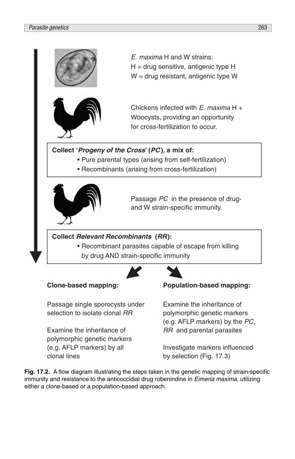
Chickens infected with E. maxima H +Woocysts, providing an opportunityfor cross-fertilization to occur.
E. maxima H and W strains:H = drug sensitive, antigenic type HW = drug resistant, antigenic type W
Passage PC in the presence of drug-and W strain-specific immunity.
Collect Relevant Recombinants (RR ): • Recombinant parasites capable of escape from killing by drug AND strain-specific immunity
Clone-based mapping:
Passage single sporocysts underselection to isolate clonal RR
Examine the inheritance ofpolymorphic genetic markers(e.g. AFLP markers) by allclonal lines
Population-based mapping:
Examine the inheritance ofpolymorphic genetic markers(e.g. AFLP markers) by the PC,RR and parental parasites
Investigate markers influencedby selection (Fig. 17.3)
Collect ‘Progeny of the Cross’ (PC ), a mix of: • Pure parental types (arising from self-fertilization) • Recombinants (arising from cross-fertilization)
Fig. 17.2. A flow diagram illustrating the steps taken in the genetic mapping of strain-specificimmunity and resistance to the anticoccidial drug robenindine in Eimeria maxima, utilizingeither a clone-based or a population-based approach.
Parasite genetics 263

occur. Among the first experimental evidence supporting the occurrence ofgenetic recombination within the eimerian genome was the generation of multi-drug-resistant parasites by co-infecting chickens with two differentially resistantstrains of E. maxima or E. tenella (Jeffers, 1974; Joyner and Norton, 1975,1976). Further reports based upon the recombination of loci associated withprecocious development and enzyme markers followed (Jeffers, 1976;Rollinson et al., 1979; Sutton et al., 1986).
Given evidence that a locus encoding a trait may be transferred geneticallybetween strains, that locus may be mapped within the donor genome, asdescribed for E. tenella (Shirley and Harvey, 2000). Observations of escapefrom strain-specific immunity by two strains of E. maxima in Line C chickensrevealed a phenotype akin to that of resistance/susceptibility to anticoccidialtreatment (Smith et al., 2002).
We therefore hypothesized that loci integral to escape from strain-specificimmune killing may be accessible through a genetic mapping strategy (Blake etal., 2004). Previous observations of recombination between genetic markers ofstrain-specific immunity and resistance to pyrimethamine in the murinemalarial parasite Plasmodium berghei supported the application of such anapproach (Oxbrow, 1973). A similar approach for the murine malarial parasiteP. chabaudi chabaudi has been independently reported elsewhere (Culleton etal., 2005; Martinelli et al., 2005).
Genetic mapping of loci encoding immunoprotective antigens of E. maxima
Generation and isolation of recombinant E. maxima
The phenotypically divergent H and W strains of E. maxima, characterized bydifferential escape from killing by strain-specific immunity and robenidine, wereused in these studies (Fig. 17.1; Blake et al., 2004). Oocysts excreted followingconcurrent infection with the E. maxima H and W strains were harvested in theusual manner (Long et al., 1976).
These parasites, termed the Progeny of the Cross (PC), were expected torepresent the progeny of self-fertilization by either strain, together with a poolof recombinant parasites arising from cross-fertilization (Fig. 17.2). Passageinfection with the PC through a selective double barrier consisting of strain-specific immunity lethal to the W parent and dietary robenidine lethal to the Hparent yielded a population of parasites recombinant at loci integral to escapefrom both barriers, proving in the process that these loci segregateindependently. Those parasites capable of escape from the double barrier weretermed Relevant Recombinants (RR).
Subsequent passage of a RR parasite population in the presence of eitherindividual component of the double barrier revealed that escape from neitherdrug, nor immunity, was fixed in the population after a single selective passage(Table 17.1). An important feature of the eimerian life cycle is that meiosisoccurs during sporulation, an environmental stage of the life cycle removedfrom selective pressures such as host immunity or chemotherapy. Hence, it
264 D.P. Blake et al.

appears that escape from killing by drug or immune selection are bothmultilocus traits which may be disrupted by resegregation. However, RR werealways isolated from the PC in the presence of double barrier selection, evenwhen using an oocyst dose as low as 100/bird, implicating a limited number ofgenetic loci in escape from both barriers (Blake et al., 2004). The finding thatRR escape the drug barrier better than the immune barrier suggests that thelarger proportion of those loci that encode molecules important for escape fromdouble barrier selection encode antigens exposed to deleterious strain-specificselection.
Genetic mapping analyses of populations
Classically, genetic mapping strategies with protozoan parasites have beenbased upon the clonal progeny of a genetic cross (Wellems et al., 1991; Shirleyand Harvey, 2000; Su et al., 2002; Hunt et al., 2004). However, whilstachievable through passage of a single sporocyst, cloning the Eimeria species isnot a trivial undertaking (Shirley and Harvey, 1996).
As an alternative we used an approach based upon populations of pure andrecombinant parasites generated and isolated under a variety of selection regimes(Fig. 17.2). Central to this approach is the hypothesis that genetic markers notphysically linked within the genome to loci under deleterious selection will beinherited by progeny parasites irrespective of the influence of that selection. Incontrast, a genetic marker that is physically linked to a locus under deleteriousselection close enough not to be separated by the majority of recombinationevents in a genetically diverse haploid population will be reduced, or lostaltogether, from the subset of that population capable of escaping the selection(see Fig. 17.3).
Whole-genome genetic fingerprinting of parasite populations using theamplified fragment length polymorphism (AFLP) technique generated markersfrom total genomic DNA extracted from oocysts of either parent and multipleindependent PC and RR lineages (Vos et al., 1995; Blake et al., 2003).Comparison between AFLP fingerprints revealed five patterns of markerinheritance (Fig. 17.4).
Table 17.1. Phenotypic characterisation of a Relevant Recombinant(RR) parasite population: escape from single and double barrierselection as a percentage of oocyst excretion following comparableinfection in the absence of selection. Birds exposed to a mixed doseof the parental parasites (H + W) demonstrated the integrity of eachbarrier (from Blake et al., 2004).
Parasite Barrier Escape (%)
RR Drug 72.4a
RR Immunity 39.4b
RR Drug + Immunity 21.1c
H+W Drug + Immunity 0.0d
Different superscript letters indicate significant differences (p < 0.05).
Parasite genetics 265

By far the most common pattern represented a non-polymorphic markerfound in both parental E. maxima strains which persisted in all progenypopulations, irrespective of selection (e.g. pattern 1, Fig. 17.4). The secondmost common patterns were strain-specific markers inherited by all progenypopulations (e.g. patterns 2 and 3, Fig. 17.4). The inheritance pattern of thesemarkers indicates no physical linkage to loci under drug or immune selection.
In contrast, a subset of markers (currently a total of 38) was clearlycorrelated with the deleterious effects of double barrier selection (e.g. pattern 4,Fig. 17.4). The use of single barriers (drug or W strain-specific immunity)allowed clear attribution of the markers to either of the two phenotypes presentin the double barrier. At present two markers display an inheritance pattern thatrelates to the drug resistant phenotype whereas 36 can be related to theimmune-escape phenotype.
This bias in marker association supports our earlier observations with thecapacity of RR to escape single barriers and suggests more independent loci
H1
H2
H3H4
H5
W1
W2W3W4W5W6
W1
W2W3W4W5W6
H1
H2
H3H4
H5
H1
H2
H3H4
H5
W1
W2W3W4W5W6
AFLP fingerprints: Hand W strain parents.
Parent-specificmarkers labelledH1–5 or W1–6
H W PC RR
PC fingerprint
All parentalmarkers
represented
RR fingerprint
All parental markersrepresented EXCEPT
those physically linked toloci under negative
selection by the doublebarrier (e.g. W1)
Genetic cross Double barrier selection
Fig. 17.3. AFLP analyses of populations. The inheritance of AFLP markers amplified fromthe parental H and W strains of Eimeria maxima used in the production of a genetic crosswas compared between the Progeny of the Cross (PC) and those recombinants within thePC capable of reproducing in the presence of barriers lethal to either individual parent (theRelevant Recombinants, RR). Genetic markers lost under selection by either strain-specificimmunity or dietary robenidine are physically linked to those loci under negative selection.
266 D.P. Blake et al.

encode strain-specific protective antigens than drug resistance. The markersassociated with immunity have been subjected to further analysis. Using acombination of: (i) probing Southern blotted digested parental genomic DNAresolved by pulsed field gel electrophoresis; and (ii) a BAC library generatedfrom the W strain parent, it is clear that the AFLP markers group in regions ofthe parasite genome. At present, markers can be linked to four regions of thegenome, supporting the premise that strain-specific protective immunity isrestricted to very few antigen-encoding loci.
CONCLUSION
For many years the Eimeria species have been subject to detailed study ofvariables that affect immune protection in hosts of differing genotype against
Ha P1.1b R1.1b P2.1a P2-Ima R2.1a R3.1a R3.2a R4.1a R5.1a R5.2a
1
2
3
4
Wa P2.2a P2-Da
Fig. 17.4. AFLP analysis of a population-based DNA mapping panel. H, Eimeria maxima Hstrain parent; W, E. maxima W strain parent; P, Progeny of the Cross (PC) population; R,Relevant Recombinant (RR) population. Lane identifiers: first number is the independentpopulation replicate number; second number is the generation number (under the selectiveconditions specified). -D, selected through the single barrier of dietary drug (66 ppm robenidine);-Im, selected through the single barrier of W strain-specific immunity. a Derived against a LineC host genetic background; b derived against a Line 151 host genetic background.
The characteristics of each of the five band patterns arising from AFLP analysis of the DNAmapping panel: 1, a non-polymorphic band consistently amplified from all parasite populations,regardless of selection regime. 2, a polymorphic band specific to the H strain; this band wasretained by all of the RR populations and therefore did not mark a region of the genomenegatively selected in the presence of dietary robenindine. 3, a polymorphic band specific to theW strain; this band was retained by all of the RR populations and therefore did not mark a regionof the genome negatively selected in the presence of W strain-specific immunity. 4, apolymorphic band specific to the W strain; amplified from all PC progeny populations andretained following exposure to the single barrier of robenidine; this band was lost followingexposure to the single barrier of W strain-specific immunity with or without robenidine; henceimplicated as a marker associated with immune killing of the W strain. 5 (not shown), apolymorphic band specific to the H strain; amplified from all PC progeny populations and retainedfollowing exposure to the single barrier of W strain-specific immunity; this band was lost followingexposure to robenidine; hence implicated as a marker associated with killing by robenidine.
Parasite genetics 267

homologous and heterologous parasite challenge (e.g. Rose and Long, 1962;Norton and Hein, 1976; Long and Millard, 1979; Shirley and Hoyle, 1981;Lillehoj, 1988; Bumstead and Millard, 1992; Fitz-Coy, 1992; Martin et al., 1997;Smith et al., 2002). More recently, techniques to characterize and manipulatenucleic acids have progressed rapidly (e.g. Jeffreys et al., 1985; Vos et al., 1995).Together, the combination of the discriminating strain-specific immune protectionexhibited by certain host lines of chicken and the ability to generate genome-widegenetic markers in the absence of detailed sequence data provided anopportunity to utilize immunological characters as genetic markers in the searchfor immunoprotective antigens (Walker, 1968; Oxbrow, 1973; Vos et al., 1995;Smith et al., 2002). The development of a mapping strategy targetingpopulations, not clones, has expedited these studies (Blake et al., 2004).
It is clear that many Eimeria antigens are recognized by the host immuneresponse but it is equally apparent that few of these confer protection. Thedevelopment of a genetic approach in identifying protective, antigen-encodingloci represents a major step forward in the development of new vaccinesprotective against the Eimeria species. Moreover, the proof of principle for thegenetic approach described here offers the opportunity to apply this methodologymore broadly in other parasitic diseases of veterinary and medical importance.
ACKNOWLEDGEMENTS
This work was primarily funded by the BBSRC (grant no. 201/S15343; M.W.S.and A.L.S.), with some support from DEFRA (grant code OD0534; A.L.S.).
The authors would like to thank members of the enteric immunology andparasitology groups for their help and input.
REFERENCES
Abrahamsen, M.S., Templeton, T.J., Enomoto, S., Abrahante, J.E., Zhu, G., Lancto, C.A., Deng,M., Liu, C., Widmer, G., Tzipori, S. et al. (2004) Complete genome sequence of theapicomplexan, Cryptosporidium parvum. Science 304, 441–445.
Bedrnik, P., Hiepe, T., Mielke, D. and Drossigk, U. (1995) Antigens and immunisation proceduresin the development of vaccines against poultry coccidiosis. COST 89/820. In: Eckert, J.,Braun, R., Shirley, M. and Coudert, P. (eds) Biotechnology: Guidelines on Techniques inCoccidiosis Research. European Commission, Luxembourg, pp. 176–189.
Berriman, M., Ghedin, E., Hertz-Fowler, C., Blandin, G., Renauld, H., Bartholomeu, D.C.,Lennard, N.J., Caler, E., Hamlin, N.E., Haas, B. et al. (2005) The genome of the Africantrypanosome Trypanosoma brucei. Science 309, 416–422.
Blake, D.P., Smith, A.L. and Shirley, M.W. (2003) Amplified fragment length polymorphismanalyses of Eimeria spp.: an improved process for genetic studies of recombinant parasites.Parasitology Research 90, 473–475.
Blake, D.P., Hesketh, P., Archer, A., Carroll, F., Smith, A.L. and Shirley, M.W. (2004) Parasitegenetics and the immune host: recombination between antigenic types of Eimeria maxima asan entree to the identification of protective antigens. Molecular and Biochemical Parasitology138, 143–152.
268 D.P. Blake et al.

Blake, D.P., Hesketh, P., Archer, A., Carroll, F., Shirley, M.W. and Smith, A.L. (2006) Theinfluence of immunizing dose size and schedule on immunity to subsequent challenge withantigenically distinct strains of Eimeria maxima. Avian Pathology 34, 489–494.
Bumstead, N. and Millard, B. (1992) Variation in susceptibility of inbred lines of chickens to sevenspecies of Eimeria. Parasitology 104, 407–413.
Chapman, H. (1976) Eimeria tenella: experimental studies on the development of resistance torobenidine. Parasitology 73, 265–273.
Chapman, H. (1997) Biochemical, genetic and applied aspects of drug resistance in Eimeriaparasites of the fowl. Avian Pathology 26, 221–244.
Cuckler, A.C. and Malanga, C.M. (1955) Studies on drug resistance in Coccidia. Journal ofParasitology 41, 302–311.
Culleton, R., Martinelli, A., Hunt, P. and Carter, R. (2005) Linkage group selection: rapid genediscovery in malaria parasites. Genome Research 15, 92–97.
El-Sayed, N.M., Myler, P.J., Bartholomeu, D.C., Nilsson, D., Aggarwal, G., Tran, A.N., Ghedin,E., Worthey, E.A., Delcher, A.L., Blandin, G. et al. (2005) The genome sequence ofTrypanosoma cruzi, etiologic agent of Chagas disease. Science 309, 409–415.
Fitz-Coy, S. (1992) Antigenic variation among strains of Eimeria maxima and E. tenella of thechicken. Avian Diseases 36, 40–43.
Gardner, M.J., Hall, N., Fung, E., White, O., Berriman, M., Hyman, R.W., Carlton, J.M., Pain, A.,Nelson, K.E., Bowman, S. et al. (2002) Genome sequence of the human malaria parasitePlasmodium falciparum. Nature 419, 498–511.
Hodgson, J.N., Ball, S.J., Ryan, K.C. and Warren, E.W. (1969) The incidence of drug-resistantstrains of Eimeria in chickens in Great Britain, 1966. British Veterinary Journal 125, 31–35.
Hunt, P., Martinelli, A., Fawcett, R., Carlton, J., Carter, R. and Walliker, D. (2004) Gene syntenyand chloroquine resistance in Plasmodium chabaudi. Molecular and BiochemicalParasitology 136, 157–164.
Ivens, A.C., Peacock, C.S., Worthey, E.A., Murphy, L., Aggarwal, G., Berriman, M., Sisk, E.,Rajandream, M.A., Adlem, E., Aert, R. et al. (2005) The genome of the kinetoplastidparasite, Leishmania major. Science 309, 436–442.
Jeffers, T.K. (1974) Genetic transfer of anticoccidial drug resistance in Eimeria tenella. Journal ofParasitology 60, 900–904.
Jeffers, T. (1976) Genetic recombination of precociousness and anticoccidial drug resistance inEimeria tenella. Zeitschrift fur Parasitenkunde 50, 251–255.
Jeffreys, A.J., Wilson, V. and Thein, S.L. (1985) Hypervariable ‘minisatellite’ regions in humanDNA. Nature 314, 67–73.
Joyner, L. (1957) Induced drug-fastness to nitrofurazone in a laboratory strain of Eimeria tenella.Journal of Parasitology 69, 1415.
Joyner, L.P. (1969) Immunological variation between two strains of Eimeria acervulina.Parasitology 59, 725–732.
Joyner, L. and Norton, C. (1975) Transferred drug-resistance in Eimeria maxima. Parasitology 71,385–392.
Joyner, L. and Norton, C. (1976) Further observations on transferred drug resistance in Eimeriamaxima. Parasitology, Abstracts 73, III.
Lee, E. and Fernando, M. (1978) Immunogenicity of a single sporocyst of Eimeria maxima.Journal of Parasitology 64, 483–485.
Lillehoj, H. (1988) Influence of inoculation dose, inoculation schedule, chicken age, and hostgenetics on disease susceptibility and development of resistance to Eimeria tenella infection.Avian Diseases 32, 437–444.
Long, P. and Millard, B. (1979) Immunological differences in Eimeria maxima: effect of a mixedimmunizing inoculum on heterologous challenge. Parasitology 79, 451–457.
Long, P., Joyner, L., Millard, B. and Norton, C. (1976) A guide to laboratory techniques used inthe study and diagnosis of avian coccidiosis. Folia Veterinaria Latina 6, 201–217.
Parasite genetics 269
Martin, A., Danforth, H., Barta, J. and Fernando, M. (1997) Analysis of immunological cross-protection and sensitivities to anticoccidial drugs among five geographical and temporalstrains of Eimeria maxima. International Journal for Parasitology 27, 527–533.
Martinelli, A., Cheesman, S., Hunt, P., Culleton, R., Raza, A., Mackinnon, M. and Carter, R.(2005) A genetic approach to the de novo identification of targets of strain-specific immunityin malaria parasites. Proceedings of the National Academy of Sciences USA 102, 814–819.
Melby, P., Ogden, G., Flores, H., Zhao, W., Geldmacher, C., Biediger, N., Ahuja, S., Uranga, J.and Melendez, M. (2000) Identification of vaccine candidates for experimental visceralleishmaniasis by immunization with sequential fractions of a cDNA expression library.Infection and Immunity 68, 5595–5602.
Melby, P., Yang, J., Zhao, W., Perez, L. and Cheng, J. (2001) Leishmania donovani p36(LACK)DNA vaccine is highly immunogenic but not protective against experimental visceralLeishmaniasis. Infection and Immunity 69, 4719–4725.
Norton, C. and Hein, H. (1976) Eimeria maxima: a comparison of two laboratory strains with afresh isolate. Parasitology 72, 345–354.
Norton, C. and Joyner, L. (1975) The development of drug-resistant strains of Eimeria maxima inthe laboratory. Parasitology 71, 153–165.
Oxbrow, A. (1973) Strain specific immunity to Plasmodium berghei: a new genetic marker.Parasitology 67, 17–27.
Pain, A., Renauld, H., Berriman, M., Murphy, L., Yeats, C.A., Weir, W., Kerhornou, A., Aslett, M.,Bishop, R., Bouchier, C. et al. (2005) Genome of the host-cell transforming parasite Theileriaannulata compared with T. parva. Science 309, 131–133.
Profous-Juchelka, H., Liberator, P. and Turner, M. (1988) Identification and characterization ofcDNA clones encoding antigens of Eimeria tenella. Molecular and Biochemical Parasitology30, 233–241.
Richie, T. and Saul, A. (2002) Progress and challenges for malaria vaccines. Nature 415,694–701.
Rollinson, D., Joyner, L. and Norton, C. (1979) Eimeria maxima: the use of enzyme markers todetect the genetic transfer of drug resistance between lines. Parasitology 78, 361–367.
Rose, M. and Long, P. (1962) Immunity to four species of Eimeria in fowls. Immunology 5,79–92.
Shibui, A., Ohmori, Y., Suzuki, Y., Sasaki, M., Nogami, S., Sugano, S. and Watanabe, J. (2001)Effects of DNA vaccine in murine malaria using a full-length cDNA library. ResearchCommunications in Molecular Pathology and Pharmacology 109, 147–157.
Shirley, M. and Bellatti, M. (1988) Live attenuated coccidiosis vaccine: selection of a secondprecocious line of Eimeria maxima. Research in Veterinary Science 44, 25–28.
Shirley, M. and Harvey, D. (1996) Eimeria tenella: infection with a single sporocyst gives a clonalpopulation. Parasitology 112, 523–528.
Shirley, M. and Harvey, D. (2000) A genetic linkage map of the apicomplexan protozoan parasiteEimeria tenella. Genome Research 10, 1587–1593.
Shirley, M. and Hoyle, S. (1981) The antigenicity of Eimeria maxima populations obtained fromcommercial farms. Journal of Parasitology 67, 587–588.
Smith, A., Hesketh, P., Archer, A. and Shirley, M. (2002) Antigenic diversity in Eimeria maximaand the influence of host genetics and immunisation schedule on cross-protective immunity.Infection and Immunity 70, 2472–2479.
Smooker, P., Setiady, Y., Rainczuk, A. and Spithill, T. (2000) Expression library immunisationprotects mice against challenge with virulent rodent malaria. Vaccine 18, 2533–2540.
Su, C., Howe, D.K., Dubey, J.P., Ajioka, J.W. and Sibley, L.D. (2002) Identification ofquantitative trait loci controlling acute virulence in Toxoplasma gondii. Proceedings of theNational Academy of Sciences USA 99, 10753–10758.
270 D.P. Blake et al.

Sutton, C.A., Shirley, M.W. and McDonald, V. (1986) Genetic recombination of markers forprecocious development, arprinocid resistance, and isoenzymes of glucose phosphate iso-merase in Eimeria acervulina. Journal of Parasitology 72, 965–967.
Vos, P., Hogers, R., Bleeker, M., Reijans, M., van de Lee, T., Hornes, M., Frijters, A., Pot, J.,Peleman, J., Kuiper, M. and Zabeau, M. (1995) AFLP: a new technique for DNA fingerprint-ing. Nucleic Acids Research 23, 4407–4414.
Walker, P. (1968) Investigational problems and the mechanisms of inheritance in blood protozoa.In: Weinman, D. and Ristic, M. (eds) Infectious Blood Diseases of Man and Animals: DiseasesCaused by Protista. Academic Press, New York, pp. 367–391.
Wallach, M., Smith, N.C., Petracca, M., Miller, C.M., Eckert, J. and Braun, R. (1995) Eimeriamaxima gametocyte antigens: potential use in a subunit maternal vaccine against coccidiosisin chickens. Vaccine 13, 347–354.
Warren, E., Ball, S. and MacKenzie, D. (1966) The incidence of drug-resistant strains of Eimeriaspecies in chickens in Great Britain, 1964/65. British Veterinary Journal 122, 534–543.
Wellems, T.E., Walker-Jonah, A. and Panton, L.J. (1991) Genetic mapping of the chloroquine-resistance locus on Plasmodium falciparum Chromosome 7. Proceedings of the NationalAcademy of Sciences USA 88, 3382–3386.
Xu, P., Widmer, G., Wang, Y., Ozaki, L.S., Alves, J.M., Serrano, M.G., Puiu, D., Manque, P.,Akiyoshi, D., Mackey, A.J. et al. (2004) The genome of Cryptosporidium hominis. Nature431, 1107–1112.
Parasite genetics 271

This page intentionally left blank

PART VI
Immunological and pathogen control

This page intentionally left blank

CHAPTER 18Developments and pitfalls of feedacidification in controlling gut pathogens in poultry, with emphasis on Salmonella
F. van Immerseel,1* I. Gantois,1 L. Bohez,1L. Timbermont,1 F. Boyen,1 I. Hautefort,2J.C.D. Hinton,2 F. Pasmans,1 F. Haesebrouck1
and R. Ducatelle1
1Department of Pathology, Bacteriology and Avian Diseases, ResearchGroup, Veterinary Public Health and Zoonoses, Faculty of VeterinaryMedicine, Ghent University, Ghent, Belgium; 2Molecular MicrobiologyGroup, Institute of Food Research, Norwich, UK; *e-mail: [email protected]
ABSTRACT
Acidification of drinking water and feed has been used for years in poultry.Drinking water acidification increases the threshold of bacterial infection due tothe bactericidal activity of the acids added. Feed supplementation with powderproducts also increases the threshold of bacterial infection due to itsbacteriostatic/bactericidal activity in the crop. For drinking water and powderform feed supplementation, the choice of the acids used is dependent on theirability to kill bacteria. Currently, coated or impregnated acid products are alsoon the market, with the aim of allowing acids to penetrate further down thegastrointestinal tract.
These products thus aim to change the ratio of organic acids in the gut,and thus the concentration changes achieved are more subtle. It was shownthat exposure of Salmonella to low concentrations of acetic acid increasedinvasion of the bacterium in intestinal epithelial cells, while propionic andbutyric acid decreased invasion at non-growth inhibitory concentrations.
This is due to changes in virulence gene expression of the genes of theSalmonella Pathogenicity Island I. Gene expression profiling by microarraytechnology has shown that low concentrations of butyric acid result in a specificdownregulation of Salmonella Pathogenicity Island I genes, directly responsiblefor invasion, while regular metabolic genes are not altered in expression,making the butyric acid effect highly specific.
© CAB International 2006. Avian Gut Function in Health and Disease (ed. G.C. Perry) 275

It was also shown that supplementation of acetic acid-coated products inthe feed increased colonization of the chicken gut by Salmonella, and thatcoated propionic and butyric acid products decreased colonization of thechicken intestinal tract. Moreover, it was shown that coated butyric aciddecreased faecal shedding in a seeder model in broilers throughout the rearingperiod up to slaughter age. The possibility of differential effects of organic acidson virulence of other pathogens and on performance of the animals is underresearch.
It was concluded that, instead of empirically using organic acid com-pounds as additives in feed and water of poultry, well-formulated and well-balanced preparations of organic acids should be developed, based onscientific data.
USE OF ORGANIC ACIDS IN POULTRY FEED
Organic acids have been widely used as feed and drinking water additives forvarious reasons in different farm animal species. Many products are on themarket, consisting of mixtures of organic acids in different concentrations andformulations. Organic and short-chain fatty acid (SCFA) mixtures have beenproduced aiming at enhancing digestibility and diet palatability, thus improvingfeed conversion and growth rates of animals, including pigs and poultry. Acidmixtures are sometimes included in feed to control mould, and claims even foregg production increases using acids have been made.
When searching the scientific, peer-reviewed literature however,information on these potential beneficial characteristics is lacking and it isimpossible to define the optimal acid mixture for achieving certain aims.Whatever the claims, it is clear that SCFA, medium-chain fatty acids (MCFA)and other organic acids have a more or less pronounced antimicrobial activity,depending on both the concentration of the acid and the bacterial species thatis exposed to the acid.
Indeed, for certain acids there is a large difference in minimal inhibitoryconcentrations (MIC) when different bacterial species are exposed, while forcertain bacterial species there are large differences in MIC values when thesebacteria are exposed to different organic acids (Nakai and Siebert, 2002).
Some organic acids that are used as feed additives, however, do not seemto have any significant antimicrobial activity in the concentration range that issuitable for use in feed. Short-chain fatty acids are bacteriostatic or bactericidalin vitro, provided that there are sufficient undissociated acid molecules presentand that they are in contact with the bacteria for a sufficiently long time(Thompson and Hinton, 1997). Lowering the pH increases the concentrationof undissociated molecules. Therefore, the products will be more efficient at lowpH.
This was proved in in vitro experiments by a decreased growth rate ofSalmonella Typhimurium at decreasing pH levels in the presence of volatilefatty acids (VFA) (Durant et al., 1999). The SCFA diffuse into the bacterial cellin undissociated form. Inside the bacterial cell the acid dissociates, resulting in
276 F. Van Immerseel et al.

reduction of intracellular pH and anion accumulation (Van der Wielen et al.,2000). Concentrations of VFA sufficient to cause growth inhibition ofEscherichia coli in vitro immediately slowed the rates of RNA, DNA, protein,lipid and cell wall synthesis (Cherrington et al., 1990). Since synthesis ofmacromolecules other than DNA is not completely inhibited, and since therewas no loss in membrane integrity, bacterial mass increased without celldivision in the presence of VFA (Cherrington et al., 1990, 1991).
Another common field of application for SCFA or other fatty acids is inpathogen control, especially Salmonella. This is the main focus of commercialproducts, claiming a (partial) control of shedding or colonization of the chickengut. Drinking water additives are widely applied, and it is clear that when theconcentration of certain acids in the drinking water is high, the pH of thedrinking water will be sufficiently low to kill the bacteria that are present. Thiswill thus create a barrier at the drinking water level, and recontamination of thebirds due to the presence of pathogens in the drinking water (e.g. from thelitter) can be controlled in this way to some extent. Powder-form feed additiveshave also been used widely to reduce Salmonella contamination.
The action of these compounds will not take place in the feed itself due tothe lack of water for carrying out their activity. Once taken up by the animalthey can become active. It is, however, the case that their action will most likelybe limited to the crop and the stomach, since it has been shown that onecannot detect the acids further down in the gastrointestinal tract due toresorption (Thompson and Hinton, 1997).
More recently, new technologies have allowed the development offormulations that release the acids further down the gastrointestinal tract. Theseformulations include acids coated on to or impregnated into microbeads.Whether this really is the case remains to be seen, but these kinds ofpreparations are surely capable of bringing probiotic strains into the gut ofanimals (Kailasapathy, 2002).
Considering the above-mentioned data, it is thus advisable that research iscarried out on the use of acids for a specific purpose. Preferably, the acidsshould be tested one by one prior to the study of the more complicated effectsof mixtures. In this way one can try to find the optimal composition andformulation designed for achieving a specific aim.
In this chapter, details are given on the evaluation of the SCFA formic,acetic, propionic and butyric acid for their ability to control Salmonella inpoultry. A study was conducted to assess the antimicrobial activity ofSalmonella and the effects of the single SCFA on Salmonella virulence geneexpression and invasion into intestinal epithelial cells. Furthermore, the effectsof the different single acids on early colonization of the caeca and internalorgans in young chickens were evaluated. The effect of the formulation inSalmonella control was tested using (powder form and impregnated) butyricacid, and long-term shedding of Salmonella was followed in broilers usingbutyric acid as a feed additive.
These data should provide information on how to optimize SCFApreparations for Salmonella control in poultry. Before detailed studies on theuse of SCFA in Salmonella control are presented, an introduction to this food-
Feed acidification and gut pathogens 277

contaminating pathogen and its pathogenesis is discussed, in order to underlinethe importance of having well-developed solutions for Salmonella control inpoultry and to inform the reader about some important characteristics of theSalmonella bacteria, which will be used below in the discussion on the use ofacids in Salmonella virulence.
SALMONELLA: AN IMPORTANT PATHOGEN IN POULTRY
Public health consequences
Salmonella is a foodborne pathogen for humans. Poultry is the main source ofSalmonella, causing human infections. The emergence of S. Enteritidis hasraised concern in the last two decades, due to its frequent transmission tohumans and its ability to cause severe disease in man. A major problem is thetropism of S. Enteritidis for the reproductive organs of the hen and thecontamination of eggs. Nevertheless poultry meat can also be contaminated.
In Europe, contamination of poultry is low in the Scandinaviancountries but higher in the Mediterranean region. While contamination ofbreeders is generally low, a considerably high level of positive layer flocks isdetected. In addition, Southern Europe has the highest percentage ofpositive flocks, ranging from 5 to 10%. Concerning table eggs, highpercentages of Salmonella-positive table eggs are reported in SouthernEurope (between 3 and 8%), while in the rest of Europe less than 1% of theeggs were positive.
The serotype Enteritidis is predominant in layers (57.7%) and in table eggs(72.9%) (European Commission, 2002). Broilers are also frequentlycontaminated by Salmonella. Scandinavian countries reported very lowpercentages of positive flocks, while Western European countries reportedhigher levels of contamination, with large differences between differentcountries, ranging between 1 and 11% in 2002.
Contamination levels of poultry meat are dramatically high in Europe. In2002, in all European countries except Scandinavia, about 10–15% of thepoultry meat at the retail level was positive. Also, Enteritidis is one of themost frequently isolated serotypes in broilers (10.8%) and in broiler meat(11.1%), next to a wide range of other serotypes (European Commission,2002).
In Europe most human Salmonella isolates are currently of the serotypeEnteritidis. In England and Wales S. Enteritidis isolates became the mostfrequent, with a percentage of 70.7% of human isolates in 1997. In recentyears, fortunately, there has been a general downward trend in the number ofhuman cases in Europe. In 2002, more than 145,000 cases of humansalmonellosis were reported in 15 member states of the EU, compared with afigure of more than 200,000 cases in 1997. In the EU and Norway, about 70%of all isolates in 2002 were of the serotype Enteritidis and 17% were ofserotype Typhimurium (European Commission, 2002), making it very clearthat these serotypes need be controlled.
278 F. Van Immerseel et al.

The intestinal phase of infections with broad host-range Salmonella serotypes
Salmonella bacteria can colonize a wide range of host species, dependent onthe serotype. Salmonella serotypes that are exclusively associated with oneparticular host species are referred to as being host-restricted, such as S.Gallinarum in poultry. Serotypes that usually induce a self-limitinggastroenteritis in a broad range of unrelated host species, whilst being capableof inducing systemic disease in a wide range of host animals, are termedunrestricted or broad host-range serotypes (Uzzau et al., 2000). Examples areS. Enteritidis and S. Typhimurium, thus these are the serotypes leading tohuman infections.
Chickens are usually infected by oral uptake of bacteria from theenvironment. Salmonella bacteria are able to survive gastric acidity and cantherefore pass through the stomach to reach the intestinal tract of the animal(Kwon and Ricke, 1998). In the chicken, the caeca are the predominant sites ofSalmonella colonization (Desmidt et al., 1997, 1998). The bacteria can adhereto intestinal epithelial cells by specific adhesion–receptor interactions in whichbacterial fimbriae and mannosylated residues on the mucous and resorptiveepithelial cells are involved (Dibb-Fuller et al., 1999; Vimal et al., 2000).
A crucial step in the pathogenesis of Salmonella is the process of invasioninto intestinal epithelial cells. Upon contact with intestinal epithelial cells,Salmonella bacteria inject a set of bacterial proteins into host cells via thebacterial Pathogenicity Island I (SPI-1) Type-3 secretion system (TTSS) (Zhouand Galán, 2001). Type-3 secretion systems are specialized, needle-likestructures located on the bacterial membrane whose core function is theinjection of bacterial proteins into eukaryotic cells (Hueck, 1998; Galán andCollmer, 1999; Cornelis and Van Gijsegem, 2000).
The Pathogenicity Island I of Salmonella is a large genetic element on thechromosome encoding multiple proteins, necessary to assemble a complexTTSS apparatus (structural proteins), regulatory proteins as well as someeffector proteins, injected by the needle complex into intestinal epithelial cells(Lostroh and Lee, 2001). The effector proteins of SPI-1 that are injected in theepithelial cells interact with host proteins of the intestinal epithelial cells.
The main effect of the injected proteins is the rearrangement of thecytoskeleton of the intestinal epithelial cells, in such a way that bacteria areengulfed by ruffles on the host cell membrane, resulting in uptake by theepithelial cell. This is an active process mediated by the Salmonella proteinsthat are injected in the intestinal epithelial cells. It is called invasion.
The proteins that are encoded by the Salmonella Pathogenicity Island Ialso play a role in the attraction of immune cells to the gut wall. Macrophagesmay take up bacteria penetrating through the caecal mucosa, which is the startof the systemic phase of the infection. This systemic phase of the infection andthe mechanism of egg contamination are not discussed here, but moreinformation on these topics can be read in review articles by Hensel (2000) andDe Buck et al. (2004).
Feed acidification and gut pathogens 279

Prevention and control
In 2003, the European Parliament and the Council of the European Unionintroduced Regulation (EC) No 2160/2003 to ensure that proper and effectivemeasures were taken to detect and control Salmonella and other zoonoticagents at all relevant stages of production, processing and distribution,particularly at the level of primary production – including in feed – in order toreduce their prevalence and the risk they pose to public health.
National control programmes should provide for the detection of zoonosesand zoonotic agents in accordance with certain minimal sampling require-ments. These programmes should also define the respective responsibilities ofcompetent authorities and specify the control measures to be taken followingthe detection of zoonoses and zoonotic agents, in particular to protect publichealth. For example, with broilers, national control programmes should includea policy that fresh poultry meat may not be placed on the market for humanconsumption unless Salmonella is absent in 25 g, starting from December2010. Thus the infection needs to be controlled.
Different measures are available to control Salmonella in poultry. It is notwithin the scope of this chapter to develop this topic in detail but, for theinterested reader, reviews on the use of vaccines and feed additives for controlof Salmonella in poultry are available (Van Immerseel et al., 2002, 2005). Inthe text below, the use of SCFA in Salmonella control is discussed, based ondata generated at the Department of Pathology, Bacteriology and AvianDiseases of the Faculty of Veterinary Medicine, Ghent University, Belgium.
SHORT-CHAIN FATTY ACIDS AND THEIR EFFECTS ON SALMONELLAVIRULENCE, WITH EMPHASIS ON INVASION
In vitro analysis of the antimicrobial characteristics of SCFA was performed bymaking growth curves at different pH values by Durant et al. (1999) and byVan Immerseel et al. (2003a) for S. Typhimurium and S. Enteritidis,respectively. Propionic acid was slightly more antimicrobial than were the otheracids. Antimicrobial activity of the SCFA was limited, if present at all, at pH 7. Itis concluded, therefore, that the antimicrobial activity against Salmonellacannot be used as a criterion in choosing an appropriate SCFA for use as afeed additive to control Salmonella in poultry.
Effects of pre-incubation of S. Enteritidis in growth medium, supplementedwith the different SCFA, on invasion of Salmonella into intestinal epithelial cellswere evaluated by different authors. Van Immerseel et al. (2003b) used humanintestinal epithelial cells (T84 cell line) for this purpose. The cells were grown incell culture plates until a confluent layer of cells was reached.
Simultaneously, for each SCFA, three Luria-broth (LB) solutions weremade with the following concentrations: 25, 50 and 100 mM. Luria-brothmedium without supplements was used as control. The pH of all solutions wasbrought to 6 and 7 by addition of NaOH and HCl, respectively. Osmolarity of
280 F. Van Immerseel et al.
all solutions was adjusted to the same value (610 mmol/kg) by addition of NaClin solutions with lower osmolarity. This was done because differences inosmolarity influence invasion of Salmonella (Galán and Curtiss, 1990).
The bacteria were grown for 20 h in LB, whereafter the suspension wasdiluted 1:50 in the SCFA solutions, the compositions of which were describedabove. After 4 h of incubation at 37°C suspensions were centrifuged and dilutedin cell culture medium. The cfu/ml value was determined by plating 6 � 20 �l ofa dilution series of the suspensions on BGA, whereafter the plates were incubatedovernight at 37°C. The suspensions were held at 4°C until they were used in theassay. The bacterial suspensions were diluted to a density of 5.106 cfu/ml.
The growth medium of the epithelial cells was then replaced by 1 ml of thediluted bacterial suspensions. This was centrifuged for 10 min at 1500 rpm,whereafter the plates were incubated for 1 h at 37°C and 5% CO2. Then cellswere rinsed three times, cell culture medium with gentamicin (50 �g/ml) wasadded to kill extracellular bacteria and the plates were incubated for 1 h at37°C and 5% CO2. Hereafter, cells were rinsed three times with PBS and lysedwith 1% Triton X-100 in distilled water. From this lysate, a tenfold dilutionseries was made. From each dilution, 6 � 20 �l were inoculated on BGA, todetermine the cfu/ml intracellular S. enteritidis.
As can be seen in Fig. 18.1, a decreased percentage of invaded bacteriawas detected when bacteria were pre-incubated in propionate- and butyrate-supplemented media at pH 6, relative to that of bacteria pre-incubated inacetate- and formate-supplemented medium. These differences were significant(p < 0.05) in all cases within the same concentration of the VFA. Thepropionate- and butyrate-exposed bacteria also invaded significantly less (p <0.05) than did the control bacteria. When the test was performed at pH 7,similar results were detected (data not shown).
The use of primary chicken caecal epithelial cells resulted in the sameconclusions (Van Immerseel et al., 2004). Moreover, unpublished data from ourlaboratory showed that the effects of the SCFA on Salmonella invasion arevalid for multiple serotypes, including Hadar and Infantis.
This phenomenon was explained by changes in expression patterns ofgenes of the Salmonella Pathogenicity Island I (SPI-1). As already discussedabove, the SPI-1 genes are directly involved in the invasive phenotype. Thegene island consists of regulatory genes, genes encoding structural componentsof the needle complex and genes encoding effector proteins that are inserted inthe eukaryotic cells. The hilA gene is the crucial regulator of the SPI-1 and is atranscriptional activator that can be influenced by numerous other regulatoryproteins.
The molecular mechanism of acetate-induced increases in invasion hasbeen elucidated. Lawhon et al. (2002) showed that acetate, after conversion toacetyl-phosphate, activates the BarA/SirA two-component system byphosphorylation of BarA and subsequently SirA. Activation of SirA results inenhanced transcription of hilA, the key regulator of SPI-1.
Our laboratory (Gantois et al., 2006) showed that hilA is downregulatedwhen Salmonella is grown in butyrate-supplemented growth media. This wasshown when plasmids were constructed with transcriptional fusions between
Feed acidification and gut pathogens 281

the promoter of hilA and the reporter genes luxCDABE, and thus real-timeluminescence measurement was used as a marker of hilA expression.
Furthermore, transcriptomic analysis of both S. Typhimurium and S.Enteritidis grown in media supplemented with low doses of butyric acidshowed a specific downregulation of the whole SPI-1 without alterations inmetabolic gene expression, compared with the control conditions. Also hilD, apositive regulator of hilA, and HU, a nucleoid-binding protein (and previouslyshown to be involved in virulence gene regulation), was downregulated(Gantois et al., 2006).
These data suggest that butyric acid or one of its metabolic intermediatesspecifically interacted with one of the Salmonella proteins that regulate SPI-1.Most probably a simple molecular mechanism is involved, as for acetate, butthe exact biochemical/molecular basis is still unclear.
To facilitate completion of the effects of SCFA on virulence, it has to bementioned that pre-incubation of Salmonella with SCFA also influences acidresistance and intracellular survival in macrophages (Kwon and Ricke, 1998).For example, acid resistance is increased when the bacteria are in contact for asufficiently long time with acetic acid. The exact relevance of these effects on invivo colonization needs to be clarified.
gcf gfgfgc gca
0
1
2
3
4
5
6
7
8
0 25 50 100
Inva
sion
(%
)
Concentration (mM)
b
b b
dc
e e
caad
Fig. 18.1. Salmonella Enteritidis bacteria were pre-incubated for 4 h at 37°C in LB mediumwith short-chain fatty acids concentrations of 25, 50 and 100 mM. Column 1, formic acid;Column 2, acetic acid; Column 3, propionic acid; Column 4, butyric acid. Following this, aninvasion assay was performed in 24-well cell culture plates. The pH of the growth mediumwas 6. Invasion was calculated as the percentage of invaded bacteria relative to the initialnumber of bacteria inoculated in one well of the cell culture plate (y-axis). Differentconcentrations of short-chain fatty acids are given in the x-axis. Values that do not share thesame superscripts are statistically significant different from each other (p < 0.05).
282 F. Van Immerseel et al.
Importance of invasion into intestinal epithelial cells in vivo
Since invasion of intestinal epithelial cells is influenced differently by thedifferent SCFA, a study was carried out to assess the exact role of invasion inboth the colonization of chicken gut and internal organs and shedding. The keyregulatory protein of SPI-1, hilA, was therefore deleted in the Salmonellachromosome, and the mutant was shown to have lost its ability to invadeepithelial cells in vitro. An in vivo study was thus carried out with the wild-typeS. Enteritidis strain and its hilA mutant strain (Bohez et al., 2006).
Therefore, 1-day-old chicks were randomly divided into two groups of 39and orally inoculated with 108 colony-forming units (CFU) of S. Enteritidis76Sa88 parent strain and S. Enteritidis �hilA mutant in a volume of 0.2 ml ofLB broth. At days 2, 4, 6, 9, 15, 20 and 28 post-hatch, cloacal swabs weretaken for the detection of Salmonella. Four chickens per group were euthanizedat days 3, 4, 6, 9, 15, 20 and 28 post-hatch and samples of caeca, liver andspleen were taken for bacteriological analysis.
As shown in Fig. 18.2, cloacal swabs taken 1 day after infection revealedthat all animals were positive after direct plating for both inoculated groups. Allchicks infected with S. Enteritidis 76Sa88 parent strain shed the bacteria infaeces throughout the study. The number of animals shedding the �hilA mutantbacteria in the faeces was significantly lower compared with the number infectedwith the parent strain from day 5 post-inoculation onwards. The chickens didnot shed the �hilA strain any more at 27 days post-inoculation.
Bacteriological analysis of the caeca showed peak values of about log10 8CFU/g organ at 2 days post-infection for both S. Enteritidis 76Sa88 parent strainand S. Enteritidis �hilA mutant (Fig. 18.3). Subsequently, the numbers of the�hilA mutant bacteria in the caeca decreased. Statistically significant differences(p < 0.005) between both groups were observed from day 5 following infection,with the exception of day 8 post-infection. At 3 weeks post-infection, the parentstrain still efficiently colonized the caeca (c. log10 6 CFU/g), whereas the birdsinfected with the �hilA mutant had largely cleared the infecting bacteria, asthese were detected only after enrichment At the age of 28 days only two out offour chicks were positive at enrichment level (Bohez et al., 2006).
It is thus concluded that intestinal epithelial cell invasion is important forlong-term colonization of the chicken gut and the shedding pattern. SinceSCFA have effects on invasion, this effect can have significance in vivo.
Use of short-chain fatty acids in Salmonella control in vivo
To study the use of SCFA to control Salmonella in vivo and to generate data onthe differences between the acids with respect to Salmonella control, it is ofutmost importance that these products are compared within the sameexperiment using standardized infection protocols.
In an experiment using SPF layer chickens, the different SCFA werecompared using the micro-encapsulated form. For all products, 0.5% ofmicropearls was included in the feed, so that final in-feed concentrations of
Feed acidification and gut pathogens 283

SCFA were 0.217% for formic acid, 0.242% for acetic acid, 0.275% forpropionic acid and 0.156% for butyric acid. For this experiment, the chickenswere randomly divided into five groups each of 20 chickens.
From the day of hatch, four groups received feed supplemented with theabove-described feed additives, while one group received unsupplementedfeed. The animals were orally inoculated with 2�103 cfu S. Enteritidis 76Sa88on days 5 and 6. At day 7, cloacal swabs were taken from five animals pergroup to detect Salmonella bacteria. At day 8, chickens were euthanased byintravenous T61 injection. Samples of caecum, liver and spleen were taken forbacteriological analysis. One day after the second infection, 4/5 animals of thegroup that had received acetic acid as feed supplement had positive cloacalswabs, compared with none of the animals in any of the other groups.
Table 18.1 shows the number of animals classified according to caecalbacterial count of Salmonella (log-scaled intervals). In the control group, thecaeca of 6/20 animals were positive only after enrichment, while 9/20 animalshad > 106 cfu/g Salmonella in their caeca. In the group that had receivedformic acid as feed supplement, 10/20 animals had > 106 cfu/g Salmonella inthe caeca, and only one animal was positive after enrichment. Thus, a highernumber of animals in the formic acid-treated group had intermediateSalmonella titres, compared with the control animals.
In the group receiving acetic acid as feed supplement, 16/20 animals had aSalmonella count of > 106 cfu/g, from which 13 had > 107 cfu/g caecum. In
n = 39 n = 27 n = 19 n = 14 n = 10 n = 6 n = 3
n = 8
n = 12n = 16
n = 20
n = 30
n = 39
n = 25
0
20
40
60
80
100
120
0 5 10 15 20 25 30
Age (days)
Pos
itive
faec
al s
hedd
ing
(%)
Fig. 18.2. Percentages of chickens having a Salmonella-positive cloacal swab. Animals wereinfected at day of hatch with 108 cfu S. Enteritidis 76Sa88 parent strain (● direct plating) and108 cfu S. Enteritidis �hilA mutant (� direct plating, � after enrichment). The numbers ofchickens are represented by n.
284 F. Van Immerseel et al.

the propionic acid-supplemented group, seven animals had > 106 cfu ofSalmonella/g caecum, while 8/20 animals were only positive after enrichment.Finally, in the group receiving butyric acid-impregnated microbeads, noanimals had a caecal Salmonella count of > 106 cfu/g, while 11/20 animalswere positive only after enrichment.
Table 18.2 shows the number of animals classified according to bacterialcounts of Salmonella in liver and spleen. In the control and the butyric acid
0
2
4
6
8
10
12
0 5 10 15 20 25 30
Age (days)
log 10
CF
U/g
cae
ca
Fig. 18.3. Log10 cfu of S. Enteritidis 76Sa88 parent strain (●) and S. Enteritidis �hilA mutant(�) in the caeca of chicks dosed orally at 1 day of age and then euthanased at different timeintervals. Each time point represents four chicks, with the exception of three chicks at days20 and 28 in the S. Enteritidis 76Sa88 parent strain-infected group.
Table 18.1. Colonization of the caeca at day 8 of life (inoculation with 103 cfu S. Enteritidis76Sa88 on days 5 and 6) in chickens fed a diet supplemented with formic, acetic, propionicand butyric acids or no feed additives (control). Figures represent numbers of chickens in agroup of 20 having a given number of Salmonella bacteria in the caeca.
Controla Formic acida Acetic acidb Propionic acidac Butyric acidc
Negative 0 0 0 0 0Positive after enrichment 6 1 1 8 11102–103 cfu/g 0 1 1 1 2103–104 cfu/g 0 4 0 1 1104–105 cfu/g 3 2 0 2 3105–106 cfu/g 2 2 2 1 3106–107 cfu/g 8 7 3 3 0> 107 cfu/g 1 3 13 4 0
a,b,c Groups not having equal superscripts are statistically significantly different.
Feed acidification and gut pathogens 285

groups only a few animals had a bacterial count of > 102 cfu/g of spleen, butfive animals in the propionic acid group had Salmonella numbers between 102
and 103 cfu/g spleen. In the formic acid group, 11/20 animals had aSalmonella count of > 102 cfu/g, of which six had a count of > 103 cfu/g andone of more than 104 cfu/g. In the acetic acid group, 17/20 animals had > 102
cfu/g spleen, of which 13 had > 103 cfu/g, seven more than 104 cfu/g and twoanimals had even more than 105 cfu/g spleen. For the liver, comparable resultswere obtained.
Mean log cfu/g of caeca, liver and spleen for the different animal groupsare shown in Fig. 18.4. The mean log cfu S. Enteritidis/g caecum wassignificantly higher in the acetic acid-treated group than in all other groups. Thebutyric acid-treated group had a significantly lower mean log cfu/g caecumthan all other groups, except for the propionic acid-treated group. For liver andspleen, the acetic and formic acid-treated groups had significantly higher valuesof mean log cfu/g compared with all other groups.
Butyric acid thus decreases the invasion of Salmonella in vitro anddecreases Salmonella colonization of the caeca in chickens early after infectionof 1-day-old chicks. It was not yet known whether the formulation of thebutyric acid feed additive affects the protective effect. Therefore, an infectionexperiment was performed with young chickens to study the differencesbetween powdered and coated butyric acid as protection against Salmonellacolonization of the caeca and internal organs of chickens.
In an experimental study, SPF chickens were randomly divided into fourgroups, each of 25 chickens. From the day of hatch, three groups received feedsupplemented with different additives. The first group received the powderedform of butyric acid at a concentration of 0.63 g/kg. The second group receivedthe coated product at a concentration of 2.5 g/kg. The third group received0.315 g/kg of the powdered and 1.25 g/kg of the coated product.
All three groups thus received the same amount of the active productbutyric acid (or sodium salt) i.e. 0.63g/kg. One group received unsupplemented
Table 18.2. Colonization of the liver and spleen at day 8 of life (inoculation with 103 cfu S.Enteritidis 76Sa88 on days 5 and 6) in chickens fed a diet supplemented with formic, acetic,propionic and butyric acids or no feed additives. Figures represent numbers of chickens in agroup of 20 having a given number of Salmonella bacteria in the liver (L) and spleen (S).
Formic Acetic Propionic ButyricControl acid acid acid acid
La Sa Lbd Sb Lb Sc Lad Sa La Sa
Negative 0 8 0 0 0 0 1 5 0 7Positive after enrichment 19 10 11 9 8 3 14 10 20 12102–103 cfu/g 0 0 6 4 6 4 5 5 0 1103–104 cfu/g 1 1 3 6 3 6 0 0 0 0104–105 cfu/g 0 1 0 1 2 5 0 0 0 0> 105 cfu/g 0 0 0 0 1 2 0 0 0 0
a,b,c,d Within each organ, groups not having equal superscripts are statistically significantlydifferent.
286 F. Van Immerseel et al.

feed. The animals were orally inoculated with 106 cfu S. Enteritidis 76Sa88 onday 5. At day 6, cloacal swabs were taken to detect Salmonella bacteria. At day8, chickens were euthanased and samples of caecum, liver and spleen weretaken for bacteriological analysis.
Cloacal swabs taken 1 day post-infection showed that 10/25 animals hadshed Salmonella in the control group, 8/25 animals in the group receiving thepowdered form of butyric acid, 4/25 in the group receiving coated butyric acidand 3/25 in the group receiving the combined product as feed additive.
Caecal counts revealed more distinct differences. As can be seen fromTable 18.5, in the group of animals receiving the coated form of butyric acid orthe combination of the powdered and coated forms, a small number of animalshad > 106 cfu/g caecum (4 and 3/25, respectively). The control group and thegroup receiving the powdered form of butyric acid had a rather higher numberof animals with > 106 cfu/g caecum (10 and 12/25, respectively). Colonizationof the caeca by groups that had received coated butyrate or the combinedpowdered/coated form was significantly lower than either the control group orthe group that had received the powdered form of butyrate.
Concerning splenic colonization, there was a significantly lowercolonization of the group that had received the combined butyrate product asfeed additive compared with the control group or the group that had received
0
1
2
3
4
5
6
7
8
9
Con For Ace But Prop
Treatment
Mea
n lo
g cf
u/g
a
aa
a
a
aa
bcc
b
b
c
bac
ac
Fig. 18.4. The mean log cfu/g in caeca (black), liver (white) and spleen (grey) at day 8 oflife of chickens that were orally inoculated with 103 cfu S. Enteritidis 76Sa88 on days 5 and6. Chickens received diets supplemented with formic acid (For), acetic acid (Ace),propionic acid (Prop), butyric acid (But) and non-supplemented feed (Con). Betweenconditions, for each organ, values not having the same superscripts are statisticallysignificantly different.
Feed acidification and gut pathogens 287

the powdered form of butyrate (Table 18.4). Liver colonization was similar(data not shown).
From the data above, it can be concluded that butyric acid, impregnated inmicrobeads, decreases colonization of Salmonella in poultry and also reducesshedding. In these trials colonization was analysed only shortly after infection inlayer-type chicks. In a further study the protective effect in broilers wasexamined and this was performed over a longer period.
For this, Ross broiler chickens were randomly divided into two groups of50 chickens each. From the day of hatch, one of the groups received a wheat-based starter diet until day 10, after which the diet was changed to a wheat-based grower diet. In the other group the feed was supplemented with 2.5 g/kg
Table 18.3. Colonization of the caeca at day 8 of life (inoculation with106 cfu S. Enteritidis 76Sa88 on day 5) in chickens fed a diet supple-mented with butyric acid in powdered form, coated form, a combinationof half-doses of powdered and coated forms (Combi) or no feed addi-tives (Control). Concentration of the active product butyric acid was0.63 g/kg feed in each group. Figures represent numbers of chickens ina group of 25 having a given number of Salmonella bacteria in thecaeca.
Controla Powdereda Coatedb Combib
Negative 0 0 2 1Positive after enrichment 6 8 12 8< 104 cfu/g 2 1 3 2104–105 cfu/g 3 1 0 6105–106 cfu/g 4 3 4 5> 106 cfu/g 10 12 4 3
Groups with different superscripts are significantly different.
Table 18.4. Colonization of the spleen at day 8 of life (inoculation with106 cfu S. Enteritidis 76Sa88 on day 5) in chickens fed a diet supple-mented with butyric acid in powdered form, coated form, a combinationof half-doses of powder and coated form (Combi) or no feed additives(Control). Concentration of the active product butyric acid was 0.63 g/kgfeed in each group. Figures represent numbers of chickens in a groupof 25 that were negative or had a given amount of Salmonella bacteriain the spleen.
Controla Powdereda Coateda Combib
Negative 12* 10 12 15Positive after enrichment 2 1 4 7< 103 cfu/g 3 3 1 2103–104 cfu/g 6 10 4 1> 104 cfu/g 2 1 4 0
a,b Groups not having equal superscripts are statistically significantlydifferent (p < 0.05).
288 F. Van Immerseel et al.

of the coated butyric acid product. Ten out of 50 animals of both groups wereorally inoculated with 105 cfu S. Enteritidis 76Sa88 at day 5 post-hatch. Ondays 6, 9, 13, 20, 27, 34 and 41 cloacal swabs were taken for bacteriologicalanalysis. At day 42, the animals were killed by intravenous injection of ananaesthetic and samples of caeca were taken for bacteriological analysis.
In the control group, the percentage of chickens having Salmonella-positive cloacal swabs after direct plating increased to 35–40% at 1 and 3weeks after seeder bird inoculation (days 12 and 27 of age). At slaughter,however, only 4.8% of birds were positive in direct plating. In the group ofanimals that had received butyric acid as feed additive, 15% of the animals hadSalmonella-positive cloacal swabs after direct plating 1 and 3 weeks afterseeder bird inoculation (days 12 and 27 of age), whereas the percentage ofanimals having Salmonella-positive cloacal swabs after direct plating decreasedto 2.2% at slaughter age (Fig. 18.5). Differences between both groups werestatistically significant at day 9 (p < 0.1) and days 12 and 27 (p < 0.05).
Analysis of total numbers of positive cloacal swabs (including those positiveat enrichment level) showed an increase to 97% in the proportion of chickenshaving Salmonella-positive cloacal swabs at day 20 of age (day 15 post-inoculation of seeders) in the control group. Thereafter, a decrease to 36% wasobserved by slaughter age. In the group of animals that received butyric acid asfeed additive the percentage of chickens having Salmonella-positive cloacalswabs increased to 48% at day 20 (day 15 post inoculation of seeders) andthen decreased to 6% at slaughter age. Differences between both groups werestatistically significant at all time points starting from day 9 of age (day 4 post-inoculation of seeder birds; p < 0.05).
Bacteriological analysis of caeca at slaughter age showed 68% of theanimals as being positive for Salmonella at enrichment level (no directpositives) in both the control group and the group that had received butyricacid as a feed additive.
CONCLUSION
Organic acids, and in particular SCFA, are currently being used as feedadditives in a wide range of applications. For most applications, combinationsof acids are empirically chosen and placed on the market. Data presented inthis chapter clearly illustrate that, for Salmonella control, both the nature of theacid and the formulation of the feed additive play a significant role. Indeed,while butyric acid decreased virulence gene expression and invasion ofSalmonella in intestinal epithelial cells, acetic acid increased both virulencegene expression and invasion.
When coated organic acids were used as feed additives, the differencesbetween acetic and butyric acid concerning effects on the potential ofSalmonella to invade epithelial cells were reflected in differences in colonizationof the caeca in chickens. Indeed, coated acetic acid in the feed resulted in amore than a 10,000-fold increase in the number of bacteria colonizing thecaeca in comparison with coated butyric acid, and 100-fold increases
Feed acidification and gut pathogens 289

0
5
10
15
20
25
30
35
40
45
50
0 2 4 6 8 10 12 14 16 18 20 22 24 26 28 30 32 34 36 38 40 42
Pos
itive
afte
r di
rect
pla
ting
(%)
*
**
0
10
20
30
40
50
60
70
80
90
100
0 2 4 6 8 10 12 14 16 18 20 22 24 26 28 30 32 34 36 38 40 42
Age (days)
Pos
itive
afte
r en
richm
ent (
%)
*
*
* *
* *
a
b
Fig. 18.5. The number of positive cloacal swabs during time (days) is shown after directinoculation on brilliant green agar (a) and after enrichment (b). Infection was carried out usinga seeder model, infecting ten animals with 105 cfu S. Enteritidis 76Sa88 at day 5 post-hatch,and housing these animals with 40 non-inoculated broilers. Animals were given feedsupplemented with coated butyric acid at 0.63 g/kg butyric acid (�) or unsupplemented feed(●). * Denotes significant differences between groups.
290 F. Van Immerseel et al.

compared with control animals. In the powdered form, butyric acid did not aidin controlling Salmonella colonization when animals were infected orally, incontrast to the coated form.
This clearly showed that the formulation is a very important feature thathas to be optimized. Although the formulation of feed additives in control cancertainly still be improved, it seems to be the case that coated propionic orbutyric acid should be included in these preparations. Acetic acid needs to beavoided for this purpose, based on the above-mentioned results.
One aspect that certainly needs further attention in the future is the role ofthe microbiota in the endogenous production of SCFA and the way in whichgut microbiota can be shifted to become butyric acid producing. It seems to bethe case that Salmonella takes advantage of the large amount of acetic acid inthe gut and responds with increased invasion of the gut cells. Butyric acidfermentation can be improved by manipulation of the feed, and by theaddition of proper prebiotic substrates for certain microbiotic populations.
As an alternative to the use of SCFA preparations in the feed, simple feedcomposition changes could be used to improve Salmonella control. Also, theimpact of SCFA on the virulence of other bacterial pathogens, such asClostridium perfringens, needs to be clarified.
Finally, the effect of changes in acid composition in the gut on the gutepithelial cells can also have an influence on the outcome of an infection andon performance parameters. It has been shown in humans and mice thatintestinal epithelial cell proliferation is increased and that gut damage isresolved faster when butyric acid is present, thus improving digestibility (Hodin,2000).
REFERENCES
Bohez, L., Ducatelle, R., Pasmans, F., Botteldoorn, N., Haesebrouck, F. and Van Immerseel, F.(2006) Salmonella enterica serovar enteritidis colonization of the chicken caecum requiresthe hila regulatory protein. Veterinary Microbiology 116, 202–210.
Cherrington, C.A., Hinton, M. and Chopra, I. (1990) Effect of short-chain organic acids onmacromolecular synthesis in Escherichia coli. Journal of Applied Bacteriology 68, 69–74.
Cherrington, C.A., Hinton, M., Pearson, G. and Chopra, I. (1991) Short-chain organic acids atpH 5.0 kill Escherichia coli and Salmonella spp. without causing membrane perturbation.Journal of Applied Bacteriology 70, 161–165.
Cornelis, G.R. and Van Gijsegem, F. (2000) Assembly and function of type III secretory systems.Annual Reviews in Microbiology 54, 735–774.
De Buck., J., Van Immerseel, F., Haeseboruck, F. and Ducatelle, R. (2004) Colonization of thechicken reproductive tract and egg contamination by Salmonella. Journal of AppliedMicrobiology 97, 233–245.
Desmidt, M., Ducatelle, R. and Haesebrouck, F. (1997) Pathogenesis of Salmonella Enteritidisphage type four after experimental infection of young chickens. Veterinary Microbiology 56,99–109.
Desmidt, M., Ducatelle, R. and Haesebrouck, F. (1998) Immunohistochemical observations in thecaeca of chickens infected with Salmonella Enteritidis phage type four. Poultry Science 77,73–74.
Feed acidification and gut pathogens 291

Dibb-Fuller, M.P., Allen-Vercoe, E., Thorns, C.J. and Woodward, M.J. (1999) Fimbriae- andflagella-mediated association with and invasion of culture epithelial cells by SalmonellaEnteritidis. Microbiology 145, 1023–1031.
Durant, J.A., Lowry, V.K., Nisbet, D.J., Stanker, L.H., Corrier D.E. and Ricke, S.C. (1999) Short-chain fatty acids affect cell-association and invasion of Hep-2 cells by SalmonellaTyphimurium. Journal of Environmental Science and Health B 34, 1083–1099.
European Commission (2002) Trends and sources of zoonotic agents in animals, feedingstuffs,food and man in the European Union and Norway in 2002. Health and ConsumerProtection Directorate-General. Salmonella and Food-borne Diseases – Zoonoses Reports for2002.
Galán, J.E. and Collmer, A. (1999) Type III secretion machines: bacterial devices for proteindelivery into host cells. Science 284, 1322–1328.
Galán, J.E., Curtiss, R. 3rd. (1990) Expression of Salmonella Typhimurium genes required for inva-sion is regulated by changes in DNA supercoiling. Infection and Immunity 58, 1879–1885.
Gantois, I., Ducatelle, R., Pasmans, F., Haesebrouck, F., Hautefort, I., Thompson, A., Hinton, J.C.and Van Immerseel, F. (2006) Butyrate specifically down-regulates Salmonella pathogenicityisland 1 gene expression. Applied Environmental Microbiology 72, 946–949.
Hensel, M. (2000) Salmonella Pathogenicity Island 2. Molecular Microbiology 36, 1015–1023.Hodin, R. (2000) Maintaining gut homeostasis: the butyrate NF-B connection. Gastroenterology
118, 798–801.Hueck, C.J. (1998) Type III secretion systems in bacterial pathogens of animals and plants.
Microbiology and Molecular Biology Reviews 62, 379–433.Kailasapathy, K. (2002) Microencapsualtion of probiotic bacteria: technology and potential appli-
cations. Current Issues in Intestinal Microbiology 32, 39–48.Kwon, Y.M. and Ricke, S.C. (1998) Induction of acid resistance of Salmonella Typhimurium by
exposure to short-chain fatty acids. Applied and Environmental Microbiology 64, 3458–3463.Lawhon, S.D., Maurer, R., Suyemoto, M. and Altier, C. (2002) Intestinal short-chain fatty acids
alter Salmonella Typhimurium invasion gene expression and virulence through BarA/SirA.Molecular Microbiology 46, 1451–1461.
Lostroh, C.P. and Lee, C.A. (2001) The Salmonella Pathogenicity Island-1 type III secretionsystem. Microbes and Infection 3, 1281–1291.
Nakai, S.A. and Siebert, K.J. (2002) Validation of bacterial growth inhibition models based onmolecular properties of organic acids. International Journal of Food Microbiology 2678, 1–7.
Thompson, J.L. and Hinton, M. (1997) Antibacterial activity of formic acid and propionic acid inthe diet of hens on Salmonellae in the crop. British Poultry Science 38, 59–65.
Uzzau, S., Brown, D.J., Wallis, T., Rubino, S., Leori, G., Bernard, S., Casadesús, J., Platt, D.J.and Olsen, J.E. (2000) Host-adapted serotypes of Salmonella enterica. Epidemiology andInfection 125, 229–255.
Van der Wielen, P.W., Biesterveld, S., Notermans, S., Hofstra, H., Urlings, B.A. and van Knapen,F. (2000) Role of volatile fatty acids in development of the cecal microflora in broilerchickens during growth. Applied and Environmental Microbiology 66, 2536–2540.
Van Immerseel, F., Cauwerts, K., Devriese, L.A., Haesebrouck, F. and Ducatelle, R. (2002) Feedadditives to control Salmonella in poultry. World’s Poultry Science Journal 58, 501–513.
Van Immerseel, F., De Buck, J., Pasmans, F., Velge, P., Bottreau, E., Fievez, V., Haesebrouck, F.and Ducatelle, R. (2003a) Invasion of Salmonella Enteritidis in avian intestinal epithelial cellsin vitro is influenced by short-chain fatty acids. International Journal of Food Microbiology85, 237–248.
Van Immerseel, F., De Buck, J., Pasmans, F., Rychlik, I., Volf, J., Sevcik, M., Haesebrouck, F. andDucatelle, R. (2003b) The choice of volatile fatty acids for supplementation of poultry feed toreduce Salmonella. 14th European Symposium on Poultry Nutrition, Lillehammer, Norway,pp. 160–161.
292 F. Van Immerseel et al.

Van Immerseel, F., De Buck, J., De Smet, I., Pasmans, F., Haesebrouck, F. and Ducatelle, R.(2004) Interaction of butyric and acetic acid-treated Salmonella with chicken primary caecalepithelial cells in vitro. Avian Diseases 48, 384–391.
Van Immerseel, F., Methner, U., Rychlik, I., Nagy, B., Velge, P., Martin, G., Foster, N., Ducatelle,R. and Barrow, P.A. (2005) Vaccination and early protection against non-host-specificSalmonella serotypes in poultry; exploitation of innate immunity and microbial metabolicactivity. Epidemiology and Infection 133, 959–978.
Vimal, D.B., Khullar, M., Gupta, S. and Ganguly, N.K. (2000) Intestinal mucus: the binding sitesfor Salmonella Typhimurium. Molecular and Cellular Biochemistry 204, 107–117.
Zhou, D. and Galán, J. (2001) Salmonella entry into host cells: the work in concert of type IIIsecreted effector proteins. Microbes and Infection 3, 1293–1298.
Feed acidification and gut pathogens 293

CHAPTER 19Competitive exclusion in poultryproduction
C. SchneitzOrion Corporation, Orion Pharma Animal Health, Turku, Finland; e-mail: [email protected]
ABSTRACT
The concept of Competitive Exclusion or CE, sometimes also described as the‘Nurmi concept’, involves treatment of newly hatched chickens with mixedbacterial preparations originating from the gut of healthy adult birds to controlintestinal disturbances. The treatment has also been given successfully to olderbirds after therapeutic doses of antibiotics to regenerate the intestinalmicroflora. CE treatment is usually applied by spraying, but can also be givenvia the first drinking water.
The concept of CE was originally developed to control Salmonella ingrowing chickens but has, with time, been expanded to involve several otherenteropathogens like chicken and human pathogenic Escherichia coli,Clostridium perfringens, Listeria and Campylobacter. The chicken intestinalflora has also been shown to be efficacious in other avian species like turkey,quail and pheasant. The mechanisms by which the intestinal bacteria inhibitthe invasion and proliferation of enteropathogens include competition forreceptor sites, elaboration of inhibitory compounds and competition fornutrients or stimulation of the immune system.
In addition to pathogen control, it has been shown in field studies that CEtreatment enhances the growth and decreases the mortality of birds andimproves the feed conversion. The nutritional effects of the treatment havebeen confirmed in well-controlled laboratory studies. Increased bacterialresistance to antibiotics in humans has caused an increase in public and officialinterest in abandonment of the use of growth-promoting antibiotics.Competitive exclusion is a reliable alternative because the treatment is fullybiological, it does not leave any residues and only one treatment on the day ofhatch is normally enough.
294 © CAB International 2006. Avian Gut Function in Health and Disease (ed. G.C. Perry)

INTRODUCTION
In 1971 the Finnish broiler industry suffered from a severe outbreak ofSalmonella infantis. More than 80% of the broiler flocks were Salmonellapositive. At the same time the incidence of human cases caused by the sameserotype increased dramatically. The problem with Salmonella-infected poultryflocks could not be solved using antibiotics, so research was started at theNational Veterinary and Food Research Institute under the leadership ofProfessor Esko Nurmi to find a different approach.
In 1952 Milner and Schaffer had infected chicks with S. Typhimurium andobserved that the resistance of young chickens to Salmonella increased withage, but the researchers had no explanation for this. The theory of Nurmi wasthat newly hatched chickens are sensitive to infection by Salmonella and otherenteropathogens because of delayed establishment of normal intestinalmicroflora due to lack of contact with the brooding hen. Nurmi and hisresearch group were the first to demonstrate experimentally that administrationof intestinal contents from a healthy adult hen to newly hatched chickensprotected the latter from colonization by Salmonella. This finding waspublished in Nature in 1973 (Nurmi and Rantala), and triggered worldwideresearch around this subject.
The competitive exclusion concept can be defined as ‘the earlyestablishment of an adult-type intestinal microflora to prevent subsequentcolonization by enteropathogens’. The concept was originally designed toprevent the colonization of foodborne Salmonellae in the gut of youngchickens but has, with time, been extended also to cover other human andpoultry pathogens like chicken and human pathogenic E. coli, Campylobacter,Cl. perfringens and Listeria. Improvement in bird performance has also beenshown in well-controlled laboratory-scale studies and also in the field (reviewedby Pivnick and Nurmi, 1982; Schleifer, 1985; Mead and Impey, 1987; Stavricand D’Aoust, 1993; Mead, 2000; Schneitz and Mead, 2000; Schneitz, 2005).
In this chapter, the development and applicability of the CE concept inpoultry production – as well as factors affecting the efficacy of the treatment infield conditions – are reviewed and discussed.
THE DEVELOPMENT OF THE CE CONCEPT
The phenomenon by which the mature intestinal microflora protects the hostagainst invading pathogens is called competitive exclusion (CE), or the ‘Nurmiconcept’ (Pivnick and Nurmi, 1982). Greenberg was the first to use the term‘competitive exclusion’, in 1969 (Greenberg, 1969). He studied the intestinalflora of maggots of blowflies and claimed that competitive exclusion of S.Typhimurium from the maggots was so effective that the organism survived inthe gut only if the normal microbiota was simplified or eliminated.
Luckey (1963) had demonstrated a similar phenomenon in higher animals.Colonization resistance, a term synonymous with CE, was introduced by vander Waaij et al. in 1971 when examining the intestinal flora of mice. The term
Competitive exclusion 295

competitive exclusion was used in relation to poultry for the first time by Lloydet al. in 1974.
Mixed cultures
In their first study, Nurmi and Rantala used diluted material from the crop,small intestine and caeca of adult, healthy cocks (Nurmi and Rantala, 1973).The results of this study showed that 1-day-old chicks became very wellprotected against relatively high doses of S. infantis. Later, the same workerssuccessfully used broth cultures of the alimentary tract of adult chickens(Rantala and Nurmi, 1973).
Although little is known about the essential properties of the protectiveorganisms, the one certainty is that protection depends upon the oraladministration of viable bacteria (Mead, 2000). According to Rantala (1974),the protective material has to be cultured under anaerobic conditions to beeffective, and the results of our unpublished studies support this proposition.However, according to the results of recent studies, aerobically incubatedcaecal contents gave equal protection to that of anaerobically incubatedcontents against S. Typhimurium, S. infantis, S. agona and S. Enteritidis innewly hatched chickens (de Oliveira et al., 2000; Filho et al., 2003). However,this does not nullify the previous conclusions, because static broth culturesdevelop very low redox potential due to the many facultative anaerobic strainsdwelling in the inoculum.
Constantly effective CE preparations and products have always been moreor less unknown mixed cultures. Pure culture preparations that are equal inefficacy to mixed cultures have not yet been developed. In addition, theyapparently lose their efficacy upon storage (Gleeson et al., 1989; Stavric,1992).
Since 1987, five commercial CE products have been developed: AviFree®,Aviguard®, Broilact®, MSC® and Preempt® or CF-3® (DeLoach 29). These areall mixed cultures derived from the caecal contents and/or wall of domesticfowl. According to the knowledge of the author, MSC® and Preempt® havenow been withdrawn from the market. These commercial CE products havebeen reviewed recently (Schneitz, 2005) and will not be discussed here.
Pure cultures
The use of isolates from native intestinal microflora differs in some respectsfrom the application of conventional probiotics in Salmonella control inpoultry. Probiotics are used to enhance the growth performance of foodanimals or to control conditions such as scouring, and are given via feed orwater over a longer period of time (Mead, 2000). The anti-Salmonella activityof several probiotics containing Bacillus, Enterococcus and Lactobacillus spp.was tested in laboratory-scale trials by Hinton and Mead (1991). No protectioncould be shown. Similar results were presented by Stavric et al. (1992) while
296 C. Schneitz
testing the efficacy of a probiotic containing L. acidophilus and Bifidobacteriumbifidum, and a commercial yoghurt fermented with L. acidophilus andBifidobacterium spp.
The competitive exclusion properties involving one single dose on the dayof hatch have recently been tested on several pure cultures. Shin et al. (2002)tested the ability of a L. fermentum culture to prevent colonization by S.Typhimurium. The results showed that chicks inoculated with L. fermentum onday 1 and challenged with S. Typhimurium on day 3 had decreased caecalcolonization of Salmonella, but the reduction was only 1.02 or 0.15 log10 units.La Ragione et al. (2004) tested the efficacy of L. johnsonii F19785 in reducingthe colonization and shedding of S. Enteritidis, E. coli O78:K80 and C.perfringens in poultry when given on the day of hatch as a single dose.
According to the results there were no significant effects against S.Enteritidis, whereas colonization of the small intestine by E. coli O78K80 wasreduced significantly. No differences could be observed in caecal and coloniccounts between treated and control birds. However, the test organism was ableto reduce the counts of Cl. perfringens when given to 20-day-old chicks as asingle dose. When L. salivarius CTC2197 was given to newly hatched chickenstogether with S. Enteritidis as a single dose, the pathogen was completelyremoved from the birds after 21 days (Pascual et al., 1999).
A L. reuteri pure culture produced by Biogaia Biologics AB is marketed asa CE product. The protective effect of L. reuteri against enteropathogens isbased on its ability to produce reuterin, a broad-spectrum antibiotic (Talarico etal., 1988). Very little information is available concerning the use of thisorganism in poultry and its ability to prevent intestinal colonization withenteropathogens. However, it is claimed that in ovo treatment with L. reuterireduces chick mortality caused by Salmonella (Dunham et al., 1994). Inanother study, however, L. reuteri given in ovo to turkey poults had only aminimal effect against S. Typhimurium (Edens et al., 1991, 1994, 1997).
Hofacre et al. (2000) tested the efficacy of a commercially produced, freeze-dried L. acidophilus culture against S. kedougou in 1-day-old turkey poults. Thewater mixture of the culture was both sprayed on the birds and given in thedrinking water during the first 4 days of the rearing period – as suggested by themanufacturer – but no protection against Salmonella could be shown.
THE MECHANISM OF CE TREATMENT
The mechanism of the protective effect of CE is unknown. One of the mostimportant factors, evidently, is competition for receptor sites on the intestinalmucosa. Seuna (1979), Soerjadi et al. (1981) and Stavric (1987) showed thatprotection of chicks against oral Salmonella challenge starts to becomeapparent within only 1–2 h of CE treatment. A similar observation was madeby Mead et al. (1989b) when studying the effect of CE treatment on thetransmission of S. Enteritidis in chicken transport boxes.
Besides competition for the adherence sites on the mucosa, there are manyother factors which could be involved in the process, e.g.: (i) local immunity;
Competitive exclusion 297
(ii) pH and Eh (redox potential); (iii) peristalsis, diet and body temperature ofthe host; and (iv) inhibitory substances such as bacteriocins, H2S,deconjugated bile acids and fatty acids (Meynell, 1963; Savage, 1977; Barneset al., 1979; Mead and Barrow, 1990; Corrier et al., 1995a, b; van der Wielenet al., 2000).
van der Wielen et al. (2000) showed that viable counts of Bacteroides spp.and Eubacterium spp. were established after 14 days at stable levels in the caecalcontents of chicks, and propionic and butyric acids were detected in 12–15 day-old chickens. Simultaneously, a decrease in the numbers of Enterobacteriaceaewas noticed. Additionally, pure cultures of Enterobacteriaceae isolated from thecaecal contents were grown in the presence of volatile fatty acids. Growth ratesand maximal optical density decreased when the strains grew in the presence ofincreasing volatile fatty acid concentrations. Hume et al. (1998) have shown thatchicks challenged 4 h after CE treatment are relatively well protected againstSalmonella colonization. The increase of the caecal propionic acid concentrationswas also significant within 1 day post-treatment.
THE APPLICATION OF CE TREATMENT
For prophylactic use, chicks should be Salmonella-free prior to treatment.Seuna (1979) showed that treatment given only 1 h before the Salmonellachallenge also afforded protection against higher doses of the challengeorganism. The Salmonella-reducing effect of CE treatment has also beenshown in those flocks that were Salmonella positive already in the hatchery.
The results of a field study conducted by Palmu and Camelin (1997)showed that, of 34 CE-treated flocks, 13% tested Salmonella positive at 1 day-old but only 6% of them were found positive at 45 days-of-age. Of the 34control flocks 25% were positive at day-old and 42% at 45. The CE treatmentalso significantly reduced contamination of neck skin samples taken at theprocessing plant.
Bolder et al. (1995) also found that treatment of Salmonella-positive chickswith a CE preparation reduced the level of infection. Reynolds et al. (1997)found that combined treatment of 11 infected flocks with enrofloxacin and CEresulted in a long-term reduction of Salmonella in two, and short-termreduction in birds from another five, trials.
Both drinking-water administration and spraying in the hatchery aresuitable means for dosing birds under field conditions. Initially, the only way ofadministering CE preparations in the field was via the first drinking water. Aslaughterhouse in Finland monitored the effect of CE treatment by comparingthe incidence of Salmonella infection in treated and untreated flocks. During a3-year period from 1986 to 1988 they gave CE treatment to 400 broiler flocksand left 192 untreated. Of the treated flocks, 6.5% were Salmonella positive atthe time of slaughter, while the corresponding figure for the untreated flockswas 21% (Nurmi et al., 1990).
The drinking-water method was used even more successfully in Sweden byWierup et al. (1988, 1992), when of 179 CE-treated flocks only one was
298 C. Schneitz

Salmonella positive at slaughter. The same method was used by Martin et al. in2000.
However, there are several disadvantages to this method. One is thatsometimes some chicks fail to drink and protection may spread unevenlyamong the flock (Schneitz et al., 1991). Then, the most oxygen-sensitiveanaerobic organisms in the treatment preparation are likely to die, and theproduct becomes ineffective before all the chicks have received an adequatedose (Seuna et al., 1978). Additionally, chicks may be exposed to Salmonelladuring transportation to the farm, and even earlier if there is verticaltransmission from infected breeders. In the former case, treatment on the farmvia drinking water would be too late.
The idea of aerosol use as a method of administering CE prepartions wasfirst created by Pivnick and Nurmi (1982). Later, Goren et al. (1984b, 1988)developed a method of spray application to treat newly hatched chicks in thehatchery, either in the hatchers themselves or in delivery boxes. Spraying in thehatcher followed by drinking water administration on the farm was used byBlankenship et al. (1993) to assure maximum efficacy of the treatment.
Manual spray application employing a hand-held garden spray device(Schneitz et al., 1990) preceded automated spray cabinets; this latter methodoffers a much more rapid and even treatment to each box of chickens(Schneitz, 1992). Spray application does not have any adverse effects on thehealth or performance of the birds during grow-out (Corrier et al., 1995a).
The possibility of chicks becoming infected in the hatchers has encouragedresearchers to look for a method of administration that would enable the birdsto be treated prior to hatch (Cox et al., 1990, 1991). Cox and Bailey (1993)developed an in ovo method in which the CE preparation was introduced intoeither the air cell or amnion of the egg a few days before hatching. However,the use of a caecal culture resulted in depressed hatchability when the materialwas introduced into the air cell. Introducing the preparation into the amnionkilled all the embryos (Cox et al., 1992; Cox and Bailey, 1993). Similar resultswere obtained with the commercial CE product Broilact® by Meijerhof andHulet (1997).
However, the results of our recent unpublished studies have shown that it ispossible to develop effective mixed culture preparations that do not affect thehatchability.
PATHOGEN AND HOST SPECIFICITY OF CE TREATMENT
Studies carried out by several research groups around the world have shownthat the CE concept applies to all serotypes of Salmonella that are capable ofintestinal colonization in the chick.
In addition to the ability to control Salmonella infections in poultry, it hasbeen shown experimentally that CE treatment also protects chicks againstpathogenic E. coli (Soerjadi et al., 1981; Weinack et al., 1981, 1982, 1984;Stavric et al., 1992; Hakkinen and Schneitz, 1996; Hofacre et al., 2002),Yersinia enterocolitica (Soerjadi-Liem et al., 1984b) and C. jejuni (Soerjadi et
Competitive exclusion 299

al., 1982; Soerjadi-Liem et al., 1984a; Hakkinen and Schneitz, 1999; Stern etal., 2001).
In addition, CE treatment decreases mortality due to necrotic enteritis andhepatitis and reduces levels of caecal Cl. perfringens, which is considered to beone of the causative factors in necrotic enteritis (Barnes et al., 1980;Snoeyenbos et al., 1983; Elwinger et al., 1992; Craven et al., 1999; Kaldhusdalet al., 2001). In a 7-day study, CE treatment also significantly reduced caecalcolonization by L. monocytogenes (Hume et al., 1998), though according toanother study, most chicks were able to eliminate the organism from the bodywithin 9 days without any treatment (Husu et al., 1990).
Protection of newly hatched chicks against Salmonella colonization, usingmaterial from adult birds of the same species, seems to be independent ofbreed, strain or sex of bird, even though individual differences exist with respectto protective capability. Chickens can be protected to some extent by themicroflora of a few other species of birds (Snoeyenbos et al., 1979; Weinack etal., 1982; Impey et al., 1984), but material from other animals, e.g. horse andcow, has been shown to be ineffective (Rantala and Nurmi, 1973).
Weinack et al. (1982), Impey et al. (1984) and Schneitz and Nuotio (1992)showed that native chicken flora (Broilact®) and turkey microflora providedreciprocal protection in chicks and turkey poults. Cox et al. (2001)demonstrated that CE cultures generated from mucosal scrapings of turkeyseffectively controlled Salmonella in turkeys during brooding. Hofacre et al.(2000) showed that fresh turkey caecal material was significantly moreprotective in turkeys than the commercial CE product, Aviguard®. Bamba et al.(1997) showed that the S. Typhimurium count decreased significantly inartificially challenged Japanese quails that had been treated on day one withchicken caecal contents. In a recent study, pheasant chicks were successfullyprotected against S. infantis by Broilact® (Schneitz and Renney, 2003).
EFFECTS OF CE TREATMENT ON BIRD PERFORMANCE
CE treatment has also been shown to enhance bird performance parameters.According to Goren et al. (1984b), an improvement in growth rate wasobserved in commercial broiler flocks sprayed with an undefined CEpreparation. Corrier et al. (1995a) reported an improvement in the efficiency offeed utilization in broiler flocks that were given CE treatment on the day ofhatch. An improvement in bird performance in terms of higher body weight,better feed conversion and lower mortality was reported by Abu-Ruwaida et al.(1995). Higher bodyweight and lower mortality were also noticed by Bolder etal. (1995).
To explain the nutritional effects of the treatment, a laboratory-scale studywas conducted with Broilact®. Broiler chicks were treated orally on the day ofhatch and ileal and caecal samples were taken at 12 and 31 days-of-age. Theresults of the study showed that CE treatment decreased the viscosity of theileal contents and increased the faecal dry matter content significantly. It alsoimproved the ME value of the feed by 1.6%, increased the concentration of
300 C. Schneitz

propionic acid in the caeca and decreased that of butyric acid in the ilealcontents significantly (Schneitz et al., 1998).
These results were supported by another study performed by Bilal et al.(2000); the results of this study showed that Broilact® improved the growth rateand increased the faecal dry matter content of broilers numerically, but thedifference was not significant. At the end of the experiment, total digestibility at35 days-of-age in the group treated with Broilact® was significantly higher thanin the other groups.
FACTORS THAT MAY AFFECT THE EFFICACY OF CE TREATMENT
Because of the fact that the use of growth-promoting antibiotics was bannedwithin the European Union from 1 January 2006, their possible effect on CEtreatment is not discussed here. On the other hand, anticoccidials which are stillcommonly used have not been found to decrease the efficacy of CE treatment.
With regard to therapeutic use of antibiotics, the results of a pilot-scalestudy indicated that 5 days’ (days 1–5) treatment of newly hatched chicks witheither furazolidone or trimethoprim-methoxasole sulphate did not eliminate theCE effect of Broilact® (Bolder and Palmu, 1995). CE treatment has also beengiven successfully to older birds after therapeutic doses of antibiotics toregenerate the intestinal microflora (Johnson, 1992; Uyttebroek et al., 1992;Humbert et al., 1997; Reynolds et al., 1997).
The effect of in ovo administration of gentamicin or ceftiofur on theefficacy of CE application has been speculated upon. McReynolds et al. (2000)reported that in ovo administration of either ceftiofur or gentamicin resulted inmarked depressions in caecal propionate levels, an indicator of Preempt®
establishment, in chicks treated orally on day 1 with the CE product comparedwith non-injected control chicks. On the other hand, Bailey and Line (2001)demonstrated that gentamicin, at a rate of 0.4 mg/egg administered in ovo onday 18, had no adverse effect on the CE product MSC®.
Other factors that can reduce the efficacy of CE treatment include stressand disease. Starving chicks for the first 24 h of life also has a negative effect(Goren et al., 1984a) whereas, in older birds, the protective flora is moredifficult to disrupt (Snoeyenbos et al., 1985). With the 1 day-old chick,physiological stress induced by high or low environmental temperatures orremoval of feed and water either interfered with the colonization of protectiveorganisms or reduced the protection provided by these organisms; however,there was no obvious effect at 2 weeks of age (Weinack et al., 1985).
Lafont et al. (1983) studied CE-treated chicks that were carrying lownumbers of Salmonellae in their intestines and administered oocytes of Eimeriatenella at a level known to produce caecal coccidiosis. The birds then shedlarge numbers of Salmonellae for more than 2 weeks. Exposure of CE-treatedchicks to aerosols of Mycoplasma gallisepticum and/or infectious bronchitisvirus increased the number of birds shedding pathogenic E. coli or S.Typhimurium, following a challenge 2 days after protective treatment (Weinacket al., 1984).
Competitive exclusion 301

Induced moulting of White Leghorn layers and subjecting market-agebroilers to feed withdrawal have also been shown to increase both the numbersof Salmonellae in the gastrointestinal tract and the proportion of infectedindividuals (Holt and Porter, 1993; Holt et al., 1995; Macri et al., 1997;Raminez et al., 1997; Corrier et al., 1999).
To evaluate the effect of pre-slaughter feed withdrawal on Salmonellacaecal contamination on broiler chickens and the ability of CE treatment toprevent it, the following pilot-scale trial was conducted:
At 1 day-of-age 180 broiler chicks were challenged with S. Enteritidis,approximately 104 per bird, divided between six small, environmentallycontrolled houses, 30 per house, two houses per treatment. Commercialrearing conditions were simulated. At 42 days-of-age, the birds in four houseswere placed on feed withdrawal. For the first 6 h two of the four houses hadaccess to regular tap water and two houses received Broilact®-suspended water(approx. 1 mg of lyophilized Broilact® per bird). Two houses had access to feedand water. Six h post-feed withdrawal, all birds were loaded into transportboxes and placed in a pick-up truck, where they were held until the total feedwithdrawal time was 12 h, after which time the birds were killed.
Caeca were collected from 20 birds per house and Salmonella wasdetermined from the caecal contents using conventional methods. Salmonella-positive birds were found neither in the Broilact®-treated houses nor in thehouses that were not on feed withdrawal, whereas 30% (12/40) of theuntreated chicks that were on feed withdrawal were Salmonella positive. Inaccordance with the findings of Corrier et al. (1999), the results of this trialindicate that birds can become infected with Salmonella by consumingcontaminated rearing house litter during feed withdrawal. Administration of CEtreatment material in the drinking water during feed withdrawal can prevent thebirds from becoming infected. However, more work is needed to prove theresults of this trial.
Seuna (1979) showed that treatment given only 1 h before the Salmonellachallenge afforded protection also to higher doses of the challenge organism.However, commercial hatchery environments, as well as the breeder birds, maybe contaminated with Salmonella (Cox et al., 1990, 1991), which can alsoreduce the efficacy of CE treatment (Bailey et al., 1998).
DISCUSSION AND CONCLUSIONS
The understanding of the significance of the intestinal flora in protection of thehost animal against enteropathogens arose from the work of Elie Metchnikoff, aRussian biologist working at the Pasteur Institute at the turn of the 19th century(Metchnikoff, 1908). He suggested that fermented milk containing so-called‘lactic acid bacteria’ was responsible for the health of Bulgarian farmers. It wasassumed that the introduction of benign, non-toxigenic gut microflora createdconditions that favoured good health. Milner and Schaffer had alreadyobserved, in 1952, that the bird’s natural resistance to Salmonella infectionincreased with age (Milner and Shaffer, 1952), but nearly 20 years passed
302 C. Schneitz

before attention was drawn to the conditions under which chicks were hatchedand reared, having no contact with the brooding hen.
The observation that oral administration of adult intestinal microflora tonewly hatched chicks increased their resistance to colonization by foodpoisoning Salmonellae was first presented in Finland and published in Naturein 1973 by Nurmi and Rantala. Several research groups around the world haveconfirmed the validity of the CE concept in poultry production and a numberof profound reviews on the topic have been written over the years (Pivnick andNurmi, 1982; Schleifer, 1985; Mead and Impey, 1987; Stavric and D’Aoust,1993; Mead, 2000; Schneitz and Mead, 2000; Schneitz, 2005).
The widest experience concerning the effect of this treatment in poultryproduction is probably in Finland, which continues to be the only countrywhere the entire chain, from grandparents to meat birds, are given CEtreatment on the day of hatch. The use of CE began in 1976 when theNational Veterinary and Food Research Institute started the production anddelivery of CE broth cultures.
Broilact® was developed under leadership of Professor Nurmi andlaunched in 1987 as a broth culture, but from 1994 onwards it has been soldfreeze-dried. CE treatment has been found to contribute significantly to thedecrease in incidence of Salmonella-contaminated flocks and carcasses inFinland (Hirn et al., 1992). The incidence of Salmonella-contaminated broilerflocks has stayed, on average, below 1.0% (National Food Agency).
The incidence of Campylobacter-infected broiler flocks was tested during,June, July and August in 2004 and, on average, 7.3% of the flocks were foundto be positive. The number of infected flocks was highest during July andAugust, being 8.8 and 10.1%, respectively. In June the percentage of infectedflocks was 3.1%. Outside of the summer period Campylobacter-positive poultryflocks are seldom found in Finland (National Veterinary and Food ResearchInstitute). It has been suggested that the long-term use of CE cultures in Finnishbroiler flocks has contributed to the low incidence of Campylobacter in broilersin Finland (Aho and Hirn, 1988).
Five different commercial CE products have been developed since 1987:AviFree®, Aviguard®, Broilact®, MSC® and Preempt®. They are all mixedcultures derived from the caecal contents and/or wall of healthy, adult chickens.Of these commercial products, MSC® and Preempt® have been withdrawnfrom the market.
To standardize the methods used to evaluate different CE preparations,Mead et al. (1989a) described a recommended assay. Newly hatched chicksare treated orally on day 1, challenged orally with Salmonella 24 h later andexamined 5 days post-challenge to determine both the proportion of positivebirds in treated and control groups and the levels of Salmonella carriage ininfected individuals. The efficacy of the treatment is determined by thecalculation of an Infection Factor (IF) value, which is the geometric mean ofthe number of Salmonellae/g of caecal contents for all chicks in a particulargroup.
There would be obvious advantages in being able to use CE preparationsunder commercial conditions with completely defined strain composition.
Competitive exclusion 303

Inclusion of any potential poultry pathogen could be avoided with certaintyand the quality control of the treatment product during manufacture would bealleviated (Mead, 2000). However, consistent protection of newly hatchedchicks against Salmonella has been obtained only with undefined mixedcultures. Fully effective defined preparations have not yet been developed. Thework is difficult because the mechanism of CE is poorly understood, as are thebacteria involved in the process. Only defined cultures containing largenumbers of strains from different genera have been comparable to undefinedcultures in their efficacy (Stavric, 1992).
Competitive exclusion is a very effective measure for protection of newlyhatched chicks, turkey poults, quails and pheasants – and possibly other gamebirds, too, against Salmonella and other enteropathogens. The results of studiesperformed by several research groups around the world and the 10-year fieldexperience in Finland show that both manual and automated spray applicationare effective means of dosing newly hatched chicks, though spray application ispreferred (Mead, 2000).
Obviously, CE treatment is effective against all host non-specificSalmonellae able to colonize the alimentary tract of the bird. There are alsoindications that current treatment products may be of value in controllinginfections with other enteropathogens, including Cl. perfringens and chickenand human pathogenic E. coli. Protection against Campylobacter has alsobeen shown with two of the existing CE products (Hakkinen and Schneitz,1999; Stern et al., 2001).
However, to improve results a different composition of protective bacteriamay be needed, because of the specific location of Campylobacter in themucous layer of the caecal crypts (Beery et al., 1988). Furthermore, thetreatment efficacy is unaffected by breed, strain or sex of the bird. Bothimprovement in bird growth and feed utilization and a reduction in themortality of chicks are also of great importance.
The efficacy of the CE treatment may be affected by antimicrobials, stress,disease, moulting, feed withdrawal and infected breeders and contaminatedhatchery area. To overcome the problem with hatchery contamination, the inovo application method was developed by Cox and Bailey (1993). However,the use of a caecal culture containing highly proteolytic organisms andabundant gas-formers resulted in depressed hatchability when the material wasintroduced into the air cell. Introducing the preparation into the amnion killedall the embryos. Adverse effects on hatchability can be avoided by excludingstrongly proteolytic and gas-forming organisms (unpublished results).
WHO have suggested that there should be a special category for thelicensing of CE products termed normal gut flora, and the European Union iscurrently reviewing its position on CE, according to Wray and Davies (2000).
Removal of antibiotic growth promoters is likely to increase the variabilityof broiler performance and lead to the use of therapeutic doses of antibioticsunder prescription (Bedford, 2000). According to present knowledge on theefficacy of the competitive exclusion concept, commercial, safety-approved CEproducts could be integrated into management programmes for commercialpoultry, as has already been done in Finland.
304 C. Schneitz

REFERENCES
Abu-ruwaida, A.S., Husseini, M. and Banat, I.M. (1995) Salmonella exclusion in broiler chicks bythe competitive action of adult gut microflora. Microbios 83, 59–69.
Aho, M. and Hirn, J. (1988) Prevelance of campylobacteria in the Finnish broiler chicken chainfrom the producer to the consumer. Acta Veterinaria Scandinavica 29, 451–462.
Bailey, J.S. and Line, E. (2001) In ovo gentamicin and Mucosal Starter Culture to controlSalmonella in broiler production. Journal of Applied Poultry Research 10, 376–379.
Bailey, J.S., Cason, J.A. and Cox, N.A. (1998) Effect of Salmonella in young chicks on competitiveexclusion. Poultry Science 77, 394–399.
Bamba, H., Toyoshima, K. and Kamiya, M. (1977) Protective properties of the treatments of cecalcontents on Salmonella Typhimurium colonization in the bowel of growing quail chicks.Research Bulletin of the Aichi-ken Agricultural Research Centre 29, 355–358.
Barnes, E.M., Impey, C.S. and Srevens, J.H. (1979) Factors affecting the incidence and anti-Salmonella activity of the anaerobic caecal flora of the young chick. Journal of Hygiene,Cambridge 82, 263–283.
Barnes, E.M., Impey, C.S. and Cooper, D.M. (1980) Manipulation of the crop and intestinal floraof the newly hatched chick. American Journal of Clinical Nutrition 33, 2426–2433.
Bedford, M. (2000) Removal of antibiotic growth promoters from poultry diets: implications andstrategies to minimise subsequent problems. World’s Poultry Science Journal 56, 347–365.
Beery, J.T., Hugdahl, M.B. and Doyle, M.P. (1988) Colonization of gastrointestinal tracts of chicksby Campylobacter jejuni. Applied and Environmental Microbiology 54, 2365–2370.
Bilal, T., Özpinar, H., Kutay, C., Eseceli, H. and Abas, I. (2000) The effects of Broilact® onperformance and feed digestibility of broilers. Archiv für Geflügelkunde 64, 134–138.
Blankenship, L.C., Bailey, J.S., Cox, N.A., Stern, N.J., Brewer, R. and Williams, O. (1993) Two-step mucosal competitive exclusion flora to diminish Salmonellae in commercial broilerchickens. Poultry Science 72, 1667–1672.
Bolder, N.M. and Palmu, L. (1995) Effect of antibiotic treatment on competitive exclusion againstSalmonella Enteritidis PT4 in broilers. The Veterinary Record 137, 350–351.
Bolder, N.M., Vereijken, P.F.G., Putirulan, F.F. and Mulder, R.W.A.W. (1995) The effect of com-petitive exclusion on the Salmonella contamination of broilers (a field study). In: Briz, R.C.(ed.) Proceedings of the 2nd annual meeting of EC COST Working Group, No. 2. GraficasImprinter, Zaragoza, Spain, pp. 89–97.
Corrier, D.E., Nisbet, D.J., Scanlan, C.M., Hollister, A.G., Caldwell, D.J., Thomas, L.A., Hargis,B.M., Tomkins, T. and Deloach, J.R. (1995a) Treatment of commercial broiler chickens witha characterized culture of cecal bacteria to reduce Salmonellae colonization. Poultry Science74, 1093–1101.
Corrier, D.E., Nisbet, D.J., Scanlan, C.M., Hollister, A.G. and Deloach, J.R. (1995b) Control ofSalmonella Typhimurium colonization in broiler chicks with continuous-flow characterizedmixed culture of cecal bacteria. Poultry Science 74, 916–924.
Corrier, D.E., Byrd, J.A., Hargis, B.M., Hume, M.E., Bailey, R.H. and Stanker, L.H. (1999)Presence of Salmonella in the crop and caeca of broiler chickens before and after pre-slaughter feed withdrawal. Poultry Science 78, 45–49.
Cox, N.A. and Bailey, J.S. (1993) Introduction of bacteria in ovo. USA Patent No. 5,206,015.Cox, N.A., Bailey, J.S., Mauldin, J.M. and Blankenship, L.C. (1990) Presence and impact of
Salmonellae contamination in commercial broiler hatcheries. Poultry Science 69,1606–1609.
Cox, N.A., Bailey, J.S., Mauldin, J.M. and Blankenship, L.C. (1991) Extent of Salmonellae conta-mination in breeder hatcheries. Poultry Science 70, 416–418.
Cox, N.A., Bailey, J.S., Mauldin, J.M. and Blankenship, L.C. (1992) In ovo administration of acompetitive exclusion treatment to broiler embryos. Poultry Science 71, 1781–1784.
Competitive exclusion 305

Cox, N.A., Bailey, J.S. and Stern, N.J. (2001) Effectiveness on an undefined mucosal competitiveexclusion treatment to control Salmonella in turkeys during brooding. Journal of AppliedPoultry Research 10, 319–322.
Craven, S.E., Stern, N.J., Coc, N.A., Bailey, J.S. and Berrang, M. (1999) Cecal carriage ofClostridium perfringens in broiler chickens given Mucosal Starter Culture. Avian Diseases 43,484–490.
De Oliveira, G.H., Berchieri, A.J.R. and Barrow, P.A. (2000) Prevention of Salmonella infectionby contact using intestinal flora of adult birds and/or a mixture of organic acids. BrazilianJournal of Microbiology 31, 116–120.
Dunham, H.J., Edens, F.W., Casas, I.A. and Dobrogosz, W.J. (1994) Efficacy of Lactobacillusreuteri as a probiotic for chickens and turkeys. Microbial Ecology in Health and Disease 7,52–53.
Edens, F.W., Parkhurst, C.R. and Joyce, K. (1991) Lactobacillus reuteri and whey reduceSalmonella colonization in the caeca of turkey poults. Poultry Science 70, 158.
Edens, F.W., Casas, I.A., Parkhurst, C.R. and Joyce, K. (1994) Reduction of egg-borne E. coli-associated chick mortality by in-hatcher exposure to Lactobacillus reuteri. Poultry Science 73,79.
Edens, F.W., Parkhurst, C.R. and Casas, I.A. (1997) Principles of ex ovo competitive exclusionand in ovo administration of Lactobacillus reuteri. Poultry Science 76, 179–196.
Elwinger, K., Schneitz, C., Berndtson, E., Fossum, O., Teglöf, B. and Engström, B. (1992) Factorsaffecting the incidence of necrotic enteritis, caecal carriage of Clostridium perfringens andbird performance in broiler chicks. Acta Veterinaria Scandinavica 33, 369–378.
Filho, R.L.A., Sampaio, H.M., Barros, M.R., Gratâo, P.R. and Cataneo, A. (2003) Use of cecalmicroflora cultured under aerobic or anaerobic conditions in the control of experimentalinfection of chicks with Salmonella Enteritidis. Veterinary Microbiology 92, 237–244.
Gleeson, T.M., Stavric, S. and Blanchfield, B. (1989) Protection of chicks against Salmonellainfection with a mixture of pure cultures of intestinal bacteria. Avian Diseases 33, 636–642.
Goren, E., De Jong, W.A., Doornenbal, P., Koopman, J.P. and Kennis, H.M. (1984a) Protection ofchicks against Salmonella infantis infection induced by strict anaerobically cultured intestinalmicroflora. The Veterinary Quarterly 6, 22–26.
Goren, E., De Jong, W.A., Doornenbal, P., Koopman, J.P. and Kennis, H.M. (1984b) Protectionof chicks against Salmonella infection induced by spray application of intestinal microflora inthe hatchery. The Veterinary Quarterly 6, 73–79.
Goren, E., De Jong, W.A., Doornenbal, P., Bolder, N.M., Mulder, R.W.A.W. and Jansen, A.(1988) Reduction of Salmonella infection of broilers by spray application of intestinalmicroflora: a longitudinal study. The Veterinary Quarterly 10, 249–255.
Greenberg, B. (1969) Salmonella suppression by known populations of bacteria in flies. Journalof Bacteriology 99, 629–635.
Hakkinen, M. and Schneitz, C. (1996) Efficacy of a commercial competitive exclusion productagainst chicken pathogenic Escherichia coli and E. coli O157:H7. The Veterinary Record139, 139–141.
Hakkinen, M. and Schneitz, C. (1999) Efficacy of a commercial competitive exclusion productagainst Campylobacter jejuni. British Poultry Science 40, 619–621.
Hinton, M. and Mead, G.C. (1991) Salmonella control in poultry: the need for the satisfactoryevaluation of probiotics for this purpose. Letters in Applied Microbiology 13, 49–50.
Hirn, J., Nurmi, E., Johansson, T. and Nuotio, L. (1992) Long-term experience with competitiveexclusion and salmonellas in Finland. International Journal of Food Microbiology 15, 281–285.
Hofacre, C.L., Primm, N.D., Vance, K., Goodwin, M.A. and Brown, J. (2000) Comparison of alyophilized chicken-origin competitive exclusion culture, a lyophilized probiotic, and freshturkey cecal material against Salmonella colonization. Journal of Applied Poultry Research 9,195–203.
306 C. Schneitz

Hofacre, C.L., Johnson, A.C., Kelly, B.J. and Froyman, R. (2002) Effect of a commercial com-petitive exclusion culture on reduction of colonization of an antibiotic-resistant pathogenicEscherichia coli in day-old broiler chickens. Avian Diseases 46, 198–202.
Holt, P.S. and Porter, J.R. (1993) Effect of induced molting on the recurrence of a previousSalmonella Enteritidis infection. Poultry Science 72, 2069–2078.
Holt, P.S., Macri, P. and Porter, J.R. (1995) Microbial analysis of the early Salmonella Enteritidisinfection in molted and unmolted hens. Avian Diseases 39, 55–63.
Humbert, F., Carraminana, F., Lalande, F. and Salvat, G. (1997) Bacteriological monitoring ofSalmonella Enteritidis carrier birds after decontamination using enrofloxacin, competitiveexclusion and movement of birds. The Veterinary Record 141, 297–299.
Hume, M.E., Byrd, J.A., Stanker, L.H. and Ziprin, R.L. (1998) Reduction of caecal Listeria mono-cytogenes in Leghorn chicks following treatment with a competitive exclusion culture(PreemptTM). Letters in Applied Microbiology 26, 432–436.
Husu, J.R., Beery, J.T., Nurmi, E. and Doyle, M.P. (1990) Fate of Listeria monocytogenes in orallydosed chicks. International Journal of Food Microbiology 11, 259–269.
Impey, C.S., Mead, G.C. and George, S.M (1984) Evaluation of treatment with defined andundefined mixtures of gut microorganisms for preventing Salmonella colonization in chicksand turkey poults. Food Microbiology 1, 143–147.
Johnson, C.T. (1992) The use of an antimicrobial and competitive exclusion combination inSalmonella-infected pullet flocks. International Journal of Food Microbiology 15, 293–298.
Kaldhusdal, M., Schneitz, C., Hofshagen, M. and Skjerve, E. (2001) Reduced incidence ofClostridium perfringens-associated lesions and improved performance in broiler chickenstreated with normal intestinal bacteria from adult fowl. Avian Diseases 45, 149–156.
Lafont, J.P., Brée, A., Naciri, M., Yvoré, P., Guillot, J.F. and Chaslus-Dancla, E. (1983)Experimental study of some factors limiting ‘competitive exclusion’ of Salmonella inchickens. Research in Veterinary Science 34, 16–20.
La Ragione, R.M., Narbad, A., Gasson, M.J. and Woodward, M.J. (2004) In vivo characterizationof Lactobacillus johnsonii F19785 for use as a defined competitive exclusion agent againstbacterial pathogens in poultry. Letters in Applied Microbiology 38, 197–205.
Lloyd, A.B., Cumming, R.B. and Kent, R.D. (1974) Competitive exclusion as exemplified bySalmonella Typhimurium. Proceedings of the Australian Poultry Science Convention,Hobart, Tasmania, pp. 185–186.
Luckey, T.D. (1963) Germ-free Life and Gnotobiology. Academic Press, New York.Macri, N.P., Porter, J.R. and Holt, P.S. (1997) The effects of induced molting on the severity of
acute intestinal inflammation caused by Salmonella Enteritidis. Avian Diseases 41,117–124.
Martin, C., Dunlap, E., Caldwell, S., Barnhart, E., Keith, N. and Deloach, J.R. (2000) Drinkingwater delivery of a defined competitive exclusion culture (Preempt®) in 1-day-old broilerchicks. Journal of Applied Poultry Research 9, 88–91.
McReynolds, J.L., Caldwell, D.Y., Barnhart, E.T., Deloach, J.R., McElroy, A.P., Moore, R.W.,Hargis, B.M. and Caldwell, D.J. (2000) The effect of in ovo or day-of-hatch subcutaneousantibiotic administration on competitive exclusion culture (Preempt®) establishment inneonatal chickens. Poultry Science 79, 1524–1530.
Mead, G.C. (2000) Prospects for ‘competitive exclusion’ treatment to control salmonellas andother foodborne pathogens in poultry. The Veterinary Journal 159, 111–123.
Mead, G.C. and Barrow, P.A. (1990) Salmonella control in poultry by ‘competitive exclusion’ orimmunization. Letters in Applied Microbiology 10, 221–227.
Mead, G.C. and Impey, C.S. (1987) The present status of the Nurmi Concept for reducingcarriage of food-poisoning Salmonellae and other pathogens in live poultry. In: Smulders,F.J.M. (ed.) Elimination of Pathogenic Organisms from Meat and Poultry. Elsevier SciencePublishers BV, Amsterdam, pp. 57–77.
Competitive exclusion 307

Mead, G.C., Barrow, P.A., Hinton, M.H., Humbert, F., Impey, C.S., Lahellec, C., Mulder,R.W.A.W., Stavric, S. and Stern, N.J. (1989a) Recommended assay for treatment of chicks toprevent Salmonella colonization by ‘competitive exclusion’. Journal of Food Protection 52,500–502.
Mead, G.C., Schneitz, C., Nuotio, L.O. and Nurmi, E.V. (1989b) Treatment of chicks using com-petitive exclusion to prevent transmission of Salmonella Enteritidis in delivery boxes. 19thInternational Congress of the World Veterinary Poultry Association, Brighton, UK, p. 115(poster abstract).
Meijerhof, R. and Hulet, R.M. (1997) In ovo injection of competitive exclusion culture in broilerhatching eggs. Journal of Applied Poultry Research 6, 260–266.
Metchnikoff, E. (1908) Prolongation of Life. G.P. Putnam and Sons, New York.Meynell, G.G. (1963) Antibacterial mechanisms of the mouse gut. II. The role of Eh and volatile
fatty acids in the normal gut. British Journal of Experimental Pathology 44, 209–219. Milner, K.C. and Shaffer, M.F. (1952) Bacteriologic studies of experimental Salmonella infections
in chick. Journal of Infectious Diseases 90, 81.Nurmi, E.V. and Rantala, M. (1973) New aspects of Salmonella infection in broiler production.
Nature 241, 210.Nurmi, E.V., Schneitz, C. and Mäkelä, P.H. (1987) Process for the production of a bacterial
preparation for the prophylaxis of intestinal disturbances in poultry. USA Patent No. 4,689,226.Nurmi, E.V., Nuotio, L., Schneitz, C., Hakkinen, C. and Hirn, J. (1990) Prevention of Salmonella
and Campylobacter infections in poultry by competitive exclusion. Public Health of PoultryMeat and Egg Consumption, International Symposium, 6–7 June, 1990, Wiesbaden,Germany.
Palmu, L. and Camelin, I. (1997) The use of competitive exclusion in broilers to reduce the levelof Salmonella contamination on the farm and at the processing plant. Poultry Science 76,1501–1505.
Pascual, M., Hugas, M., Badiola, J.I., Monfort, J.M. and Garriga, M. (1999) Lactobacillussalivarius CTC2197 prevents Salmonella Enteritidis colonization in chickens. Applied andEnvironmental Microbiology November, 4981–4986.
Pivnick, H. and Nurmi, E. (1982) The Nurmi Concept and its role in the control of Salmonella inpoultry. In: Davies, R. (ed.) Developments in Food Microbiology 1. Applied SciencePublishers Ltd., Barking, UK, pp. 41–70.
Raminez, G.A., Sarlin, L.L., Caldwell, D.J., Yezak, C.R., Hume, D.E., Corrier, D.E., Deloach, J.R.and Hargis, B.M. (1997) Effect of feed withdrawal on the incidence of Salmonella in thecrops and caeca of market age broiler chickens. Poultry Science 76, 654–656.
Rantala, M. (1974) Cultivation of a bacterial flora able to prevent the colonization of Salmonellainfantis in the intestines of broiler chickens, and its use. Acta Pathologica MicrobiologicaScandinavica. Section B 82, 75–80.
Rantala, M. and Nurmi, E. (1973) Prevention of the growth of Salmonella infantis in chicks by theflora of the alimentary tract of chickens. British Poultry Science 14, 627–630.
Reynolds, D.J., Davies, R.H., Richards, M. and Wray, C. (1997) Evaluation of combined anti-biotic and competitive exclusion treatment in broiler breeder flocks infected with Salmonellaenterica serovar Enteritidis. Avian Pathology 26, 83–95.
Savage, D.C. (1977) Microbial ecology of the gastrointestinal tract. Annual Review ofMicrobiology 31, 107–133.
Schleifer, J.H. (1985) A review of the efficacy and mechanism of competitive exclusion for thecontrol of Salmonella in poultry. World’s Poultry Science Journal 41, 72–83.
Schneitz, C. (1992) Automated droplet application of a competitive exclusion preparation. PoultryScience 71, 2125–2128.
Schneitz, C. (2005) Competitive exclusion in poultry: 30 years of research. Food Control 16,657–667.
308 C. Schneitz

Schneitz, C. and Mead, G.C. (2000) Competitive exclusion. In: Wray, C. and Wray, A. (eds)Salmonella in Domestic Animals. CAB International, Wallingford, UK, pp. 301–322.
Schneitz, C. and Nuotio, L. (1992) Efficacy of different microbial preparations for controllingSalmonella colonization in chicks and turkey poults by competitive exclusion. British PoultryScience 33, 207–211.
Schneitz, C. and Renney, D.J. (2003) Effect of a commercial competitive exclusion product onthe colonization of Salmonella infantis in day-old pheasant chicks. Avian Diseases 47,1448–1451.
Schneitz, C., Hakkinen, M., Nuotio, L., Nurmi, E. and Mead, G. (1990) Droplet application forprotecting chicks against Salmonella colonization by competitive exclusion. The VeterinaryRecord 126, 510.
Schneitz, C., Nuotio, L., Kiiskinen, T. and Nurmi, E. (1991) Pilot-scale testing of the competitiveexclusion method in chickens. British Poultry Science 32, 877–880.
Schneitz, C., Kiiskinen, T., Toivonen, V. and Näsi, M. (1998) Effect of Broilact® on the physico-chemical conditions and nutrient digestibility in the gastrointestinal tract of broilers. PoultryScience 77, 426–432.
Seuna, E. (1979) Sensitivity of young chickens to Salmonella Typhimurium var. Copenhagen andS. infantis infection and the preventive effect of cultured intestinal microflora. Avian Diseases23, 392–400.
Seuna, E., Raevuori, M. and Nurmi, E. (1978) An epizootic of Salmonella Typhimurium var.Copenhagen in broilers and the use of cultured chicken intestinal flora for its control. BritishPoultry Science 19, 309–314.
Shin, J.W., Kang, J.K.J., Jang, K.-I. and Kim, K.Y. (2002) Intestinal colonization characteristics ofLactobacillus spp. isolated from chicken caecum and competitive inhibition againstSalmonella Typhimurium. Journal of Microbiology and Biotechnology 12, 576–582.
Snoeyenbos, G.H., Weinack, O.M. and Smyser, C.F. (1979) Further studies on competitive ex-clusion for controlling salmonellas in chickens. Avian Diseases 24, 904–914.
Snoeyenbos, G.H., Weinack, O.M. and Soerjadi, A.S. (1983) Our current understanding of therole of native microflora in limiting some bacterial pathogens of chickens and turkeys.Australian Veterinary Poultry Association and International Union of Immunological Societies,Proceedings No. 66, Disease Prevention and Control in Poultry Production. Sydney,Australia, pp. 45–51.
Snoeyenbos, G.H., Weinack, O.M., Soerjadi-Liem, A.S., Miller, B.M., Woodward, D.E. andWeston, C.R. (1985) Large-scale trials to study competitive exclusion of Salmonella inchickens. Avian Diseases 29, 1004–1011.
Soerjadi, A.S., Stehman, S.M., Snoeyenbos, G.H., Weinack, O.M. and Smyser, C.F. (1981) Somemeasurements of protection against paratyphoid Salmonella and Escherichia coli by com-petitive exclusion in chickens. Avian Diseases 24, 706–712.
Soerjadi, A.S., Snoeyenbos, G.H. and Weinack, O.M. (1982) Intestinal colonization and com-petitive exclusion of Campylobacter fetus subsp. jejuni in young chicks. Avian Diseases 26,520–524.
Soerjadi-Liem, A.S., Snoeyenbos, G.H. and Weinack, O.M. (1984a) Comparative studies oncompetitive exclusion of three isolates of Campylobacter fetus subsp. jejuni in chickens bynative gut microflora. Avian Diseases 28, 139–146.
Soerjadi-Liem, A.S., Snoeyenbos, G.H. and Weinack, O.M. (1984b) Establishment and com-petitive exclusion of Yersinia enterocolitica in the gut of monoxenic and holoxenic chicks.Avian Diseases 28, 256–260.
Stavric, S. (1987) Microbial colonization control of chicken intestine using defined cultures. FoodTechnology 41, 93–98.
Stavric, S. (1992) Defined cultures and prospects. International Journal of Food Microbiology 15,245–263.
Competitive exclusion 309

Stavric, S. and D’Aoust, J.-Y. (1993) Undefined and defined bacterial preparations for the com-petitive exclusion of Salmonella in poultry – a review. Journal of Food Protection 56,173–180.
Stavric, S., Buchanan, B. and Gleeson, T.M. (1992a) Competitive exclusion of Escherichia coliO157:H7 from chicks with anaerobic cultures of faecal microflora. Letters in AppliedMicrobiology 14, 191–193.
Stavric, S., Gleeson, T.M., Buchanan, B. and Blanchfield, B. (1992b) Experience of the use ofprobiotics for Salmonella control in poultry. Letters in Applied Microbiology 14, 69–71.
Stern, N.J., Cox, N.A., Bailey, J.S., Berrang, M.E. and Musgrove, M.T. (2001) Comparison ofmucosal competitive exclusion and competitive exclusion treatment to reduce Salmonellaand Campylobacter spp. colonization in broiler chickens. Poultry Science 80, 156–160.
Talarico, T.L., Casas, I.A., Chung, T.C. and Dobrogosz, W.J. (1988) Production and isolation ofreuterin, a growth inhibitor produced by Lactobacillus reuteri. Antimicrobial Agents andChemotherapy 32, 1854–1858.
Uyttebroek, E., Devriese, L.A., Desmidt, M., Ducatelle, R. and Haesebrouck, F. (1992) Efficacy ofearly versus delayed treatment of Salmonella Enteritidis infection in replacement pullets.Proceedings, Posters, of International Symposium on Salmonella and Salmonellosis (posterabstract). Imprimerie Guivarch, Saint-Brieuc, France, p. 176.
van der Waaij, D., Berghuis-de Vries, J.M. and Lekkerkerk-van der Wees, J.E.C. (1971)Colonization resistance of the digestive tract in conventional and antibiotic-treated mice.Journal of Hygiene 69, 405–511.
van der Wielen, P.W.J.J., Biesterveld, S., Notermans, S., Hofstra, H., Urlings, B.A.P. and vanKnapen, F. (2000) Role of volatile fatty acids in development of the cecal microflora in broilerchickens during growth. Applied and Environmental Microbiology 66, 2536–2540.
Weinack, O.M., Snoeyenbos, G.H., Smyser, C.F. and Soerjadi, A.S. (1981). Competitive ex-clusion of intestinal colonization of Escherichia coli in chicks. Avian Disease 25, 696–705.
Weinack, O.M., Snoeyenbos, G.H., Smyser, C.F. and Soerjadi, A.S. (1982) Reciprocal competi-tive exclusion of Salmonella and Escherichia coli by native intestinal microflora of thechicken and turkey. Avian Diseases 26, 585–595.
Weinack, O.M., Snoeyenbos, G.H., Smyser, C.F. and Soerjadi-Liem, A.S (1984) Influence ofMycoplasma gallisepticum, infectious bronchitis, and cyclophosphamide on chickens pro-tected by native intestinal microflora against Salmonella Typhimurium or Escherichia coli.Avian Diseases 28, 416–425.
Weinack, O.M., Snoeyenbos, G.H., Soerjadi-Liem, A.S. and Smyser, C.F. (1985) Influence oftemperature, social and dietary stress on development and stability of protective microflorain chickens against S. Typhimurium. Avian Diseases 29, 1177–1183.
Wierup, M., Wold-Troell, M., Nurmi, E. and Hakinen, M. (1988) Epidemiological evaluation ofthe Salmonella-controlling effect of a nationwide use of a competitive exclusion culture inpoultry. Poultry Science 67, 1026–1033.
Wierup, M., Wahlström, H. and Engström, B. (1992) Experience of a 10-year use of competitiveexclusion treatment as part of the Salmonella control programme in Sweden. InternationalJournal of Food Microbiology 5, 287–291.
Wray, C. and Davies, R.H. (2000) Competitive exclusion: an alternative to antibiotics. TheVeterinary Journal 159, 107–108.
310 C. Schneitz

CHAPTER 20Campylobacters and their bacteriophage in poultry
P.L. Connerton and I.F. Connerton*Division of Food Sciences, School of Biosciences, University of Nottingham,Loughborough, UK; *e-mail: [email protected]
ABSTRACT
Mounting levels of resistance to antimicrobial agents by clinically importantbacterial pathogens have led scientists to review alternative methods for theircontrol, including bacteriophage therapy. Bacteriophage are viral parasitesthat are ubiquitous in the environment. Although they are specific to their hostbacteria, as a group they infect almost all bacterial genera, includingCampylobacter. Those specific to Campylobacter are frequently found in theintestinal contents of poultry, along with their hosts. They are particularlyprevalent in free-range and organic birds exposed to the environment.Bacteriophage may be transferred from the intestinal contents of birds tothe surface of poultry meat, at slaughter. Here they are able to survive formore than 10 days and are therefore naturally present on food for humanconsumption.
Experimental trials have indicated the possibility of their use as therapeuticagents. Reductions in Campylobacter numbers in chicken caecal contents ofbetween log10 2 and log10 5 colony-forming units (CFU)/g have been achieved.Phage intervention, carried out just prior to slaughter, could therefore reducethe numbers of campylobacters entering the human food chain. A possibledrawback of phage therapy is the acquisition of resistance by the targetcampylobacters, although recent research has indicated that this may not be asproblematic as initially suggested. Phage may also be applied to the surface ofpoultry meat to reduce Campylobacter numbers. This review seeks to explorewhat is known about the Campylobacter phage present in poultry and theirpotential as therapeutic agents.
INTRODUCTION
Bacteriophage were first reported in 1896 by Ernest Hankin, who noted thatsomething in the waters of the Ganges had an antibacterial effect against Vibrio
© CAB International 2006. Avian Gut Function in Health and Disease (ed. G.C. Perry) 311
cholerae (Hankin, 1896). The word phage comes from the Greek ‘phagein’ – toeat. The discovery that phage were, in fact, viruses was made independently byFrederick Twort in 1915 (Twort, 1915) and Felix D’Herelle in 1917 (D’Herelle,1922). It was Felix D’Herelle who first suggested bacteriophage therapy as ameans of treating or preventing bacterial infection. Bacteriophage offer severalintrinsic advantages in that they are specific to their target host bacteria and willnot directly cause wholesale changes in non-target microflora that may bepresent.
Unlike antimicrobial treatments, these bystanders will not be placed understrong selective pressure to develop resistance that has the potential to spreadfrom one environment to another. The acquisition of antibiotic resistance is nota barrier to the application of bacteriophage, as their natural diversity andrapid replication give them the potential to combat many types of resistance.Bacteriophage are already present in the environment to such an extent thattheir use will not constitute the addition of any new biologically active product,with the consequent benefit of the associated low risk of unwanted side effects,such as an allergic response.
Bacteriophage, like other viruses, are composed of nucleic acid surroundedby a protein coat (the capsid). The first stage of the bacteriophage life cycleusually involves absorption to a specific cell surface component of their hostbacteria, followed by penetration of the nucleic acid through the cell wall. Thenucleic acid then directs synthesis of further phage nucleic acid and proteincapsids using the host cell biosynthetic apparatus. Once the phage particleshave been assembled the cell envelope is ruptured (lysed) to release the phageprogeny. Phage that undergo this type of life cycle without any otherintermediate form are called lytic phage.
However, a group of phage called the temperate phage undergo analternative life cycle where the nucleic acid can become incorporated into thehost cell genome, where it remains and is replicated along with its hostbacterium. This process is called lysogeny. Lysogenized phage can sometimesbe induced to re-enter the lytic life cycle. When this happens they may carryadditional excised host bacterial DNA, which can be introduced into a new hostfollowing infection by the phage. For this reason lysogenic phage are notsuitable for phage therapy as they have the potential to transfer geneticmaterial from one bacterium to another. These genetic traits may includevirulence factors and antibiotic resistance. It is for this reason that lytic phageshould be carefully selected in order to take advantage of the benefits ofbacteriophage therapy.
CAMPYLOBACTER PHAGE
The first reports of the isolation of Campylobacter phage were in the 1960s,where phage against Vibrio coli (now known as C. coli) or V. fetus (now knownas C. fetus) were isolated from cattle and pigs (Fletcher and Bertschinger, 1964;Firehammer and Border, 1968; Fletcher, 1968). In the early 1980s C. jejunithat were reported as having been isolated from aborted sheep fetuses were
312 P.L. Connerton and I.F. Connerton

found to induce lysogenic phage when treated with mitomycin C (Bryner et al.,1982). Campylobacter phage were also reported to play a role in the auto-agglutination of cells, which interfered with attempts to serotypeCampylobacter isolates (Ritchie et al., 1982). Poultry manure was one of themain sources of bacteriophage isolated in order to develop phage typingsystems for C. jejuni and C. coli (Grawjewski et al., 1985; Salama et al., 1989;Khakhria and Lior, 1992; Frost et al., 1999).
More recently, studies of the incidence of Campylobacter phage in UKpoultry and in retail poultry products have been carried out (Atterbury et al.,2003 a, b, 2005; El-Shibiny et al., 2005) as a prelude to investigating their usein phage therapy aimed at reducing campylobacters in poultry, and theirsubsequent entering of the human food chain from this source (Connerton andConnerton, 2005).
Characteristics of Campylobacter phage
Some of the phage used in the UK phage typing system (Frost et al., 1999)were characterized and subdivided into three groups based on their genomesize, determined by pulsed field gel electrophoresis (PFGE; Sails et al., 1998).Group I phage were those with head diameters of 140.6 and 143.8 nm andgenome sizes in excess of 320 kb (Sails et al., 1998). However, in our hands,using slightly different PFGE running conditions, the genome sizes of thesesame phage (NCTC 12676 and NCTC 1277) were found to be approximately530 and 560 kb, respectively (our unpublished observations).
Group II phage had head diameters of 99 nm and average genome size of184 kb (Sails et al., 1998). The Group III phage had average head diameters of100 nm and genomes sizes averaging 138 kb. Campylobacter phage are quitelarge compared to those that infect other genera, particularly those in Group I.This is especially notable considering the small physical size (0.2–0.5 �mdiameter and approximately 5 �m length) of campylobacters.
In common with many other types of phage, genomic DNA fromCampylobacter phage is refractory to digestion by many restriction enzymes.All of the phage isolated from poultry and characterized in our laboratory havebeen members of either Group II or Group III (Atterbury et al., 2003a;Connerton et al., 2004; El-Shibiny et al., 2005; Loc Carrillo et al., 2005). Theimplication of this is that the Group I phage are uncommon in UK poultry. TheGroup II and III phage were all lytic phage typical of the Myoviridae family(International Committee on the Taxonomy of Viruses, 2004). Lysogenic phagehave, however, been isolated from C. fetus (Ritchie et al., 1982) and from C.jejuni isolated from aborted sheep fetuses (Bryner et al., 1982).
Recent genome sequence data (Fouts et al., 2005) showed the presence ofMu-like phage sequences in C. jejuni RM1221 and that these sequences werepresent in other C. jejuni strains but not in the prototype genomic sequence ofC. jejuni 11168 (Parkhill et al., 2000). The majority of the open reading frameswithin the prophage regions are hypothetical, so it is unknown what possiblefunction the genes encoded by these sequences have, if any.
Campylobacters and their bacteriophage 313

Survival of Campylobacter phage
The ability of phage to survive processing is an important aspect of theirpotential use in the biocontrol of Campylobacter in poultry production.Procedures have been developed to recover Campylobacter bacteriophagefrom chilled and frozen retail poultry and have been validated using acharacterized Campylobacter phage NCTC 12674 (Atterbury et al., 2003a).Survival experiments demonstrated that bacteriophage can be recovered up to10 days following inoculation, which is well beyond the stated shelf life of theproduct.
Phage have the advantage that they are fairly robust in nature andtherefore can be simply added to drinking water and in feed provided theintended targets are intestinal bacteria. However, some phage may be sensitiveto the low pH encountered in the stomach or proventriculus (Leverentz et al.,2003; Loc Carrillo, 2004). These problems can be overcome through the use ofantacid or by selection of appropriate low pH-tolerant phage. Antacids such asMaalox (aluminium and magnesium hydroxide) or calcium carbonate havebeen used to improve the ability of phage to survive low acidity in digestivesystems (Smith et al., 1987b; Koo et al., 2001).
Incidence of Campylobacter phage in UK poultry
The incidence of Campylobacter phage, in intensively reared chickens, wasdetermined between August and September 2002, from 22 farms belonging tothree national poultry producers based in the UK (Atterbury et al., 2005).Campylobacter jejuni was isolated from 63% (129/205) of the caeca of thesebirds and lytic bacteriophage of C. jejuni were isolated from 20% (41/205).Enumeration of campylobacters determined that the mean number in thepresence of bacteriophage was log10 5.1 cfu/g of caecal contents, while it waslog10 6.9 cfu/g when phage were absent (Atterbury et al., 2005). This log10 1.8cfu/g difference was a significant (p < 0.001) reduction in numbers comparingcaecal contents with or without phage. Clearly, the presence of phage alreadyinfluence the numbers of campylobacters in poultry without any therapeuticintervention.
Organic flocks are generally close to 100% colonized by Campylobacter(Heuer et al., 2001). The incidence of bacteriophage in Campylobacter-positive organic birds was found to be 51% (El-Shibiny et al., 2005).Presumably this figure is higher than that in intensively reared birds (20%;Atterbury et al., 2005) due to organic birds having greater exposure to theenvironment and therefore to a greater range of Campylobacter types and theirphage.
The study of a broiler chicken barn, in which flocks were naturally infectedwith Campylobacter and bacteriophage, indicated that the phage and its hostCampylobacter could be carried over from one flock to the next (Connerton etal., 2004). The role of developing bacteriophage resistance was investigatedand the phage-sensitive hosts were found to be succeeded by unrelated
314 P.L. Connerton and I.F. Connerton
campylobacters that were insensitive to the resident phage. This successionoccurred due to incursion of new genotypes rather than de novo developmentof resistance. The implication from this work was that while campylobacters likeother bacteria can mutate to become resistant to phage, this may have negativeconsequences in terms of fitness. The role of developing resistance will bediscussed further below.
BACTERIOPHAGE THERAPY
Using bacteriophage to control bacterial pathogens is not a new idea, butWestern countries have been somewhat slow to embrace bacteriophagetherapy. This is primarily because antibiotics were, in the past, deemed to besuperior and were developed and refined on a large scale. There was also alack of consistent evidence of phage efficacy. However, former Soviet Unioncountries have embraced phage therapy and developed procedures over manyyears (Sulakvelidze et al., 2001).
The failure of Western scientists to achieve consistent results initially mayhave resulted from a general lack of understanding of phage biology andreplication dynamics. Phage replication is critically dependent on bacterialdensity. There is predicted to be a distinct threshold above which phagenumbers increase and below which they decrease, termed the phageproliferation threshold (Payne and Jansen, 2003). The outcome of phagetherapy also depends on the various life history parameters, including theinoculum size and the inoculum timing (Payne and Jansen, 2001; Weld et al.,2004).
From the previous sections it is clear that the use of bacteriophage toreduce the numbers of campylobacters entering the food chain at farm level isa feasible strategy. Reductions in the numbers of campylobacters in chickenscould lead to a measurable reduction in carcass contamination (Connerton andConnerton, 2005). With more than 80% of birds in the UK harbouring theseorganisms as a part of their normal intestinal flora (Newell and Wagenaar,2000; Corry and Atabay, 2001), a reduction of Campylobacter from poultrysources is certainly a desirable objective. The fact that the bacteria are presentin the intestines of poultry at very high densities – ranging from log10 4 to log108 (Rudi et al., 2004) – is a factor that makes phage treatment feasible.
In addition, bacteriophage can readily be isolated from poultry excreta,which means that their potential therapeutic application in animals would notintroduce any new biological entity into the food chain. A growing number ofstudies have successfully used phage to treat animal diseases (reviewed byBarrow, 2001; Joerger, 2003; Payne and Jansen, 2003). These includeapplications to treat Salmonella (Berchieri et al., 1991; Sklar and Joerger,2001) and E. coli (Barrow et al., 1998; Huff et al., 2003, 2004) infections ofyoung chickens.
The use of phage to control Campylobacter in chickens differs from theseprevious studies as campylobacters may be considered a natural part of thecommensal microflora of poultry species, and not specifically as pathogens.
Campylobacters and their bacteriophage 315

However, as noted earlier, concerns have been raised that campylobacters willsimply become resistant to bacteriophage, rendering this strategy ineffective inthe long term (Barrow, 2001). This will be discussed further in the next section.
To test the efficacy of phage therapy for Campylobacter it was firstnecessary to design and evaluate experimental models of Campylobacterinfection in chickens (Newell and Wagenaar, 2000). This is particularlyimportant as strains of C. jejuni are variable in their ability to colonize chickens(Ringoir and Korolik, 2003). Once the colonization model has been establishedit was important to test candidate bacteriophage for their efficacy in vitro priorto use in experimental birds. This has been done in our laboratory with phageisolates from broiler chickens (Loc Carrillo et al., 2005).
Following the in vitro experiments, bacteriophage were administered to theCampylobacter-colonized birds at three different doses (log10 5, log10 7 andlog10 9 PFU) in antacid suspension. The numbers of campylobacters in thecaecal contents of phage-treated and untreated birds were determined at 24 hintervals over 5 days. The bacteriophage-treated birds showed a markedreduction in numbers of campylobacters, particularly for the first 48 h aftertreatment. The reduction in caecal C. jejuni numbers varied with the phage fromlog10 2 to log10 5 (per g of caecal contents) compared with controls. The successof treatment was dependent on the bacteriophage and the colonization strainused, with some bacteriophage being considerably more virulent than others.
A Campylobacter bacteriophage isolated from poultry meat rather thanpoultry excreta was found to be ineffective in a similar trial, despite the fact thatthe phage was virulent in vitro on laboratory media (Atterbury, 2003).Optimization of dose and selection of appropriate phage were found to be thekey elements in the use of phage therapy in reducing campylobacters in broilerchickens.
Wagenaar et al. (2005) reported the use of the Campylobacter-typingphage 69 (NCTC 12669) to prevent, as well as to reduce, Campylobactercolonization of broiler chickens. The administration of phage resulted in a log10 3 decline in C. jejuni caecal counts. Preventative phage treatment delayedthe onset of C. jejuni colonization, and the peak titres remained log10 2 lowerthan the controls. There were no adverse health effects from the phagetreatments. A second phage (NCTC 12671) was added to the original, in anattempt to counter any tendency towards development of phage resistance inthe Campylobacter population. However, this strategy did not result in anysignificant increase in efficacy. The use of these model systems providesvaluable data and indicates the potential of phage therapy to reducecampylobacters.
ACQUISITION OF PHAGE RESISTANCE
Bacteria rapidly mutate to become resistant to bacteriophage in vitro on laboratorymedia (Adams, 1959). However, bacteriophage constantly evolve to circumventhost barriers to infection. This leads to an evolutionary balance that allows bothhost and prey to proliferate, otherwise one or the other would not survive.
316 P.L. Connerton and I.F. Connerton

The numbers of phage-resistant campylobacters present in theexperimental birds following bacteriophage therapy was determined and foundto be < 4% (Loc Carrillo et al., 2005). Interestingly, these resistant isolatesappeared to be compromised in their ability to colonize a further set ofexperimental birds, rapidly reverting back to the phage-sensitive phenotype inthe absence of phage. In contrast, when phage resistant isolates were obtainedby mixing host and phage in vitro and culturing on laboratory media, thefrequency of resistance was 11% and the resistant phenotype was maintainedon subculture (Loc Carrillo et al., 2005). This level of mutation means thatphage-resistant types can soon dominate phage-challenged laboratory cultures,but this does not happen in chickens.
Other studies have noted decreased fitness when bacteria acquire phageresistance through mutation (Smith et al., 1987a; Park et al., 2000). This isoften because phage have evolved to target essential structures present on theirouter surface as their receptors to gain entry to the cell. If bacteria alter thesestructures through mutation it may prevent phage entry but can also bedetrimental to their fitness, such that they do not survive in competitive orenvironmental challenge. This may not always be apparent when grown onlaboratory media that provide an ideal growth medium.
In the wider environment, field studies indicate that phage may enable thesuccession of new phage-insensitive bacterial types rather than the selectionand proliferation of phage-resistant mutants. The use of a cocktail of phagewith diverse specificity will prevent this type of succession occurring followingphage therapy.
PHAGE TREATMENT OF POULTRY MEAT
Bacteriophage have been successfully applied as a decontamination techniqueto reduce C. jejuni and Salmonella Enteriditis on poultry meat underexperimental conditions (Atterbury et al., 2003b; Goode et al., 2003). Themost effective treatments involved using high doses of phage. Campylobactersare generally believed to be unable to multiply under refrigeration conditions,so the effects are likely to be limited to ‘lysis from without’ or by prevention ofregrowth once suitable conditions for growth are provided, i.e. whencontaminated food is consumed. Phage inoculated on to the surface of skincontaminated with Campylobacter exhibit a control effect even in the absenceof host growth.
CONCLUSIONS AND FURTHER WORK
Research in any area is driven by the need to solve a particular problem. Muchof the initial studies of Campylobacter phage were carried out with the aim ofproducing reliable and practical phage-typing methods for epidemiologicaltracking of strains. At the time this work was undertaken, this aspect was aparticular problem. In recent years phage typing has largely been superseded
Campylobacters and their bacteriophage 317
by genetic methods (reviewed by Wassenaar and Newell, 2000) such asmultilocus sequence typing (MLST), fla typing, pulsed-field gel electrophoresis(PFGE) and amplified fragment length polymorphism (AFLP), all of which arehighly reproducible and widely available.
However, the rising level of resistance to various antimicrobials has fuelleda new wave of research into both Campylobacter bacteriophage and thebacteriophage of other important pathogens. There are many reviews on thesubject (Barrow, 2001; Sulakvelidze et al., 2001; Summers, 2001; Huff et al.,2005), but relatively few actual experimental trials to prove the efficacy ofphage in real situations.
As Campylobacter is a particularly good target for phage therapy, weanticipate that over the next few years more experimental ground work will beperformed with this organism. This will advance the possibility of using phagetherapy to reduce the numbers of campylobacters entering the human foodchain and causing enteritis. It has been calculated that a 2 log10 decline inCampylobacter contamination of food would result in a 30-fold decrease incases of Campylobacter foodborne disease (Rosenquist et al., 2003). Such adecrease has been shown to be feasible by our experiments (Loc Carrillo et al.,2005) and by those of Wagenaar et al. (2005).
We anticipate that different classes of phage may also be used in a varietyof ways to study Campylobacter through the development of vectors for genetransfer and transposition. Moreover, the determination of phage genomesequences may provide new opportunities for devising measures to specificallytarget the colonization of poultry by campylobacters, and possibly for thetreatment of human infection. Genes encoding new enzymes and proteins maybe discovered and an understanding of phage-mediated biological processesmay be more forthcoming.
REFERENCES
Adams, M.H. (1959) Bacteriophages. Interscience Publishers Inc., New York.Atterbury, R.J. (2003) Bacteriophage control of campylobacters in retail poultry. PhD Thesis,
University of Nottingham, UK.Atterbury, R.J., Connerton, P.L., Dodd, C.E.R., Rees, C.E.D. and Connerton, I.F. (2003a)
Isolation and characterization of Campylobacter bacteriophages from retail poultry. Appliedand Environmental Microbiology 69, 4511–4518.
Atterbury, R.J., Connerton, P.L., Dodd, C.E.R., Rees, C.E.D. and Connerton, I.F. (2003b)Application of host-specific bacteriophages to the surface of chicken skin leads to a reductionin recovery of Campylobacter jejuni. Applied and Environmental Microbiology 69,6302–6306.
Atterbury, R.J., Dillon, E., Swift, C., Connerton, P.L., Frost, J.A., Dodd, C.E.R., Rees, C.E.D. andConnerton, I.F. (2005) Correlation of Campylobacter bacteriophage with the reducedpresence of their hosts in broiler chicken ceca. Applied and Environmental Microbiology 71,4885–4887.
Barrow, P.A. (2001) The use of bacteriophages for treatment and prevention of bacterial diseasein animals and animal models of human infection. Journal of Chemical Technology andBiotechnology 76, 677–682.
318 P.L. Connerton and I.F. Connerton

Barrow, P., Lovell, M. and Berchieri, A. (1998) Use of lytic bacteriophage for control of experi-mental Escherichia coli septicemia and meningitis in chickens and calves. Clinical andDiagnostic Laboratory Immunology 5, 294–298.
Berchieri, A., Lovell, M.A. and Barrow, P.A. (1991) The activity in the chicken alimentary tract ofbacteriophages lytic for Salmonella Typhimurium. Research in Microbiology 142, 541–549.
Bryner, J.H., Ritchie, A.E. and Foley, J.W. (1982) Techniques for phage typing Campylobacterjejuni. In: Newell, D.G. (ed.) Campylobacter: Epidemiology, Pathogenisis and Biochemistry,MTP Press Ltd., Lancaster, UK, pp. 52–56.
Connerton, P.L. and Connerton, I.F. (2005) Natural Campylobacter control with bacteriophage.World Poultry (Salmonella & Campylobacter Special), 25–26.
Connerton, P.L., Loc Carrillo, C.M., Swift, C., Dillon, E., Scott, A., Rees, C.E.D., Dodd, C.E.R.,Frost, J. and Connerton, I.F. (2004) A longitudinal study of Campylobacter jejuni bacterio-phage and their hosts from broiler chickens. Applied and Environmental Microbiology 70,3877–3883.
Corry, J.E.L. and Atabay, H.I. (2001) Poultry as a source of Campylobacter and relatedorganisms. Journal of Appied Microbioogy 90, 96S–114S.
D’Herelle, F. (1922) The Bacteriophage: its Role in Immunity. Williams and Wickens Co./WaverleyPress, Baltimore, Ohio.
El-Shibiny, A., Connerton, P.L. and Connerton, I.F. (2005) Enumeration and diversity ofcampylobacters and bacteriophages isolated during the rearing cycles of free-range andorganic chickens. Applied and Environmental Microbiology 71, 1259–1266.
Firehammer, B.D. and Border, M. (1968) Isolation of temperate bacteriophages from Vibrio fetus.American Journal of Veterinary Research 29, 2229–2235.
Fletcher, R.D. (1968) Activity and morphology of Vibrio coli phage. American Journal ofVeterinary Research 26, 361–364.
Fletcher, R.D. and Bertschinger, H. (1964) A method of isolation of Vibrio coli from swine fecalmaterial by selective filtration. Zentralblatt für Veterinaeromed Reiche B 11, 469–474.
Fouts, D.E., Mongodin, E.F., Mandrell, R.E., Miller, W.G., Rasko, D.A., Ravel, J., Brinkac, L.M.,DeBoy, R.T., Parker, C.T., Daugherty, S.C., Dodson, R.J., Durkin, A.S., Madupu, R.,Sullivan, S.A., Shetty, J.U., Ayodeji, M.A., Shvartsbeyn, A., Schatz, M.C., Badger, J.H.,Fraser, C.M. and Nelson, K.E. (2005) Major structural differences and novel potentialvirulence mechanisms from the genomes of multiple Campylobacter species. Public Libraryof Science: Biology 3, e15.
Frost, J.A., Kramer, J.M. and Gillanders, S.A. (1999) Phage typing of Campylobacter jejuni andCampylobacter coli and its use as an adjunct to serotyping. Epidemiology and Infection 123,47–55.
Goode, D., Allen, V.M. and Barrow, P.A. (2003) Reduction of experimental Salmonella andCampylobacter contamination of chicken skin by application of lytic bacteriophages. Appliedand Environmental Microbiology 69, 5032–5036.
Grawjewski, B.A., Kusek, J.W. and Gelfand, H.M. (1985) Development of a bacteriophage typingscheme for Campylobacter jejuni and Campylobacter coli. Epidemiology and Infection 104,403–414.
Hankin, E.H. (1896) L’action bactericide des eaux de la Jumna et du Gange sur le vibrion ducholera. Annales de l’Institut Pasteur 10, 511.
Heuer, O.E., Pedersen, K., Andersen, J.S. and Madsen, M. (2001) Prevalence and antimicrobialsusceptibility of thermophilic Campylobacter in organic and conventional broiler flocks.Letters in Applied Microbiology 33, 269–274.
Huff, W.E., Huff, G.R., Rath, N.C., Balog, J.M. and Donoghue, A.M. (2003) Bacteriophagetreatment of a severe Escherichia coli respiratory infection in broiler chickens. Avian Diseases47, 1399–1405.
Campylobacters and their bacteriophage 319

Huff, W.E., Huff, G.R., Rath, N.C., Balog, J.M. and Donoghue, A.M. (2004) Therapeutic efficacyof bacteriophage and Baytril (enrofloxacin) individually and in combination to treat coli-bacillosis in broilers. Poultry Science 47, 1944–1947.
Huff, W.E., Huff, G.R., Rath, N.C., Balog, J.M. and Donoghue, A.M. (2005) Alternatives toantibiotics: utilization of bacteriophage to treat colibacillosis and prevent foodbornepathogens. Poultry Science 84, 655–659.
International Committee on the Taxonomy of Viruses (2004) Myoviridae (http:// www.ncbi.nlm.nih.gov/ICTVdb/Ictv/fs_myovi.htm, accessed July 2005).
Joerger, R.D. (2003) Alternatives to antibiotics: bacteriocins, antimicrobial peptides and bacterio-phages. Poultry Science 82, 640–647.
Khakhria, R. and Lior, H. (1992) Extended phage-typing scheme for Campylobacter jejuni andCampylobacter coli. Epidemiology and Infection 108, 403–414.
Koo, J., Marshall, D.L. and DePaola, A. (2001) Antacid increases survival of Vibrio vulnificus andVibrio vulnificus phage in a gastrointestinal model. Applied and Environmental Microbiology67, 2895–2902.
Leverentz, B., Conway, W.S., Camp, M.J., Janisiewicz, W.J., Abuladze, T., Yang, M., Saftner, R.and Sulakvelidze, A. (2003) Biocontrol of Listeria monocytogenes on fresh-cut produce bytreatment with lytic bacteriophages and a bacteriocin. Applied and EnvironmentalMicrobiology 69, 4519–4526.
Loc Carrillo, C.M. (2004) Bacteriophage control of campylobacters in poultry production. PhDThesis, University of Nottingham, UK.
Loc Carrillo, C.M., Atterbury, R.J., El-Shibiny, A., Connerton, P.L., Dillon, E., Scott, A. andConnerton, I.F. (2005) Bacteriophage therapy to reduce Campylobacter jejuni colonizationof broiler chickens. Applied and Environmental Microbiology 71 (11), 6554–6563.
Newell, D.G. and Wagenaar, J.A. (2000) Poultry infections and their control at farm level. In:Nachamkin, I. and Blaser, M.J. (eds) Campylobacter, 2nd edn. ASM press, Washington DC,pp. 497–510.
Park, S.C., Shimamura, I., Fukunaga, M., Mori, K.I. and Nakai, T. (2000) Isolation of bacterio-phages specific to a fish pathogen, Pseudomonas plecoglossicida, as a candidate for diseasecontrol. Applied and Environmental Microbiology 66, 1416–1422.
Parkhill, J., Wren, B.W., Mungall, K., Ketley, J.M., Churcher, C., Basham, D., Chillingworth, T.,Davies, R.M., Feltwell, T., Holroyd, S., Jagels, K., Karlyshev, A.V., Moule, S., Pallen, M.J.,Penn, C.W., Quail, M.A., Rajandream, M.A., Rutherford, K.M., van Vliet, A.H., Whitehead,S. and Barrell, B.G. (2000) The genome sequence of the foodborne pathogenCampylobacter jejuni reveals hypervariable sequences. Nature 10, 665–668.
Payne, R.J. and Jansen, V.A. (2001) Understanding bacteriophage therapy as a density-dependent kinetic process. Journal of Theoretical Biology 208, 37–48.
Payne, R.J. and Jansen, V.A. (2003) Pharmacokinetic principles of bacteriophage therapy.Clinical Pharmacokinetics 42, 315–325.
Ringoir, D.D. and Korolik, V. (2003) Colonisation phenotype and colonization potentialdifferences in Campylobacter jejuni strains in chickens before and after passage in vivo.Veterinary Microbiology 92, 225–235.
Ritchie, A.E., Bryner, J.H. and Foley, J.W. (1983) Role of DNA and bacteriophage inCampylobacter auto-agglutination. Journal of Medical Microbiology 16, 333–340.
Rosenquist, H., Nielsen, N.L. Sommer, H.M., Norrung, B. and Christensen, B.B. (2003)Quantitative risk assessment of human campylobacteriosis associated with thermophilicCampylobacter species in chickens. International Journal of Food Microbiology 83, 87–103.
Rudi, K., Hoidal, H.K., Katla, T., Johansen, B.K., Nordal, J. and Jakobsen, K.S. (2004) Directreal-time PCR quantification of Campylobacter jejuni in chicken fecal and cecal samples byintegrated cell concentration and DNA purification. Applied and Environmental Microbiology70, 790–797.
320 P.L. Connerton and I.F. Connerton

Sails, A.D., Wareing, D.R.A., Bolton, F.J., Fox, A.J. and. Curry, A. (1998) Characterisation of 16Campylobacter jejuni and C. coli typing bacteriophages. Journal of Medical Microbiology47, 123–128.
Salama, S.M., Bolton, F.J. and Hutchinson, D.N. (1989) Improved method for the isolation ofCampylobacter jejuni and Campylobacter coli bacteriophages. Letters in AppliedMicrobiology 8, 5–7.
Sklar, I.B. and Joerger, R.D. (2001) Attempts to utilize bacteriophage to combat Salmonellaenterica serovar Enteritidis infection in chickens. Journal of Food Safety 21, 15–30.
Smith, H.W., Huggins, M.B. and Shaw, K.M. (1987a) The control of experimental Escherichia colidiarrhoea in calves by means of bacteriophages. Journal of General Microbiology 133,1111–1126.
Smith, H.W., Huggins, M.B. and Shaw, K.M. (1987b) Factors influencing the survival andmultiplication of bacteriophages in calves and in their environment. Journal of GeneralMicrobiology 133, 1127–1135.
Sulakvelidze, A., Alavidze, Z. and Morris, J.G. (2001) Bacteriophage therapy. AntimicrobialAgents and Chemotherapy 45, 649–659.
Summers, W.C. (2001) Bacteriophage therapy. Annual Reviews in Microbiology 55, 437–451.Twort, F.W. (1915) An investigation on the nature of ultramicroscopic viruses. Lancet 2, 1241.Wagenaar, J.A., Van Bergen, M.A.P., Mueller, M.A., Wassenaar, T.M. and Carlton, R.M. (2005)
Phage therapy reduces Campylobacter jejuni colonization in broilers. VeterinaryMicrobiology 109 (3/4), 275–283.
Wassenaar, T.M. and Newell, D.G. (2000) Genotyping of Campylobacter spp. Applied andEnvironmental Microbiology 66, 1–9.
Weld, R.J., Butts, C. and Heinemann, J.A. (2004) Models of phage growth and their applicabilityto phage therapy. Journal of Theoretical Biology 227, 1–11.
Campylobacters and their bacteriophage 321

CHAPTER 21Breeding for disease resistance
S.C. BishopRoslin Institute, Midlothian, UK; e-mail: [email protected]
ABSTRACT
Breeding for increased resistance has a major role to play in disease control inpoultry and in the maintenance of flock health. This is because of: (i) theurgent need for new and sustainable strategies to assist in disease control; (ii)the existence of genetic variation in resistance to many economically importantdiseases in poultry; and (iii) the advent of molecular tools that enable breedersto exploit these genetic effects.
Avian diseases for which genetic variation in either resistance to infectionor the ability of the host to tolerate infection include Marek’s disease, infectiouslaryngotracheitis, avian leukosis, infectious bursal disease, avian infectiousbronchitis, Rous sarcoma, Newcastle disease, E. coli, pullorum, fowl typhoid,salmonellosis, coccidiosis and Ascaridia galli. Marek’s disease is the classicexample where genetics has assisted in disease control.
Because poultry units usually attempt to minimize exposure to infection,breeding strategies will generally be aimed at minimizing epidemic risks (orepidemic severity should an epidemic arise) rather than towards helping birdscope with endemic diseases. Therefore, selection will usually need to beundertaken on birds that are not exposed to infection, and genetic markers willgenerally be required. Commonly, genetic markers will only be associated witha proportion of the between-animal variation, therefore several markers maybe required, and to minimize risks the breeding strategy should be combinedwith other disease control approaches.
Utilizable genetic markers have been identified for several diseases,including Marek’s disease and salmonellosis. Much of current research aims toelucidate the genetic control of resistance to infectious diseases, especiallyzoonotic infections, and to refine known markers. In particular, it is predictedthat the integration of gene mapping and microarray studies will assist in thistask.
322 © CAB International 2006. Avian Gut Function in Health and Disease (ed. G.C. Perry)

INTRODUCTION
This chapter considers the role of genetics and breeding for disease resistanceas part of a wider strategy towards disease control and the maintenance offlock health. General principles in breeding for disease resistance will beoutlined, and applications to poultry diseases will be given where appropriate,although lessons from other species will be taken on board. Lastly, futureresearch requirements to enable breeding for disease resistance in practice willbe considered.
First, it is useful to define what is meant by disease and disease resistance,as two separate concepts are often confused: infection and disease itself.Infection is defined in this chapter as the colonization of a host animal by aparasite, where ‘parasite’ is a general term to describe an organism with adependence upon a host. Parasites will include pathogens or microparasiteswith a direct dependence upon the host, such as viruses, bacteria andprotozoa, as well as macroparasites that complete some part of their life cycleexternal to the host, such as helminths, flies or ticks. Disease describes the sideeffects of infection by a parasite or pathogen. Disease may take several forms –acute, sub-acute, chronic or subclinical – which may or may not be debilitating.
In terms of disease resistance, it is also necessary to distinguish betweenresistance per se and tolerance. An individual host may be infected, but sufferlittle or no harm. This is known as tolerance. In contrast, resistance is the abilityof the individual host to resist infection or control the parasite life cycle, e.g.reduce proliferation of the virus or bacterium within the host. The distinctionbetween resistance and tolerance is important when considering the impact ofselecting for disease resistance, as described below. In general terms, if geneticimprovement is made in host resistance to infection there will be a reduction inthe transmission of infection to other animals. Conversely, geneticimprovement of tolerance may reduce clinical signs of disease, but may notreduce transmission of infection to other animals.
Breeding for disease resistance should be considered as part of a widerdisease control strategy. Such strategies may include interventions or decisionsaffecting the animal, the pathogen or the environment. For example, the animalmay be influenced by means of vaccination, the selection of resistant animals orthe culling of infected animals. Pathogen-based interventions include the use ofchemotherapies, e.g. antibiotics or anthelmintics. Environmental interventionsinclude biosecurity, sanitation, improved husbandry, etc.
However, despite the range of disease control options, there are manycontinuing challenges relating to animal health and disease. Many previouslyused control strategies are now less available due to either legislation (e.g.antibiotic usage) or evolution of the pathogen in avoiding the control strategy(e.g. antibiotic or anthelmintic resistance); new issues arise continually andsome disease control problems simply remain unsolved.
Disease resistance 323
DISEASE GENETICS: GENERAL PRINCIPLES
Host genetic variation in disease resistance, i.e. in the ability of the host animaleither to control the extent of infection or to tolerate infection, appears to beubiquitous. For example, Bishop (2004) and Gibson (2002) give partialsummaries of more than 50 diseases for which there is documented or stronganecdotal evidence of genetic variation in host resistance or tolerance amongthe major domestic livestock species. These examples include all majorlivestock species, as well as all categories of parasite or pathogen.
However, it is often not clear whether the observed genetic variation is forresistance to infection, tolerance of infection or a combination of both.Nevertheless, it is probably safe to conclude that for almost every disease thathas been intensively and carefully investigated, evidence has been found forhost genetic variation in either resistance or tolerance. Further examples arelikely to be found as experimental studies become more sophisticated andexploit new genomic tools, and also as data collection from commercialpopulations increases.
Many processes may control tolerance or resistance. For example:
● The host may have an appropriately targeted immune response against thepathogen. This may enable the animal to successfully combat the infectionor avoid pathogenic effects of disease.
● The animal may have non-immune response genes that preclude infection,or limit infection in target organs. Examples include the binding proteingenes that allow specific strains of E. coli – e.g. those with K88 or F18fimbriae – to attach to the gut of the pig, resulting in pre-weaning or post-weaning diarrhoea. Absence of appropriate binding proteins results inresistance to infection.
● The animal may have physical attributes which make infection by thepathogen difficult. An example is the role of skin thickness in conferringresistance to ticks, or attributes of the mucosal surface.
● The animal may have behavioural attributes which enables it to avoidinfection. An example is the hygienic behaviour of honeybees, theirdominant natural defence against various brood diseases.
The genetic architecture of resistance or tolerance varies dramaticallyaccording to the disease. In some cases, genetic control may be duepredominantly to a single gene. Examples include resistance to E. colidiarrhoea in pigs and the PrP gene which is associated with resistance toscrapie in sheep. However, in other cases, resistance or tolerance is due to thecombined effects of several or many genes. This genetic architecture will alsodetermine whether resistance and tolerance are qualitative phenomena, as inthe case of E. coli diarrhoea in pigs, or quantitative phenomena (i.e. they showcontinuous variation from one extreme to the other). Continuous variation isexpected when resistance or tolerance is due to the combined effects of severalgenes.
324 S.C. Bishop

GENETICS OF DISEASE RESISTANCE IN POULTRY
There are a large number of diseases for which host genetic variation has beendemonstrated in poultry, including: Marek’s disease, infectious laryngotracheitis,avian leucosis, infectious bursal disease, avian infectious bronchitis, Roussarcoma, Newcastle disease, E. coli, pullorum, fowl typhoid, salmonellosis,Campylobacter, coccidiosis and Ascaridia galli. Knowledge of genes underlyinggenetic variation in resistance to these diseases is likely to increase considerablyin the next few years, due to utilization of the recently published chickengenome sequence and the accompanying dense SNP map. A selection of thesediseases is now considered.
Marek’s disease
Marek’s disease is a lymphoproliferative disease of chickens caused by the MDvirus, an oncogenic �-herpesvirus. It is characterized by immunosuppressionand the development of tumours in various organs. The economic impact ofMD on the world poultry industry is great and it is the most serious chronicdisease in the poultry industry.
Losses caused by mortality, reduced egg production and meat contaminationwere estimated to approach $1 billion (Purchase, 1985). Infections with MDV cankill 40% or more of unvaccinated flocks. The growing frequency of highly virulentMDV strains in the field and the increasing inability of MDV vaccines to providehigh levels of protection raise the need for genetic strategies to control MD.
Genetic differences in resistance to Marek’s disease have been known formore than 60 years, with conclusive evidence documented by Cole (1968),who showed strong responses to selection after only two generations. Geneticmanagement of Mareks’s disease is well established in intensive poultryproduction systems. Selection for resistance based on both response toinfection (Friars et al., 1972) and specific B alleles within the MHC (majorhistopatibility complex) (Bacon, 1987) has been used for many years to assistin disease management. Concurrent vaccination strategies, whilst critical indisease control, have possibly had deleterious consequences as evolution ofever more pathogenic strains of virus has followed each new vaccine (Witter,1998). Arguably, intensive poultry production would no longer have beenpossible in many circumstances without genetic selection.
However, genetic control of resistance of MD is considerably morecomplex than the MHC effects, as one research group (led by Cheng) hasidentified up to 14 Quantitative Trait Loci (QTL) associated with resistance toMD (Vallejo et al., 1998; Yonash et al., 1999). Bumstead (1998a) identified anadditional and separate resistance QTL. The QTL described by Yonash et al.(1999) are for nine different traits describing the proliferation of tumours,survival and viraemia, with an average of four QTL per trait (range 2–7). Thisteam of researchers has now also detected resistance QTL in commercialpopulations (Cheng, 2005), with an indication that some QTL are commonacross populations.
Disease resistance 325

It is difficult, by linkage mapping, to refine the intervals containing QTLsufficiently tightly to enable straightforward identification of candidate genes.Hence, other techniques must be used to achieve this. For example, Liu et al.(2001) described an early approach using gene expression microarraytechniques in an attempt to resolve some of the QTL identified by Cheng’sgroup. Subsequently, using a combination of microarray and virus–hostprotein–protein interaction screens, Cheng (2005) reported several novelhost–pathogen interactions, with the identification of the growth hormone geneas a candidate MD resistance gene.
Avian leukosis
Avian leukosis is an economically important virus infection that can causecancer-like disease and other production problems in meat-type chickens. Inparticular, subgroup J avian leukosis (ALV-J) is an emerging economicallyimportant viral infection. Poultry companies that produce primary breedingstock are trying to remove this virus from their chickens because it is commonlytransmitted through the egg to progeny. The main approach is to test hens andremove all those that are positive for virus. However, there is no agreement onwhich tests are best and when the tests should be performed.
In common with Marek’s disease and Rous sarcoma, MHC B alleles play amajor role in resistance to ALV viraemia and subsequent tumour development(Bacon, 1987). There is also a relationship between MHC genotype and therisk of becoming a virus shedder (Yoo and Sheldon, 1992). Additionally,resistance to infection per se for at least some strains of ALV (subgroups A andB) is inherited as a single dominant gene (described by Bumstead, 1998b), thisgene coding for the viral receptor (Young et al., 1993). This gene potentiallyallows for direct DNA-based selection for resistance to infection. However, thechromosomal location of this gene is yet to be refined. More recent studieshave confirmed an effect of various B haplotypes on immune response toinfection; however, line differences – explained by other genetic factors –appear to have a stronger influence (Williams et al., 2004; Mays et al., 2005).
Infectious bursal disease
Infectious bursal disease (IBD or Gumboro disease) is an immunosuppressiveviral disease, with clinical effects in birds over 2 weeks of age. In the clinicaldisease, morbidity can be seen in nearly 100% of the flock and mortality canrange from 0 to over 50% with some very virulent IBDV strains. The WorldOrganisation for Animal Health (OIE) estimates that IBD is present in morethan 95% of the Member Countries. The acute clinical form of IBD caused byvvIBDV isolates has been observed in over 80% of these countries. It has beenreported in Europe, Asia, Africa, South America and Central America.
Line differences in mortality have been observed with crossingexperiments, indicating the likely involvement of a single gene (Bumstead et
326 S.C. Bishop

al., 1993). Subsequently, it was shown that MHC alleles were associated withboth response to vaccination and with disease severity in deliberately infectedbirds (previously vaccinated). Additionally, large breed differences haverecently been reported in mortality rate following challenge with vvIBDV(Hassan et al., 2004).
Rous sarcoma
The Rous sarcoma virus is a retrovirus causing cancer in chickens. Whilst Roussarcoma is not an economically important disease in chickens, the study of thisvirus has made major contributions to cancer genetics. The possibility ofselecting for RSV resistance was noted by Gyles and Brown (1971), when theydeveloped lines in which the sarcomas would progress or regress. In otherwords, this genetic control is not for susceptibility to infection, but the ability tocontrol the effects of infection.
Many studies have linked genetic differences between chickens in theprogression of Rous sarcomas to specific MHC B alleles (e.g. Collins et al.,1977; Bacon et al., 1981; Hala et al., 1998; Kaufman and Venugopal, 1998), aresult often found with virus-induced conditions. However, genes outside theMHC complex tumours in chickens are likely to be involved. The results ofYoung et al. (1993) indicate a gene for resistance to infection to Rous sarcomaas well as ALV, this gene coding for the virus receptor. More recently, successfuldivergent selection for tumour progression has been reported in a populationthat was homozygous for the B-19 MHC (Pinard-van der Laan et al., 2004).
Newcastle disease
Exotic Newcastle disease is a contagious and fatal viral disease affecting allspecies of birds and is probably one of the most infectious diseases of poultryin the world. Newcastle disease is endemic in many countries of Asia, theMiddle East, Africa and Central and South America. Some European countriesare considered free of Newcastle disease.
It has long been realized that genetic variation exists between birds in theirability to cope with infection, specifically in their mortality post-infection (Gordonet al., 1971). Additionally: (i) genetic variation has been shown to exist forantibody response to NDV vaccines in turkeys (Sacco et al., 1994), withheritabilities being relatively high, i.e. > 0.3; (ii) lines of turkeys previouslyselected for various performance traits differed in their Newcastle diseasemortality (Tsai et al., 1992), with increased meat production apparently beingadversely correlated with survival; (iii) large breed differences have been reportedin mortality rate following challenge with NDV (Hassan et al., 2004); and (iv)QTL for antibody response to NDV have been located (Yonash et al., 2001).
Therefore, the possibility of selecting for resistance to Newcastle diseaseexists, although considerable refinements need to be made before it becomes a
Disease resistance 327

practical reality. This selection may be of limited utility in the USA or inWestern Europe, where the disease is successfully addressed by eradicationstrategies; however, it may be of immense importance in countries where thedisease is endemic.
Escherichia coli infections in chickens
Escherichia coli infections are of primary importance, due to effects on themortality and morbidity of chickens and, possibly more important, to thezoonotic potential of various strains of E. coli. Additionally, concerns aboutantibiotic resistance and the need to reduce antibiotic usage potentially impingeupon the control of various E. coli infections.
There has been substantial work performed on the antibody response toE. coli vaccination. Divergent selection (high and low) for antibody responsefollowing vaccination was successful, resulting in substantial differencesbetween the two lines, with antibody response itself being highly heritable(heritability > 0.4) (Heller et al., 1992; Yonash et al., 1996). Critically, theselines were simultaneously exposed to E. coli, infectious bursal disease andNewcastle disease, and they showed similar divergence in their antibodyresponse to each of these antigens (Yunis et al., 2002b), indicating that overallimmunocompetence to a variety of challenges can be increased.
Currently, the implications of these results in terms of mortality andmorbidity following infectious challenge are unclear (Yunis et al., 2002a). Initialstudies seeking genetic markers for E. coli antibody response have beensuccessful, with significant associations found both within the MHC complex(Lavi et al., 2005) and throughout the genome (Yunis et al., 2002c).
Salmonellosis in chickens
As with E. coli infections, salmonellosis is of primary concern to the poultryindustry because of pathogenic effects to poultry and, through contaminatedproducts, the introduction of food poisoning to humans. Additionally, concernsregarding antibiotic resistance and antibiotic or vaccine residues in meat areimportant. Increasing pathogen virulence, intensive husbandry practices andthe failure of pathogen eradication have led to the recognition of the need touse genetics to help control the disease.
Considerable research has shown genetic differences in resistance,particularly resistance to Salmonella enteritidis, but also to other Salmonellaspecies. Line differences in resistance are well documented (Bumstead andBarrow, 1993), and recently QTL have been found for resistance or antibodyresponse (Mariani et al., 2001; Yunis et al., 2002c; Tilquin et al., 2005).Additionally, associations have been shown with a wide variety of candidategenes (Kramer et al., 2003; Malek et al., 2004), including NRAMP (e.g. Huet al., 1997; Girard-Santosuosso et al., 2002; Lamont et al., 2002; Liu et al.,2003) and MHC polymorphisms (Liu et al., 2002).
328 S.C. Bishop

In summary, there is compelling evidence of genetic variation in resistance,with many clues as to the underlying genes. However, based on publishedresults, there is not a uniformly useful genetic test as yet, and selection mayneed to be based on several genes rather than on a single gene.
Coccidiosis in chickens
Coccidiosis is caused by Eimeria infections and is one of the most economicallyimportant diseases in modern poultry production. Replication of the asexualand/or sexual stages of the parasite in the intestine causes this disease. It hasbeen estimated that the total cost of coccidial infections in the UK (c.780million broilers) is at least £42 m per annum, of which 74% is due to subclinicaleffects on weight gain and feed conversion and 24% to the cost of prophylaxisand therapy of commercial birds. In intensively reared poultry, infections havealways been controlled primarily by prophylactic in-feed medication, butvaccination with live, virulent or attenuated parasites has a role to play. InEurope, control is threatened by the withdrawal of anti-coccidial drugs.
Chickens are infected by seven species of Eimeria. Genetic differencesbetween inbred lines of chickens in resistance to each of these species havebeen demonstrated (Bumstead and Millard, 1992) and between the Institute forAnimal Health Light Sussex chickens and these inbred lines (Smith et al.,2002). Pinard-van der Laan et al. (1998) also observed large differencesbetween various outbred lines, both in their resistance and also in the impactthat infection had on performance, and this author is currently refining QTL forresistance. A QTL associated with oocyst shedding has been reported by Zhu etal. (2003).
Ascaridia galli
One of the most common parasitic roundworms of poultry, Ascaridia galli,occurs in chickens and turkeys. Adult worms are about 3.5–8 cm long andabout the size of an ordinary pencil lead. Thus, they can be seen easily with thenaked eye. Heavily infected birds may show droopiness, emaciation anddiarrhoea. The primary damage is reduced efficiency of feed utilization, butdeath has been observed in severe infections. Specimens of this parasite arefound occasionally in eggs. The worm apparently wanders from the intestineup the oviduct, and is included in the egg contents as the egg is formed.Ascaridia infections are observed with a high prevalence in free-range (andhence organic) chickens. Available drugs remove only the adult parasite, but itis probably the immature form that produces the most severe damage.
Whilst breed differences in resistance have been known for many years,only recently has genetic variation been more formally quantified. Faecal eggcounts differ between modern breeds (Permin and Ranvig, 2001; Gauly et al.,2002; Schou et al., 2003), and they are repeatable and heritable (heritabilitybetween 0.1 and 0.2) (Gauly et al., 2002).
Disease resistance 329

GENERALIZED IMMUNITY
So far, consideration has been given to specific diseases. However, itsometimes becomes more difficult to identify overriding diseases of importancein production systems where most endemic diseases are under control. In thiscase, a more generalized approach may be desirable, e.g. looking for generalresistance to a variety of diseases, or generalized immunity.
Therefore, generalized resistance to a wide range of diseases, coveringdifferent types of parasites or pathogens, is probably too great an expectation.Mechanisms of disease resistance tend to be specific to individual classes ofdisease, and therefore it is unlikely to expect animals or breeds to be resistant toa wide range of diseases unless they have had a long evolutionary relationshipwith this disease. However, resistance to diseases caused by related parasitesdoes tend to be correlated.
For example, the genetic correlation between the resistance of ruminants todifferent species or genera of nematode parasites does tend to be stronglygenetically correlated (e.g. Bishop et al., 2004), indicating that many of themechanisms controlling resistance may be common to different types ofnematode parasites, and there is an opportunity to improve generalizedresistance to different classes of diseases. This is most often addressed throughthe concept of generalized immunity.
Generalized immunity may be considered as a combination of immuneresponses to a variety of immune system challenges, the aim being to identifyanimals that are better able to respond to a variety of challenges. The goals ofgeneralized immunity are to: (i) genetically improve the overall health statusand performance of animals, in the face of unknown infectious challenges; and(ii) attempt to provide animals with protection against accidental infection fromexisting or new diseases. The generalized immunity approach may also bedesirable if the breeder does not wish to base a genetic management strategyupon a single mode of resistance, if selection for this mode has been shown toadversely affect resistance to other diseases (although examples of thisphenomenon are rare in outbred populations). An achievable goal throughgeneralized immunity may be to breed animals better able to cope withsubclinical infections and hence having higher productivity.
The immune measurements used in generalized immunity may be specificchallenges, but more often they are non-specific challenges such as sheep redblood cells, or even simply measures of the innate immune response, e.g.natural killer (NK) cells measured in animals known to be facing subclinicalchallenges. However, it must be realized that, at the immunological level, thedistinction between selecting for resistance to a specific disease (if resistance isimmunologically controlled) and generalized immunity may not be clear. Forexample, allelic variants that affect immunity to many pathogens may be partof the innate immune system and could be selected for both when selecting forspecific resistance and also when selecting for generalized immunity.
Defining success when improving generalized immunity is difficult; the goalmay best be defined as enhanced performance in an environment withsubclinical challenges. To this end, progress has been made in pig studies. For
330 S.C. Bishop

example, phenotypic relationships have been demonstrated betweenperformance and proportions of peripheral mononuclear cells (Clapperton etal., 2005a) or of acute-phase proteins (Clapperton et al., 2005b). Additionally,the same authors have demonstrated that these immunological measurementsare heritable and genetically related to performance attributes (Clapperton etal., 2005b, 2006). Such measurements may well allow the selection of chickenswith enhanced generalized immunity.
BREEDING FOR DISEASE RESISTANCE
General principles
Genetic markers, immunological tests or indicator traits may provide the meansby which a breeder can select animals for enhanced resistance. However, thebreeding goals have to be put into the context of the overall husbandry systemand the overall disease management strategy. This will determine the precisegoals, the constraints within which the breeder is working and the actual toolsavailable. For example, poultry units usually attempt to minimize exposure toinfection, therefore breeding strategies will generally be aimed at minimizingepidemic risks (or epidemic severity should an epidemic arise) rather thanhelping birds to cope with endemic diseases.
As a consequence, selection will usually need to be undertaken on birdsthat are not exposed to infection, and genetic markers will generally berequired. Commonly, genetic markers will be associated with only a proportionof the between-animal variation, therefore several markers may be required,and to minimize risks the breeding strategy should be combined with otherdisease control approaches.
Because breeding for resistance in poultry is often associated with riskmanagement, the goal of the breeding programme may be quantified in termsof decreased epidemic risks. Thus, evaluating the success of the breedingprogramme becomes an epidemiological question, and needs to be undertakenusing epidemiological tools.
A first step in this process is the definition of the transmission pathways ofinfection (Bishop and MacKenzie, 2003), as this will encapsulate much of thedisease dynamics and define possible outcomes from the disease controlstrategy. Typical pathways are shown in simplified form in Fig. 21.1. In mostcases, only some of these pathways will be relevant to any particular disease,and the relevant pathways may well be more complex than shown here. Forexample, intermediate hosts are not shown in the diagram, and they would bea part of pathway c.
If the disease comes from a reservoir of infection outside the hostpopulation of interest, transmission of infection from this reservoir may be‘continuous’ or sporadic. Once infection is in the host population it may followseveral pathways. Typically, viral or bacterial infections will be transmitted bydirect animal-to-animal contact along pathway b, whereas macroparasitic (forexample, nematode or arthropod) infections will be transmitted along pathways
Disease resistance 331

a and c via some external host, vector or reservoir. Diseases where pathway ais important and continuous, and pathways b and c are non-existent or trivial interms of the impact of the disease, include many endemic tropical diseases.Diseases where pathway b is critical, with sporadic infection from the reservoir(pathway a), include most viral diseases affecting livestock.
The consequences of these infection pathways have been explored byBishop and Stear (1997, 1999) in the case of ruminant nematode infections,and by MacKenzie and Bishop (1999, 2001a, b) for viral infections. Theseimpacts have been put into a broader context by Bishop and Stear (2003).Reducing transmission of infection along pathways a and c will lead to so-called Type III epidemiological effects, in which a virtuous cycle of reduction ininfection and disease consequence can be achieved. Reducing transmissionalong pathway b will lead to Type II epidemiological effects, in which theoutcome of the reduction of transmission is a reduction in the frequency and/orseverity of epidemics.
A conclusion to be drawn from this approach is that outcomes of selectionshould be measured at the population – rather than at the individual animal –level. Moreover, the outcomes are nonlinear and depend upon the startingpoint. For example, a moderate improvement in animal resistance to viraldisease might either solve the disease problem or make no impact at all,depending on the nature of the disease and the initial resistance of the hostpopulation.
One parameter which helps to summarize this concept is the reproductiveratio, R0, which is defined as the average number of secondary cases ofinfection resulting from one primary case introduced into a population ofsusceptible individuals (Anderson and May, 1992). For example, if the primaryanimal infects five other animals, then R0 is five. R0 has direct application interms of defining genetically resistant populations of animals. In the case ofgenes that determine complete resistance, the number of resistant animals thatthe population as a whole must contain is a simple function of R0, as therequirement is simply to reduce R0 below 1.0.
The concept of describing the outcomes of selection for disease resistance interms of R0 has been explored by MacKenzie and Bishop (1999) and developedby Bishop and MacKenzie (2003). As described above, one aim of a selection
Host
populationReservoir
ab
c
Fig. 21.1. Summary of pathways for diseases of domestic livestock.
332 S.C. Bishop

programme can be to reduce R0 towards or even below 1.0. As R0 reduces, thelikely rate transmission of infection in a population decreases, as does the risk ofan epidemic arising in the first instance. When R0 is below 1.0, the risk of amajor epidemic is zero, but short-term minor epidemics can still arise.
Translating this theoretical framework into practice is challenging.However, Nath et al. (2004) detailed a process in which parameters describingthe transmission of infection through a population could be equated tohost–pathogen interactions, and hence to QTL or genes describing resistance.Further, they demonstrated which QTL were likely to have a greater impact interms of ameliorating diseases. This then translates into a decision tree fordetermining which types of QTL should be given priority in investigations andin breeding programmes.
Genetic improvement which results in a reduction in the clinical signs ofdisease – that is, improved tolerance of infection – will be effective in reducingthe incidence or the impact of disease in the target population. However, itmay not decrease the prevalence of the pathogen. Hence, the diseaseincidence in other populations in the same environment will not be affected. Inworst-case scenarios, the presence of infection in the environment may bemasked by the absence of symptoms in the carriers of the pathogen.
The sustainability of breeding for enhanced disease resistance is oftenchallenged, with the assertion that the pathogen will evolve in the face of thechanging host population. Pathogen evolution is of course a risk, but the risksare not easily quantified. More relevant is the question of whether pathogenevolution is more likely when genetic strategies are used than when otherstrategies are used. This question is addressed in detail by Gibson and Bishop(2005).
In summary, pathogen evolution risks are probably less than, and certainlynot greater than, the risks imposed by other disease control strategies such aschemical control or vaccination. Resistance will pose greater selection pressurethan tolerance, where the pathogen can complete its life cycle unhindered, butthis advantage of tolerance has to be balanced against the possibledisadvantages described above. A general conclusion is that to minimizepathogen evolution risks, the breeding strategy should be combined with otherdisease control approaches, thus lessening the overall risks and presenting agreater challenge to the pathogen. Such an approach will increase the effectivelifespan of all the control strategies.
Into practice
Despite the large number of well-documented examples of genetic variation inresistance, there are surprisingly few cases of structured commercial breedingprogrammes for disease resistance in any species. The best-known examplesare for nematode resistance in sheep, mastitis in dairy cattle and sheep and forMarek’s disease.
For nematode resistance there are breeding programmes underway in NewZealand, Australia and the UK; mastitis resistance, either in terms of clinical
Disease resistance 333

cases of mastitis or reduced somatic counts, is incorporated into many dairycow and sheep breeding programmes; and aspects of Marek’s diseaseresistance are a feature of modern chicken breeding. Breeding for resistance topost-weaning E. coli diarrhoea in pigs, where the causal mutation or closelylinked markers are known, has begun in some selection programmes.Additionally, scrapie resistance selection is now underway in several countriesin Western Europe, although arguably this is imposed upon breeders ratherthan applied by choice.
The reasons for the relative lack of convincing examples of breedingschemes for disease resistance are many and varied. They probably include:
1. A belief by many practitioners – veterinarians, scientists and animalproducers – in the adequacy of existing disease control strategies, even whenthese are failing.2. The lack of awareness of the opportunities. This is partly a function of theinterdisciplinary nature of the skills required to exploit these opportunities,requiring animal health experts to be familiar with genetics, and geneticists tobe familiar with animal health issues.3. The lack of infrastructure and tools to exploit the genetic opportunities.Exploitation often requires considerable information, infrastructure (technology),cooperation and will. Breeding for resistance to diseases that are absent fromproduction units will require genetic markers; it is a daunting task to findunambiguous genetic markers that are in sufficiently close linkage disequilibriumwith the causative mutation for use across populations. Likewise, breeding forresistance to an endemic disease requires considerable effort in terms of datacollection protocols and database structures.
However, the opportunities are now becoming widely recognized and thetools to tackle many of the diseases of importance are becoming available.Success still requires effort and cooperation between geneticists and animalhealth specialists. Clearly, the simplest situation occurs in structured breedingindustries such as those seen in the poultry industry, where specific markers oralleles can be fixed within breeding lines, and then propagated down through thebreeding pyramid. As described above, resolving these genetic markers to theextent where they can be easily used is a large undertaking and, as described byCheng (2005), this will often require a combination of genomics approaches,including both gene mapping and gene expression studies. Additionally, severalgenetic markers will most probably be required, as each polymorphic gene islikely to explain only a proportion of the genetic variation between host animalsin their resistance. The approach of Nath et al. (2004) will help chooseappropriate genetic markers from amongst a panel of candidates.
Finally, having implemented a breeding programme, it is necessary toregularly monitor and appraise the success of that programme. It is critical thata breeding programme for disease resistance is regularly assessed in terms ofwhether or not it is meeting its objectives of helping to improve animal healthor control the targeted disease problem, and therefore whether or not it iscontributing to sustainable disease control and livestock production.
334 S.C. Bishop

ACKNOWLEDGEMENTS
I wish to thank the BBSRC for funding.
REFERENCES
Anderson, R.M. and May, R.M. (1992) Infectious Diseases of Humans. Dynamics and Control.Oxford University Press, Oxford, UK.
Bacon, L.D. (1987) Influence of the major histocompatability complex on disease resistance andproductivity. Poultry Science 66, 802–811.
Bacon, L.D., Crittenden, L.B., Witter, R.L., Fadly, A. and Motta, J. (1981) B-haplotype influenceon Marek’s disease, Rous sarcoma, and lymphoid leukosis virus-induced tumors in chickens.Poultry Science 60, 1132–1139.
Bishop, S.C. (2004) Disease resistance: genetics. In: Pond, W.G. and Bell, A.W. (eds)Encyclopedia of Animal Science. Marcel Dekker, Inc., Ithaca, New York, pp. 288–290.
Bishop, S.C. and MacKenzie, K.M. (2003) Genetic management strategies for controllinginfectious diseases in livestock populations. Genetics, Selection, Evolution 35 (1), S3–S17.
Bishop, S.C. and Stear, M.J. (1997) Modelling responses to selection for resistance to gasto-intestinal parasites in sheep. Animal Science 64, 469–478.
Bishop, S.C. and Stear, M.J. (1999) Genetic and epidemiological relationships between pro-ductivity and disease resistance: gastointestinal parasite infection in growing lambs. AnimalScience 69, 515–524.
Bishop, S.C. and Stear, M.J. (2003) Modeling of host genetics and resistance to infectious diseases:understanding and controlling nematode infections. Veterinary Parasitology 115 (2), 147–166.
Bishop, S.C., Jackson, F., Coop, R.L. and Stear, M.J. (2004) Genetic parameters for resistance tonematode infections in Texel lambs. Animal Science 78, 185–194.
Bumstead, N. (1998a) Genomic mapping of resistance to Marek’s disease. Avian Pathology 27,S78–S81.
Bumstead, N. (1998b) Genetic resistance to avian viruses. Office International des Épizooties,Scientific and Technical Review 17, 249–255.
Bumstead, N. and Barrow, P. (1993) Resistance to Salmonella gallinarum, S. pullorum and S.Enteritidis in inbred lines of chickens. Avian Diseases 37, 189–193.
Bumstead, N. and Millard, B.J. (1992) Variation in susceptibility of inbred lines of chickens to 7species of Eimeria. Parasitology 104, 407–413.
Bumstead, N., Reece, R.L. and Cook, J.K.A. (1993) Genetic differences in susceptibility ofchicken lines to infection with infectious bursal disease virus. Poultry Science 72, 403–410.
Cheng, H.H. (2005) Integrated genomic approaches to understanding resistance to Marek’sdisease. In: Lamont, S.J., Rothschild, M.F. and Harris, D.L. (eds) Proceedings of the ThirdInternational Symposium on Genetics of Animal Health, Iowa State University, Ames, Iowa,13–15 July 2005.
Clapperton, M., Bishop, S.C., Cameron, N.D. and Glass, E.J. (2005a) Leucocyte sub-sets inLarge White pigs: associations with weight gain and food intake. Livestock ProductionScience 96, 249–260.
Clapperton, M., Bishop, S.C., Cameron, N.D. and Glass, E.J. (2005b) Associations of acute-phase protein levels with growth performance and with selection for growth performance inLarge White pigs. Animal Science 81, 213–220.
Clapperton, M., Bishop, S.C., Cameron, N.D. and Glass, E.J. (2006) Selection for lean growthand food intake levels leads to correlated changes in innate immune traits in Large Whitepigs. Animal Science 82, 1–11.
Disease resistance 335

Cole, R.K. (1968) Studies on the genetic resistance to Marek’s disease. Avian Diseases 12, 9–28.Collins, W.H., Briles, W.E., Zsigray, R.M., Dunlop, W.R., Corbett, A.C., Clark, K.K., Marks, J.L.
and McGrail, T.P. (1977) The B locus (MHC) in the chicken: association with the fate of RSV-induced tumors. Immunogenetics 5, 333–343.
Friars, G.W., Chambers, J.R., Kennedy, A. and Smith, A.D. (1972) Selection for resistance toMarek’s disease in conjunction with other economic traits in chickens. Avian Diseases 16,2–10.
Gauly, M., Bauer, C., Preisinger, R. and Erhardt, G. (2002) Genetic differences of Ascaridia galliegg output in laying hens following a single dose infection. Veterinary Parasitology 103,99–107.
Gibson, J.P. (2002) Role of genetically determined resistance of livestock to disease in thedeveloping world: potential impacts and researchable issues (Appendix). In: Perry, B.D.,Randolph, T.F., McDermott, J.J., Sones, K.R. and Thornton, P.K. (eds) Investing in AnimalHealth Research to Alleviate Poverty. International Livestock Research Institute, Nairobi,Kenya, p.14.
Gibson, J.P. and Bishop, S.C. (2005) Use of molecular markers to enhance resistance of livestockto disease: a global approach. Office International des Épizooties, Scientific and TechnicalReview 24 (1), 343–353.
Girard-Santosuosso, O., Lantier, F., Lantier, I., Bumstead, N., Elsen, J.M. and Beaumont, C.(2002) Heritability of susceptibility to Salmonella Enteritidis infection in fowls and test of therole of the chromosome carrying the NRAMP1 gene. Genetics, Selection, Evolution 34,211–219.
Gordon, C.D., Beard, C.W., Hopkins, S.R. and Siegel, H.S. (1971) Chick mortality as a criterionof selection towards resistance or susceptibility to Newcastle disease. Poultry Science 50,783–789.
Gyles, N.R. and Brown, C.J. (1971) Selection in chickens for retrogression of tumours caused byRous sarcomas. Poultry Sciences 50, 901–905.
Hala, K., Moore, C., Plachy, J., Kaspers, B., Bock, G. and Hofmann, A. (1998) Genes of chickenMHC regulate the adherence activity of blood monocytes in Rous sarcomas progressing andregressing lines. Veterinary Immunology and Immunopathology 66, 143–157.
Hassan, M.K., Afify, M.A. and Aly, M.M. (2004) Genetic resistance of Egyptian chickens toinfectious bursal disease and Newcastle disease. Tropical Animal Health and Production 36,1–9.
Heller, E.D., Leitner, G., Friedman, A., Uni, Z., Gutman, M. and Cahaner, A. (1992)Immunological parameters in meat-type chicken lines divergently selected by antibody-response to Escherichia coli vaccination. Veterinary Immunology and Immunopathology 34,159–172.
Hu, J.X., Bumstead, N., Barrow, P., Sebastiani, G., Olien, L., Morgan, K. and Malo, D. (1997)Resistance to salmonellosis in the chicken is linked to NRAMP1 and TNC. Genome Research7, 693–704.
Kaufman, J. and Venugopal, K. (1998) The importance of MHC for Rous sarcoma virus andMarek’s disease virus – some Payne-ful considerations. Avian Pathology 27, S82–S87.
Kramer, J., Malek, M. and Lamont, S.J. (2003) Association of twelve candidate gene poly-morphisms and response to challenge with Salmonella Enteritidis in poultry. Animal Genetics34, 339–348.
Lamont, S.J., Kaiser, M.G. and Liu, W. (2002) Candidate genes for resistance to SalmonellaEnteritidis colonization in chickens as detected in a novel genetic cross. VeterinaryImmunology and Immunopathology 87, S423–S428.
Lavi, Y., Cahaner, A., Pleban, T. and Pitcovski, J. (2005) Genetic variation in major histocompati-bility complex class I alpha 2 gene among broilers divergently selected for high or low earlyantibody response to Escherichia coli. Poultry Science 84, 1199–1208.
336 S.C. Bishop

Liu, H.-C, Cheng, H.H., Tirunagaru, V., Sofer, L. and Burnside, J. (2001) A strategy to identifypositional candidate genes conferring Marek’s disease resistance by integrating DNA micro-arrays and genetic mapping. Animal Genetics 32, 351–359.
Liu, W., Miller, M.M. and Lamont, S.J. (2002) Association of MHC class I and class II gene poly-morphisms with vaccine or challenge response to Salmonella Enteritidis in young chicks.Immunogenetics 54, 582–590.
Liu, W., Kaiser, M.G. and Lamont, S.J. (2003) Natural resistance-associated macrophage protein1 gene polymorphisms and response to vaccine against or challenge with SalmonellaEnteritidis in young chicks. Poultry Science 82, 259–266.
MacKenzie, K. and Bishop, S.C. (1999) A discrete-time epidemiological model to quantifyselection for disease resistance. Animal Science 69, 543–551.
MacKenzie, K. and Bishop, S.C. (2001a) Developing stochastic epidemiological models toquantify the dynamics of infectious diseases in domestic livestock. Journal of Animal Science79, 2047–2056.
MacKenzie, K. and Bishop, S.C. (2001b) Utilising stochastic genetic epidemiological models toquantify the impact of selection for resistance to infectious diseases in domestic livestock.Journal of Animal Science 79, 2057–2065.
Malek, M., Hasenstein, J.R. and Lamont, S.J. (2004) Analysis of chicken TLR4, CD28, MIF, MD-2, and LITAF genes in a Salmonella Enteritidis resource population. Poultry Science 83,544–549.
Mariani, P., Barrow, P.A., Cheng, H.H., Groenen, M.A.M., Negrini, R. and Bumstead, N. (2001)Localization to chicken Chromosome 5 of a novel locus determining salmonellosis resistance.Immunogenetics 53, 786–791.
Mays, J.K., Bacon, L.D., Pandiri, A.R. and Fadly, A.M. (2005) Response of white leghornchickens of various B haplotypes to infection at hatch with subgroup J avian leukosis virus.Avian Diseases 49, 214–219.
Nath, M., Woolliams, J.A. and Bishop, S.C. (2004) Identifying critical parameters in the dynamicsand control of microparasite infection using a stochastic epidemiological model. Journal ofAnimal Science 82, 384–396.
Permin, A. and Ranvig, H. (2001) Genetic resistance to Ascaridia galli infections in chickens.Veterinary Parasitology 102, 101–111.
Pinard-van der Laan, M.H., Monvoisin, J.L., Pery, P., Hamet, N. and Thomas, M. (1998)Comparison of outbred lines of chickens for resistance to experimental infection withcoccidiosis (Eimeria tenella). Poultry Science 77, 185–191.
Pinard-van der Laan, M.H., Soubieux, D., Merat, L., Bouret, D., Luneau, G., Dambrine, G. andThoraval, P. (2004) Genetic analysis of a divergent selection for resistance to Rous sarcomasin chickens. Genetics, Selection, Evolution 36, 65–81.
Purchase, H.G. (1985) Clinical disease and its economic impact. In: Payne, L.N. (ed.) Marek’sDisease, Scientific Basis and Methods of Control. Martinus Nkjhoff Publishing, Boston,Massachusetts, pp. 17–42.
Sacco, R.E., Nestor, K.E., Saif, Y.M., Tsai, H.J., Anthony, N.B. and Patterson, R.A. (1994) Geneticanalysis of antibody responses of turkeys to Newcastle disease virus and Pasteurellamultocida vaccines. Poultry Science 73, 1169–1174.
Schou, T., Permin, A., Roepstorff, A., Sorensen, P. and Kjaer, J. (2003) Comparative geneticresistance to Ascaridia galli infections of 4 different commercial layer-lines. British PoultryScience 44, 182–185.
Smith, A.L., Hesketh, P., Archer, A. and Shirley, M.W. (2002) Antigenic diversity in Eimeriamaxima and the influence of host genetics and immunization schedule on cross-protectiveimmunity. Infection and Immunity 70, 2472–2479.
Tilquin, P., Barrow, P.A., Marly, J., Pitel, F., Plisson-Petit, F., Velge, P., Vignal, A., Baret, P.V.,Bumstead, N. and Beaumont, C. (2005) A genome scan for quantitative trait loci affectingthe Salmonella carrier-state in the chicken. Genetics, Selection, Evolution 37, 539–561.
Disease resistance 337

Tsai, H.J., Saif, Y.M., Nestor, K.E., Emmerson, D.A. and Patterson, R.A. (1992) Genetic variationin resistance of turkeys to experimental infection with Newcastle disease virus. AvianDiseases 36, 561–565.
Vallejo, R.L., Bacon, L.D., Liu, H.-C., Witter, R.L., Groenen, M.A.M., Hillel, J. and Cheng, H.H.(1998) Genetic mapping of quantitative trait loci affecting susceptibility to Marek’s disease-induced tumours in F2 intercross chickens. Genetics 148, 349–360.
Williams, S.M., Reed, W.M., Bacon, L.D. and Fadly, A.M. (2004) Response of white leghornchickens of various genetic lines to infection with avian leukosis virus subgroup J. AvianDiseases 48, 61–67.
Witter, R.L. (1998) The changing landscape of Marek’s disease. Avian Pathology 27, S46–S53.Yonash, N., Leitner, G., Waiman, R., Heller, E.D. and Cahaner, A. (1996) Genetic differences and
heritability of antibody response to Escherichia coli vaccination in young broiler chicks.Poultry Science 75, 683–690.
Yonash, N., Bacon, L.D., Witter, R.L. and Cheng, H.H. (1999) High-resolution mapping andidentification of new quantitative trait loci (QTL) affecting susceptibility to Marek’s disease.Animal Genetics 30, 126–135.
Yonash, N., Cheng, H.H., Hillel, J., Heller, D.E. and Cahaner, A. (2001) DNA microsatelliteslinked to quantitative trait loci affecting antibody response and survival rate in meat-typechickens. Poultry Science 80, 22–28.
Yoo, B.H. and Sheldon, B.L. (1992) Association of the major histocompatibility complex withavian leukosis virus infection in chickens. British Poultry Science 33, 613–620.
Young, J.A.T., Bates, P. and Varmus, H.E. (1993) Isolation of a gene that confers susceptibility toinfection by subgroup A avian leucosis and sarcoma viruses. Journal of Virology 67,1811–1816.
Yunis, R., Ben-David, A., Heller, E.D. and Cahaner, A. (2002a) Antibody responses and morbidityfollowing infection with infectious bronchitis virus and challenge with Escherichia coli, in linesdivergently selected on antibody response. Poultry Science 81, 149–159.
Yunis, R., Ben-David, A., Heller, E.D. and Cahaner, A. (2002b) Genetic and phenotypic corre-lations between antibody responses to Escherichia coli, infectious bursa disease virus (IBDV),and Newcastle disease virus (NDV), in broiler lines selected on antibody response toEscherichia coli. Poultry Science 81, 302–308.
Yunis, R., Heller, E.D., Hillel, J. and Cahaner, A. (2002c) Microsatellite markers associated withquantitative trait loci controlling antibody response to Escherichia coli and SalmonellaEnteritidis in young broilers. Animal Genetics 33, 407–414.
Zhu, J.J., Lillehoj, H.S., Allen, P.C., Van Tassell, C.P., Sonstegard, T.S., Cheng, H.H., Pollock, D.,Sadjadi, M., Min, W. and Emara, M.G. (2003) Mapping quantitative trait loci associated withresistance to coccidiosis and growth. Poultry Science 82, 9–16.
338 S.C. Bishop

PART VII
Monitoring and practicalexperience

This page intentionally left blank

CHAPTER 22The EU perspective on the monitoring ofzoonoses and zoonotic agents
S. IdeiEuropean Commission Health and Consumer Protection Directorate,Brussels; e-mail: [email protected]
ABSTRACT
The mission statement of the Directorate General of Health and ConsumerProtection (DG SANCO) is to ensure a high level of protection of consumerhealth and safety.
The monitoring of zoonoses along the entire food chain and in humans isnecessary to assess related risks. Zoonoses present, particularly at the level ofprimary production, must be adequately controlled.
The Commission’s objective is to recast the existing Community legislationin order to better harmonize the monitoring of zoonoses and to broaden thescope of control measures of zoonoses, starting with the reduction ofSalmonella occurence in different animal populations.
INTRODUCTION
Objective of the monitoring of zoonoses and zoonotic agents
The protection of human health against diseases and infections transmissiblebetween animals and humans is of paramount importance. Zoonoses can betransmitted through food, contact with domestic and wild animals and petanimal populations. Zoonoses may cause not only severe human suffering butalso economic losses to food business operators.
A brief history
Council Directive 92/117/EEC (1992) laid down compulsory monitoring forfour pathogens.
© CAB International 2006. Avian Gut Function in Health and Disease (ed. G.C. Perry) 341

Monitoring was mostly based on national systems. Monitoring according tothis Directive included the collection of data on animals, food, feed and onhuman cases of zoonoses, while control meant rules on Salmonella control inbreeding flocks of Gallus gallus. Member States were required to submit to theCommission the national measures that they had taken to achieve theobjectives of the Directive, and to draw up plans for the control of Salmonellain poultry flocks. Detailed Community minimum measures to eradicate S.Enteritidis and S. Typhimurium in breeding flocks of poultry were established.These two serotypes of Salmonella account for over 70% of the totalSalmonella serotypes isolated from humans.
According to Council Directive 92/117/EEC (1992), the Commissionapproved a number of programmes in Member States to control Salmonella inpoultry. Within those approved programmes, the eradication of someSalmonella serotypes (S. Enteritidis and S. Typhimurium) from fowl breedingflocks (Gallus gallus) is co-financed by the Community.
A number of other control and eradication programmes, coveringdiseases/infections which may be transmitted directly or indirectly to humans,are also co-financed by the Community, in particular brucellosis in large andsmall ruminants, as well as tuberculosis in cattle.
THE ZOONOSES REPORT
The Annual Report on Zoonoses provides the basis for evaluation of thecurrent situation on zoonoses in the Community as a whole. However, sincethe data collection is not harmonized, the comparison between Member Statesis not always possible. In general, many cases of zoonoses in humans remainunreported and, therefore, the data collected do not give the entire picture ofthe current situation. On the other hand, trends can be assessed and, probablybecause of control measures, a decreasing trend of human cases ofsalmonellosis is observed.
The Directive required the Member States to collect, evaluate and report tothe Commission each year data on specific zoonoses and zoonotic agents. TheAnnual Community Report on Trends and Sources of Zoonoses and ZoonoticAgents in Animals, Feedingstuffs, Food and Man in the EU and Norway wasprepared on the basis of national reports and provides the basis for evaluationof the current situation on zoonoses in the Community as a whole.
This data collection covered tuberculosis due to Mycobacterium bovis,brucellosis, salmonellosis and trichinellosis, but the Directive also foresaw thatthe collection could be extended to certain additional zoonoses. The CouncilDirective 92/117/EC (1992) appointed the Institut für Veterinärmedizin inBerlin (currently Bundesinstitut für Risikobewertung) as a CommunityReference Laboratory for the Epidemiology of Zoonoses (CRL-E). Thislegislation provided the legal basis for data collection, both on zoonosesalready targeted by the Community surveillance and control programmesand on other zoonoses, in order to assess the priorities for Communityactions.
342 S. Idei

The European Commission could follow the development of theepidemiological situation in order to be able to propose appropriate measureswhen necessary.
During recent years the Commission, in collaboration with CRL-E and theMember States, has further improved the zoonoses monitoring and reportingsystem based on Council Directive 92/117/EEC (1992) and this system,together with the Annual Community Summary Report, have graduallyimproved. By 2004, the reporting system had progressed to covering 11zoonoses, as well as covering antimicrobial resistance in certain zoonoticagents, foodborne outbreaks and some demographic data. However, not allMember States delivered data in all these areas.
As a consequence, the Scientific Committee on Veterinary Measuresrelating to Public Health (SCVPH) recommended, in its opinion on zoonoses of12 April 2000, to improve in particular the measures in place to monitor andcontrol foodborne zoonoses, with special emphasis being placed on zoonoticagents of public health priority (Directive 2003/99 EC, 2003).
THE STATE OF PLAY
Council Directive 92/117/EEC (1992) was repealed and replaced – with effectfrom 12 June 2004 – by Directive 2003/99/EC (2003) on the monitoring ofzoonoses and zoonotic agents and by Regulation (EC) No. 2160/2003/EC (2003)on the control of Salmonella and other specified foodborne zoonotic agents.
The Directive on the monitoring on zoonoses and zoonotic agents
Directive 2003/99/EC (2003) of the European Parliament and of the Councilon the monitoring of zoonoses and zoonotic agents lays down obligations forMember States as well as duties for food business operators, and requires thecollection of data on the occurrence of zoonoses and zoonotic agents inanimals, food, feed and humans in order to obtain comparable data toevaluate related risks and determine the trends and sources of zoonoses.
Food business operators should: (i) keep the results and arrange for thepreservation of any relevant isolate for a period to be specified by thecompetent authority; and (ii) communicate the results or provide isolate to thecompetent authority on request.
The monitoring is mandatory for eight zoonoses (brucellosis, campylo-bacteriosis, echinococcosis, listeriosis, salmonellosis, trichinellosis, tuberculosisdue to Mycobacterium bovis and verotoxigenic Escherichia coli – later referredto as A-list zoonoses), whilst other zoonoses should be included in themonitoring and reporting according to the epidemiological situation in eachMember State (later referred to as B-list zoonoses). Foodborne outbreaks andantimicrobial resistance in Salmonella and Campylobacter are also to bereported on a mandatory basis. The epidemiological investigation of foodborneoutbreaks is also required.
The EU and zoonoses 343

The system used is based on that of the Member State, and in a few casesit is harmonized to the extent that the results from the monitoring arecomparable between the Member States. The objective is to collect relevantand comparable data to support risk assessment activities. It provides the legalbasis for: (i) harmonization of monitoring schemes; (ii) measurement of theburden of the disease and its attribution to different sources; (iii) assessment ofexposure and trends; and (iv) identification of the need for measures orevaluation of the effects of those measures.
Risk-based management requires many data. The Directive on monitoringis intended to be a tool for collection of relevant and comparable data.
If data collected through routine monitoring are not sufficient, coordinatedmonitoring programmes concerning one or more zoonoses and/or zoonoticagents may be established, especially when specific needs are identified, toassess risks or to establish baseline values related to zoonoses or zoonoticagents at the level of Member States or/at Community level.
The European Food Safety Authority
From the start of 2005, the national reports on monitoring of zoonoses andzoonotic agents during 2004, prepared by Member States, have beentransmitted to the European Food Safety Authority (hereinafter the EFSA),which assesses them and provides a Community summary report on trendsand sources of zoonoses. The EFSA was legally created from the EuropeanParliament and Council Regulation No 178/2002 of 28 January 2002 (2002).The core activity of the EFSA is to provide independent scientific advice onfood safety issues throughout the food chain and to issue scientific opinionupon request from, e.g. the European Commission.
From 2005, the EFSA has been entrusted to prepare the yearly EuropeanUnion (EU) Summary Report. The first report prepared by the EFSA, coveringthe data from 2004, was due by November 2005. The Commission hasestablished a mandate to the EFSA concerning the monitoring of zoonoses.The prioritization on monitoring schemes will be decided by the Commissionand Member States.
Reporting of data on zoonotic cases in humans
The new Directive (Directive 2003/99/EC, 2003) on monitoring highlights theimportance of cooperation at national level between authorities inanimal/feed/food/human health sectors and the continuous need for theinterpretation of data on zoonoses and zoonotic agents at national level foryearly reporting.
Results from the investigations of foodborne outbreaks will be reportedunder the zoonoses Directive, while data on zoonotic cases in humans will becollected through the Community Legislation on Communicable Diseases(Decision 2119/98/EC of the European Parliament, 1998), and the Council has
344 S. Idei
established a network for epidemiological surveillance and control ofcommunicable disease in the Community managed by the Commission.
Decision No 2119/98/EC (1998) gives the legal basis for setting up theEuropean Network for the Epidemiological Surveillance and Control ofCommunicable Diseases in humans in the European Community. The purposeof establishing such a network is to promote the collaboration between differentMember States to strengthen the coordination on infectious diseases.
In future, this network will be coordinated by the European Centre forDisease Prevention and Control (ECDC) (Regulation (EC) No. 851/2004,2004). The ECDC is a new EU agency that has been created to help strengthenEurope’s defences against infectious diseases and, furthermore, provides theframework for coordination at EU as well as international level. It will work inpartnership with national health protection bodies across Europe to strengthenand develop continent-wide disease surveillance and early warning systems.
The mission of the ECDC is defined in Regulation (EC) No 851/2004(2004) of the European Parliament and of the Council of 21 April 2004. TheCentre shall identify, assess and communicate current and emerging threats tohuman health from communicable diseases. In the case of other outbreaks ofillness of unknown origin, which may spread within or to the Community, theCentre shall act on its own initiative until the source of the outbreak is known.In the case of an outbreak, which clearly is not caused by a communicabledisease, the Centre shall act only in cooperation with the competent authorityupon request from that authority.
Reference laboratories
Guidance and assistance for analyses and testing is given by referencelaboratories.
To assist the Commission and Member States in the efficient detection andmonitoring of biological hazards present in food, especially of animal origin, sixCommunity Reference Laboratories (hereinafter CRLs) have been designatedin the area of biological risks to coordinate the work of the National ReferenceLaboratories (hereinafter NRLs).
At present there are five operational CRLs in the field of biological risks,because CRL-Epidemiology in Berlin was operational only up to the end of2004. These are divided into distinct operational areas: (i) milk and milkproducts (Paris); (ii) Salmonella (Bilthoven, Netherlands); (iii) marine biotoxins(Vigo, Spain); (iv) bacteriological and viral contaminants of molluscs(Weymouth, UK); and (v) TSE (Weybridge, UK).
The CRLs dealing with biological risks were appointed at EuropeanCommunity (EC) level, and their tasks and organization are defined in EClegislation. They receive financial assistance from the EC. They represent ECCentres of excellence on analytical methods.
The tasks of the CRLs are to provide NRLs with details of analyticalmethods and to coordinate application of methods by the NRLs, by organizingcomparative testing (ring trials/proficiency testing). Particular tasks are: (i) to
The EU and zoonoses 345

coordinate research on new methods; (ii) to conduct training for NRLs; (iii) toassist the Commission; (iv) to cooperate with laboratories in third countries;and (v) to help NRLs implement Quality Assurance.
Proficiency testing (PT) is a core mission for CRLs. Most CRLs organizePTs regularly. The CRLs usually produce the reference materials (RMs) for thePTs. They hold regular workshops and meetings to discuss PT results and carryout assessments for the implementation of reference methods.
Member States (MSs) shall organize the designation of one or more NRLsfor each CRL area of responsibility. The MSs that have more than one NRL forone area of responsibility should ensure efficient coordination. The NRLs havethe duty, in their area of competence, to collaborate with the relevant CRL andto coordinate, on a national level, the activities of laboratories responsible forthe analysis of samples taken by official controls. NRLs are responsible fororganizing comparative tests between the official national laboratories and toensure the appropriate follow-up of the test results where necessary.
The NRLs should ensure dissemination of information obtained by therelevant CRL to the competent authorities (CA) and to official nationallaboratories. NRLs should provide scientific and technical assistance to the CA.NRLs and CRLs are important components for official feed and food control bycoordinating laboratory activities with field laboratories.
CRLs need information on zoonoses and zoonotic agents for development,validation and standardization of diagnostic and analytical methods fordetection, identification and typing of zoonotic agents. In particular,information on the matrices to be considered and need for rapid methodswould be of interest. The information is also essential for keeping the CRLsabreast of developments in epidemiology. The data on zoonoses mightsometimes indicate a need to establish additional CRLs.
Microbiological criteria
The Commission has introduced other tools to control foodborne pathogensalong the food chain, particularly the revision of the legislation related tomicrobiological criteria (hereinafter MC) for foodstuffs in Communitylegislation.
A scientific opinion from the Standing Committee on Veterinary PublicHealth (SCVPH) on the evaluation of MC for food products of animal originconcluded that MC should be: (i) relevant and effective in relation to humanhealth protection; (ii) based on formal risk assessment where possible; and (iii)harmonized and uniform. The revision of MC started in 2001.
It is necessary to establish MC and temperature control requirementsbased on scientific risk assessment. Food business operators should complywith MC for foodstuffs, sampling and analysis. MC are only one part of theoverall strategy on food safety. According to the Codex Standard principles,MC shall include the foodstuffs, micro-organisms, analytical method,sampling plan, frequency, limits, stage of application and actions to be takenin case of unsatisfactory results. MC have to be applied by food business
346 S. Idei

operators in the framework of their Hazard Analysis and Critical ControlPoints (HACCP) procedures. Two kinds of criteria are applied: (i) the criteriadefining the safety of foodstuffs; and (ii) the criteria indicating theacceptability of the process.
Test results are dependent on the analytical method used. Therefore, areference method (hereinafter RM) should be associated with each criterion.Other methods can be used if they provide equivalent results, and this includesalternative methods. RM are generally EN/ISO methods.
The legal basis for the draft Commission Regulation on microbiologicalcriteria for foodstuffs (SANCO/4198/2001 Revision 21, 2001) is the Regulation(EC) No. 852/2004 (2004) of the European Parliament and of the Council of29 April 2004 on the hygiene of foodstuffs. This draft Regulation received afavourable final vote by Member States in the Standing Committee of FoodChain and Animal Health of 23 September 2005.
Regulation (EC) No. 2160/2003 (2003) on the control of Salmonella
Regulation (EC) No. 2160/2003 (2003) on the control of Salmonella and otherspecified foodborne zoonotic agents is a framework legislation that prescribesthat controls on zoonoses and zoonotic agents cover the whole food chain,particularly at the level of primary production.
The primary aim of this Regulation is to ensure that effective measures aretaken to decrease the occurrence of Salmonella serotypes of significance forpublic health (and other zoonotic agents in the future where appropriate),particularly at the level of primary production by establishing reduction targetsfor Salmonella. Progressively, different categories of poultry and pigs will becovered by Community Salmonella reduction targets.
Implementation of Regulation (EC) No. 2160/2003 (2003)
Community targets and control programmes
The first Salmonella reduction target has been established by CommissionRegulation (EC) No. 1003/2005 (2005). The Community target for thereduction of S. Enteritidis, S. Hadar, S. infantis, S. Typhimurium and S.Virchow in breeding flocks of Gallus gallus shall be a reduction of themaximum percentage of adult breeding flocks, comprising at least 250 birdsremaining positive to 1% or less by 31 December 2009.
This target was set on the basis of the collection of EU data on theprevalence of the five top serotypes in poultry breeding flocks, whereby thesamples were investigated by bacteriology, according to the provisions onmonitoring of Council Directive 92/117/EEC (1992). The Commission madepublicly available the EU report on Salmonella in breeding flocks of Gallusgallus in 2004 via its website.
For certain zoonoses and zoonotic agents, specific requirements forcontrols should be laid down, which are based on Community targets for the
The EU and zoonoses 347

reduction of the occurrence of zoonoses and zoonotic agents (Regulation (EC)No. 2160/2003, 2003).
National programmes should be operational 18 months after the setting ofthe Community targets: this means 1 January 2007 in the case of poultrybreeding flocks. In order to ensure achievement of the targets, Member Statesshould set up specific control programmes, which are to be approved by theCommission (Regulation (EC) No. 2160/2003, 2003).
Commission regulation of the use of specific control methods for the controlof Salmonella
Commission Regulation (EC) No. 1091/2005 (2005) on the use of controlmethods for the control of Salmonella specifies control methods in theframework of the national programmes for the control of Salmonella andintroduces measures on the use of antimicrobials. The EFSA recommendedthat the use of antimicrobials should be discouraged due to public health risksassociated with development, selection and spread of resistance. The use ofantimicrobials should be subject to formally defined conditions that wouldensure protection of public health, and must be fully justified in advance andrecorded by the competent authority.
Baseline surveys on Salmonella in animal populations
IN LAYING HENS. A baseline study on Salmonella in laying hens was launchedfrom 1 October 2004 for a period of 1 year, with the financial assistance of theCommunity (Commission Decision 2004/665/EEC, 2004).
The objective of this study was to estimate, across the EU, the prevalenceof Salmonella spp. in flocks of laying hens (Gallus gallus) for table eggproduction, at the end of their production period. The results shall be used toset Community targets.
IN BROILERS. The Commission Decision of 1 September 2005 (2005/636/EC,2005) has been adopted and outlines a baseline survey on Salmonella inbroilers. A 1-year study with the financial assistance of the Community shouldbe implemented by Member States from 1 October 2005. The final results ofthis survey shall also be used to set Community targets.
In 2006 similar baseline surveys will be drafted for turkeys and pigs.
REFERENCES
Commission Decision 2004/665/EEC (2004) This Decision of 22 September concerns a baselinestudy on the prevalence of Salmonella in laying flocks of Gallus gallus (notified under docu-ment number C 3512).
Commission Decision 2005/636/EC (2005) This Decision of 1 September concerns a financialcontribution by the Community towards a baseline survey on the prevalence of Salmonellaspp. in broiler flocks of Gallus gallus to be carried out in the Member States.
Commission Regulation (EC) No. 1003/2005 (2005) This Regulation of 30 June implementsRegulation (EC) No. 2160/2003 of the European Parliament and of the Council as regards a
348 S. Idei

Community target for the reduction of the prevalence of certain Salmonella serotypes inbreeding flocks of Gallus gallus, and amends Regulation (EC) No. 2160/2003.
Commission Regulation (EC) No. 1091/2005 (2005) This Regulation of 12 July implementsRegulation (EC) No. 2160/2003 of the European Parliament and of the Council as regardsrequirements for the use of specific control methods in the framework of the national pro-grammes for the control of Salmonella.
Council Directive 92/117/EEC (1992) This Directive concerns measures for protection againstspecified zoonoses and specified zoonotic agents in animals and products of animal origin inorder to prevent outbreaks of foodborne infections and intoxications (17 December).
Decision No. 2119/98/EC (1998) This Decision of the European Parliament and the Councilestablished a network for epidemiological surveillance and control of communicable diseasesin the Community.
Directive 2003/99/EC (1999) This Directive of the European Parliament and of the Council of17 November on the monitoring of zoonoses and zoonotic agents amended CouncilDecision 90/424/EEC and repealed Council Directive 92/117/EEC.
Draft Commission Regulation SANCO/4198/2001 Revision 21 (2001) Concerns microbiologicalcriteria for foodstuffs.
Regulation (EC) No. 178/2002 (2002) This Regulation of the European Parliament and Councillaid down the general principles and requirements of food law, established the EuropeanFood Authority and set down procedures in matters of food.
Regulation (EC) No. 2160/2003 (2003) This Regulation of 17 November of the EuropeanParliament and of the Council concerns the control of Salmonella and other specified food-borne zoonotic agents.
Regulation (EC) No. 851/2004 (2004) This Regulation of the European Parliament and of theCouncil of 21 April established a European centre for disease prevention and control.
Regulation (EC) No. 852/2004 (2004) This Regulation of the European Parliament and of theCouncil of 29 April concerns the hygiene of foodstuffs.
The EU and zoonoses 349

CHAPTER 23Gastrointestinal problems: the fieldexperience and what it means to thepoultry farmer
S.A. LISTERCrowshall Veterinary Services, Attleborough, UK; e-mail: [email protected]
ABSTRACT
Farmers are interested in profit, and solutions to problems that affect that profit.Gut problems and the loss of intestinal integrity are potentially very
important in maximizing that profit, as the intestinal tract is the interfacebetween a whole range of feed ingredients taken in by the bird and theirconversion into saleable meat or eggs.
As a result, the intestinal tract is arguably the most important organ of thebody. Although making up only 5% of total body weight the intestines use upto 30% of whole body oxygen consumption and protein turnover.
Added to the direct commercial penalty caused by loss of intestinalintegrity are the equally important aspects of reduced health through secondarydisease and adverse welfare (e.g. poor environment, pododermatitis), whichmay be sequelae to gut dysfunction.
Furthermore, the current draft EU Broiler Directive, which will determinefuture broiler stocking density and define standards of poultry production, willconcentrate on welfare outcomes as an audit of performance. One of theseaudits will be an assessment of footpad dermatitis, which can be related to litterconditions and hence intestinal function/dysfunction. This further strengthensthe need to understand and respond to gut problems in modern poultryproduction.
As with so many syndromes affecting poultry production, the causes of theloss of intestinal integrity are many, varied and interrelated.
The art of solving the farmer’s problems is centred around diagnosing andinterpreting the relative contribution of these various factors in specific farmingsituations. Once this has been achieved, control measures concentrating onenvironmental manipulation, the role of specific disease agents, nutritionaleffects and the use of novel products for maintaining gut flora, together withthe structured use of therapeutic antibiotics and other medicines, can beformulated.
350 © CAB International 2006. Avian Gut Function in Health and Disease (ed. G.C. Perry)

INTRODUCTION
The bird’s intestine is the industry’s method of transforming a whole range offeed ingredients into money. Anything that damages intestinal integritydamages profit. There are a wide range of factors that can disturb intestinalintegrity, including nutritional, infectious, environmental and managementchanges. The loss of antimicrobial digestive enhancers (ADEs) has onlyheightened the requirement for a holistic approach to maintaining intestinalintegrity and, hence, enabling birds to show their full potential.
THE INTESTINES: A BALANCE BETWEEN STRUCTURE AND FUNCTION
The intestinal tract is arguably the most important organ of the bird and,although making up only around 5% of total bodyweight, it uses up to 30% ofwhole body oxygen consumption and protein turnover.
Physiological damage to the digestive system can result from insults from avariety of infectious agents, toxins and other noxious chemicals and effects. Thisdamage will result in inflammation, loss of or damage to absorptive epithelial cellsand shortening of the all-important villi. Due to the dynamic nature of the organthere can be rapid repair to the villi, but frequently repopulation is with immatureand poorly absorptive epithelial cells. The end result is reduced absorption ofnutrients leading to poor growth and/or stunting, as well as difficulties inmaintaining fluid balance, leading to diarrhoea and wet litter.
DIARRHOEA, WET LITTER AND THE CONSEQUENCES
Common clinical signs associated with enteric disease include some or all ofthe following:
● Diarrhoea.● Dehydration.● Depression.● Weakness.● Reduced appetite.● Huddling.● Vocalization.● Emaciation.● Feed refusal.
The physiological damage and loss of ability to regulate fluid intake andloss can result in wet litter and difficulties in maintaining a satisfactoryenvironment, air quality and conditions underfoot as dry friable litter.
Wet litter can manifest in a number of ways:
● A general increase in moisture in the litter, with dry matter falling below50%; good litter conditions usually have a dry matter content approaching75%.
Problems in the field 351

● Wet litter restricted to service areas and under drinker lines.● Increased passing of wet droppings, scouring or diarrhoea.● Increased faecal output and caecal droppings.● Increased passing of poorly digested feed.
The consequences of diarrhoea, malabsorption and ensuing wet litter canbe grouped into three main areas:
1. Commercial effect● Loss of weight gain and performance.● Unevenness.● Increased respiratory and leg problems associated with deterioration in
litter conditions and air quality.● Increased downgrading at processing.● Increased littering-down plus associated labour costs. Possible increased
use of therapeutic antibiotics.
2. Health issues● Increased risk of coccidiosis and other protozoal disease.● Increased bacterial challenge and imbalance.● Respiratory challenges and air sac damage.● Leg problems.● Public health concerns from dirty birds presented for processing.
3. Welfare issues● Pododermatitis (foot pad lesions) and hock burn.● Other leg problems ● Breast blisters.● Soiled feathering.● Secondary disease, especially respiratory.
The recent draft EU Council Directive laying down minimum rules for theprotection of chickens kept for meat production (COM (2005) 221 final) setsout potentially punitive consequences of a high incidence of pododermatitis inbroiler flocks. It is proposed that each consignment of broilers will be assessedand scored for pododermatitis lesions. Flocks showing an unacceptableincidence of lesions may be required to reduce stocking density for futurebroiler crops on that site.
Although the cause of pododermatitis in broiler flocks is a complex issue,wet litter and its causation by environmental/management effects, together withintrinsic factors affecting intestinal integrity, present challenges for producers.
CAUSES OF LOSS OF INTESTINAL INTEGRITY
The major reasons revolve around the following aspects:
1. What you are doingProducers may be influencing the situation, e.g. lack of control of disease, poorbiosecurity or poor environmental control through inadequate or inappropriateventilation or poor drinker management.
352 S.A. Lister
2. What others are doingThis may relate to unwanted challenges from poor regional biosecurity ornutritional effects due to inappropriate diets, errors in specification, unwantedinteraction, etc.3. What we are forced to doThe loss of antimicrobial digestive enhancers due to retail pressure and/orlegislation is bound to have an effect on intestinal integrity and the ability of theintestine to recover from disease and other toxic challenges.4. Factors outside our controlAmbient temperature and climatic conditions can affect the producer’s ability tomanage a wet litter problem, especially in very cold weather where there maybe difficulties in maintaining suitable minimum ventilation or where relativehumidity is excessive.
These reasons are important in the ability to cope with wet litter problems,but the actual precipitating causes can be even more numerous and varied.
There are a number of ways of categorizing these causes, but they can bebroadly divided into external or internal effects.
External causes include:
1. Feed● Interruptions, physical quality, ingredients.● Minerals/electrolytes (e.g. potassium, sodium, chloride).● Protein – quality and quantity or digestibility.● Fats – carbohydrate source (e.g. wheat versus maize).● Mycotoxins.● Other anti-nutritive factors.
2. Drugs● Anticoccidial choice.● Feed interactions.● Feed errors.
3. Environmental● Ventilation (especially minimum ventilation).● Structural insulation.● Water and water systems.● Drinker management.● Litter type.● Ambient temperature.
Internal causes include:
1. Nutrition● Physical quality.● Effects of digestibility and viscosity of intestinal contents.● Presence/absence of antimicrobial digestive enhancers.
Problems in the field 353

2. Disease● Coccidiosis.● Other parasites.
3. Viruses● Coronaviruses.● Rotaviruses.● Enteroviruses.● Adenoviruses.● Reoviruses.● Astroviruses.● Parvoviruses.● Caliciviruses.● Picornaviruses.
4. Intrinsic effects● Malasorption syndrome.● Immunosuppression.● Bacteria and dysbacteriosis.
Feed and various feed ingredients are commonly blamed as triggers for wetlitter/scouring episodes. This is frequently prompted by a temporal relationshipwith observed clinical problems. However, on most commercial sites, feeddeliveries are such a frequent occurrence that it would be surprising if anepisode of scouring did not occur close to the time of delivery. In the case ofbroilers, feed interruption, sudden changes in physical quality and changes inraw material matrix are known to precipitate conditions such as necroticenteritis.
Changes in other factors such as mineral/electrolyte composition, proteinquality, quantity and digestibility and carbohydrate source (wheat versus maize)have also been linked to problems with intestinal stability. Bacteria in theintestines gain their energy for growth from components of the diet. Differentbacteria have different preferences for substrates, such that the chemicalcomposition of the digesta can have a potent effect on the composition of themicrobial community of the intestines.
The physical presentation of feed, pellet versus mash, grist size, wholewheat feeding and other digestibility factors which can have potent effects onviscosity and time of feed passage are all areas requiring further research,especially in a ‘post-ADE’ era.
The role of disease agents in their ability to cause insult to the intestinaltract have long been studied and full details can be found in other publications.
NOTHING IS SIMPLE!
It can be seen from the foregoing discussion that there are a vast number ofpossible factors which can have an adverse effect on intestinal integrity.Whereas, in the past, disease control was more related to ‘simple’ monofactor
354 S.A. Lister
diseases which require diagnosis and then prevention or treatment, currentsyndromes are, by their nature, multifactorial in their pathology and impact. Asa result, disease management is more focused on evaluating the extent and roleof different components and subsequent management of the syndrome, ratherthan on finding simple treatment options.
Mediation of the effects of these numerous factors is often through theirinterruption of bacterial gut homeostasis.
The chick hatches with a sterile gut which is receptive to rapid colonization,leading to the gradual development of a balanced gut microflora.
The intestinal tract and its bacterial community are as metabolically powerfulas any other organ in the body. The gut wall is the interface between a massivebacterial soup and healthy sterile tissue such as the bloodstream. Bacteria,including Clostridia, Bacillus and Staphylococcus, are known to produce toxinsthat will damage intestinal cells. Bacteria also compete with the bird for nutrients,notably energy and amino acids. In addition, they can produce microbialmetabolites that increase gut mucosal turnover and reduce growth efficiency.
Changes in the chemical and physical composition of digesta can affect thecomposition of the microbial community in the intestinal tract.
This ‘soup’ of intestinal bacteria is very complex, such that probably lessthan 10% can actually be cultured and identified using current techniques.Those that have been identified and investigated are probably the lessfastidious and more easily cultured species. This may also suggest they are thespecies least sensitive and hence influenced by changes in composition of theintestinal environment. Recent work highlighting the DNA profiles of thebacterial flora of the intestines offers the possibility of a better understanding ofchanges relating to certain diseases or dietary inputs and their effects ondigestion and performance.
The role of antibiotic digestive enhancers and the impact of their removalis of concern to the poultry industry. It is known that feeding antibiotics togerm-free birds does not induce a growth response. On the other hand,infecting germ-free birds with intestinal bacteria from normal birds results ingrowth depression. Hence, as is well understood by the industry – but perhapsnot by consumers – in-feed antibiotic digestive enhancers are growth permittingrather than growth promoting.
The absence of these products from poultry diets can lead to an imbalanceof bacteria, when the intestinal tract is exposed to some or many of the factorsdiscussed above. This condition, loosely described as ‘dysbacteriosis’, can be asequel to a number of conditions. There is usually no specific illness ormortality, but feed intake is reduced and water intake may change. There issignificant diarrhoea and frequently abnormal, foamy caecal droppings. Thiscan lead to severe wet litter and poor performance.
THE FUTURE?
This chapter has highlighted the importance of intestinal integrity to profitableand healthy production and a complexity of factors that can damage that
Problems in the field 355

integrity. However, the industry is more interested in solutions rather than incauses.
It is clear that the complexity of causation is mirrored by the difficulties infinding answers. It is imperative to maintain competitiveness and, to alleviatepossible health and welfare concerns, that progress is made. The art isestablishing that progress.
Getting a good diagnosis
In evaluating and managing such complex interactions of factors, it is necessaryto establish an assessment of which factors are involved and their relativeimportance and contribution. This can be achieved by a partnership approachbetween producers and their veterinarians to utilize detailed post-mortem dataand the results of further laboratory tests. This should also involve maximizingthe benefit of new, accurate, targeted and cost-effective techniques fordiagnosis such as Polymerase Chain Reaction (PCR) technology. This alsorequires the maintenance of centres of excellence, adequately funded, toundertake this type of work.
Therapy
Targeted therapy can be undertaken when specific bacterial or parasiticinvolvement has been identified. Antibiotics such as amoxicillin or tylosin canbe used to treat necrotic enteritis due to Clostridium perfringens infection andmay also help to re-establish gut microflora. Electrolytes can help to ensureeffective fluid balance during scouring episodes.
Vaccination
There are currently no commercially available vaccines to control enteric viralinfections in broilers. Experience in other animal species suggests this approachmay not be likely to achieve great success, but work on identifying specific viralinvolvement and candidate vaccines is in progress and should be encouraged.Bacterial vaccines such as that for Clostridium perfringens, the cause ofnecrotic enteritis, are similarly useful developments for investigation.
Stockmanship
Undoubtedly, many of the factors discussed in this chapter are outwith thecontrol of the individual producer. However, it is equally true that operating thehighest standards of management and stockmanship in all areas of productionwill reduce the impact of these factors on health, welfare and performance.
356 S.A. Lister
Biosecurity
In the absence of effective tools to treat or prevent infectious agents, the role ofbiosecurity in cleansing and disinfection and ongoing actions during the life ofthe flock must remain a priority.
The aim is to prevent the introduction of primary or contributory infectiousagents. Important areas to consider include:
● Use of protective clothing.● Use of footdips, utilizing effective disinfectants at the manufacturer’s
recommended dilution rates, regularly replenished.● Use of hand sanitizers.● Terminal cleansing and disinfection utilizing effective products with known
efficacy against targeted agents, properly applied.● Ensuring a down time sufficient to allow procedures to be undertaken
effectively.● Following a full ‘all in, all out’ approach – do not re-use old litter.
Immunomodulation
The role of products designed to boost the avian immune system should helpto reduce the effects of infectious agents on the intestinal mucosa, and this is anarea requiring further work.
Feed husbandry and ration quality
As a non-nutritionist commenting on nutritional aspects, my requirement is forstable, smooth rations with as few changes as possible to allow a consistentmicrobial gut flora and hence offer the best opportunity for maintainingintestinal integrity. This may be achieved by attention to:
● High-quality feed ingredients.● Limited raw material matrix.● Pellet quality.● Grist size.● Feed form (e.g. whole wheat feeding reduces gizzard pH and reduces gut
transit time, although the exact mechanisms of this benefit are not fullyunderstood).
● Anti-nutritive factors.● Implications of enzymes releasing elements then made ‘available’ to the
bird; possible unwanted interactions or releases.● Factors affecting gut viscosity and feed passage time.● Longer term, fundamental nutritional manipulation to reduce pressure on
the avian intestinal tract.
Problems in the field 357

Oral treatments and feed additives
In the absence of anitmicrobial digestive enhancers, it is hoped that other ‘non-antibiotic’ alternatives may be identified. Some examples include:
Undefined microbial cultures
These treatments tend to relate to competitive exclusion (CE) products. TheWHO definition for CE products is an ‘undefined preparation of live obligateand facultatively anaerobic bacterial originating from normal healthyindividuals of an avian species which is free from specific pathogenic micro-organisms and its quality control’. As a result, they represent a ‘safe’ bacterialsoup without defining exactly which bacteria are, or are not, present. Theseproducts have a proven track record for exclusion of Salmonella but their rolein deterring some of the other bacteria associated with dysbacteriosis andgeneral bacterial imbalance is less clear cut.
Defined microbial cultures or products
This covers the area of probiotics, which can be defined as ‘mono- or mixedcultures of living microorganisms which beneficially affect the host byimproving the properties of indigenous microflora’. This does not exclude CEproducts, but tends to include more specific cultures such as Lactobacillus orBifidobacter.
Prebiotics
These are food ingredients that stimulate selectively the growth and activity ofcertain bacteria, including Bifidobacter and Lactobacillus in the gut, andtherefore benefit health as probiotics. There is also some suggestion that theymay reduce the growth of pathogens such as Clostridium.
Enzymes
Supplementary enzymes are aimed at improving digestibility of nutrients andreducing gut fermentation. However, are the ‘released’ elements always goodsubstrates for the microflora of the gut? Their activity may not be selective inhelping specific bacteria.
Organic acids
These are known to reduce bacterial contamination of animal feeds, but it isnot clear whether their direct in vivo activity has any significant effect on gutpH and bacterial flora other than in the crop.
Plant products
A number of products available on the market have hinted at antimicrobialbenefits, although practical experience again suggests equivocal results.
358 S.A. Lister

CONCLUSIONS
The maintenance of intestinal integrity must be a cornerstone of effectivepoultry production. There are a multitude of different challenges, be theyinfectious, nutritional, managemental or environmental, which can disturb thatintegrity.
It is clear that there are no easy or single answers, but it is important thatproducers try to do everything they are doing just a little bit better, and for all‘stakeholders’ to take a holistic view by looking at the whole range of factorsinvolved and to attempt to influence the things they can influence to aidprogress.
Problems in the field 359

This page intentionally left blank

POSTER 1Structure and function of the ostrich(Struthio camelus var. domesticus) gut:nutritional impact
R.G. Cooper*Department of Physiology, University of Zimbabwe, Mount Pleasant Drive,Harare, Zimbabwe
Data collected from Zimbabwean farms indicated that protein (69 ± 2 g/dayfor maintenance; 140 ± 1 g/day for 1.35 kg egg mass; in 103 kg body mass; n,8) and energy requirements (12 ± 1 MJ/day for maintenance; 7 ± 1 MJ/dayfor egg production; n, 8) form an important part of layer intake. Breeder birdsrequire increased levels of calcium and phosphorus during lay. Correctnutritional proportions are necessary for high fertility after the attainment ofsexual maturity at 24 months. Impaction due to ingestion of sticks, etc. leads toimpaired peristaltic contractility and digestibility and damage to mucosae, withconsequences for reproductive performance.
The structure of the ostrich digestive system differs from that of poultry.Post-mortem examinations demonstrated the lack of a crop, with theoesophagus emptying directly into the proventriculus without any distinctdemarcation.
Function was associated with secretion of protein from glands combinedwith entrapped sloughed cells and other cellular debris, resulting in a dark,tough lining of the proventriculus and ventriculus. The green-brown colour wasattributed to the refluxed bile pigments from the duodenum. Measurements ofthe small intestine showed it to be significantly shorter than the rectum (660 ±3 cm and 853 ± 2 cm, respectively; n, 10), presumably to enable digestion ofbulky food and to facilitate fluid absorption. The round gizzard was muscularand contained pebbles for facilitating the grinding of food. The spiral fold in thecaecum was noted.
In conclusion, accurate formulation of diets is important for maximumsustainability of reproductive health and performance.
361
*Current address: Department of Applied Physiology, School of Health and Policy Studies,University of Central England, Birmingham, UK

POSTER 2Effect of metabolic efficiency on kineticdifferences in Salmonella Enteritidisshedding rates
Ellen Van EerdenAdaptation Physiology Group, Animal Sciences Group, WageningenUniversity, PO Box 338, 6700 AH Wageningen, Netherlands
It has been presumed that genetic selection for production efficiency mighthave negative consequences on other physiological processes. If immunity iscompromised because of selection, efficient animals might respond lessadequately to infectious challenges, e.g. shedding bacteria in higher numbersor for a longer period of time, or both.
Therefore, we compared shedding rates in both efficient and non-efficientpullets after a Salmonella Enteritidis (SE) challenge. Efficiency status wascalculated as residual feed intake, the difference between actual and expectedfeed intake based on metabolic body weight and growth. The terms R– andR+ indicate efficient and non-efficient animals, respectively. After 1 week ofcage adaptation 29 R– and 32 R+ pullets were challenged orally with 108 cfuSE. Shedding was determined in fresh faeces obtained daily between days 0and 27.
The results show that in the first week after inoculation, shedding rates ofSalmonella were higher in R– pullets compared with R+ pullets, and also thenumber of bacteria excreted were higher in the R– group. However, the rateand level of shedding decreased gradually in the R– group, whereas theyremained relatively high in the R+ group. Preliminary conclusion: kinetics ofshedding differs between R– and R+ pullets. Apart from being efficientgrowers, R– pullets seem to be at least as capable as , or even better than, R+pullets of mounting an adequate response to a Salmonella challenge.
362 Poster abstracts

POSTER 3Live vectored delivery of therapeutic andprophylactic proteins to the chicken gut
S.A. Sheedy,1,2 J.I. Rood2 and R.J. Moore1,2
1CSIRO Livestock Industries, Australian Animal Health Laboratory (AAHL),PO Bag 24, Geelong, Victoria 3220, Australia; 2ARC Centre for Structuraland Functional Microbial Genomics, Department of Microbiology, MonashUniversity, Victoria 3800, Australia
Successful implementation of biological vectoring strategies has the potential toreduce the use of conventional antibiotics and chemicals, and hencesignificantly to reduce animal losses and the cost of treatment of diseases suchas necrotic enteritis to the poultry industry. In this study we have identified liveEscherichia coli vectors from commercial broiler (meat) chickens and have usedthem to deliver agents that may have effects on the health status of the bird.After oral inoculation into chickens, genetically marked vector strains were ableto persist in the gastrointestinal tract of the chicken for a considerable time andcaused no ill-health effects.
Genes encoding biological agents – such as cytokine IL6 and thebacteriocin Pisicolin 126 – were introduced into the vector strains and theexpression of functional protein detected in vitro. The constructs were deliveredto the chicken gut via the E. coli vector and effective delivery of the agentassessed by its effect on the immune system and the bacterial load. A potentialnecrotic enteritis vaccine antigen was also delivered to the chicken gut via theselected live bacterial vector. This project has shown that biological agentscould be successfully delivered to the gastrointestinal tract of the chicken via theselected live bacterial vectors where they had a detectable effect on theimmune system; however, the specific agents selected may not necessarilycontrol disease in chickens under these conditions.
Poster abstracts 363

POSTER 4The development and use of moleculartools to monitor the gut microflora ofpoultry
Valeria A. Torok,1 Kathy Ophel-Keller1 andR.J. Hughes2
The Australian Poultry Cooperative Research Centre; 1SARDI, Field CropsPathology Unit, Plant Research Centre, GPO Box 397, Adelaide SA 5064,Australia; 2SARDI, Pig and Poultry Production Institute, University ofAdelaide, Roseworthy SA 5371, Australia
Gut microbiology and its role in animal health has become increasinglyimportant, particularly now that the use of antibiotics in animal feeds topromote growth and/or to prevent enteric disease is limited due to legislation insome countries and to consumer pressure generally. The microorganisms thatcolonize the gastrointestinal tract during the early post-hatch period form asynergistic relationship with their poultry host. Gastrointestinal microorganismshave a highly significant impact on uptake and utilization of energy and othernutrients, and on the response of poultry to anti-nutritional factors (such asnon-starch polysaccharides), pre- and probiotic feed additives and feedenzymes.
Microorganisms can also directly interact with the lining of thegastrointestinal tract, which may alter the physiology of the tract andimmunological status of the bird. The gastrointestinal tract contains a complexpopulation of bacteria, which can have both negative and positive effects ontheir host. However, the complexity of these interactions is not yet fullyunderstood. This report describes the development and application of terminalrestriction fragment length polymorphism (T-RFLP), a microbial profilingtechnique for examining the chicken intestinal microflora based on high-throughput, high-resolution fingerprinting of bacterial gene regions. This tool isbeing used to examine diet-induced changes in the microbial community of thechicken gut and will contribute to an increased knowledge of the chicken gutmicrobiota and, hence, a better understanding of its role in chicken nutrition.
364 Poster abstracts

POSTER 5Effects of a Clostridium perfringensinfection on the normal intestinalmicroflora of broiler chickens
L. Bjerrum,1 K. Finster2 and K. Pedersen1
1Danish Institute for Food and Veterinary Research, Hangøvej 2, 8200Aarhus N, Denmark; 2Department of Microbial Ecology, Institute of BiologicalSciences, University of Aarhus, Bldg. 540, 8000 Aarhus C, Denmark
The effect of a Clostridium perfringens infection on the composition of thebacterial community of the caecum and ileum of broiler chickens wasinvestigated. Broiler chickens were kept in isolator facilities. At day 17–20 onegroup was given feed mixed 1:1 with fresh BHI broth cultures containingapproximately 108 cfu/ml of a rifampicin-resistant Cl. perfringens type A strain.In addition, the birds were given ten times the prescribed dose of a coccidialvaccine (Paracox-8) as a predisposing factor for necrotic enteritis. Control birdswere given pure BHI broth.
At days 17, 19, 20 and 24, five birds were randomly selected and killed bycervical dislocation. The numbers of Cl. perfringens were quantified in thecontents of ileum and caecum by plate counts. In addition, material wascollected for molecular analysis of the microbial community, by PCR-denaturing gradient gel electrophoresis (DGGE), using universal 16S rDNAprimers. Birds in the group given Cl. perfringens and coccidial vaccinedeveloped subclinical necrotic enteritis, whereas no clinical or pathologicalsigns were found in control birds. The administered Cl. perfringens strain wasfound in both ileum and caecum at levels of up to 107 cfu/g content. Controlchickens also contained Cl. perfringens, but these were naturally occurringstrains.
DGGE profiles from the total bacterial community indicated that thecommunity composition was influenced by the infection, as different bandingpatterns were found in the control group compared those in infected birds.Specific DGGE bands were excised from the gel and identified by sequenceanalysis to obtain knowledge about the organisms involved.
Poster abstracts 365

POSTER 6Development of the adaptive immunesystem in broiler gut-associated lymphoidtissue during the first two weeks post-hatch
A. Friedman and E. Bar-ShiraSection of Immunology, Department of Animal Sciences, Faculty ofAgricultural, Food and Environmental Sciences, The Hebrew University ofJerusalem, PO Box 12, Rehovot, Israel
The chick’s gastrointestinal tract undergoes dramatic changes within the firstfew days of life. These include a rapid increase in digestive structures and theirfunction. Concomitant with the development of intestinal tissue, a rapiddevelopment of the gut-associated lymphoid tissue (GALT) occurs. Althoughthis lymphoid system works within and in concert with the digestive tractparenchyma, there is scant information describing the normal developmentand immunological function of the avian GALT in the immediate post-hatchperiod. Using FACS analysis, RT-PCR and ELISA, we show a unique biphasicpattern of lymphocyte colonization of the intestine and gradual maturation of Tcell functions by 2 weeks post hatch.
The first maturation phase of the adaptive GALT occurs during the firstweek of life and is characterized by establishment of lymphocytic populations inthe intestine. The second phase of maturation, which takes place during thesecond week of life, is characterized by functional maturation which isdemonstrated by increases in chIL2 and chIFN and the ability to mount ahumoral response to enterically administered proteins.
Evidence is provided to show that maturation of GALT in the hindgutprecedes that of the small intestine, is dependent upon development ofintestinal tissue and is driven by exposure to microflora. Our data stronglysuggest that, in the intestinal milieu, cellular immune responses mature earlierand are a prerequisite for humoral responses. Hence, the lack of antibodyresponse in young chicks is primarily due to immaturity of T lymphocytes.
366 Poster abstracts

POSTER 7Expression of innate immune functions indeveloping broiler gut-associatedlymphoid tissue in the immediate post-hatch period
E. Bar-Shira and A. FriedmanSection of Immunology, Department of Animal Sciences, Faculty ofAgricultural, Food and Environmental Sciences, The Hebrew University ofJerusalem, PO Box 12, Rehovot, Israel
In mucosal surfaces the innate immune system, including the epithelial lining, isthe first line of defence against invasive microorganisms prior to induction ofadaptive immunity. Previous studies from our laboratory have showed that thedevelopment of this gut-associated lymphoid tissue (GALT) is concomitant withthe structural and functional development of intestinal tissue, and that its fulladaptive immune maturation is obtained by the end of the second week post-hatch. As the maturation of the innate limb in the chick’s GALT has not beendescribed we hypothesized that, in addition to protection by maternalantibodies, it functions in providing immediate protection to post-hatch entericchallenges.
We tested the development and protective potential of the innate GALT innewly hatched chicks. RT-PCR was used to study expression of functionalgenes representing different activities of innate immune cells, and theirdistribution was studied by light microscopy. Polymorphonuclear cells increasedin number during the first 2 weeks post-hatch. Surprisingly, we found that thesecells completed their maturation in the intestine, as shown by both microscopicobservation and by expression of presenilin 1 and gallinacin.
Expression of the pro-inflammatory genes IL1�, IL8 and K203 was basal athatch but increased rapidly therafter, so that by day 2 levels were similar tothose observed on day 7. The rapid increase in expression of these genes isrelated to the exposure to environmental microflora and demonstrates thecapability of the enteric immune system to respond rapidly to inflammatorystimuli of external origin. In conclusion, innate cells in GALT complete theirmaturation in the intestine and are capable of mounting an immediateresponse to inflammatory stimuli encountered post-hatch.
Poster abstracts 367

POSTER 8Temporal expression of immunoglobulintransporter genes in broiler gut epithelialbarriers during the immediate pre- andpost-hatch period
I. Bromberger and A. FriedmanSection of Immunology, Department of Animal Sciences, Faculty ofAgricultural, Food and Environmental Sciences, The Hebrew University ofJerusalem, PO Box 12, Rehovot, Israel
Recent studies have described two types of immunoglobulin transporters inchicks: (i) the Gallus gallus polymeric Ig receptor (GG-pIgR) that bindspolymeric IgA (pIgA); and (ii) the chicken yolk sac IgY receptor (FcRY) thatbinds IgY. However, receptor-mediated transport of antibodies through gutepithelial tissue has yet to be described in poultry. As the contents of the yolksac are absorbed from the gut as well as from the yolk sac membrane, wehypothesized that the FcRY gene is expressed in epithelial cells of the intestinallining. In contrast to IgY, most IgA found in chicks is not of maternal origin.Thus, while the FcRY gene is expected to be expressed in intestinal epithelialcells, pIgR gene expression is expected to be minimal in these cells at hatch andto increase with age.
To investigate these hypotheses we determined FcRY and pIgR geneexpression in intestine, liver and cloacal bursal tissue slices sampled from pre-and post-hatched chicks. As expected, we found constant levels of FcRY geneexpression in the tested tissues during the immediate pre- and post-hatchperiod. The expression of pIgR gene, however, was low in these tissues duringlate embryonic development, but increased significantly during the first fewdays post-hatch. The increase was found to correlate with increasing IgA levelsin bile, intestinal washings and cloacal bursal secretions.
These findings are the first to demonstrate expression of immunoglobulintransporters in the chicken gut and gut-associated organs. Furthermore, thedifferential temporal expression of both transporters indicates that epithelialbarriers in the gut are coordinated with the development of the immune systemin terms of antibody availability. Interestingly, they indicate the function of thecloacal bursa as an active site of antibody production.
368 Poster abstracts

POSTER 9Strategy for the development of acompetitive exclusion product for poultrywhich meets the regulatory requirementsfor registration in the EU
V. Klose,1 M. Mohnl,1 R. Plail1 and G. Schatzmayr2
1BOKU, University of Natural Resources and Applied Life Sciences, Vienna;c/o Dep. IFA-Tulln, Division of Environmental Biotechnology, Konrad LorenzStraße 20, 3430 Tulln, Austria; 2Biomin GmbH, Industriestraße 21, 3130Herzogenburg, Austria
In the course of a project which was aimed towards the development of adefined competitive exclusion (CE) product for young chickens with EUmarketing potential, a large pool of diverse gut bacteria was isolated fromdifferent intestinal compartments from various healthy broilers. Around 500chicken isolates were collected and classified by combining multiple methodsalong the route of a polyphasic approach. A reduced number of 121representative strains could be selected for probiotic screening.
On the basis of the collected results, a multispecies combination comprisingfive probiotic strains of different species affiliation originating from the crop,jejunum, ileum and caeca of broilers was chosen for the final product. Acomplex and stepwise risk assessment was performed to guarantee the safety ofeach single strain and to meet the current regulatory demands in the EuropeanCommunity. New insights obtained in this study are presented and discussed inthe context of the main quality and food safety aspects.
Poster abstracts 369
POSTER 10Development of in vitro assays for thecharacterization of putative probioticmechanisms for Lactobacillus salivariusand Enterococcus faecium
A.J. Carter,1,2,3 R.M. La Ragione1 andM.J. Woodward1
1Department of Food and Environmental Safety, Veterinary LaboratoriesAgency (Weybridge), New Haw, Addlestone, Surrey KT15 3NB, UK; 2FoodSafety Research Group, School of Biomedical and Molecular Science,University of Surrey, Guildford, Surrey GU2 7XH, UK; 3ProbioticsInternational Ltd., Stoke-sub-Hamdon, Somerset TA14 6QE, UK
We report initial studies on the competition for pathogen receptor sites onmucosal epithelia and immunomodulation of host cells by two probiotic strains.
We used in vitro tissue culture to demonstrate the ability of Lactobacillussalivarius and Enterococcus faecium to adhere to human and avian epithelia.Both strains adhered to HEp-2 cells. E. faecium associated at approximately 1� 101 cfu/ml lower than control strain EPEC O127. whereas L. salivariusassociated 50-fold lower than EPEC O127. We have also conductedcompetition assays describing the effect of L. salivarius and E. faeciumassociation on the association of Salmonella Entertidis to HEp-2 cells, theresults of which will be presented.
In additional experiments using tissue derived from 1 day-old SPF chicks,L. salivarius associated with crop tissue at 5 � 106 cfu/ml after an originalinoculum of 5 � 108 cfu/ml. This was significantly higher than the level ofassociation observed in other tissue types. E. faecium and L. salivarius alsoelicited tissue-specific tropism to caecal tissue. L. salivarius formed micro-colonies when adhered to HEp-2 cells, as shown by scanning electronmicroscopy and Giemsa staining. whereas E. faecium did not appear to formmicrocolonies. No invasion of HEp-2 cell lines, by either L. salivarius or E.faecium, was observed by transmission electron microscopy. Modulation ofcytokine TNF-�, IL8, IL6 and IL10 responses induced by both strains in Caco-2and murine macrophage RAW is currently under investigation.
370 Poster abstracts

POSTER 11Molecular characterization of two novelpotential probiotics: Lactobacillussalivarius and Enterococcus faecium
A.J. Carter,1,2,3 R.M. La Ragione1 andM.J. Woodward1
1Department of Food and Environmental Safety, Veterinary LaboratoriesAgency (Weybridge), New Haw, Addlestone, Surrey KT15 3NB, UK; 2FoodSafety Research Group, School of Biomedical and Molecular Science,University of Surrey, Guildford, Surrey GU2 7XH, UK; 3ProbioticsInternational Ltd., Stoke-sub-Hamdon, Somerset TA14 6QE, UK
Acquisition and carriage of mobile antibiotic resistance elements by probioticbacteria is a concern regarding the safety of these commercial products.Enterococcus species have been shown to possess elements conferringresistance to glycopeptide antibiotics, resistance to which is thought to besignificantly under-reported in E. faecium. Many Lactobacillus species areintrinsically resistant to glycopeptide antibiotics but may also acquire mobileresistance elements which, as yet, is unproven. Transfer of resistance fromcommensal and probiotic bacteria to pathogens is a possibility.
In our analysis of two novel probiotic strains, we utilized 16S rRNAsequencing to confirm the identity of L. salivarius to the species level and PCRamplification of species-specific markers in the ddl gene locus to speciate. Wehave also employed a PCR-based method to demonstrate that both L.salivarius and E. faecium do not possess vanA or vanB, the genetic markers foracquired glycopeptide resistance. Also, to investigate intrinsic resistance, we arecurrently sequencing the coding region of the proposed D-Ala D-Lac ligase ofL. salivarius and intend to compare this to published database sequences.
Molecular strain typing will become an increasingly important tool for thecharacterization of probiotics, especially in the light of EU requirements. Wehave developed a rapid, PCR-based, strain typing method using the repetitiveGram-positive BOX motif. This BOX PCR method was compared to PFGEprofiles generated for our two probiotic strains.
Poster abstracts 371

POSTER 12Fermented liquid feed for organic layers
R.M. Engberg,1 S. Steenfeldt,1 N.F. Johansen2 andB.B. Jensen1
1Danish Institute of Agricultural Sciences, Research Centre Foulum, POBox 50, 8830 Tjele, Denmark; 2Danish Agricultural Advisory Service,Udkærsvej 15, 8200 Aarhus N, Denmark
In order to investigate the effect of fermented liquid feed on the activity andcomposition of the gastrointestinal microflora, an experiment was carried outwith 24 layers (Hyline Brown, 19 weeks old) housed individually in cages.Over a period of 25 days, the birds received either an organic mash feed thatwas fed dry (group 1), after natural fermentation (group 2) or afterfermentation using Lactobacillus pentosus (DSM 14025, Chr. Hansen A/S) as astarter culture (group 3). Fermented feeds were characterized by high lactic acidconcentrations (≈ 260 mmol/kg feed) and moderate amounts of acetic acid(20–30 mmol/kg feed), high numbers of lactic acid bacteria (109–1010/g feed)and a pH of about 4.5.
After 25 days, the birds were killed and the contents of crop, gizzard, ileumand caeca were collected. In crop contents the pH was about 0.5 units lower inbirds receiving fermented feed as compared to hens receiving dry feed (4.36versus 4.89). The pH in caecal contents was about 0.5 units higher followingintake of fermented feed (7.0 versus 6.5), indicating reduced microbialfermentation at this location. Fermented feed increased the numbers ofanaerobic bacteria, lactic acid bacteria and the concentrations of lactic acid inthe crop, gizzard and ileum. In caecal contents the number of Clostridiumperfringens was about ten times lower in birds fed with fermented feed. Inconclusion, fermented feed seems to improve gastrointestinal health due toboth acidification of the upper digestive tract and reduced bacterialfermentation in the lower digestive tract.
372 Poster abstracts

POSTER 13Bacteriocins reduce duodenal crypt depthand reduce Campylobacter colonization inturkey poults
K. Cole,1 M.B. Farnell,2 A.M. Donoghue,2N.J. Stern,3 E.A. Svetoch,4 B.N. Eruslanov,4Y.N. Kovalev,4 V.V. Perelygin,4 V.P. Levchuck,4I. Reyes-Herrera,1 P.J. Blore1 and D.J. Donoghue1
1Department of Poultry Science, University of Arkansas, Fayetteville,Arkansas, USA; 2Poultry Production and Product Safety Research Unit,,ARS, USDA, Fayetteville, Arkansas, USA; 3Poultry Microbiological SafetyResearch Unit, ARS, USDA, Athens, Georgia, USA; 4State ResearchCenter for Applied Microbiology, Obolensk, Russian Federation
Campylobacter is a leading cause of human foodborne illness. Recent evidencehas demonstrated that bacteriocins produced by Lactobacillus andPaenibacillus bacteria inhibit C. jejuni in broiler chicks. Campylobacter coli isthe most prevalent Campylobacter isolate found in turkeys. The objective of thepresent study was to evaluate the efficacy of bacteriocins against C. colicolonization and to evaluate the influence of bacteriocins on thegastrointestinal architecture in poults.
Turkey poults were allocated to three treatment groups (total 90 poults;30/treatment, three trials). Three days post-hatch, all poults were orallychallenged with 1.0 � 106 cfu of C. coli. On days 10–12 post-hatch, thetreatment groups were fed a commercial diet supplemented with purified,micro-encapsulated bacteriocins (500 mg/kg feed). On day 13 post-hatch,caeca were enumerated for Campylobacter and the duodenum collected formorphometric analysis.
Campylobacter was undetectable in poults supplemented with bacteriocinsin all trials, whereas untreated controls were colonized by 5–6 log10 cfu/g ofcaecal contents. Duodenum crypt depth was decreased in poults treated withbacteriocin compared to controls (p < 0.05). Intestinal crypts are an importantniche for Campylobacter colonization in poultry. The dynamic reduction incrypt depth in poults supplemented with bacteriocin may provide clues as tohow bacteriocins inhibit Campylobacter in vivo.
Poster abstracts 373

POSTER 14Effect of acetic or citric acid administrationin drinking water on performance, growthcharacteristics and ileal microflora ofbroiler chickens
H. Kermanshahi, M.R. Akbari and J.M. NadafFerdowsi University of Mashhad, PO Box 91775-1163, Mashhad, Iran
Two experiments were conducted. In each experiment, 300 day-old maleRoss broiler chicks were divided into 25 groups and a completely randomizeddesign, including five treatments and five replicates, was used. The treatmentsin the first experiment consisted of five different levels (0.0, 0.1, 0.2, 0.3 and0.4%) of acetic acid in drinking water, while in the second experiment citricacid was used at the same levels. Treatments were administered from 1–21days of age. All groups were fed a medicine-free, practical maize/soybean-based diet.
On days 14 and 28, one chicken of each replicate was weighed and killedby cervical dislocation. Ileal contents were used for microbial evaluation. Onday 49, one bird from each pen was killed to weigh gastrointestinal tract, liver,pancreas and abdominal fat. The number of total aerobes and coliforms pergram of ileal contents were enumerated on the appropriate bacteriologicalmedia. Neither acetic nor citric acid had any significant influence on feedintake, weight gain, feed:gain ratio or on the weights of body, gastrointestinaltract, liver and pancreas (p > 0.05).
Also, the differences between treatments for total aerobe and coliformcounts were not significant (p > 0.05). Although acetic acid had no significanteffect on the weight of abdominal fat, citric acid caused a significant reductionin it (p � 0.05). Under the conditions of this study, addition of acetic or citricacid to drinking water at the used levels did not affect the performance andileal microbial counts of chickens. However, citric acid may reduce abdominalfat.
374 Poster abstracts

POSTER 15Gut microflora and its relation to broilerperformance
P.J.A. Wijtten,1 L.L.M. de Lange,2 H. Panneman,3K.C.J. de Goffau4 and P. van Vugt1
1Provimi B.V., Veerlaan 17–23, 3072 AN Rotterdam, Netherlands; 2DeHeus, Rubensstraat 175, 6717 VE Ede, Netherlands; 3Dr. Van HaeringenLaboratorium B.V., Agro Business Park 100, 6708 PW Wageningen,Netherlands; 4Storteboom Group, Provincialeweg 70, 9864 PG Kornhorn,Netherlands
The gut microflora is of major importance for animal performance and health. Anovel molecular technique, Microbial Community Profiling and Characterization(MCPC), creates the possibility of obtaining detailed knowledge of thecomposition and dynamics of the intestinal microflora. We have performed anextensive field survey and a controlled experiment to assess the potential andusability of this method. The effects of feed ingredients on the intestinalmicroflora and the relation of the microflora to production parameters werestudied.
The field survey consisted of 23 flocks on different farms. At approximately8, 28 and 35 days of age, ten broilers per flock were sacrificed and swabsamples of the crop, ileal and caecal contents were taken. In the controlledexperiment the effects of dietary ingredients (avilamycin, whey protein, sucroseand lactose and combinations thereof) were tested. Each treatment wasassigned to six pens. Crop and ileal swab samples of six birds per pen weretaken at 8 and 28 days of age. The bacterial composition of the samples wasdetermined using the MCPC method, a modification of the T-RFLP (TerminalRestriction Fragment Length Polymorphism) method.
Both experiments showed that lower levels of specific peaks in the bacterialprofile are correlated with higher performance levels of the birds. Moreover, itwas found that a combination of lactose, sucrose and whey protein significantlydecreased the level of those bacterial peaks and improved weight gain and feedconversion ratio. These results indicate that the bacteria responsible for thesepeaks in the bacterial profile negatively affect broiler performance.
Poster abstracts 375

POSTER 16Organic acids in poultry diets: improvingthe decision-making process
S. Keller,1 D.S. Parker,2 P. Buttin2 andC. Schasteen3
1Röthel GmbH, Schwänheit 10, 34281 Gudensberg, Germany; 2NovusEurope, 200 Ave Marcel Thiry, 1200 Brussels, Belgium; 3Novus InternationalInc., 20 Research Park Drive, St Charles, Missouri 63304, USA
The EU-wide ban on the use of in-feed, sub-therapeutic levels of antibiotics hasfocused interest on the role of organic acids in animal feeds. Apart fromoptimizing feed hygiene there is also the potential for encouraging a favourablemicroflora in the digestive tract.
There are a number of factors to be considered when developing a strategyfor acid use:
1. The level of inclusion that will ensure a consistent response – in some casesthe actual concentration and acid composition in commercial blends is notstated, providing no information upon which to make decisions on theoptimum inclusion rate.2. Single acid or blend – blends of methionine hydroxy analogue (Alimet®)*and formic acid, for example, have been shown at acidic pH in vitro to besignificantly more effective against Salmonella Enteriditis than the individualacids under the same conditions.3. The use of free acid or salt – whereas the use of acid salts has advantages forhandling, data from in vitro experiments show that acid salts may be less effectiveat modifying the microbial population than inclusion of the free acid in feed.4. Encapsulated or protected acids – this approach will only be cost-effective ifthe protective matrix is truly acid resistant and also if the blend of acidsreleased is active at the pH of the target intestinal site. This will be primarilydetermined by the pKa value of the organic acids present in the matrix and thetarget microflora.
This poster reports on developments in in vitro testing systems whichprovide a means of evaluating individual organic acids and blends for use inpoultry rations.
376 Poster abstracts
*Alimet® is a registered trademark of Novus International Inc., and is registered in the USAand in other countries.
POSTER 17MintrexTM Zn and MintrexTM Cu organictrace minerals improve intestinal strengthand immune response to coccidiosisinfection and/or vaccination in broilers
J.D. Richards, T.R. Hampton, C.W. Wuelling, M.E. Wehmeyer and J.J. DibnerNovus International Inc., 20 Research Park Drive, St Charles, Missouri63304, USA
MintrexTM* Zn and MintrexTM Cu are organic zinc and copper sources, respectively,with 2-hydroxy-4(methylthio) butanoic acid (HMTBa) as the organic ligand. Bothwere tested for the ability to improve intestinal breaking strength (IBS) andimmune response to a coccidiosis challenge and/or vaccination.
In experiment one, Cobb 500 broilers were fed diets that were zincdeficient (35 ppm), supplemented with 70 ppm zinc sulphate or 70 ppm zincsulphate + 35 ppm organic zinc (zinc-methionine or Mintrex Zn).
Half of the birds were vaccinated with a three-species (Eimeria tenella,maxima and acervulina) coccidiosis vaccine (AdventTM,** Coccidiosis Control)at hatch, and challenged with all three species on day 24. Mintrex Zn and zinc-methionine both significantly increased IBS in both vaccinated and challengedbirds. Only Mintrex Zn increased IBS in both unvaccinated and unchallengedbirds. Only Mintrex Zn birds exhibited significantly improved post-vaccinationantibody response to an E. tenella antigen.
In experiment two, broilers were fed a low copper (9 ppm) diet or a dietsupplemented with 25 ppm copper from either copper sulphate, a copperproteinate, copper lysine or Mintrex Cu. All birds were vaccinated with Advent,and challenged as above. Mintrex Cu birds exhibited greater IBS than all othertreatments. Eimeria tenella lesion scores were significantly reduced in MintrexCu-treated birds and were numerically lower than with the other coppersources. Only Mintrex Cu gave a significant improvement in anticoccidial(MIC2) antibody response. Therefore, Mintrex Zn and Mintrex Cu providesignificant immune and intestinal health benefits to broilers.
Poster abstracts 377
* MintrexTM is a trademark of Novus International, Inc.** Advent® is a trademark of Viridus Animal Health, LLC, and is registered in the USA and in
other countries.
POSTER 18The antimicrobial and acidification effectsof Activate WD, an organic acid blendcontaining Alimet® with other organicacids, in poultry drinking water
C. Schasteen,1 D. Parker,2 S. Keller,3 Jennifer Wu,1M. Davis1 and S.P. Fiene1
1Novus International Inc., 20 Research Park Drive, St Charles, Missouri63304, USA; 2Novus Europe, 200 Ave Marcel Thiry, 1200 Brussels,Belgium; 3Röthel GmbH, Schwänheit, 34281 Gudensberg, Germany
The antimicrobial properties of DL-2-hydroxy-4-(methylthio)butanoic acid(HMTBA or Alimet®) have previously been documented in culture broth. Wehave also evaluated the effect of formic, fumaric, propionic, butyric, lactic,HMTBA and HMTBA-containing organic acid blends (Activate™) onSalmonella growth in feed using a simulation of conditions (moisture and pH)encountered in the stomach and proximal to the small intestine.
The addition of HMTBA to individual or a blend of organic acids resultedin a more-than-additive increase in antibacterial effect against Salmonella inthis assay. We have also done field testing of our HMTBA-containing organicacid blend for the drinking water, Activate WD. Three trials (one 22K broilerhouse treated and one untreated per trial) at different locations were conductedwith Activate WD (0.04–0.08%, pH ~3) included in the drinking water for thelast 10–14 days prior to processing.
Cloacal swabs of birds were positive for Salmonella prior to startingActivate WD, while swabs taken after treatment and prior to transporting thebirds to the processing facility were found to be Salmonella negative. This doesnot guarantee the chickens have no Salmonella present in their intestine, butindicates that they were not shedding. Liveweight increases were seen in allthree trials, with an average increase of 140.7 g/bird compared to untreatedhouses. The water intake of the Activate WD-treated birds was no different tothat of the untreated controls. The presence of HMTBA-containing organic acidblends in the feed and/or water provides antimicrobial and acidificationbenefits.
378 Poster abstracts
*Alimet® is a registered trademark of Novus International Inc., and is registered in the USAand in other countries.

POSTER 19Microbial diversity of the dominantbacterial communities in Ross 308 broilerchickens reared in different Europeancountries
A. Knarreborg, K. Hoyebye, N. Milora andT.D. LeserDepartment of Health and Nutrition, RDA, Chr. Hansen A/S, 10–12 BoegeAllé, 2970 Hoersholm, Denmark
Baseline studies of the structure and variation of the dominant bacterialcommunity are necessary to be able to detect changes in the intestinalmicrobiota of broiler chickens caused by probiotics, Trials, with identicalexperimental designs, were conducted in four European countries. Each trialinvolved Ross 308 male chicks, in eight replicate pens with an approximatedfinal stocking density of 32 kg BW/m2 and a trial duration of 0–35 days. Thechickens were offered country-specific standard diets devoid of antimicrobials.
At slaughter, ileal contents from three birds/pen were collected for geneticprofiling of the dominant microbial composition. PCR-Denaturing Gradient GelElectrophoresis analyses (DGGE) using universal primers were performed. Themethod was refined by adjusting the DNA concentration before and after PCRamplification. Further, a molecular standard ladder was applied. The bandingprofiles were compared using the Dice similarity coefficient (Dsc) with theBioNumerics software package. When all samples were compared, the profilesclustered according to trial location with average Dsc of 0.60. Within each triallocation, distinct profile separations were obtained between pens, indicating asmaller difference within pens (average Dsc ≈ 0.80) than between pens(average Dsc ≈ 0.50).
In conclusion, the dominant bacterial community in the ileum of broilerchickens was clearly governed by external factors, e.g. feeding, managementand climatic conditions. However, with the relatively high homogeneity foundwithin trial locations, effects on the dominant part of the microbiota byprobiotics that improve animal health and growth performance are likely to bedetected using the present experimental approach.
Poster abstracts 379

POSTER 20The effect of exogenous enzymesupplementation of barley- and oat-baseddiets on fermentation in the broiler chickengastrointestinal tract
D. Józefiak,1 A. Rutkowski,1 O. Højberg,2B.B. Jensen2 and R.M. Engberg2
1Department of Animal Nutrition and Feed Management, AugustCieszkowski Agricultural University, ul.Wolynska 33, 60-637 Poznan,Poland; 2Department of Animal Nutrition and Physiology, Danish Institute ofAgricultural Sciences, PO Box 50, 8830 Tjele, Denmark
The objective of this experiment was to estimate the effect of different dietaryfibre fractions from barley and oat and microbial enzyme supplementation (ES)on the fermentation in the broiler chicken gastrointestinal tract. The experiment,lasting 5 weeks, was performed on 384 1 day-old Cobb 500 cockerels, dividedrandomly into four experimental groups of 12 cages (replicates) each. Barleyand oats constituted the majority of carbohydrates in the diets. Experimentaldiets were either non-supplemented or supplemented with a commercial �-glucanase (1g/kg feed Avizyme® 1100, Danisco, UK, which contained 100 U/gendo 1,3(4)-�-glucanase; 300 U/g endo 1,4-�-xylanase and 800 U/g protease).
The fermentation in the different gastrointestinal segments (crop, gizzard,ileum, caeca) was estimated in terms of the concentrations of short-chain fattyacids (SCFA) and lactic acid (LA) and pH value. Irrespective of the type ofcereal, ES increased the lactic acid concentration and lowered the pH value inthe crop content. No such changes in fermentation were observed in thecontents of gizzard and ileum. The total concentration of SCFA was found to bethe highest in the caeca, followed by the crop, ileum and gizzard.
In all gastrointestinal segments, the pH value depended on the cereal type(p < 0.05). The location of the crop anterior to the rest of the gastrointestinaltract allows it to influence the composition of the intestinal flora of the host.The observed higher LA and acetic acid concentrations in the crop of the ESbirds suggest a stimulation of the growth and activity of lactic acid bacteria atthis location. In conclusion, it can be stated that barley- and oat-based diets forbroiler chickens can be modified by �-glucanase supplementation leading toacidification of the crop contents.
380 Poster abstracts

POSTER 21The effect of exogenous enzymesupplementation of barley- and oat-baseddiets on microbiotic populations in broilerchicken caeca
D. Józefiak,1 A. Rutkowski,1 O. Højberg,2 B.B.Jensen2 and R.M. Engberg2
1Department of Animal Nutrition and Feed Management, AugustCieszkowski Agricultural University, ul.Wolynska 33, 60–637 Poznan,Poland; 2Department of Animal Nutrition and Physiology, Danish Institute ofAgricultural Sciences, PO Box 50, 8830 Tjele, Denmark
The objective of this experiment was to estimate the effect of different dietary fibrefractions from barley and rye and microbial enzyme supplementation (ES) on themicrobiotic populations in broiler chicken caeca. The experiment, lasting 5 weeks,was performed on 384 1 day-old Cobb 500 cockerels, divided randomly into fourexperimental groups of 12 cages (replicates) of eight birds each. Barley and ryeconstituted the majority of carbohydrates in the diets. The experimental diets wereeither non-supplemented or supplemented with a commercial �-glucanase orxylanase (1g/kg feed Avizyme® 1100, Danisco UK, which contained 100 U/g endo1,3(4)-�-glucanase; 300 U/g endo 1,4-�-xylanase and 800 U/g protease) orxylanase (1g/kg feed Avizyme® 1300, Danisco UK, which contained 2500 U/gendo 1,4-�-xylanase and 800 U/g protease).
Bacterial populations were estimated by fluorescence in situ single cellhybridization (FISH). Microbial diversity was determined by the terminalrestriction fragment length polymorphism (T-RFLP). The Clostridiumcoccoides–Eubacterium rectale group constituted the majority of the totalmicrobiota in all treatments (9.5–10.6%). This was followed by Bacteroides sp.(6.2–8.4%); Lactobacillus sp./Enterococcus sp. (1.8–3.9%); Enterobacteriaceaesp. (0.2–0.4%) and Bifidobacterium sp. (0.2–3.7%). The ES reduced thenumber of Enterobacteriaceae sp. in chickens receiving both diets (0.4 versus0.2%). In chickens fed barley, the ES increased the numbers of Bifidobacteriumsp. (0.61 versus 3.77%) and reduced the numbers of Lactobacillussp./Enterococcus sp. (3.1 versus 1.8%).
Poster abstracts 381

POSTER 22Effects of specific oligosaccharides (Bio-Mos®) on gut microbial populations and immunity
A. Kocher,1 L. Nollet1 and L.A. Tucker2
1Alltech Biotechnology Centre, Dunboyne, Co. Meath, Republic of Ireland;2Cundy Technical Services, Henderson, Auckland, New Zealand
Immunity is recognized as being intimately associated with feed-influenced gutbacterial populations. Recent studies indicate how commercial oligosaccharideproducts (Bio-Mos™, Alltech Inc., USA) influence bacterial populations andimmune function, demonstrated via vaccine efficacy.
A small-scale trial using 300 mixed-sex Cobb broilers examined three diets:negative control, positive AGP control (Avilamycin 10 ppm) and MOS (Bio-Mos, 2 kg/t 0–21 days, 1 kg/t 22–42 days). Each diet was fed to 100 birdshoused in floor pens. Performance was measured at 0–42 days. At 42 dayscaeca were excised and contents used to determine bacterial populations.
MOS significantly improved body weight and FCR of broiler chickens at 21and 42 days compared to both the untreated control and the Avilamycin-fedgroups (p < 0.001), and decreased mortality by up to 4%. MOS significantlyincreased Lactobacilli and Bifidobacteria (p < 0.05) and decreased Clostridiumperfringens and coliforms compared to both controls.
The impact of MOS supplementation on the efficacy of Newcastle Diseasevaccination was examined. Forty-eight mixed-sex Ross broiler chickens, housedin floor pens, were fed either a negative control or Bio-Mos (1 kg/t) diet from8–48 days. Birds were vaccinated with NewVac by the ocular–conjunctivalroute at 15 and 33 days. Antibody titres were analysed via ELISA (ELI-NDV,Pasteur Institute) or IHA-ND (Romvac, Romania) kits (ten birds/treatment).Data showed that birds receiving Bio-Mos had significantly higher antibodytitres as determined by IHA at 21 days (p = 0.044), and as determined by bothmethods at 48 days (p = 0.045 and 0.009 for ELISA and IHA, respectively).
This research demonstrated that specific oligosaccharides (Bio-Mos) caninfluence bacterial populations and impact immunity via vaccination.
382 Poster abstracts
POSTER 23Plant extracts from Australian nativeplants as alternatives to antibiotic growthpromoters in feed for broiler chickens
J.K. Vidanarachchi,1 L.L. Mikkelsen,1 I. Sims,2P.A. Iji1 and M. Choct3
1School of Rural Science and Agriculture, University of New England,Armidale, NSW 2351, Australia; 2Carbohydrate Chemistry, IndustrialResearch Limited, PO Box 31-310, Lower Hutt, New Zealand; 3Australian Poultry CRC, University of New England, PO Box U242,Armidale, NSW 2351, Australia
The aim of the present study was to investigate the effect of water-solublecarbohydrate extracts from the Australian cabbage tree (Cordyline australis),Acacia tree (Acacia pycnantha) and the seaweed Wakame (Undaria pinnatifida)on growth performance and intestinal microflora of broiler chickens.
One-day-old male broiler chickens (Cobb 500) were fed one of eightexperimental diets (n, 6, eight birds per replicate) from days 1–42.
The diets were: (i) a non-supplemented control diet; (ii) the control dietsupplemented with 45 ppm Zn-bacitracin; or (iii) each of the plant extracts at 5g/kg or 10 g/kg feed. Weight and feed intake per pen were recorded everyweek. On day 35, two birds from each replicate were killed and digesta fromthe ileum and caeca collected for bacterial enumeration. The plant extracts didnot improve body weight gain, but both levels of Acacia extract significantlyimproved (p < 0.05) FCR compared with the negative control group. Theantibiotic treatment group (positive control) significantly improved (p < 0.05)body weight gain and feed conversion.
The plant extracts had no effect on the total number of anaerobic bacteria,but significantly increased (p < 0.05) the number of Lactobacilli, especially inthe caeca. Cabbage tree extract at 10 g/kg and seaweed extract at 5 g/kgsignificantly reduced the number of coliform bacteria in the ileum. The numberof Clostridium perfringens was significantly reduced in the ileum by Acaciaextract, but this effect did not persist into the caeca. The antibiotic-treatedgroup reduced the number of Cl. perfringens in both ileum and caecacompared with the negative control group. The number of Bifidobacteria wasunaffected and below detection levels (103 cfu/g digesta) in most samples.
Poster abstracts 383
POSTER 24Effects of oxidation and vitamin E inclusionon resistance to haemolysis, intestinalmicrobiota, faecal coccidial counts andepithelial structure of broiler chickens
J.A. Choque-López, E.G. Manzanilla, A. Gomez de Segura, M.D. Baucells and A.C. BarroetaDepartment of Animal Sciences and Food, Faculty of Veterinary Medicine,Universitat Autònoma de Barcelona, 08193 Bellaterra, Barcelona, Spain
A trial was carried out to study the effect of vitamin E supplementation oroxidized fat on chicken health status. A total of 45 commercial Ross broilerchickens were allocated to one of three dietary treatments (15animals/treatment) from 4–18 days of age. Experimental diets were: (i) SO, 6%sunflower oil; (ii) OE, oil plus 400 ppm of �-tocopherol acetate; and (iii) OO,6% sunflower oil oxidized by heating at 185°C for 18 h. Resistance tohaemolysis, ileal microbiota, faecal coccidial concentrations and epithelialstructure was studied, from five chickens/treatment, at days 11 and 18 of age.
Resistance to haemolysis was increased by vitamin E inclusion at days 11(p = 0.008) and 18 (p = 0.150) but was not affected by oxidation of the fat.Concerning microbiota and coccidial counts, no significant differences werefound at day 11, but at day 18 each diet promoted a totally different microbialpopulation. In particular, the OO diet promoted lower counts for all studiedbacteria: Enterococcus (p = 0.017), Enterobacterium (p = 0.012),Lactobacillus (p = 0.007) and Gram-negative anaerobes (p = 0.14).
On the other hand, the OE diet promoted only a decrease in Enterococcus(p = 0.017). Concerning coccidiosis, both oxidation and vitamin E inclusionpromoted an increase in coccidia, counts being more pronounced in the caseof the OE diet (p = 0.0005). Epithelial structure, villous height, crypt depth andintra-epithelial lymphocytes were not affected by the treatments.
384 Poster abstracts

POSTER 25Molecular epidemiology, gastrointestinalecology and interventions for commensalhuman foodborne bacterial pathogens inthe chicken at the Poultry MicrobiologicalSafety Research Unit, ARS, USA
B.S. SealPoultry Microbiological Safety Research Unit, Russell Research Center,Agricultural Research Service, USDA, 950 College Station Road, Athens,Georgia 30605, USA
Campylobacter spp., Salmonella spp. and Clostridium perfringens, the threeleading causes of human bacterial foodborne illness, are commonly associatedwith normal poultry gastrointestinal flora. Additionally, Listeria monocytogenesis considered to be the deadliest bacterial foodborne pathogen. Our researchunit determined, using DNA sequence correlated to rep-PCR analysis,speciation of Campylobacter spp., serotyping of Salmonella spp. and source-tracking for Cl. perfringens.
Using suppressive–subtractive hybridization and proteomics, genesassociated with membrane protein glycosylation were identified that mightpromote colonization of Campylobacter jejuni in the chicken. Microbial ecologyof the chicken gastrointestinal tract, reproductive tract, internal organs andexogenous biofilms was assayed utilizing real-time PCR and biophotonicsmethods.
Furthermore, investigators in the unit have developed bacteriocins(antibacterial peptides) and identified bacteriophages as potentially effectiveintervention strategies for alternatives to antibiotics in animal feeds. This wasaccomplished by assaying bacteria, such as Paenibacillus polymyxa, for anti-Campylobacter spp. replication and by isolating lytic bacteriophage specific forCampylobacter spp. and Cl. perfringens from poultry processing plants andsewage. The bacteriophages have DNA genomes that are approximately 50 kbin size. Investigators have also improved cultural methods for Campylobacterspp. obtained from poultry by developing a chromogenic selective platingmedium, Campy-Line agar.
Poster abstracts 385

POSTER 26Rep-PCR subtyping of type A Clostridiumperfringens isolates from healthy andnecrotic broilers and their environs rearedunder drug-free conditions
G.R. Siragusa,1 A. Aryaie2 and M.G. Wise1
1Agricultural Research Service, USA Department of Agriculture, RussellResearch Center, 950 College Station Road, Athens, Georgia 30605, USA;2Department of Microbiology, University of Georgia, Athens, Georgia 30605,USA
A library of Clostridium perfringens (Cp) isolates from healthy and necroticbroiler chickens from 13 different drug-free farms (no antibiotic growthpromotants), darkling beetles and larvae were examined for geneticrelationships using rep-PCR (repetitive element, DiversiLab system) molecularsubtyping. Two major groupings were observed: Group I consisted of 73%(eight of 11) Cp of necrotic origin; Group II was composed of 54% (20/37) Cpisolates of necrotic origin.
Within Group I, isolates were distributed between intestinal, caecal andlarval origin from five different growout houses and ranged in similarity from80 to 99% (Pearson’s Correlation index). From Group II, isolates were within70% similarity and of caecal, intestinal and beetle origin; clustering of isolatesfrom necrotic birds, healthy birds and both was observed.
It appears that, outside of the two major subgroupings I and II and thethree Group II minor clades, four main groupings were observed within thiscollection. Although from different growout houses representing 13 differentfarms, at least one of the clades (labelled IIB) consisted only of Cp isolates fromhealthy broiler intestinal/caecal origin and a second clade (labelled IIA1a) wascomposed of 70% Cp isolates of necrotic origin. These were not affiliated withone single house or farm. While the significance of these cladistic observationsremains to be studied, it is our hypothesis that focusing study on Cp subtypesof necrotic enteritis linkage will lead to a better understanding of those Cpstrains most involved in necrotic enteritis in drug-free rearing operations.
386 Poster abstracts
POSTER 27Quantitative assessment of the gutmicrobial flora of broilers with and withoutantibiotic growth promotants, determinedby quantitative, real-time PCR
M.G. Wise and G.R. SiragusaAgricultural Research Service, USA Department of Agriculture, RussellResearch Center, 950 College Station Road, Athens, Georgia, 30605, USA
The contribution of major bacterial groups to the broiler intestinal ecosystemwas determined from samples of chickens reared with and without antibioticgrowth promotants (AGPs). Using 16S rRNA-DNA primers and quantitative, realtime PCR, the following groups were estimated in terms of relative abundance ofsignature sequences: the Clostridium leptum subgroup, the Cl. coccoidessubgroup, the Bacteroides group, Bifidobacterium ssp., Enterobacteriaceae, theLactobacilli, the Cl. perfringens subgroup, Enterococcus spp., Veillonella spp.,Atopobium spp., Campylobacter spp. and the domain bacteria.
Broiler chickens were obtained at 7, 14 and 21 days of age from each oftwo broiler farms (conventional and drug free). Ileal and caecal samples wereremoved, iced and frozen (–80°C) until DNA was extracted. Ileal communitieswere dominated by the Lactobacilli and Enterobacteriaceae, with enterics muchmore numerous in drug-free birds than in those reared with AGPs at the 7 and14 day time periods.
With caecal samples, successional patterns were similar in conventionaland drug-free birds: the Enterobacteriaceae dominated at day 7 and werereplaced by obligate anaerobes of the Cl. leptum subgroup, the Cl. coccoidessubgroup and the Bacteroides group by day 14. Numerically lesser groups weremore abundant in drug-free caeca. It was observed that days 14 and 21 drug-free caecal samples were positive for Campylobacter spp. sequence, while noCampylobacter-positive samples were found in the conventional house afterday 7. We plan to utilize this method in conjunction with a necrotic enteritismodel to determine the gut profile during the time period preceding necroticenteritis.
Poster abstracts 387

POSTER 28Dietary probiotic changes the luminal,mucosally associated and caecalmicroflora
A. Smirnov, R. Perez and Z. UniDepartment of Animal Sciences, Faculty of Agricultural, Food andEnvironmental Sciences, The Hebrew University of Jerusalem, PO Box 12,Rehovot, Israel
The commensal microbial populations have an important metabolic andprotective function in the chicken gastrointestinal tract. In this study, the effectof dietary probiotic on the relative amounts of several jejunal luminal bacterial(JLB), mucosally associated bacterial (MAB) and caecal bacterial (CB) specieswere examined. For the experiment, 1 day-old Cobb chicks were fed dietscontaining either probiotic (Pro, Primalac 2 g/kg) or antibiotic growth promoter(AGP, Virgniamycin 20 mg/kg). A control group was fed a diet with noadditives. Samples were taken at 7 and 28 days of age.
Relative amounts of the bacterial species were analysed by PCRamplification of the bacterial 16s ribosomal DNA (rDNA). Lactobacillus spp. werepresent in JLB and MAB fractions, with no differences between the treatments.However, Lactobacillus spp. in the CB fraction were increased at 7 days by 31%and at 28 days by 44% in the Pro group compared with the controls (p < 0.05).Bifidobacterium spp. at age 7 days were present only in the JLB fraction of allPro and AGP chickens, and only in a few control chicks. Bifidobacterium spp. atday 28 were present in all fractions of Pro and control chicks and wereundetectable in the AGP group. Clostridium spp. at 7 days were detected in onlythe JLB and CB fractions, while at 28 days all fractions contained Clostridiumspp., with decreased amounts in the MAB and JLB of the AGP group comparedwith the controls (73% and 70%, respectively; p < 0.05).
Clostridium perfringens were detected only at 7 days in the JLB fraction,with no differences between the treatments. The results showed that in the earlystages of chicken growth all bacterial species examined were present in thejejunal luminal content, while mucosa and caeca were colonized gradually. Thismight be related to the developmental changes occurring in the intestine.Probiotic supplementation led to early colonization and increased amount ofLactobacillus spp. in the caeca, which might have a beneficial effect on thechicken gut function and health.
388 Poster abstracts

POSTER 29Mucin dynamics in the chicken jejunumfollowing dietary mannoligosaccharides(Bio Mos) and antibiotic growth promoter(Virginiamycin) supplementation
A. Smirnov, R. Perez, M. Thein and Z. UniDepartment of Animal Sciences, Faculty of Agricultural, Food andEnvironmental Sciences, The Hebrew University of Jerusalem, PO Box 12, Rehovot, Israel
The mucous layer plays an important protective role in the chicken smallintestine. In this study, the effect of mannoligosaccharides and antibiotic growthpromoter on mucin dynamics in the chicken jejunum was examined. Cobbchickens at day 1 were fed diets containing either mannoligosaccharides (BioMos 2 g/kg) or antibiotic growth promoter (AGP, Virginiamycin 20 mg/kg). Acontrol group was fed a diet with no additives. The jejunum was sampled at age7, 14 and 28 days. Mucin gene expression was determined by semiquantitativeRT-PCR. The mucous-adherent layer thickness was measured according toCorne’s method. Feeding BioMos led to an increase in mucin mRNA expressionat day 7 (by 146%) and day 28 (by 154%) compared with the controls (p <0.05).
Following enhanced mucin gene expression an increased thickness of themucous-adherent layer was found at days 7, 14 and 28 (81, 22 and 60%greater than controls, respectively). Dietary AGP caused a 30% decrease at 14days and a 30% increase in mucin gene expression at 28 days compared to thecontrol, while mucous-adherent layer thickness was greater by 45% at 7 daysand by 32% at 28 days compared with the controls (p < 0.05). Results showthat both Bio Mos and AGP had a stimulating effect on the mucin dynamics inthe chicken small intestine, either directly or by the changes in the intestinalmicroflora. A change in mucin dynamics may modify the mucous defensivebarrier and have important physiological implications.
Poster abstracts 389

POSTER 30Characterization of bacterial community inthe ileum of broiler chickens
W. McBurney,1 G.W. Tannock1 and V. Ravindran2
1Department of Microbiology, University of Otago, Dunedin, New Zealand;2Institute of Food, Nutrition and Human Health, Massey University,Palmerston North, New Zealand
In common with the gut microflora of other animal species, a large proportionof the bacteria that reside in the distal gut of broilers have not yet beencultivated in the laboratory. This has necessitated the application of nucleicacid-based, culture-independent methods of analysis of the gut microflora. Inthis study, denaturing gradient gel electrophoresis (DGGE) of DNA fragmentsobtained by polymerase chain reaction (PCR) amplification was used to definethe microfloral profile in the ileal contents of healthy broilers.
The birds were fed maize–soy diets without or with an in-feed antibiotic(zinc bacitracin; 100 mg/kg diet) in a 6-week trial, and contents from theterminal ileum were obtained on days 1 and 2, and thereafter at weeklyintervals. Bacterial DNA was extracted from each ileal sample and the V2–V3regions of the 16S ribosomal RNA gene were amplified by PCR using bacterialprimers HDA1-GC and HDA2. The 16S rDNA fragments in the PCR productswere separated by DGGE to generate a profile of the bacterial community inileal samples. DNA fragments of interest were cut from the DGGE gel andsequenced to permit bacterial identification.
The microfloral profile progressed from a simple collection of bacterialspecies containing Enterococci and Escherichia coli at day 1 to a profile inwhich Lactobacilli were predominant from week 3. Particularly noticeable inbirds aged 1 week of age were DNA fragments representing Gram-positivecocci (Enterococci/Streptococci), the appearance of L. aviarius in the ileumfrom week 4 and L. salivarius from week 3. The segmented, filamentous ilealorganism that attaches to the ileal mucosa was particularly apparent in birdsaged 1 week. The impact of zinc bacitracin on the composition of the ilealmicroflora was negligible except at day 2, when Clostridium perfringens wasdetected in the untreated birds but not in the treated birds.
390 Poster abstracts

POSTER 31Performance and development of digestiveorgans and gut morphology of broilers feddiets based on maize or wheat
V. Ravindran and Y.B. WuInstitute of Food, Nutrition and Human Health, Massey University,Palmerston North, New Zealand
The performance of broiler chickens fed diets containing viscous grains (wheat)is generally poorer than those fed diets containing non-viscous grains (maize),and the hypothesis that these differences in performance are related todifferences in relative digestive tract size and intestinal morphology was testedin this study. Two experimental diets containing wheat and maize as the cerealbase were formulated. All three diets were formulated to contain similar levelsof metabolizable energy and amino acids. The wheat-based diet wassupplemented with a commercial xylanase. Each diet was fed to four replicategroups (46 birds/replicate) from days 1–35 post-hatching. Body weights andfeed intake were recorded at weekly intervals, and the feed per gain wascalculated on a pen basis.
The weights of digestive organs and tracts were obtained from two birdsper replicate pen at weekly intervals. On day 21, jejunal segments wereremoved from two birds per replicate pen for gut morphological measurements.Weight gain and feed intake of birds were unaffected ( p > 0.05) by the cerealtype, but feed efficiency of birds fed the maize-based diet was better (p < 0.05)than for those fed the wheat-based diet (1.60 versus 1.66 g/g).
In general, the type of cereal base had no effects (p > 0.05) on thedevelopment or relative weights of the proventriculus, gizzard, liver, pancreasand small intestine. The villus height tended (p = 0.07) to be greater and thegoblet cell numbers lower (p < 0.05) in birds fed the maize-based dietcompared with those fed the wheat-based diet. No significant differences (p >0.05) were observed in epithelial thickness and crypt depth between the twodietary types. These results show that the differences in feed efficiency ofbroilers fed maize- and wheat-based diets may be explained, at least in part, bydifferences in gut morphology.
Poster abstracts 391
POSTER 32Effect of Virginiamycin on microbiota infive intestinal locations in broiler chickenusing chaperonin-60 sequencing andquantitative PCR
A.G. Van Kessel,1 T.J. Dumonceaux,1 J.E. Hill2 andS.M. Hemmingsen2
1Department of Animal and Poultry Science, University of Saskatchewan,51 Campus Drive, Saskatoon S7N 5AB, Canada; 2National ResearchCouncil Canada, Plant Biotechnology Institute, Saskatoon, Canada
The inclusion of antibiotic growth promoters (AGP) at subtherapeutic levels inpoultry feeds can positively affect animal health and growth. These effects arepresumed to be indirect and to result from changes in the host gastrointesinalmicrobiota. To improve our understanding of the intestinal microbiota and theeffect of AGP, bacterial flora were characterized in five different gastrointestinaltract locations (duodenal loop, mid-jejunum, proximal ileum, ileocaecal junctionand caecum) in twenty 47 day-old broiler chickens selected from a larger studyin which maize/soybean-based diets, with and without virginiamycin, were fed.
Ten chaperonin-60 universal target (cpn60 UT) libraries were prepared,corresponding to each intestinal location within diet, and a total of 10,932nucleotide sequences determined. A total of 370 distinct cpn60 UT sequenceswere identified, which ranged in frequency of recovery from 1 to 4753. Smallintestinal libraries were dominated by Lactobacillales-like species, accountingfor 90% of sequences. Sequence diversity was much higher in the caecum andincluded members of the Clostridiales (68% of sequences), Lactobacillales(25% of sequences) and Bacteroidetes (6% of sequences) families.
The effects of virginiamycin were investigated by quantitative PCR (qPCR)enumeration of 15 individual bacterial targets selected based on frequency ofsequence recovery. Virginiamycin increased the abundance of many of thetargets in the proximal gastrointestinal tract (duodenal loop to proximal ileum),with fewer targets affected in the distal regions (ileocaecal junction andcaecum). These findings provide improved profiling of the composition of thechicken intestinal microbiota and indicate that microbial responses to AGP aremost prominent in the proximal small intestine.
392 Poster abstracts

POSTER 33Effect of low-protein diets of differingdigestibility on necrotic enteritis in broiler chickens
T.E. Warren, D.C. Wilkie, J. Dahiya, A.G. Van Kessel and M.D. DrewDepartment of Animal and Poultry Science, University of Saskatchewan, 51 Campus Drive, Saskatoon S7N 5AB, Canada
An experiment was performed to determine the effect of protein digestibility onintestinal Clostridium perfringens populations in broiler chickens. Canolaprotein concentrate (CPC) was autoclaved for 40 min and this treatmentreduced the ileal digestibility of crude protein from 770 to 700 g/kg. Malebroiler chickens (n, 144) were fed a commercial medicated starter diet on days1–14. From day 14 onwards the birds were switched to one of three iso-energetic experimental diets. A control diet was formulated to contain 186 g/kgcrude protein, 11 g/kg total lysine and 8.4 g/kg ileal digestible lysine, with allother amino acids balanced for ideal protein using CPC as the primary proteinsource.
A second diet was formulated to contain total amino acids equal to thecontrol diet using the heat-damaged CPC as the primary protein source. A thirddiet based on heat-damaged CPC was formulated to contain ileal digestibleamino acids equal to the control diet. Birds were orally gavaged with 1.0 ml of aculture of Cl. perfringens on days 14–21 and euthanased on day 28.
The birds were dull and depressed for the first 4–6 days after gavaging,with 9.7% mortality. No significant differences (p > 0.05) were observed in theileal or caecal Cl. perfringens or Lactobacilli counts or growth performanceduring the experiment. The results suggest that the difference in proteindigestibility due to heat treatment of CPC did not affect the intestinal Cl.perfringens populations and may not be a contributing factor in thedevelopment of necrotic enteritis in broiler chickens.
Poster abstracts 393
POSTER 34The effect of hen egg antibodies onClostridium perfringens colonization in thegastrointestinal tract of broiler chickens
D.C. Wilkie, A.G. Van Kessel, T. Dumonceaux andM.D. DrewDepartment of Animal and Poultry Science, University of Saskatchewan, 51 Campus Drive, Saskatoon S7N 5AB, Canada
Two feeding trials were performed to assess the ability of hen egg antibodies(HEA) to reduce intestinal colonization by Clostridium perfringens in broilerchickens. Antibodies against Cl. perfringens or cholera toxin (negative control)were obtained from the eggs of laying hens hyperimmunized using a Cl.perfringens bacterin or cholera toxin. A preliminary experiment was conductedto determine the in vivo activity of the administered antibody along the lengthof the intestine and, although antibody activity declined from proximal to distalregions of the intestine, it remained detectable in the caecum.
In the first experiment there was no significant reduction in the number ofCl. perfringens in the birds fed the diet amended with the anti-Cl. perfringensHEA compared with the birds that received the anti-cholera toxin HEA (p >0.05). In the second experiment there was a significant decrease in Cl.perfringens intestinal populations 72 h after treatment, as assessed by cultured-based enumeration (p < 0.05), but not by molecular enumeration using real-time, quantitative PCR.
Antibody-mediated agglutination of Cl. perfringens cells may havecontributed to the lower counts obtained using culture-based enumeration. Intrial 2, intestinal lesion scores were higher (p < 0.05) in the birds that hadreceived the anti-Cl. perfringens HEA. This work suggests that HEA raisedagainst a Cl. perfringens bacterin did not reduce the level of Cl. perfringensintestinal colonization and, conversely, may exacerbate the severity of necroticenteritis.
394 Poster abstracts

POSTER 35Effect of dietary glycine on intestinalpopulations of Clostridium perfringensand Lactobacillus in broiler chickens
J.P. Dahiya,1 D. Hoehler,2 D.C. Wilkie,1A.G. Van Kessel1 and M.D. Drew1
1Department of Animal and Poultry Science, University of Saskatchewan,51 Campus Drive, Saskatoon S7N 5AB, Canada; 2Degussa Corporation,1701 Barrett Lakes Boulevard, Kennesaw, Georgia 30144, USA
Previously we reported a significant correlation between gut populations ofClostridium perfringens, the causative agent of necrotic enteritis (NE), anddietary glycine level. To confirm these results, an experiment was conducted toexamine the effect of dietary glycine on gut Cl. perfringens populations, � toxinproduction and necrotic enteritis lesion scores in broiler chickens. From 14 daysof age, 24 groups of four birds were given six different ideal, protein-balanceddiets (19 or 23% CP) formulated to contain 5.0, 7.5, 10.0, 15.0, 20.0 or 40.0g/kg glycine. Diets were isocaloric (13.4 MJ/kg ME) and met or exceeded NRCnutrient requirements.
All birds were orally challenged with Cl. perfringens Type A broth culturedaily on days 14–21 and killed on day 28. The majority of birds showedclinical signs of necrotic enteritis for the first 5–7 days after gavaging and therewas 4.16% mortality. The lesion score was significantly higher (p < 0.05) inbirds fed 40.0 g/kg glycine diets compared with those fed the other diets.
Glycine supplementation resulted in a significant (p < 0.05) quadraticresponse for Cl. perfringens growth in the caecum with maximum response at35.1 g/kg dietary glycine concentration. Lactobacillus counts in the ileum andcaecum declined significantly (p < 0.05) in birds fed 40 g/kg glycine dietscompared with those in the other treatment groups. We conclude that glycinemay be an important determinant of Cl. perfringens numbers in the intestinaltract and of the levels of morbidity and mortality due to necrotic enteritis inbroiler chickens.
Poster abstracts 395

POSTER 36Effect on dietary AME and nitrogenretention in broilers previously fed diets containing phytase
V. Pirgozliev,1 O. Oduguwa,2 T. Acamovic1 andM.R. Bedford3
1Avian Science Research Centre, Scottish Agricultural College, West MainsRoad, Edinburgh EH9 3JG, UK; 2University of Agriculture, Abeokuta,Nigeria; 3 Zymetrics Inc., Chestnut House, Beckhampton, Marlborough,Wiltshire SN8 1QJ, UK
Dietary constituents interact with the epithelial tissue of the gastrointestinal tract(GIT) and with the microflora, therein causing variation in the excretion ofendogenous material which may influence the health and wellbeing of poultry.Enzyme supplementation may improve performance and microbial profiles andmay reduce endogenous losses, improving the health of the avian GIT.
A precision feeding experiment was conducted with broilers, which hadpreviously been fed diets containing phytase, to study the effects on dietaryapparent metabolizable energy (AME), nitrogen retention and endogenouslosses. Sixty-four female Ross broilers, weighing approximately 2.5 kg andpreviously fed with four experimental diets: (i) control diet based on amaize/soy diet; (ii) control + 250 IU phytase; (iii) control + 500 IU phytase;and (iv) control + 2500 IU phytase were used. Thirty-two birds (eight fromeach of the enzyme treatments) were fed a standard withdrawal diet, whileanother 32 received glucose. Birds from each of the previous treatments werereplicated eight times in a randomized block design.
The AME and N retention was significantly higher (p < 0.001) for broilerswhich had been previously fed diets supplemented with 250 and 500 IUphytase compared with birds fed diets containing 0 and 2500 IU phytase.Interestingly, and in line with our hypothesis, there was a negative, linearrelationship between sialic acid excreted (p < 0.001; R2 = 0.60) and AME. Theresult of this study suggests that moderate phytase supplementation prior toconsumption of diets without supplemental enzymes may improve gut health,reduce endogenous losses of the birds and improve nutrient utilization.
396 Poster abstracts

POSTER 37Potential for natural products to controlgrowth of Clostridium perfringens inchickens
J. Zrustova,1 K. Svoboda,2 M. Ritter1 andJ.D. Brooker1
1Animal Health Group, 2Life Sciences Group, Scottish Agricultural College,West Mains Rd, Edinburgh EH9 3JG, UK
Clostridium perfringens causes necrotic enteritis, a disease that affects thepoultry industry both subclinically and as overt disease. Enteric infection withCl. perfringens is currently controlled by antimicrobial ionophores, but theincidence may increase with their proposed ban. We have investigated the useof certain plant secondary products as potential agents to reduce the risk of Cl.perfringens colonization in chickens.
Studies in vitro were carried out with Cl. perfringens Type A, andcommensal gut microflora from chickens, using a fermentation model that wedeveloped previously. Four essential oils, three condensed tannins and 14different natural product feed additives were tested.
The results show that the fermentative activity of Cl. perfringens wasinhibited by all essential oils tested, with the most effective being lemon myrtle(Backhousia citriadora), with an MIC of 0.05% v/v of culture medium. This wasat least twice as effective as tea tree (Leptospermum) oil. In contrast, thefermentative activity of commensal chicken gut microflora was not affecteduntil at least 0.1% v/v, and the MIC was > 0.2% v/v. Condensed tannins werealso effective as anticlostridial agents, with MICs of 0.6–1% w/v of the culturemedium; feed intake impairment in vivo did not occur until at least 3% w/vcondensed tannin. Of 14 natural product feed additives tested, three wereeffective at preventing growth of Cl. perfringens.
The data show that several essential oils, tannins and other naturalproducts have the potential to reduce the risk of enteric disease, andapplication in vivo now needs to be tested.
Poster abstracts 397
POSTER 38The supplementation of chicken diets withBacillus subtilis
J. McLean,1 T. Acamovic,1 S. Hansen,2M. Moerk-Jensen2 and I. Nevison3
1Avian Science Research Centre, Scottish Agricultural College, West MainsRoad, Edinburgh EH9 3JG, UK; 2Chr. Hansen A/S, Boege Alle 10–12, 2970Hoersholm, Denmark; 3BioSS, Hannah Research Institute, Ayr KA6 5HL,UK
The effects of dietary supplementation of Bacillus subtilis on performance ofchickens fed wheat/soybean-based diets over a 35-day grower period wereassessed. Day-old male chickens (Ross 308, n, 1600; four dietary treatments �eight replicates) in a randomised block design in a controlled-environmenthouse were fed ad libitum with a starter feed (days 0–14) and grower feed(days 14–35). Diets were designed to be identical within phase (calculated ME;12.2 and 12.8 MJ/kg and determined CP; 234 and 228 g/kg DM; starter andgrower, respectively). Diets were supplemented with the B. subtilis spores at 0,500, 1000 and 4000 g/tonne (0, 0.8 � 106, 1.6 � 106 and 6.4 � 106 CFU/g ofdiet).
Mortality (5% overall) was low and was not influenced by dietary treatment.The determined spore content of the diets approximated that expected. Thefeed and caecal spore counts were positively correlated (correlation coefficient0.998) and regression analysis indicated a highly significant linear relationship(R2 = 0.99, p < 0.01).
Supplementation of the wheat/soybean meal-based diets with B. subtilisspores improved overall (0–35 days) feed intake (6.8%, p < 0.001) and growth(5.1%, p = 0.002), although FCR was slightly poorer (1.6%, p = 0.003)compared to the birds fed the unsupplemented control diet. The modifiedEuropean Efficiency Factor tended to increase with Bacillus supplementationcompared to the unsupplemented control group (395 versus 388, p = 0.384).The beneficial effects on intake and body weight were consistent throughout thestudy and were apparent in the first week of life.
398 Poster abstracts

POSTER 39Diet affects gut structure and absorptivecapacity: gaining strength from a universal method
E. Clarke and C. DrakleyDepartment of Animal Physiology, School of Biosciences, University ofNottingham, Sutton Bonington Campus, Loughborough, LeicestershireLE12 5RD, UK
The growth rate to market of broilers is now so rapid that any detrimentalchange in the growth and absorptive capacity of the gut can, potentially, have ahuge effect on performance. Many mainstream dietary components containharmful levels of anti-nutritive factors (ANF), from the �-glucanase in barley tothe trypsin inhibitors of soy. Enzymes (and other supplements) are increasinglybeing used to counteract the effects of these ANFs and enable broilers to growat their full potential. But how are these detrimental and positive effects of dietmeasured? Various methods exist, including: (i) direct measurements of the sizeof the absorptive surface of the villi (length, width height measurements, etc.);(ii) epithelial cell migration; (iii) measurement of crypt cell proliferation rate byradio-labelled thymidine; or (iv) metaphase arrest.
The importance of gut health is well accepted, but difficulty in defining theeffects of diet on gut health means that this important area is often neglected inmainstream poultry nutrition. However, it could be a really powerful tool in theassessment of dietary impact on performance. At present there is no oneuniversally accepted method, so students and researchers new to the area haveno clear guidance on the best approach, often resulting in yet another hybridmethod being born. This poster aims to kick-start debate around the mainpublished approaches, leading to a universally accepted method of allowingthe results of different studies to be compared directly.
Poster abstracts 399

POSTER 40Development of a model for inducingsubclinical necrotic enteritis in chickens
R.M. McDevitt, J. Brooker, T. Acamovic and N.H.C. SparksAvian Science Research Centre, Scottish Agricultural College, West MainsRoad, Edinburgh EH9 3JG, UK
Subclinical necrotic enteritis (NE), caused by Clostridium perfringens, affectsthe health and productivity of chickens. The correlation between Cl.perfringens colonization and small intestinal damage was evaluated in 168chickens. Birds were fed a diet of wheat, soy meal, plus fish meal (100 g/kg)and carboxymethyl cellulose (7 g/kg). Birds were divided into four treatments:(i) basal diet (C–); (ii) basal diet plus the in-feed antibiotic monensin (C+); and(iii) two treatments of C– plus an inflammation agent (IA) – either dextransulphate (DS) or DS plus trinitrobenzene sulphonic acid (TNBS).
All chickens received a background dose of Cl. perfringens (104 cfu) at 4days of age. The DS and DS + TNBS birds were dosed with IA from 11–13days of age. Lesion score and Cl. perfringens numbers were evaluated in allgroups at 2, 5, 7, 15 and 22 days post-dosing. Overall, 27% of C+ birds hadlesions, compared with 50% of C–, 56% of DS and 66% of DS + TNBS birds.Mean lesion score was three times higher in DS (0.80) and DS + TNBS (0.85)compared with C+ (0.24) birds, and was intermediate in C– (0.57).Clostridium perfringens numbers were greater in DS and DS + TNBS birdscompared with either C+ or C–. Lesion score and Cl. perfringens numberswere positively correlated in this model of subclinical NE.
400 Poster abstracts

POSTER 41Effect of dietary dried whey and probioticon performance and humoral immuneresponse in broilers
M. Torki and B. MolanapourAnimal and Poultry Science Department, Agricultural Faculty, RaziUniversity, Imam Avenue, Kermanshah, Iran
Lactose is fermented to lactic acid and volatile fatty acids (VFA), which maystimulate the colonization of Lactobacilli in the intestinal tract. Dried wheycontains approximately 65% lactose. This study was conducted to evaluate theeffect of dietary supplementation of dried whey and probiotic on broilerperformance and antibody response to Newcastle disease virus. A basal dietwas supplemented with dried whey and probiotic in a 3 � 2 factorialarrangement (0, 1.5 and 3% dried whey with or without probiotic). ProbioticBioplus 2B® contains Bacillus subtilis and B. licheniformis (1.6�109 CFU/g foreach) at 0.1 and 0.04%, respectively.
During days 7–21 and 22–49 of age, 252 7-day-old Ross broiler chicks, amixture of both sexes, were utilized for the 42-day experimental period. Thechicks were randomly allocated to 18 pens containing 14 chicks, each withthree replicates and assigned to receive one of the six dietary treatments.Chicks were immunized (eye dropper) against NDV (B1) at 12 d, followed by abooster (La Sota) at 36 days. Blood samples were collected (two birds per pen)at 13, 23, 31 and 37 days post-primary immunization.
At day 42, the chicks fed on 15 g/kg dried whey diet had significantlyhigher (p < 0.05) body weight (BW) than the control chicks. Probiotic had nosignificant effect on BW, feed intake or feed:gain ratio, except for feed intake inthe growing period for the chicks fed on a diet supplemented by probiotic,where there was a significantly lowered feed intake than in control chicks. Driedwhey and probiotic had no significant effect on antibody response. The resultsof this study show that including dried whey at up to 15 g/kg diet, especially ingrowing diets, can improve the BW of chicks.
Poster abstracts 401

POSTER 42Effect of feeding enzyme-supplementedrice bran on the performance of broilerchicks
M. Torki and M. FalahatiAnimal and Poultry Science Department, Agricultural Faculty, RaziUniversity, Imam Avenue, Kermanshah, Iran
Rice bran contains non-starch polysaccharides (NSPs), of which arabinose andxylose are predominant. Much of the 16–18 g phosphorus (P)/kg in rice branoccurs as phytic acid P. Not only is the availability of P very low in rice bran,but some other minerals can be complexes with phytate and this availability isfurther reduced. The influence of various dietary enzyme supplementations onthe performance of 216 unsexed Ross broiler chicks fed on wheat/soybeanmeal/rice bran-based diets was investigated.
The experiment was conducted as a 4 � 3 factorial arrangement oftreatments. Within the factorial, four iso-nitrogenous and iso-energetic (12.12MJ/kg ME and 208 g crude protein) NRC-recommended diets containing fourlevels of rice bran (0, 7.5, 15.0 and 22.5%) were evaluated. Each diet wassupplemented with either phytase (0.5 g Natuphos/kg of diet), NSPase (0.17 gof Grindazyme GP 15,000/kg of diet) or no enzyme as control.
There was a significant effect of enzyme on body weight gain during theperiod of 21–42 days of age (p < 0.05). There was a significant effect of ricebran on feed intake between 21–42 days of age (p < 0.05). Even though dieteffects on feed conversion efficiency (FCE) during the periods of 7–21 and21–42 days of age were significant (p < 0.05), there was no significant effect (p> 0.05) of dietary rice bran on FCE between 42–49 days of age. FCE wassignificantly influenced by the enzyme inclusion (p < 0.01).
402 Poster abstracts

POSTER 43Gut health and poultry meat quality
T.I. FotinaVeterinary Medicine Department, Sumy State University of Agriculture,40021 Sumy, Ukraine
A healthy bird can live in equilibrium with Escherichia coli organisms in itslower gut, the acidic conditions of the higher gut preventing colonization of thegut lining and penetration into deeper tissues and bloodstream. Certain strainsare more important than others and mediate their effects through the release ofdamaging toxins, which lead to tissue damage, toxaemia and death. E. coli islying in wait, waiting for a change in the delicate balance between the bird’simmune system and organisms in the intestines or environment. But moreover,when the balance in the gut is changed and E. coli starts to cause problems, thebiochemical composition of meat could be changed also.
In our experiment we studied changes in meat quality during eschirichiosis.We established that the moisture content of meat increased by 1.77–2.27%,protein content by 1.02–3.0% while fat and calorie content decreased by 4.2%and 130.66–134.01 KJ, correspondingly. Essential amino acids concentrationdecreased by 1.08–1.27% and non-essential amino acid content increased by2.58–2.69%. Mineral content (Ca, K, Al, Cu, Co), vitamin concentrations(vitamins A, E) and biological value of meat were all decreased in the meatfrom infected poultry. So we suggested that gut health is important to thehealth status of poultry and poultry product quality. Experimental work hasalso shown that water is a potent vector for spreading a variety of bacterialspecies, including Salmonella, Campylobacter and Pasteurella – as well asE. coli – from bird to bird in a house within a short space of time.
Poster abstracts 403
POSTER 44Changes in morphological properties ofthe small intestine of broiler chickens fedon diets containing higher levels ofsoluble, non-starch polysaccharides and enzymes
F. Shariatmadari,1 S.D. Sharifi,1 M. Teshfam2 andA. Yaghobfar3
1Faculty of Agriculture, Tarbiat Modarres University, Tehran, Iran; 2Faculty ofVeterinary Medicine, University of Tehran, Tehran, Iran; 3Animal ScienceResearch Institute, Karaj, Iran
This experiment was carried out to investigate the effect of including hull-lessbarley (0, 10, 20 and 30%) and enzymes (0, 300 and 600g/ton) on broilerchicken performance. Nine hundred and sixty broiler chicks in a 4�3 factorialarrangement (each treatment consisted of four replicates with 20 birds in eachreplicate) were reared up to 49 days of age.
The results showed that an increase in concentration of soluble NSPs(barley) depressed the performances (body weight and feed conversion ratio)of broiler chickens (p < 0.01). Enzymes had beneficial effects only at lowerbarley inclusion level (10%) and not at the higher level (30%). The significantlower apparent digestibility of nutrients from diets containing higher barleyinclusion levels is suggested to be the main factor for the depression effect onperformances reported above.
Increasing NSP concentration increased the weight of small intestine (p <0.01), decreased the percentage of tongue-shaped villi (but increased othertypes – leaf, finger, bridge convoluted) and reduced the heights and widths ofvilli significantly (p < 0.01). Enzyme supplementation of diets had little effecton the morphological properties of the small intestine. NSP concentration hadno effect on E. coli, Lactobacillus or Clostridium count in caeca up to 21 daysof age, but the Lactobacillus count increased while E. coli and Clostridiumdecreased significantly.
It is concluded that the most adverse effect seen by the presence of barleyin the diet is due to morphological damages of the digestive tract and thatenzymes could, to some extent, alter the negative effect of NSP presence atlower levels in later stages of growth.
404 Poster abstracts

POSTER 45Sequence analysis of the toll-like receptor7 from gallinaceous and non-gallinaceousspecies
D.R. KapczynskiSoutheast Poultry Research Laboratory, USDA-ARS, 934 College StationRoad, Athens, Georgia 30605, USA
The immune system can be divided into two functional components – theinnate and adaptive – which differ in their mechanism of pathogen recognition.The innate immune response is responsible for detecting invadingmicroorganisms during the initial stages of infection, which is a crucialdeterminant of disease resistance or susceptibility. The innate system usesgerm-line encoded receptors, termed pattern-recognizing receptors (PRR), torecognize highly conserved, pathogen-associated molecular patterns (PAMP)from infectious agents. The Toll-like receptor (TLR) family members aremembrane-bound PRR responsible for recognizing the presence of invadingmicroorganisms through PAMP. Recently, several chicken TLR have beenidentified, including TLR7, which is reported to be involved in recognition ofribonucleic acid components characteristic of viral genomes (e.g. ssRNA).
In this study, we amplified a 416 bp product of TLR7 using RT-PCR withmRNA derived from 13 different avian species. The nucleotide and predictedamino acid sequences were compared to previously reported TLR 7 sequencesfrom different sources. Preliminary results indicate 99% similarity betweenchicken and turkey species, and 64 or 70% similarity to human and murinespecies, respectively. The phylogenetic relationship of various TLR7 from avianand mammalian origin will be discussed.
Poster abstracts 405

POSTER 46Pathogenic characterization of infectiousbursal disease virus isolated from broilerchickens in Bangladesh
M.A. Islam,1 M.M. Khatun,1 M.M. Rahman,1 M.R.Islam2 and M.T. Hossain1
1Department of Microbiology and Hygiene, Bangladesh AgriculturalUniversity, Mymensingh-2202, Bangladesh; 2Department of Pathology,Bangladesh Agricultural University, Mymensingh-2202, Bangladesh
Infectious bursal disease (IBD) is an acute, contagious, viral disease of youngchickens (3–6 weeks of age) characterised by diarrhoea, dehydration, voidingof blood and straining during defaecation and inflammation, followed byatrophy of the bursa of Fabricius. IBD is caused by a double-stranded RNAvirus belonging to the family Birnaviridae. IBD is endemic in Bangladesh andcauses serious economic losses in the poultry industry due to high morbidityand mortality.
Therefore, an attempt was undertaken to establish pathogeniccharacterization of infectious bursal disease virus (IBDV) from a field outbreakof IBD. Bursae of Fabricius (BF) were collected from broiler chickens during afield outbreak of IBD at the Bangladesh Agricultural University (BAU) poultryfarm. Isolation of IBDV from bursal tissues was performed in chickens (5 weeksof age) by inoculation of 10% bursal homogenate via the intranasal, intraocularand intracloacal routes. Agar gel immunodiffusion (AGID) test was performedto detect viral antigen in the BF. Five IBDV isolates were obtained by chickeninoculation.
Clinical signs manifested by chickens included reduced feed and waterintake, marked depression and diarrhoea. One hundred per cent morbidity and10% mortality were recorded. Ulcerative enteritis, oedematous swelling of theBF with yellowish and gelatinous transudate covering the serosal surface wereobserved at day 3 post-hatch. Histopathological lesions of BF consisted ofextensive lymphoid necrosis and complete lymphoid depletion. It may beconcluded from the results that the field outbreak of IBD at the BAU poultryfarm was caused by a very virulent pathotype of IBDV.
406 Poster abstracts

POSTER 47Gut health and nutrition
T. Acamovic and J.D. BrookerAvian Science Research Centre, Scottish Agricultural College, West MainsRoad, Edinburgh, EH9 3JG, UK; (address for communication) SAC, AyrCampus, Ayr, KA6 5HW, UK; e-mail: [email protected]
The provision of nutrients in a suitable form, balance and availability isessential for the normal health of animals. Similarly, since the gastrointestinaltract (GIT) and organs constitute about 25–30% of the body weight of poultry,the requirement for nutrients by these organs is variable, but substantial. Therequirement of the GIT for energy amounts to 20–25% of the bird’s totalenergy. If there is perturbation in the health of the gut and/or the supply ofnutrients, those nutrient requirements and utilization will change substantially,thereby altering flock productivity and the financial viability of poultryproduction.
There are, therefore, inextricable interactions between the GIT, thenutrients present therein and anti-nutrients or toxins, as well as the microflorathat will influence GIT health and, ultimately, bird growth. The physical natureof nutrients will also have an impact. The health of poultry flocks, especially inEurope, is now becoming influenced much more by management as well asvariation in the quantity and quality of nutrients from different ingredients.Thus, the use of ingredients that, in the past, had little effect on nutrientutilization by poultry, may now have greater effects because of the absence ofin-feed antibiotics.
Recently, there has been an increased focus on the interactions betweeningredients, nutrients and GIT health, as well as nutrient utilization, microfloralchanges and the factors that influence these. Aspects of these will be reviewedand presented in this poster.
Poster abstracts 407

This page intentionally left blank

INDEX
409
absorption, intestinalconstraining factor for growth 45dietary effects 50–53effect of early feeding 48effect of fasting 49–50effect of heat stress 53–55evolutionary adaptation 46–48luminal nutrient concentrations 55–58ontogenetic adaptation 48–49passive paracellular absorption 57significance for commercial production 44sugar: effect of sodium intake 51–53
Acacia pycnantha 378–379acetic acid 284, 285(tab) 286, 287(fig) 372acids, organic 276–278, 280–282, 358,
373–374, 375Activate WD 375adenoviruses 232–233, 234AdventTM 374–375aldosterone 51–53, 70–75Alimet® 375amino acids 143, 173–175, 176–179aminopeptidase 34ammonia: in broiler houses 176amylase 187animal protein growth factor 4Annual Report on Zoonoses 342–343antibiotics: as growth promoters
active ingredients of withdrawn products6(tab)
discovery, use and ban 4–5effects on competitive exclusion treatment
301
effects on gut microflora 133–134,382–383, 386–387
effects on mucin dynamics 384holo-analytical empirical antibiotic models
see holo-analysisimplications of ban 14replacement by plant extracts 378–379resistance in enteric bacteria 5, 118,
119(fig)Seven Question Test for replacement
candidates 24–25antibodies
cell interactions involved in production92–93
classes 93development of response 95–96effect of vitamin A deficiency 214hen egg antibodies to reduce clostridial
colonization 388maternal 96, 200
apparent metabolizable energy (AME) 389–390arginine 176–177arginine vasotocin (AVT) 77, 78–79, 197, 202Ascaridia galli 329astroviruses 235ATPase, Na+/K+ 34avian leukosis 326avidin 216Aviguard® 300
B cells 92–93Bacillus subtilis 391

bacitracin: product cycle 8(tab)bacteria see flora, gastrointestinal; pathogens,
bacterialbacteriocins 145, 363, 371, 381bacteriophages see under virusesbarrier, mucosal 35, 139–141, 214basis caeci 67(fig)betaine 215bile acids 187Bio-Mos® 378–379, 384biofilms, intestinal 116, 118, 147biosecurity 357biotin 216bismuth 140bran, rice 393–394Broilact® 299, 300–301, 301, 303Broiler Directive (EU) 350bursa of Fabricius 89–90, 94
infectious bursal disease virus (IBDV) 232,326–327, 396–397
butyrate 115–116, 280–282, 285–291
C-type lectins 90caeca
activity of microflora 198–199effect of non-starch polysaccharidases
165–166effects of dietary protein 200–201microbial community surveys 129–131morphology 67(fig)Salmonella colonization 279, 283tonsils 89, 94–95
major site of bacterial invasion 245, 246transport systems 70–71
calcium 203Campylobacter 140–141, 142, 143, 145, 253,
381bacteriophage 312–317competitive exclusion treatment 303effect of bacteriocins 371interactions with GIT 147
carbohydrate: impact on wet litter 201chelators, iron 143chloride: secretion 76–77cholecystokinin 187citric acid 372citrinin 203Clostridia 132, 133(fig) 364–365, 382
control with natural products 390effect of dietary glycine 388–389
effect of hen egg antibodies on colonization388
coccidiosis 96, 97, 374–375control 260–261effect of betaine 215effect of vitamin A deficiency on mortality
214effect of vitamin E 380Elmeria drug resistance 261–262Elmeria genetic recombination 262, 264immunity and disease resistance 261,
263(fig) 264–267, 329coccidiostats 6, 7(tab) 134colibacillosis 251colon 67(fig) 70, 71, 72(fig) 73–74, 77–78competition: for nutrients by microbes
142–143competitive exclusion 135, 141, 142, 358,
368application of treatment 298–299development of concept 295–297effect on bird performance 300–301factors affecting efficacy 301–302, 304mechanism of protective effect 297–298pathogen and host specificity 299–300standardization of evaluation methods 303
copper 374–375antimicrobial effects 216–217in pig feed 18(tab)toxic effects in gizzard 212–213
coprodeum 67(fig) 68(fig) 69, 71–73, 74(fig)76, 77
Cordyline australis 378–379coronaviruses 231, 235–236, 238–239corpus caeci 67(fig)corticosterone 77–78crop 161–162, 185crypts, intestinal 32–33, 371cycles, product 7, 8(tab)cytokines 86, 92, 213, 246, 248, 363
defensins 91denaturing gradient gel electrophoresis
111–113, 364–365diagnosis, importance of 356direct-fed microbes (DFM) 135Directive 2003/99/EC 343, 344–345Directive 70/524 (EU) 7–8, 9Directive 92/117/EEC (1992) 341–342
410 Index

diseaseseffects on competitive exclusion treatment
301resistance see resistance viral see under viruses
dysbacteriosis 355
electrogenic sodium channels (ENaCs) 69–70,71, 75
electrophoresis, denaturing gradient gel111–113, 364–365
Elmeria spp. see coccidiosisenergy, apparent metabolizable 389–390enteritis, necrotic 300, 382, 387, 390
experimental model 392Enterococcus spp. 253–254, 368–370enterocytes
density 32effect of non-starch polysaccharidases 164effect of vitamin A deficiency 213in chronic heat stress 54proliferation and migration 32–33protective effect of betaine 215virus replication in differentiated villous
enterocytes 237–238enzymes
development of intestinal mucosal activity33–34
heat sensitivity 191supplementation 358, 377–378, 393–394,
395Erysipelothrix rhusopathiae 254Escherichia coli 134, 248–251, 394
resistance to E. coli 328synergy with viruses 238–239
European Centre for Disease Prevention andControl 345
European Food Safety Authority 344European Union
Broiler Directive 350Directive 2003/99/EC 343, 344–345Directive 70/524 7–8, 9Directive 92/117/EEC (1992) 341–342monitoring of zoonoses see zoonoses:
monitoring (EU) practical aspects of current legislation
10–12problems of definition and nomenclature
15–16
Regulation 1831/2003 8–10Regulation 2160/2003 (EU) 347–348Regulation 852/2004 (EU) 347
evolution 46–48excretion, urinary 197
faeces 198–199fasting 49–50, 94fat, abdominal: of broilers 174fatty acids, polyunsaturated (PUFA) 202,
211(tab) 218fatty acids, short-chain 115–116, 117(fig)
143–144antibacterial properties 280–282
in vivo control of Salmonella 283–290as feed supplements 276–278
fatty acids, volatile 201Fc receptors 90feeding, early 36, 39–40, 48feeds, commercial
effects of heat on nutritional value190–191
feed passage studies 198fermented liquid feed for layers 370grinding 162–163
fine vs. coarse format 184–187impact on wet litter problem 199–203pellets
compared to mash diets 187–190quality 185
ration quality 357use of organic acid additives 276–278
control of Salmonella 283–290fermentation, microbial 66, 377
effect of non-starch polysaccharidases165–166
fibre 50–51, 186fimbriae 146, 279Finland 303FISH (fluorescent in situ hybridization)
110(tab) 111, 115–116, 117(fig)flagella 146–147flora, gastrointestinal
and broiler performance 372–373antibiotic resistance in commensal microbes
118, 119(fig)biofilm-associated populations 116, 118butyrate producers 115–116, 117(fig)cultured isolates 114–115
Index 411

flora, gastrointestinal continueddifferences between large and small
intestine 108diversity 376dysbacteriosis 355effect of Bio-Mos® 378–379effect of dietary glycine 388–389effect of enzyme supplementation 278effect of fermented liquid feed 370effect of hen egg antibodies 388effect of non-starch polysaccharidases
164–166effect of plant extracts 378–379effect of vitamin E 380effects of acetic or citric acid 372effects of antimicrobials 133–134effects of non-starch polysaccharidases
161–162effects on the host 125growth 126–127ileal 385information from 16S rRNA sequences
and bacterial nomenclature 132–133and prevention of wet litter 204interspecies diversity 108–110limitations 110principle 108, 127–128rapid profiling approaches 111–113specific probes and primers 110–111,
115–116, 117(fig) 128–129surveys of ileum and caecum 129–131
initial exposure of host 125–126, 135interaction with mucin 140interaction with mucosal immune system
145interactions with micronutrients 216–217metagenomics 114molecular tools for monitoring 363–364protective role against pathogens 141–143stable isotope probing 113–114targeting of functionally relevant genes 114with antibiotic growth promoters 382–383,
383–384with probiotics 383–384see also competitive exclusion
formic acid 284, 285(tab) 286, 287(fig)
GALT (gut-associated lymphoid tissue) seelymphoid tissue, gut-associated(GALT)
Ganges 311
genes, immunoglobulin transporter 367genetics: of disease resistance see under
resistancegizzard
as food intake regulator 188–190copper toxicity 212–213effect of vit. E and/or Se deficiency 215effects of non-starch polysaccharidases
162–163effects of pelleted diets 188structure and function 186–187
glucanase 377–378glucose: transport kinetics 54glutamine 215–216glycine 388–389glycoproteins: in mucin 35goblet cells 35–36, 37(fig) 94, 213guanylin-like endogenous compounds 77
haemolysis 380haemorrhagic enteritis virus 229, 230, 232,
234, 237, 239–240, 250–251heat stress 53–55, 57, 196Helicobacter pylori 140helicopter disease 230heterophils 91, 145, 248hexose: absorption 49–50Hjarre’s disease 248holo-analysis
applications 21–23independent variables used 20models
broiler feed antibiotics 19–20broiler feed phytase 21(tab)Cu in pig feed 18(tab)pronutrient admixtures 23–24proteinuria in broilers 22(tab)
numbers of tests used 19(tab)principles 17–18structure and scope 18(tab)
housinglighting 161wet litter see litter, wet
ileum see under intestine, smallimmune system and immunity
adaptive immunity 91–93, 365bacterial sampling through retrograde
peristalsis 89
412 Index
balance of tolerance and response 86–87,200
components of gut immune responses 86development
directed by first exposure to microflora126
of gut immune system 93–95, 365, 366of immune responses to model antigens
95–96of responses to pathogens 96–98
distribution of gut lymphoid tissue 87,88(fig) 89
effect of Bio-Mos® 378–379effect of oligosaccharides 378–379effect of probiotics 393effect of vit. E and/or Se
deficiency/supplementation 215effects of critical amino acids 176–178effects of micronutrients 211(tab)effects of vit. A deficiency 213–214genetic basis of generalized immunity
330–331genetic mapping of immunity and resistance
Elmeria 263(fig) 264–267immunomodulatory effects of
micronutrients 217–218innate immunity 366interaction with gastrointestinal microflora
145to Elmeria spp. 261
immunoglobulin transporter genes 367infectious bronchitis virus (IBV) 231infectious bursal disease virus (IBDV) 232,
396–397resistance 326–327
infectious stunting syndrome 230inflammation 91insulin-like growth factor I (IGF-I) 175integrity, intestinal: causes of loss 352–354interleukin (IL)-2 92intestine, lower
growth of microflora 126lymphoid tissue 89–90microbial community 108, 198–199microbial community surveys 129–131morphology 66–68regulatory function 66role of non-starch polysaccharidases
165–166transport systems 68–69
chloride secretion 76–77
effects of dietary salt and aldosterone69–75, 78–79
effects of other hormones 77–79potassium transport 76proton secretion 76role of actin cytoskeleton 75see also caeca
intestine, smallabsorption of nutrients see absorption,
intestinaldevelopment
crypt formation and enterocyteproliferation/migration 32–33
effect of early feeding 36, 39–40effect of vitamin A deficiency 213goblet cells 35–36, 37(fig)mucosal enzyme activity 33–34villi: growth and morphology 30–32,
38(fig)effect of fasting 50effects of non-starch polysaccharidases
163–165, 395growth of microflora 126–127ileum
effect of arginine vastocin 78effect of monensin on microflora 134effects of acetic or citric acid on
microflora 372microbial community surveys 129,
130(tab) 385jejunum
effect of arginine vastocin 78enterocytes at and post hatching 33(fig)morphology in chronic heat stress 54mucin dynamics 384Salmonella infection 249(fig)villi in broiler embryo 31(fig)
lymphoid tissue 87, 89microbial community 108nutrient absorption see absorption
iron 142–143bacterial requirements 216
isoleucine: effects on immunity 177, 178(tab)
jejunum see under intestine, small
kidneys 197, 202–203koilin 186
lactic acid 161–162lactobacilli 134, 140, 297, 368–370, 388–389
Index 413

lactose 144lectins, C-type 90legislation (EU): feed additives
complications and contradictions 11–12current principles 9–10history 7–9
leukosis, avian 326lighting: effect on feeding behaviour 161lipids: impact on wet litter 201–202litter, wet
consequences 351–352influencing factors
carbohydrate 201lipid 201–202mineral balance 202–203nephrotoxins 203protein 199–201water balance 196–199
nutritional intervention 203–204optimum moisture content 196
lumen, intestinal: nutrient concentrations 55–58lymphocytes
B cells 92–93effect of vitamin A deficiency 213–214export of thymic and bursal cells 94functional maturation 95NK cells 90–91T cells 91–92
lymphoid tissue, gut associated (GALT)development 93–95location in the gut 87, 88(fig) 89
macrophageseffect of betaine 215in Salmonella infections 248surface receptors 90
maggots: competitive exclusion 295magnesium 203malabsorption syndrome 230mash diets see under feeds, commercialmeat, poultry
decontamination with bacteriophage 317effect of gut health on quality 394
Meckel’s diverticulum 89metagenomics 114metals, heavy 213Metchnikoff, Elie 302mice: competitive exclusion 295Microbial Community Profiling and
Characterization 372–373
micronutrientsabsorption affected by infections 218–219deficiencies and excesses 212–216immunomodulatory effects 217–218impact on gut health 211(tab)interactions with gut microflora 216–217
microorganisms see flora, gastrointestinal;pathogens; viruses
mills: roller vs. hammer 185minerals
impact on gut health 211(tab)impact on wet litter problem 202–203
MintrexTM Zn and Cu 374–375mitochondria-rich (MR) cells 67–68
effect of dietary salt 72, 74(fig)models, holo-analytical see holo-analysismonensin 134monitoring: of zoonoses see zoonoses:
monitoring (EU)moulting, induced 302MSC® 301mucins 139–141
dynamics in jejunum 384gene expression 39(fig)glycoproteins 35pre- and post-hatching 94types 35
necrotic enteritis 300, 382, 387, 390experimental model 392
nematodes 332nephrotoxins 203Newcastle disease 327–328nitrogen 200–201
retention 389–390NK cells 90–91nomenclature and definitions 14–17
bacteria 132–133non-nutrients 16, 391–392non-starch polysaccharide (NSP) enzymes
product cycle 8(tab)role in caecum 165–166role in crop 161–162role in proventriculus and gizzard 162–163role in small intestine 163–165, 395
Nurmi effect see competitive exclusion
ochratoxin 203oesophagus
tonsils 87, 89vitamin E toxicity 213
414 Index

oils, essential 390oligonucleotide probing 110(tab) 111oligosaccharides 378–379, 384oosporin 203osmoregulation 196–199
osmotic loss of water 70ostriches 361–362overconsumption 188–190ovotransferrin 216
pale bird syndrome 230pancreas 187Paneth cells 91parathyroid hormone 203parvoviruses 237Pasteurella multocida 251–252Pathogenicity Island I 279, 281pathogens, bacterial
antimicrobial effects of micronutrients216–217
bacterial antibiotic resistance 5, 118,119(fig)
biofilm formation 147Campylobacter see Campylobacter
development of immunity to 96–98dynamics of human pathogens in avian GIT
146–147effect on micronutrient absorption
218–219Escherichia coli 134, 238–239, 248–251Gram-positive organisms 253–254interactions with mucin 140mechanisms of control
bacteriocins 145bacteriophages 145–146gut mucosal barrier 35, 139–141interaction of microflora and immune
system 145natural products 390organic acids 276–278role of microflora 141–143short-chain fatty acids 143–144summary table 148(tab)
mechanisms of invasion 245Pasteurella multocida 251–252protective roles of avidin and ovotransferrin
216pathogens, parasitical see coccidiosispathogens, viral see virusespellets see under feeds, commercial
peristalsis, retrograde 86, 198–199as means of bacterial sampling 89
Peyer’s patches 89major site of bacterial invasion 245
phytase 389–390holo-analytical model 21(tab)
Pisicolin 126 363polymerase chain reaction (PCR): principle
128polymorphonuclear leukocytes 91polysaccharidases, non-starch see non-starch
polysaccharide (NSP) enzymespolyunsaturated fatty acids see fatty acids,
polyunsaturated (PUFA)potassium: transport systems 76poult enteritis 230, 249prebiotics 16–17, 358
to increase short-chain fatty acids 144precautionary principle 8–9Preempt® 301probiotics 16, 141, 297, 358, 383–384
and kinetics of natural gut bacterial growth135
effect on immunity and performance 393mechanisms 368–370to increase short-chain fatty acids 144
product cycles 7, 8(tab)proficiency testing 346profitability: effect of amino acid density in diet
173–174prolactin 77, 78proliferating cell nuclear antigen 93–94pronutrients
admixtures 23–24definition and concept 15–16
propionic acid 280, 285, 286, 287(fig)protease 377–378protein
impact on wet litter 199–201low protein diets and necrotic enteritis 387
protons: secretion 76proventriculitis, virally induced 230–233proventriculus 162–163
ratio, reproductive 332–333Regulation 1831/2003 (EU) 8–10Regulation 2160/2003 (EU) 347–348Regulation 852/2004 (EU) 347renin-angiotensin-aldosterone system 51–53reoviruses 97, 231, 236
Index 415

reproductive ratio 332–333resistance
breeding for resistancegeneral principles 331–333practical aspects 33–334
definition and concept 323genetic basis 324
Ascaridia galli 329avian leukosis 326coccidiosis 329Escherichia coli 328generalized immunity 330–331infectious bursal disease virus (IBDV)
326–327Marek’s disease 325–326Newcastle disease 327–328Rous sarcoma 327Salmonella spp. 328–329
of bacteria to bacteriophage 316–317respiration, water loss during 196retrograde peristalsis see peristalsis, retrogradeRibosomal Database Project II 110–111ribosomal RNA: 16S rRNA sequences
and bacterial nomenclature 132–133and prevention of wet litter 204interspecies diversity in gut microbial
communities 108–110limitations 110principle of PCR 128rapid profiling approaches 111–113specific probes and primers 110–111
used in surveys in broilers 128, 129(tab)surveys of ileum and caecum 129–131
rice bran 393–394rotaviruses 235, 238Rous sarcoma 327runting stunting syndrome (RSS) 230
Salmonella spp. 97, 144bacteriophage decontamination of poultry
meat 317inhibition by organic acids 276–277,
280–282intestinal phase of infection 249(fig) 279,
283pathogenesis 246–248prevention and control 280
competitive exclusion see competitiveexclusion
short-chain fatty acids 283–290
public health impact 278Regulation 2160/2003 (EU) 347–348resistance to the bacterium 328–329shedding rates 362
salt, dietaryeffects on lower intestine 66–67, 69–75toxicity 213
selection: for disease resistance 331selenium 214–215septicaemia 250–251Seven Question Test 24–25short-chain fatty acids 115–116, 117(fig)
143–144siderophores 143single-strand conformational polymorphism
(SSCP) 113skin, water loss from 196–197small intestine see intestine, smallsodium
and wet litter problem 202–203effect on sugar absorption 51–53electrogenic sodium channels (ENaCs)
69–70, 71, 75sodium-glucose transporter (SGLT) 34
adaptation to sodium intake 51–53, 75colonic isoform 75, 78effect of fasting 49in chronic heat stress 54–55
soy 199–200stable isotope probing 113–114Standing Committee on Veterinary Public
Health 346steatorrhoea 202stockmanship 356Streptococcus spp 253–254stress
and E. coli infection 250chronic heat stress
effect on intestinal absorption 53–55luminal fluid composition 57
effects on competitive exclusion treatment301
sucrase-isomaltase 33–34supplements, dietary
effects on mucin dynamics 140–141Swann Committee 5Sweden: ban on antibiotic growth promoters 5
T cells 91–92T-RFLP profiling 113
416 Index

tannins 390temperature
effects of heat on nutritional value of feeds190–191
see also heat stressthirst 196threonine 178–179thymus 94
indicator of marginal nutrient deficiencies212
tolerance, immunological 96Toll-like receptors 90, 91, 395–396tonsils, caecal 89, 94–95
major site of bacterial invasion 245, 246tonsils, oesophageal 87, 89toroviruses 236transmission: of infection 331–332trypsin inhibitor (in soy) 199–200tryptophan 179turkey coronavirus (TCV 235–236, 238–239
Undaria pinnatifida 378–379
vaccination 97–98, 356and vit. E/Se supplementation 215
vagus nerve 187valine 177–178variables: holo-analytical models 20vectors, therapeutic 363Vibrio cholerae 311villi
effect of vitamin A deficiency 213lower intestine
effect of dietary salt 66–67, 73small intestine
enterocyte proliferation and migration32–33
growth and morphological development30–32, 38(fig)
species differences 47virginiamycin 384, 386–387viruses
bacteriophages 145–146, 312–317disease control 240–241disease mechanisms 229economic effects of infection 228
enteritisassociated viruses 234–237pathogenesis 237–240
transmissable viral proventriculitis (TVP)aetiological factors 230–231associated viruses 231–233pathogenesis 233
types of viral diseases 229–230, 396–397viscosity, intestinal 163–164, 190–191, 201,
385–386vitamins
heat sensitivity 191immunomodulatory effects 218impact on gut health 211(tab)vitamin A 213–214
effect of infections on absorption 219vitamin C 215vitamin E 163, 380
deficiency 214–215toxicity 213
volatile fatty acids 201
wakame seaweed 378–379water
absorption inhibited by prolactin 78and osmoregulation 196–199
osmotic loss 70deprivation 77faecal 198
weight, body: effect of amino acid density indiet 173, 175
wet litter see litter, wet
xylanase 140–141, 377–378see also non-starch polysaccharide (NSP)
enzymes
zinc 374–375antimicrobial effects 217
zoonoses: monitoring (EU)Annual Report on Zoonoses 342–343Directive 2003/99/EC 343, 344–345European Food Safety Authority 344history 341–342microbiological criteria 346–347objective 341reference laboratories 345–346
Index 417