Embed Size (px)
Citation preview
SHORT COMMUNICATION
AZFc deletion detected in a newborn with prenatally diagnosedYq deletion
Andras Toth1*, Erika P. Tardy1, Sandor Gombos2, Krisztina Hajdu1, Jozsef Batorfi1 and Csilla Krausz3,4
1Semmelweis University, Faculty of Health Sciences, Department of Obstetrics and Gynaecology, Budapest, Hungary2St Rokus Hospital, Department of Obstetrics and Gynaecology, Budapest, Hungary3Immunogenetique Humain, Institut Pasteur, Paris, France4Andrology Unit, University of Florence, Florence, Italy
A case of prenatally diagnosed Yq deletion is described. Fluorescence in situ hybridisation (FISH) was usedto identify the abnormal chromosome and to exclude mosaicism. Based on the cytogenetic result and theultrasound investigation the pregnancy was continued. A newborn with normal male genitalia wasdelivered. Microdeletion analysis of the Yq showed the absence of the AZFc region. This type of deletionhas been described as being associated with azoospermia or oligozoospermia with a progressive decrease ofsperm number over time. Long-term andrological follow-up of the newborn will be necessary with eventualcryoconservation of sperm at early adulthood. The present report proposes that AZF analysis combinedwith FISH has an important role in accurate genetic counselling in sex chromosome anomalies. Copyright# 2001 John Wiley & Sons, Ltd.
KEY WORDS: Y chromosome; AZF; FISH; prenatal diagnosis; infertility
INTRODUCTION
A structural anomaly of the Y chromosome inprenatal diagnosis is a difficult dilemma concerningthe prognosis on fetal sexual differentiation andreproductive counselling. The euchromatic region ofthe Y chromosome mainly contains genes involved inspermatogenesis, therefore deletion of this part of theY chromosome can lead to infertility. Chromosomeswith structural anomalies tend to become unstablecausing mosaicism with the 45,X cell line. The rate andthe distribution of 45,X cells among tissues willdetermine the phenotype. It is extremely difficult topredict the phenotype of a fetus with conven-tional cytogenetic techniques. Recently, a number ofmethods have become available to characterise markerchromosomes, and to obtain more information aboutthe presence of mosaicism. The present report describea fetus with prenatally diagnosed Y chromosomalabnormality, and discusses the consequences of geneticcounselling.
CASE REPORT
The mother was a 37-year-old gravida 1, para 0.The 39-year-old father has three healthy childrenfrom a previous marriage. The family history wasunremarkable. Amniocentesis was performed at 17
weeks of gestation because of advanced maternal age.Repeated ultrasound investigation at 21 weeksrevealed a phenotypic male fetus without any sign ofTurner syndrome. No other dysmorphic features ormalformations were observed. The pregnancy con-tinued uneventfully to delivery. A 3550 g infant withnormal male external genitalia was born at term withgood Apgar scores (9 and 10 at 1 and 5 min,respectively). At delivery a blood sample was takenfrom the umbilical cord for cytogenetic and moleculargenetic analysis. One year after delivery, physicalexaminations revealed that the penis and scrotum werenormal.
MATERIALS AND METHODS
Cytogenetic analysis
Chromosome analysis from amniotic fluid cells, cordblood lymphocyte cultures, and the father’s peripheralblood lymphocyte cultures were prepared by routinemethods and were evaluated by G- and Q-bandingmethods.
Fluorescence in situ hybridisation
Fluorescence in situ hybridisation (FISH) was per-formed on cultured amniotic cells, cord blood cells anda buccal smear from the baby. Probes for loci DYZ3,DYZ1 and DXZ1 (ONCOR) were used on the samepreparations. The hybridisation procedure was carried
*Correspondence to: A. Toth, Semmelweis University, Faculty ofHealth Sciences, Department of Obstetrics and Gynaecology, H-1135Budapest Szabolcs u. 35, Hungary. E-mail: [email protected]
PRENATAL DIAGNOSIS
Prenat Diagn 2001; 21: 253–255.DOI: 10.1002/pd.35
Copyright # 2001 John Wiley & Sons, Ltd. Received: 12 June 2000Revised: 16 October 2000
Accepted: 4 November 2000

out according to Pinkel et al. (1986) and themanufacturer’s guidelines.
Y Chromosome microdeletion analysis
Genomic DNA was extracted from cord blood andfrom peripheral blood from the father. Four anony-mous STS markers (sY84, sY87, sY127, sY142, sY152,sY158) and a DAZ gene marker (sY254) were testedby polymerase chain reaction (PCR). The STS markersand the conditions of amplifications were as describedpreviously (Krausz et al., 1999). The PCR experimentcontained one male with no deletion of the Yq, onefemale and water controls (positive, negative andblank, respectively).
RESULTS
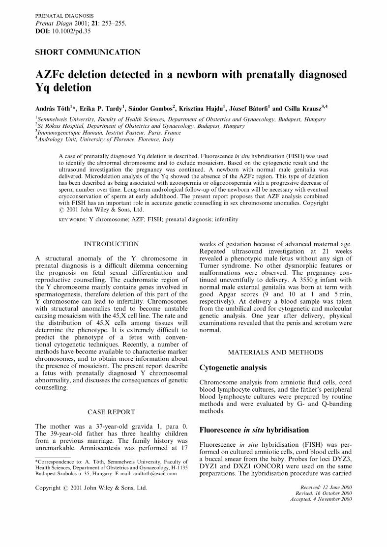
Cytogenetic analysis with GTG-banding showed a46,X,+mar chromosome constitution in 15 meta-phases of amniotic cells (Figure 1). Probes for the Xcentromere (DXZ1) and Y centromere (DYZ3) weresimultaneously hybridised in all 102 interphase amnio-tic cell nuclei showing no sign of mosaicism. The probefor the Y heterochromatic region DYZ1 did not showany signal in evaluated cells. The FISH was repeatedfrom the cord blood of the newborn, and the earlierresult was confirmed on metaphase nuclei (Figure 2)and on an additional 250 interphase nuclei. There wasno sign of mosaicism on the buccal smear preparationeither. The markers sY84 and sY87 from AZFa andsY127 and sY142 from AZFb were present. The DAZgene (sY254) and two other AZFc markers, sY152and sY158, were deleted. The final interpretationof the karyotype is 46,X,+mar. ish del(Y)(q11.22)(DYZ1x,DYZ3+). Karyotype analysis of the child’sfather revealed normal Y chromosome by G- andQ-banding. All the Yq STSs markers tested in thechild’s father gave the expected product of amplifica-tion, therefore no microdeletion was found.
DISCUSSION
In 46,X,+mar constitution, the marker chromosomeis usually derived from the X or Y chromosome. FISHis an important tool in the identification of sexchromosome markers (Schwartz et al., 1997). Unlikemolecular genetic methods, in situ hybridisation doesnot require additional samples as cells prepared forcytogenetic analysis are suitable for FISH, too. The
determination of the origin and structure of markerchromosomes is especially crucial for genetic counsel-ling in prenatal cases. The SRY gene is located close tothe centromere on the short arm of the Y chromosome(Simpson et al., 1997), so if this region of the marker ispresent then a male phenotype is expected. The DYZ3DNA probe is informative since it hybridises close tothis region. Y-derived marker chromosomes may beisochromosomes of the short arm (Slim et al., 1994;Wang et al., 1995). Wang et al. (1995) concluded thatthere is a greater risk for phenotypic abnormality withi(Yp) than with del(Yq). On the basis of themorphology of the marker in the present case adeletion was proposed, and this hypothesis wasconfirmed by the presence of STSs located in AZFaand b regions on the long arm.
The majority of patients with a sex chromosomemarker have a mosaic karyotype resulting in a wideclinical spectrum from normal male to Turner’ssyndrome. According to recent studies, about one-quarter of patients with Turner phenotype haveY-derived DNA fragments (Patsalis et al., 1998).Hoshi et al. (1998) published a prenatally diagnosedcase where two-thirds of amniotic fluid cells contained45,X and one-third 46,X,+mar. chromosome comple-ment, where the marker originated from the Ychromosome. An infant with normal male externalgenitalia was delivered. Reddy et al. (1998) examinedthe distribution of mosaic clones in the differentgonadal cell types of five patients having 45,X/46,XYmosaicism. Two patients were female with gonadaldysgenesis, two male patients had ambiguous genitalia
Figure 1—GTG-banded partial karyotype from amniocyte cultureof the fetus. As a consequence of the deletion the Y chromosome issmaller than the normal chromosomes 21 and 22
Figure 2—Identification of the marker chromosome using fluores-cence in situ hybridisation (FISH) with DYZ3 and DYZ1 probes onmetaphase nuclei prepared from the cord blood. There is a singlebright signal produced by DYZ3 probe which hybridises to thecentromeric region of the Y chromosome. The probe for the Yheterochromatic region DYZ1 did not show any signal. Inset showsa control patient with a normal Y chromosome. Note the doublesignal since both DYZ3 and DYZ1 probes hybridise. The single anddouble signals can be easily distinguished even in interphase nuclei
A. TOTH ET AL.254
Copyright # 2001 John Wiley & Sons, Ltd. Prenat Diagn 2001; 21: 253–255.

and mixed gonadal dysgenesis, and one infertile malehad an atrophic testis. They concluded that thephenotype not only depends on the percentage ofXY cells, but also on the distribution pattern in thegenital ridge. There was no concordance between thepercentage of mosaicism in the peripheral blood andgonads in each case. As a consequence, it is importantto exclude mosaicism as reliably as possible. FISH wasalso useful from this point of view since the evaluationof several hundred cells increases the reliability of theprognosis.
The present patient had the DYZ3 signal in all ofthe investigated cells, and the ultrasound at 21gestational weeks confirmed the diagnosis – a newbornwith normal male genitalia was expected. The preg-nancy was continued because no major anomaly wasprobable. Infertility was supposed as a long-term effectof the chromosome aberration, but considering theadvanced gestational age interruption of the preg-nancy was not advised. In order to give correct geneticcounselling DNA analysis for AZF deletions wasperformed from the cord blood of the newborn. It isknown that three loci named azoospermia factor(AZF) AZFa, AZFb and AZFc involved in spermato-genesis are mapped in the non-fluorescent portion ofthe long arm of the Y chromosome (Yq) (Vogt et al.,1996). Deletions of these regions are associated withimpaired spermatogenesis. Deletions range in sizefrom large, cytogenetically detectable anomalies tomicrodeletions which can be detected only by mole-cular biological techniques. The AZFc locus is locatedclose to the heterochromatic region, and its deletion isassociated with different phenotypes ranging fromazoospermia to oligozoospermia, while deletions inAZFa and AZFb are usually associated with Sertolicell only syndrome type I and with spermatogenicarrest, respectively. The deletion found in the presentpatient removes the whole AZFc region while theAZFa and AZFb regions are intact. The infertilephenotype cannot be exactly predicted, but consider-ing the type of deletion the spermatogenic failure canbe of a different entity. Since a progressive changefrom oligozoospermia to azoospermia is possible over
time in the case of AZFc deletions (Krausz andMcElreavey, 1999), semen analysis with cryoconserva-tion of sperm at early adulthood is recommended. Thepresent report illustrates the role of AZF deletionanalysis combined with cytogenetic analysis (FISH) inaccurate genetic counselling.
ACKNOWLEDGEMENT
C.K. has been supported by Grant No. 281b of theTelethon, Italy.
REFERENCES
Hoshi N, Tonoki H, Handa Y, et al. 1998. Prenatal identification ofmos 45,X/46,X,+mar in a normal male baby by cytogenetic andmolecular analysis. Prenat Diagn 18: 1316–1322.
Krausz C, McElreavey K. 1999. Y chromosome and male infertility.Front Biosci 4: 1–8.
Krausz C, Quintana-Murci L, Barbaux S, et al. 1999. A highfrequency of Y chromosome deletions in males with non-idiopathic infertility. J Clin Endocrinol Metab 84: 3606–3612.
Patsalis PC, Sismani C, Hadjimarcou MI, et al. 1998. Detection andincidence of cryptic Y chromosome sequences in Turner syndromepatients. Clin Genet 53: 249–257.
Pinkel D, Straume T, Gray W. 1986. Cytogenetic analysis usingquantitative, high-sensitivity, fluorescence hybridization. ProcNatl Acad Sci U S A 83: 2934–2938.
Reddy KS, Sulcova V. 1998. Pathogenesis of 45,X/46,XY gonadalmosaicism. Cytogenet Cell Genet 82: 52–57.
Schwartz S, Depinet TW, Leana-Cox J, et al. 1997. Sex chromosomemarkers: characterization using fluorescence in situ hybridizationand review of the literature. Am J Med Genet 71: 1–7.
Simpson E, Scott D, Chandler P. 1997. The male-specific histocom-patibility antigen, H-Y: a history of transplantation, immuneresponse genes, sex determination and expression cloning. AnnuRev Immunol 15: 39–61.
Slim R, Soulie J, Hotmar J, Lecolier B, Bercau G, Bernheim A.1994. Prenatal identification of an isochromosome for the shortarm of the Y i(Yp) by cytogenetic and molecular analyses. PrenatDiagn 14: 23–28.
Vogt PH, Edelmann A, Kirsch S, et al. 1996. Human Y chromosomeazoospermia factors (AZF) mapped to different subregions inYq11. Hum Mol Genet 5: 933–943.
Wang RB, Loh-Chung Y, Willow P, Falk RE, Willams J. 1995.Prenatal identification of i(Yp) by molecular cytogenetic analysis.Prenat Diagn 15: 1115–1119.
PRENATALLY DIAGNOSED Yq DELETION 255
Copyright # 2001 John Wiley & Sons, Ltd. Prenat Diagn 2001; 21: 253–255.


![Lesions of the Rete Testis in Mice Exposed Prenatally to … · [CANCER RESEARCH 45, 5145-5150, October 1985] Lesions of the Rete Testis in Mice Exposed Prenatally to Diethylstilbestrol](https://img.pdfslide.net/doc/110x75/5fc722183cfb0439ef1b1dc9/lesions-of-the-rete-testis-in-mice-exposed-prenatally-to-cancer-research-45-5145-5150.jpg)