Embed Size (px)
Citation preview

AZOOSPERMIA NON OSTRUTTIVA: LA RISPOSTA ANDROLOGICA DI MICROTESE ALLA PROPOSTA
GINECOLOGICA DI AID: TIP & TRICKS ANDROLOGICI E BIOLOGICI
Fabrizio M. Castiglioni
Dipartimento di
Andrologia – Urologia e
Procreazione Assistita
Clinica San Carlo,
Paderno Dugnano
(Milano)

Non-obstructive azoospermia (NOA) is an unfavorableprognostic condition for male infertility since spermatogenesisis disrupted
Men with NOA have no treatment options other than attempting testicular sperm retrieval coupled with intracytoplasmic sperm injection
The technique of testicular sperm extraction (TESE) via an open testicular biopsy was described for obstructiveazoospermia (OA) by Schoysman et al. as well as Craft et al. and subsequently by Silber et al. and Devroey et al. for NOA.
Ashraf et al. 2013; Schoysman et al. 1993; Craft et al 1993; Silber et al. 1995;Devroey et al 1995
Testicular sperm extraction (TESE)
Testicular biopsy can be part of intracytoplasmic sperm injection (ICSI)
treatment in patients with clinical evidence of NOA. Testicular sperm
extraction (TESE) is the technique of choice. Spermatogenesis may be
focal, which means that in about 50% of men with NOA, spermatozoa can
be found and used for ICSI.
There is a good correlation between the histology found upon diagnostic
biopsy and the likelihood of finding mature sperm cells during testicular
sperm retrieval and ICSI.
However, no threshold value has been found for FSH, inhibin B, or testicular
volume and successful sperm harvesting. When there are complete AZFa
and AZFb microdeletions, the likelihood of sperm retrieval is virtually zero
and therefore TESE procedures are contraindicated.
Microsurgical TESE yields the highest sperm retrieval rates, and multiple TESE
is superior to conventional TESE. Microsurgical TESE should be preferred in
severe cases of non-obstructive azoospermia.

AID
???


Tecniche di Recupero
Altre Indicazioni: Anejaculazione – Necrozoospermia – Azoospermia virtuale – Criptozoospermia (?)

Probabilità di recupero positivo (SRR):nelle OA: 98% ( 100%)nelle NOA: 35% (30-40%).
Nelle NOA, Prelievi Multipli (multiple TESE) possono migliorare il tasso di recupero
La cTESE (open single biopsy) è una procedura molto usata sia
per le OA che per le NOA: in quest’ultimo caso si basa sull’assunto
di una distribuzione intratesticolare multifocale e/o omogenea della
spermatogenesi residua.
OA NOA

MicroTESE was described by Schlegel in 1999 as a combination of Simple
TESE and Multiple TESE, but assisted with an operative microscope.In many cases of NOA, a “patchy distribution” of areas with residual spermatogenesis
was shown. (Tournaye, 1995; Hauser, 1998).
Therefore, very soon MicroTESE has been reported to offer a higher SRR than Single
TESE and Multiple random TESE. (Okada, 2002; Tsujimura, 2002).
“MicroTESE is based on the principle of identifying the mostadvanced pattern, though notnecessarily the predominantpattern, of spermatogenesis in the testis” (Schlegel, 1999).
MAJOR ADVANTAGES:
1) Identification of individual clumps of tubules with better spermatogenesis, so maximizing sperm recovery
2) Excision of single tubules, so providing the biologist with less testicular tissue to dissect in order to find sperms
3) The optimal visualization of the intratesticular and the subalbugineal terminal vessels allows the preservation of blood supply with minimal damage to the testis.

MICROTESE: Surgical TechniqueThe testis is widely opened (for 2/3 or 3/4 of itscircumference) along an equatorial or para-equatorial plane, avoiding any parenchymalstretching. A vast exposition of the seminiferoustubules is obtained, following the physiologicdistribution of the intratesticular vessels (5->15 x).
Some Authors use a longitudinal incision, whichnevertheless makes more difficult to respect thefine internal arterial supply of the testis, mainlythat involving the posterior and inferior gonadalportions.
Seminiferous tubules are curled-up inside lobules,which are separated by thin septa; the subtlevessels run parallel to lobules and septa.
A careful and fine microdissection allows to accessto the deeper sections of tubules of a lobule, andto many of more internal lobules. Respectingblood supply of lobules, and avoiding any roughdisconnection of tubules from tunica albuginea(rich of easily bleeding vessels) are mandatory.

MICROTESE: Surgical Technique
In many NOA testes, foci with residual spermatogenesis are heterogeneouslydistributed; therefore, microdissection must be extremely exploratory. Operatingwith a microscope at 15x – 24x(36x) magnification allows to identify the moreopaque and larger tubules, more probably those hosting mature spermatogeneticcells. These tubules are removed and passed to the biologist in the theatre, for ameticulous sperm search.

MICROTESE: Surgical TechniqueAt the end, testicular tissue is irrigated with Ringer solution plus gentamycin.Haemostasis is performed by gently pressing parenchyma for 3-4’ by a gauze wetwith antibiotic and, if necessary, using the bipolar thermal device (0.3 mm tip).Albugineal incision is repaired with continuous suture (VicrylTM 5-0 or 4-0), followedby closure of tunica vaginalis, infusion of betamethasone solution into vaginal cavity(Colpi, 2010) to prevent pain and adhesions, and closure of dartos and skin.
If surgery has been made meticulously, post-operative progress is actually painless,and any scar will result invisible at ultrasonography three months later.
Colpi, Clinical Andrology, 2010
Later: Ice bag on the scrotum and bed rest for 24 hours.


Bernie et al, 2015

SRR del 35% con cTESE e 52% con MicroTESE.
Con MicroTESE si recupera 1.5 volte in più rispetto a TESE.
SSR del 56% con cTESE e 28% con TESA.
Con TESE si ottiene un recupero positivo 2 volte in più che con TESA.
Bernie et al, 2015

According to an old paper, testicular histopathology seemsto be the best predictor for a testicular SR in NOA, with:
Sensitivity= 58.8; Specificity= 88.5; PPV= 83.3; NPV= 68.7
PREDICTIVE FACTORS IN SUCCESSFUL TESTICULAR SPERM RECOVERY
Histopathology findings sperm recovery
Hypospermatogenesis 100%
Incomplete SCOS 86%
Complete SCOS 19%
Incomplete Maturation Arrest 62%
Complete Maturation Arrest 48%Tournaye, 1996
SRR stratified by histology: 100% in hypospermatogenesis, 46% in MA and 33% in SCO.
Hauser et al, 2006
Unfortunately, histological exam is usually available after SR, apart from few cases already submitted to testis biopsy.

MicroTESE
Richiede:- strumentazione microchirurgica
- microscopio operatore (fino a 24X)

Our Operative Times
(da MicroTESE No. 601 to No. 720)Procedure Mean operative time
(minutes)
Monolateral MicroTESE with successful retrieval 86.9
(60-140)
Bilateral MicroTESE (successful or unsuccessful retrieval) 125.8
(85-180)
Monolateral MicroTESE plus Contralateral cTESE 106.6
(94-131)
Durata media:
- 1.8 h (range 0.5–6.6 h) in caso di recupero positivo
- 2.7 h (range 0.8–7.5 h) in caso di recupero negativo
Ramasamy, 2011
Richiede:
- Anestesia generale
- Tempi operatori maggiori della cTESE

150 consecutive MicroTESE performed
by the same surgeon
SSR
Group A (first 50) 32%
Group B (middle 50) 44%
Group C (last 50) 48%
(P < 0.05)
Schlegel sostiene che lo SRR continua lentamente a migliorare finchè l’esperienza
microchirurgica supera i 500 casi.
Superata tale soglia, cresce l’abilità nell’individuare anche minimali differenze nel
calibro dei tubuli e si riducono i tempi operatori.
Ishikawa (2010) ha dimostrato che la curva di apprendimento consente di
ottenere tassi di recupero significativamente migliori dopo i primi 100 casi.
Richiede:- ESPERIENZA DEL CHIRURGO OPERATORE
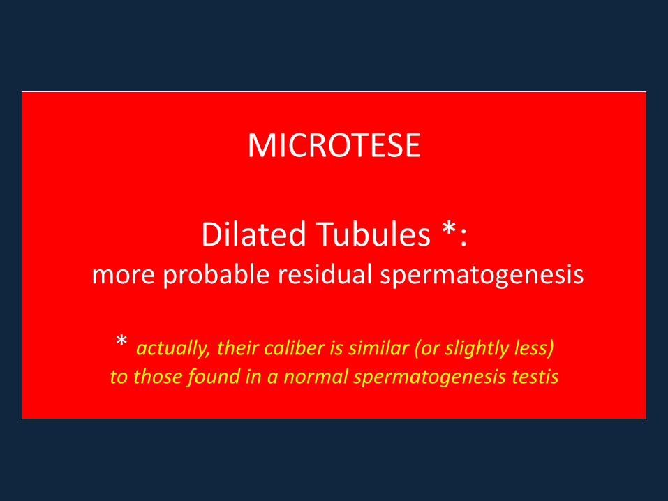
MICROTESE
Dilated Tubules *:more probable residual spermatogenesis
* actually, their caliber is similar (or slightly less)
to those found in a normal spermatogenesis testis


• METTERE FOTO


«The seminiferous tubule caliber pattern as evaluated at high magnificationduring microdissection testicular sperm extraction predicts sperm retrievalin patients with non-obstructive azoospermia».
E. Caroppo, E.M. Colpi, G. Gazzano, L. Vaccalluzzo, E. Piatti, G. D’Amato, G.M. Colpi Andrology, 2018
We retrospective reviewed the clinical data of 143 male patients: 64 underwentunilateral and 79 bilateral microTESE, for a total amount of 222 testis. (from January2015 to July 2017).
During all procedures we carefully recorded the caliber pattern of all tubules retrieved:dilated tubules (DT), not dilated tubules (NDT) and slightly dilated tubules (SDT).
The embryologists were blinded to the pattern of tubules retrieved during MicroTESE.
A fragment of one or more of the tubules of the same diameter (DT, SDT or NDT) foundwas sent for histological analysis (conducted by examining at least 100 different tubulesections).
All surgical procedures were performed by the same urologist.All testis specimen processing were made by the same two embryologists.
All histological analysis were made by a unique pathologist.
RELEVANCE OF TUBULAR CALIBER LOOK
DURING MICROTESE

«The seminiferous tubule caliber pattern as evaluated at high magnification during
microdissection testicular sperm extraction predicts sperm retrieval in patients with non-
obstructive azoospermia».E. Caroppo, E.M. Colpi, G. Gazzano, L. Vaccalluzzo, E. Piatti, G. D’Amato, G.M. Colpi
Successful sperm retrieval Sperm retrieval failure P
Patients age (years) 36 (33-40) [26-62] 35 (33-38) [26-46] 0.23
Testis volume (ml) 7,5 (6-9) [1.5-17] 7.1 (5.5-8.5) [1.6 – 17] 0.15
FSH mIU/ml 21 (16-28) [1.8-47.6] 20.8 (14.7-29.5) [1.47-68.7] 0.96
LH mIU/ml 6.9 (4,4-10) [0.2-27.4] 6.2 (4.4-10.9) [0.5-48] 0.944
Total testosterone (ng/dl) 422 (350-550) [220-890] 380 (277-497) [134-860] 0.007
Testis histology <0.0001#
SCO (%) 45 (31.5%) 98 (68,5%)
Focal SCO (%) 9 (100%) 0 (0%)
MA (%) 11 (38%) 18 (62%)
Hypo (%) 27 (96,4%) 1 (3.6%)
Hyalinosis (%) 2 (22,2%) 7 (77,8%)
Carcinoma In Situ (%) 1 (50%) 1 (50%)
Tubules caliber pattern <0.0001§
Dilated (%) 63 (90%) 7 (10%)
Slightly dilated (%)(at 24x) 25 (47%) 28 (53%)
Not dilated (%) 7 (7%) 92 (93%)
Sperm count per tubule caliber pattern <0.0001#
Dilated 50000 (9000-300000) [500-5,2x10⁶] /
Slightly dilated 1000 (500-1000) [500-450000] /
Not dilated tubules 500 (500-500) [up to 500] /
Spermatozoi recuperati in 95 dei 222 testicoli (42,8%), cioè in 83 dei 143 pazienti (58,0%).
Recuperi positivi: nel 90% dei Tubuli Dilatati,
nel 47% dei Tubuli Apparentemente Dilatati (a 24X), e
nel 7% dei Tubuli Non Dilatati (p<0.0001).
Andrology, 2018
= 68,9% = 35,3%

Stepwise binary logistic regression revealed that only the seminiferous tubules patternand testis histology were significantly predictive of SSR: the combination of both variatescorrectly classified 86,8% of testes, with an excellent diagnostic accuracy asdemonstrated by ROC AUC estimate computed on the predictive probability (0.93).
Model Chi-Square P Cox& Snell R
Square
Nagelkerke R
Square
% cases
correctly
predicted
Intercept odds
ratio
AUC
Prediction of sperm retrieval success
Histology 63.329 <0.0001 0.25 0.336 72.7 38.9 0.7
Tubules pattern 133.731 <0.0001 0.45 0.608 82.4 0.84 0.89
Tub. pattern plus
Histology
156.749 <0.0001 0.51 0.68 86.8 28.4 0.93
Prediction of tubules caliber pattern*
Histology 33.843 <0.0001 0.143 0.2 75.9 0.85 0.67


In sala operatoria deve essere presente un microscopio ottico per i Biologi
La presenza di due biologi consente di dissociare i tubuli di più prelievi rapidamente e
dare in pochi minuti una risposta sulla presenza o assenza di spermatozoi nelle
microaliquote di tessuto in esame.
Preparazione del campione da MicroTESE
Courtesy by Elisabetta Piatti e Giuseppe Caminiti

Dissociazione meccanica dei tubuliLa dissociazione avviene secondo il metodo di Schlegel (con vetrini sterili o lame di bisturi utilizzate come i vetrini) per far fuoriuscire il contenuto dei tubuli seminiferi. Si depositano più prelievi in differenti piastre Petri da 60mm.
Si passa più volte la sospensione ottenuta in angiocatetere (sec. Schlegel)e una goccia da ciascuna piastra viene esaminata al microscopio a 40x aluce diretta per dare indicazioni se continuare con i prelievi.
Il vetrinopotràesserepoifissato ecolorato.
Courtesy by Elisabetta Piatti e Giuseppe Caminiti

Al termine della procedura di prelievo il materiale deve risultare
sminuzzato completamente e reso il più possibile omogeneo.
Il contenuto delle diverse piastre può essere o meno unificato.
Si prepara il campione lavando con terreno HTF contenente albumina
per centrifugazione a 600g e si concentra lasciando un volume finale
in funzione del pellet ottenuto (in genere tra 0,3 e 1,5 ml).
Preparazione all’uso a fresco
o alla crioconservazione
Courtesy by Elisabetta Piatti e Giuseppe Caminiti

Per dare una corretta quantificazione degli spermatozoi presenti si
utilizza una camera di conta (Makler o Burker).
In caso di numero scarso di spermatozoi si fornisce una stima semi-
quantitativa andando a valutare il numero medio (cioè di più letture) di
spermatozoi presenti in un volume di 10 μl sottostante ad un vetrino
copri-oggetto da 22x22 mm a ingrandimento 40X, analizzando in modo
sistematico l’ intera superficie.
Stima della concentrazione di
spermatidi maturi recuperati
Courtesy by Elisabetta Piatti e Giuseppe Caminiti

In funzione del numero di spermatozoi ottenuto si crioconserva il
campione con congelamento rapido, in modo da rendere possibile il
recupero di un numero adeguato di spermatozoi per un ciclo di
ICSI, evitando tuttavia di dover sprecare materiale in eccesso.
Crioconservazione
Courtesy by Elisabetta Piatti e Giuseppe Caminiti

Il recupero di una buona percentuale di spermatozoi vitali dopo
crioconservazione potrebbe essere stimato valutando la vitalità
pre-congelamento quando possibile.
È noto che esistono fattori intrinseci (apoptosi abortiva, difetti
maturativi, patologie a carico dell’ epididimo) che rendono più
vulnerabili gli spermatozoi alla frammentazione e questo influisce
inevitabilmente sulla qualità degli spermatozoi recuperati.
Vari tipi di trattamento del materiale prelevato e il congelamento per
vitrificazione potrebbero essere modificazioni utili all’ottenimento di
un maggior numero di spermatozoi utilizzabili per ICSI.
Proposte di Ottimizzazione
Courtesy by Elisabetta Piatti e Giuseppe Caminiti

“Salvage micro-dissection testicular sperm extraction: outcome in men with non-obstructive azoospermia
with previous failed sperm retrievals”. (Kalsi et al, 2014) Positive SRR in 27 out of 58 cases (46.5%), without any correlation with FSH levels.
This finding confirms data in previous papers, reporting positive SRRsfrom 45% (Tsujimura, 2006) to 60% (Ramasamy, 2007).
About half of NOA patients are hypogonadic, and require endocrine post-surgical follow-up, and testosterone replacement when needed.
Bobjer, 2012
MICROTESE in PREVIOUS TESE FAILURES
GENERAL WARNING in NOA PATIENTS

-> Limiting patient exposure to certain harmful physical such as heat (Jung & Schuppe 2007), and chemical agents
-> Supplementation therapy (Nutraceuticals ?)
-> Treating NOA due to Hypogonadism
-> Pre-treating NOA with low testosterone (Raman 2002, Schlegel
2009, Cavallini 2011
Dabaja & Schlegel 2013
TRYING TO INCREASE Successful Testicular Sperm Retrievals
PREOPERATIVE MEASURES

“The testicular sperm extraction
procedure should be offered
to all men with NOA, but should
only be undertaken in a Centre
with expertise in MicroTESE
and where an ICSI laboratory
with expertise in handling
these samples is available”
(Canadian Guidelines, Jarvi et al. 2015)