Embed Size (px)
Citation preview
K
Protein Expression and Purification 18, 310–315 (2000)doi:10.1006/prep.1999.1195, available online at http://www.idealibrary.com on
Bacterial Expression and Purification of the ArabidopsisNADPH–Cytochrome P450 Reductase ATR2
Anna K. Hull and John L. Celenza1
Department of Biology, Boston University, 5 Cummington Street, Boston, Massachusetts 02215
Received October 6, 1999, and in revised form December 5, 1999
cbhbeitfctsfA
tiPdc
I
An N-terminally modified form of the ArabidopsisNADPH–cytochrome P450 ATR2 (ATR2mod) was ex-pressed from the tactac promoter in Escherichia colito obtain high yields of the enzyme. The N-terminalmodification eliminates the predicted chloroplasttransit peptide of ATR2 allowing for more efficientexpression. ATR2mod was purified from membraneextracts using a 2*,5*-ADP–agarose affinity column.The specific activity of the purified ATR2mod for cy-tochrome c reduction was 9.4 mmol min21 mg21 and the
m for cytochrome c reduction was 15 6 2 mM. Thepurified NADPH–cytochrome P450 reductase was ableto support function of CYP79B2. © 2000 Academic Press
Cytochrome P450s have crucial roles in metabolicpathways of all known species. In plants, cytochromeP450s are involved in a range of secondary metabolicpathways, including the biosynthesis of brassinosteroids,flavonoids, lignins, coumarins, and other phenylpro-panoids (1). In addition, numerous novel cytochromeP450s with unknown functions continue to be identi-fied as plant genomes are sequenced (http://drnelson.utmem.edu/CytochromeP450.html). Cytochrome P450s areheme-containing monooxygenases that require the fla-voprotein NADPH–cytochrome P450 reductase to carryout oxidation reactions. The two proteins are found to-gether in membrane-bound enzyme complexes in whichNADPH–cytochrome P450 reductase is responsible fortransferring electrons from NADPH to the cytochromeP450 [(2) and references therein]. Animals have a singleisoform of P450 reductase, while as many as three iso-forms have been found within different plant species(Jerusalem artichoke, maize, Catharanthus roseus, andVicia sativa (3)]. This suggests that in plants cytochromeP450-mediated reactions are compartmentalized into dif-
1 To whom correspondence should be addressed. Fax: (617) 353-
6340. E-mail: [email protected].310
ferent organellar membranes and/or that plants have ahigh demand for cytochrome P450-derived metabolites.
Genes encoding two isoforms of P450 reductase,ATR1 and ATR2, have been cloned from the Arabidop-sis thaliana ecotype Ler2 [Accession Nos. X66016 andX66017 (4)]. ATR1 and ATR2 were independentlycloned from the Col-0 ecotype and were named AR1and AR2, respectively (5). In this paper we refer to thegenes as ATR1 and ATR2 because the difference insequence between the two ecotypes is negligible. Thetwo isoforms encoded by ATR1 and ATR2 are only 63%identical at the amino acid level and are predicted tohave different subcellular locations (4). ATR1 andATR2 have been expressed previously in yeast (4) andinsect cells (5) and both isoforms equally support theactivity of CYP73A5 in vivo and in vitro.
It is estimated that when the Arabidopsis genome isompletely sequenced, over 300 cytochrome P450s wille found (http://drnelson.utmem.edu/CytochromeP450.tml). With such a large number of cytochrome P450seing identified in plants, in vitro assays constitute anssential tool for characterization of these enzymes. Anntegral component of any cytochrome P450 assay ishe addition of P450 reductase. While P450 reductasesrom any species appear to function with any cyto-hrome P450, there may be cases where it is desirableo use a reductase from the same or closely relatedpecies (6,7). Therefore, we have created a plasmid toacilitate the expression and purification of activeTR2 from Escherichia coli.We chose to work with ATR2 because it is predicted
o be targeted to the chloroplast. We have recentlydentified the activity of two Arabidopsis cytochrome450s, CYP79B2 and CYP79B3, which are also pre-icted to be targeted to the chloroplast. These cyto-hrome P450s catalyze the conversion of tryptophan to
2 Abbreviations used: Ler, Landsberg erecta; Col-0, Columbia;PTG, isopropyl-b-thiogalactopyranoside; Ap, ampicillin; DTT, di-
thiothreitol; CHAPS, 3-[(3-cholamidopropyl)-dimethylammonio]-1-
propanesulfonic acid; FMN, flavinmononucleotide.1046-5928/00 $35.00Copyright © 2000 by Academic Press
All rights of reproduction in any form reserved.
NoCMsa
C
(A
o1btaMocsb
311ARABIDOPSIS P450 REDUCTASE
indole-3-acetaldoxime (Hull and Celenza, unpub-lished). When we reconstituted CYP79B2 andCYP79B3 activity in vitro it was logical to use ATR2,which would be their most likely in vivo partner. Thepurification protocol from E. coli described here pro-vides an alternative to the previously reported yeastand baculovirus expression systems. This method pro-vides an inexpensive approach to obtaining large quan-tities of active ATR2 without requiring specialized ex-pertise.
MATERIALS AND METHODS
Reagents and Molecular Biological Methods
Restriction enzymes were purchased from New En-gland Biolabs (Beverly, MA). [a-32P]dCTP was from
EN Life Sciences Products (Boston, MA). Oligonucle-tides were from Genemed Inc. (South San Francisco,A). Chemicals were purchased from Sigma (St. Louis,O), unless noted to the contrary. Unless otherwise
tated all media and molecular biological methods ares described (8).
loning of the ATR2 cDNA
An Arabidopsis lYES cDNA library (9) was probedwith a 500-bp 32P-labeled polymerase chain reactionPCR) product encoding an N-terminal fragment ofTR2. The oligonucleotides ATR2-F (59-CTCATGGC-
AGCAATCATCAAAGG-39) and ATR2-R (59-GAATCT-CGCTGCATTGTCGGTAGG-39) were used to createthe probe using standard Hot Start PCR (10). SixcDNA clones were identified and subcloned into pBlue-script KS1 (Stratagene, La Jolla, CA) using EcoRI.Restriction digest analysis revealed that all six cDNAswere of the ATR2 gene.
DNA Sequencing
Sequencing of the largest cDNA clone confirmed thatwe had obtained the entire coding region of ATR2.Oligonucleotides T3, T7, AO15 (59-GAATGACAGAG-GAGAATGGC-39), AO16 (59-GTGAAACTGTAGATGA-AGC-39), and AO17 (59-CACGTCACATGTGCACTGG-39) were used for sequencing. Sequencing was carriedout using the ABI PRISM Big Dye Terminator CycleSequencing Ready Reaction kit (PE Biosystems, FosterCity, CA) according to the manufacturer’s instructions.The samples were analyzed on an ABI PRISM 377XLDNA sequencer (PE Biosystems) at the ACGT Se-quencing Facility of Boston University.
59-End Modification of ATR2
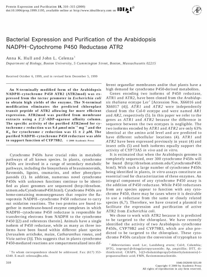
The first 135 bp of ATR2, which encode the predictedchloroplast localization sequence, were deleted usingPCR mutagenesis as outlined in Fig. 1 and as described
below. The resulting ATR2mod was subcloned into pC-Wori1 (11).
Oligonucleotides AO12 and AO13 (59-GCTGAAG-GATCCCATATGCTTATAGAGAATCGTCAATTCG-39and 59-CGGTAGGATCCCCATCTCCATATGTGGC-39,respectively) were used to amplify bases 118–523 ofthe cDNA introducing BamHI ends and a novel NdeIsite at position 130 bp of the cDNA. This BamHI frag-ment was cloned into the pBluescript KS1 vector andsequenced using T3 and T7 primers (Fig. 1A, step a).ATR2mod was assembled by subcloning into pCWori1the 39 region beginning with the native NdeI site at 499bp and ending in the XbaI site of the pBluescript KS1vector (Fig. 1A, step b). The mutagenized 59 fragmentwas subcloned upstream of the 39 sequence using NdeI(Fig. 1A, step c).
Expression of ATR2mod in E. coli
ATR2mod was expressed from the pCWori1 vectorin DH5a cells by IPTG induction as follows. A culturewas grown overnight in LB containing 100 mg/ml Apand then diluted 1:100 into Terrific broth supple-mented with 100 mg/ml Ap and 1 mg/ml riboflavin. Cellswere grown at 37°C at 225 rpm to an A550 of 0.9, 1 mMIPTG was added, and the culture was grown at 30°C,190 rpm for 48 h. The cells were collected by centrifu-gation at 4000 rpm for 15 min at 4°C, washed twicewith 4°C MOPS buffer (100 mM MOPS, 10% glycerol,0.2 mM DTT, 1 mM EDTA, adjusted to pH 7.3 withNaOH), and resuspended in MOPS buffer before soni-cation on ice in a Fisher Scientific 550 sonic dismem-brator (Fisher Scientific, Suwanee, GA) 30 3 2-s burstsn setting 3. The cell lysate (purification step 1, Table) was assayed for cytochrome c reduction activity (seeelow). The membranes were collected by centrifuga-ion in a Sorvall SA-600 rotor at 17,500 rpm for 45 mint 4°C and the membrane proteins solubilized in 4°Cops buffer containing 0.5% CHAPS by slow rocking
n ice for 2 h. After solubilization the sample wasentrifuged as before and the supernatant, containingolubilized membrane proteins (purification step 2, Ta-le 1), was applied to a 5-ml 29,59-ADP–agarose column
equilibrated with affinity buffer [50 mM Tris (pH 7.7),0.1 mM EDTA, 0.05 mM DTT, 10% glycerol, 0.1% Tri-ton X-100 (12)] at a flow rate of 0.2 ml/min. All affinitychromatography steps were carried out at 4°C. Thecolumn was washed with affinity buffer containing 5mM adenosine and then eluted with affinity buffercontaining 2 mM 29-AMP. One-milliliter fractions werecollected and assayed for cytochrome c reductase activ-ity. The two fractions with the highest specific activitywere pooled and dialyzed overnight at 4°C in twochanges of dialysis buffer [30 mM potassium phos-phate (pH 7.8), 20% glycerol, 0.1 mM EDTA, 2.0 mMFMN]. The protein content in the column-purified frac-

dcww
a
c
dNtp
312 HULL AND CELENZA
tion (purification step 3, Table 1) was measured usingthe Bio-Rad protein assay (Bio-Rad, Hercules, CA) anda bovine serum albumin standard. To monitor purifi-cation, samples were examined by SDS–PAGE (7.5%gel). Coomassie-stained gels were analyzed using Bio-Rad MultiAnalyst software.
Assay Conditions
Cytochrome c reductase assays were performed asescribed by (13). Briefly, 1 mg protein and 42.6 mMytochrome c in 0.3 M potassium phosphate (pH 7.7)ere mixed in a 1-ml cuvette and 100 mg of NADPHas added to start the reaction. The change in A550 was
recorded for 2 min at 30°C in a Perkin–Elmer Lambda5 UV/VIS spectrophotometer. Cytochrome c reductasectivity was linear for .2.5 min.
FIG. 1. Construction of ATR2mod expression plasmid. (A) Steps inescribed under Materials and Methods. PCR primers (arrows) anddeI site. The hatched box indicates the predicted chloroplast transi
heir predicted amino acid sequences. The predicted chloroplast tranCWori1 is in bold. Sequences derived from pCWori1 and ATR2mo
The Km for cytochrome c was calculated using 5, 6,10, 20, or 60 mM cytochrome c and 0.12 mM NADPH.The rate of cytochrome c reduction was calculated us-ing an extinction coefficient of 21 mM21 cm21. All con-entration points were done in triplicate.CYP79B2 was expressed in E. coli from pCWori1
(Hull and Celenza, unpublished) and membrane ex-tracts were prepared as described above for ATR2mod.These extracts were then mixed with 12 nmol of puri-fied ATR2mod. The proteins were reconstituted by in-cubation for 10 min at 30°C in 70 ml 50 mM potassiumphosphate (pH 7.9) containing 4 mg dilauroylphos-phatidylcholine. Tryptophan was added to a final con-centration of 750 mM and the reaction was initiated bythe addition of NADPH to a final concentration of 1
embly of ATR2mod. a, b, and c refer to the steps of the constructionlevant restriction enzyme sites are indicated. * indicates the addedptide. (B) DNA sequences of the 59 end of ATR2 and ATR2mod and
peptide is underlined (4). The NdeI site used to clone ATR2mod intore indicated with arrows. Amino acid positions are shown at right.
assre
t pesitd a
mM. For each time point, a fraction of the reaction was
o
H
(ciefsp
c
313ARABIDOPSIS P450 REDUCTASE
extracted with ethyl acetate. The extracts were appliedto an aluminum-backed F254 Merck Silica60 TLCplate (Fisher Scientific) and separated in 50:35:10ethyl acetate:chloroform:formic acid. The dried TLCplate was visualized under UV fluorescence using aBio-Rad Fluor-S and Bio-Rad MultiAnalyst software.Indole-3-acetaldoxime was used as a standard and wassynthesized as previously described (14,15). Gas chro-matography–mass spectrometry analysis of the syn-thesized indole-3-acetaldoxime confirmed its identityas represented by ions at m/z 174 and 130 (16).
RESULTS AND DISCUSSION
To date, the two Arabidopsis NADPH–cytochromeP450 reductases, ATR1 and ATR2, have been ex-pressed and purified from yeast (4) and from insectcells (5). Although these methods have proven quitevaluable in the characterization of ATR1 and ATR2,both techniques require familiarity with the respectiveexpression system. We have developed an E. coli ex-pression system for ATR2 that in three simple purifi-cation steps yields high quantities of purified ATR2and only requires basic competency in molecular biol-ogy. We chose to work with ATR2 because it is pre-dicted to be chloroplast localized (4) and we are inter-ested in characterizing the activity of chloroplast-localized cytochrome P450s.
ATR2 Isolation
ATR2 was cloned from an Arabidopsis cDNA libraryof the Col-0 ecotype using an ATR2 PCR product asdescribed under Materials and Methods. The cDNAsequence was identical to the previously publishedCol-0 sequence and confirms that there is a small se-quence difference between the Col-0 and Ler ecotypes(5). The new ATR2 cDNA clone contained the entirecoding region and an additional 160 bp of 59 and 200 bpf 39 noncoding sequence.
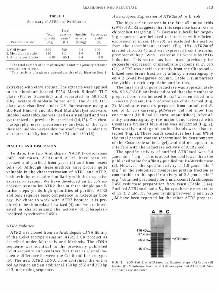
TABLE 1
Summary of ATR2mod Purification
Purification step
Totalprotein
(mg)
Totalenzymeunitsa
(U)
Specificactivity(U/mg)
Percentageyieldb
(%)
1. Cell lysate 1890 730 0.4 1002. Membrane fraction 110 111 1.0 153. Affinity purification 6.89 65.1 9.4 8.9
a The total number of units of enzyme. 1 unit 5 1 mmol cytochromereduced per minute.b Total activity of a given step/total activity of purification Step 1.
eterologous Expression of ATR2mod in E. coli
The high serine content in the first 45 amino acids29%) of ATR2 suggests that this sequence has a role inhloroplast targeting (17). Because subcellular target-ng sequences are believed to interfere with efficientxpression in E. coli (18–20), we excluded this portionrom the recombinant protein (Fig. 1B). ATR2modtarted at codon 45 and was expressed from the tactacromoter of the pCWori1 vector in DH5a cells by IPTG
induction. This vector has been used previously forsuccessful expression of membrane proteins in E. coli(21). ATR2 was purified directly from the Chaps-solu-bilized membrane fraction by affinity chromatographyon a 29,59-ADP–agarose column. Table 1 summarizesthe yields at each step of the purification.
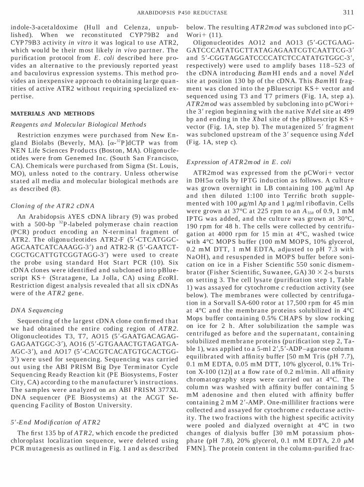
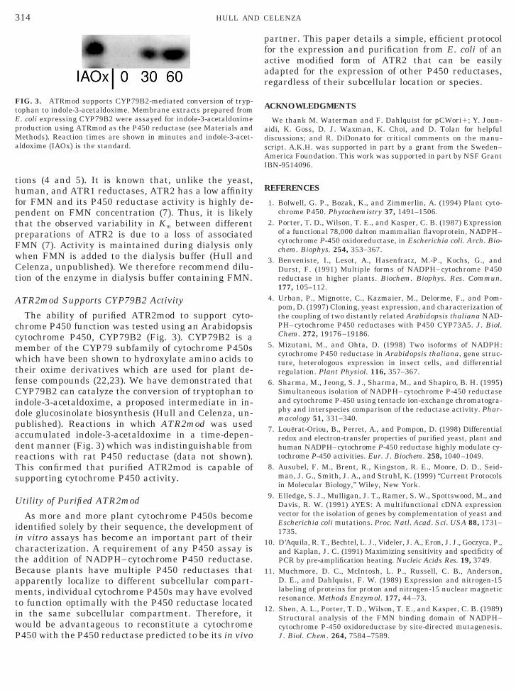
The final yield of pure reductase was approximately9%. SDS–PAGE analysis indicated that the membranepreparations from induced E. coli were enriched in a;74-kDa protein, the predicted size of ATR2mod (Fig.2). Membrane extracts prepared from uninduced E.coli or E. coli carrying pCWori1 did not show thisenrichment (Hull and Celenza, unpublished). After af-finity chromatography the major band detected withCoomassie brilliant blue stain was ATR2mod (Fig. 2).Two weakly staining unidentified bands were also de-tected (Fig. 2). These bands constitute less than 6% ofthe total protein content (determined by densitometryof the Coomassie-stained gel) and did not appear tointerfere with the reductase activity of ATR2mod.
The specific activity of purified ATR2mod was 9.4mmol min21 mg21. This is about fourfold lower than thepublished value for affinity-purified rat P450 reductase(2). However, the specific activity of 1.0 mmol min21
mg21 in the solubilized membrane protein fraction iscomparable to the specific activity of 2.8 mmol min21
mg21 obtained previously for a microsomal ArabidopsisP450 reductase preparation from yeast (Table 1) (4).Purified ATR2mod had a Km for cytochrome c reductionof 15 6 2 mM. Km values ranging between 3 and 22.5mM have been reported for the other ATR2 prepara-
FIG. 2. SDS–PAGE of ATR2mod purification steps. (A) Crude celllysate. (B) Membrane fraction. (C) Affinity-purified ATR2mod. Sizestandards are indicated.
ctBamtiwP
pfaar
1
1
314 HULL AND CELENZA
tions (4 and 5). It is known that, unlike the yeast,human, and ATR1 reductases, ATR2 has a low affinityfor FMN and its P450 reductase activity is highly de-pendent on FMN concentration (7). Thus, it is likelythat the observed variability in Km between differentpreparations of ATR2 is due to a loss of associatedFMN (7). Activity is maintained during dialysis onlywhen FMN is added to the dialysis buffer (Hull andCelenza, unpublished). We therefore recommend dilu-tion of the enzyme in dialysis buffer containing FMN.
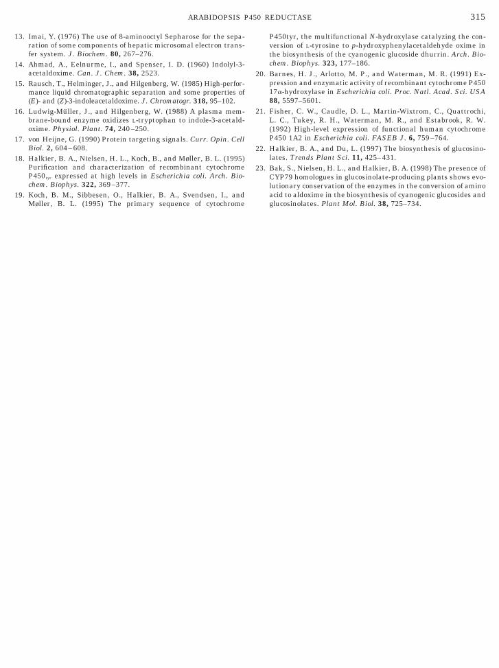
ATR2mod Supports CYP79B2 Activity
The ability of purified ATR2mod to support cyto-chrome P450 function was tested using an Arabidopsiscytochrome P450, CYP79B2 (Fig. 3). CYP79B2 is amember of the CYP79 subfamily of cytochrome P450swhich have been shown to hydroxylate amino acids totheir oxime derivatives which are used for plant de-fense compounds (22,23). We have demonstrated thatCYP79B2 can catalyze the conversion of tryptophan toindole-3-acetaldoxime, a proposed intermediate in in-dole glucosinolate biosynthesis (Hull and Celenza, un-published). Reactions in which ATR2mod was usedaccumulated indole-3-acetaldoxime in a time-depen-dent manner (Fig. 3) which was indistinguishable fromreactions with rat P450 reductase (data not shown).This confirmed that purified ATR2mod is capable ofsupporting cytochrome P450 activity.
Utility of Purified ATR2mod
As more and more plant cytochrome P450s becomeidentified solely by their sequence, the development ofin vitro assays has become an important part of theirharacterization. A requirement of any P450 assay ishe addition of NADPH–cytochrome P450 reductase.ecause plants have multiple P450 reductases thatpparently localize to different subcellular compart-ents, individual cytochrome P450s may have evolved
o function optimally with the P450 reductase locatedn the same subcellular compartment. Therefore, itould be advantageous to reconstitute a cytochrome450 with the P450 reductase predicted to be its in vivo
FIG. 3. ATRmod supports CYP79B2-mediated conversion of tryp-tophan to indole-3-acetaldoxime. Membrane extracts prepared fromE. coli expressing CYP79B2 were assayed for indole-3-acetaldoximeproduction using ATRmod as the P450 reductase (see Materials andMethods). Reaction times are shown in minutes and indole-3-acet-aldoxime (IAOx) is the standard.
artner. This paper details a simple, efficient protocolor the expression and purification from E. coli of anctive modified form of ATR2 that can be easilydapted for the expression of other P450 reductases,egardless of their subcellular location or species.
ACKNOWLEDGMENTS
We thank M. Waterman and F. Dahlquist for pCWori1; Y. Joun-aidi, K. Goss, D. J. Waxman, K. Choi, and D. Tolan for helpfuldiscussions; and R. DiDonato for critical comments on the manu-script. A.K.H. was supported in part by a grant from the Sweden–America Foundation. This work was supported in part by NSF GrantIBN-9514096.
REFERENCES
1. Bolwell, G. P., Bozak, K., and Zimmerlin, A. (1994) Plant cyto-chrome P450. Phytochemistry 37, 1491–1506.
2. Porter, T. D., Wilson, T. E., and Kasper, C. B. (1987) Expressionof a functional 78,000 dalton mammalian flavoprotein, NADPH–cytochrome P-450 oxidoreductase, in Escherichia coli. Arch. Bio-chem. Biophys. 254, 353–367.
3. Benveniste, I., Lesot, A., Hasenfratz, M.-P., Kochs, G., andDurst, F. (1991) Multiple forms of NADPH–cytochrome P450reductase in higher plants. Biochem. Biophys. Res. Commun.177, 105–112.
4. Urban, P., Mignotte, C., Kazmaier, M., Delorme, F., and Pom-pom, D. (1997) Cloning, yeast expression, and characterization ofthe coupling of two distantly related Arabidopsis thaliana NAD-PH–cytochrome P450 reductases with P450 CYP73A5. J. Biol.Chem. 272, 19176–19186.
5. Mizutani, M., and Ohta, D. (1998) Two isoforms of NADPH:cytochrome P450 reductase in Arabidopsis thaliana, gene struc-ture, heterologous expression in insect cells, and differentialregulation. Plant Physiol. 116, 357–367.
6. Sharma, M., Jeong, S. J., Sharma, M., and Shapiro, B. H. (1995)Simultaneous isolation of NADPH–cytochrome P-450 reductaseand cytochrome P-450 using tentacle ion-exchange chromatogra-phy and interspecies comparison of the reductase activity. Phar-macology 51, 331–340.
7. Louerat-Oriou, B., Perret, A., and Pompon, D. (1998) Differentialredox and electron-transfer properties of purified yeast, plant andhuman NADPH–cytochrome P-450 reductase highly modulate cy-tochrome P-450 activities. Eur. J. Biochem. 258, 1040–1049.
8. Ausubel, F. M., Brent, R., Kingston, R. E., Moore, D. D., Seid-man, J. G., Smith, J. A., and Struhl, K. (1999) “Current Protocolsin Molecular Biology,” Wiley, New York.
9. Elledge, S. J., Mulligan, J. T., Ramer, S. W., Spottswood, M., andDavis, R. W. (1991) lYES: A multifunctional cDNA expressionvector for the isolation of genes by complementation of yeast andEscherichia coli mutations. Proc. Natl. Acad. Sci. USA 88, 1731–1735.
0. D’Aquila, R. T., Bechtel, L. J., Videler, J. A., Eron, J. J., Goczyca, P.,and Kaplan, J. C. (1991) Maximizing sensitivity and specificity ofPCR by pre-amplification heating. Nucleic Acids Res. 19, 3749.
1. Muchmore, D. C., McIntosh, L. P., Russell, C. B., Anderson,D. E., and Dahlquist, F. W. (1989) Expression and nitrogen-15labeling of proteins for proton and nitrogen-15 nuclear magneticresonance. Methods Enzymol. 177, 44–73.
12. Shen, A. L., Porter, T. D., Wilson, T. E., and Kasper, C. B. (1989)Structural analysis of the FMN binding domain of NADPH–cytochrome P-450 oxidoreductase by site-directed mutagenesis.J. Biol. Chem. 264, 7584–7589.
1
1
1
1
1
2
2
2
2
315ARABIDOPSIS P450 REDUCTASE
13. Imai, Y. (1976) The use of 8-aminooctyl Sepharose for the sepa-ration of some components of hepatic microsomal electron trans-fer system. J. Biochem. 80, 267–276.
4. Ahmad, A., Eelnurme, I., and Spenser, I. D. (1960) Indolyl-3-acetaldoxime. Can. J. Chem. 38, 2523.
5. Rausch, T., Helminger, J., and Hilgenberg, W. (1985) High-perfor-mance liquid chromatographic separation and some properties of(E)- and (Z)-3-indoleacetaldoxime. J. Chromatogr. 318, 95–102.
16. Ludwig-Muller, J., and Hilgenberg, W. (1988) A plasma mem-brane-bound enzyme oxidizes L-tryptophan to indole-3-acetald-oxime. Physiol. Plant. 74, 240–250.
7. von Heijne, G. (1990) Protein targeting signals. Curr. Opin. CellBiol. 2, 604–608.
8. Halkier, B. A., Nielsen, H. L., Koch, B., and Møller, B. L. (1995)Purification and characterization of recombinant cytochromeP450tyr expressed at high levels in Escherichia coli. Arch. Bio-chem. Biophys. 322, 369–377.
9. Koch, B. M., Sibbesen, O., Halkier, B. A., Svendsen, I., andMøller, B. L. (1995) The primary sequence of cytochrome
P450tyr, the multifunctional N-hydroxylase catalyzing the con-version of L-tyrosine to p-hydroxyphenylacetaldehyde oxime inthe biosynthesis of the cyanogenic glucoside dhurrin. Arch. Bio-chem. Biophys. 323, 177–186.
0. Barnes, H. J., Arlotto, M. P., and Waterman, M. R. (1991) Ex-pression and enzymatic activity of recombinant cytochrome P45017a-hydroxylase in Escherichia coli. Proc. Natl. Acad. Sci. USA88, 5597–5601.
1. Fisher, C. W., Caudle, D. L., Martin-Wixtrom, C., Quattrochi,L. C., Tukey, R. H., Waterman, M. R., and Estabrook, R. W.(1992) High-level expression of functional human cytochromeP450 1A2 in Escherichia coli. FASEB J. 6, 759–764.
2. Halkier, B. A., and Du, L. (1997) The biosynthesis of glucosino-lates. Trends Plant Sci. 11, 425–431.
3. Bak, S., Nielsen, H. L., and Halkier, B. A. (1998) The presence ofCYP79 homologues in glucosinolate-producing plants shows evo-lutionary conservation of the enzymes in the conversion of aminoacid to aldoxime in the biosynthesis of cyanogenic glucosides and
glucosinolates. Plant Mol. Biol. 38, 725–734.