Embed Size (px)
Citation preview
Histol Histopathol (2001) 16: 997-1003
001: 10.14670/HH-16.997
http://www.hh.um.es
Histology and Histopathology Cellular and Molecular Biology
Comparison of NADPH diaphorase activity in the brains of hamsters infected with scrapie strains 139H or 263K or with normal hamster brain homogenate X. Yel, H.C. Meekerl, A.C. Scallet2 and R.I. Carp1 1 New York State Institute for Basic Research in Developmental Disabilities, Staten Island, NY, USA and
2Division of Neurotoxicology, FDNNational Center for Toxicological Research, Jefferson, AR, USA
Summary. Previous studies showed that the histopathological changes found in the brains of scrapieinfected animals included amyloid plaque formation, vacuolation , gliosis and neuronal and neurite degeneration. There were differences in the histopathological findings as a function of the scrapie strain-host combination. NADPH-diaphorase (NADPHd) has been shown to be a selective histoch emical marker for neurons containing nitric oxide (NO) synthase. Neuronal cell damage caused by NOS in brain has been reported to be associated with many neurodegenerative diseases. In this study, we used NADPH-d histostaining to investigate changes in the NOS system in brains of 139H- and 263K-infected hamsters and compared the results to normal hamster brain (NHB) injected animals. We observed that some of the NADPH-d histostaining neurons in the cortex of scrapie-infected hamsters appeared to be atrophic: the neurons were smaller and had fewer neurites. The NADPH-d histostaining intensity of neurons or astrocytes in septum, thalamus , hypothalamus and amygdala of 139H- and 263K-infected hamsters was greater than in control hamsters. Astrocytes in the thalamus, hypothalamus and lower part of the cortex (layers 4 to 6) in 263K-infected hamsters were more intensely stained for NADPH-d than in either 139Hinfected hamsters or controls. Our results suggest that changes in NADPH-d system might playa role in the diversity of scrapie induced neurodegenerative changes.
Key words: NADPH, Scrapie, Hamster, Astrocytosis, Vacuolation
Offprint requests to: Dr. Xuemin Ye, New York State Institute for Basic
Research in Developmental Disabilities, 1050 Forest Hill Road, Staten
Island, NY, 10314, USA. Fax: 718-698-0896. e-mail: [email protected]
Introduction
Nitric oxide (NO) is a neurotransmitter, but it also has the potential to act as a toxic gas with free radical properties. NO plays a crucial role in many phys iological events, including vasodilatation , neurotransmission, platelet aggregation , and immune activation . NO also plays a role in neurodegenerative changes. NO is produced by nitric oxide synthase (NOS) using L-arginine, NADPH and molecular oxygen. A number of studies have reported that the neuronal enzyme NADPH-d is NOS (Hope et aI., 1991). This enzyme is responsible for the calcium/calmodulindependent formation of citrulline and NO from arginine. Neuronal constitutive, or brain nitric oxide synthase (nNOS, bNOS or NOS I) was found in brain and peripheral nervous system (Sessa, 1994; Norman and Cameron, 1996). Endothelial constitutive nitric oxide synthase (eNOS or NOS III) exists in endothelial cells of vessels (Sessa, 1994; Norman and Cameron, 1996). These two isoforms require calcium calmodulin for activation. Activated glial cells , m acrophages , hepatocytes, and neutrophils synthesize NO by an inducible calcium-independent form of NOS (mac NOS, iNOS or NOS II) (Knowles and Moncada, 1994; Sessa, 1994; Norman and Cameron, 1996). Recent studies have implicated NO in several mechanisms related to neuronal degeneration and synaptic plasticity. On the other hand, neurons containing NADPH-d enzyme selectively resist the toxic effects of excitatory amino acids (Beal et aI., 1986; Koh et aI., 1986) and hypoxia (Uemura et aI., 1990). NADPH-d containing neurons were also found to survive the degenerative processes of Huntington's (Ferrante et aI., 1985) and Alzheimer 's (Kowall and Beal , 1988) disease in select areas. In Huntington 's disease, for example, up to 95 % of striatal neurons degenerate , while virtually all NADPH-d containing striatal neurons survive.
Scrapie is the archetypal unconventional slow infection disease, and is characterized by vacuolization, gliosis, neuronal and neurite degeneration, and in some

NADPH-d in scrapie-infected hamsters
instances, arnyloid plaque formation (DeArmond et al., 1992). Recent studies demonstrated that the activity of NOS is impaired in the brains of mice infected with experimental scrapie a s well a s in scrapie-infected neuroblastoma cells (ScN2a) (Ovadia et al . , 1996; Keshet et al., 1999). It has also been reported that NO produced by iNOS causes damage to brain cells. NO synthesized by iNOS in reactive astrocytes of ME7 scrapie-infected C57BL mice is reported to be a part of the neurodegenerative mechanisms in scrapie infection (Ju et al., 1998). In the current study, we used NADPH-d histostaining to investigate changes in the NOS system in brains of 139H and 263K scrapie-infected hamsters. The results were compared with pathological findings in these scrapie strain-host combinations.
caudal to the frontal end of the fixed brain and were s t a ined by the NADPH-d his tochemist ry method (Scherer-Singler e t al . , 1983). To demonstra te the NADPH-d reaction, the free-floating sections were incubated in 0.1M phosphate buffer, pH 7.4, containing 0.3% Triton X-100, 0.1 mglml nitroblue tetrazolium and 1.0 mglml b-NADPH at 37 "C for 30-60 min. Following the reaction, sections were rinsed in phosphate buffer, pH 7.4, and mounted onto chrome-alum coated slides. The slides were air-dried overnight, some of the sections were then counterstained with neutral red. The slides were then rinsed in distilled water and dried again. Coverslips were then mounted directly with Permount. The sections were examined under a light microscope. P h o t o m i c r o g r a p h s w e r e t aken wi th a Car1 Ze i s s Axiophot.
Materials and methods Results
Female , w e a n l i n g Syr ian hams te r s , s t r a in LVGILAK, were obtained from Charles River Breeding Farms (Wilmington, MA) and maintained in rooms in which the temperature, humidity, and light cycle (12 h on, 1 2 h off) were automatically controlled. Each cage contained no more than three animals. The hamsters were given food and water ad libitum. The experiment was approved by the Institutional Animal Care and Use Committee in our institute.
The hamsters were injected intracerebrally with scrapie strain 139H or 263K or with NHB homogenates. There were ten animals per group. The incubation period of the 139H- in fec ted hams te r s ( 1 3 5 2 5 days ) is approximately twice as long as that of the 263K-infected hamsters (6524 days). At the t ime of sacrifice, the animals were anesthetized with pentobarbital sodium (50 mglml ; A b b o t t , Nor th C h i c a g o , IL) and in jec ted intraperitoneally at a dose rate of 100 mglkg of body weight. Anesthetized animals were perfused via the heart with normal saline (15 mllmin) at room temperature for 2 - 3 min, fo l lowed b y pe r fus ion wi th 4 % para - fo rmaldehyde and 0 . 0 5 % g lu ta ra ldehyde in 0 .1M phosphate-buffered saline (PBS) (pH 7.4, 15 mllmin) for 15 min at room temperature. The brains were removed and f ixed in 4 % para fo rmaldehyde , 0 . 0 5 % g lu ta ra ldehyde in 0 . 1 M sod ium phospha te buffer overnight. Half brain was used for cryostat sections; the other half was paraffin embedded and then sectioned. Frozen sections from fixed scrapie and control brains were examined with NADPH-d histostaining. Paraffin sec t ions (6 p m th ickness ) w e r e used fo r rou t ine histopathological studies. NOS containing cryostat sections (20 p m thickness) were cut serially from the
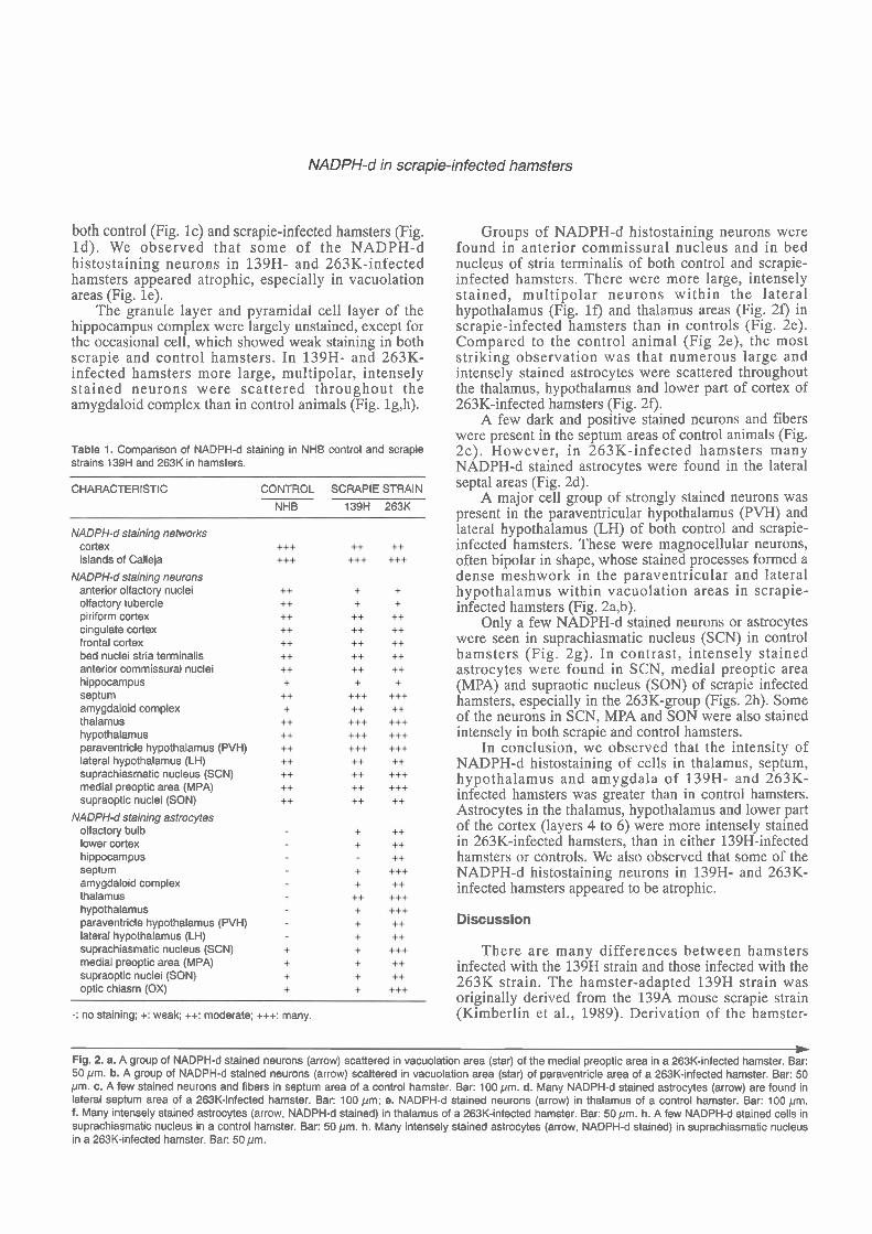
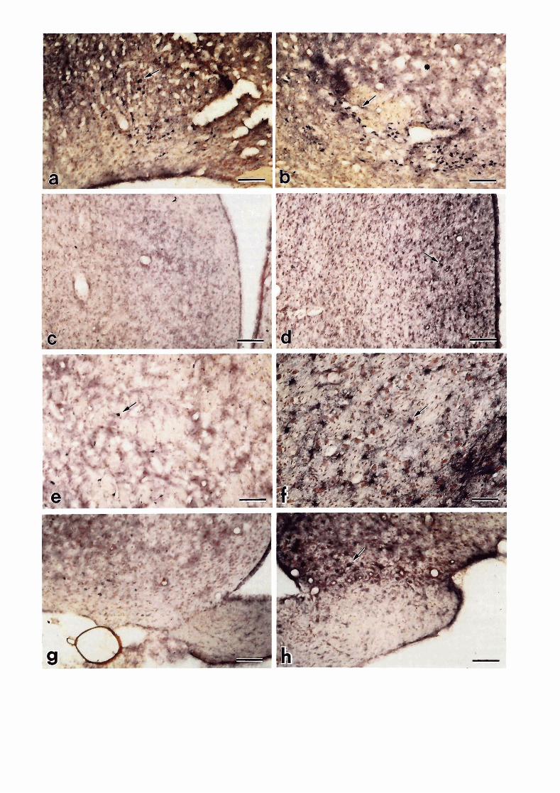
The results of this study are summarized in Table 1. In control animals , moderate ly in tense NADPH-d histostaining neurons were present within the anterior olfactory nuclei, olfactory tubercle, and lateral olfactory tract; the stained neurons surrounded unstained neurons (Fig. l a ) . A t more caudal levels, large. multipolar neurons were found more frequently scattered within the gray matter of the dorsal, lateral, ventral and medial anterior olfactory nuclei. These neurons had somewhat rounded cell bodies with prominent long, thick, sparsely branching dendrites. Similar cells were found within the gray matter of the piriform, cingulate and frontal cortex, and within the subcortical areas. Moderately dense networks of fine, varicose and non-varicose fibers were present throughout all neocortical regions and in all cortical layers. NADPH-d neurons were also present but slightly fewer within the main olfactory bulb in 139H- and 263K-infected hamsters (Fig. lb). In some cases a few NADPH-d neurons and astrocytes were found in vacuolation areas of 263K- and 139H-infected hamsters. But most of the time increased NADPH-d staining was not directly correlated with vacuolation in scrapie- infected hamsters. Some of the NADPH-d neurons showed atrophy, in which the neurons appeared smaller and had fewer neurites. There were fewer NADPH-d histostaining neurite networks in cortex of scrapie- infected hamsters than in controls.
Within the olfactory tubercle, the most striking feature was the intense staining of the islands of Calleja. The granule cells themselves appeared to be unstained, but th ree o r four of the i s l ands of Cal le ja w e r e surrounded by a most intense staining fiber network in
W Fig. 1. a. Coronal section through the anterior olfactory nucleus of a control animal shows many stained neurons (arrow). Bar: 200 pm; b. NADPH-d staining neurons (arrow) are seen in vacuolation areas (star) of a 263K-infected hamster. Bar: 200 pm. c. NADPH-d staining in islands of Calleja (arrow) in a control hamster. Bar: 200pm. d. The olfactory tubercle and islands of Calleja (arrow) are vacuolated (star) in a 263K-infected hamster. Bar: 100pm; e. Some of the NADPH-d stained neurons (arrow) showing atrophy in vacuolation areas (star) in a 263K-infected hamster. Bar: 50 pm; f. A group of NADPH-d stained neurons (arrow) scattered in vacuolation area (star) of lateral hypothalamus of a 263K-infected hamster. Bar: 50 pm g. A few NADPH-d stained neurons in amygdaloid complex in a control hamster. Bar: 100pm. h. Many NADPH-d stained neurons (arrow) in amygdaloid complex in a 139H-infected hamster. Bar: 100pm.
NADPH-d in scrapie-infected hamsters
both control (Fig. lc) and scrapie-infected hamsters (Fig. Id) . We observed that some of the NADPH-d histostaining neurons in 139H- and 263K-infected hamsters appeared atrophic, especially in vacuolation areas (Fig. le).
The granule layer and pyramidal cell layer of the hippocampus complex were largely unstained, except for the occasional cell, which showed weak staining in both scrapie and control hamsters. In 139H- and 263K- infected hamsters more large, rnultipolar, intensely stained neurons were scattered throughout the amygdaloid complex than in control animals (Fig. lg,h).
Table 1. Comparison of NADPH-d staining in NHB control and scrapie strains 139H and 263K in hamsters.
CHARACTERISTIC
NADPH-d staining networks cortex islands of Calleja
NADPH-d staining neurons anterior olfactory nuclei olfactory tubercle piriform cortex cingulate cortex frontal cortex bed nuclei stria terminalis anterior commissural nuclei hippocampus septum amygdaloid complex thalamus hypothalamus paraventricle hypothalamus (PVH) lateral hypothalamus (LH) suprachiasmatic nucleus (SCN) medial preoptic area (MPA) supraoptic nuclei (SON)
NADPH-d staining astrocytes olfactory bulb lower cortex hippocampus septum amygdaloid complex thalamus hypothalamus paraventricle hypothalamus (PVH) lateral hypothalamus (LH) suprachiasmatic nucleus (SCN) medial preoptic area (MPA) supraoptic nuclei (SON) optic chiasm (OX)
CONTROL SCRAPIE STRAIN
NHB 139H 263K
-: no staining; +: weak; ++: moderate; +++: many.
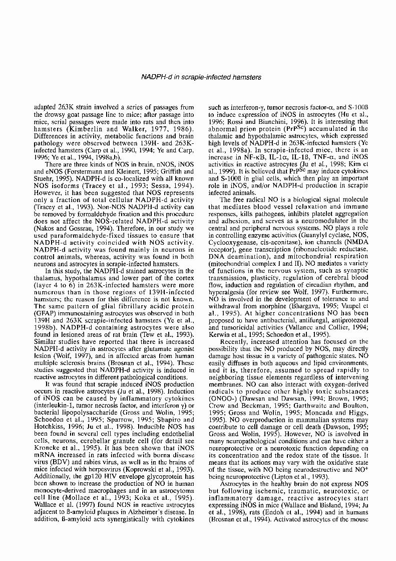
Groups of NADPH-d histostaining neurons were found in anterior commissural nucleus and in bed nucleus of stria terminalis of both control and scrapie- infected hamsters. There were more large, intensely s tained, multipolar neurons within the lateral hypothalamus (Fig. If) and thalamus areas (Fig. 2 0 in scrapie-infected hamsters than in controls (Fig. 2e). Compared to the control animal (Fig 2e), the most striking observation was that numerous large and intensely stained astrocytes were scattered throughout the thalamus, hypothalamus and lower part of cortex of 263K-infected hamsters (Fig. 29.
A few dark and positive stained neurons and fibers were present in the septum areas of control animals (Fig. 2c). However, in 263K-infected hamsters many NADPH-d stained astrocytes were found in the lateral septal areas (Fig. 2d).
A major cell group of strongly stained neurons was present in the paraventricular hypothalamus (PVH) and lateral hypothalamus (LH) of both control and scrapie- infected hamsters. These were magnocellular neurons, often bipolar in shape, whose stained processes formed a dense meshwork in the paraventricular and lateral hypothalamus within vacuolation areas in scrapie- infected hamsters (Fig. 2a,b).
Only a few NADPH-d stained neurons or astrocytes were seen in suprachiasmatic nucleus (SCN) in control hamsters (Fig. 2g). In contrast, intensely stained astrocytes were found in SCN, medial preoptic area (MPA) and supraotic nucleus (SON) of scrapie infected hamsters, especially in the 263K-group (Figs. 2h). Some of the neurons in SCN, MPA and SON were also stained intensely in both scrapie and control hamsters.
In conclusion, we observed that the intensity of NADPH-d histostaining of cells in thalamus, septum, hypothalamus and amygdala of 139H- and 263K- infected hamsters was greater than in control hamsters. Astrocytes in the thalamus, hypothalamus and lower part of the cortex (layers 4 to 6) were more intensely stained in 263K-infected hamsters, than in either 139H-infected hamsters or controls. We also observed that some of the NADPH-d histostaining neurons in 139H- and 263K- infected hamsters appeared to be atrophic.
Discussion
There are many differences between hamsters infected with the 139H strain and those infected with the 263K strain. The hamster-adapted 139H strain was originally derived from the 139A mouse scrapie strain (Kimberlin et al., 1989). Derivation of the hamster-
* Fig. 2. a. A group of NADPH-d stained neurons (arrow) scattered in vacuolation area (star) of the medial preoptic area in a 263K-infected hamster. Bar: 50 pm. b. A group of NADPH-d stained neurons (arrow) scattered in vacuolation area (star) of paraventricle area of a 263K-infected hamster. Bar: 50 pm. c. A few stained neurons and fibers in septum area of a control hamster. Bar: 100 pm. d. Many NADPH-d stained astrocytes (arrow) are found in lateral septum area of a 263K-infected hamster. Bar: 100 pm; e. NADPH-d stained neurons (arrow) in thalamus of a control hamster. Bar: 100 pm. f. Many intensely stained astrocytes (arrow, NADPH-d stained) in thalamus of a 263K-infected hamster. Bar: 50 pm. h. A few NADPH-d stained cells in suprachiasmatic nucleus in a control hamster. Bar: 50 pm. h. Many intensely stained astrocytes (arrow, NADPH-d stained) in suprachiasmatic nucleus in a 263K-infected hamster. Bar: 50 pm.
NADPH-d in scrapie-infected hamsters
adapted 263K strain involved a series of passages from the drowsy goat passage line to mice; after passage into mice, serial passages were made into rats and then into hamsters (Kimberlin and Walker, 1977, 1986). Differences in activity, metabolic functions and brain pathology were observed between 139H- and 263K- infected hamsters (Carp et al., 1990, 1994; Ye and Carp, 1996; Ye et al., 1994, 1998a,b).
There are three kinds of NOS in brain, nNOS, iNOS and eNOS (Forstermann and Kleinert, 1995; Griffith and Stuehr, 1995). NADPH-d is CO-localized with all known NOS isoforms (Tracey et al., 1993; Sessa, 1994). However, it has been suggested that NOS represents only a fraction of total cellular NADPH-d activity (Tracey et al., 1993). Non-NOS NADPH-d activity can be removed by formaldehyde fixation and this procedure does not affect the NOS-related NADPH-d activity (Nakos and Gossrau, 1994). Therefore, in our study we used paraformaldehyde-fixed tissues to ensure that NADPH-d activity coincided with NOS activity. NADPH-d activity was found mainly in neurons in control animals, whereas, activity was found in both neurons and astrocytes in scrapie-infected hamsters.
In this study, the NADPH-d stained astrocytes in the thalamus, hypothalamus and lower part of the cortex (layer 4 to 6) in 263K-infected hamsters were more numerous than in those regions of 139H-infected hamsters; the reason for this difference is not known. The same pattern of glial fibrillary acidic protein (GFAP) immunostaining astrocytes was observed in both 139H and 263K scrapie-infected hamsters (Ye et al., 1998b). NADPH-d containing astrocytes were also found in lesioned areas of rat brain (Tew et al., 1993). Similar studies have reported that there is increased NADPH-d activity in astrocytes after glutamate agonist lesion (Wolf, 1997), and in affected areas from human multiple sclerosis brains (Brosnan et al., 1994). These studies suggested that NADPH-d activity is induced in reactive astrocytes in different pathological conditions.
It was found that scrapie induced iNOS production occurs in reactive astrocytes (Ju et al., 1998). Induction of iNOS can be caused by inflammatory cytokines (interleukin-l, tumor necrosis factor, and interferon y) or bacterial lipopolysaccharide (Gross and Wolin, 1995; Schoedon et al., 1995; Sparrow, 1995; Shapiro and Hotchkiss, 1996; Ju et al., 1998). Inducible NOS has been found in several cell types including endothelial cells, neurons, cerebellar granule cell (for detail see Kroncke et al., 1995). It has been shown that iNOS mRNA increased in rats infected with borna disease virus (BDV) and rabies virus, as well as in the brains of mice infected with herpesvirus (Koprowski et al., 1993). Additionally, the gp120 HIV envelope glycoprotein has been shown to increase the production of NO in human monocyte-derived macrophages and in an astrocytoma cell line (Mollace et al., 1993; Koka et al., 1995). Wallace et al. (1997) found NOS in reactive astrocytes adjacent to 0-amyloid plaques in Alzheimer's disease. In addition, 0-amyloid acts synergistically with cytokines
such as interferon-y, tumor necrosis factor-a, and S-1000 to induce expression of iNOS in astrocytes (Hu et al., 1996; Rossi and Bianchini, 1996). It is interesting that abnormal prion protein (prpSc) accumulated in the thalamic and hypothalamic astrocytes, which expressed high levels of NADPH-d in 263K-infected hamsters (Ye et al., 1998a). In scrapie-infected mice, there is an increase in NF-KB, IL - l a , IL-1l3, TNF-a, and iNOS activities in reactive astrocytes Ju et al., 1998; Kim et al., 1999). It is believed that PrPgC may induce cytokines and S-1000 in glial cells, which then play an important role in iNOS, andlor NADPH-d production in scrapie infected animals.
The free radical NO is a biological signal molecule that mediates blood vessel relaxation and immune responses, kills pathogens, inhibits platelet aggregation and adhesion, and serves as a neuromodulator in the central and peripheral nervous systems. NO plays a role in controlling enzyme activities (Guanylyl cyclase, NOS, Cyclooxygenase, cis-aconitase), ion channels (NMDA receptor), gene transcription (ribonucleotide reductase, DNA deamination), and mitochondrial respiration (mitochondrial complex I and 11). NO mediates a variety of functions in the nervous system, such as synaptic transmission, plasticity, regulation of cerebral blood flow, induction and regulation of circadian rhythm, and hyperalgesia (for review see Wolf, 1997). Furthermore, NO is involved in the development of tolerance to and withdrawal from morphine (Bhargava, 1995; Vaupel et al., 1995). At higher concentrations NO has been proposed to have antibacterial, antifungal, antiprotozoal and tumoricidal activities (Vallance and Collier, 1994; Kenvin et al., 1995; Schoedon et al., 1995).
Recently, increased attention has focused on the possibility that the NO produced by NOS, may directly damage host tissue in a variety of pathogenic states. NO easily diffuses in both aqueous and lipid environments, and it is, therefore, assumed to spread rapidly to neighboring tissue elements regardless of intervening membranes. NO can also interact with oxygen-derived radicals to produce other highly toxic substances (ONOO-) (Dawsan and Dawsan, 1994; Brown, 1995; Crow and Beckman, 1995; Garthwaite and Boulton, 1995; Gross and Wolin, 1995; Moncada and Higgs, 1995). NO overproduction in mammalian systems may contribute to cell damage or cell death (Dawson, 1995; Gross and Wolin, 1995). However, NO is involved in many neuropathological conditions and can have either a neuroprotective or a neurotoxic function depending on its concentration and the redox state of the tissue. It means that its actions may vary with the oxidative state of the tissue, with NO being neurodestructive and NO+ being neuroprotective (Lipton et al., 1993).
Astrocytes in the healthy brain do not express NOS but following ischemic, traumatic, neurotoxic, or inflammatory damage, reactive astrocytes start expressing iNOS in mice (Wallace and Bisland, 1994; Ju et al., 1998), rats (Endoh et al., 1994) and in humans (Brosnan et al., 1994). Activated astrocytes of the mouse
1003
NADPH-d in scrapie-infected hamsters
hippocampus contain high levels of NADPH-d (Wallace and Fredens, 1992). In scrapie-infected mice, astrocytic iNOS activity in the brain is increased, and i t was suggested that the increase in NO is responsible for the cerebral disorders induced by scrapie (Ju et al., 1998). In other studies, in scrapie-infected mice and hamsters, nNOS activity in the brain was markedly inhibited (Ovadia et al., 1996). It was suggested that prpC may play a role in the targeting of nNOS to its proper subcellular localization; the nNOS impairment might be due to prpC dysfunction in scrapie-infected animals (Keshet et al., 1999). In this study, we observed that some of the NADPH-d histostaining neurons appeared to be atrophic and that there were fewer NADPH-d histostaining neurons and neurite networks in some areas of the brain in scrapie-infected hamsters compared to controls. NOS expression is induced in neurons of the hypothalamus following neuronal injury (Lumme et al., 1997). In the current study, we observed that the NADPH-d activity was higher in septum, thalamus, hypothalamus and amygdala of 139H- and 263K- infected hamsters than in control hamsters. The increase in NADPH-d activity in astrocytes and neurons in this study probably represents iNOS induction. However, the increased NADPH-d activity was not directly correlated with brain pathological changes such as vacuolation. The question of whether the increased NADPH-d activity is involved in a neuroprotective or a neurodestructive function requires further study. It may depend on its location, its concentration and the redox state of the tissue. Our results suggest that changes in NADPH-d system might play a role in the diversity of scrapie pathology.
Acknowledgements. The authors wish to thank Ms. Joanne Stocker for editing and excellent assistance in preparation of this manuscript. The authors appreciate the excellent assistance of Ms. Mary Ellen Cafaro with the layout of figures and Ms. Sharon Mathier for graphics preparation.
References
Beal M.F.. Kowall N.W., Ellison D.W., Mazurek M.F., Swartz K.J. and Martin J.B. (1986). Replication of the neurochemical characteristics of Huntington's disease by quinolinic acid. Nature 321, 168-1 71.
Bhargava H.N. (1995). Attenuation of tolerance to, and physical dependence on, morphine in the rat by inhibition of nitric oxide synthase. Gen. Pharmacol. 26, 1049-1053.
Brosnan C.F., Battistini L., Raine CS., Dickson D.W., Casadevall A. and Lee S.C. (1994). Reactive nitrogen intermediates in human neuropathology-An overview. Dev. Neurosci. 16, 152-161.
Brown G.C. (1995). Nitric oxide regulates mitochondrial respiration and cell functions by inhibiting cytochrome oxidase. FEBS Lett. 369, 136-1 39.
Carp R.I., Kirn Y.S. and Callahan S.M. (1990). Pancreatic lesions and hypoglycemia-hyperinsulinemia in scrapie-injected hamsters. J. Infect. Dis. 161, 462-466.
the strain-host combinations. In: Slow infections of the central nervous system. Bjornsson J., Carp R.I., Love A. and Wisniewski H.W. (eds). Ann. NY Acad. Sci. 724, 221 -234.
Crow J.P. and Beckman J.S. (1995). The role of peroxynitrite in nitric oxide-mediated toxicity. Curr. Top. Microbial. Immunol. 196, 57-73.
Dawson V.L. (1995). Nitric oxide: role in neurotoxicity. Clin. Exp. Pharmacol. Physiol. 22, 305-308.
Dawson T.M, and Dawson V.L. (1994). Nitric oxide: actions and pathological roles. The Neuroscientist Preview Issue, 9-20.
DeArmond S.J., Kristensson K. and Bowler R.P. (1992). PrPSc causes nerve cell death and stimulates astrocyte proliferation: a paradox. In: Progress in brain research. Vol. 94. Yu A.C.H., Hertz L., Norenberg M.D., Sykova E. and Waxman S.G. (eds). Elsevier. Amsterdam. pp 437-446.
Endoh M., Maiese K. and Wagner J. (1994). Expression of the inducible form of nitric oxide synthase by reactive astrocytes after transient global ischemia. Brain Res. 651, 92-100.
Ferrante R.J., Kowall M.W., Beal M.F., Richardson Jr. E.P., Bird E.D. and Martin J.B. (1985). Selective sparing of a class of striatal neurons in Huntington's disease. Science 230, 561 -563.
Forstermann U. and Kleinert H. (1995). Nitric oxide synthase: Expression and expressional control of the three isoforms. Naunyn- Schmiedeberg's Arch. Pharmacol. 352, 351-364.
Garthwaite J. and Boulton C.L. (1995). Nitric oxide signaling in the central nervous system. Annu. Rev. Physiol. 57, 683-706.
Griffith O.W. and Stuehr D.J. (1995). Nitric oxide synthases: Properties and catalytic mechanism. Annu. Rev. Physiol. 57, 707-736,
Gross S.S. and Wolin M.S. (1995). Nitric oxide: pathophysiological mechanisms. Annu. Rev. Physiol. 57, 737-769.
Hope B.T., Mecheal G.J., Knigge K.M. and Vincent S.R. (1991). Neuronal NADPH-diaphorase is a nitric oxide synthase. Proc. Natl. Acad. Sci. USA 88,281 1-281 4.
Hu J., Castets F., Guevarra J.L and Van Eldick L.J. (1996). S-1000 stimulates inducible nitric oxide synthase activity and mRNA levels in rat cortical astrocytes. J. Biol. Chem. 271, 2543-2547.
Ju W.K., Park K.J., Choi E.K., Kirn J., Carp R.I., Wisniewski H.M. and Kirn Y.S. (1998). Expression of inducible nitric oxide synthase in the brains of scrapie-infected mice. J. Neurovirol. 4, 445-450.
Kerwin Jr. J.F., Lancaster Jr. J.R. and Feldman P.L. (1995). Nitric oxide: A new paradigm for second messengers. J. Med. Chem. 38, 4343- 4362.
Keshet G.I., Ovadia H., Taraboulos A. and Gabizon R. (1999). Scrapie- induced mice and PrP knockout mice share abnormal localization and activity of neuronal nitric oxide synthase. J. Neurochem. 72, 1224-1231.
Kimberlin R.H. and Walker C.A. (1977). Characteristics of a short incubation model of scrapie in the golden hamster. J. Gen. Virol. 34, 295-304.
Kimberlin R.H. and Walker C.A. (1986). Pathogenesis of scrapie (strain 263K) in hamsters infected intracerebrally, intraperitoneally or intraocularly. J. Gen.Virol. 67, 255-263.
Kimberlin R.H., Walker C.A. and Fraser H. (1989). The genomic identity of different strains of mouse scrapie in expressed in hamsters and preserved on reisolation in mice. J. Gen. Virol. 70, 2017-2025.
Kirn J.I., Ju W.K., Choi J.H., Kirn J.. Choi E.K., Carp R.I., Wisniewski H.M. and Kirn Y.S. (1999). Expression of cytokine genes and increased nuclear factor-kappa B activity in the brains of scrapie- infected mice. Mol. Brain Res. 73. 17-27.
Carp R.I., Ye X., Kascsak R.J. and Rubenstein R. (1994). The nature of Knowles R.G. and Moncada S. (1994). Nitric oxide synthases in
NADPH-d in scrapie-infected hamsters
mammals. Biochem. J. 298, 249-258. Koh H.Y., Peters S, and Choi D.W. (1986). Neurons containing NADPH-
diaphorase are selectively resistant to quinolinate toxicity. Science 234, 73-76.
Koka P., He K., Zack J.A., Kitchen S., Peacok W., Fried I.. Tran T., Yashar S.S. and Merrill J.E. (1995). HIV-I envelope proteins induce IL- l , TNFa and nitric oxide in glial cultures derived from fetal, neonatal and adult human brain. J. Exp. Med. 182, 941-952.
Koprowski H., Zheng Y.M., Heber-Katz E., Fraser N., Rorke L., Fu Z.F., Hanlon C. and Dietzschold B. (1993). In vivo expression of inducible nitric oxide synthase in experimentally induced neurologic diseases. Proc. Natl. Acad. Sci. USA 90, 3024-3027.
Kowall N.W. and Beal M.F. (1988). Cortical somatostatin, neuropeptide Y and NADPH diaphorase neurons: Normal anatomy and alterations in Alzheimer's disease. Ann. Neurol. 23, 105-1 14.
Kroncke K.D., Fehsel K. and Kolb-Bachofen V. (1995). Inducible nitric oxide synthase and its product nitric oxide, a small molecule with complex biological activities. Biol. Chem. Hoppe-Seyle 376, 327- 343.
Lipton S.A., Choi Y.B., Pan Z.H., Lei S.Z., Chen H.S., Sucher N.J., Loscalzo J., Singel D.J. and Stamler J.S. (1993). A redox-base mechanism for the neuroprotective and neurodestructive effects of nitric oxide and related nitroso-compounds. Nature 364, 626-632.
Lumme A., Vanhatalo S., Sadeniemi M. and Soinila S. (1997). Expression of nitric oxide synthase in hypothalamic nuclei following axonal injury or colchicine treatment. Exp. Neurol. 144, 248- 257.
Mollace V., Colasanti M,, Persichini T., Bagetta G., Lauro G.M. and Nistico G. (1993). HIV gp120 glycoprotein stimulates the inducible isoform of NO synthase in human cultured astrocytoma cells. Biochem. Biophys. Res. Commun. 194. 439-445.
Moncada S. and Higgs E.A. (1995). Molecular mechanisms and therapeutic strategies related to nitric oxide. FASEB J. 9, 1319- 1330.
Nakos G. and Gossrau R. (1994). When NADPH diaphorase (NADPHd) works in the presence of formaldehyde, the enzyme appears to visualize selectively cells with constitutive nitric oxide synthase (NOS). Acta Histochem. 96, 335-343.
Norman J.E. and Cameron I.T. (1996). Nitric oxide in the human uterus. Rev. Report l , 61 -68.
Ovadia H., Rosenmann H., Shezen E., Halimi M,. Ofran I. and Gabizon R. (1996). Effect of scrapie infection on the activity of neuronal nitric- oxide synthase in brain and neuroblastoma cells. J. Biol. Chem. 271, 16856-1 6861.
Rossi F. and Bianchini E. (1996). Synergistic induction of nitric oxide by B-amyloid and cytokines in astrocytes. Biochem. Biophys. Res. Commun. 225,474-478.
Scherer-Singler U,, Vincent S.R., Kimura H. and McGeer E.G. (1983). Demonstration of a unique population of neurons with NADPH- diaphorase histochemistry. J. Neurosci. Meth. 9, 229-234.
Schoedon G.. Schneemann M,, Walter R., Blau N., Hofer S. and Schaffner A. (1995). Nitric oxide and infection: Another view. Clin.
Infect. Dis. 21, 152-157. Sessa W.C. (1994). The nitric oxide synthase family of proteins. J. Vasc.
Res. 31, 131-143. Shapiro K.B. and Hotchkiss J.H. (1996). Induction of nitric oxide
synthesis in murine macrophages by Helicobacter pylori. Cancer Lett. 102, 49-56.
Sparrow J.R. (1995). Inducible nitric oxide synthase in the central nervous system. J. Mol. Neurosci. 5, 219-229.
Tew E.M.M., Saffrey M.J., Anderson P.N. and Burnstock G. (1993). Postnatal rat NADPH-diaphorase-containing myenteric neurons extend processes when transplanted into adult rat corpus striatum. Exp. Neurol. 124, 265-273.
Tracey W.R., Nakane M,, Pollock J.S. and Forstermann U. (1993). Nitric oxide synthases in neural cells, macrophages and endothelium are NADPH diaphorase, but represent only a fraction of total cellular NADPH diaphorase activity. Biochem. Biophys. Res. Commun. 195, 1035-1 040.
Uemura Y., Kowall N.W. and Beal M.F. (1990). Selective sparing of NADPH-diaphorase-somatostatin-neuropeptide Y neurons in ischemia gerbil striatum. Ann. Neurol. 27, 620-625.
Vallance P. and Collier J. (1994). Biology and clinical relevance of nitric oxide. Br. Med. J. 309, 435-457.
Vaupel D.B.. Kimes A.S. and London E.D. (1995). Comparison of 7- nitroindazoles with other nitric oxide synthase inhibitors as attenuators of opioid withdrawal. Psychophamacology (Berl.) 118, 361 -368.
Wallace M.N. and Bisland S.K. (1994). NADPH-diaphorase activity in activated astrocytes represents inducible nitric oxide synthase. Neuroscience 59, 905-919.
Wallace M.N. and Fredens K. (1992). Activated astrocytes of the mouse hippocampus contain high levels of NADPH-diaphorase. NeuroReport3, 953-956.
Wallace M.N., Geddes J.G.. Farquhar D.A. and Masson M.R. (1997). Nitric oxide synthase in reactive astrocytes adjacent to B-amyloid plaques. Exp. Neurol. 144, 266-272.
Wolf G. (1997). Nitric oxide and nitric oxide synthase: biology, pathology, localization. Histol. Histopathol. 12, 251-261.
Ye X. and Carp R.I. (1996). Pathological changes in the pituitaries of female hamsters infected with the 139H strain of scrapie. J.Comp. Pathol. 11 4,291 -304.
Ye X., Carp R.I., Yu Y., Kozielski R. and Kozlowski P. (1994). Hyperplasia and hypertrophy of B-cells in the islets of Langerhans in hamsters infected with the 139H strain of scrapie. J. Comp. Pathol. 1 10, 169-1 83.
Ye X., Scallet A.C., Kascsak R.J. and Carp R.I. (1998a). Astrocytosis and amyloid deposition in scrapie-infected hamsters. Brain Res. 809, 277-287.
Ye X., Scallet A.C., Kascsak R.J. and Carp R.I. (1998b). Astrocytosis and proliferating cell nuclear antigen expression in brain of scrapie- infected hamsters. J. Mol. Neurosci. 11. 253-263.
Accepted May 18,2001