Embed Size (px)
Citation preview
CentralBringing Excellence in Open Access
Journal of Cardiology & Clinical Research
Cite this article: Olsen T, Gill S (2016) Bartonella Endocarditis Imitating Anca-Associated Vasculitis. J Cardiol Clin Res 4(1): 1054.
*Corresponding author
Thomas Olsen, Department of Cardiology, Odense University Hospital, Sdr. Boulevard 29, 5000 Odense C, Denmark, Tel: 45-26351337; Email:
Submitted: 12 January 2016
Accepted: 22 February 2016
Published: 23 February 2016
Copyright© 2016 Olsen et al.
OPEN ACCESS
Keywords•Bartonella•Infectious endocarditis•ANCA-associated vasculitis•Glomerulonephritis•Blood culture negative endocarditis•Bicuspid aortic valve
Case Report
Bartonella Endocarditis Imitating Anca-Associated VasculitisThomas Olsen* and Sabine GillDepartment of Cardiology, Odense University Hospital, Denmark
Abstract
We describe a case of Bartonella endocarditis and crescentic glomerulonephritis. Initially, the patient was suspected of microscopic polyangiitis (MPA) with involvement of the aortic valve due to high levels of anti-neutrophil cytoplasmic antibodies (ANCA) and negative blood cultures. The patient’s condition worsened on the initiated treatment with steroids and cyclophosphamide, which lead to further analysis of the blood. Polymerase chain reaction (PCR) and specific testing revealed high titers of Bartonellahenselae. The patient was treated with antibiotics and valve replacement surgery with gradually full recovery.
ABBREVIATIONSIE: Infective Endocarditis; MPA: Microscopic Polyangiitis;
BCNE: Blood Culture Negative Endocarditis; PR3: Proteinase 3; Ab: Antibody; ANCA: Anti-Neutrophil Cytoplasmic Antibodies MPO: Myelo Per Oxidase; ELISA: Enzyme-Linked Immunosorbent Assay; ANA: Anti Nuclear Antibodies; GBM: Glomerular Basement Membrane; CSD: Cat Scratch Disease; SBE: Subacute Bacterial Endocarditis; PCR: Polymerase Chain Reaction; TOE: Transesophageal Echocardiography
INTRODUCTIONInfective Endocarditis (IE) is a serious disease, often
challenging to diagnose and treat. The prognosis depends on specific treatment with antibiotics, valve replacement if indicated, the degree of peripheral embolism, especially central nervous system embolism and/ or vascular complications, i.e. heart failure complications. Specific microbial diagnosis is crucial for the prognosis, but is often challenging to obtain.
Blood culture-negative endocarditis (BCNE) is defined as endocarditis where it is not possible to grow a causative microorganism from a blood sample with the usual laboratory methods. BCNE is reported with an incidence varying from 2.5-31 % of all cases of endocarditis [1] and is rare in our region [2,3]. BCNE is classified into three different categories; 1) BCNE due to prior antibiotic treatment; 2) BCNE due to infection with fastidious microorganisms and 3) True BCNE due to intracellular bacteria that cannot be cultured with the normal methods [4].
True BCNE is mainly caused by Coxiellaburnetiiog Bartonellaspecies (spp.).
Bartonella spp. normally causes a minor self-limiting disease,
but in rare cases it may cause IE. To date, 23 Bartonella spp. are known but only a few have been associated with human IE B. quintana being the most frequent followed by B. henselae [5].
Bicuspid aortic valve is the most common congenital heart lesion and is found in 1- 2% of the population and has a strong male predominance. A bicuspid aortic valve tends to develop calcification which leads to either aortic stenosis or aortic regurgitation in nearly all patients. About 7-25 % develops IE, mostly in the fourth or fifth decade of life and bicuspid aortic valve IE accounts for about 12 % of all IE on native valves [6].
We present a rare case of IE due to Bartonella henselae on a bicuspid aortic valve, initially mimicking microscopic polyangiitis (MPA) which delayed the diagnosis due to the rarity of this infection.
CASE PRESENTATIONA 45-year old man, without prior medical history except
a rash on the legs a few weeks before, was admitted to a local hospital after 2 weeks of illness with severe fatigue, general malaise, dyspnea, fever, tender legs, night sweats and 3 kg of weight gain. Physical examination revealed an early mild diastolic murmur in the third left intercostal space. The patient was transferred to our University Hospital due to acute renal failure. Initial transoesophageal echocardiography (TOE) showed slight aortic valve regurgitation and a bicuspid aortic valve. The patient was treated for acute glomerulonephritis with steroids and cyclophosphamide and discharged to ambulatory follow-up. One week later the patient was readmitted due to severe dyspnea and a weight gain of 5 kg.
Initial investigations revealed: Blood pressure 150/94mmHg; temperature 37.6°C. Hemoglobin 7.2mmol/L; platelets 62*10E9/L;
CentralBringing Excellence in Open Access
Olsen et al. (2016)Email:
2/4J Cardiol Clin Res 4(1): 1054 (2016)
Leukocytes 5.3*10E9/L; albumin 26g/L; Lactate dehydrogenase 269 U/L; Creatinine: 264 mmol/L; CRP levels of 42mg/L; reticulocytes 93*10E9/L. Spot urine was positive with protein and blood. Plain chest radiographs showed a slightly enlarged heart, and slight pulmonary congestion. CT-scan showed discrete ascites and splenomegaly. Ultrasound of the kidneys revealed no abnormalities. Further blood samples showed a strongly positive semi quantitative result for c-ANCA, while p-ANCA was negative. Additional analysis with ELISA showed a positive PR3-Ab, while MPO-Ab was negative. Supplementary serology showed negative ANA (Antinuclear antibodies), negative anti DNA antibodies and negative GBM (glomerular basement membrane) antibody, normal IgA & IgM, while IgG was slightly elevated. A bone marrow aspiration showed reactive bone marrow with no signs of lymphoma. Kidney biopsy revealed focal necrotizing nephritis with scattered crescents. Immunohistochemistry staining revealed a diffuse IgG reaction in all kinds of tissue without accentuating in glomeruli while IgA was negative. There were no signs of monoclonal immunoglobulins, Hantavirus, Epstein Barr virus or cytomegalovirus.
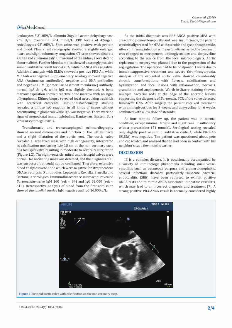
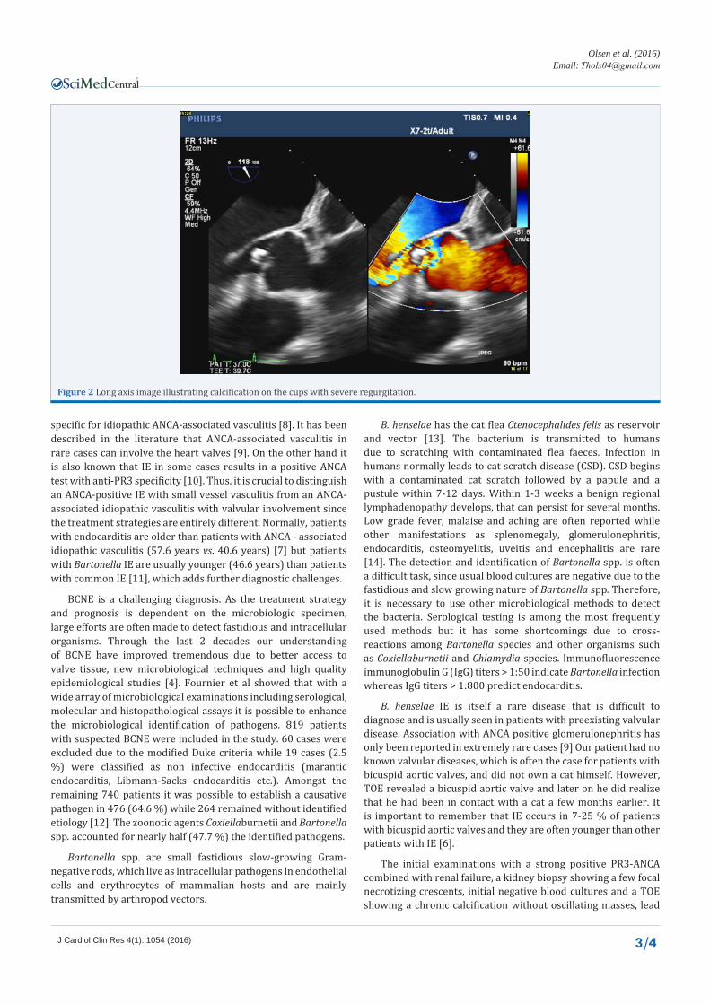
Transthoracic and transoesophageal echocardiography showed normal dimensions and function of the left ventricle and a slight dilatation of the aortic root. The aortic valve revealed a large fixed mass with high echogenicity, interpreted as calcification measuring 1.6x0.5 cm at the non-coronary cusp of a bicuspid valve resulting in moderate to severe regurgitation (Figure 1,2). The right ventricle, mitral and tricuspid valves were normal. No oscillating mass was detected, and the diagnosis of IE was suspected but could not be confirmed. Therefore, extensive blood analyses were done which were negative for streptococcus DNAse, retolysin O antibodies, Leptospira, Coxiella, Brucella and Bartonella serologies. Immunofluorescence microscopi revealed Bartonellahenselae IgM 160 (ref: < 64) and IgG 32.000 (ref: < 512). Retrospective analysis of blood from the first admission showed Bartonellahenselae IgM negative and IgG 16.000 g/L.
As the initial diagnosis was PR3-ANCA positive MPA with crescentic glomerulonephritis and renal insufficiency, the patient was initially treated for MPA with steroids and cyclophosphamide. After confirming infection with Bartonella henselae, the treatment was changed to meropenem, aminoglycosides and doxycycline according to the advice from the local microbiologists. Aortic replacement surgery was planned due to the progression of the regurgitation. The operation had to be postponed 1 week due to immunosuppressive treatment and severe thrombocytopenia. Analysis of the explanted aortic valve showed considerably chronic transformations with fibrosis, calcifications and hyalinization and focal lesions with inflammation, necrosis, granulation and angiogenesis. Warth in-Starry staining showed multiple bacterial rods at the edge of the necrotic lesions supporting the diagnosis of Bartonella. PCR of the valve detected Bartonella DNA. After surgery the patient received treatment with aminoglycosides for 3 weeks and doxycycline for 6 weeks combined with a low dose of steroids.
At four months follow up, the patient was in normal condition, except minimal fatigue and slight renal insufficiency with a p-creatinine 171 mmol/L. Serological testing revealed only slightly positive semi quantitative c-ANCA, while PR-3-Ab (ELISA) was negative. The patient was questioned about pets and cat scratch and realized that he had been in contact with his neighbor’s cat a few months earlier.
DISCUSSION IE is a complex disease. It is occasionally accompanied by
a variety of immunologic phenomena including small vessel vasculitis such as cutaneous purpura and glomerulonephritis. Several infectious diseases, particularly subacute bacterial endocarditis (SBE), have been reported to exhibit positive ANCA tests and to mimic ANCA-associated idiopathic vasculitis, which may lead to an incorrect diagnosis and treatment [7]. A strong positive PR3-ANCA result is normally considered highly
Figure 1 Bicuspid aortic valve with calcification on the non coronary cusp.
CentralBringing Excellence in Open Access
Olsen et al. (2016)Email:
3/4J Cardiol Clin Res 4(1): 1054 (2016)
specific for idiopathic ANCA-associated vasculitis [8]. It has been described in the literature that ANCA-associated vasculitis in rare cases can involve the heart valves [9]. On the other hand it is also known that IE in some cases results in a positive ANCA test with anti-PR3 specificity [10]. Thus, it is crucial to distinguish an ANCA-positive IE with small vessel vasculitis from an ANCA-associated idiopathic vasculitis with valvular involvement since the treatment strategies are entirely different. Normally, patients with endocarditis are older than patients with ANCA - associated idiopathic vasculitis (57.6 years vs. 40.6 years) [7] but patients with Bartonella IE are usually younger (46.6 years) than patients with common IE [11], which adds further diagnostic challenges.
BCNE is a challenging diagnosis. As the treatment strategy and prognosis is dependent on the microbiologic specimen, large efforts are often made to detect fastidious and intracellular organisms. Through the last 2 decades our understanding of BCNE have improved tremendous due to better access to valve tissue, new microbiological techniques and high quality epidemiological studies [4]. Fournier et al showed that with a wide array of microbiological examinations including serological, molecular and histopathological assays it is possible to enhance the microbiological identification of pathogens. 819 patients with suspected BCNE were included in the study. 60 cases were excluded due to the modified Duke criteria while 19 cases (2.5 %) were classified as non infective endocarditis (marantic endocarditis, Libmann-Sacks endocarditis etc.). Amongst the remaining 740 patients it was possible to establish a causative pathogen in 476 (64.6 %) while 264 remained without identified etiology [12]. The zoonotic agents Coxiellaburnetii and Bartonella spp. accounted for nearly half (47.7 %) the identified pathogens.
Bartonella spp. are small fastidious slow-growing Gram-negative rods, which live as intracellular pathogens in endothelial cells and erythrocytes of mammalian hosts and are mainly transmitted by arthropod vectors.
B. henselae has the cat flea Ctenocephalides felis as reservoir and vector [13]. The bacterium is transmitted to humans due to scratching with contaminated flea faeces. Infection in humans normally leads to cat scratch disease (CSD). CSD begins with a contaminated cat scratch followed by a papule and a pustule within 7-12 days. Within 1-3 weeks a benign regional lymphadenopathy develops, that can persist for several months. Low grade fever, malaise and aching are often reported while other manifestations as splenomegaly, glomerulonephritis, endocarditis, osteomyelitis, uveitis and encephalitis are rare [14]. The detection and identification of Bartonella spp. is often a difficult task, since usual blood cultures are negative due to the fastidious and slow growing nature of Bartonella spp. Therefore, it is necessary to use other microbiological methods to detect the bacteria. Serological testing is among the most frequently used methods but it has some shortcomings due to cross-reactions among Bartonella species and other organisms such as Coxiellaburnetii and Chlamydia species. Immunofluorescence immunoglobulin G (IgG) titers > 1:50 indicate Bartonella infection whereas IgG titers > 1:800 predict endocarditis.
B. henselae IE is itself a rare disease that is difficult to diagnose and is usually seen in patients with preexisting valvular disease. Association with ANCA positive glomerulonephritis has only been reported in extremely rare cases [9] Our patient had no known valvular diseases, which is often the case for patients with bicuspid aortic valves, and did not own a cat himself. However, TOE revealed a bicuspid aortic valve and later on he did realize that he had been in contact with a cat a few months earlier. It is important to remember that IE occurs in 7-25 % of patients with bicuspid aortic valves and they are often younger than other patients with IE [6].
The initial examinations with a strong positive PR3-ANCA combined with renal failure, a kidney biopsy showing a few focal necrotizing crescents, initial negative blood cultures and a TOE showing a chronic calcification without oscillating masses, lead
Figure 2 Long axis image illustrating calcification on the cups with severe regurgitation.
CentralBringing Excellence in Open Access
Olsen et al. (2016)Email:
4/4J Cardiol Clin Res 4(1): 1054 (2016)
Olsen T, Gill S (2016) Bartonella Endocarditis Imitating Anca-Associated Vasculitis. J Cardiol Clin Res 4(1): 1054.
Cite this article
to initial diagnosis of PR3-ANCA positive MPA. When the patient was treated with immunosuppressive therapy his condition worsened and the diagnosis was reconsidered. After addition of antibiotics and a surgical replacement of his degenerated aortic valve the patient gradually recovered. The patient was diagnosed with Bartonella IE due to elevated B. henselaeIgG titers in the blood and a Warthin-Starry staining and PCR analysis of the explanted valve supporting the diagnosis. Retrospective analysis of the blood from admission confirmed the diagnosis.
This case demonstrates that IE and ANCA-associated vasculitis may have overlapping clinical manifestations like fever, glomerulonephritis, splenomegaly and dyspnea. Nevertheless is it at outmost importance to make the correct diagnosis since the treatment is completely different. It is important to consider that idiopathic ANCA-associated vasculitis can have valvular involvement and also that IE can have elevated antineutrophilic cytoplasmic -PR3 antibodies. At the same time it is important to remember that between 2 and 31 % of all IE are blood culture-negative and nearly half of these cases are due to zoonotic agents like C. burnetii and Bartonella spp. which can be identified with immunological testing. Furthermore, is it important that patients with bicuspid aortic valves have an increased risk of developing IE and typical are of younger age that other IE patients. It is therefore recommended that all glomerulonephritis with simultaneous affected valves undergo advanced blood analysis, i.e. culturing, serology, and immunological studies before the diagnosis IE is discarded. We also recommend immunological testing for Coxiella and Bartonella species in all blood culture-negative endocarditis. At the same time we suggest that all patients with bicuspid aortic valves are informed about their larger risk and symptoms of infective endocarditis and are instructed in frequent dentist examinations and careful dental hygiene.
ACKNOWLEDGEMENTSWe acknowledge Edin Colic, M.D. from the Nephrology
Department for collaborating in the diagnosis and treatment of this patient.
REFERENCES1. Brouqui P, Raoult D. Endocarditis due to rare and fastidious bacteria.
Clin microbial Rev. 2001; 14: 177-207.
2. ÖzcanC, Asmar A, Gill S, Thomassen A, Diederichsen A. The value of FDG-PET/CT in the diagnostic work-up of extra cardiac infectious manifestations in infectious endocarditis. Int J Cardiovasc Imaging. 2013; 29: 1629-1637.
3. Le V, Gill S. Serious complications after infective endocarditis. Dan Med Bul. 2010; 57: A4192.
4. Tattevin P, Watt G, Revest M, Arvieux C, Fournier PE. Update on blood culture-negative endocarditis. Med Mal infect. 2015; 45: 1-8.
5. Edouard S, Nabet C, Lepidi H, Fornier P-E, Raoult D. Bartonella, et al. A Common Cause of Endocarditis: a Report on 106 Cases an Review. J ClinMicrobiol. 2015; 53: 824-829.
6. Lamas CC, Eykyn S. Bicuspid Aortic Valve- A silent Danger: Analysis of 50 Cases of Infective Endocarditis. Clin Infect Dis. 2000; 30: 336-341.
7. Chirinos JA, Corrales-Medina VF, Garcia S, Lichtstein DM, Bisno AL Chakko S, et al. Endocarditis associated with antineutrophilic cytoplasmic antibodies: a case report and review of the literature. ClinRheumatol. 2007; 26: 590–595.
8. Lane SK, Gravel JW jr. Clinical utility of common serum rheumatologic test. Am FamPhysican. 2002; 65: 1073-1080.
9. Lacoste C, Mansencal N, Ben m´rad M, Goulon-Goeau C, Cohen P, Guillevin L, et al. Valvular involvement in ANCA-associated systemic vascultis: a case report and literature review. BMC MusculoskeletDisord. 2011; 12: 50.
10. Mahr A, Batteux F, Tubiana S, Goulvestre C, Wolff M, Papo T, et al. Brief report: prevalence of antineutrophil cytoplasmic antibodies in infective endocarditis. Arthritis Rheumatol. 2014; 66: 1672-1677.
11. Fournier PE, Leliervre H, Eykyn SV, Mainard JL, Marrie TJ, Bruneel F, et al. Epidemiologic and clinical characteristics of Bartonella quintana and Bartonella henselae endocarditis: a study of 48 patients. Medicine (Baltimore). 2001; 80: 245-251.
12. Fournier PE, Thuny F, Richet H, Lepidi H, Casalta JP, Arzouni JP, et al. Comprehensive Diagnostic Strategy for Blood Culture–Negative Endocarditis: A Prospective Study of 819 New Cases. Clin Infect Dis. 2010; 51: 131-140.
13. Pennisi MG, Marsilio F, Hartmann K, Lloret A, Addie D, Belák S et al. Bartonella species infection in cats: ABCD guidelines on prevention and management. J Feline Med Surg. 2013; 15: 563-569.
14. Chomel BB, Boulouis HJ, Maruyama S, Breitschwerdt EB. Bartonella spp, in pets and effect on human health. Emerg Infect Dis. 2006; 12: 389-394.