Embed Size (px)
Citation preview

Page 1/19
Comprehensive Treatment for Major Salivary Gland CarcinomaBased on Intensity-Modulated Radiotherapy with or withoutRadical SurgeryZichen Qiu
Sun Yat-sen University A�liated Tumor Hospital: Sun Yat-sen University Cancer CenterZheng Wu
hu nan sheng zhong liu yi yuan: Hunan Cancer HospitalFeifei Lin
Sun Yat-sen University A�liated Tumor Hospital: Sun Yat-sen University Cancer CenterLei Wang
Sun Yat-sen University A�liated Tumor Hospital: Sun Yat-sen University Cancer CenterDehuan Xie
Sun Yat-sen University A�liated Tumor Hospital: Sun Yat-sen University Cancer CenterWanqin Cheng
Shunde Hospital of Southern Medical UniversityShaowen Lyu
Maastricht University Medical Centre+: Maastricht Universitair Medisch Centrum+Xuekui Liu
Sun Yat-sen University A�liated Tumor Hospital: Sun Yat-sen University Cancer CenterMingli Wang
Sun Yat-sen University A�liated Tumor Hospital: Sun Yat-sen University Cancer CenterJiang Hu
Sun Yat-sen University A�liated Tumor Hospital: Sun Yat-sen University Cancer CenterYalan Tao
Sun Yat-sen University A�liated Tumor Hospital: Sun Yat-sen University Cancer CenterYong Su ( [email protected] )
Sun Yat-sen University Cancer Center https://orcid.org/0000-0002-8888-8694
Research
Keywords: Intensity-modulated radiotherapy, Lymphoepithelial carcinoma, Major salivary gland carcinoma, Target volumedelineation
Posted Date: November 11th, 2021
DOI: https://doi.org/10.21203/rs.3.rs-1042786/v1
License: This work is licensed under a Creative Commons Attribution 4.0 International License. Read Full License
Page 2/19
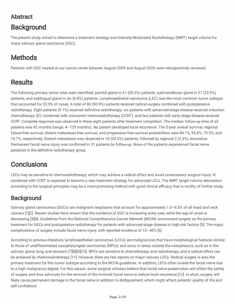
Abstract
BackgroundThe present study aimed to determine a treatment strategy and Intensity-Modulated Radiotherapy (IMRT) target volume formajor salivary gland carcinoma (SGC).
MethodsPatients with SGC treated at our cancer center between August 2009 and August 2020 were retrospectively reviewed.
ResultsThe following primary tumor sites were identi�ed: parotid gland in 61 (69.3%) patients, submandibular gland in 21 (23.9%)patients, and sublingual gland in six (6.8%) patients. Lymphoepithelial carcinoma (LEC) was the most common tumor subtypethat accounted for 23.9% of cases. A total of 80 (90.9%) patients received radical surgery combined with postoperativeradiotherapy. Eight patients (9.1%) received de�nitive radiotherapy: six patients with advanced-stage disease received inductionchemotherapy (IC) combined with concurrent chemoradiotherapy (CCRT), and two patients with early-stage disease receivedCCRT. Complete response was observed in these eight patients after treatment completion. The median follow-up time of allpatients was 42 months (range: 4–129 months). No patient developed local recurrence. The 5-year overall survival, regionalfailure-free survival, distant metastasis-free survival, and progression-free survival probabilities were 84.1%, 95.6%, 75.3%, and75.7%, respectively. Distant metastasis was observed in 18 (20.5%) patients, followed by regional 2 (2.3%) recurrence.Permanent facial nerve injury was con�rmed in 31 patients by follow-up. None of the patients experienced facial nerveparalysis in the de�nitive radiotherapy group.
ConclusionsLECs may be sensitive to chemoradiotherapy, which may achieve a radical effect and avoid unnecessary surgical injury. ICcombined with CCRT is expected to become a new treatment strategy for advanced LECs. The IMRT target volume delineationaccording to the surgical principles may be a more promising method with good clinical e�cacy that is worthy of further study.
BackgroundSalivary gland carcinomas (SGCs) are malignant neoplasms that account for approximately 1.0–8.5% of all head and neckcancers [1][2]. Recent studies have shown that the incidence of SGC is increasing every year, while the age of onset isdecreasing [3][4]. Guidelines from the National Comprehensive Cancer Network (NCCN) recommend surgery as the primarytreatment for SGCs and postoperative radiotherapy for patients with advanced-stage disease or high-risk factors [5]. The majorcomplications of surgery include facial nerve injury, with reported incidence of 12–40% [6].
According to previous literature, lymphoepithelial carcinomas (LECs) are malignancies that have morphological features similarto those of undifferentiated nasopharyngeal carcinomas (NPCs) and occur in areas outside the nasopharynx, such as in thesalivary gland, lung, and stomach [7][8][9][10]. NPCs are sensitive to chemotherapy and radiotherapy, and a radical effect canbe achieved by chemoradiotherapy [11]. However, there are few reports on major salivary LECs. Radical surgery is also theprimary treatment for this tumor subtype according to the NCCN guidelines. In addition, LECs often invade the facial nerve dueto a high malignancy degree. For this reason, some surgical scholars believe that facial nerve preservation will affect the safetyof surgery and thus advocate for the removal of the involved facial nerve to reduce local recurrence [12]. In short, surgery willlikely cause permanent damage to the facial nerve in addition to dis�gurement, which might affect patients’ quality of life andself-con�dence.
Page 3/19
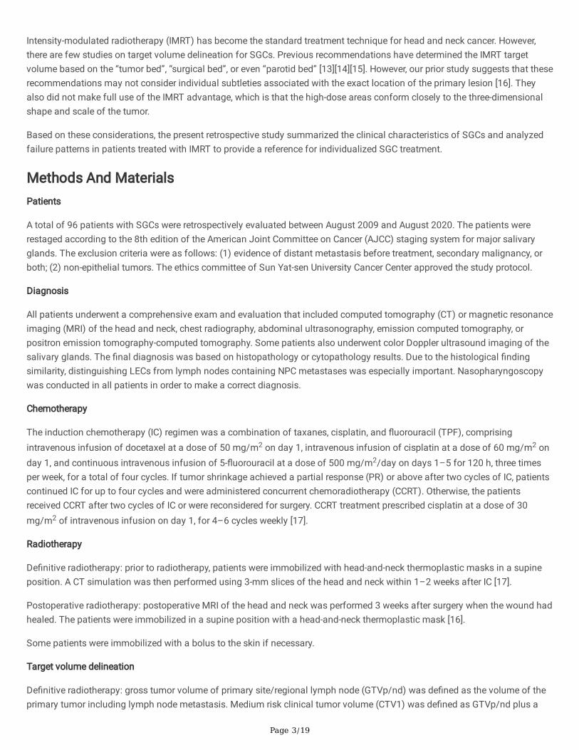
Intensity-modulated radiotherapy (IMRT) has become the standard treatment technique for head and neck cancer. However,there are few studies on target volume delineation for SGCs. Previous recommendations have determined the IMRT targetvolume based on the “tumor bed”, “surgical bed”, or even “parotid bed” [13][14][15]. However, our prior study suggests that theserecommendations may not consider individual subtleties associated with the exact location of the primary lesion [16]. Theyalso did not make full use of the IMRT advantage, which is that the high-dose areas conform closely to the three-dimensionalshape and scale of the tumor.
Based on these considerations, the present retrospective study summarized the clinical characteristics of SGCs and analyzedfailure patterns in patients treated with IMRT to provide a reference for individualized SGC treatment.
Methods And MaterialsPatients
A total of 96 patients with SGCs were retrospectively evaluated between August 2009 and August 2020. The patients wererestaged according to the 8th edition of the American Joint Committee on Cancer (AJCC) staging system for major salivaryglands. The exclusion criteria were as follows: (1) evidence of distant metastasis before treatment, secondary malignancy, orboth; (2) non-epithelial tumors. The ethics committee of Sun Yat-sen University Cancer Center approved the study protocol.
Diagnosis
All patients underwent a comprehensive exam and evaluation that included computed tomography (CT) or magnetic resonanceimaging (MRI) of the head and neck, chest radiography, abdominal ultrasonography, emission computed tomography, orpositron emission tomography-computed tomography. Some patients also underwent color Doppler ultrasound imaging of thesalivary glands. The �nal diagnosis was based on histopathology or cytopathology results. Due to the histological �ndingsimilarity, distinguishing LECs from lymph nodes containing NPC metastases was especially important. Nasopharyngoscopywas conducted in all patients in order to make a correct diagnosis.
Chemotherapy
The induction chemotherapy (IC) regimen was a combination of taxanes, cisplatin, and �uorouracil (TPF), comprisingintravenous infusion of docetaxel at a dose of 50 mg/m2 on day 1, intravenous infusion of cisplatin at a dose of 60 mg/m2 onday 1, and continuous intravenous infusion of 5-�uorouracil at a dose of 500 mg/m2/day on days 1–5 for 120 h, three timesper week, for a total of four cycles. If tumor shrinkage achieved a partial response (PR) or above after two cycles of IC, patientscontinued IC for up to four cycles and were administered concurrent chemoradiotherapy (CCRT). Otherwise, the patientsreceived CCRT after two cycles of IC or were reconsidered for surgery. CCRT treatment prescribed cisplatin at a dose of 30mg/m2 of intravenous infusion on day 1, for 4–6 cycles weekly [17].
Radiotherapy
De�nitive radiotherapy: prior to radiotherapy, patients were immobilized with head-and-neck thermoplastic masks in a supineposition. A CT simulation was then performed using 3-mm slices of the head and neck within 1–2 weeks after IC [17].
Postoperative radiotherapy: postoperative MRI of the head and neck was performed 3 weeks after surgery when the wound hadhealed. The patients were immobilized in a supine position with a head-and-neck thermoplastic mask [16].
Some patients were immobilized with a bolus to the skin if necessary.
Target volume delineation
De�nitive radiotherapy: gross tumor volume of primary site/regional lymph node (GTVp/nd) was de�ned as the volume of theprimary tumor including lymph node metastasis. Medium risk clinical tumor volume (CTV1) was de�ned as GTVp/nd plus a
Page 4/19
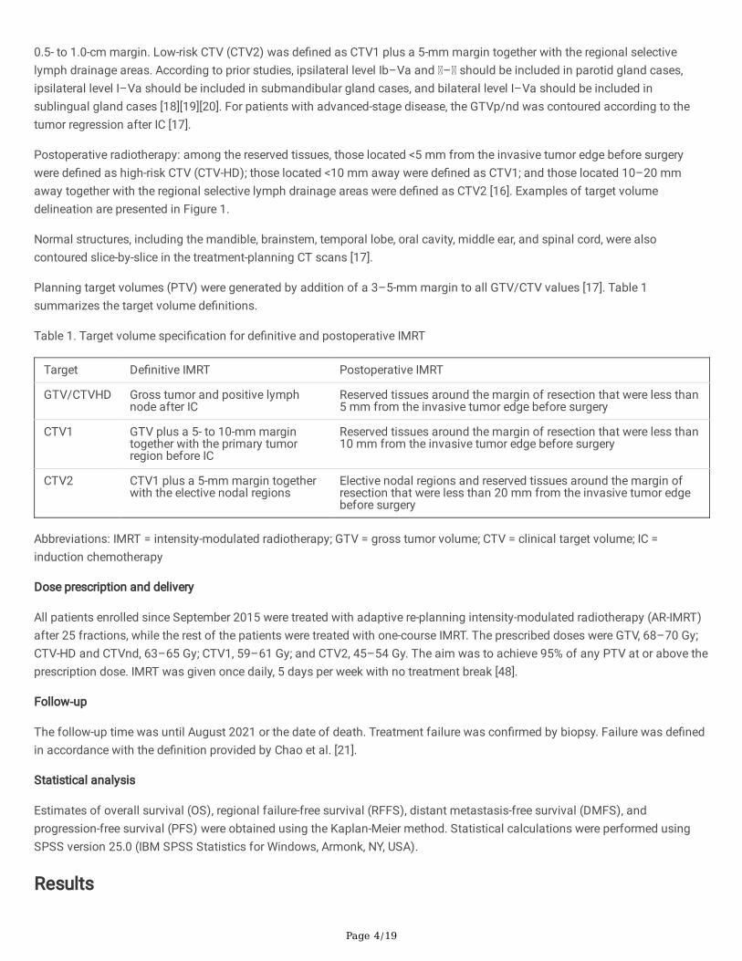
0.5- to 1.0-cm margin. Low-risk CTV (CTV2) was de�ned as CTV1 plus a 5-mm margin together with the regional selectivelymph drainage areas. According to prior studies, ipsilateral level Ib–Va and – should be included in parotid gland cases,ipsilateral level I–Va should be included in submandibular gland cases, and bilateral level I–Va should be included insublingual gland cases [18][19][20]. For patients with advanced-stage disease, the GTVp/nd was contoured according to thetumor regression after IC [17].
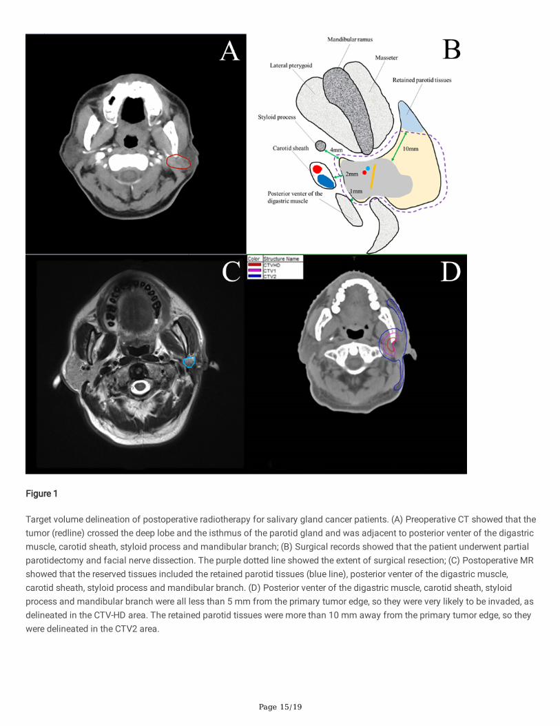
Postoperative radiotherapy: among the reserved tissues, those located <5 mm from the invasive tumor edge before surgerywere de�ned as high-risk CTV (CTV-HD); those located <10 mm away were de�ned as CTV1; and those located 10–20 mmaway together with the regional selective lymph drainage areas were de�ned as CTV2 [16]. Examples of target volumedelineation are presented in Figure 1.
Normal structures, including the mandible, brainstem, temporal lobe, oral cavity, middle ear, and spinal cord, were alsocontoured slice-by-slice in the treatment-planning CT scans [17].
Planning target volumes (PTV) were generated by addition of a 3–5-mm margin to all GTV/CTV values [17]. Table 1summarizes the target volume de�nitions.
Table 1. Target volume speci�cation for de�nitive and postoperative IMRT
Target De�nitive IMRT Postoperative IMRT
GTV/CTVHD Gross tumor and positive lymphnode after IC
Reserved tissues around the margin of resection that were less than5 mm from the invasive tumor edge before surgery
CTV1 GTV plus a 5- to 10-mm margintogether with the primary tumorregion before IC
Reserved tissues around the margin of resection that were less than10 mm from the invasive tumor edge before surgery
CTV2 CTV1 plus a 5-mm margin togetherwith the elective nodal regions
Elective nodal regions and reserved tissues around the margin ofresection that were less than 20 mm from the invasive tumor edgebefore surgery
Abbreviations: IMRT = intensity-modulated radiotherapy; GTV = gross tumor volume; CTV = clinical target volume; IC =induction chemotherapy
Dose prescription and delivery
All patients enrolled since September 2015 were treated with adaptive re-planning intensity-modulated radiotherapy (AR-IMRT)after 25 fractions, while the rest of the patients were treated with one-course IMRT. The prescribed doses were GTV, 68–70 Gy;CTV-HD and CTVnd, 63–65 Gy; CTV1, 59–61 Gy; and CTV2, 45–54 Gy. The aim was to achieve 95% of any PTV at or above theprescription dose. IMRT was given once daily, 5 days per week with no treatment break [48].
Follow‐up
The follow-up time was until August 2021 or the date of death. Treatment failure was con�rmed by biopsy. Failure was de�nedin accordance with the de�nition provided by Chao et al. [21].
Statistical analysis
Estimates of overall survival (OS), regional failure-free survival (RFFS), distant metastasis-free survival (DMFS), andprogression-free survival (PFS) were obtained using the Kaplan-Meier method. Statistical calculations were performed usingSPSS version 25.0 (IBM SPSS Statistics for Windows, Armonk, NY, USA).
Results
Page 5/19
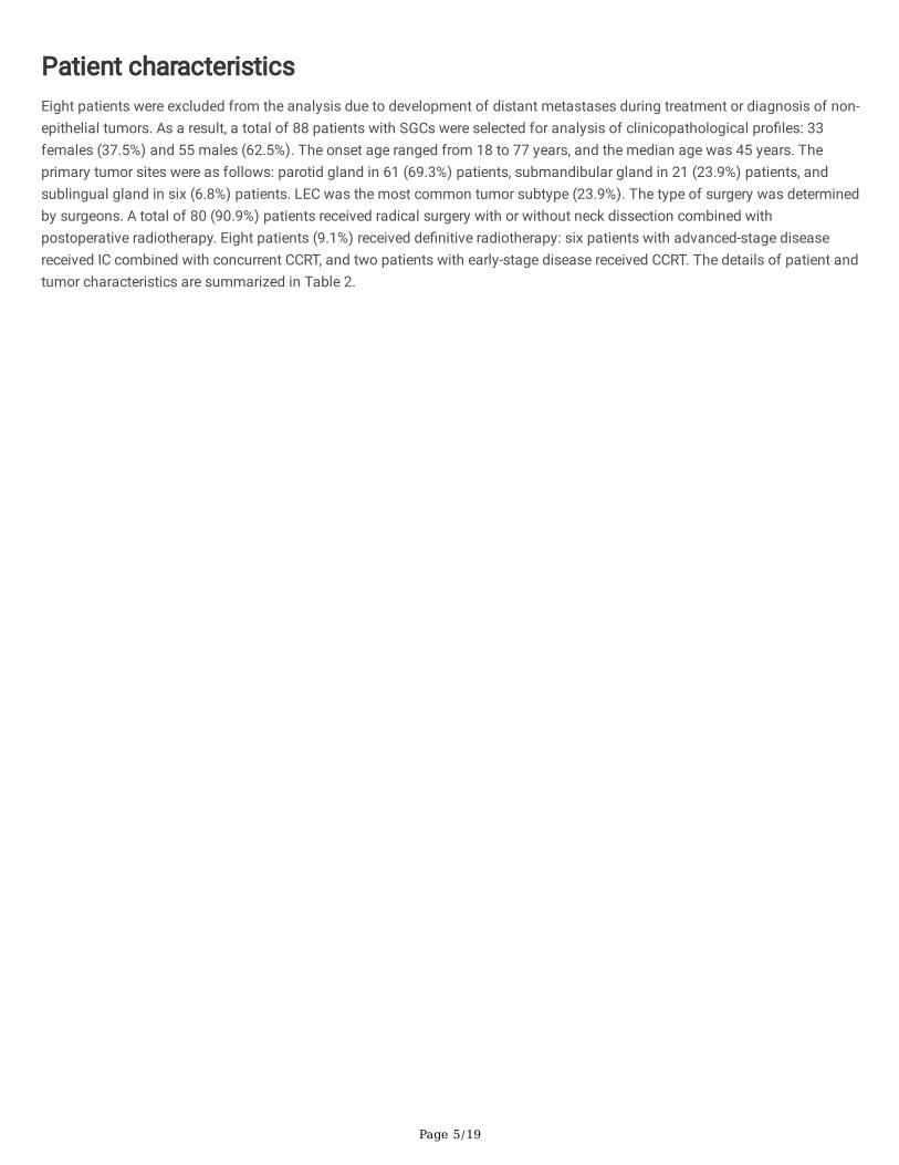
Patient characteristicsEight patients were excluded from the analysis due to development of distant metastases during treatment or diagnosis of non-epithelial tumors. As a result, a total of 88 patients with SGCs were selected for analysis of clinicopathological pro�les: 33females (37.5%) and 55 males (62.5%). The onset age ranged from 18 to 77 years, and the median age was 45 years. Theprimary tumor sites were as follows: parotid gland in 61 (69.3%) patients, submandibular gland in 21 (23.9%) patients, andsublingual gland in six (6.8%) patients. LEC was the most common tumor subtype (23.9%). The type of surgery was determinedby surgeons. A total of 80 (90.9%) patients received radical surgery with or without neck dissection combined withpostoperative radiotherapy. Eight patients (9.1%) received de�nitive radiotherapy: six patients with advanced-stage diseasereceived IC combined with concurrent CCRT, and two patients with early-stage disease received CCRT. The details of patient andtumor characteristics are summarized in Table 2.
Page 6/19
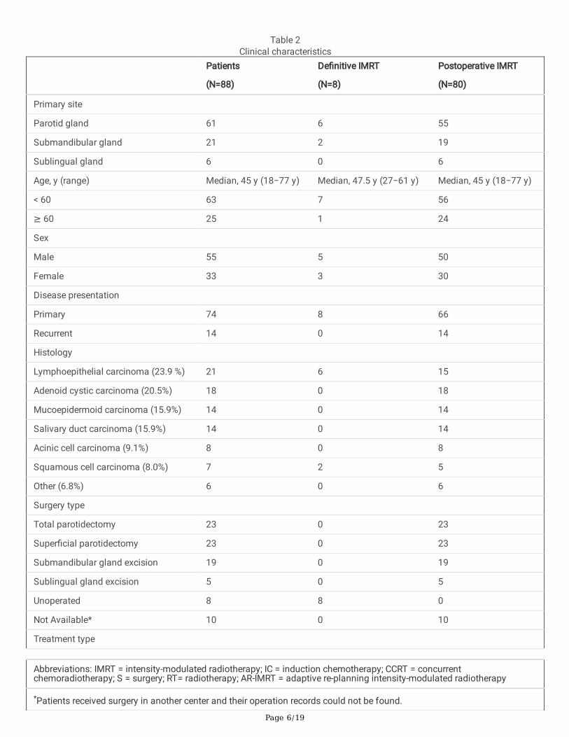
Table 2Clinical characteristics
Patients
(N=88)
De�nitive IMRT
(N=8)
Postoperative IMRT
(N=80)
Primary site
Parotid gland 61 6 55
Submandibular gland 21 2 19
Sublingual gland 6 0 6
Age, y (range) Median, 45 y (18−77 y) Median, 47.5 y (27−61 y) Median, 45 y (18−77 y)
< 60 63 7 56
≥ 60 25 1 24
Sex
Male 55 5 50
Female 33 3 30
Disease presentation
Primary 74 8 66
Recurrent 14 0 14
Histology
Lymphoepithelial carcinoma (23.9 %) 21 6 15
Adenoid cystic carcinoma (20.5%) 18 0 18
Mucoepidermoid carcinoma (15.9%) 14 0 14
Salivary duct carcinoma (15.9%) 14 0 14
Acinic cell carcinoma (9.1%) 8 0 8
Squamous cell carcinoma (8.0%) 7 2 5
Other (6.8%) 6 0 6
Surgery type
Total parotidectomy 23 0 23
Super�cial parotidectomy 23 0 23
Submandibular gland excision 19 0 19
Sublingual gland excision 5 0 5
Unoperated 8 8 0
Not Available* 10 0 10
Treatment type
Abbreviations: IMRT = intensity-modulated radiotherapy; IC = induction chemotherapy; CCRT = concurrentchemoradiotherapy; S = surgery; RT= radiotherapy; AR-IMRT = adaptive re-planning intensity-modulated radiotherapy
*Patients received surgery in another center and their operation records could not be found.
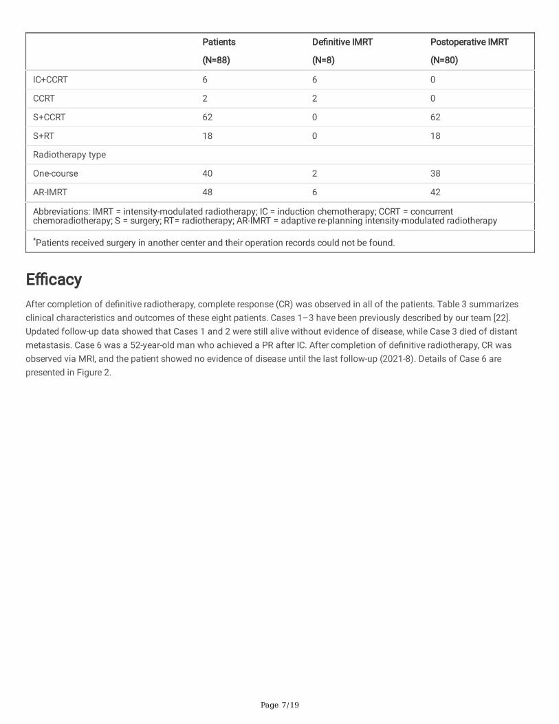
Page 7/19
Patients
(N=88)
De�nitive IMRT
(N=8)
Postoperative IMRT
(N=80)
IC+CCRT 6 6 0
CCRT 2 2 0
S+CCRT 62 0 62
S+RT 18 0 18
Radiotherapy type
One-course 40 2 38
AR-IMRT 48 6 42
Abbreviations: IMRT = intensity-modulated radiotherapy; IC = induction chemotherapy; CCRT = concurrentchemoradiotherapy; S = surgery; RT= radiotherapy; AR-IMRT = adaptive re-planning intensity-modulated radiotherapy
*Patients received surgery in another center and their operation records could not be found.
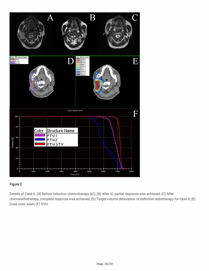
E�cacyAfter completion of de�nitive radiotherapy, complete response (CR) was observed in all of the patients. Table 3 summarizesclinical characteristics and outcomes of these eight patients. Cases 1–3 have been previously described by our team [22].Updated follow-up data showed that Cases 1 and 2 were still alive without evidence of disease, while Case 3 died of distantmetastasis. Case 6 was a 52-year-old man who achieved a PR after IC. After completion of de�nitive radiotherapy, CR wasobserved via MRI, and the patient showed no evidence of disease until the last follow-up (2021-8). Details of Case 6 arepresented in Figure 2.
Page 8/19
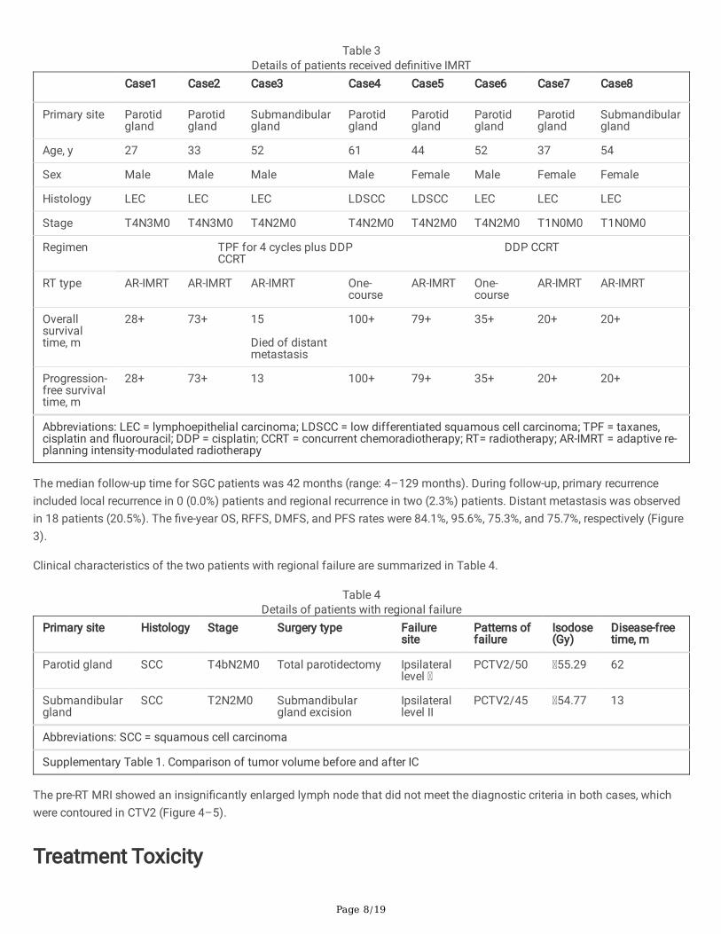
Table 3Details of patients received de�nitive IMRT
Case1 Case2 Case3 Case4 Case5 Case6 Case7 Case8
Primary site Parotidgland
Parotidgland
Submandibulargland
Parotidgland
Parotidgland
Parotidgland
Parotidgland
Submandibulargland
Age, y 27 33 52 61 44 52 37 54
Sex Male Male Male Male Female Male Female Female
Histology LEC LEC LEC LDSCC LDSCC LEC LEC LEC
Stage T4N3M0 T4N3M0 T4N2M0 T4N2M0 T4N2M0 T4N2M0 T1N0M0 T1N0M0
Regimen TPF for 4 cycles plus DDPCCRT
DDP CCRT
RT type AR-IMRT AR-IMRT AR-IMRT One-course
AR-IMRT One-course
AR-IMRT AR-IMRT
Overallsurvivaltime, m
28+ 73+ 15
Died of distantmetastasis
100+ 79+ 35+ 20+ 20+
Progression-free survivaltime, m
28+ 73+ 13 100+ 79+ 35+ 20+ 20+
Abbreviations: LEC = lymphoepithelial carcinoma; LDSCC = low differentiated squamous cell carcinoma; TPF = taxanes,cisplatin and �uorouracil; DDP = cisplatin; CCRT = concurrent chemoradiotherapy; RT= radiotherapy; AR-IMRT = adaptive re-planning intensity-modulated radiotherapy
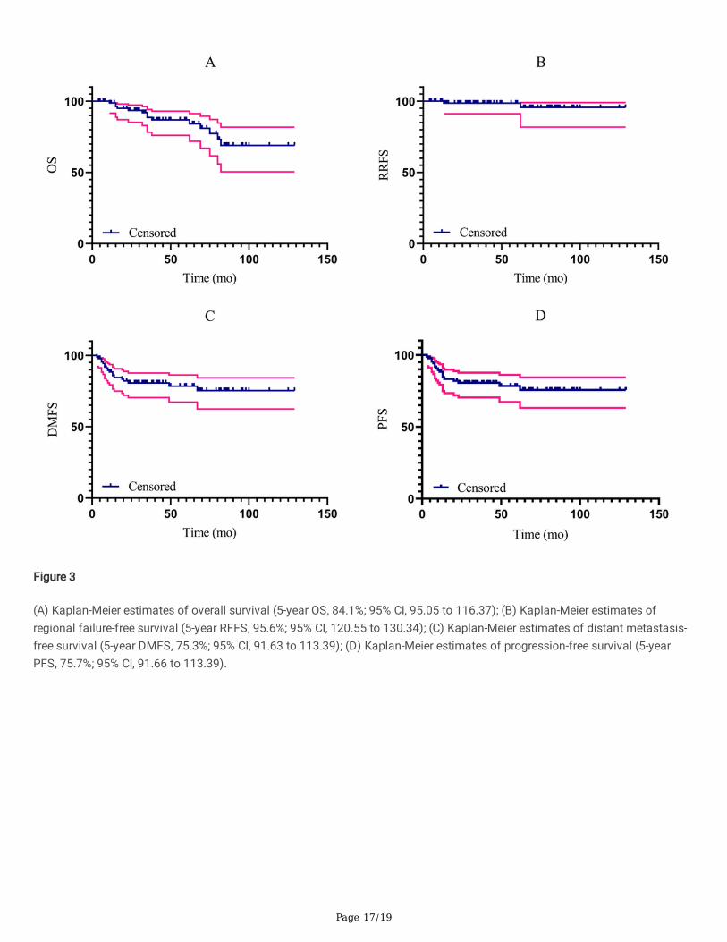
The median follow-up time for SGC patients was 42 months (range: 4–129 months). During follow-up, primary recurrenceincluded local recurrence in 0 (0.0%) patients and regional recurrence in two (2.3%) patients. Distant metastasis was observedin 18 patients (20.5%). The �ve-year OS, RFFS, DMFS, and PFS rates were 84.1%, 95.6%, 75.3%, and 75.7%, respectively (Figure3).
Clinical characteristics of the two patients with regional failure are summarized in Table 4.
Table 4Details of patients with regional failure
Primary site Histology Stage Surgery type Failuresite
Patterns offailure
Isodose(Gy)
Disease-freetime, m
Parotid gland SCC T4bN2M0 Total parotidectomy Ipsilaterallevel
PCTV2/50 55.29 62
Submandibulargland
SCC T2N2M0 Submandibulargland excision
Ipsilaterallevel II
PCTV2/45 54.77 13
Abbreviations: SCC = squamous cell carcinoma
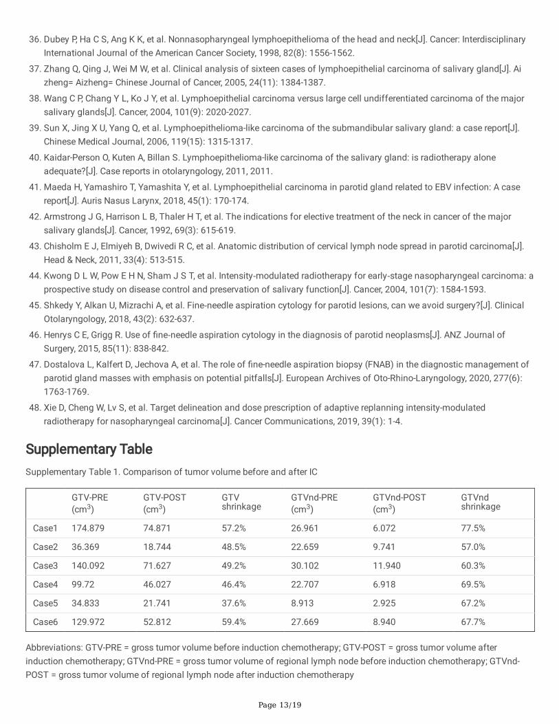
Supplementary Table 1. Comparison of tumor volume before and after IC
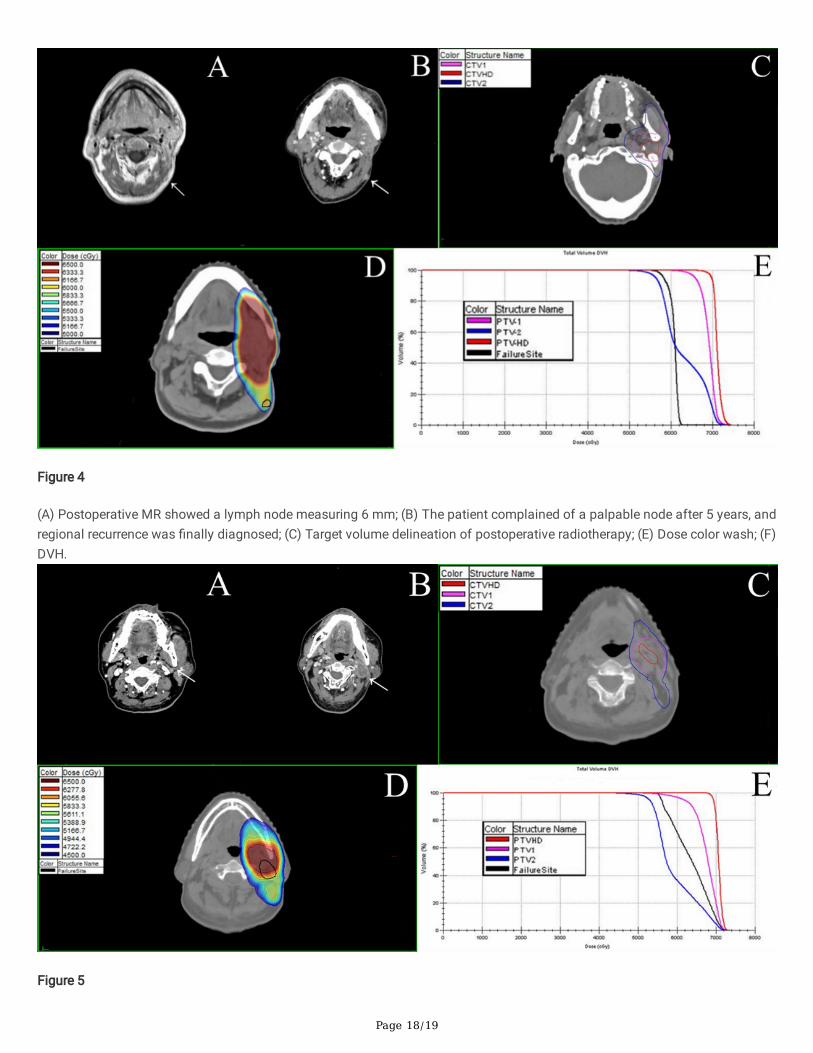
The pre-RT MRI showed an insigni�cantly enlarged lymph node that did not meet the diagnostic criteria in both cases, whichwere contoured in CTV2 (Figure 4–5).
Treatment Toxicity
Page 9/19
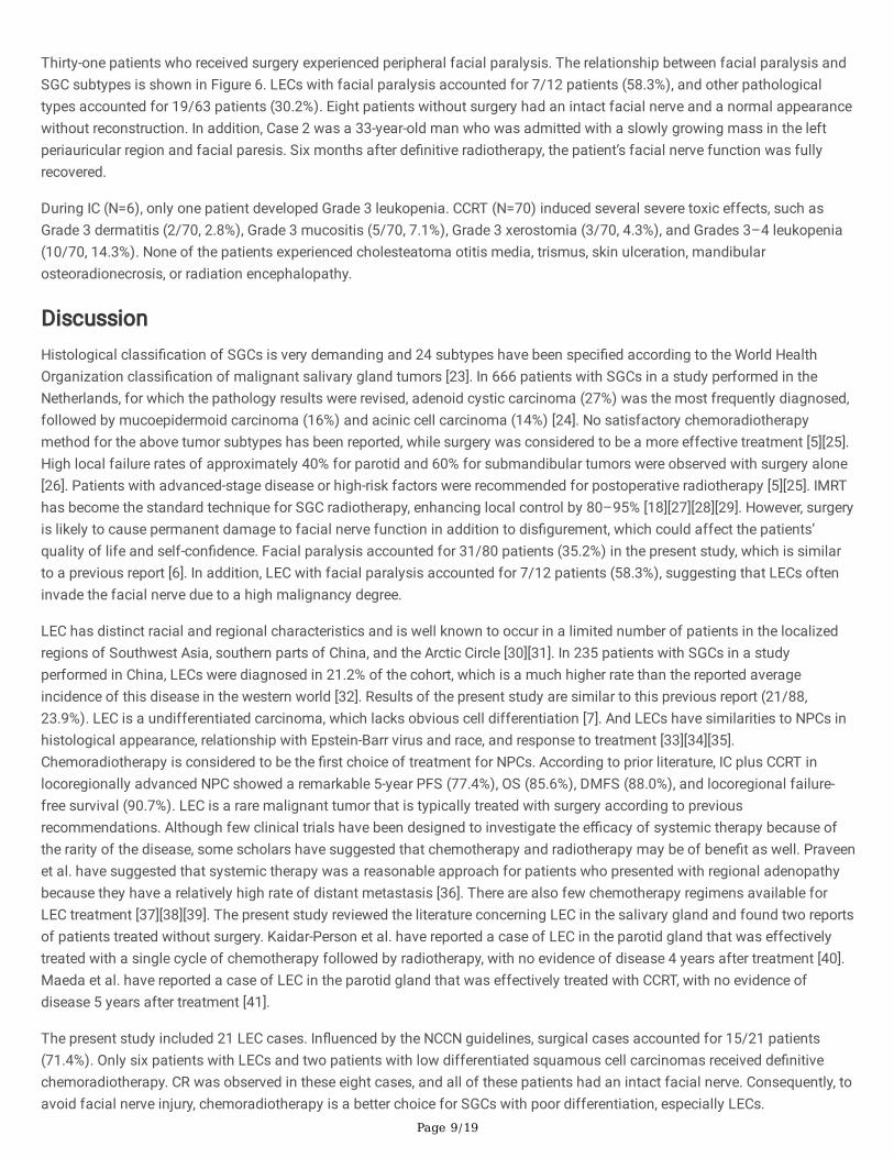
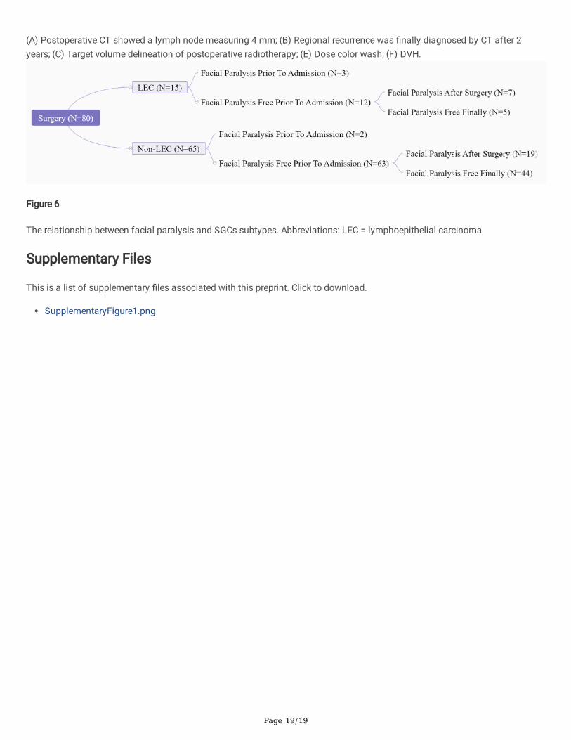
Thirty-one patients who received surgery experienced peripheral facial paralysis. The relationship between facial paralysis andSGC subtypes is shown in Figure 6. LECs with facial paralysis accounted for 7/12 patients (58.3%), and other pathologicaltypes accounted for 19/63 patients (30.2%). Eight patients without surgery had an intact facial nerve and a normal appearancewithout reconstruction. In addition, Case 2 was a 33-year-old man who was admitted with a slowly growing mass in the leftperiauricular region and facial paresis. Six months after de�nitive radiotherapy, the patient’s facial nerve function was fullyrecovered.
During IC (N=6), only one patient developed Grade 3 leukopenia. CCRT (N=70) induced several severe toxic effects, such asGrade 3 dermatitis (2/70, 2.8%), Grade 3 mucositis (5/70, 7.1%), Grade 3 xerostomia (3/70, 4.3%), and Grades 3–4 leukopenia(10/70, 14.3%). None of the patients experienced cholesteatoma otitis media, trismus, skin ulceration, mandibularosteoradionecrosis, or radiation encephalopathy.
DiscussionHistological classi�cation of SGCs is very demanding and 24 subtypes have been speci�ed according to the World HealthOrganization classi�cation of malignant salivary gland tumors [23]. In 666 patients with SGCs in a study performed in theNetherlands, for which the pathology results were revised, adenoid cystic carcinoma (27%) was the most frequently diagnosed,followed by mucoepidermoid carcinoma (16%) and acinic cell carcinoma (14%) [24]. No satisfactory chemoradiotherapymethod for the above tumor subtypes has been reported, while surgery was considered to be a more effective treatment [5][25].High local failure rates of approximately 40% for parotid and 60% for submandibular tumors were observed with surgery alone[26]. Patients with advanced-stage disease or high-risk factors were recommended for postoperative radiotherapy [5][25]. IMRThas become the standard technique for SGC radiotherapy, enhancing local control by 80–95% [18][27][28][29]. However, surgeryis likely to cause permanent damage to facial nerve function in addition to dis�gurement, which could affect the patients’quality of life and self-con�dence. Facial paralysis accounted for 31/80 patients (35.2%) in the present study, which is similarto a previous report [6]. In addition, LEC with facial paralysis accounted for 7/12 patients (58.3%), suggesting that LECs ofteninvade the facial nerve due to a high malignancy degree.
LEC has distinct racial and regional characteristics and is well known to occur in a limited number of patients in the localizedregions of Southwest Asia, southern parts of China, and the Arctic Circle [30][31]. In 235 patients with SGCs in a studyperformed in China, LECs were diagnosed in 21.2% of the cohort, which is a much higher rate than the reported averageincidence of this disease in the western world [32]. Results of the present study are similar to this previous report (21/88,23.9%). LEC is a undifferentiated carcinoma, which lacks obvious cell differentiation [7]. And LECs have similarities to NPCs inhistological appearance, relationship with Epstein-Barr virus and race, and response to treatment [33][34][35].Chemoradiotherapy is considered to be the �rst choice of treatment for NPCs. According to prior literature, IC plus CCRT inlocoregionally advanced NPC showed a remarkable 5-year PFS (77.4%), OS (85.6%), DMFS (88.0%), and locoregional failure-free survival (90.7%). LEC is a rare malignant tumor that is typically treated with surgery according to previousrecommendations. Although few clinical trials have been designed to investigate the e�cacy of systemic therapy because ofthe rarity of the disease, some scholars have suggested that chemotherapy and radiotherapy may be of bene�t as well. Praveenet al. have suggested that systemic therapy was a reasonable approach for patients who presented with regional adenopathybecause they have a relatively high rate of distant metastasis [36]. There are also few chemotherapy regimens available forLEC treatment [37][38][39]. The present study reviewed the literature concerning LEC in the salivary gland and found two reportsof patients treated without surgery. Kaidar-Person et al. have reported a case of LEC in the parotid gland that was effectivelytreated with a single cycle of chemotherapy followed by radiotherapy, with no evidence of disease 4 years after treatment [40].Maeda et al. have reported a case of LEC in the parotid gland that was effectively treated with CCRT, with no evidence ofdisease 5 years after treatment [41].
The present study included 21 LEC cases. In�uenced by the NCCN guidelines, surgical cases accounted for 15/21 patients(71.4%). Only six patients with LECs and two patients with low differentiated squamous cell carcinomas received de�nitivechemoradiotherapy. CR was observed in these eight cases, and all of these patients had an intact facial nerve. Consequently, toavoid facial nerve injury, chemoradiotherapy is a better choice for SGCs with poor differentiation, especially LECs.

Page 10/19
There is still confusion concerning the optimal radiation target volume for SGCs. As reported previously, the present study reliedon surgical principles to determine the IMRT target volume [16]. The 5-year OS, RFFS, DMFS, and PFS were 84.1%, 95.6%,75.3%, and 75.7%, respectively. No patient developed local recurrence, and the main cause of failure within the study cohortwas distant metastasis, which suggested that the method was reasonable and worthy of further research.
The present study included 14 patients with recurrent SGCs after primary surgery. In general, the �rst treatment plays a majorrole in cancer. But perhaps because of the special anatomic location of the salivary glands, secondary operation pluspostoperative radiotherapy for recurrent SGCs also showed good clinical outcomes. Consequently, patients with recurrent SGCsare expected to strive for radical treatment.
The pre-RT MRI showed an insigni�cantly enlarged lymph node that did not meet the diagnostic criteria for both regionalfailures, which were contoured in CTV2 (Figure 4–5). According to a previous report, high rates of implicit metastasis ofapproximately 12–45% were observed for lymph nodes in SGCs cases, suggesting that it is very important in clinical practice todetermine whether the lymph nodes have been spared or not [42][43]. However, the optimal treatment for risky lymph nodes thatdo not meet the diagnostic criteria remains to be determined. Guidelines from the NCCN recommend prescription doses of 44–50 Gy and 54–63 Gy for low and intermediate risk sites of suspected subclinical spread, respectively [5]. In addition, recurrenceobserved in a previous study occurred in a cervical lymph node that was not signi�cantly enlarged, but was probably involved,and received a radiation dose of about 64 Gy [44]. Consequently, to control the more than microscopic disease, a dose of 63-65Gy has been irradiated for the risky lymph nodes in the following treatment.
The present study has some limitations. First, it was a retrospective study from a single center, and further prospectivemulticenter studies are needed. Second, patients without surgery received lesion site �ne needle aspiration biopsy in our study.According to previous reports, �ne needle aspiration biopsy showed a sensitivity and a speci�city of 41.7–92.8% and 93.9–98.5%, respectively [45][46][47]. Whether intraoperative frozen sections should be performed to obtain more pathologicalinformation is need to be studied further.
ConclusionsLECs may be sensitive to chemoradiotherapy, which may achieve a radical effect and avoid unnecessary surgical injury. ICcombined with CCRT is expected to become a new treatment strategy for advanced LECs. The IMRT target volume delineationaccording to the surgical principles may be a more promising method with good clinical e�cacy that is worthy of further study.
AbbreviationsIMRT: intensity-Modulated Radiotherapy, SGC: major salivary gland carcinoma, LEC: lymphoepithelial carcinoma, IC: inductionchemotherapy, CCRT: concurrent chemoradiotherapy, NCCN: National Comprehensive Cancer Network, NPC: nasopharyngealcarcinoma, AJCC: American Joint Committee on Cancer, CT: computed tomography, MRI: magnetic resonance imaging, TPF:taxanes, cisplatin, and �uorouracil, GTVp/nd: gross tumor volume of primary site/regional lymph node, CTV1: medium riskclinical tumor volume, CTV2: low risk clinical tumor volume, CTV-HD: high-risk clinical tumor volume, PTV: planning targetvolumes, OS: overall survival, RFFS: regional failure-free survival, DMFS: distant metastasis-free survival, PFS: progression-freesurvival, CR: complete response, PR: partial response
DeclarationsEthics approval and consent to participate: This study was approved by the Medical Ethics Committee of A�liated Hospital ofSun Yat-sen University Cancer Center, and the need for written informed consent was waived.
Consent for publication: Not applicable.
Availability of supporting data and materials: All data generated or analyzed during this study are included in this publishedarticle.
Page 11/19
Competing interests: The authors declare that they have no competing interests.
Authors’ Contributions: ZQ YS: conception and design; ZQ WC FL SL XL: data collection, statistical analysis; ZQ ZW:manuscript preparation; ZW LW DX: manuscript editing; YT YS: quality control of data and manuscript review; MW JH: radiationtherapy planning.
All authors read and approved the �nal manuscript.
Acknowledgments: The authors declare that they have no funding.
References1. Bjørndal K, Krogdahl A, Therkildsen M H, et al. Salivary gland carcinoma in Denmark 1990–2005: Outcome and prognostic
factors: Results of the Danish Head and Neck Cancer Group (DAHANCA)[J]. Oral Oncology, 2012, 48(2): 179-185.
2. Gatta G, Guzzo M, Locati L D, et al. Major and minor salivary gland tumours[J]. Critical Reviews in Oncology/Hematology,2020, 152: 102959.
3. Del Signore A G, Megwalu U C. The rising incidence of major salivary gland cancer in the United States[J]. Ear, Nose &Throat Journal, 2017, 96(3): E13-E16.
4. Ata-Ali J, Zurriaga O, Alberich C. Incidence and survival rates for malignant salivary gland tumors[J]. Journal of OralScience, 2016, 58(1): 67-73.
5. National Comprehensive Cancer Network. Head and Neck Cancers (Version 3.2021).https://www.nccn.org/professionals/physician_gls/pdf/head-and-neck.pdf. Accessed April 27, 2021.
�. Guntinas-Lichius O, Silver C E, Thielker J, et al. Management of the facial nerve in parotid cancer: preservation or resectionand reconstruction[J]. European Archives of Oto-rhino-laryngology, 2018, 275(11): 2615-2626.
7. Hilderman W C, Gordon J S, Large Jr H L, et al. Malignant lymphoepithelial lesion with carcinomatous componentapparently arising in parotid gland. A malignant counterpart of benign lymphoepithelial lesion?[J]. Cancer, 1962, 15(3):606-610.
�. Saku T, Cheng J, Jen K Y, et al. Epstein-Barr virus infected lymphoepithelial carcinomas of the salivary gland in the Russia-Asia area: a clinicopathologic study of 160 cases[J]. Arkhiv Patologii, 2003, 65(2): 35-39.
9. Pittaluga S, Wong M P, Chung L P, et al. Clonal Epstein-Barr virus in lymphoepithelioma-like carcinoma of the lung[J]. TheAmerican Journal of Surgical Pathology, 1993, 17(7): 678-682.
10. Shibata D, Tokunaga M, Uemura Y, et al. Association of Epstein-Barr virus with undifferentiated gastric carcinomas withintense lymphoid in�ltration. Lymphoepithelioma-like carcinoma[J]. The American Journal of Pathology, 1991, 139(3): 469.
11. Li W F, Chen N Y, Zhang N, et al. Concurrent chemoradiotherapy with/without induction chemotherapy in locoregionallyadvanced nasopharyngeal carcinoma: Long–term results of phase 3 randomized controlled trial[J]. International Journalof Cancer, 2019, 145(1): 295-305.
12. Whelan A, Al-Sayed A A, Bullock M, et al. Primary parotid lymphoepithelial carcinoma: A case report and literature review ofa rare pathological entity[J]. International Journal of Surgery Case Reports, 2020, 72: 610-614.
13. Kaur J, Goyal S, Muzumder S, et al. Outcome of surgery and post-operative radiotherapy for major salivary glandcarcinoma: ten year experience from a single institute[J]. Asian Paci�c Journal of Cancer Prevention, 2014, 15(19): 8259-8263.
14. Hosni A, Huang S H, Goldstein D, et al. Outcomes and prognostic factors for major salivary gland carcinoma followingpostoperative radiotherapy[J]. Oral Oncology, 2016, 54: 75-80.
15. Nutting C M, Morden J P, Beasley M, et al. Results of a multicentre randomised controlled trial of cochlear-sparing intensity-modulated radiotherapy versus conventional radiotherapy in patients with parotid cancer (COSTAR; CRUK/08/004)[J].European Journal of Cancer, 2018, 103: 249-258.
Page 12/19
1�. Lyu S, Wu Z, Xie D, et al. Clinical target volume delineation of postoperative intensity-modulated radiotherapy for majorsalivary gland tumours according to surgical principles: an innovative method[J]. Journal of Cancer Research and ClinicalOncology, 2021: 1-10.
17. Wang L, Wu Z, Xie D, et al. Reduction of target volume and the corresponding dose for the tumor regression �eld afterinduction chemotherapy in locoregionally advanced nasopharyngeal carcinoma[J]. Cancer Research and Treatment:O�cial Journal of Korean Cancer Association, 2019, 51(2): 685.
1�. Terhaard C H J, Lubsen H, Rasch C R N, et al. The role of radiotherapy in the treatment of malignant salivary glandtumors[J]. International Journal of Radiation Oncology* Biology* Physics, 2005, 61(1): 103-111.
19. Ferlito A, Pellitteri P K, Robbins K T, et al. Management of the neck in cancer of the major salivary glands, thyroid andparathyroid glands[J]. Acta Oto-laryngologica, 2002, 122(6): 673-678.
20. Armstrong J G, Harrison L B, Thaler H T, et al. The indications for elective treatment of the neck in cancer of the majorsalivary glands[J]. Cancer, 1992, 69(3): 615-619.
21. Chao K S C, Ozyigit G, Tran B N, et al. Patterns of failure in patients receiving de�nitive and postoperative IMRT for head-and-neck cancer[J]. International Journal of Radiation Oncology* Biology* Physics, 2003, 55(2): 312-321.
22. Lv S, Xie D, Wu Z, et al. Is surgery an inevitable treatment for advanced salivary lymphoepithelial carcinoma? Three casereports[J]. Ear, Nose & Throat Journal, 2020: 0145561320923170.
23. Leivo I. Insights into a complex group of neoplastic disease: advances in histopathologic classi�cation and molecularpathology of salivary gland cancer[J]. Acta Oncologica, 2006, 45(6): 662-668.
24. Terhaard C H J, van der Schroeff M P, van Schie K, et al. The prognostic role of comorbidity in salivary gland carcinoma[J].Cancer: Interdisciplinary International Journal of the American Cancer Society, 2008, 113(7): 1572-1579.
25. Geiger J L, Ismaila N, Beadle B, et al. Management of salivary gland malignancy: ASCO Guideline[J]. Journal of ClinicalOncology, 2021: JCO. 21.00449.
2�. Spiro R H. Salivary neoplasms: overview of a 35‐year experience with 2,807 patients[J]. Head & Neck Surgery, 1986, 8(3):177-184.
27. Therkildsen M H, Christensen M, Andersen L J, et al. Salivary gland carcinomas: Prognostic factors[J]. Acta Oncologica,1998, 37(7-8): 701-713.
2�. Chen A M, Bucci M K, Weinberg V, et al. Adenoid cystic carcinoma of the head and neck treated by surgery with or withoutpostoperative radiation therapy: Prognostic features of recurrence[J]. International Journal of Radiation Oncology*Biology* Physics, 2006, 66(1): 152-159.
29. Poulsen M G, Pratt G R, Kynaston B, et al. Prognostic variables in malignant epithelial tumors of the parotid[J].International Journal of Radiation Oncology* Biology* Physics, 1992, 23(2): 327-332.
30. Nielsen N H Ø, Mikkelsen F, Hansen J P H. Incidence of salivary gland neoplasms in Greenland with special reference to ananaplastic carcinoma[J]. Acta Pathologica Microbiologica Scandinavica Section A Pathology, 1978, 86(1‐6): 185-193.
31. Leung S Y, Chung L P, Yuen S T, et al. Lymphoepithelial carcinoma of the salivary gland: in situ detection of Epstein-Barrvirus[J]. Journal of Clinical Pathology, 1995, 48(11): 1022-1027.
32. Li F, Zhu G, Wang Y, et al. A clinical analysis of 37 cases with lymphoepithelial carcinoma of the major salivary glandtreated by surgical resection and postoperative radiotherapy: A single institution study[J]. Medical Oncology, 2014, 31(5):957.
33. Cleary K R, Batsakis J G. Undifferentiated carcinoma with lymphoid stroma of the major salivary glands[J]. Annals ofOtology, Rhinology & Laryngology, 1990, 99(3): 236-238.
34. Hamilton-Dutoit S J, Therkildsen M H, Nielsen N H, et al. Undifferentiated carcinoma of the salivary gland in GreenlandicEskimos: demonstration of Epstein-Barr virus DNA by in situ nucleic acid hybridization[J]. Human Pathology, 1991, 22(8):811-815.
35. Lanier A P, Clift S R, Bornkamm G, et al. Epstein-Barr virus and malignant lymphoepithelial lesions of the salivary gland[J].Arctic Medical Research, 1991, 50(2): 55-61.
Page 13/19
3�. Dubey P, Ha C S, Ang K K, et al. Nonnasopharyngeal lymphoepithelioma of the head and neck[J]. Cancer: InterdisciplinaryInternational Journal of the American Cancer Society, 1998, 82(8): 1556-1562.
37. Zhang Q, Qing J, Wei M W, et al. Clinical analysis of sixteen cases of lymphoepithelial carcinoma of salivary gland[J]. Aizheng= Aizheng= Chinese Journal of Cancer, 2005, 24(11): 1384-1387.
3�. Wang C P, Chang Y L, Ko J Y, et al. Lymphoepithelial carcinoma versus large cell undifferentiated carcinoma of the majorsalivary glands[J]. Cancer, 2004, 101(9): 2020-2027.
39. Sun X, Jing X U, Yang Q, et al. Lymphoepithelioma-like carcinoma of the submandibular salivary gland: a case report[J].Chinese Medical Journal, 2006, 119(15): 1315-1317.
40. Kaidar-Person O, Kuten A, Billan S. Lymphoepithelioma-like carcinoma of the salivary gland: is radiotherapy aloneadequate?[J]. Case reports in otolaryngology, 2011, 2011.
41. Maeda H, Yamashiro T, Yamashita Y, et al. Lymphoepithelial carcinoma in parotid gland related to EBV infection: A casereport[J]. Auris Nasus Larynx, 2018, 45(1): 170-174.
42. Armstrong J G, Harrison L B, Thaler H T, et al. The indications for elective treatment of the neck in cancer of the majorsalivary glands[J]. Cancer, 1992, 69(3): 615-619.
43. Chisholm E J, Elmiyeh B, Dwivedi R C, et al. Anatomic distribution of cervical lymph node spread in parotid carcinoma[J].Head & Neck, 2011, 33(4): 513-515.
44. Kwong D L W, Pow E H N, Sham J S T, et al. Intensity‐modulated radiotherapy for early‐stage nasopharyngeal carcinoma: aprospective study on disease control and preservation of salivary function[J]. Cancer, 2004, 101(7): 1584-1593.
45. Shkedy Y, Alkan U, Mizrachi A, et al. Fine‐needle aspiration cytology for parotid lesions, can we avoid surgery?[J]. ClinicalOtolaryngology, 2018, 43(2): 632-637.
4�. Henrys C E, Grigg R. Use of �ne‐needle aspiration cytology in the diagnosis of parotid neoplasms[J]. ANZ Journal ofSurgery, 2015, 85(11): 838-842.
47. Dostalova L, Kalfert D, Jechova A, et al. The role of �ne-needle aspiration biopsy (FNAB) in the diagnostic management ofparotid gland masses with emphasis on potential pitfalls[J]. European Archives of Oto-Rhino-Laryngology, 2020, 277(6):1763-1769.
4�. Xie D, Cheng W, Lv S, et al. Target delineation and dose prescription of adaptive replanning intensity-modulatedradiotherapy for nasopharyngeal carcinoma[J]. Cancer Communications, 2019, 39(1): 1-4.
Supplementary TableSupplementary Table 1. Comparison of tumor volume before and after IC
GTV-PRE(cm3)
GTV-POST(cm3)
GTVshrinkage
GTVnd-PRE(cm3)
GTVnd-POST(cm3)
GTVndshrinkage
Case1 174.879 74.871 57.2% 26.961 6.072 77.5%
Case2 36.369 18.744 48.5% 22.659 9.741 57.0%
Case3 140.092 71.627 49.2% 30.102 11.940 60.3%
Case4 99.72 46.027 46.4% 22.707 6.918 69.5%
Case5 34.833 21.741 37.6% 8.913 2.925 67.2%
Case6 129.972 52.812 59.4% 27.669 8.940 67.7%
Abbreviations: GTV-PRE = gross tumor volume before induction chemotherapy; GTV-POST = gross tumor volume afterinduction chemotherapy; GTVnd-PRE = gross tumor volume of regional lymph node before induction chemotherapy; GTVnd-POST = gross tumor volume of regional lymph node after induction chemotherapy
Page 14/19
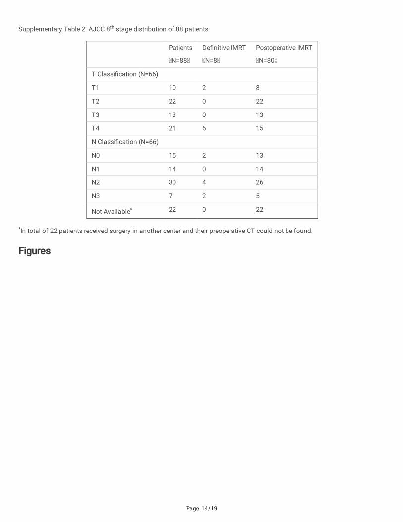
Supplementary Table 2. AJCC 8th stage distribution of 88 patients
Patients
N=88
De�nitive IMRT
N=8
Postoperative IMRT
N=80
T Classi�cation (N=66)
T1 10 2 8
T2 22 0 22
T3 13 0 13
T4 21 6 15
N Classi�cation (N=66)
N0 15 2 13
N1 14 0 14
N2 30 4 26
N3 7 2 5
Not Available* 22 0 22
*In total of 22 patients received surgery in another center and their preoperative CT could not be found.
Figures
Page 15/19
Figure 1
Target volume delineation of postoperative radiotherapy for salivary gland cancer patients. (A) Preoperative CT showed that thetumor (redline) crossed the deep lobe and the isthmus of the parotid gland and was adjacent to posterior venter of the digastricmuscle, carotid sheath, styloid process and mandibular branch; (B) Surgical records showed that the patient underwent partialparotidectomy and facial nerve dissection. The purple dotted line showed the extent of surgical resection; (C) Postoperative MRshowed that the reserved tissues included the retained parotid tissues (blue line), posterior venter of the digastric muscle,carotid sheath, styloid process and mandibular branch. (D) Posterior venter of the digastric muscle, carotid sheath, styloidprocess and mandibular branch were all less than 5 mm from the primary tumor edge, so they were very likely to be invaded, asdelineated in the CTV-HD area. The retained parotid tissues were more than 10 mm away from the primary tumor edge, so theywere delineated in the CTV2 area.
Page 16/19
Figure 2
Details of Case 6. (A) Before induction chemotherapy (IC); (B) After IC, partial response was achieved; (C) Afterchemoradiotherapy, complete response was achieved; (D) Target volume delineation of de�nitive radiotherapy for Case 6; (E)Dose color wash; (F) DVH.
Page 17/19
Figure 3
(A) Kaplan-Meier estimates of overall survival (5-year OS, 84.1%; 95% CI, 95.05 to 116.37); (B) Kaplan-Meier estimates ofregional failure-free survival (5-year RFFS, 95.6%; 95% CI, 120.55 to 130.34); (C) Kaplan-Meier estimates of distant metastasis-free survival (5-year DMFS, 75.3%; 95% CI, 91.63 to 113.39); (D) Kaplan-Meier estimates of progression-free survival (5-yearPFS, 75.7%; 95% CI, 91.66 to 113.39).
Page 18/19
Figure 4
(A) Postoperative MR showed a lymph node measuring 6 mm; (B) The patient complained of a palpable node after 5 years, andregional recurrence was �nally diagnosed; (C) Target volume delineation of postoperative radiotherapy; (E) Dose color wash; (F)DVH.
Figure 5
Page 19/19
(A) Postoperative CT showed a lymph node measuring 4 mm; (B) Regional recurrence was �nally diagnosed by CT after 2years; (C) Target volume delineation of postoperative radiotherapy; (E) Dose color wash; (F) DVH.
Figure 6
The relationship between facial paralysis and SGCs subtypes. Abbreviations: LEC = lymphoepithelial carcinoma
Supplementary Files
This is a list of supplementary �les associated with this preprint. Click to download.
SupplementaryFigure1.png