Embed Size (px)
Citation preview
Summary. Basement membranes (BMs) constitute adistinct compartment of the extracellular matrix (ECM).All BMs show a similar structural appearance but differin molecular composition. These variations have criticalfunctional implications. The aim of this study is toestablish the pattern of the tomato lectin (Lycopersiconesculentum agglutinin - LEA) binding sites in the BMsof the developing chick embryo (stages 4-21, Hamburgerand Hamilton, 1951) in order to achieve a betterunderstanding of the molecular heterogeneity of BMs.The study was performed with transmission electronmicroscopy (TEM) histochemistry, and confocal lasermicroscopy.
TEM showed that LEA bound to the lamina densaand to the lamina fibroreticularis of the BMs. Throughthe period studied, most of the LEA binding appeared inthe ectodermal BM and its derivatives. In the limb bud,LEA binding to the ectoderm BM was more intense inthe ventral half than in the dorsal half. Furthermore,LEA allowed the early (HH16) detection of thetransverse fibrillar tracts. In the lens and in the inner earprimordium, the BMs were LEA positive through theplacode and cup stages. The binding was progressivelyreduced through the vesicle stage. The BMs of theolfactory primordium, and of the Rathke’s pouch werepositive. In contrast, the BMs of the developing centralnervous system were negative. The BMs of both theparaxial and the lateral plates of the mesoderm werenegative, whereas the notochord and the BM of theWolffian duct were positive. The endodermal BM and itsderivatives were negative. The ECM located between thefusing endocardial tubes, and the BM of the fusion zoneof the paired aortae, were positive. This suggested anactive role of the LEA-positive glycoproteins in thefusion of endothelia. Our results show the heterogeneity
of the chick embryo BMs during development. Inaddition, LEA constitutes an excellent marker for theprimordial germ cells.
Key words: Chick development, Basement membrane,Lycopersicum esculentum agglutinin, Wolff duct,Primordial cells
Introduction
The extracellular matrix (ECM) is a complexassemblage of molecules. The different molecularcomponents interact both with each other and with cellsby means of integrin receptors (Hynes, 1992), tomodulate a wide range of cellular functions. Therepertoire of integrin expression and ECM compositionappear to dictate whether a cell will survive, proliferate,or exit the cell cycle and differentiate in response tosoluble factors (Beauvais-Jouneau and Thiery, 1997).Furthermore, some of the sulphated glycosaminoglycans(GAGs) present in the ECM interact with diverse growthfactors to become part of their signalling pathway.Changes in GAG composition may function as criticaltemporal regulators of the activity of growth factors, andas morphogen signalling during embryogenesis (Allenand Rapraeger, 2003).
Basement membranes (BMs) are layers ofspecialized ECM associated with different cell typessuch as epithelial and endothelial cells. They have a cell-supporting role, are involved in the maintenance andregulation of the cellular phenotypes (Streuli, 1996), andappear to control macromolecular passage throughepithelia and endothelia (Robinson and Walton, 1989).Electron microscope studies have shown few significantstructural differences between the BMs from differentanatomic sites (Inoue, 1989). However, the molecularcomposition of BMs is extremely heterogeneous(Damjanov, 1990; Erickson and Cauchman, 2000).
Basement membrane heterogeneity during chick development as shown by tomato (Lycopersicon esculentum) lectin bindingJ.L. Ojeda and J.M. IcardoDepartment of Anatomy and Cell Biology, Faculty of Medicine, University of Cantabria, Santander, Spain
Histol Histopathol (2006) 21: 237-248
Offprint requests to: Prof. José L. Ojeda, Departamento de Anatomía yBiología Celular, Facultad de Medicina, c/ Cardenal Herrera Oria, s/n,39011-Santander, Spain. e-mail: [email protected]
http://www.hh.um.es
Histology andHistopathology
Cellular and Molecular Biology
Several isoforms of laminin, collagen IV andentactin/nidogen (Ekblom et al., 1988; Sanes et al.,1990; Kohfeldt et al., 1998; Pierce et al., 2000; Virtanenet al., 2003), as well as different proteoglycans (Ericksonand Couchman, 2000), are normal components of theBMs. Variations in sugar composition, size, andsulfatation pattern of the constituents of the proteoglycanchains, can generate a wide array of molecular species(Gallagher, 1986; Sanes et al., 1990). A large body ofevidence indicates that the histochemical characteristicsof BMs vary from one organ to another; that differentanatomic structures within the same organ may beinvested with different BMs; and, finally, that BMcomposition changes considerably during developmentand in relation to adult stages (Wan et al., 1984; Leu andDamjanov, 1988; Ojeda et al., 1993). The knowledge ofthe temporal sequence in which the developmentalheterogeneity of the BMs is established is necessary tounderstand the possible regulatory roles played by BMs.
The carbohydrate moieties of the BMglycoconjugates vary during development. Thesechanges have been implicated in cell-to-cell and in cell-matrix interactions (Shur, 1989). Also, carbohydratedomains may serve as markers of several physiologicaland pathological conditions (Rademacher et al., 1988;Salamat et al., 1993). Lectins are ubiquitous proteins ofnon-immune origin which show a high specificity forcarbohydrate moieties. This quality of lectins has provedvery useful in detecting both cellular and extracellularcarbohydrate changes in BMs (Lis and Sharon, 1986).Distinct changes in the ability of BMs to bind todifferent lectins have been detected in the course of themorphogenesis of several organs (Gallagher, 1986;Laitinen et al., 1987; Ojeda et al., 1993). However,systematic studies of the BM glycosilation patternduring chick development are scarce.
The Lycopersicon esculentum (tomato) agglutinin(LEA) is a lectin which binds specifically to the poly-N-acetyl D-glucosamine sugar residues (Nachbar andOppenheim, 1982) (nominal carbohydrate specificity:GlcNAcß1, 4GlcNAcß1, 4GlcNAcß1, 4GlcNAc>GlcNAcß1, 4GlcNAcß1, 4GlcNAc>GlcNAcß1,4GlcNAc, - Goldstein and Poretz, 1986). This lectin hasfrequently been used as a marker for microglial cells(Acarin et al., 1994; Andjelkovic et al., 1998) and fornon-professional phagocytes (Andjelkovic et al., 1998;Navarro et al., 2003). In the early chick embryo, LEAhas shown affinity for the primordial germ cells (stage 4-5, Hamburger and Hamilton, 1951) and for the mostlateral regions of the epiblast (6HH-7HH) (Griffith andSanders, 1991). The developmental distribution ofseveral lectins has been the subject of previous studies(Griffith and Sanders, 1991; Ojeda et al., 1993; Wilsonand Wyatt, 1995; Yao et al., 1996; Götz andQuondamatteo, 2001). However, very little is known ofthe changes which may occur in the distribution of LEAduring development (see: Griffith and Sanders, 1991;Hentschel and Walther, 1993). The aim of the presentstudy is to establish the pattern of LEA-binding sites
during the development of the chick embryo (4HH-21HH). Special emphasis has been placed on BMs. Abetter understanding of BM heterogeneity will provideinsights into the regulatory roles of BMs duringdevelopment.
Materials and methods
Fertilised White Leghorn chicken eggs wereincubated at 37.5°C in a humidified commercial eggincubator. Embryos ranging from stages HH4 to HH21(Hamburger and Hamilton, 1951) were used accordingto the following procedures.
Fluorescence microscopy
The embryos were fixed in cold ethanol-glacialacetic acid (99:1), dehydrated in cold ethanol,transferred to cold xylene and embedded in diethyleneglycol diestearate (DGD) (Polysciences, Inc.Warrington,PA), as previously described (Ojeda et al., 1989).Briefly, the embryos were transferred from the xylene toa mixture of xylene/DGD of 2:1 (10 min.), and then 1:2(10 min.), followed by two changes of 100% DGD (15min. each). During the embedding procedure, the DGDwas maintained at 60°C to keep it molten. Afterinfiltration, the embryos were transferred to flat siliconeembedding moulds filled with freshly melted DGD. Theblocks were allowed to solidify at room temperature, andwere stored at 4ºC until sectioning.
Semi-thin sections with a nominal thickness of 1-2µm were obtained on a Jeol Jum-7 ultramicrotome usingglass knives with a water bath. The sections wereremoved from the water with a glass rod and transferredto a water drop on a clean slide. The slides were placedon a warm plate (40°C) until dry. They were then storedat 4°C overnight. Dewaxed sections were stained withLEA fluorescein isothiocyanate (FITC)-conjugated(Sigma) (50µg/ml in phosphate buffered saline [PBS])for 30 min, followed by three washes with PBS (10 min.each). In addition, selected sections were stained with apropidium iodide solution (1µg/ml) for 3 min. at roomtemperature to visualize the cell nucleus. All the stainingprocedures were carried out in the dark at roomtemperature. The slides were mounted with theantifading medium Vectashield (Vector, Burlingame,CA). All sections were examined with a confocal lasermicroscope (MRC 1024; BioRad) using argon (488nm)and HeNe (543nm) lasers. The specificity of the lectinbinding was tested by incubating the FITC-conjugatedlectin with 0.1M solution of N-acetyl D-glucosamine(Sigma) for 30 min. Then, selected sections wereincubated with the mix lectin-sugar (Acarin et al., 1994).All the controls were routinely negative.
Enzymatic digestion
Prior to LEA staining, a duplicate series of sectionswas treated with neuraminidase in order to expose the
238
LEA binding to developing basement membranes
penultimate carbohydrate residues blocked by sialic acid(Uehara et al., 1985). These sections were washed in0.05M acetate buffer pH 5.3. Neuraminidase fromClostridium perfringens (Type V, Sigma) was used at theconcentration of 1U/ml, diluted in acetate buffer for 30min., at 37°C.
Electron microscopy histochemistry
To identify the LEA-positive sites of the BMs,several embryos in different stages were processed forlectin electron microscopy histochemistry. The embryoswere fixed by immersion (2-3 h) in cold cacodylate-buffered 2% paraformaldehyde and 0.5%glutaraldehyde, pH 7.3, and washed with PBS for 2 h.The embryos were then dehydrated in graded methanolat -20°C, and embedded in Lowicryl K4M at -20°C.Ultra-thin sections were cut with a Leica Ultracut UCTultratome, mounted on nickel grids, treated with 1%BSA-PBS for 10 min, and incubated with biotinylatedLEA (20µg/ml) (Sigma) in 0.1BSA-PBS for 1h. Afterrinsing in PBS, the sections were incubated withstreptavidin conjugated with 10-nm gold particles(Sigma) diluted 1:15 in PBS. The sections were thenwashed, air-dried, contrasted with uranyl acetate andlead citrate, and examined with a Philips SEM 501microscope.
Results
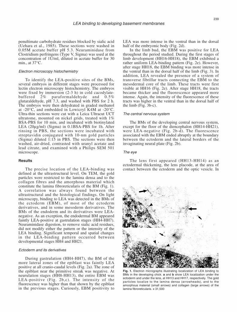
The precise location of the LEA-binding wasdefined at the ultrastructural level. On TEM, the goldparticles were restricted to the lamina densa and to thecollagen fibres and the amorphous material whichconstitute the lamina fibroreticularis of the BM (Fig. 1).A correlation was always found between theultrastructural and the histological findings. On lightmicroscopy, binding to LEA was detected in the BMs ofthe ectoderm (EBM), of most of the ectodermderivatives, and in some mesoderm derivatives. TheBMs of the endoderm and its derivatives were LEA-negative. As an exception, the endodermal BM appearedfaintly LEA-positive at gastrulation stages (HH4-HH7).Neuraminidase digestion, to remove sialic acid residues,did not modify either the pattern or the intensity of theLEA binding. Significant temporal and spatial changesin the LEA-binding pattern occurred betweendevelopmental stages HH4 and HH21.
Ectoderm and its derivatives
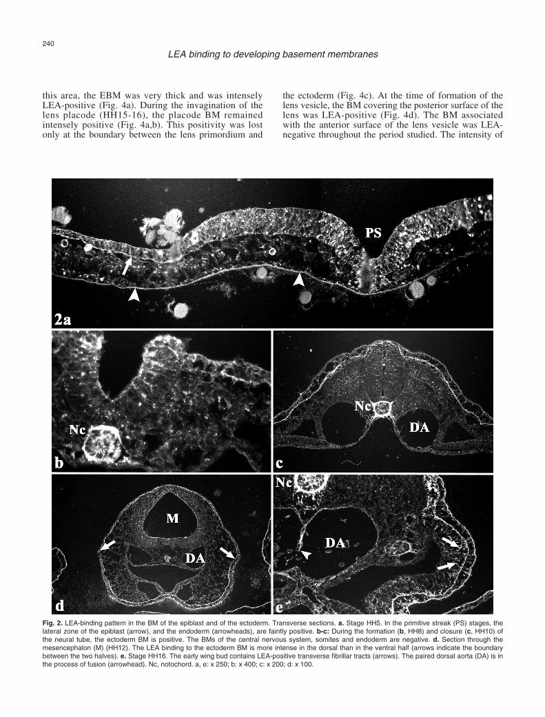
During gastrulation (HH4-HH7), the BM of themore lateral zones of the epiblast was faintly LEApositive at all cranio-caudal levels (Fig. 2a). The zone ofthe epiblast near the primitive streak was negative. Atneurulation stages (HH8-HH13), the entire EBM wasLEA-positive (Fig. 2b,c). The intensity of thefluorescence was higher than that shown by the epiblastin the previous stages. Curiously, EBM positivity to
LEA was more intense in the ventral than in the dorsalhalf of the embryonic body (Fig. 2d).
In the limb bud, the EBM was positive for LEAthroughout the period studied. During the first stages oflimb development (HH16-HH18), the EBM exhibited arather uniform LEA-binding pattern (Fig. 2e). However,after stage HH18, the EBM binding was more intense inthe ventral than in the dorsal half of the limb (Fig. 3). Inaddition, LEA revealed the presence of a system oftransverse fibrillar tracts connecting the EBM to themesodermal core of the limb. These tracts were firstvisible at HH16 (Fig. 2e). After stage HH18, the tractsbecame thicker and the fluorescence appeared moreintense. Again, the intensity of the fluorescence of thesetracts was higher in the ventral than in the dorsal half ofthe limb (Fig. 3b-c).
The central nervous system
The BMs of the developing central nervous system,except for the floor of the diencephalon (HH14-HH21),were LEA-negative (Fig. 2b-d). The fluorescenceassociated with the EBM ended abruptly at the boundarybetween the ectoderm and the lateral borders of theinvaginating neural plate (Fig. 2b).
The eye
The lens first appeared (HH13-HH14) as anectodermal thickening, the lens placode, at the area ofcontact between the ectoderm and the optic vesicle. In
239
LEA binding to developing basement membranes
Fig. 1. Electron micrographs illustrating localization of LEA binding toBMs in the developing chick. a and b show LEA localization under theectoderm and under the lens, at HH13 and HH17, respectively. The goldparticles localize to the lamina densa (arrowheads), and to theamorphous material (small arrows) and collagen (large arrows) of thelamina fibroreticularis. x 31,500
this area, the EBM was very thick and was intenselyLEA-positive (Fig. 4a). During the invagination of thelens placode (HH15-16), the placode BM remainedintensely positive (Fig. 4a,b). This positivity was lostonly at the boundary between the lens primordium and
the ectoderm (Fig. 4c). At the time of formation of thelens vesicle, the BM covering the posterior surface of thelens was LEA-positive (Fig. 4d). The BM associatedwith the anterior surface of the lens vesicle was LEA-negative throughout the period studied. The intensity of
240
LEA binding to developing basement membranes
Fig. 2. LEA-binding pattern in the BM of the epiblast and of the ectoderm. Transverse sections. a. Stage HH5. In the primitive streak (PS) stages, thelateral zone of the epiblast (arrow), and the endoderm (arrowheads), are faintly positive. b-c: During the formation (b, HH8) and closure (c, HH10) ofthe neural tube, the ectoderm BM is positive. The BMs of the central nervous system, somites and endoderm are negative. d. Section through themesencephalon (M) (HH12). The LEA binding to the ectoderm BM is more intense in the dorsal than in the ventral half (arrows indicate the boundarybetween the two halves). e. Stage HH16. The early wing bud contains LEA-positive transverse fibrillar tracts (arrows). The paired dorsal aorta (DA) is inthe process of fusion (arrowhead). Nc, notochord. a, e: x 250; b: x 400; c: x 200; d: x 100.
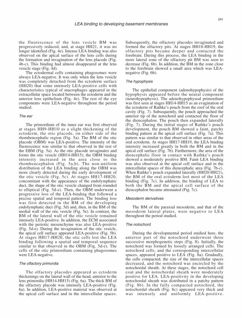
the fluorescence of the lens vesicle BM wasprogressively reduced, and, at stage HH21, it was nolonger identified (Fig. 4e). Intense LEA binding was alsoobserved on the apical surface of the lens cells duringthe formation and invagination of the lens placode (Fig.4b-c). This binding had almost disappeared at the lensvesicle stage (Fig. 4d).
The ectodermal cells containing phagosomes werealways LEA-negative. It was only when the lens vesiclewas completely detached from the ectoderm surface(HH20) that some intensely LEA-positive cells withcharacteristics typical of macrophages appeared in theextracellular space located between the ectoderm and theanterior lens epithelium (Fig. 4e). The rest of the eyecomponents were LEA-negative throughout the periodstudied.
The ear
The primordium of the inner ear was first observedat stages HH9-HH10 as a slight thickening of theectoderm, the otic placode, on either side of therhombencephalic region (Fig. 5a). The BM of the oticplacode (OBM) was LEA-positive. The intensity of thefluorescence was similar to that observed in the rest ofthe EBM (Fig. 5a). As the otic placode invaginates andforms the otic vesicle (HH14-HH15), the OBM bindingintensity increased in the area close to therhombencephalon (Fig. 5a,b). The non-uniformdistribution of the LEA binding along the OBM wasmore clearly detected during the early development ofthe otic vesicle (Fig. 5c). At stages HH17-HH20,concomitant with the appearance of the endolymphaticduct, the shape of the otic vesicle changed from roundedto elliptical (Fig. 5d-e). Then, the OBM underwent aprogressive loss of the LEA-binding that followed aprecise spatial and temporal pattern. The binding losswas first detected in the BM of the developingendolymphatic duct (Fig. 5d) and, then, in the BM of themedial wall of the otic vesicle (Fig. 5e). In contrast, theBM of the lateral wall of the otic vesicle remainedintensely LEA-positive. In addition, the ECM associatedwith the periotic mesenchyme was also LEA-positive(Fig. 5d-e). During the invagination of the otic vesicle,the apical cell surface appeared LEA-positive (Fig. 5b).At stages HH17-HH20, the otic cells lost the LEAbinding following a spatial and temporal sequencesimilar to that observed in the OBM (Fig. 5d-e). Thecells of the otic primordium containing phagosomeswere LEA-negative.
The olfactory primordia
The olfactory placodes appeared as ectodermthickenings on the lateral wall of the head, anterior to thelens primordia (HH14-HH15) (Fig. 6a). The thick BM ofthe olfactory placode was intensely LEA-positive (Fig.6a). In addition, LEA-positive material was observed atthe apical cell surface and in the intercellular spaces.
Subsequently, the olfactory placodes invaginated andformed the olfactory pits. At stages HH18-HH19, theolfactory pits became deeper and contacted theforebrain. During this process, the LEA binding in themore lateral zone of the olfactory pit BM was seen todecrease (Fig. 6b). In addition, the BM in the zone closeto the forebrain showed a small area which was LEA-negative (Fig. 6b).
The hypophysis
The epithelial component (adenohypophysis) of thehypophysis appeared before the neural component(neurohypophysis). The adenohypophyseal primordiumwas first seen at stages HH14-HH15 as an evagination ofthe ectoderm of Rathke’s pouch from the roof of the oralcavity (Fig. 7). Subsequently, the pouch approached theanterior tip of the notochord and contacted the floor ofthe diencephalon. The pouch then expanded laterally(Fig. 7). During the initial stages of Rathke’s pouchdevelopment, the pouch BM showed a faint, patchybinding pattern at the apical cell surface (Fig. 7a). Thispattern was similar to that observed in other areas of theoral ectoderm. At stages HH17-HH19, the LEA bindingintensity increased greatly in both the BM and in theapical cell surface (Fig. 7b). Furthermore, the area of thediencephalic floor in contact with Rathke’s pouchshowed a moderately positive BM. Faint LEA bindingwas also observed at the apical cell surface and in theintercellular spaces of this diencephalic area (Fig. 7b-c).When Rathke’s pouch expanded laterally (HH20-HH21),the BM of the oral ectoderm lost most of the LEAbinding (Fig. 7c). In addition, the binding of LEA toboth the BM and the apical cell surface of thediencephalon became attenuated (Fig. 7c).
Mesoderm derivatives
The BM of the paraxial mesoderm, and that of themesoderm lateral plates, were negative to LEAthroughout the period studied.
The notochord
During the developmental period studied here, theanterior part of the notochord underwent threesuccessive morphogenetic steps (Fig. 8). Initially, thenotochord was formed by loosely arranged cells. Thenotochord cells, and the corresponding intercellularspaces, appeared positive to LEA (Fig. 8a). Gradually,the cells compacted, the size of the intercellular spacesdecreased, and the notochord was encircled by thenotochordal sheath. At these stages, the notochord cellcoat and the notochordal sheath were moderatelypositive for LEA. LEA-positivity in the developingnotochordal sheath was distributed in a patchy pattern(Fig. 8b). In the fully compacted notochord, thenotochordal sheath (Fig. 8c) appeared very thick andwas intensely and uniformly LEA-positive.
241
LEA binding to developing basement membranes
242
LEA binding to developing basement membranes
Fig. 3. Longitudinal sections through the wing bud (HH20). a. Panoramic view. AER, apical ectodermal ridge. Wd, Wolffian duct. b-c. Detail of thedorsal (b) and ventral (c) halves. The fluorescence is more intense in the ectoderm BM of the ventral half (large arrows) than in the dorsal half (smallarrows). The BM of the APR is also positive. Arrowheads, fibrillar tracts. a: x150 ; b-c: x 650
Fig. 4. LEA-binding pattern in the developing eye. Transverse sections.a. Stage HH10. The area of contact between the optic vesicle (OV) and theectoderm (E) is positive before the formation of the lens placode. b. Developing lens placode (LP) (HH13). c. Invaginating lens placode (HH15). Notethe loss of fluorescence in the EBM at the boundary between the LP and the ectoderm (arrows). Arrowhead in b and c indicate LEA binding to theapical cell surface. d. Lens vesicle stage (HH18). Large arrow, BM of the posterior lens surface. Small arrow, lens stalk. e. Developing lens (HH20).Note the loss of LEA binding in the posterior lens BM (arrow). Asterisk, differentiating lens fibers. Inset: Subectodermal (E) macrophages are intenselyLEA-positive. E, ectoderm. a, d, e: x 200; b, c: x 250. Inset: x 650
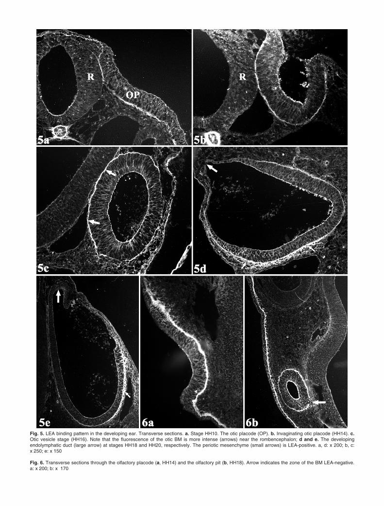
Fig. 5. LEA binding pattern in the developing ear. Transverse sections. a. Stage HH10. The otic placode (OP). b. Invaginating otic placode (HH14). c.Otic vesicle stage (HH16). Note that the fluorescence of the otic BM is more intense (arrows) near the rombencephalon; d and e. The developingendolymphatic duct (large arrow) at stages HH18 and HH20, respectively. The periotic mesenchyme (small arrows) is LEA-positive. a, d: x 200; b, c: x 250; e: x 150
Fig. 6. Transverse sections through the olfactory placode (a, HH14) and the olfactory pit (b, HH18). Arrow indicates the zone of the BM LEA-negative.a: x 200; b: x 170
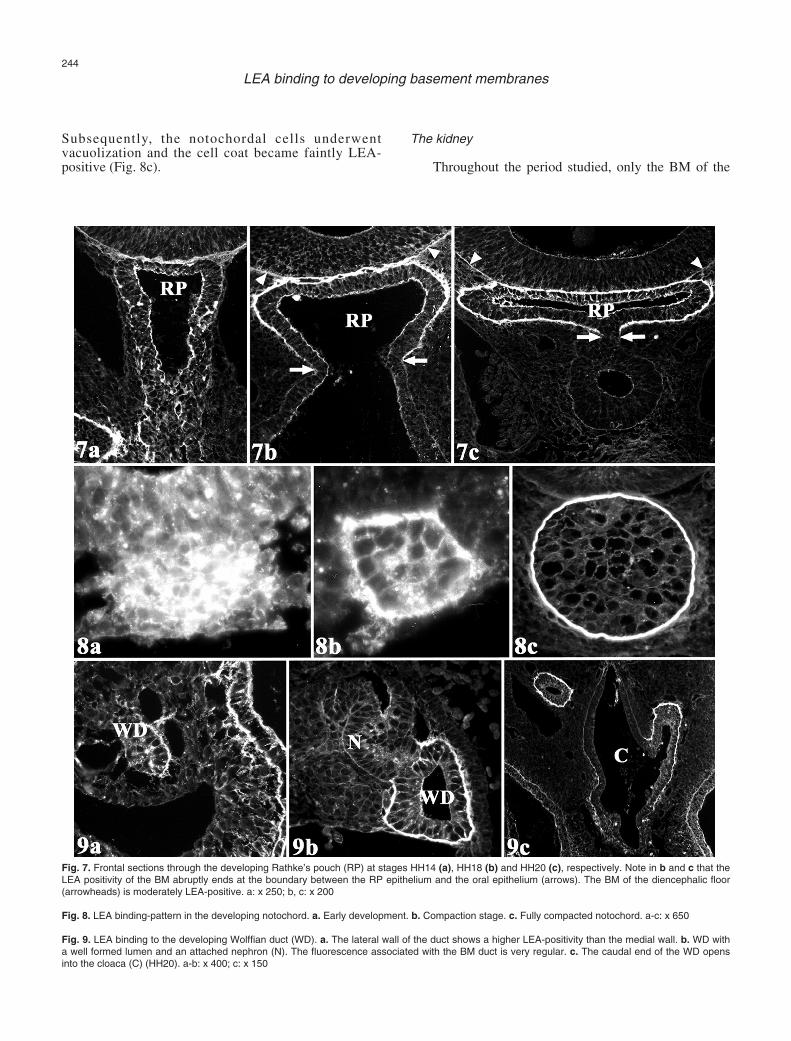
Subsequently, the notochordal cells underwentvacuolization and the cell coat became faintly LEA-positive (Fig. 8c).
The kidney
Throughout the period studied, only the BM of the
244
LEA binding to developing basement membranes
Fig. 7. Frontal sections through the developing Rathke’s pouch (RP) at stages HH14 (a), HH18 (b) and HH20 (c), respectively. Note in b and c that theLEA positivity of the BM abruptly ends at the boundary between the RP epithelium and the oral epithelium (arrows). The BM of the diencephalic floor(arrowheads) is moderately LEA-positive. a: x 250; b, c: x 200
Fig. 8. LEA binding-pattern in the developing notochord. a. Early development. b. Compaction stage. c. Fully compacted notochord. a-c: x 650
Fig. 9. LEA binding to the developing Wolffian duct (WD). a. The lateral wall of the duct shows a higher LEA-positivity than the medial wall. b. WD witha well formed lumen and an attached nephron (N). The fluorescence associated with the BM duct is very regular. c. The caudal end of the WD opensinto the cloaca (C) (HH20). a-b: x 400; c: x 150
Wolffian duct was LEA-positive (Fig. 9). The Wolffianduct first appeared (HH10-HH12) as a solid cell cordoriginating from the intermediate mesoderm. At thesestages, the Wolffian duct BM exhibited an asymmetricLEA-binding pattern (Fig. 9a). While the lateral ductzone was moderately positive, the ventromedial zonewas only faintly positive. Faint LEA-binding was alsoobserved in the intercellular spaces. After stage HH15,concomitant with the appearance of the lumen in theWolffian duct, the intensity of the LEA binding to theduct BM increased (Fig. 9b). The caudal end of theWolffian duct opened into the dorsolateral wall of thecloaca at stages HH20-HH21. Then, the LEA binding tothe duct BM ended abruptly at the boundary between theWolffian epithelium and the cloacal epithelium (anendoderm derivative) (Fig. 9c).
Heart and blood vessels
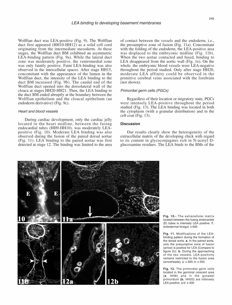
During cardiac development, only the cardiac jellylocated in the heart midline, between the fusingendocardial tubes (HH9-HH10), was moderately LEA-positive (Fig. 10). Moderate LEA binding was alsoobserved during the fusion of the paired dorsal aortae(Fig. 11). LEA binding to the paired aortae was firstdetected at stage 12. The binding was limited to the area
of contact between the vessels and the endoderm, i.e.,the presumptive zone of fusion (Fig. 11a). Concomitantwith the folding of the endoderm, the LEA-positive areawas displaced to the embryonic midline (Fig. 11b).When the two aortae contacted and fused, binding toLEA disappeared from the aortic wall (Fig. 1e). On thewhole, the embryonic blood vessels were LEA-negativethroughout the period studied. Only after stage HH20,moderate LEA affinity could be observed in theprimitive cerebral veins associated with the forebrain(not shown).
Primordial germ cells (PGCs)
Regardless of their location or migratory state, PGCswere intensely LEA-positive throughout the periodstudied (Fig. 13). The LEA binding was located in boththe cytoplasm (with a granular distribution) and in thecell coat (Fig. 13).
Discussion
Our results clearly show the heterogeneity of theextracellular matrix of the developing chick with regardto its content in glycoconjugates rich in N-acetyl D-glucosamine residues. The LEA binds to the BMs of the
245
LEA binding to developing basement membranes
Fig. 10.- The extracellular matrixlocated between the fusing endocardial(E) tubes is intensely LEA positive. F,endodermal foregut. x 600
Fig. 11. Modifications of the LEA-binding pattern during the formation ofthe dorsal aorta. a. In the paired aorta,only the presumptive zone of fusion(arrow) is positive for LEA (Compare tofigure 2c). b. During the approachingof the two vessels, LEA-positivityremains restricted to the fusion area(arrowheads). a: x 300; b: x 250
Fig. 12. The primordial germ cellslocated in the germinal crescent area(a, HH8) and in the gonadalprimordium (b, HH20) are intenselyLEA positive. a-b: x 400
ectoderm and its derivatives (excluding the BM of thedeveloping nervous system), and to the BMs of severalmesodermal derivatives. However, the BMs of theendoderm and its derivatives were LEA-negativethroughout the period studied.
Our results on the LEA binding affinity to the BMsof the ectoderm and of the developing nervous systemare in agreement with previous studies (Griffith andSanders, 1991). During the development of the lens, oticand nasal placodes, the LEA-binding pattern followed asimilar trend, with LEA localized to the BM and theapical cell surface. A similar pattern was also observedduring the formation of Rathke’s pouch. The depositionof LEA-positive glycoconjugates at the apical side of theinvaginating epithelia supports the notion (Sinning andOlson, 1988; Hilfer and Randolph, 1993; Yao et al.,1996) that glycoproteins constitute an important(physical and/or chemical) factor in the signallingpathway during the formation of the placodes. Theprogressive loss of LEA binding in these structures mustbe considered a sign of developmental maturation. Theloss of LEA binding could be explained by sialylationmasking. Masking of sugar residues has been reportedduring the differentiation of several tissues (Zieske andBernstein, 1982; Ojeda et al., 1993). However, we didnot detect any LEA binding after neuraminidasedigestion. Another possibility is that the LEA-positiveglycoproteins could have been replaced by newlysynthesized glycoproteins during the differentiationprocess. However, the loss of LEA binding in the BM ofthe lens vesicle occurred both in the undifferentiatedanterior epithelium and in the more differentiatedposterior epithelium. Finally, pre-existing LEA-positiveglycoproteins could have been degraded byglycosidases. While we favour the last hypothesis,experimental testing is necessary to prove the existenceof glycosidase activity.
The non-uniform distribution of the LEA-bindingsites along the EBM during limb development should beemphasized. The dorso-ventral asymmetric distributionof the LEA-binding pattern observed in the EBM has notpreviously been described for lectins with N-acetylglucosamine specificity (WGA and WGAs). Ourresults provide further support in favour of the existenceof a dorso-ventral compartmentalization in thedeveloping chick limb. According to Altabef et al.(1997, 2000), the ectoderm layer may be bisected by aplane passing from the forelimb to the hindlimb region,and thus dividing the limb into two cell lineagerestriction compartments, dorsal and ventral. Indeed, theapical ectodermal ridge of the developing limb is formedat the boundary between these two compartments. Onthe other hand, the LEA detects the fibrillar tractsextended between the EBM and the mesodermal core ofthe developing limb earlier (HH18) than previously(HH26) reported (Hurle et al., 1988). Furthermore, weshow that the tracts are thicker (and more intenselyfluorescent) in the ventral than in the dorsal half of thelimb. Again, this speaks in favour of the existence of a
dorso-ventral compartmentalization in the developingchick limb. The lectins of the LEA group appear to be abetter tool to study the development of the limb fibrillartracts. To gain a complete understanding of thedevelopmental and compositional changes undergone bythese fibrils is of crucial importance since they appear tomodulate the activity of the intercellular signals duringthe development of the limb (Arteaga-Solis et al., 2001).
The notochord and the BM of the Wolffian ductwere the mesodermal derivatives that showed thegreatest LEA positivity. The increasing amount of LEA-positive glycoconjugates may represent a factor ofstabilization during notochord compaction. Our resultson the LEA binding to the chick notochord differ fromprevious (Griffith and Sanders, 1991) observations.However, these differences could be explained by thedifferent lectins used. The human notochord has beenshown to be positive for wheat germ agglutinin (WGA)(Götz and Quondamatteo, 2001). Both WGA and LEAbelong to the N-acetylglucosamine group.
Between stages HH14 and HH21, the Wolffian ductBM was LEA positive. However, both the pronephricand mesonephric nephrons were negative. To ourknowledge, this is the first lectin-binding study of theWolffian duct. Throughout the period studied, theWolffian duct underwent compaction of the loosemesenchyme into a solid epithelium, generation of alumen (canalization), establishment of the cell polarity,and cranio-caudal migration of the Wolffian duct tip tojoin the cloaca. Several ECM glycoproteins, especiallyfibronectin and laminin (Jacob et al., 1991), appear toplay an important role in all these processes (Hay,1991).Our results indicate that glycoconjugates having N-acetyl D-glucosamine residues play a key role during thedevelopment of the Wolffian duct.
The presence of LEA-positive material associatedwith the fusion of endothelia is worth comment. Duringheart development, LEA-positive material accumulatesbetween the fusing endocardial tubes (the rest of theextracellular matrix -cardiac jelly- of the early heart isnegative for LEA). During the fusion of the pairedaortae, LEA positivity is restricted to the BM of thefusion area. In the two cases, the presence of LEA-positive material may mediate the fusion process. At thetime of endocardial fusion, apoptosis of the midlineendocardium plays a decisive role in the fusion process(Ojeda and Hurle, 1975, 1981). Endothelial cell deathalso occurs during formation of the single aorta (Berry,1973). The concurrent accumulation of LEA-positivematerial and the presence of endothelial apoptosissuggests a causal relationship. It is well established thatthe ECM has an essential function in the control of cellsurvival. The ECM signals for cell survival are specificand are mediated, at least in part, by the integrin familyof cell surface receptors (Prince et al., 2002). The lateappearance of moderate LEA affinity in the wall of thecerebral veins suggests that the presence ofglycoproteins rich in N-acetyl D-glucosamine residuesmay constitute a sign of maturation of the venous
246
LEA binding to developing basement membranes
endothelium (see: Navarro et al., 2003). The PGCs are intensely LEA-positive from the time
of their appearance in the central zone of the areapellucida (Griffith and Saunders, 1991). These cellsexpress LEA-positive sites along their entire migratorypath, and when they settle into the germinal crescents.This suggests that the presence of glycoproteinscontaining N-acetyl D-glucosamine residues may playan important role in the processes of cell surfacerecognition and cell migration during PGC development.LEA is revealed as an excellent marker for PGCs in thechick embryo.
Apoptotic cell areas appear in several developingstructures such as the lens vesicle (García-Porrero et al.,1979), the otic vesicle (Lang et al., 2000), and the neuraltube (Weil et al., 1997; Hirata and Hall, 2000). In theseareas, healthy neighbouring cells function as non-professional phagocytes. While LEA has been defined asa macrophage marker in several animal species(Andjelkovic et al., 1998; Navarro et al., 2003), we didnot observe any LEA binding associated with non-professional phagocytes. However, a few LEA positivecells with the characteristics of free macrophages wereobserved in the final stages of lens development. Ourresults suggest that the presence of N-acetyl D-glucosamine residues may constitute an importantdifference between the two types of phagocytes.
Finally, the nature of the glycoproteins recognizedby the LEA binding remains mostly unknown. Previousstudies have identified lymphocyte cell surface(Kilpatrick et al., 1986) and sperm adhesion (Zhang andMartin-DeLeon, 2003) molecules as LEA targets. Wehave shown here that LEA binds to the collagenassociated with the lamina fibroreticularis of the BMs(presumably, some type IV-collagen isoform). We couldalso infer that, during the fusion process of the heartanlage, LEA may be binding to carbohydrate chainsassociated to the fibronectin molecule. Monoclonalantibodies recognize large fibronectin deposits in thisarea of the developing heart (Icardo and Manasek,1983). In addition, the LEA could be recognizing theglycoprotein fibrillin in the developing limb (see:Handford et al., 2000), and laminin-1 receptors at thecell surface (Botti et al., 2003). However, fullidentification of the LEA-bounded glycoproteins is outof the scope of the present paper.
Acknowledgements. Supported by grant CGL2004-06306-C02-01/BOSfrom the “Ministerio de Ciencia y Tecnología“.
References
Acarin L., Vela J.M., González B. and Castellano B. (1994).Demonstration of poly-N-acetyl lactosamine residues in ameboidand ramified microglial cells in rat brain by tomato lectin binding. J.Histochem. Cytochem. 42, 1033-1041.
Allen B.L. and Rapraeger A.C. (2003). Spatial and temporal expressionof heparan sulfate in mouse development regulates FGF and FGF
receptor assembly. J. Cell Biol. 10, 637-648.Altabef M., Clarke J.D.W. and Tickle C. (1997). Dorso-ventral
ectodermal compartments and origin of apical ectodermal ridge indeveloping chick limb. Development 124, 4547-4556.
Altabef M., Logan C., Tickle C. and Lumsden A. (2000). Engrailed-1missexpression in chick embryos prevents apical ridge formation butpreserves segregation of dorsal and ventral compartments. Dev.Biol. 222, 307-316.
Andjelkovic A.V., Nikolic B., Pachter J.S. and Zecevic N. (1998).Macrophages/microglial cells in human central nervous systemduring development: an immunohistochemical study. Brain Res.814, 13-25.
Arteaga-Solis E., Gayraud B., Lee S.Y., Shum L., Sakai L. and RamirezF. (2001). Regulation of limb patterning by extracellular microfibrils.J. Cell Biol. 154, 275-281.
Beauvais-Jouneau A. and Thiery J.-P. (1997). Multiple roles for integrinsduring development. Biol. Cell. 89, 5-11.
Berry C.L. (1973). Growth, development and healing of large arteries.Ann. R. Coll. Surg. Engl. 53, 246-257.
Botti J., Musset M., Moutsita R., Aubery M. and Derappe C. (2003). Twolaminin receptors with N-acetylglucosamine-binding specificity.Biochimie 85, 231-239.
Damjanov I. (1990). Heterogeneity of basement membranes in normaland pathological altered tissues. Virchows Arch. (A). 416, 185-188.
Ekblom M., Falk M., Salmivirta K., Durbeej M. and Ekblom P. (1998).Laminin isoforms and epithelial development. Ann. NY Acad. Sci.857, 194-211.
Erickson A.C. and Couchman J.R. (2000). Still more complexity inmammalian basement membranes. J. Histochem. Cytochem. 48,1291-1306.
Gallagher B.C. (1986). Basal laminar thinning in branchingmorphogenesis of the chick lung as demonstrated by lectin probes.J. Embryol. Exp. Morphol. 94, 173-188
García-Porrero J., Collado J.A. and Ojeda J.L. (1979). Cell death duringdetachment of the lens rudiment from ectoderm in the chick embryo.Anat. Rec. 193, 791-803.
Goldstein I.J. and Poretz R.D. (1986). Isolation, physicochemicalcharacterization, and carbohydrate-binding specificity of lectins. In:The lectins: Properties, functions, applications in biology andmedicine. Liener I.E., Sharon N. and Goldstein I.J. (eds). AcademicPress. London. pp 94-95.
Götz W. and Quondamatteo F. (2001). Glycoconjugate distribution inearly human notochord and axial mesenchyme. Acta Histochem.103, 21-35.
Griffith C.M. and Sanders E.J. (1991). Changes in glycoconjugateexpression during early chick embryo development: a lectin-bindingstudy. Anat. Rec. 231, 238-250.
Hamburger V. and Hamilton H.L. (1951). A series of normal stages inthe development of the chick embryo. J. Morphol. 88, 49-92.
Handford P.A., Downing A.K., Reinhardt D.P. and Sakai L.Y. (2000)Fibrillin: from domain structure to supramolecular assembly. MatrixBiol. 19, 457-470.
Hay E.D. (1991). Collagen and other matrix glycoproteins inembryogenesis. In: Cell biology of extracellular matrix. Hay E.D.(ed). Plenum Press. New York. pp 419-456.
Hentschel H. and Walther P. (1993). Heterogeneous distribution ofglycoconjugates in the kidney of dogfish Scyliorhinus caniculus (L.)with reference to changes in the glycosilation pattern duringontogenic development of the nephron. Anat. Rec. 235, 21-32.
247
LEA binding to developing basement membranes
Hilfer S.R. and Randolph G.J. (1993). Immunolocalization of basallamina components during development of chick otic and opticprimordia. Anat. Rec. 235, 443-452.
Hirata M. and Hall B.K. (2000). Temporospatial patterns of apoptosis inchick embryos during the morphogenetic period of development. Int.J. Dev. Biol. 44, 747-768.
Hurle J.M., Ros M.A. and Hinchliffe J.R. (1988). Spatial and temporalchanges in the pattern of glycosilation of the developing chick limbtissue components as revealed by fluorescent conjugated lectinprobes. Cell Differ. 24, 149-158.
Hynes R.O. (1992). Integrins: versatility, modulation, and signaling incell adhesion. Cell 69, 11-25.
Icardo J.M. and Manasek F.J. (1983). Fibronectin distribution duringearly chick embryo heart development. Dev. Biol. 95, 19-30.
Inoue S. (1989). Ultrastructure of basement membranes. Int. Rev. Cytol.117, 57-98.
Jacob M., Christ B., Jacob H.J. and Poelmann R.E. (1991). The role offibronectin and laminin in development and migration of the avianWolffian duct with reference to somitogenesis. Anat. Embryol. 183,385-395.
Kilpatrick D.C., Graham C. and Urbaniak S.J. (1986). Inhibition ofhuman lymphocyte trasformation by tomato lectin. Scand. J.Immunol. 24, 11-19.
Kohfeldt E., Sasaki T., Gohring W. and Timpl R. (1998). Nidogen-2: anew basement membrane protein with diverse binding properties. J.Mol. Biol. 282, 99-109.
Lait inen L., Virtanen I. and Saxén L. (1987). Changes in theglycosylation pattern during embryonic development of mousekidney as revealed with lectin conjugates. J. Histochem. Cytochem.35, 55-65.
Lang H., Bever M.M. and Fekete D.M. (2000). Cell proliferation and celldeath in the developing chick inner ear: spatial and temporalpatterns. J. Comp. Neurol. 417, 205-220.
Leu F.J. and Damjanov I. (1988). Protease treatment combined withimmunohistochemistry reveals heterogeneity of normal andneoplastic basement membranes. J. Histochem. Cytochem. 36, 213-320.
Lis H. and Sharon N. (1986). Lectins as molecules and as tools. Ann.Rev. Biochem. 55, 35-42.
Nachbar M.S. and Oppenheim J.D. (1982). Tomato (Lycopersiconesculentum) lectin. Methods Enzymol. 83, 363-368.
Navarro M., DeRuiter M.C., Carretero A. and Ruberte J. (2003).Microvascular assembly and cell invasion in chick mesonephrosgrafted onto chorioallantoic membrane. J. Anat. 202, 213-225.
Ojeda J.L. and Hurle J.M. (1975). Cell death during the formation oftubular heart of the chick embryo. J. Embryol. Exp. Morph. 33, 523-534.
Ojeda J.L. and Hurle J.M. (1981). Establishment of the tubular heart.Role of cell death. In: Perspectives in cardiovascular research, Vol.5 mechanisms of cardiac morphogenesis and teratogenesis.Pexieder T. (ed). Raven Press. New York. pp. 101-113.
Ojeda J.L., Ros M.A. and Icardo J.M. (1989). A technique forfluorescence microscopy in semithin sections. Stain Technol. 64,243-248.
Ojeda J.L., Ros M.A. and Icardo J.M. (1993). Lectin-binding sites during
postnatal differentiation of normal and cystic rabbit renal corpuscles.Anat. Embryol. 187, 539-547.
Pierce R.A., Griffin G.L., Miner J.H. and Senior R.M. (2000). Expressionpatterns of laminin alpha1 and alpha5 in human lung duringdevelopment. Am. J. Respir. Cell. Mol. Biol. 23, 742-747.
Prince J.M., Klinowska T.C.M., Marshman E., Lowe E.T., Mayer U.,Miner J., Aberdam D., Vestweber D., Gusterson B. and Streuli C.H.(2002). Cell-matrix interactions during development and apoptosis ofthe mouse mammary gland in vivo. Dev. Dyn. 223, 497-516.
Rademacher T.W., Parek R.B. and Dwek R.A. (1988). Glycobiology.Annu. Rev. Biochem. 57, 785-838.
Robinson G.B. and Walton H.A. (1989). Glomerular basementmembranes as a compressible ultrafilter. Microvascular Res. 38, 36-48.
Salamat M., Gotz W., Werner J. and Herken R. (1993). Ultrastructurallocalization of lectin-binding sites in different basement membranes.Histochem. J. 25, 464-468.
Sanes J.R., Engvall E., Butkowski R. and Hunter D. (1990). Molecularheterogeneity of basal laminae: isoforms of laminin and collagen IVat the neuromuscular junction and elsewhere. J. Cell Biol. 111,1685-1699.
Shur B.D. (1989). Glycoconjugates as mediators of cellular interactionsduring development. Curr. Opin. Cell Biol. 1, 905-912.
Sinning A.R. and Olson M.D. (1988). Surface coat material associatedwith the developing otic placode/vesicle in the chick. Anat. Rec. 220,198-207.
Streuli C. (1996). Basement membrane as a differentiation and survivalfactor. In: The laminins. Ekblom P. and Timpl R. (eds). HarwoodAcademic. Reading. pp 217-233.
Uehara F., Muramatsu T., Sameshina M., Kawano K., Koide H. andOhaba N. (1985) Effects of neuraminidase on lectin binding sites inphotoreceptor cells of monkey retina. Jpn. J. Ophthalmol. 29, 54-62.
Virtanen I., Korhonen M., Petajaniemi N., Karhunen T., Thornell L.E.,Sorokin L.M. and Konttinen Y.T. (2003). Laminin isoforms in fetaland adult human adrenal cortex. J. Clin. Endocrinol. Metab. 88,4960-4966.
Wan Y.J., Wu T.C., Chung A.E. and Damjanov I. (1984). Monoclonalantibodies to laminin reveal the heterogeneity of basementmembranes in the developing and adult mouse tissues. J. Cell Biol.98, 971-979.
Weil M., Jacobson M.D. and Raff M.C. (1997). Is programmed cell deathrequired for neural tube closure?. Curr. Biol. 7, 281-284.
Wilson D.B. and Wyatt D.P. (1995). Patterns of lectin binding duringmammalian neurogenesis. J. Anat. 186, 209-216.
Yao R., Alcala J. and Maisel H. (1996). Developmental changes inglycoconjugate composition during chick lens morphogenesis. Exp.Eye Res. 62, 419-431.
Zhang H. and Martin-DeLeon P.A. (2003). Mouse epididymal Spam1(pH-20) is released in the luminal fluid with its lipid anchor. J. Androl.24, 51-58.
Zieske J.D. and Bernstein I.A. (1982). Modification of cell surfaceglycoproteins: Addition of fucosyl residues during epidermaldifferentiation. J. Cell Biol. 95, 626-631.
Accepted October 24, 2005
248
LEA binding to developing basement membranes