Embed Size (px)
Citation preview
Catalog No. 552364
BD Cytometric Bead Array (CBA) Mouse Inflammation KitInstruction Manual
BD CBA Mouse Inflammation Kitii
BD flow cytometers are Class 1 Laser Products.For Research Use Only. Not for use in diagnostic or therapeutic procedures.© 2014 Becton, Dickinson and Company. All rights reserved. No part of this publication may bereproduced, transmitted, transcribed, stored in retrieval systems, or translated into any language orcomputer language, in any form or by any means: electronic, mechanical, magnetic, optical, chemical,manual, or otherwise, without prior written permission from BD Biosciences.Purchase does not include or carry any right to resell or transfer this product either as a stand-alone product or as a component of another product. Any use of this product other than the permitted use without the express written authorization of Becton, Dickinson and Company is strictly prohibited.BD, BD Logo and all other trademarks are property of Becton, Dickinson and Company. © 2014 BD
Trademarks
FCAP Array is a trademark of Soft Flow Hungary Ltd.
Mac is a trademark of Apple Computer, Inc., registered in the US and other countries.
Falcon® is a registered trademark of Corning Incorporated.
History
Revision Date Change made
23-12720-00 Rev. 01 1/2011 New document
23-12720-01 Rev. 01 2/2012 Updated image
Rev. 2 4/2015 Update hazard statements
For Research Use Only. Not for use in diagnostic or therapeutic procedures.
Contents
Chapter 1: About this kit . . . . . . . . . . . . . . . . . . . . . . . . . . . . . . . . . . . . 5Purpose of this kit . . . . . . . . . . . . . . . . . . . . . . . . . . . . . . . . . . . . . . . 6Limitations . . . . . . . . . . . . . . . . . . . . . . . . . . . . . . . . . . . . . . . . . . . . 8Kit contents . . . . . . . . . . . . . . . . . . . . . . . . . . . . . . . . . . . . . . . . . . . . 9Storage and handling . . . . . . . . . . . . . . . . . . . . . . . . . . . . . . . . . . . . 11
Chapter 2: Before you begin . . . . . . . . . . . . . . . . . . . . . . . . . . . . . . . . 13Workflow overview . . . . . . . . . . . . . . . . . . . . . . . . . . . . . . . . . . . . . 14Required materials . . . . . . . . . . . . . . . . . . . . . . . . . . . . . . . . . . . . . . 15
Chapter 3: Assay preparation . . . . . . . . . . . . . . . . . . . . . . . . . . . . . . . 17Preparing Mouse Inflammation Standards . . . . . . . . . . . . . . . . . . . . 18Mixing Mouse Inflammation Capture Beads . . . . . . . . . . . . . . . . . . 20Diluting samples . . . . . . . . . . . . . . . . . . . . . . . . . . . . . . . . . . . . . . . 21
Chapter 4: Assay procedure . . . . . . . . . . . . . . . . . . . . . . . . . . . . . . . . 23Performing the Mouse Inflammation Assay . . . . . . . . . . . . . . . . . . . 24Data analysis . . . . . . . . . . . . . . . . . . . . . . . . . . . . . . . . . . . . . . . . . . 27
Chapter 5: Performance . . . . . . . . . . . . . . . . . . . . . . . . . . . . . . . . . . . . 29Theoretical limit of detection . . . . . . . . . . . . . . . . . . . . . . . . . . . . . . 30Recovery . . . . . . . . . . . . . . . . . . . . . . . . . . . . . . . . . . . . . . . . . . . . . 31Linearity . . . . . . . . . . . . . . . . . . . . . . . . . . . . . . . . . . . . . . . . . . . . . 32Specificity . . . . . . . . . . . . . . . . . . . . . . . . . . . . . . . . . . . . . . . . . . . . 33Precision . . . . . . . . . . . . . . . . . . . . . . . . . . . . . . . . . . . . . . . . . . . . . 35
Chapter 6: Reference . . . . . . . . . . . . . . . . . . . . . . . . . . . . . . . . . . . . . . 37Troubleshooting . . . . . . . . . . . . . . . . . . . . . . . . . . . . . . . . . . . . . . . 38References . . . . . . . . . . . . . . . . . . . . . . . . . . . . . . . . . . . . . . . . . . . . 40

For Research Use Only. Not for use in diagnostic or therapeutic procedures.
1About this kit
This section covers the following topics:
Purpose of this kit (page 6)
Limitations (page 8)
Kit contents (page 9)
Storage and handling (page 11)
BD CBA Mouse Inflammation Kit6
For Research Use Only. Not for use in diagnostic or therapeutic procedures.
Purpose of this kit
Use of the kit The BD™ CBA Mouse Inflammation Kit can be used to quantitatively measure Interleukin-6 (IL-6), Interleukin-10 (IL-10), Monocyte Chemoattractant Protein-1 (MCP-1), Interferon- (IFN-), Tumor Necrosis Factor (TNF), and Interleukin-12p70 (IL-12p70) protein levels in a single sample. The kit performance has been optimized for analysis of specific proteins in tissue culture supernatants, EDTA plasma, and serum samples. The kit provides sufficient reagents for 80 tests.
Principle of CBAassays
BD CBA assays provide a method of capturing a soluble analyte or set of analytes with beads of known size and fluorescence, making it possible to detect analytes using flow cytometry.
Each capture bead in the kit has been conjugated with a specific antibody. The detection reagent provided in the kit is a mixture of phycoerythrin (PE)-conjugated antibodies, which provides a fluorescent signal in proportion to the amount of bound analyte.
When the capture beads and detector reagent are incubated with an unknown sample containing recognized analytes, sandwich complexes (capture bead + analyte + detection reagent) are formed. These complexes can be measured using flow cytometry to identify particles with fluorescence characteristics of both the bead and the detector.
Chapter 1: About this kit 7
For Research Use Only. Not for use in diagnostic or therapeutic procedures.
Principle of thetest
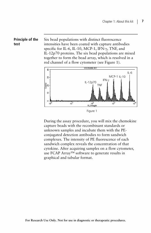
Six bead populations with distinct fluorescence intensities have been coated with capture antibodies specific for IL-6, IL-10, MCP-1, IFN-, TNF, and IL-12p70 proteins. The six bead populations are mixed together to form the bead array, which is resolved in a red channel of a flow cytometer (see Figure 1).
During the assay procedure, you will mix the chemokine capture beads with the recombinant standards or unknown samples and incubate them with the PE-conjugated detection antibodies to form sandwich complexes. The intensity of PE fluorescence of each sandwich complex reveals the concentration of that cytokine. After acquiring samples on a flow cytometer, use FCAP Array™ software to generate results in graphical and tabular format.
IL-6IL-10MCP-1
TNF
IFN-IL-12p70
Figure 1
BD CBA Mouse Inflammation Kit8
For Research Use Only. Not for use in diagnostic or therapeutic procedures.
Advantages overELISA
The broad dynamic range of fluorescence detection via flow cytometry and the efficient capturing of analytes via suspended particles enable BD CBA assays to measure the concentration of an unknown in substantially less time and using fewer sample dilutions compared to conventional ELISA methodology.
The required sample volume is approximately one-sixth the quantity necessary for conventional ELISA assays due to the detection of six analytes in a single sample.
A single set of diluted standards is used to generate a standard curve for each analyte.
A BD CBA experiment takes less time than a single ELISA and provides results that would normally require six conventional ELISAs.
Limitations
Assay limitations The theoretical limit of detection of the BD CBA Mouse inflammation Kit is comparable to conventional ELISA, but due to the complexity and kinetics of this multi-analyte assay, the actual limit of detection in a given experiment may vary slightly. Note the reduced sensitivity of the Mouse MCP-1 assay. See Theoretical limit of detection (page 30) and Precision (page 35).
The BD CBA assay is not recommended for use on stream-in-air instruments for which signal intensities may be reduced, adversely affecting assay sensitivity. Stream-in-air instruments include the BD FACStar™ Plus, BD FACSVantage™, and BD Influx™ flow cytometers (BD Biosciences).
Chapter 1: About this kit 9
For Research Use Only. Not for use in diagnostic or therapeutic procedures.
Serum spike recoveries for IL-10 and TNF are lower than for the other proteins in this assay. This variation is due to assay conditions and serum proteins. It may affect quantitation of these proteins in serum samples.
Quantitative results or protein levels for the same sample or recombinant protein run in ELISA and BD CBA assays may differ. A spike recovery assay can be performed using an ELISA standard followed by BD CBA analysis to assess possible differences in quantitation.
This kit is designed to be used as an integral unit Do not mix components from different batches or kits.
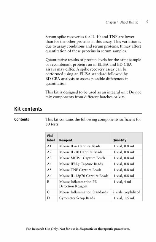
Kit contents
Contents This kit contains the following components sufficient for 80 tests.
Viallabel Reagent Quantity
A1 Mouse IL-6 Capture Beads 1 vial, 0.8 mL
A2 Mouse IL-10 Capture Beads 1 vial, 0.8 mL
A3 Mouse MCP-1 Capture Beads: 1 vial, 0.8 mL
A4 Mouse IFN- Capture Beads 1 vial, 0.8 mL
A5 Mouse TNF Capture Beads 1 vial, 0.8 mL
A6 Mouse IL-12p70 Capture Beads 1 vial, 0.8 mL
B Mouse Inflammation PE Detection Reagent
1 vial, 4 mL
C Mouse Inflammation Standards 2 vials lyophilized
D Cytometer Setup Beads 1 vial, 1.5 mL
BD CBA Mouse Inflammation Kit10
For Research Use Only. Not for use in diagnostic or therapeutic procedures.
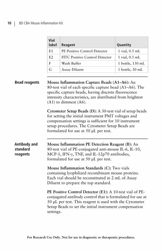
Bead reagents Mouse Inflammation Capture Beads (A1–A6): An 80-test vial of each specific capture bead (A1–A6). The specific capture beads, having discrete fluorescence intensity characteristics, are distributed from brightest (A1) to dimmest (A6).
Cytometer Setup Beads (D): A 30-test vial of setup beads for setting the initial instrument PMT voltages and compensation settings is sufficient for 10 instrument setup procedures. The Cytometer Setup Beads are formulated for use at 50 µL per test.
Antibody andstandardreagents
Mouse Inflammation PE Detection Reagent (B): An 80-test vial of PE-conjugated anti-mouse IL-6, IL-10, MCP-1, IFN-, TNF, and IL-12p70 antibodies, formulated for use at 50 µL per test.
Mouse Inflammation Standards (C): Two vials containing lyophilized recombinant mouse proteins. Each vial should be reconstituted in 2 mL of Assay Diluent to prepare the top standard.
PE Positive Control Detector (E1): A 10-test vial of PE-conjugated antibody control that is formulated for use at 50 µL per test. This reagent is used with the Cytometer Setup Beads to set the initial instrument compensation settings.
E1 PE Positive Control Detector 1 vial, 0.5 mL
E2 FITC Positive Control Detector 1 vial, 0.5 mL
F Wash Buffer 1 bottle, 130 mL
G Assay Diluent 1 bottle, 30 mL
Viallabel Reagent Quantity
Chapter 1: About this kit 11
For Research Use Only. Not for use in diagnostic or therapeutic procedures.
FITC Positive Control Detector (E2): A 10-test vial of FITC-conjugated antibody control that is formulated for use at 50 µL per test. This reagent is used with the Cytometer Setup Beads to set the initial instrument compensation settings.
Buffer reagents Wash Buffer (F): A 130-mL bottle of phosphate buffered saline (PBS) solution (1X), containing protein and detergent, used for wash steps and to resuspend the washed beads for analysis.
Assay Diluent (G): A 30-mL bottle of a buffered protein solution (1X) used to reconstitute and dilute the Mouse Inflammation Standards and to dilute test samples.
Note: Source of all serum proteins is from USDA inspected abattoirs located in the United States.
Storage and handling
Storage Store all kit components at 2 to 8°C. Do not freeze.
Warning Components A1–A6, B, D, E1, E2, F, and G contain sodium azide. Sodium azide yields a highly toxic hydrazoic acid under acidic conditions. Dilute azide compounds in running water before discharging to avoid accumulation of potentially explosive deposits in plumbing.
Mouse Inflammation Standards (component 51-9000148) contains 0.02% (w/w) of a CMIT/MIT mixture (3:1), which is a mixture of: 5-chloro-2-methyl-4-isothiazolin-3-one [EC No 247-500-7] and 2-methyl-4-isothiazolin-3-one [EC No 220-239-6] (3:1).
Hazardstatements
May cause an allergic skin reaction.
BD CBA Mouse Inflammation Kit12
For Research Use Only. Not for use in diagnostic or therapeutic procedures.
Precautionarystatements
Wear protective gloves / eye protection.
Wear protective clothing.
Avoid breathing mist/vapours/spray.
If skin irritation or rash occurs: Get medical advice/attention.
IF ON SKIN: Wash with plenty of water.
Dispose of contents/container in accordance with local/regional/national/international regulations.
For Research Use Only. Not for use in diagnostic or therapeutic procedures.
2Before you begin
This section covers the following topics:
Workflow overview (page 14)
Required materials (page 15)
BD CBA Mouse Inflammation Kit14
For Research Use Only. Not for use in diagnostic or therapeutic procedures.
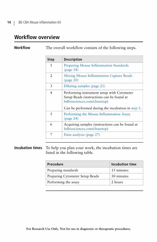
Workflow overview
Workflow The overall workflow consists of the following steps.
Incubation times To help you plan your work, the incubation times are listed in the following table.
Step Description
1 Preparing Mouse Inflammation Standards (page 18)
2 Mixing Mouse Inflammation Capture Beads (page 20)
3 Diluting samples (page 21)
4 Performing instrument setup with Cytometer Setup Beads (instructions can be found at bdbiosciences.com/cbasetup)
Can be performed during the incubation in step 5.
5 Performing the Mouse Inflammation Assay (page 24)
6 Acquiring samples (instructions can be found at bdbiosciences.com/cbasetup)
7 Data analysis (page 27)
Procedure Incubation time
Preparing standards 15 minutes
Preparing Cytometer Setup Beads 30 minutes
Performing the assay 2 hours

Chapter 2: Before you begin 15
For Research Use Only. Not for use in diagnostic or therapeutic procedures.
Required materials
Materialsrequired but notprovided
In addition to the reagents provided in the BD CBA Mouse inflammation Kit, the following items are also required:
A dual-laser flow cytometer equipped with a 488-nm or 532-nm and a 633-nm or 635-nm laser capable of distinguishing 576-nm, 660-nm, and >680-nm fluorescence. The following table lists examples of compatible instrument platforms.
Falcon® 12 × 75-mm sample acquisition tubes for a flow cytometer (Catalog No. 352008)
15-mL conical, polypropylene tubes (Falcon, Catalog No. 352097), or equivalent
Note: FCAP Array software (Catalog No. 641488 [PC] or 645447 [Mac®])
Flow cytometerReporterchannel Bead channels
BD FACSArray™ Yellow Red
BD FACSCanto™ platformBD™ LSR platformBD FACSAria™ platform PE APC
BD FACSCalibur™ (single laser)BD FACSCalibur (dual laser) FL2
FL3FL4
Note: Visit bdbiosciences.com/cbasetup for setup protocols.
BD CBA Mouse Inflammation Kit16
For Research Use Only. Not for use in diagnostic or therapeutic procedures.
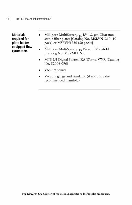
Materialsrequired forplate loader-equipped flowcytometers
Millipore MultiScreenHTS-BV 1.2-µm Clear non-sterile filter plates [Catalog No. MSBVN1210 (10 pack) or MSBVN1250 (50 pack)]
Millipore MultiScreenHTS Vacuum Manifold (Catalog No. MSVMHTS00)
MTS 2/4 Digital Stirrer, IKA Works, VWR (Catalog No. 82006-096)
Vacuum source
Vacuum gauge and regulator (if not using the recommended manifold)
For Research Use Only. Not for use in diagnostic or therapeutic procedures.
3Assay preparation
This section covers the following topics:
Preparing Mouse Inflammation Standards (page 18)
Mixing Mouse Inflammation Capture Beads (page 20)
Diluting samples (page 21)
BD CBA Mouse Inflammation Kit18
For Research Use Only. Not for use in diagnostic or therapeutic procedures.
Preparing Mouse Inflammation Standards
Purpose of thisprocedure
The Mouse Inflammation Standards are lyophilized and should be reconstituted and serially diluted immediately before mixing with the Capture Beads and the PE Detection Reagent.
You must prepare fresh cytokine standards to run with each experiment. Do not store or reuse reconstituted or diluted standards.
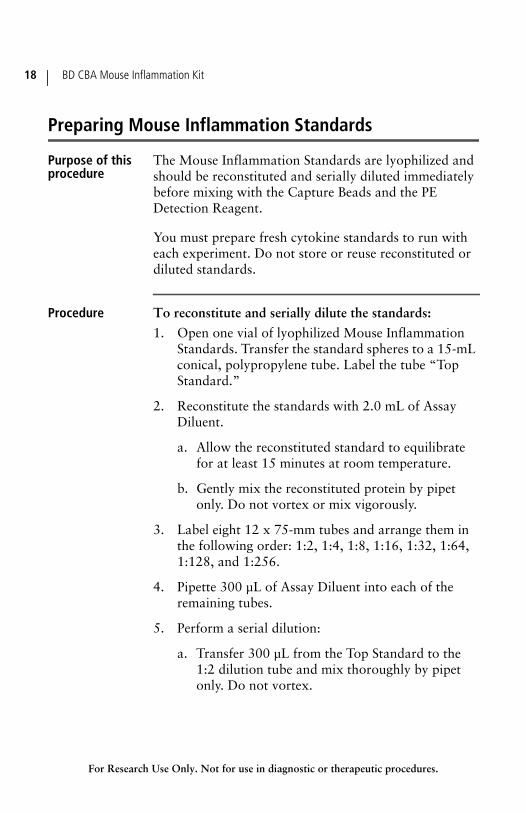
Procedure To reconstitute and serially dilute the standards:
1. Open one vial of lyophilized Mouse Inflammation Standards. Transfer the standard spheres to a 15-mL conical, polypropylene tube. Label the tube “Top Standard.”
2. Reconstitute the standards with 2.0 mL of Assay Diluent.
a. Allow the reconstituted standard to equilibrate for at least 15 minutes at room temperature.
b. Gently mix the reconstituted protein by pipet only. Do not vortex or mix vigorously.
3. Label eight 12 x 75-mm tubes and arrange them in the following order: 1:2, 1:4, 1:8, 1:16, 1:32, 1:64, 1:128, and 1:256.
4. Pipette 300 µL of Assay Diluent into each of the remaining tubes.
5. Perform a serial dilution:
a. Transfer 300 µL from the Top Standard to the 1:2 dilution tube and mix thoroughly by pipet only. Do not vortex.
Chapter 3: Assay preparation 19
For Research Use Only. Not for use in diagnostic or therapeutic procedures.
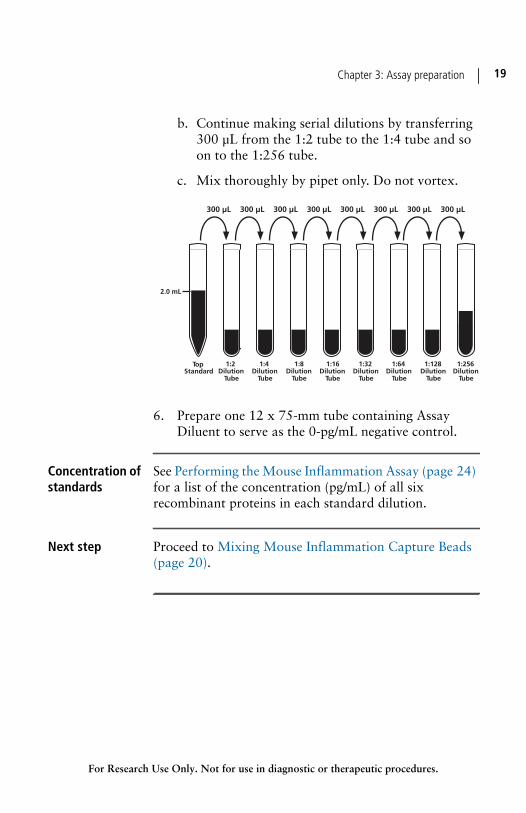
b. Continue making serial dilutions by transferring 300 µL from the 1:2 tube to the 1:4 tube and so on to the 1:256 tube.
c. Mix thoroughly by pipet only. Do not vortex.
6. Prepare one 12 x 75-mm tube containing Assay Diluent to serve as the 0-pg/mL negative control.
Concentration ofstandards
See Performing the Mouse Inflammation Assay (page 24) for a list of the concentration (pg/mL) of all six recombinant proteins in each standard dilution.
Next step Proceed to Mixing Mouse Inflammation Capture Beads (page 20).

BD CBA Mouse Inflammation Kit20
For Research Use Only. Not for use in diagnostic or therapeutic procedures.
Mixing Mouse Inflammation Capture Beads
Purpose of thisprocedure
The Capture Beads are bottled individually (A1–A6). You must pool all six bead reagents immediately before using them in the assay.
Procedure To mix the Capture Beads:
1. Determine the number of assay tubes (including standards and controls) that are required for the experiment (for example, 8 unknowns + 9 standard dilutions + 1 negative control = 18 assay tubes).
2. Vigorously vortex each Capture Bead suspension for 3 to 5 seconds before mixing.
Note: The antibody-conjugated beads will settle out of suspension over time. Vortex the vial immediately before taking a bead-suspension aliquot.
3. Add a 10-µL aliquot of each Capture Bead, for each assay tube to be analyzed, into a single tube labeled “Mixed Capture Beads” (for example, 10 µL of IL-6 Capture Beads × 18 assay tubes = 180 µL of IL-6 Capture Beads required).
4. Vortex the bead mixture thoroughly.
Next step The mixed Capture Beads are now ready to be transferred to the assay tubes. Discard excess mixed Capture Beads. Do not store after mixing.
To begin the assay, proceed to Performing the Mouse Inflammation Assay (page 24). If you need to dilute samples having high-protein concentration, proceed to Diluting samples (page 21).
Chapter 3: Assay preparation 21
For Research Use Only. Not for use in diagnostic or therapeutic procedures.
Diluting samples
Purpose of thisprocedure
The standard curve for each protein covers a defined set of concentrations from 20 to 5,000 pg/mL. It might be necessary to dilute test samples to ensure that their median fluorescence values fall within the range of the generated standard curve. For best results, dilute samples that are known or assumed to contain high levels of a given protein.
Procedure To dilute samples with a known high-protein concentration:
1. Dilute the sample by the desired dilution factor (ie, 1:2, 1:10, or 1:100) using the appropriate volume of Assay Diluent.
2. Mix sample dilutions thoroughly.
Next step Perform instrument setup using the Cytometer Setup Beads. For details, go to bdbiosciences.com/cbasetup and select the appropriate flow cytometer under CBA Kits: Instrument Setup.
Of, if you wish to begin staining your samples for the assay, proceed to Performing the Mouse Inflammation Assay (page 24), and you can perform instrument setup during the 2-hour staining incubation.

For Research Use Only. Not for use in diagnostic or therapeutic procedures.
4Assay procedure
This section covers the following topics:
Performing the Mouse Inflammation Assay (page 24)
Data analysis (page 27)
BD CBA Mouse Inflammation Kit24
For Research Use Only. Not for use in diagnostic or therapeutic procedures.
Performing the Mouse Inflammation Assay
Purpose of thisprocedure
Prepare the standards as described in Preparing Mouse Inflammation Standards (page 18).
Mix the Capture Beads as described in Mixing Mouse Inflammation Capture Beads (page 20).
If necessary, dilute the unknown samples. See Diluting samples (page 21)
Procedure fortubes
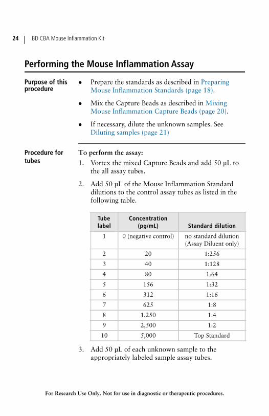
To perform the assay:
1. Vortex the mixed Capture Beads and add 50 µL to the all assay tubes.
2. Add 50 µL of the Mouse Inflammation Standard dilutions to the control assay tubes as listed in the following table.
3. Add 50 µL of each unknown sample to the appropriately labeled sample assay tubes.
Tubelabel
Concentration(pg/mL) Standard dilution
1 0 (negative control) no standard dilution (Assay Diluent only)
2 20 1:256
3 40 1:128
4 80 1:64
5 156 1:32
6 312 1:16
7 625 1:8
8 1,250 1:4
9 2,500 1:2
10 5,000 Top Standard
Chapter 4: Assay procedure 25
For Research Use Only. Not for use in diagnostic or therapeutic procedures.
4. Add 50 µL of the Mouse Inflammation PE Detection Reagent to all assay tubes.
5. Incubate the assay tubes for 2 hours at room temperature, protected from light.
Note: If you have not yet performed cytometer setup, you may wish to do so during this incubation.
6. Add 1 mL of Wash Buffer to each assay tube and centrifuge at 200g for 5 minutes.
7. Carefully aspirate and discard the supernatant from each assay tube.
8. Add 300 µL of Wash Buffer to each assay tube to resuspend the bead pellet.
Procedure forfilter plates
To perform the assay:
1. Wet the plate by adding 100 µL of wash buffer to each well.
2. Place the plate on the vacuum manifold.
3. Aspirate for 2 to 10 seconds until the wells are drained.
4. Remove the plate from the manifold, then blot the bottom of the plate on paper towels.
5. Add 50 µL of each of the following to the wells in the filter plate:
• Capture Beads (vortex before adding)
• Standard or sample (add standards from the lowest concentration to the highest, followed by samples)
• Mouse Inflammation PE Detection Reagent
6. Cover the plate and shake it for 5 minutes at 1,100 rpm on a plate shaker.
BD CBA Mouse Inflammation Kit26
For Research Use Only. Not for use in diagnostic or therapeutic procedures.
7. Incubate the plate for 2 hours at room temperature on a non-absorbent, dry surface.
Note: Place the plate on a non-absorbent, dry surface during incubation. Absorbent or wet surfaces can cause the contents of the wells to leak.
8. Remove the cover from the plate and apply the plate to the vacuum manifold.
9. Vacuum aspirate for 2 to 10 seconds until the wells are drained.
10. Remove the plate from the manifold, then blot the bottom of the plate on paper towels after aspiration.
11. Add 120 µL of wash buffer to each well to resuspend the beads.
12. Cover the plate and shake it for 2 minutes at 1,100 rpm before you begin sample acquisition.
Next step Acquire the samples on the flow cytometer. For details, go to bdbiosciences.com/cbasetup and select the appropriate flow cytometer under CBA Kits: Instrument Setup.
CBA samples must be acquired on the same day they are prepared. Prolonged storage of samples, once the assay is complete, can lead to increased background and reduced sensitivity.
To facilitate the analysis of samples using the FCAP Array software, we recommend the following guidelines:
Acquire standards from lowest (0 pg/mL) to highest (Top Standard) concentration, followed by the test samples.
If running sample dilutions, acquire sequentially starting with the most concentrated sample.
Chapter 4: Assay procedure 27
For Research Use Only. Not for use in diagnostic or therapeutic procedures.
Store all FCS files (standards and samples) in a single folder.
When you are finished acquiring samples, proceed to Data analysis (page 27).
Data analysis
How to analyze Analyze BD CBA Mouse inflammation Kit data using FCAP Array software. For instructions on analysis, go to bdbiosciences.com/cbasetup and see the Guide to Analyzing Data from BD CBA Kits Using FCAP Array Software.
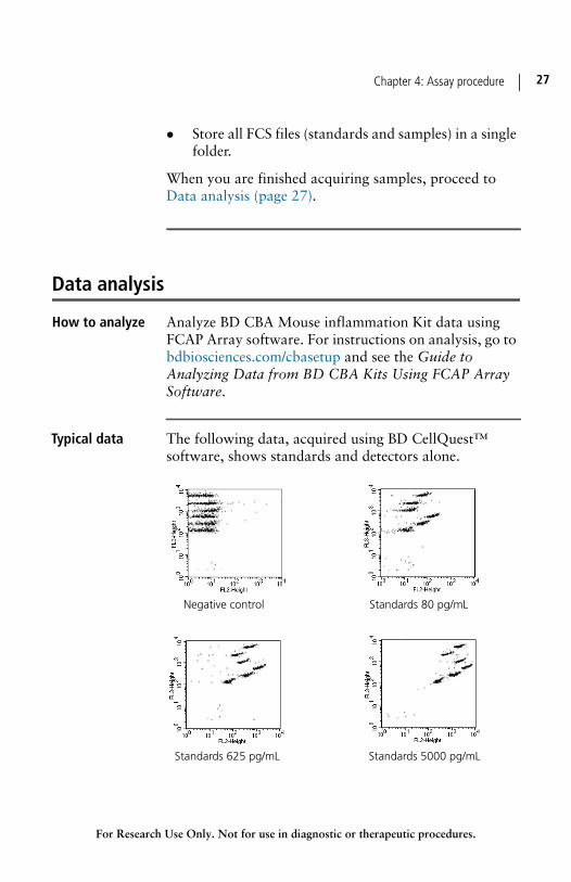
Typical data The following data, acquired using BD CellQuest™ software, shows standards and detectors alone.
Negative control Standards 80 pg/mL
Standards 625 pg/mL Standards 5000 pg/mL
BD CBA Mouse Inflammation Kit28
For Research Use Only. Not for use in diagnostic or therapeutic procedures.
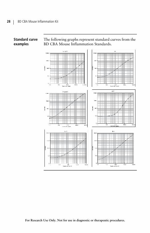
Standard curveexamples
The following graphs represent standard curves from the BD CBA Mouse Inflammation Standards.
For Research Use Only. Not for use in diagnostic or therapeutic procedures.
5Performance
This section covers the following topics:
Theoretical limit of detection (page 30)
Recovery (page 31)
Linearity (page 32)
Specificity (page 33)
Precision (page 35)
BD CBA Mouse Inflammation Kit30
For Research Use Only. Not for use in diagnostic or therapeutic procedures.
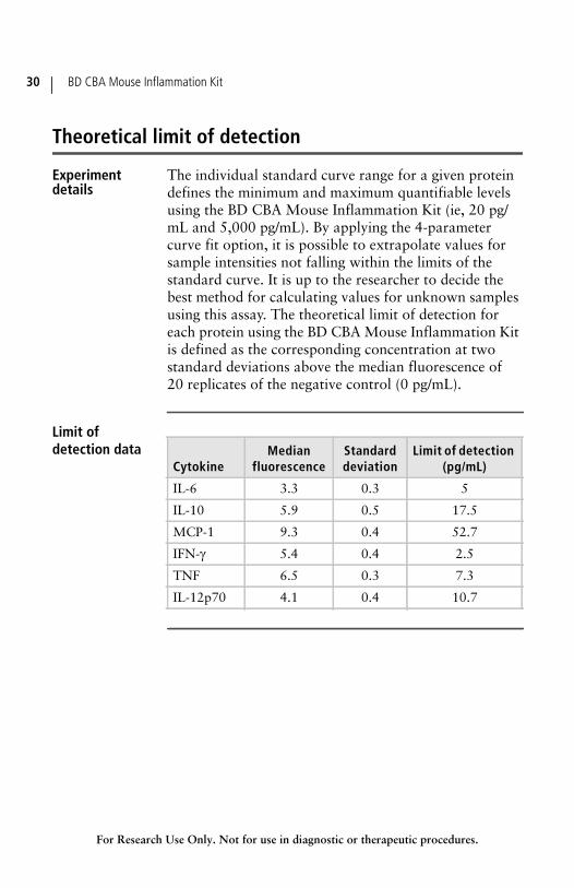
Theoretical limit of detection
Experimentdetails
The individual standard curve range for a given protein defines the minimum and maximum quantifiable levels using the BD CBA Mouse Inflammation Kit (ie, 20 pg/mL and 5,000 pg/mL). By applying the 4-parameter curve fit option, it is possible to extrapolate values for sample intensities not falling within the limits of the standard curve. It is up to the researcher to decide the best method for calculating values for unknown samples using this assay. The theoretical limit of detection for each protein using the BD CBA Mouse Inflammation Kit is defined as the corresponding concentration at two standard deviations above the median fluorescence of 20 replicates of the negative control (0 pg/mL).
Limit ofdetection data
CytokineMedian
fluorescenceStandarddeviation
Limit of detection(pg/mL)
IL-6 3.3 0.3 5
IL-10 5.9 0.5 17.5
MCP-1 9.3 0.4 52.7
IFN- 5.4 0.4 2.5
TNF 6.5 0.3 7.3
IL-12p70 4.1 0.4 10.7
Chapter 5: Performance 31
For Research Use Only. Not for use in diagnostic or therapeutic procedures.
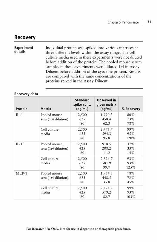
Recovery
Experimentdetails
Individual protein was spiked into various matrices at three different levels within the assay range. The cell culture media used in these experiments were not diluted before addition of the protein. The pooled mouse serum samples in these experiments were diluted 1:4 in Assay Diluent before addition of the cytokine protein. Results are compared with the same concentrations of the proteins spiked in the Assay Diluent.
Recovery data
Protein Matrix
Standardspike conc.
(pg/mL)
Observed ingiven matrix
(pg/mL) % Recovery
IL-6 Pooled mouse sera (1:4 dilution)
2,50062580
1,990.3458.462.3
80%73%78%
Cell culture media
2,50062580
2,476.7594.395.8
99%95%120%
IL-10 Pooled mouse sera (1:4 dilution)
2,50062580
918.5208.211.2
37%33%14%
Cell culture media
2,50062580
2,326.7581.999.7
93%93%125%
MCP-1 Pooled mouse sera (1:4 dilution)
2,50062580
1,954.5448.535.8
78%72%45%
Cell culture media
2,50062580
2,474.2579.282.7
99%93%103%
BD CBA Mouse Inflammation Kit32
For Research Use Only. Not for use in diagnostic or therapeutic procedures.
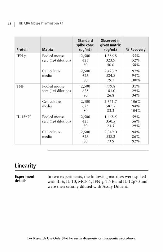
Linearity
Experimentdetails
In two experiments, the following matrices were spiked with IL-6, IL-10, MCP-1, IFN-, TNF, and IL-12p70 and were then serially diluted with Assay Diluent.
IFN- Pooled mouse sera (1:4 dilution)
2,50062580
1,386.8323.946.6
55%52%58%
Cell culture media
2,50062580
2,423.9584.879.7
97%94%100%
TNF Pooled mouse sera (1:4 dilution)
2,50062580
779.8181.026.8
31%29%34%
Cell culture media
2,50062580
2,651.7587.583.3
106%94%104%
IL-12p70 Pooled mouse sera (1:4 dilution)
2,50062580
1,468.5350.523.5
59%56%29%
Cell culture media
2,50062580
2,349.0538.273.9
94%86%92%
Protein Matrix
Standardspike conc.
(pg/mL)
Observed ingiven matrix
(pg/mL) % Recovery
Chapter 5: Performance 33
For Research Use Only. Not for use in diagnostic or therapeutic procedures.
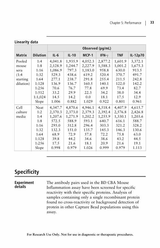
Linearity data
Specificity
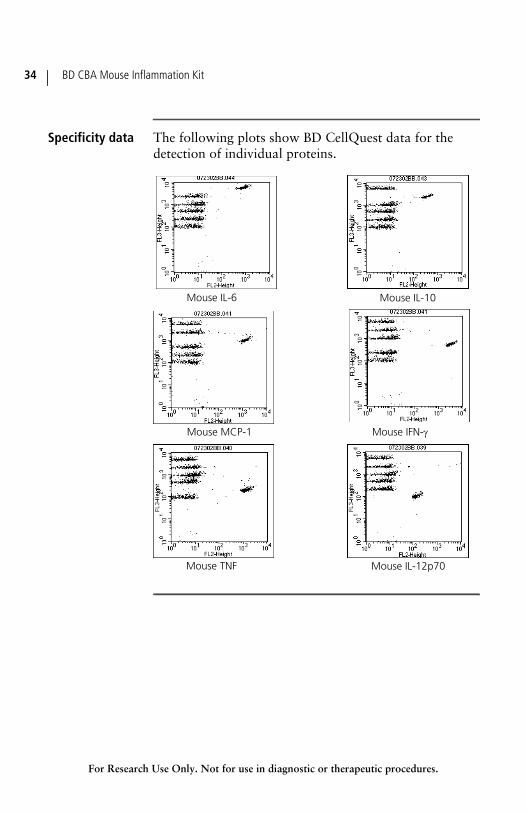
Experimentdetails
The antibody pairs used in the BD CBA Mouse Inflammation assay have been screened for specific reactivity with their specific proteins. Analysis of samples containing only a single recombinant protein found no cross-reactivity or background detection of protein in other Capture Bead populations using this assay.
Matrix Dilution
Observed (pg/mL)
IL-6 IL-10 MCP-1 IFN- TNF IL-12p70
Pooled mouse sera (1:4 starting dilution)
1:41:81:161:321:641:1281:2561:512
1:1,024Slope
4,041.82,128.91,086.9529.3277.1136.970.633.214.51.006
1,935.91,244.7797.3438.6238.7136.776.729.914.2
0.882
4,052.32,227.91,183.0619.2291.8160.577.822.30.0
1.029
2,877.21,588.3958.8520.4255.4140.169.934.218.10.922
1,601.91,001.2630.0370.7211.5122.073.438.017.50.801
3,372.11,675.3913.5491.7242.8142.282.734.412.90.961
Cell culture media
Neat1:21:41:81:161:321:641:1281:256Slope
4,347.72,370.31,207.6572.5295.0132.368.939.817.50.998
4,870.62,373.01,271.9588.9312.8151.072.944.221.6
0.979
4,946.12,379.31,202.2593.1296.4135.757.834.618.11.026
4,518.42,392.41,255.9640.7303.3145.372.238.620.90.999
4,407.92,576.81,330.1616.1321.2146.375.843.221.60.979
4,615.72,426.81,203.6588.7328.2130.663.044.519.11.113
BD CBA Mouse Inflammation Kit34
For Research Use Only. Not for use in diagnostic or therapeutic procedures.
Specificity data The following plots show BD CellQuest data for the detection of individual proteins.
Mouse IL-6 Mouse IL-10
Mouse MCP-1 Mouse IFN-
Mouse TNF Mouse IL-12p70
Chapter 5: Performance 35
For Research Use Only. Not for use in diagnostic or therapeutic procedures.
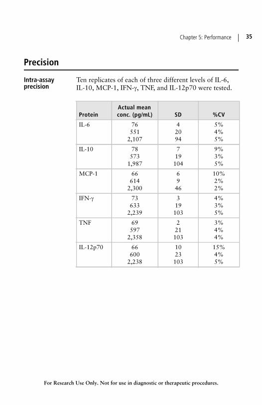
Precision
Intra-assayprecision
Ten replicates of each of three different levels of IL-6, IL-10, MCP-1, IFN-, TNF, and IL-12p70 were tested.
ProteinActual meanconc. (pg/mL) SD %CV
IL-6 76551
2,107
42094
5%4%5%
IL-10 78573
1,987
719104
9%3%5%
MCP-1 66614
2,300
6946
10%2%2%
IFN- 73633
2,239
319103
4%3%5%
TNF 69597
2,358
221103
3%4% 4%
IL-12p70 66600
2,238
1023103
15%4%5%
BD CBA Mouse Inflammation Kit36
For Research Use Only. Not for use in diagnostic or therapeutic procedures.
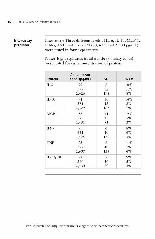
Inter-assayprecision
Inter-assay: Three different levels of IL-6, IL-10, MCP-1, IFN-, TNF, and IL-12p70 (80, 625, and 2,500 pg/mL) were tested in four experiments.
Note: Eight replicates (total number of assay tubes) were tested for each concentration of protein.
ProteinActual meanconc. (pg/mL) SD % CV
IL-6 79557
2,426
862198
10%11%8%
IL-10 71581
2,329
1045162
14%8%7%
MCP-1 58598
2,431
113351
19%5%2%
IFN- 75631
2,425
640120
8%6%5%
TNF 75592
2,697
840155
11%7% 6%
IL-12p70 72590
2,430
73070
9%5%3%
For Research Use Only. Not for use in diagnostic or therapeutic procedures.
6Reference
This section covers the following topics:
Troubleshooting (page 38)
References (page 40)
BD CBA Mouse Inflammation Kit38
For Research Use Only. Not for use in diagnostic or therapeutic procedures.
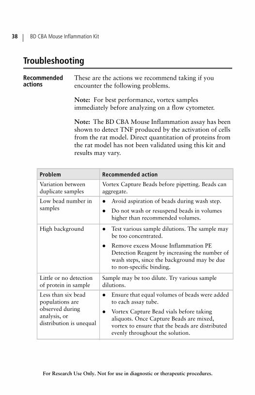
Troubleshooting
Recommendedactions
These are the actions we recommend taking if you encounter the following problems.
Note: For best performance, vortex samples immediately before analyzing on a flow cytometer.
Note: The BD CBA Mouse Inflammation assay has been shown to detect TNF produced by the activation of cells from the rat model. Direct quantitation of proteins from the rat model has not been validated using this kit and results may vary.
Problem Recommended action
Variation between duplicate samples
Vortex Capture Beads before pipetting. Beads can aggregate.
Low bead number in samples
Avoid aspiration of beads during wash step.
Do not wash or resuspend beads in volumes higher than recommended volumes.
High background Test various sample dilutions. The sample may be too concentrated.
Remove excess Mouse Inflammation PE Detection Reagent by increasing the number of wash steps, since the background may be due to non-specific binding.
Little or no detection of protein in sample
Sample may be too dilute. Try various sample dilutions.
Less than six bead populations are observed during analysis, or distribution is unequal
Ensure that equal volumes of beads were added to each assay tube.
Vortex Capture Bead vials before taking aliquots. Once Capture Beads are mixed, vortex to ensure that the beads are distributed evenly throughout the solution.
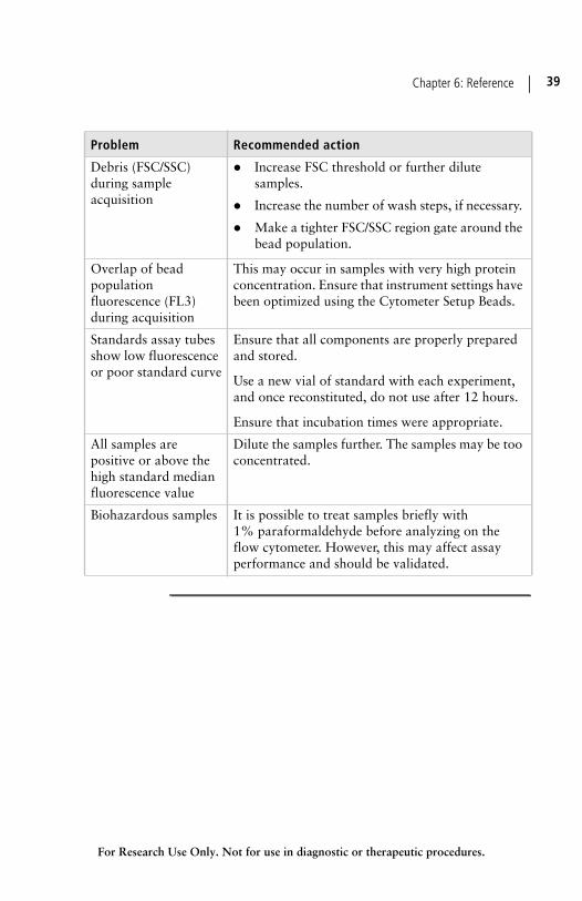
Chapter 6: Reference 39
For Research Use Only. Not for use in diagnostic or therapeutic procedures.
Debris (FSC/SSC) during sample acquisition
Increase FSC threshold or further dilute samples.
Increase the number of wash steps, if necessary.
Make a tighter FSC/SSC region gate around the bead population.
Overlap of bead population fluorescence (FL3) during acquisition
This may occur in samples with very high protein concentration. Ensure that instrument settings have been optimized using the Cytometer Setup Beads.
Standards assay tubes show low fluorescence or poor standard curve
Ensure that all components are properly prepared and stored.
Use a new vial of standard with each experiment, and once reconstituted, do not use after 12 hours.
Ensure that incubation times were appropriate.
All samples are positive or above the high standard median fluorescence value
Dilute the samples further. The samples may be too concentrated.
Biohazardous samples It is possible to treat samples briefly with 1% paraformaldehyde before analyzing on the flow cytometer. However, this may affect assay performance and should be validated.
Problem Recommended action
BD CBA Mouse Inflammation Kit40
For Research Use Only. Not for use in diagnostic or therapeutic procedures.
References
Relatedpublications
1. Bishop JE, Davis KA. A flow cytometric immunoassay for 2-microglobulin in whole blood. J Immunol Methods. 1997;210:79–87.
2. Camilla C, Defoort JP, Delaage M, et al. A new flow cytometry-based multi-assay system. 1. Application to cytokine immunoassays. Cytometry Suppl. 1998;8:132.
3. Carson R, Vignali D. Simultaneous quantitation of 15 cytokines using a multiplexed flow cytometric assay. J Immunol Methods. 1999;227:41–52.
4. Chen R, Lowe L, Wilson JD, et al. Simultaneous quantification of six human cytokines in a single sample using microparticle-based flow cytometric technology. Clin Chem. 1999;45:1693–1694.
5. Collins DP, Luebering BJ, Shaut DM. T-lymphocyte functionality assessed by analysis of cytokine receptor expression, intracellular cytokine expression, and femtomolar detection of cytokine secretion by quantitative flow cytometry. Cytometry. 1998;33:249–255.
6. Fulton RJ, McDade RL, Smith PL, Kienker LJ, Kettman JR Jr. Advanced multiplexed analysis with the FlowMetrix system. Clin Chem. 1997;43:1749–1756.
7. Kricka LJ. Simultaneous multianalyte immunoassays. In: Diamandis EP, Christopoulos TK, eds. Immunoassay. Academic Press. 1996:389–404.
Chapter 6: Reference 41
For Research Use Only. Not for use in diagnostic or therapeutic procedures.
8. Lund-Johansen F, Davis K, Bishop J, de Waal Malefyt R. Flow cytometric analysis of immunoprecipitates: high-throughput analysis of protein phosphorylation and protein-protein interactions. Cytometry. 2000;39:250–259.
9. McHugh TM. Flow microsphere immunoassay for the quantitative and simultaneous detection of multiple soluble analytes. Methods Cell Biol. 1994;42:575–595.
10. Oliver KG, Kettman JR, Fulton RJ. Multiplexed analysis of human cytokines by use of the FlowMetrix system. Clin Chem. 1998;44:2057–2060.
11. Stall A, Sun Q, Varro R, et al. A single tube flow cytometric multibead assay for isotyping mouse monoclonal antibodies. Abstract LB77. Experimental Biology Meeting 1998 (late-breaking abstracts).
12. Cook EB, Stahl JL, Lowe L, et al. Simultaneous measurement of six cytokines in a single sample of human tears using microparticle-based flow cytometry: allergics vs non-allergics. J Immunol Methods. 2001;254:109–118.
13. Dotti G, Savoldo B, Takahashi S, et al. Adenovector-induced expression of human-CD40-ligand (hCD40L) by multiple myeloma cells: A model for immunotherapy. Exp Hematol. 2001;29:952–961.
BD CBA Mouse Inflammation Kit42
For Research Use Only. Not for use in diagnostic or therapeutic procedures.
Notes

552364Rev# 2
United States877.232.8995
Canada866.979.9408
Europe32.2.400.98.95
Japan0120.8555.90
Asia/Pacific65.6861.0633
Latin America/Caribbean55.11.5185.9995
Becton, Dickinson and CompanyBD Biosciences2350 Qume DriveSan Jose, CA 95131 USA(US) Ordering 855.236.2772Technical Service 877.232.8995Fax [email protected]