Embed Size (px)
Citation preview

Accepted Manuscript
Benign Prognosis of Early Repolarization Pattern in Elite Athletes After a LongFollow-Up
Ricard Serra-Grima, Maite Doñate, Jesús Álvarez-García, Ana Barradas-Pires,Andreu Ferrero, Lidia Carballeira, Teresa Puig, Enrique Rodríguez, Juan Cinca
PII: S0002-9343(14)00512-9
DOI: 10.1016/j.amjmed.2014.06.017
Reference: AJM 12578
To appear in: The American Journal of Medicine
Received Date: 15 April 2014
Revised Date: 4 June 2014
Accepted Date: 4 June 2014
Please cite this article as: Serra-Grima R, Doñate M, Álvarez-García J, Barradas-Pires A, FerreroA, Carballeira L, Puig T, Rodríguez E, Cinca J, Benign Prognosis of Early Repolarization Patternin Elite Athletes After a Long Follow-Up, The American Journal of Medicine (2014), doi: 10.1016/j.amjmed.2014.06.017.
This is a PDF file of an unedited manuscript that has been accepted for publication. As a service toour customers we are providing this early version of the manuscript. The manuscript will undergocopyediting, typesetting, and review of the resulting proof before it is published in its final form. Pleasenote that during the production process errors may be discovered which could affect the content, and alllegal disclaimers that apply to the journal pertain.

MANUSCRIP
T
ACCEPTED
ACCEPTED MANUSCRIPT
1
Benign Prognosis of Early Repolarization Pattern in Elite Athletes
After a Long Follow-Up
Ricard Serra-Grima 1, Maite Doñate 1, Jesús Álvarez-García 1, Ana Barradas-Pires
1, Andreu Ferrero 2, Lidia Carballeira 1, Teresa Puig 2, Enrique Rodríguez 1, Juan
Cinca 1
1 Department of Cardiology, Hospital de la Santa Creu i Sant Pau, IIb-Sant Pau,
Universitat Autónoma de Barcelona, Barcelona, Spain
2 Department of Epidemiology and Health, Hospital de la Santa Creu i Sant Pau,
Universitat Autónoma de Barcelona, Barcelona, Spain
Word count: 2984
All authors report no conflict of interest
This study was supported in part by grants from Fundació Fútbol Club Barcelona,
Barcelona, Spain Address for correspondence:
Jesús Álvarez-García, MD,
Department of Cardiology
Hospital de la Santa Creu i Sant Pau
St. Antoni M. Claret 167, 08025, Barcelona, Spain
Phone: +34935565940, fax: +34935565603, e-mal: [email protected]

MANUSCRIP
T
ACCEPTED
ACCEPTED MANUSCRIPT
2
ABSTRACT
Background Analyze the prevalence, clinical characteristics, and long-term outcome of
early repolarization pattern (ERP) in elite athletes during professional activity and after
retirement. ERP is considered a benign variant of the ECG, more frequent in athletes.
However, prospective studies suggested that ERP is associated with an increased risk of
sudden cardiac death (SCD).
Methods and Results A cohort of 299 elite athletes recruited between 1960-1999 was
retrospectively analyzed. Athletes were eligible if they had participated for at least six
consecutive months in high-competition and retired for a minimum of five years before
inclusion. Clinical data and ECG were abstracted from the clinical records using a
questionnaire and outcomes after a mean follow-up of 24 years were registered. Among
the 299 athletes, 66% were men with a mean age of 20 (SD 6.4) years. ERP was found
in 31.4% of participants and it was located in lateral ECG leads in 57.4% of cases, in
inferior in 6.4%, and in both leads in the remaining 36.2%. After retirement ERP still
persisted in 21.1% of athletes. Predictive factors for the persistence were: Sokolov-Lyon
index ≥ 3,5 mV (OR 4.35; CI95% 1.43-13.24; p=0.010), sinus bradycardia (OR 2.56;
CI95% 1.09-5.99; p=0.031), and presence of ERP during the sportive career (OR 20.35;
CI95% 8.54-48.51; p<0.001). After a mean follow-up of 24 years no episodes of SCD
occurred.
Conclusion A third part of elite athletes presented ERP. This pattern persisted in most
of participants after retirement and was not associated with fatal events after a long
follow-up.
Keywords Early repolarization pattern, athletes, electrocardiogram, sudden cardiac
death.

MANUSCRIP
T
ACCEPTED
ACCEPTED MANUSCRIPT
3
INTRODUCTION
Early repolarization pattern (ERP) is defined as a broad positive deflection
originating from an elevated J-point in inferior or lateral ECG leads (1). This ECG
pattern has been historically considered a normal variant in the general population (2),
being more prevalent in males, African-Americans, and in young trained athletes
especially those engaged in endurance disciplines (3, 4). However, emerging data from
case-control (5, 6) and prospective cohort (7) studies suggested that ERP in the inferior
ECG leads is associated with an increased risk of sudden cardiac death (SCD). Although
SCD is relatively rare in athletes, an association between ERP in inferior leads and SCD
has been recently reported (8). Moreover, recent refined analyses of the morphology of
ST-segment following the early repolarization waveforms have afforded new light in the
stratification of individuals at higher risk of arrhythmic death. Indeed,
ascending/upsloping ST-segments identified individuals with benign prognosis whereas
horizontal/descending ST variant was associated with a high risk of arrhythmic death
(9-11). Although almost 50% of cases of SCD in athletes there were not an identifiable
cardiac disorder (12), studies in patients with acute myocardial infarction have shown
that the presence of horizontal/descending ST-segment in the ECG recorded before the
ischemic event increased the risk for occurrence of ventricular fibrillation (13, 14). The
magnitude of ERP and the intensity of the training are closely related (15), but the
factors determining the persistence of ERP in athletes after cessation of high-level
training are not well known. Therefore, the aim of our study was to analyze the
prevalence and clinical characteristics of ERP in active athletes and the very long-term
prognosis after their retirement.

MANUSCRIP
T
ACCEPTED
ACCEPTED MANUSCRIPT
4
METHODS
Study population
This is a retrospective longitudinal study which included 299 Caucasian high level
competition athletes referred to the Health and Sport Unit of Consell Català de l´Esport
of Barcelona City, Medical Service of Barcelona Football Club, and High Performance
Center at Sant Cugat del Vallés, Barcelona, between 1960 to 1999. Athletes were
included if they fulfilled the following criteria: a) participation in elite competitions
while in active training. We defined competitive athletes and official sports competition
as previously defined by the Study Group of Sports Cardiology of the European Society
of Cardiology (16); b) maintenance of a high level of competition training for at least
six consecutive months, and c) retirement from intensive training for a minimum of five
years before inclusion in the study. This study was approved by our institutional review
committee and written consent was obtained from all participants.
Clinical variables
Demographic and clinical baseline information was abstracted from the clinical
records using a structured questionnaire. This included the type of sport performed, the
number of hours of weekly training during competitions periods, and the number of
years participating in high level competition. The level of training during the high
competition period was classified dichotomously according to a 12 weekly hours
training cut-off. A resting 12-lead ECG was obtained during the baseline visit.
In the follow-up visit, we recorded a complete clinical history, ECG, cardiovascular
events (SCD, myocardial infarction, and stroke), number of years since retirement from
high competition, and information on current physical activity categorized as high >5
hours a week; low <5 hours a week. Follow-up period was calculated as the time
(years) elapsed between the baseline and the follow-up visit.

MANUSCRIP
T
ACCEPTED
ACCEPTED MANUSCRIPT
5
ECG variables
The presence of ERP and its morphologic characteristics were stated according to the
work of Tikkanen et al (9). ERP is defined by upright deviation of the J-point of at least
1 mm (0.1 mV) from baseline appearing either as QRS complex slurring (a smooth
transition from the QRS to the ST segment), QRS complex notching (a positive J
deflection inscribed on the S wave), or as discrete QRS complex contour (ERP after
signal return to baseline). These patterns could be observe in the inferior ECG leads (II,
III and aVF), lateral leads (I and aVL, V4-V6) or in both groups of leads. ST-segment
patterns after the J point were coded as: (1) horizontal/descending or (2)
upsloping/rapidly ascending. The upsloping/rapidly ascending ST segment was defined
as ≥0.1 mV elevation of ST segment within 100 ms after the J point or a persistently
elevated ST segment of ≥0.1 mV throughout the ST segment. Horizontal/descending
type was defined as <0.1 mV elevation of the ST segment within 100 ms after the J
point. The baseline was defined as the level between 2 T-P intervals. Sinus bradycardia
was categorized as: light (51-60 bpm); moderate (41-50 bpm); and severe (≤40 bpm)
(17). QT interval duration was corrected according to the Bazett´s formula. Left bundle
branch block (LBBB) was defined by a prolonged QRS duration of 120 ms or more
associated with broad, notched R wave without q waves in leads I, aVL, and V6 and a
rS pattern in lead V1. Right BBB (RBBB) was characterized by prolonged QRS
duration of 120 ms or more associated with an R, rSR´, or qR wave in lead V1; wide,
slurred S waves in leads I, aVL, V5, and V6 ; and a wide terminal r wave in aVR. The
presence of left ventricular (LV) hypertrophy at the ECG was defined by a Sokolov-
Lyon index ≥ 3,5 mV.
Echocardiographic variables

MANUSCRIP
T
ACCEPTED
ACCEPTED MANUSCRIPT
6
Since echocardiographic examination was not feasible at the beginning of the
inclusion period (1960) and was not routinely performed in athletes thereafter, we only
include in this study the recordings obtained in 64% of participants during the follow-up
visit.
Data analysis
Descriptive analyses were performed at the first step. Categorical variables were
described by frequencies and percentages. Statistical differences were analyzed using a
2x2 table test and the χ2 test. Continuous variables were described by the mean and
standard deviation and statistical differences were analyzed using the student's t test in
the case of a normal distribution. To identify independent predictors of ERP in the
follow-up visit, a multivariable logistic regression model was constructed, adjusting for
the covariates statistically significant at the univariate analysis (p value less than 0.20 as
a criterion of entry into multivariate analysis). Additionally, the final model was
adjusted for those variables categorized as clinically relevant. Significant predictors of
ERP were expressed in terms of odds ratio and 95% confidence intervals (CI). To assess
the predictive ability of our model, we calculated the area under the receiver operating
characteristics curve (ROC) following a nonparametric distribution assumption. A p
value of less than 0.05 was considered statistically significant. Data were analyzed
using the statistical package SPSS for Windows (IBM, SPSS Statistics, 21 version).

MANUSCRIP
T
ACCEPTED
ACCEPTED MANUSCRIPT
7
RESULTS Clinical characteristics
Among the 299 elite athletes included in the study, 94 (31.4%) presented ERP on
admission. As shown in Table 1, athletes with and without ERP showed comparable
demographic profile except for a higher incidence of males in the former group.
Likewise, both groups of athletes showed comparable distribution of competition
categories and similar volume of physical training. Our study participants had been in
the elite sport over 3 years on average and most of them trained more than 12 hours a
week. The sports spectrum was very broad, but Athletics were practiced by about 45%
of participants.
ECG
As shown in Table 2, the presence of ERP was significantly associated with
sinus bradycardia, criteria of LV hypertrophy, and a longer QRS complex duration.
However, there were not differences in the corrected QTc interval and the presence of
RBBB. The ERP was observed in lateral leads in 57.4% of cases, in inferior leads in
6.4%, and in both regions in 36.2%. The large majority of cases presented an
ascending/upsloping ST-segment with a J-point elevation between 0.1 to 0.2 mV from
baseline. In the group of ERP, the pattern of the QRS morphology was discrete in the
41.2% of cases and slurred and notched in about 30%. There were 2 cases of
horizontal/descending ST-segment, more apparent in the inferior leads. Figure 1
illustrates a typical example of ERP with ascending/upsloping ST-segment.

MANUSCRIP
T
ACCEPTED
ACCEPTED MANUSCRIPT
8
Follow-up
The mean follow-up was 24 years (SD 7.6) with a minimum and a maximum of
10 and 47 years respectively. Mean age of participants at the second visit was 45 years
(SD 9.3, interquartile range 24-75). After competition retirement, 30% of individuals of
the non-ERP group and 50% of those with ERP continued to maintain a significant level
of training (>5 hours/week) (28.5% vs 47.6%, p 0.004). As shown in Table 3, the
prevalence of cardiovascular risk factors and cardiovascular events in our series was
very low. Non-fatal myocardial infarction occurred in two athletes and none of the
participants died. We observed a prevalence of the ERP in 63 out of 299 athletes
(21.1%), mainly located at the lateral (50.8%) and inferior-lateral leads (47.6%). The
presence of bradycardia was significantly higher in the ERP group (71.4% vs. 41.9%,
p<0.001). Among the 63 athletes who presented RP in the follow-up, 51 already showed
this pattern in the time of enrollment (persistent ERP), while in 12 of them this pattern
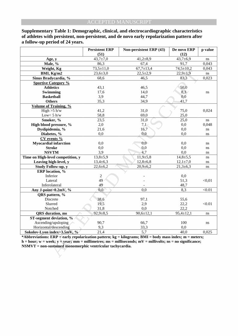
appeared de novo. Supplementary Table 1 summarizes the demographic, clinical, and
electrocardiographic characteristics of athletes with these three ERP forms. The total
number of competitions and medals won in national and international championships by
the athletes included in our study are summarized in Supplementary Table 2.
The echocardiographic examination was performed in 64% of cases during the follow-
up visit. Athletes with or without ERP showed comparable LV ejection fraction (74±10
vs. 71±9%), thickness of LV septum (9±1 vs. 9±2 mm), thickness of posterior LV wall
(9±1 vs. 8±2 mm), and left atrial diameter (33±5 vs. 32±6 mm). The LV diameter was
larger in the ERP group (50±6 vs. 48±5 mm, p <0.05).
The multivariate analysis showed that the predictors of current ERP were a Sokolov-
Lyon index ≥ 3,5 mV, sinus bradycardia at second visit and ERP during elite activity
(Table 4). However, male sex and the volume of training either during high level

MANUSCRIP
T
ACCEPTED
ACCEPTED MANUSCRIPT
9
competition or after retirement was not a significant predictor. Figure 2 shows the ROC
curve of the model to predict the presence of ERP in the follow-up ECG (AUC 0.89; CI
95% 0.85-0.94, p <0.001).
MANUSCRIP
T
ACCEPTED
ACCEPTED MANUSCRIPT
10
DISCUSSION
Main findings
This study shows that one third of elite athletes present ERP during their active
competition life and this ECG pattern is associated with a benign prognosis after a mean
follow-up period of 24 years. In 97.5% of cases the ERP was associated with
ascending/upsloping ST-segment and only 2.5% presented horizontal/descending ST-
segment pattern. Moreover, the study denotes that the ERP persists after professional
retirement in a high proportion of cases.
Prevalence of ERP and related factors
The prevalence of ERP in the general population is largely variable as it is
influenced among others by race, age, level of physical activity, characteristics of the
QRS complex and by the definition of ERP itself (18). In a recent meta-analysis based
on 7 prospective observational cohort studies and on 3 case-control studies, the
prevalence of ERP varied from 0.9% to 31% (19). Applying a stricter criterion, the
prevalence of ERP was 18.6% in a series of 5069 participants of the CARDIA cohort
with a mean age of 25 years and black race in 40% (20). The prevalence of ERP among
athletes is higher than that observed in the general population. In our elite athletes the
ERP was present in 31% of cases and this prevalence was similar to that observed in a
cohort of 503 athletes recruited between 2000-2010 at University of Miami (3), and also
comparable with a study on 879 colleague athletes registered between 2006 and 2010 at
Harvard University (15). In the multivariate analysis, the predictive factors for the
presence of ERP after competition retirement were a Sokolov-Lyon index ≥ 3.5 mV,
sinus bradycardia, and presence of ERP during the sporting career. The most common

MANUSCRIP
T
ACCEPTED
ACCEPTED MANUSCRIPT
11
type of sportive activity associated with ERP in our series was athletics (45%).
However, this association was not statistically significant because of the wide spectrum
of modalities, intensity of training, and physiological characteristics of the exercise
(isovolumetric or isotonic) performed by our athletes.
The mechanism by which intensive physical training could favor the
development of ERP is apparently related to a concomitant increased vagal tone.
Indeed, it has been extensively documented that sinus bradycardia is one of the most
prevailing electrophysiological feature in active athletes (21), and that the degree of
sinus bradycardia depends on intensity of physical training (22). Parasympathetic
modulation increases regional electrophysiological differences and repolarization
dispersion, which might result in an elevation of the ST and J point and prominent T
waves (23). Experimental studies in dogs have raised the hypothesis that ERP is caused
by a transmural voltage gradient created by a spike-and-dome action potential which is
present in the epicardium but absent in the endocardium (24). The latter is mediated by
transitory outward potassium current (Ito). This hypothesis is supported by the
observation of mutations affecting the potassium currents in patients with ERP suffering
ventricular arrhythmias (25, 26)
Prognosis of ERP
Studies conducted in the general population have reported cases of SCD
secondary to idiopathic ventricular fibrillation in individuals with ERP (5, 6). More
recently, in a meta-analysis based on seven prospective observational cohort studies and
3 case-control studies assessing the association between the ERP and risk for cardiac
death, arrhythmia death, or all-cause death, showed that ERP was associated with
increased risk and a low intermediate absolute incidence rate of arrhythmia death (19).

MANUSCRIP
T
ACCEPTED
ACCEPTED MANUSCRIPT
12
In these reports the presence of J-point elevation > 0.1 mV in the inferior leads and the
notching configuration was associated with an increased risk for arrhythmia death.
The estimated annual incidence of SCD in athletes is very low (1/75,000 to
1/200,000) (27, 28, 29). In a case-control study including 21 athletes with SCD and 365
healthy athletes with previous ECG screening, the J wave and/or QRS slurring was
found more frequently among athletes with SCD than in controls (8). However, in a
prospective study in a cohort of 503 athletes with a 30% of ERP prevalence, there were
no cases of family history of SCD and there were no cases of SCD or symptomatic
ventricular tachyarrhythmia during the 10 years period of ECG screening (3). Likewise,
in a cross-sectional cohort of 879 newly matriculated student athletes with ERP mainly
of the inferior subtype, there were no cases of SCD and unexplained syncope or
hospitalization for cardiovascular causes after a mean follow-up of about 21 months
(15). In these series the incidence of ERP increased after a load period of intensive
physical training. A major contribution in risk stratification in subjects with ERP was
the observation of an association between the morphology of the ST-segment and the
hazard ratio of arrhythmic death (9). Indeed, young athletes with horizontal/descending
ST-segment had an increased HR of arrhythmic death than subjects with
ascending/upsloping ST-segment pattern. These findings were later confirmed in case-
control series in the general population (10, 11). Our study supports the benign
prognosis of ERP with upsloping ST-segment in elite athletes during the largest follow-
up period ever reported (mean follow-up of 24 years).
In summary, this retrospective cohort of 299 elite athletes of Caucasian ethnicity shows
that ERP was present in one third of the participants and that this ECG pattern was not
associated with fatal events after a mean follow-up of about 24 years. In 97.5% of cases

MANUSCRIP
T
ACCEPTED
ACCEPTED MANUSCRIPT
13
the ERP was associated with ascending/upsloping ST-segment and only 2.5% presented
horizontal/descending ST-segment pattern. The ERP persisted in most of cases after
sportive retirement and was highly associated with sinus bradycardia. The predictive
factors for the presence of ERP after the retirement were a Sokolov-Lyon index ≥ 3,5
mV, current sinus bradycardia, and presence of ERP during the elite activity.
Study Limitations
The sample size of our cohort was relatively modest, but the strength of the data
presented here is founded by the very long follow-up period of the study. All of our
participants were of Caucasian ethnicity thus present data can not be extrapolated to
other races since a higher association of ERP with African ethnicity has been recognized
(3, 4).
Echocardiographic studies were not feasible at the early beginning of the
inclusion period, and were not routinely performed thereafter. However, the recordings
obtained during the follow-up visit in 64% of athletes suggested that the ERP is not
associated with underlying structural heart disease, as has been previously recognized
(15).

MANUSCRIP
T
ACCEPTED
ACCEPTED MANUSCRIPT
14
ACKNOWLEDGMENTS
We appreciate the assistance provided by Mrs Monica Ortega, member of Health and
Sport Unit of Consell Català de l´Esport of Barcelona City.

MANUSCRIP
T
ACCEPTED
ACCEPTED MANUSCRIPT
15
REFERENCES
1. Shipley R, Hallaran W. The four lead electrocardiogram in 200 normal men and
women. Am Heart J. 1936;11:325-345.
2. Wasserburger RH, Alt WJ. The normal RS-T segment elevation variant. Am J
Cardiol. 1961;8:184-192.
3. Junttila MJ, Sager SJ, Freiser M, et al. Inferolateral early repolarization in
athletes. J Interv Card Electrophysiol 2011;31:33–38.
4. Higgins JP. Normal resting electrocardiographic variants in young athletes. Phys
Sports Med 2008;36:69–75.
5. Haissaguerre M, Derval N, Sacher F, et al. Sudden cardiac arrest associated with
early repolarization. N Engl J Med. 2008;358:2016 –2023.
6. Rosso R, Kogan E, Belhassen B, et al. J-point elevation in survivors of primary
ventricular fibrillation and matched control subjects: incidence and clinical
significance. J Am Coll Cardiol. 2008;52:1231–1238.
7. Tikkanen JT, Anttonen O, Junttila MJ, et al. Long-term outcome associated with
early repolarization on electrocardiography. N Engl J Med. 2009;361:2529–
2537.
8. Cappato R, Furlanello F, Giovinazzo V, et al. J wave, QRS slurring, and ST
elevation in athletes with cardiac arrest in the absence of heart disease: marker
of risk or innocent bystander? Circ Arrhythm Electrophysiol 2010;3:305–311.

MANUSCRIP
T
ACCEPTED
ACCEPTED MANUSCRIPT
16
9. Tikkanen JT, Junttila MJ, Anttonen O, et al. Early repolarization:
electrocardiographic phenotypes associated with favorable long-term outcome.
Circulation 2011;123(23):2666-73.
10. Rosso R, Glikson E, Belhassen B, et al. Distinguishing “benign” from
“malignant early repolarization”: the value of the ST-segment morphology.
Heart Rhythm 2012;9(2):225-229.
11. Kim SH, Kim do Y, Kim HJ, et al. Early repolarization with horizontal ST
segment may be associated with aborted sudden cardiac arrest: a retrospective
case control study. BMC Cardiovasc Disord. 2012;11;12:122.
12. Maron BJ, Doerer JJ, Haas TS, et al. Sudden deaths in young competitive
athletes: analysis of 1866 deaths in the United States, 1980–2006. Circulation.
2009;119:1085–1092.
13. Tikkanen JT, Wichmann V, Junttila MJ, et al. Association of Early
Repolarization and Sudden Cardiac Death During an Acute Coronary Event.
Circ Arrhythm Electrophysiol 2012;5:714-718.
14. Naruse Y, Tada H, Harimura Y, et al. Early Repolarization Is an Independent
Predictor of Occurrences of Ventricular Fibrillation in the Very Early Phase of
Acute Myocardial Infarction. Circ Arrhythm Electrophysiol 2012;5:506-513.
15. Noseworthy PA, Weiner R, Kim J, et al. Early Repolarization Pattern in
Competitive Athletes: Clinical Correlates and the Effects of Exercice Training.
Circ Arrhythm Electrophysiol. 2011;4;432-440.
16. Corrado D, Pelliccia A, Bjørnstad HH, et al. Cardiovascular pre-participation
screening of young competitive athletes for prevention of sudden death:

MANUSCRIP
T
ACCEPTED
ACCEPTED MANUSCRIPT
17
proposal for a common European protocol. Consensus Statement of the Study
Group of Sport Cardiology of the Working Group of Cardiac Rehabilitation and
Exercise Physiology and the Working Group of Myocardial and Pericardial
Diseases of the European Society of Cardiology. Eur Heart J. 2005
Mar;26(5):516-24.
17. Straus HC, Bigger JT, Saroff AL, Guiardina ECU. Electrophysiologic evaluation
of sinus node function in patients with sinus sonde dysfunction. Circulation
1976;53:763.
18. Rosso R, Halkin A, Viskin S. J waves and early repolarization: do not confuse
me with the facts! Heart Rhythm 2012;9(10):1603-1604.
19. Wu SH, Lin X, Cheng Y, et al. Early repolarization pattern and risk for
arrhythmia death: a meta-analysis. J Am Coll Cardiol 2013;61(6):645-50.
20. Walsh JA 3rd, Ilkhanoff L, Soliman EZ, et al. Natural history of the early
repolarization pattern in a biracial cohort: CARDIA (Coronary artery risk
development in young adults) Study. J Am Coll Cardiol 2013;61(8):863-9.
21. Viitasalo MT, Kala R, Eisalo A. Ambulatory electrocardiographic recording in
endurance athletes. Br Heart J 1982;47:213-240.
22. Serra-Grima R, Puig T, Doñate M, et al. Long-term Follow-Up of Bradycardia in
Elite Athletes. Int Sports Med 2008;29: 934-937.
23. Barbosa EC, Bomfim AS, Benchimol-Barbosa PR, Ginefra P. Ionic mechanisms
and vectorial model of early repolarization pattern in the surface
electrocardiogram of the athlete. Ann Noninvasive Electrocardiol.2008:13;301–
307.

MANUSCRIP
T
ACCEPTED
ACCEPTED MANUSCRIPT
18
24. Yan GX, Antzelevitch C. Cellular basis for the electrocardio-graphic J wave.
Circulation 1996;93(2):372-9.
25. Haïssaguerre M, Chatel S, Sacher F, et al. Ventricular fibrillation with prominent
early repolarization associated with a rare variant of KCNJ8/KATP channel. J
Cardiovasc Electrophysiol 2009;20(1):93-8.
26. Medeiros-Domingo A, Tan BH, Crotti L, et al. Gain-of- function mutation,
S422L, in the KCNJ8-encoded cardiac K(ATP) channel Kir6.1 as a pathogenic
substrate for J-wave syndromes. Heart Rhythm 2010;7(10): 1466-71.
27. Corrado D, Basso C, Schiavon M., Thiene G. Screening for hypertrophic
cardiomyopathy in young athletes. N Engl J Med 1998, 339(6), 364–369.
28. Maron BJ. Sudden death in young athletes. N Engl J Med 2003, 349(11), 106-
1075.
29. Viskin S, Rosso R, Halkin A. Making sense of early repolarization. Heart
Rhythm 2012;9(4):566-569.

MANUSCRIP
T
ACCEPTED
ACCEPTED MANUSCRIPT
19
FIGURE LEGENDS
Figure 1: Disappearance of early repolarization pattern after a follow-up of 17
years
Panel A shows an illustrative example of an ascending/upsloping ST segment type of
early repolarization pattern, in the lateral ECG leads of a marathon runner in 1983.
Panel B shows the ECG of the same athlete, five years after retirement from the
practice of elite sport.
Figure 2: Receiver operating characteristics curve of the model
The graphic shows the ROC curve of the model predicting the presence of ERP in the
follow-up ECG (AUC 0.89; CI 95% 0.85-0.94, p <0.001).

MANUSCRIP
T
ACCEPTED
ACCEPTED MANUSCRIPTTable 1: Demographic and sportive characteristics of 299 elite athletes with and without early repolarization pattern at study inclusion
No ERP
(205)
ERP
(94) p value
ECG Localization of ERP p value
Inferior (6) Lateral (54) Inferolateral (34)
Age, years 20,1±6,4 20,5±6,4 ns 21,3±9,2 20,3±6,5 20,7±5,8 ns
Male, % 60,5 77,7 0,004 66,7 75,9 82,4 0,027
Weight, Kg 63,7±12,8 61,9±10,9 ns 56,2±9,9 61,0±10,7 64,4±10,9 ns
Height, cm 170,7±11,1 170,1±9,2 ns 166,2±11,8 170,1±9,3 171,0±8,7 Ns
BMI, Kg/m 2 21,1±2,6 21,0±2,2 ns 19,8±1,7 20,8±2,2 21,5±2,3 Ns
Sportive Category
Athletics, %
Swimming, %
Basketball, %
Others, %
45,4
13,2
6,8
34,6
44,7
16,0
4,3
35,1
ns
50,0
16,7
0,0
33,3
50,0
9,3
5,6
35,2
35,3
26,5
2,9
35,3
ns
Volume of Training
High (>12 h/w), %
Low (<12 h/w), %
79,5
20,5
84,9
15,1
ns
100
0
86
14
80,6
19,6
ns
Time on High-level
Competition, y 3,5±5,1 3,8±4,8 ns 6,3±9,9 3,0±4,2 4,6±4,4 ns
*Abbreviations: ERP = early repolarization pattern; BMI = body mass index; BSA = body surface area; Kgs = kilograms; m = meters; h = hour; w = week; y = year; ns = no significance.

MANUSCRIP
T
ACCEPTED
ACCEPTED MANUSCRIPTTable 2: ECG data of 299 elite athletes with and without early repolarization pattern at study inclusion
No ERP
(205)
ERP
(94) p value
ECG Localization of ERP p value
Inferior (6) Lateral (54) Inferolateral (34)
HR, bpm 50,5±24,8 47,0±19,5 ns 42,7±21,7 46,8±20,6 47,9±17,9 ns
Sinus Bradycardia, %
Light
Moderate
Severe
63,9
38,6
36,4
25
87,2
39,5
43,2
17,3
<0,001
66,7
33,3
50
16,7
85,2
34,1
50
15,9
94,1
48,4
32,3
19,4
<0,001
Any J-point�0.2mV, %
- 8,2 - 0 6,3 12,9 <0,001
QRS pattern, % Discrete Slurred
Notched
-
-
41,2
29,4
29,4
-
50
33,3
16,7
47,9
22,9
29,2
29
38,7
32,3
<0,001
QRS duration, ms 82±8 92±8 <0,001 90±6 91±8 95±8 ns
ST-segment pattern, %
Ascending/upsloping
Horizontal/descending
-
-
97,5
2,5
-
83,3
16,7
97,7
2,3
100
0
0,059
Sokolov-Lyon >3.5 mV, % 10,2 40,5 <0,001 33,3 38,3 45,2 <0,001
QT, ms 336,8±145,1 260,9±136,4 ns 348,3±171,4 352,9±143,0 375,3±121,9 ns
QTc, ms 331,3±142,5 337,1±126,8 ns 320,3±157,1 331,9±134,1 348,2±112,2 ns
RBBB, % 4,4 6,4 ns 0 7,4 5,9 ns
*Abbreviations: ERP = early repolarization pattern; HR = heart rate; bpm = beats per minute; ms = milliseconds; mV = millivolts; RBBB = right bundle branch block; ns = no significance

MANUSCRIP
T
ACCEPTED
ACCEPTED MANUSCRIPT Table 3: Clinical and sportive characteristics of 299 elite athletes after a mean follow-up of 24 years
ERP (63) No ERP (236) p value
ECG Localization of ERP
p value Inferior (1) Lateral (32) Inferolateral (30)
Age, years 43,7±9,75 45,0±7,5 ns 55,00 43,12±7,64 43,87±7,28 ns
Male, % 87,3 60,2 ns 100 84,4 90,0 ns
Weight, Kg 73,7±72,1 72,1±14,4 ns 55,00 73,91±10,95 74,10±10,32 ns
BMI, Kg/m 2 23,5±2,8 23,5±3,5 ns 19,00 23,31±3,05 23,93±2,53 ns
Sinus Bradycardia, % 71,4 41,9 0,000 100 68,8 73,3 ns
Sportive Category %
Athletics
Swimming,
Basketball
Others
46
14,3
3,2
36,5
45,3
13,6
6,8
34,3
ns
100
0,0
0,0
0,0
50,0
15,6
6,2
341,4
40,0
13,3
3,2
40,0
ns
Volume of Training %
High (>5 h/w)
Low (<5 h/w)
47,6
52,4
28,5
71,5
0,004
100
0
46,9
53,1
46,7
53,3
ns
Smoker, %
High Blood Pressure, %
Dyslipidemia, %
Diabetes, %
3.2
1,6
17,5
0,0
17,5
8,9
12,3
0,9
0,041
0,048
ns
ns
0,00
0,00
0,00
0,00
6,2
0,00
18,8
0,00
0,00
3,3
16,7
0,00
ns
ns
ns
ns
CV events %
Myocardial infarction
Stroke
NSMVT
0,0
0,0
3,2
0,9
0,9
1,7
ns
ns
ns
0,00
0,00
0,00
0,00
0,00
3,1
0,00
0,00
3,3
ns
ns
ns
Time on High-Level
Competition, y
Leaving High Level, y
13,9±5,7
13,2±6,4
11,9±5,9
16,5±8,5
0,022
0,004
22,0±0,0
16,0±0,0
12,9±5,4
13,2±6,6
14,6±5,9
13,1±6,4
ns
ns
Study Follow-up, y 22,4±6,2 24,7±7.9 0,030 32,0±0,0 22,1±6,7 23,4±5,6 ns
*Abbreviations: ERP = early repolarization pattern; BMI = body mass index; BSA = body surface area; Kgs = kilograms; m = meters; HR = heart rate; bpm = beats per minute; ms = milliseconds; h = hour; w = week; y = years; NSMVT = non-sustained monomorphic ventricular tachycardia.
MANUSCRIP
T
ACCEPTED
ACCEPTED MANUSCRIPTSupplementary Table 1: Demographic, clinical, and electrocardiographic characteristics of athletes with persistent, non-persistent, and de novo early repolarization pattern after a follow-up period of 24 years.
Persistent ERP (51)
Non-persistent ERP (43) De novo ERP (12)
p value
Age, y 43,7±7,0 41,2±8,9 43,7±6,9 ns Male, % 86,3 67,4 91,7 0,043
Weight, Kg 73,5±11,0 67,7±13,4 74,5±10,2 0,043 BMI, Kg/m2 23,6±3,0 22,5±2,9 22,9±1,9 ns
Sinus Bradycardia, % 68,6 46,5 83,3 0,023 Sportive Category %
Athletics Swimming Basketball
Others
43,1 17,6 3,9 35,3
46,5 14,0 44,7 34,9
50,0 8,3 0,0 41,7
ns
Volume of Training, % High >5 h/w Low< 5 h/w
41,2 58,8
31,0 69,0
75,0 25,0
0,024
Smoker, % High blood pressure, %
Dyslipidemia, % Diabetes, %
23,5 2,0 21,6 0,0
31,0 7,1 16,7 0,0
25,0 0,0 0,0 0,0
ns 0,048
ns ns
CV events % Myocardial infarction
Stroke NSVTM
0,0 0,0 3,9
0,0 0,0 4,7
0,0 0,0 0,0
ns ns ns
Time on High-level competition, y Leaving high-level, y
13,8±5,9 13,4±6,3
11,9±5,8 12,8±6,8
14,8±5,5 12,1±7,0
ns ns
Study Follow-up, y 22,6±6,2 20,9±6,2 21,3±6,3 ns ERP location, %
Inferior Lateral
Inferolateral
2 49 49
- - -
0,0 51,3 48,7
<0,01
Any J-point>0.2mV, % 0,0 0,0 8,3 <0.01 QRS pattern, %
Discrete Slurred Notched
38,6 19,5 31,8
97,1 2,9 0,0
55,6 22,2 22,2
<0.01
QRS duration, ms 92,9±8,5 90,6±12,1 95,4±12,1 ns ST-segment deviation, %
Ascending/upsloping Horizontal/descending
90,7 9,3
66,7 33,3
100 0,0
ns
Sokolov-Lyon index>3.5mV, % 21,4 5,7 40,0 0,025 *Abbreviations: ERP = early repolarization pattern; kg = kilograms; BMI = body mass index; m = meters; h = hour; w = week; y = year; mm = millimetres; ms = milliseconds; mV = millivolts; ns = no significance; NSMVT = non-sustained monomorphic ventricular tachycardia.

MANUSCRIP
T
ACCEPTED
ACCEPTED MANUSCRIPT

MANUSCRIP
T
ACCEPTED
ACCEPTED MANUSCRIPT

MANUSCRIP
T
ACCEPTED
ACCEPTED MANUSCRIPT
• One third of elite athletes presented the benign early repolarization pattern (upsloping ST-segment) whereas the one related to sudden cardiac death risk (rectified/descending ST-segment) was rare.
• Early repolarization pattern persisted in half of athletes after professional sport retirement and no episodes of sudden cardiac death were observed after 24 years of follow-up.