Embed Size (px)
Citation preview

Proc. Natl. Acad. Sci. USAVol. 92, pp. 10413-10417, October 1995Plant Biology
Benzoic acid 2-hydroxylase, a soluble oxygenase from tobacco,catalyzes salicylic acid biosynthesis
(Nicotiana tabacum/tobacco mosaic virus/cytochrome P450/acquired resistance)
JOSEi LEON, VLADIMIR SHULAEV, NASSER YALPANI*, MICHAEL A. LAWTON, AND ILYA RASKINtAgBiotech, Center for Agricultural Molecular Biology, Rutgers University, P.O. Box 231, New Brunswick, NJ 08903-0231
Communicated by Eric E. Conn, University of California, Davis, CA, July 31, 1995 (received for review February 1, 1995)
ABSTRACT Benzoic acid 2-hydroxylase (BA2H) cata-lyzes the biosynthesis of salicylic acid from benzoic acid. Theenzyme has been partially purified and characterized as asoluble protein of 160 kDa. High-efficiency in vivo labeling ofsalicylic acid with 1802 suggested that BA2H is an oxygenasethat specifically hydroxylates the ortho position of benzoicacid. The enzyme was strongly induced by either tobaccomosaic virus inoculation or benzoic acid infiltration of to-bacco leaves and it was inhibited by CO and other inhibitorsof cytochrome P450 hydroxylases. The BA2H activity wasimmunodepleted by antibodies raised against SU2, a solublecytochrome P450 from Streptomyces griseolus. The anti-SU2antibodies immunoprecipitated a radiolabeled polypeptide ofaround 160 kDa from the soluble protein extracts of L-[35S]_methionine-fed tobacco leaves. Purified BA2H showed CO-difference spectra with a maximum at 457 nm. These datasuggest that BA2H belongs to a novel class of soluble, highmolecular weight cytochrome P450 enzymes.
Salicylic acid (SA), a phenolic compound ubiquitously distrib-uted in plants (1), has been proposed as a likely endogenoussignal in the induction of plant resistance to pathogens (2, 3).The observed increase in endogenous SA levels in pathogen-inoculated and uninoculated leaves was sufficient to inducesystemic acquired resistance and synthesis of pathogenesis-related proteins (4). Work with transgenic tobacco plantsexpressing a bacterial gene encoding salicylate hydroxylasedemonstrated that SA accumulation is required for the induc-tion of systemic acquired resistance of tobacco against tobaccomosaic virus (5). However, grafting experiments suggestedthat, at least in tobacco, SA may not be a primary mobile signalin systemic acquired resistance (6).
In plants, SA is synthesized from trans-cinnamic acid bydecarboxylation to benzoic acid (BA) and further 2-hydroxy-lation of BA to SA (7). The final step is catalyzed by benzoicacid 2-hydroxylase (BA2H), an enzyme that is constitutivelyexpressed in tobacco but is highly induced by inoculation withtobacco mosaic virus (TMV) or application of BA (8). Theinduction results, in part, from enhanced de novo synthesis ofBA2H protein. NAD(P)H or reduced methyl viologen canserve as in vitro reductants for BA2H.
Hydroxylation reactions are very important in the biosyn-thesis and metabolism of plant phenolic compounds. Most ofthese hydroxylations are catalyzed by cytochrome P450 mo-nooxygenases (9). These enzymes form a superfamily of 50- to60-kDa heme-containing proteins that have been extensivelystudied in animals and bacteria, but less well studied in plants.In eukaryotes, almost all known cytochrome P450s are micro-somal proteins (10). In contrast to eukaryotic systems, mostbacterial cytochrome P450 monooxygenases are soluble pro-teins (11-13). More than 200 P450 genes have been classifiedinto subfamilies according to homology of their amino acid
sequences (14). The homology between different forms ofcytochrome P450 in eukaryotes and prokaryotes is very lim-ited. However, some cases of antigenic crossreactivity betweenbacterial and plant cytochrome P450s have been reported (15).Here we report the isolation and biochemical characteriza-
tion ofBA2H, an unusual, high molecular weight, soluble P450oxygenase from tobacco which catalyzes the formation of SAfrom BA. This enzyme may play a key role in the activation ofplant defenses against pathogens.
MATERIALS AND METHODS
Plant Culture and TMV Inoculation. Seeds of tobacco,Nicotiana tabacum L. (cv. Xanthi-nc, NN genotype), were sownand grown as described (4). The expanded leaves of 6- to8-week-old plants were inoculated with 5 ,ug of the Ul strainof TMV or mock-inoculated without virus as indicated. Theplants were incubated for 4 days at 32°C and then shifted to24°C for up to 10 hr as described (7, 8). For experiments on invivo inhibition of SA synthesis, tobacco leaves, attached to theplant, were syringe-infiltrated with 50 ,uM tetcyclacis in 5 mMpotassium phosphate buffer (pH 5.5) or incubated inside thesame transparent sealed chamber used for 1802 labeling ex-periments (see below), containing 0.5% or 5% (vol/vol) CO inpurified air. The gas mixture was replaced every 24 hr for 3days.SA Labeling with 1802. A fully expanded tobacco leaf was
inoculated with S ,tg of TMV. Twenty-four hours later, theinoculated leaf was placed in a transparent sealed chambercontaining 20% 1802, 80% N2, and 0.1% CO2. A controlledflow of gases at 500 ml-min-' through the leaf chamber wasprovided by an ASU (MF) air supply unit (Analytical Devel-opment, Kent, U.K.). Excess moisture was removed by passingthe gases through a Drierite column. Temperature inside theleaf chamber was maintained at 22-24°C. Five days afterinoculation, SA was extracted from the inoculated leaf, aspreviously described (4), methylated with ethereal diazometh-ane, and analyzed by HPLC and GC-MS. GC was performedwith a DB-5MS capillary column (30 m x 0.32 mm, 0.25-,umfilm thickness; J. & W. Scientific, Rancho Cordova, CA) witha Varian 3400 gas chromatograph connected directly into theion source of a Finnigan-Mat 8230 high-resolution double-focusing magnetic-sector mass spectrometer (Finnigan-Mat,San Jose, CA) via a heated transfer line maintained at 280°C(16). A Finnigan Mat SS 300 data system was used for dataacquisition and processing.Measurement ofPhenolic Compounds and BA2H and BA4H
Activities by HPLC. The phenolic content of tissue samples wasdetermined after separation on a C18 reverse-phase HPLC
Abbreviations: BA, benzoic acid; BA2H, benzoic acid 2-hydroxylase;SA, salicylic acid; TMV, tobacco mosaic virus.*Present address: Department of Biotechnology Research, PlantBreeding Division, Pioneer HI-BRED International, Inc., P.O. Box1004, Johnston, IA.
tTo whom reprint requests should be addressed.
10413
The publication costs of this article were defrayed in part by page chargepayment. This article must therefore be hereby marked "advertisement" inaccordance with 18 U.S.C. §1734 solely to indicate this fact.
Dow
nloa
ded
by g
uest
on
Janu
ary
1, 2
020

Proc. Natl. Acad. Sci. USA 92 (1995)
column and detection by on-line UV absorption and fluores-cence detectors (7). BA2H activity was measured by usingHPLC to monitor BA-to-SA conversion (8). The 4-hydroxylationof BA was quantified by the formation ofp-hydroxybenzoic aciddetected by UV absorption. Total SA (the sum of free andconjugated SA) was measured following chemical hydrolysis(base and acid) of leaf extracts (7, 8).
Preparation of Soluble and Microsomal Protein Fractionsfrom Leaf Extracts. Leaves from plants inoculated with TMVand subjected to temperature shift for 10 hr were harvested,frozen in liquid nitrogen, and ground to a fine powder in a coldmortar. The pulverized tissue was suspended in 3 ml of 20 mMtriethanolamine buffer, pH 7.4/14 mM 2-mercaptoethanol/1mM phenylmethanesulfonyl fluoride (standard buffer) per g oftissue fresh weight. The suspension was mixed, filtered throughfour layers of cheesecloth, and centrifuged at 10,000 x g for 15min. The resulting supernatant is referred to as crude extract.After centrifugation at 100,000 x g for 60 min, the supernatantcontained the soluble proteins and the pellet contained mi-crosomes.
Purification of BA2H. The soluble protein fraction ofTMV-inoculated tobacco leaves was subjected to two sequen-tial ammonium sulfate precipitations at 40% and 75% satu-ration. The 75% pellet was resuspended and desalted in aSephadex G-25 column. After filtration through a 0.2-,umlow-protein-binding filter, the proteins were loaded on a MonoQ FPLC ion-exchange column (Pharmacia). A Rainin FPLC/HPLC system (Rainin, Woburn, MA) controlled by Dynamaxchromatography software was used. After sample injection,the column was washed isocratically for 12 min with 20 mMtriethanolamine buffer (pH 7.3) at a flow rate of 1.5 ml-min-1.The proteins were eluted with a 0-1.4 M KCI gradient (60 ml)in 20 mM triethanolamine buffer (pH 7.3) for 40 min and1.5-ml fractions were analyzed for BA2H activity. The fractionseluted at 0.55 M KC1, containing maximum BA2H activity, werepooled, concentrated by ultrafiltration, and filtered through a0.2-,um low-protein-binding filter. The sample was injected ontoa Superose-12 FPLC gel filtration column and eluted at a flowrate of 0.75 ml-min-1 with 20 mM triethanolamine buffer (pH7.3). Fractions (0.75 ml) were assayed for BA2H activity. Totalprotein elution profiles were monitored with an on-line ISCOAU-6 UV detector (ISCO) connected to the FPLC unit.
Immunoprecipitation of Soluble BA2H. The soluble fraction(1 ml) from tobacco leaf extracts was incubated with rabbitantibodies against cytochromes P450 from avocado or Strep-
92100
@80so
X TU
-40 85
tomyces griseolus for 5 hr at 4°C. Thereafter, 40 ,ul of a proteinA-agarose suspension [15 ,ul of beads in 25 ,ul of 10 mMTris HCl buffer, pH 7.4/150 mM NaCl/2 mM EDTA/0.2%(vol/vol) Nonidet P-40; buffer A] was added and the mixturewas incubated overnight at 4°C with continuous rotation. Thenthe samples were centrifuged for 5 min at 10,000 x g and thesupernatants were used to measure the remaining BA2Hactivity. The pellets were washed twice with 1 ml of buffer A,once with 1 ml of buffer A containing 500mM NaCl, and oncewith 10 mM Tris HCl buffer (pH 7.4). The washed beads wereboiled for 10 min in SDS sample buffer and analyzed bySDS/PAGE.
Labeling of BA2H. Immediately before the temperatureshift from 32°C to 24°C, the petiole of each excised mock- orTMV-inoculated tobacco leaf was immersed in 200 ,ul of 50mM potassium phosphate buffer (pH 7.0) containing 1 mMEDTA and 0.5 mCi of L-[35S]methionine (517 Ci-mol-1; 1 Ci= 37 GBq). After solution was taken up (40-50 min), the leafwas fed twice with 200 ,ul of buffer and incubated undercontinuous light for 6 hr at 24°C. Thereafter, the leaf tissue wasfrozen in liquid nitrogen and extracts were prepared as de-scribed above.
In Vitro CO Treatment and Spectral Analysis. CO wasbubbled separately, in light or dark, through the BA2Hreaction mixture containing the substrates without NADPHand through the crude soluble protein extract from tempera-ture-shifted TMV-inoculated leaves. After 5 min of bubbling,the solutions were combined (3 volumes of reaction mixtureand 1 volume of protein extract) and the reaction was startedby the addition of 1 volume of NADPH solution. The finalconcentration of all components was as described (8). Mixtureswere incubated for 30 min at 30°C in light or dark. CO wasbubbled for 3 min through the sodium dithionite-reducedpurified BA2H and the CO-difference absorption spectrum(400-550 nm) was recorded against the reduced BA2H solu-tion as baseline in a Beckman DU 64 UV-visible spectropho-tometer.
RESULTSSA Biosynthesis Is Catalyzed by a CO/Tetcyclacis-Sensitive
Oxygenase. GC-MS analysis of the methyl ester derivative ofSA, isolated by HPLC from the TMV-inoculated tobacco leafincubated in a sealed chamber initially containing 20% 1802,showed the presence of a molecular ion at m/z 154 (Fig. 1) that
154
mlz
FIG. 1. Electron-impact mass spectral analysis of HPLC-purified 1802-labeled SA methyl ester from TMV-inoculated tobacco leaf. The numberscorrespond to the mass of molecular ions and their fragments. The chemical structure of the molecular ions and fragments from unlabeled and'802-labeled SA methyl ester are included.
10414 Plant Biology: Leo'n et al.
Dow
nloa
ded
by g
uest
on
Janu
ary
1, 2
020
Proc. Natl. Acad. Sci. USA 92 (1995) 10415
U-
0) 40-
c 30-cD0o 20-
2 10-IL
0-
0.06-
6)
c 0.04-CD0
< 0.02-CD
2OL
0-
A T
B _
CONTROL TET
- 2000 E0)
cn- 1500 3~c
- 1000 oC.
m- 500 aD
CD
Time after inoculation (days)
FIG. 2. Effect of tetcyclacis (TET) on the levels of free BA and SAin uninoculated and TMV-inoculated tobacco leaves. (Left) Free BA(A) and free SA (B) content in uninoculated healthy leaves. Leaveswere infiltrated with 50 ,uM tetcyclacis and harvested 24 hr later.(Right) Time course of free BA (C) and free SA (D) accumulation inTMV-inoculated leaves. Leaves were inoculated with TMV at time 0and syringe-infiltrated with 50 ,uM tetcyclacis twice a day. The meanvalues ± SE corresponding to three replicates are shown. A similartrend was observed when total phenolic contents were measured (datanot shown). FW, fresh weight.
is shifted by 2 mass units compared with the molecular ion atm/z 152 of unlabeled methyl SA. This suggests that only oneatom of heavy oxygen was incorporated into SA. The locationof the heavy oxygen atom in the molecule was determined fromthe fragmentation pattern of methylated SA. The loss of amethanol group from SA methyl ester gives an ion at m/z 122(Fig. 1) which is still shifted by 2 mass units compared with theion at m/z 120 from fragmented unlabeled SA methyl ester(Fig. 1). This ion fragments further by elimination of two COmolecules. The elimination of the first CO produces an ion atm/z 92 which is not shifted in the spectrum of labeled SAmethyl ester. Since it was shown, by using 14C-labeled SA, thatthe first CO group is lost by a ring carbon (17), the loss of 180at this step suggests that a heavy oxygen atom was incorporatedinto the 2-hydroxyl group of SA. Utilization of molecularoxygen for the in vivo ortho-hydroxylation of BA is consistentwith BA2H being an oxygenase.CO inhibited the accumulation of SA in TMV-inoculated
plants. Addition of 0.5% or 5% (vol/vol) CO to air for 3 daysreduced SA content in TMV-inoculated leaves by 60% and72%, respectively, when compared with an air control which
Table 1. Tetcyclacis blocks the induction of BA2H and theaccumulation of SA
Total SABA2H activity, content,
Treatment nmolhr-'lg-l FW Ag-g- I FW
Mock 1.23 ± 0.12 2.65 ± 0.31TMV 8.53 ± 2.41 16.71 ± 2.65TMV + TET 3.00 + 0.85 2.03 ± 0.15
Tobacco Xanthi-nc (NN) leaves were inoculated TMV or withbuffer (mock inoculation), incubated for 4 days at 32°C, and shifted to24°C for 10 hr. TMV + TET corresponds to TMV-inoculated leavesthat were treated with 50 ,uM tetcyclacis just before temperature shift.Values of BA2H activity and total SA content are the mean ± SE offour replicate samples. The experiment was repeated once with similarresults. FW, fresh weight.
Table 2. Rates of 2- and 4-hydroxylation of benzoic acid withsoluble and microsomal fractions of proteins from tobaccoleaf extracts
2-Hydroxylation, 4-Hydroxylation,Preparation nmol-hr- l nmol-hr-I
Crude extract 126.3 ± 18.6Soluble fraction 72.6 ± 8.4Microsomal fraction 0.6 ± 0.1 1046.7 ± 456.6
The crude extract (66 ml) was obtained from 20 g of tobacco leaves.Ultracentrifugation of crude extract resulted in the separation of thesoluble (66 ml) and the microsomal (resuspended in 1 ml of standardbuffer) fractions. Reaction mixtures contained 0.1 ml of the variousprotein fractions. Total 2- and 4-hydroxylating activities are expressedas the mean of triplicates ± SE. The experiment was repeated threetimes with similar results. -, Not detected.
averaged 2.27 ± 0.35 ,ug g-1 fresh weight (n = 3). Tetcyclacis,a more specific norbornadiene inhibitor of cytochrome P450s,caused a 12-fold accumulation of free BA over the levelsdetected in control plants (Fig. 2A) and a parallel 35%decrease in the content of free SA (Fig. 2B) when infiltratedinto healthy tobacco leaves at 50 ,uM concentration. Similarly,2 days after TMV inoculation, the levels of free BA increaseddramatically in tetcyclacis-infiltrated leaves compared with theuntreated plants (Fig. 2C). High levels of BA persisted for atleast 6 days after inoculation. Six days after inoculation, thefree SA content of TMV-inoculated tetcyclacis-treated plantswas 29% of that in untreated plants inoculated with TMV (Fig.2D). Consistently, the treatment of inoculated leaves withtetcyclacis inhibited the induction of BA2H activity and thesubsequent accumulation of SA to the levels similar to thosedetected in mock-inoculated plants (Table 1).
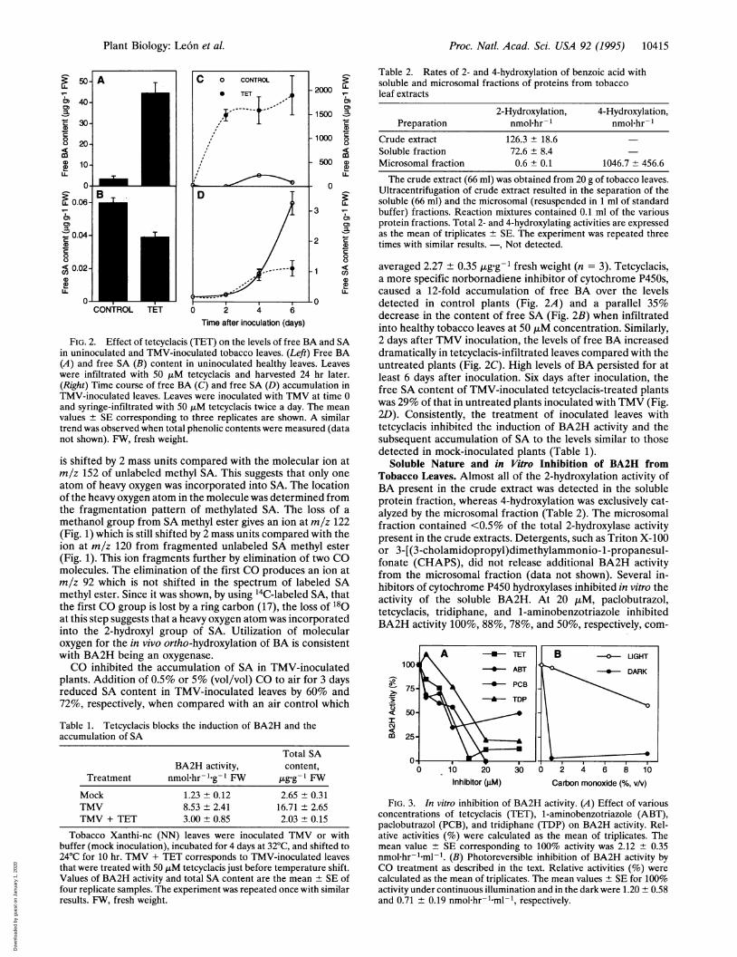
Soluble Nature and in Vitro Inhibition of BA2H fromTobacco Leaves. Almost all of the 2-hydroxylation activity ofBA present in the crude extract was detected in the solubleprotein fraction, whereas 4-hydroxylation was exclusively cat-alyzed by the microsomal fraction (Table 2). The microsomalfraction contained <0.5% of the total 2-hydroxylase activitypresent in the crude extracts. Detergents, such as Triton X-100or 3-[(3-cholamidopropyl)dimethylammonio-1-propanesul-fonate (CHAPS), did not release additional BA2H activityfrom the microsomal fraction (data not shown). Several in-hibitors of cytochrome P450 hydroxylases inhibited in vitro theactivity of the soluble BA2H. At 20 ,u M, paclobutrazol,tetcyclacis, tridiphane, and 1-aminobenzotriazole inhibitedBA2H activity 100%, 88%, 78%, and 50%, respectively, com-
1004
a-O
.5
co
75-
50-
25-
0 10 20Inhibitor (gM)
304
B UGHT
DARK
2 4 6 8 10
Carbon monoxide (%, v/v)
FIG. 3. In vitro inhibition of BA2H activity. (A) Effect of variousconcentrations of tetcyclacis (TET), 1-aminobenzotriazole (ABT),paclobutrazol (PCB), and tridiphane (TDP) on BA2H activity. Rel-ative activities (%) were calculated as the mean of triplicates. Themean value SE corresponding to 100% activity was 2.12 + 0.35nmol hr-l-ml-1. (B) Photoreversible inhibition of BA2H activity byCO treatment as described in the text. Relative activities (%) werecalculated as the mean of triplicates. The mean values + SE for 100%activity under continuous illumination and in the dark were 1.20 0.58and 0.71 + 0.19 nmolPhr-i.ml-1, respectively.
A *-TET\*--- ABT
* PCB
-A-- TDP
Plant Biology: Le6n et al.
Dow
nloa
ded
by g
uest
on
Janu
ary
1, 2
020
Proc. Natl. Acad. Sci. USA 92 (1995)
E
-aE._
ClIem
0.1
0.
0.0
C
0 5 10 15 20 25Fraction number
Time aftertemperature shift
(h)
30
A
11a
0-
IcIcm
0
cocmcc
1-
CL
B
Time after BAinfiltration
(h)
0 2 4 6 8 10 0 0.5 1 3 5
11
kDa
205116.580
49.5- 32.5- 27.5
I I
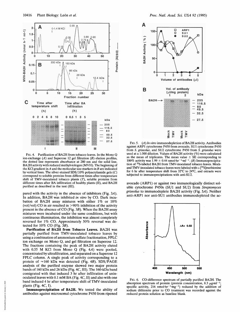
FIG. 4. Purification of BA2H from tobacco leaves. In the Mono Qion-exchange (A) and Superose 12 gel filtration (B) elution profiles,the dotted line represents absorbance at 280 nm and the solid line,BA2H activity with reduced methylviologen (MVH). The beginning ofthe KCl gradient inA and the molecular size markers in B are indicatedby vertical lines. The silver-stained SDS/10% polyacrylamide gels (C)correspond to soluble proteins from different times after temperatureshift of TMV-inoculated tobacco plants (7), soluble proteins fromdifferent times after BA infiltration of healthy plants (II), and BA2Hpurified as described in the text (III).
pared with the activity in the absence of inhibitors (Fig. 3A).In addition, BA2H was inhibited in vitro by CO. Dark incu-bation of BA2H assay mixtures with either 1% or 10%(vol/vol) CO in air resulted in >90% inhibition of the activitypresent in the absence of CO (Fig. 3B). When the BA2H assaymixtures were incubated under the same conditions, but withcontinuous illumination, the inhibition was almost completelyreversed for 1% CO. Approximately 50% reversal was de-tected for 10% CO (Fig. 3B).
Purification of BA2H from Tobacco Leaves. BA2H was
partially purified from TMV-inoculated tobacco leaves byusing a combination ofammonium sulfate fractionation, FPLCion exchange on Mono Q, and gel filtration on Superose 12.The fractions containing the peak of BA2H activity elutedwith 0.55 M KCl from Mono Q (Fig. 4A) were pooled,concentrated by ultrafiltration, and separated on a Superose 12FPLC column. A single peak of activity corresponding to a
protein of - 160 kDa was detected (Fig. 4B). SDS/PAGEanalysis of the purified enzyme showed two major proteinbands of 160 kDa and 26 kDa (Fig. 4C, III). The 160-kDa bandcomigrated with that induced 3 hr after infiltration of unin-oculated leaves with 0.1 mM BA (Fig. 4C, II) and also with oneband induced 4 hr after temperature shift of TMV-inoculatedplants (Fig. 4C, I).
Immunoprecipitation of BA2H. We tested the ability ofantibodies against microsomal cytochrome P450 from ripened
1 2 3 4 5
Volume of antibodies (li)
Vol. of antibody(gl/mg protein)
1 2
BA2H-1 i_ 'W_
kDa
- 205- 116.5- 80-49.5- 32.5
- 27.5
> ,>
FIG. 5. (A) In vitro immunodepletion of BA2H activity. Antibodiesagainst ARP1 cytochrome P450 from avocado, SUl cytochrome P450from S. griseolus, and SU2 cytochrome P450 from S. griseolus wereused at a 1:500 dilution. Values of BA2H activity (%) were calculatedas the mean of triplicates. The mean value ± SE corresponding to100% activity was 1.99 ± 0.44 nmol hr- Lml-1. (B) Immunoprecipita-tion of 35S-labeled BA2H from TMV-inoculated tobacco leaves. Mock-and TMV-inoculated tobacco leaves were labeled with L-[35S]methioninefor 6 hr after temperature shift from 32°C to 24°C, and extracts were
subjected to immunoprecipitations with anti-SU2.
avocado (ARP1) or against two immunologically distinct sol-uble cytochrome P450s (SUl and SU2) from Streptomycesgriseolus to immunodeplete BA2H activity (Fig. SA). Neitheranti-ARP1 nor anti-SUl antibodies immunodepleted the ac-
~~~~~AA=0.02
0£I
400 450 500 550Wavelength (nm)
FIG. 6. CO-difference spectrum of partially purified BA2H. Theabsorption spectrum of protein (protein concentration, 8.3 tg ml-1;specific activity, 216 nmol.hr-.mg-1) reduced by the addition ofsodium dithionite prior to CO treatment was recorded against thereduced protein solution as baseline blank.
200 kDa A l5OkDa5- B 1.A29 kDa
1D 124 kDaf15
10416 Plant Biology: Leo'n et al.
I
Dow
nloa
ded
by g
uest
on
Janu
ary
1, 2
020

Proc. Natl. Acad. Sci. USA 92 (1995) 10417
tivity significantly. However, 2 ,l of 1:500-diluted antibodiesagainst soluble SU2 completely immunodepleted the solubleBA2H activity. Incubation of mock- and TMV-inoculatedtobacco leaves with L-[35S]methionine, and subsequent immu-noprecipitation with anti-SU2, identified a radiolabeled pro-tein of '160 kDa that was specifically induced in TMV-inoculated leaves (Fig. SB).
CO-Difference Spectrum. Absorption spectral analysis ofthe purified enzyme after treatment with CO was performed.Fig. 6 shows the CO-difference absorption spectrum (400-550nm) of sodium dithionite-reduced and CO-treated BA2Hagainst the reduced BA2H protein solution as baseline. A peakof maximum absorption was detected at 457 nm. Although thepeak was shifted by 7 nm when compared with the usual 450nm maximum for other P450s, the BA2H absorption peak wasspecific for the CO-treated reduced BA2H. This peak was notdetected when the untreated reduced protein was scannedagainst the oxidized protein solution (data not shown).
DISCUSSIONThis paper describes the characterization of BA2H at thebiochemical and molecular levels. Our data on the function ofthis enzyme in hydroxylating BA, coupled with its biochemicalproperties, suggest that BA2H is a soluble cytochrome P450oxygenase, most likely a monooxygenase. We have demon-strated that BA2H acts as an oxygenase by in vivo labeling ofthe hydroxyl group of SA synthesized de novo with 1802 (Fig.1). The enzyme was inhibited by standard cytochrome P450inhibitors both in vivo and in vitro. The in vivo inhibition bytetcyclacis was accompanied by an accumulation of BA anddecrease in SA when compared with the control untreatedleaves (Fig. 2; Table 1). Tetcyclacis and other specific inhibi-tors of cytochrome P450s, such as paclobutrazol, tridiphane,and 1-aminobenzotriazole (17-20), inhibited BA2H activity invitro at concentrations below 30 ,uM (Fig. 3A). In addition, theactivity showed photoreversible inhibition by CO (Fig. 3B), afundamental property of P450 enzymes. When reduced BA2Hwas treated with CO, a specific CO-difference absorptionspectrum with a maximum at 457 nm was observed (Fig. 6).The 7-nm shift of the absorption maximum from the 450 nmcharacteristic of other P450s may be explained by slightdifferences in the structure of the BA2H chromophore and itsmolecular environment. For example, certain changes in theN-terminal amino acid sequence of the human P450 2D6 shiftthe maximum absorption for the CO-Fe2+ complex by as muchas 6 nm (18).BA2H specifically hydroxylates the ortho position of BA
(Table 2). We have partially purified BA2H from TMV-inoculated leaves by using a combination of ion-exchange andgel filtration FPLC (Fig. 4). The purified enzyme solutioncontained mostly BA2H but the recovery of activity was low(<1% of the total activity present in crude extracts). The factthat both crude soluble protein extracts and the purifiedpreparations of BA2H utilize NADPH or sodium dithionite-reduced methyl viologen as reductants (ref. 8 and data notshown) strongly supports our suggestion that BA2H is catal-itically self-sufficient. A soluble high molecular weight cyto-chrome P450 monooxygenase from Bacillus megaterium(P450BM-3) is also a catalytically self-sufficient single protein(13, 19).BA2H activity was immunodepleted by polyclonal antibod-
ies raised against SU2, a soluble cytochrome P450 that isinduced in S. griseolus by the herbicide chlorimuron ethyl (20)(Fig. 5A). This provides additional evidence that BA2H isrelated to other P450 proteins. In vivo labeling of plant tissueswith L-[35S]methionine followed by immunoprecipitation withanti-SU2 (Fig. SB) and partial purification of the enzyme fromTMV-inoculated leaves (Fig. 4) identified BA2H as a 160-kDa
protein. Immunoprecipitation of BA2H by antibodies againstsoluble SU2 from S. griseolus, and not by microsomal cyto-chrome P450 from avocado, suggests that BA2H is moreclosely related to bacterial than to plant cytochromes P450. Itis particularly interesting that tobacco BA2H is a solubleprotein not associated with microsomes (Table 2). Most sol-uble cytochrome P450 enzymes characterized so far are ofbacterial origin (11-13). To our knowledge, the only solubleeukaryotic cytochrome P450 has been identified in Fusariumoxysporum (21). Therefore, BA2H may be the first solublecytochrome P450 identified in higher plant or animal systems.Interest in this enzyme is warranted because of its key role inthe synthesis of SA, an important signal in plant-pathogeninteractions. The isolation of the BA2H gene(s) from plantsmay provide a powerful tool to manipulate SA levels in plants,thereby altering resistance to pathogens.
We thank Dr. Dan O'Keefe (DuPont de Nemours, Wilmington, DE)for the generous supply of antibodies against cytochromes P450 fromavocado and S. griseolus and Dr. Stewart Frear (U.S. Department ofAgriculture/Agricultural Research Station, Fargo, ND) for the kindgift of cytochrome P450 inhibitors. We thank Dr. Peter Day for readingthis manuscript and for his useful suggestions. This research wassupported by grants from the U.S. Department of Agriculture (Com-petitive Research Grants Office) and by the Division of EnergyBiosciences of the U.S. Department of Energy. Additional support wasprovided by the New Jersey Agricultural Experiment Station and theNew Jersey Commission for Science and Technology.
1. Raskin, I., Skubatz, H., Tang, W. & Meeuse, B. J. D. (1990)Ann.Bot. 66, 369-373.
2. Malamy, J., Carr, J. P., Klessig, D. K. & Raskin, I. (1990) Science250, 1002-1004.
3. Metraux, J. P., Signer, H., Ryals, J., Ward, E., Wyss-Benz, M.,Gaudin, J., Raschdorf, K., Schmid, E., Blum, W. & Inverardi, B.(1990) Science 250, 1004-1006.
4. Yalpani, N., Silverman, P., Wilson, T. M. A., Kleier, D. A. &Raskin, I. (1991) Plant Cell 3, 809-818.
5. Gaffney, T., Friedrich, L., Vernooij, B., Negrotto, D., Nye, G.,Ukness, S., Ward, E., Kesmann, H. & Ryals, J. (1993) Science 261,754-756.
6. Vernooij, B., Friedrich, L., Morse, A., Reist, R., Kolditz-Jawhar,R., Ward, E. R., Ukness, S., Kessmann, H. & Ryals, J. (1994)Plant Cell 6, 959-965.
7. Yalpani, N., Le6n, J., Lawton, M. A. & Raskin, I. (1993) PlantPhysiol. 103, 315-321.
8. Le6n, J., Yalpani, N., Raskin, I. & Lawton, M. A. (1993) PlantPhysiol. 103, 323-328.
9. Donaldson, R. P. & Luster, D. G. (1991) Plant Physiol. 96,669-674.
10. Ortiz de Montellano, P. R. (1986) Cytochrome P-450: Structure,Mechanisms and Biochemistry (Plenum, New York), pp. 217-271.
11. Gunsalus, I. C., Meeks, J. R., Lipscomb, J. D., Debrunner, P. &Munck, E. (1974) in Molecular Mechanisms of Oxygen Activation,ed. Hayaishi, 0. (Academic, New York), pp. 559-613.
12. Sligar, S. G. & Murray, R. I. (1986) in Cytochrome P-450: Struc-ture, Mechanisms and Biochemistry, ed. Ortiz de Montellano,P. R. (Plenum, New York), pp. 161-216.
13. Fulco, A. J. (1991) Annu. Rev. Pharmacol. Toxicol. 31, 177-203.14. Nelson, D. R., Kamataki, T., Waxman, D. J., Guenguerich, P.,
Estabrook, R. W., Feyereisen, R., Gonzalez, F. J., Coon, M. J.,Gunsalus, I. C., Gotoh, O., Okuda, K. & Nebert, D. W. (1993)DNA Cell Biol. 12, 1-51.
15. Stewart, C. B. & Schuler, M. A. (1989) Plant Physiol. 90, 534-541.16. Yalpani, N., Shulaev, V. & Raskin, I. (1993) Phytopathology 83,
702-708.17. Occolowitz, J. L. (1968) Chem. Commun. 20, 1226-1227.18. Gillam, E. M. J., Guo, Z., Martin, M. V., Jenkins, C. M. &
Guenguerich, F. P. (1995)Arch. Biochem. Biophys. 319, 540-550.19. Narhi, L. 0. & Fulco, A. J. (1986) J. Biol. Chem. 261, 7160-7169.20. O'Keefe, D. P., Romesser, J. A. & Leto, K. J. (1988) Arch.
Microbiol. 149, 406-412.21. Nakahara, K., Tanimoto, T., Hatano, K., Usuda, K. & Shoun, H.
(1993) J. Biol. Chem. 268, 8350-8355.
Plant Biology: Leo'n et al.
Dow
nloa
ded
by g
uest
on
Janu
ary
1, 2
020


![Directed hydroxyl of 16S rRNAusing Fe(II) tethered to S4Proc. NatL Acad Sci USA92 (1995) activity. Electrosprayionization massspectrometryofS4and [(BABE)-Cys3t]S4, wasperformedonaFinnigan-MAT(San](https://img.pdfslide.net/doc/110x75/61150f171ae55d5bfa0c2133/directed-hydroxyl-of-16s-rrnausing-feii-tethered-to-s4-proc-natl-acad-sci-usa92.jpg)