Embed Size (px)
Citation preview
Proc. Natl. Acad. Sci. USAVol. 92, pp. 1108-1112, February 1995Biochemistry
A sauvagine/corticotropin-releasing factor receptor expressed inheart and skeletal muscle
[urotensin I/seven transmembrane domain/a-helical corticotropin-releasing factor-(9-41)]
ToSHIMITSu KISHIMOTO*, RICHARD V. PEARSE Ilt, CHIJEN R. LIN*, AND MICHAEL G. ROSENFELDt*Eukaryotic Regulatory Biology Program, Cellular and Molecular Medicine, tBiomedical Sciences Graduate Program, and tHoward Hughes Medical Institute,University of California at San Diego, 9500 Gilman Drive, CMM Room 345, School of Medicine, La Jolla, CA 92093-0648
Contributed by Michael G. Rosenfeld, November 2, 1994
ABSTRACT Corticotropin-releasing factor (CRF) medi-ates many critical aspects of the physiological response tostress. These effects are elicited by binding to specific high-affinity receptors, which are coupled to guanine nucleotidestimulatory factor (Gs)-response pathways. Recently, a geneencoding a receptor for CRF, expressed in pituitary and thecentral nervous system (PC-CRF receptor), was isolated andcharacterized. Here we report the identification and charac-terization of a second, distinct CRF receptor that is expressedprimarily in heart and skeletal muscle and exhibits a specificligand preference and antagonist sensitivity compared withthe PC-CRF receptor. We refer to this second receptor as theheart/muscle (HM)-CRF receptor.
Corticotropin-releasing factor (CRF) (1) is a member of afamily of peptides from different species that act as agonists ofthe CRF receptor. These peptides include the frog skinpeptide, sauvagine (2), and the teleost fish urophysis peptide,urotensin I (3). CRF is a 41-amino acid hypothalamic neu-ropeptide that plays a central role in coordinating the com-munications between endocrine, nervous, and immune sys-tems to achieve homeostasis in response to environmentaladversities (4, 5). The peptide was originally characterized inthe hypothalamo-hypophyseal system but was later found tobe widely distributed throughout the central nervous system(CNS), where it appeared to function as a neurotransmitter orneuromodulator (6). In the CNS, CRF initiates the hypo-thalamic-pituitary-adrenal axis by stimulating the release ofadrenocorticotropin (ACTH) and ,B-endorphin from the an-terior pituitary. ACTH stimulates adrenal cortex to secretecorticosteroids that, in turn, elicit a wide range of biologicalresponses and exert negative feedback on the hypothalamusand pituitary (4, 5, 7). Both sauvagine and urotensin I havebeen shown to stimulate the hypothalamic-pituitary-adrenalaxis after i.v. administration (8). Intracerebroventricular ad-ministration of CRF provokes stress-like responses includingactivation of the sympathetic nervous system, resulting in anelevation of plasma epinephrine, norepinephrine, and glucose,which results in increased heart rate and mean arterial bloodpressure (9, 10). Outside the CNS, CRF immunoreactivity isdetectable in multiple peripheral organs, including placenta,adrenal medulla, pancreas, lung, stomach, duodenum, andliver (4, 5). i.v. administration of CRF, sauvagine, and uro-tensin I has been shown to elicit peripheral systemic responses,including vasodilation (11).CRF functions by binding to a membrane-bound receptor
that is coupled to the guanine nucleotide stimulatory factor(G,) signaling protein, resulting in increased intracellularcAMP levels (12, 13). The relative density of CRF receptors ishighest in the anterior and intermediate lobes of the pituitary(14, 15), moderate in discrete areas ofCNS (16), and lower, but
detectable, in spleen (17). Recently, cDNAs encoding a CRFreceptor expressed primarily in pituitary and the CNS (PC-CRF receptor) have been identified and characterized (18-20). The PC-CRF receptor belongs to a subfamily of receptorsfor peptides including vasoactive intestinal polypeptide [VIP(21)], secretin (22), calcitonin (23), and growth hormone-releasing factor [GRF (24-26)]. The PC-CRF receptormRNAwas found to be most highly expressed in the cerebellum andpituitary and found at lower levels in other brain areas,intestine, and testes. This receptor is undetectable in othertissues examined, including heart and skeletal muscle (18-20).However, physiological studies have indicated the presence ofa CRF-responsive receptor that mediates a positive inotropiceffect in isolated heart (27) and myocardium (28) and sup-pression of vascular leakage in skeletal muscle (29). Here wereport the identification and characterization of another re-ceptor that mediates intracellular responses induced by sau-vagine, urotensin I, and CRF. This receptor is expressedspecifically in heart and skeletal muscle and, as such, is termedthe heart and muscle CRF receptor (HM-CRF receptor).
MATERIALS AND METHODSGenomic DNA and cDNA Cloning. Two million plaques
from a mouse 129 genomic lambda FIXII library werescreened by a 32P-labeled rat PC-CRF receptor cDNA (18)1.4-kb Hind III/BamHI fragment as a probe with 50% (wt/vol)formamide, 5x SSPE (lx SSPE is 0.15 M NaCl/10 mMsodium phosphate, pH 7.4/1 mM EDTA), 3x Denhardt'ssolution, 0.5% SDS, and 100 ,ug of salmon sperm DNA per mlat 37°C. Membranes were washed in 2x SSC/0.05% SDS at37°C. The positive clones were then subjected to restrictionendonuclease mapping and DNA sequencing, identifying aPC-CRF receptor gene-related clone (HM-CRF receptorgene). A 0.5-kb Pst I fragment of HM-CRF receptor gene wasused to screen a mouse heart, random-primed and oligo(dT)-primed cDNA library (Clontech) under the conditions de-scribed above for the genomic library screening. However, thehybridization temperature was 42°C, and membranes werewashed with 0.1 x SSC solution at 42°C.
Northern Blot Analysis. Poly(A)+ RNAs from a selection ofmouse tissues were immobilized and hybridized at 42°C with50% formamide using a 32P-labeled 0.5-kb Pst I fragment ofHM-CRF receptor gene as a probe. This fragment spanssequence from transmembrane (TM) V to VII and includes aminimum intron sequence based on comparison to the codingsequence of PC-CRF receptor. The blot was washed twice atroom temperature in 2x SSC/0.05% SDS for 20 min and twiceat 50°C in 0.1 x SSC/0.1% SDS for 20 min.
Abbreviations: CRF, corticotropin-releasing factor; PC-CRF recep-tor, pituitary/central nervous system CRF receptor; HM-CRF recep-tor, heart/muscle CRF receptor; CNS, central nervous system; ACTH,adrenocorticotropin; VIP, vasoactive intestinal polypeptide; GRF,growth hormone-releasing factor; TM, transmembrane; ahel, a helical.
1108
The publication costs of this article were defrayed in part by page chargepayment. This article must therefore be hereby marked "advertisement" inaccordance with 18 U.S.C. §1734 solely to indicate this fact.
Dow
nloa
ded
by g
uest
on
Mar
ch 1
9, 2
020
Dow
nloa
ded
by g
uest
on
Mar
ch 1
9, 2
020
Dow
nloa
ded
by g
uest
on
Mar
ch 1
9, 2
020
Proc. Natl. Acad Sci USA 92 (1995) 1109
Expression and Functional Assay of the Cloned Receptor.The full-length cDNA (HM-CRF receptor) was subcloned intoexpression vector pCEP4 (Invitrogen) and was transfected intoCV-1 and 293-EBNA kidney cells (Invitrogen) by calciumphosphate precipitation. Stable transfectants were selected inthe presence of hygromycin B at 200 ,ug/ml. Cells expressingeither the HM-CRF receptor or the PC-CRF receptor (18)were treated with 50 ,uM 3-isobutylmethyl-1-methylxanthine(Sigma) with or without the CRF receptor antagonist a-helical(ahel) CRF-(9-41) (Peninsula Laboratories) for 20 min at37°C. Peptides were added and incubated for an additional 20min at 37°C. The cells were extracted, and levels of intracel-lular cAMP were determined in triplicate by using a[3H]cAMP assay system (Amersham), as described (18).
RESULTSCloning of HM-CRF Receptor Gene. In an attempt to
identify additional members of the CRF class of receptors, wescreened a mouse genomic library under low-stringency con-ditions by using a full-length rat PC-CRF receptor cDNA as aprobe. Fourteen independent clones were obtained, of whichsix hybridized with an oligonucleotide derived from PC-CRFreceptor sequence within TM VI-VII region. Restrictionendonuclease mapping and DNA sequencing revealed, inaddition to the expected PC-CRF receptor gene, a second genecontaining a nucleotide sequence different from, but homol-ogous to, the PC-CRF receptor.To determine the expression pattern of the HM-CRF re-
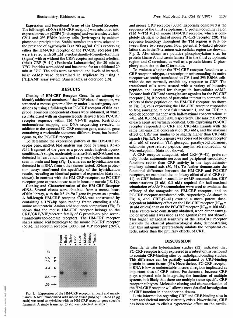
ceptor gene, mRNA blot analysis was done by using a 0.5-kbPst I fragment of the gene as a probe under high-stringencyconditions. A single, moderately intense 3-kb mRNA band wasdetected in heart and muscle, and very weak hybridization wasseen in brain and lung (Fig. 1), whereas no hybridization wasdetected in mRNA from other tissues tested. RNase protec-tion assays confirmed the specificity of the hybridizationresults, revealing an identical pattern of expression (data notshown). In contrast with the HM-CRF receptor, no PC-CRFreceptor gene expression was seen in heart or muscle (18, 19).
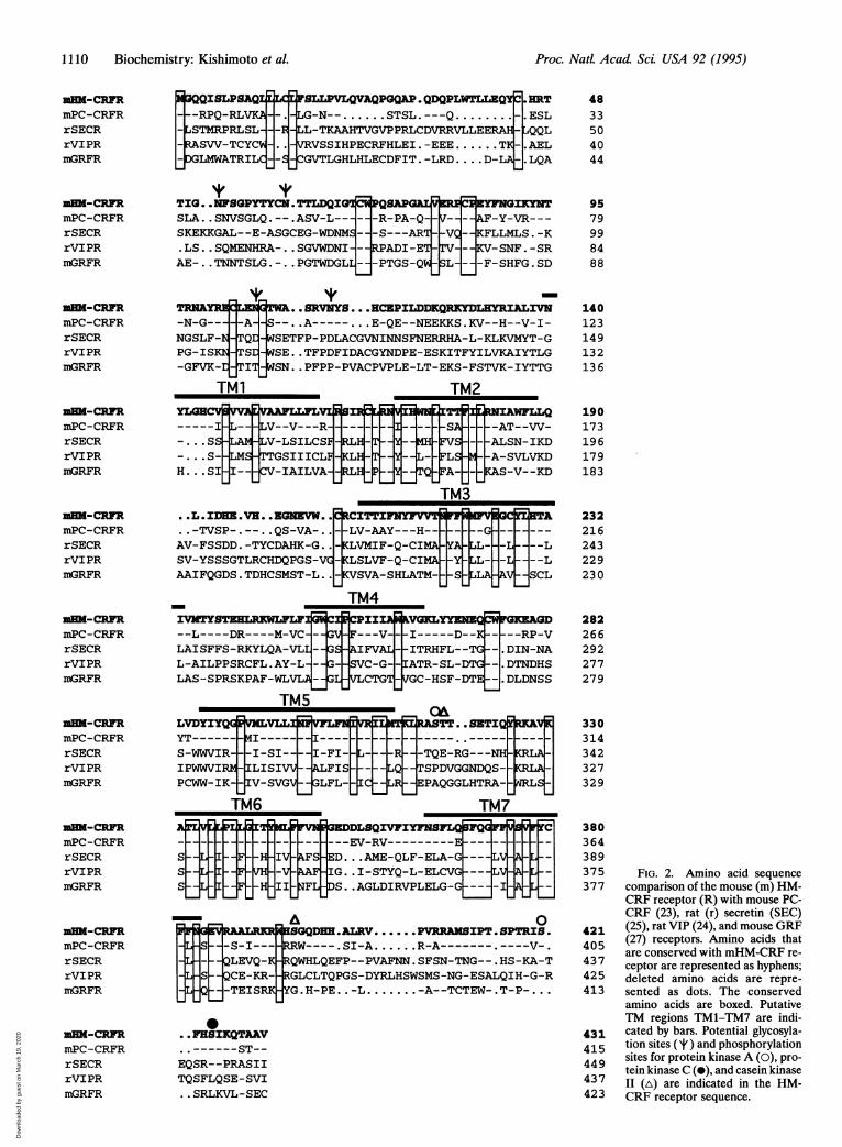
Cloning and Characterization of the HM-CRF ReceptorcDNA. Several clones were obtained from a mouse heartcDNA library, with the 0.5-kb Pst I gene fragment as a probe.A full-length HM-CRF receptor cDNA was constructed bycontaining a 1293-bp open reading frame encoding a 431-amino acid protein. Amino acid sequence comparison (Fig. 2)shows that the mouse HM-CRF receptor belongs to theCRF/GRF/VIP/secretin family of G protein-coupled seven-transmembrane-domain receptors. The HM-CRF receptorshares amino acid homology to the mouse PC-CRF receptor(66%), rat secretin receptor (30%), rat VIP receptor (26%),
-t: c (1m ,2 OI mcn) ) 1- -5 Cp* * * * * *C *--
kb
9.5 -4
7.5 _-
4.4_--b
2.4 -_
.35 -_
FIG. 1. Expression of the HM-CRF receptor in heart and muscletissues. A blot immobilized with mouse tissue poly(A)+ RNAs (2 ,geach) was used to hybridize with an HM-CRF receptor gene-specificfragment. A single transcript (3 kb) was detected, as shown.
and mouse GRF receptor (30%). Especially conserved is thesequence of the third cytoplasmic, G protein-coupling region(TM V-TM VI) of mouse HM-CRF receptor, which is com-pletely identical to that of mouse PC-CRF receptor (20). Thesequence homology throughout the TM regions is 82% be-tween these two receptors. Four potential N-linked glycosy-lation sites in the N-terminus extracellular region are shown inFig. 2. Also shown are putative phosphorylation sites forprotein kinase A and casein kinase II in the third cytoplasmicregion and C terminus, as well as a protein kinase C phos-phorylation site in the C terminus.To evaluate whether the HM-CRF receptor functions as a
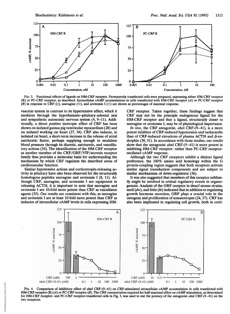
CRF receptor subtype, a transcription unit encoding the entirereceptor was stably transfected to CV-1 and 293-EBNA cells,which do not normally exhibit any response to CRF. Thetransfected cells were treated with a variety of bioactivepeptides and assayed for changes in intracellular cAMP.Because both CRF and sauvagine are agonists for the PC-CRFreceptor (18), it became of particular interest to compare theeffects of these peptides on the HM-CRF receptor. As shownin Fig. 3A, cells expressing the HM-CRF receptor respondedto frog sauvagine, teleost fish urotensin I, and rat CRF in adose-dependent manner with half-maximal concentrations at-0.1 nM, 0.3 nM, and 3 nM, respectively. The maximal effectsof each agent are virtually identical. Cells expressing PC-CRFreceptor responded to CRF and CRF-like ligands with thesame half-maximal concentration (0.3 nM), and the maximaleffect of CRF was similar to or slightly higher than CRF-likeligands (Fig. 3B). No response was observed by either receptorat 1 ,uM of secretin, VIP, glucagon, parathyroid hormone,calcitonin gene-related peptide, amylin, adrenomedulin, orMet-enkephalin (data not shown).A CRF receptor antagonist, ahel CRF-(9-41), preferen-
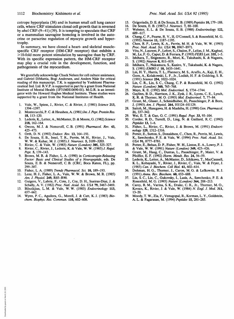
tially blocks autonomic nervous and peripheral vasodilatoryfunctions rather than CRF activity in the hypothalamic-pituitary-adrenal axis (30, 31). To further demonstrate thefunctional difference between the HM-CRF and PC-CRFreceptors, we examined the inhibitory effect of ahel CRF-(9-41) on CRF-induced intracellular cAMP accumulation. CRFconcentrations (3 nM, 0.3 nM) required for half-maximalstimulation of cAMP accumulation were used to evaluate theefficacy of the antagonist on HM-CRF receptor- and onPC-CRF receptor-transfected cells, respectively. As shown inFig. 4, ahel CRF-(9-41) exerted a more potent dose-dependent inhibitory effect on the HM-CRF receptor (IC50 =10 nM or less) than on the PC-CRF receptor (IC5o = 100 nM).These values were consistently obtained, when either sauvag-ine or urotensin I was used as the agonist (data not shown).TIhis higher antagonist sensitivity of the HM-CRF receptorparallels the classical pharmacological data, demonstratingthat this antagonist preferentially inhibits the peripheral ef-fects, rather than the pituitary effects, of CRF.
DISCUSSIONRecently, in situ hybridization studies (32) indicated thatPC-CRF receptor is only expressed in a subset of tissues foundto contain CRF-binding sites by radioligand-binding studies.This difference can be partially explained by CRF-bindingprotein in some tissues (33). Nevertheless, PC-CRF receptormRNA is low or undetectable in several regions implicated asimportant sites of CRF action. Furthermore, because CRFplays a pivotal role in integrating the functions of multiplesystems, it is likely that there are multiple tissue-specific CRFreceptor subtypes. Molecular cloning and characterization ofthe HM-CRF receptor will allow a more detailed investigationof CRF function in nonendocrine tissues.
Little information regarding CRF and CRF-binding sites inheart and skeletal muscle currently exists. Nevertheless, CRFhas been shown to elicit a hypotensive effect on the cardio-
Biochemistry: Kishimoto et aL
Dow
nloa
ded
by g
uest
on
Mar
ch 1
9, 2
020
1110 Biochemistry: Kishimoto et al.
_XQQISLPSAQLLLC_FSLLPVLQVAQPGQAP .QDQPLWTILZQYC.HRT--RPQ-RLVKA _._LG-N--........ STSL.---Q ........-_ESLLSTMRPRLSL-R--_LL-TKAAHTVGVPPRLCDVRRVLLEERAHj-_QQL_RASVV-TCYCW4-..I-IVRVSSIHPECRFHLEI.-EEE ...... TK_.AEL_DGLMWATRILC_J-S_CGVTLGHLHLECDFIT.-LRD....D-LA-J.LQA
TIG. .NFSPY TTL.DQIG PQ0AP GIKYNTSLA..SNVSGLQ.--.ASV-L---I-_]-R-PA-Q-_-- F-Y-VR---SKEKKGAL--E-ASGCEG-WDNMS--_-S---AR1 -VQ _KFLLMLS.-K.LS..SQMENHRA-..SGVWDNI- --RPADI-E1 TV--- KV-SNF.-SRAE-..TNNTSLG.-..PGTWDGLL ---PTGS-Q' _SL -F-SHFG.SD
if 'I -TRN&YRS C w G A...RVNY8...HCZPILDDKQRKYDLHYRIALIVN-N-G-- -A --..A-----...E-QE--NEEKKS.KV--H--V-I-NGSLF- _TQD SETFP-PDLACGVNINNSFNERRHA-L-KLKVMYT-GPG-IS _TSD E..TFPDFIDACGYNDPE-ESKITFYILVKAIYTLG-GFVK- NTIT..PFPP-PVACPVPLE-LT-EKS-FSTVK-IYTTG
TM1 TM2
-----IHL g--V---R- ---|--...S V-LSILCSFg
-...S-- >MSTGSIIICL L-Kt- --YH... SI-I- -C-IAILVA.IJRLHYJPYJ'
..L.IDHE.VH..EGNZVW..
..-TVSP-.--..QS-VA-..AV-FSSDD.-TYCDAHK-G..SV-YSSSGTLRCHDQPGS-VCAAIFQGDS.TDHCSMST-L..
U
IVMTYSBTHLRKWLFLjr--L----DR----M-VC-LAISFFS-RKYLQA-VLIL-AILPPSRCFL.AY-L-LAS-SPRSKPAF-WLVLU
TM5
- _+SMH _FL--FLTQ_FA
TMsJ.-rJ.NUZNv v-
-LV-AAY---H--KLVMIF-Q-CIMAKLSLVF-Q-CIMAKVSVA-SHLATM-
TM4o
F---V-FAIFVAL|SVC-G--
VLCTGT
-U-I-
VIEaVLLIMI------I-SI--ILISIVWIV-SVGX
I[
TM6
-S-I---QLEVQ-KQCE-KR--TEISRR
BP VFIlFNI----I-FI-ikLFISGLFL-
L-
IC
'YAS
CRNAWFLLQ---AT--VW-
-FALSN-IKD_ A-SVLVKD_JKAS-V--KD
-LF-I---L
IJSC--L
VSCL--GLL-ELL-
WVGKLYYEQJ ED-I-----D-- ---RP-V-ITRHFL--TGF-I. DIN-NAIATR-SL-DTG -.DTNDHSVGC-HSF-DTE DLDNSS
0 OA#T_RASTT.. ETIQ
- -TQE-RG---NH_LQ__TSPDVGGNDQS-_LR__EPAQGGLHTRA-Mc7
TM7F I~GEDDLSQIVFIYFNS FQG---H --EV-RV --------- __kF D...AME-QLF-ELA- ---LAFHIG..I-STYQ-L-ELCVGj---NFLIDS..AGLDIRVPLELG-G-
F,
LVL'V-I
a 0jHSGQDEH.ALRV......PVRRAMSIPT.SPTRIS.IRRW--.SI-A ......R-A---------V-.4RQWHLQEFP--PVAFNN.SFSN-TNG--.HS-KA-TGLCLTQPGS-DYRLHSWSMS-NG-ESALQIH-G-R
JG.H-PE. .......-L-A--TCTEW-.T-P-...
0..FHSIKQTAAV..------ST--EQSR--PRASIITQSFLQSE-SVI..SRLKVL-SEC
4833504044
9579998488
140123149132136
190173196179183
232216243229230
282266292277279
330314342327329
380364389375377
421405437425413
431415449437423
FIG. 2. Amino acid sequencecomparison of the mouse (m) HM-CRF receptor (R) with mouse PC-CRF (23), rat (r) secretin (SEC)(25), rat VIP (24), and mouse GRF(27) receptors. Amino acids thatare conserved with mHM-CRF re-ceptor are represented as hyphens;deleted amino acids are repre-sented as dots. The conservedamino acids are boxed. PutativeTM regions TM1-TM7 are indi-cated by bars. Potential glycosyla-tion sites (It ) and phosphorylationsites for protein kinase A (0), pro-tein kinase C (0), and casein kinaseII (A) are indicated in the HM-CRF receptor sequence.
MM-CRFRmPC-CRFRrSECRrVIPRmGRFR
-CRFRmPC-CRFRrSECRrVIPRmGRFR
m-CRFRmPC-CRFRrSECRrVIPRmGRFR
zM-CRFRmPC-CRFRrSECRrVIPRmGRFR
-CRmmPC-CRFRrSECRrVIPRmGRFR
m-CRFRmPC-CRFRrSECRrVIPRmGRFR
-CRFRmPC-CRFRrSECRrVIPRmGRFR
m-CRFRmPC-CRFRrSECRrVIPRmGRFR
m-CRFRmPC-CRFRrSECRrVIPRmGRFR
m-CRmmPC-CRFRrSECRrVIPRmGRFR
LVDYIYQGiYT------S-WWVIR-+IPWWVIRMPCWW-IK-
-I- U-
Ssslsl
IW_WPA
Li
Proc. Natl. Acad ScL USA 92 (1995)
VRFL
-1
Dow
nloa
ded
by g
uest
on
Mar
ch 1
9, 2
020
Proc. Natt Acad Sci USA 92 (1995) 1111
Concentration, nM Concentration, nM
FIG. 3. Functional effects of ligands on HM-CRF receptor. Permanently transfected cells were prepared, expressing either HM-CRF receptor(R) or PC-CRF receptor, as described. Intracellular cAMP accumulations in cells transfected with HM-CRF receptor (A) or PC-CRF receptor(B) in response to CRF (o), sauvagine (0), and urotensin I (0) are shown as percentages of maximal response.
vascular system in contrast to its hypertensive effect, which itmediates through the hypothalamic-pituitary-adrenal axisand sympathetic autonomic nervous system (4, 9-11). Addi-tionally, a direct positive inotropic effect of CRF has beenshown on isolated guinea pig ventricular myocardium (28) andon isolated working rat heart (27, 34). CRF also induces, inisolated rat heart, a short-term increase in the release of atrialnatriuretic factor, perhaps supplying enough to modulateblood pressure through its diuretic, natriuretic, and vasodila-tory actions (34). The identification of the HM-CRF receptoras another member of the CRF/GRF/VIP/secretin receptorfamily thus provides a molecular basis for understanding themechanism by which CRF regulates the described areas ofcardiovascular function.
Similar hypotensive actions and corticotropin-releasing ac-tivity in pituitary have also been observed for the structurallyhomologous peptides sauvagine and urotensin I (8, 11). Al-though CRF, sauvagine, and urotensin I are equipotent inreleasing ACTH, it is important to note that sauvagine andurotensin I are 10-fold more potent than CRF as vasodilatoragents (35). Our results are consistent with this, as sauvagineand urotensin I are at least 10-fold more potent than CRF asinducers of intracellular cAMP levels in cells expressing HM-
CRF receptor. Taken together, these findings suggest thatCRF may not be the principle endogenous ligand for theHM-CRF receptor and that a ligand, structurally closer tosauvagine or urotensin I, may be of physiological importance.
In vivo, the CRF antagonist, ahel CRF-(9-41), is a morepotent inhibitor of CRF-induced hypotension and tachycardiathan of CRF-induced elevations of plasma ACTH and 13-en-dorphin (30, 31). In accordance with those studies, our resultsshow that the antagonist ahel CRF-(9-41) is more potent ininhibiting HM-CRF receptor- rather than PC-CRF receptor-mediated cAMP response.Although the two CRF receptors exhibit a distinct ligand
preference, the 100% amino acid homology within the Gprotein-coupling region suggests that both receptors activatesimilar signal transduction components and are subject tosimilar mechanisms of down-regulation (36).
It was also suggested that members of this receptor subfam-ily might be involved in critical regulatory events in organo-genesis. Analysis of the GRF receptor in dwarf mouse strains,snell (dw), and little (lit) indicated that in addition to regulatinggrowth hormone secretion, GRF plays a crucial role in theontogeny and proliferation of somatotropes (24, 37). CRF hasalso been implicated in regulating cell growth, both in corti-
125
ct
0c 75
> 50v
CRF (nM) 100ahel CRF-(9-41) (nM)
30.1 1 10 100 1000
Eel 75 -
E
~50 -
25 -
0CRF (nM) 10 0.3
ahel CRF-(9-41) (nM) 0.1 1 10 100 1000
FIG. 4. Comparison of inhibitory effect of ahel CRF-(9-41) on CRF-stimulated intracellular cAMP accumulation in cells transfected withHM-CRF receptor (R) (A) or PC-CRF receptor (B). The CRF concentration required for half-maximal effect on cAMP stimulation, as determinedfor HM-CRF receptor- and PC-CRF receptor-transfected cells in Fig. 3, was used to test the potency of the antagonist ahel CRF-(9-41) on thetwo receptors.
Biochemistry: Kishimoto et aL
Dow
nloa
ded
by g
uest
on
Mar
ch 1
9, 2
020
1112 Biochemistry: Kishimoto et al.
cotrope hyperplasia (38) and in human small cell lung cancercells, where CRF stimulates clonal cell growth that is reversedby ahel CRF-(9-41) (39). It is tempting to speculate that CRFor a mammalian sauvagine homolog is involved in the auto-crine or paracrine regulation of myocyte growth and hyper-trophy.
In summary, we have cloned a heart- and skeletal muscle-specific CRF receptor (HM-CRF receptor) that exhibits a>10-fold more potent stimulation by sauvagine than by CRF.With its specific expression pattern, the HM-CRF receptormay play a crucial role in the development, function, andpathogenesis of the myocardium.
We gratefully acknowledge Chuck Nelson for cell culture assistance,and Gabriel DiMattia, Bogi Andersen, and Anders Naar for criticalreading of this manuscript. T.K. is supported by Yoshitomi Pharma-ceutical Ind. Ltd. R.V.P. is supported in part by a grant from NationalInstitute of Mental Health (lF31MH10690-01). M.G.R. is an investi-gator with the Howard Hughes Medical Institute. These studies weresupported by a grant from the National Institutes of Health.
1. Vale, W., Spiess, J., Rivier, C. & Rivier, J. (1981) Science 213,1394-1397.
2. Montecucchi, P. C. & Heushen, A. (1981) Int. J. Pept. Protein Res.18, 113-120.
3. Lederis, K., Letter, A., McMaster, D. & Moore, G. (1982) Science218, 162-164.
4. Owens, M. J. & Nemeroff, C. B. (1991) Pharmacol. Rev. 43,425-473.
5. Orth, D. N. (1992) Endocr. Rev. 13, 164-191.6. De Souza, E. B., Insel, T. R., Perrin, M. H., Rivier, J., Vale,
W. W. & Kuhar, M. J. (1985) J. Neurosci. 5, 3189-3203.7. Rivier, C. & Vale, W. (1983) Nature (London) 305, 325-327.8. Rivier, C., Rivier, J., Lederis, K. & Vale, W. W. (1983) J. Regul.
Pept. 5, 139-143.9. Brown, M. R. & Fisher, L. A. (1990) in Corticotropin-Releasing
Factor: Basic and Clinical Studies of a Neuropeptide, eds. DeSouza, E. B. & Nemeroff, C. B. (CRC, Boca Raton, FL), pp.299-307.
10. Fisher, L. A. (1989) Trends Pharmacol. Sci. 10, 189-193.11. Lenz, H. J., Fisher, L. A., Vale, W. W. & Brown, M. R. (1985)
Am. J. Physiol. 249, R85-R90.12. Guigere, V., Labrie, F., Cote, J., Coy, D. H., Sueiras-Diaz, J. &
Schally, A. V. (1982) Proc. Natl. Acad. Sci. USA 79, 3467-3469.13. Bilezikjian, L. M. & Vale, W. W. (1983) Endocrinology 113,
657-662.14. Wynn, P. C., Aguilera, G., Morell, J. & Catt, K. J. (1983) Bio-
chem. Biophys. Res. Commun. 110, 602-608.
15. Grigoriadis, D. E. & De Souza, E. B. (1989)Peptides 10, 179-188.16. De Souza, E. B. (1987) J. Neurosci. 7, 88-100.17. Webster, E. L. & De Souza, E. B. (1988) Endocrinology 122,
609-617.18. Chang, C. P., Pearse, R. V., II, O'Connell, S. & Rosenfeld, M. G.
(1993) Neuron 11, 1187-1195.19. Chen, R. P., Lewis, K. A., Perrin, M. H. & Vale, W. W. (1993)
Proc. Nat!. Acad. Sci. USA 90, 8967-8971.20. Vita, N., Laurent, P., Lefort, S., Chalon, P., Lelias, J. M., Kaghad,
M., Le, F. G., Caput, D. & Ferrara, P. (1993) FEBS Lett. 335, 1-5.21. Ishihara, T., Shigemoto, R., Mori, K., Takahashi, K. & Nagasta,
S. (1992) Neuron 8, 811-819.22. Ishihara, T., Nakamura, S., Kaziro, Y., Takahashi, K. & Nagata,
S. (1991) EMBO J. 10, 1635-1641.23. Lin, H. Y., Harris, T. L., Flannery, M. S., Aruffo, A., Kaji, E. H.,
Gorn, A., Kolakowski, L. F., Jr., Lodish, H. F. & Goldring, S. R.(1991) Science 254, 1022-1024.
24. Lin, C. R., Lin, S. C., Chang, C. P. & Rosenfeld, M. G. (1992)Nature (London) 360, 765-768.
25. Mayo, K. E. (1992) Mol. Endocrinol. 5, 1734-1744.26. Gaylinn, B. D., Harrison, J. K., Zysk, J. R., Lyons, C. E., Lynch,
K. R. & Thorner, M. 0. (1993) Mol. Endocrinol. 7, 77-84.27. Grunt, M., Glaser, J., Schmidhuber, H., Pauschinger, P. & Born,
J. (1993) Am. J. Physiol. 264, H1124-H1129.28. Saitoh, M., Hasegawa, H. & Mashiba, H. (1990) Gen. Pharmacol.
21, 337-342.29. Wei, E. T. & Gao, G. C. (1991) Regul. Pept. 33, 93-104.30. Corder, R. D., Turnill, D., Ling, N. & Gaillard, R. C. (1992)
Peptides 13, 1-6.31. Fisher, L., Rivier, C., Rivier, J. & Brown, M. (1991) Endocri-
nology 129, 1312-1316.32. Potter, E., Sutton, S., Donaldson, C., Chen, R., Perrin, M., Lewis,
K., Sawchenko, P. E. & Vale, W. (1994) Proc. Natl. Acad. Sci.USA 91, 8777-8781.
33. Potter, E., Behan, D. P., Fisher, W. H., Linton, E. A., Lowry, P. J.& Vale, W. W. (1991) Nature (London) 349, 423-426.
34. Grunt, M., Haug, C., Duntas, L., Pauschinger, P., Maier, V. &Pfeiffer, E. F. (1992) Horm. Metab. Res. 24, 56-59.
35. Lederis, K., Letter, A., McMaster, D., Ichikawa, T., MacCannell,K. L., Kobayashi, Y., Rivier, J., Rivier, C., Vale, W. & Fryer, J.(1983) Can. J. Biochem. Cell Biol 61, 602-614.
36. Dohman, H. G., Thorner, J., Caron, M. G. & Lefkowitz, R. J.(1991) Annu. Rev. Biochem. 60, 653-688.
37. Lin, S. C., Lin, C., Gukovsky, I., Lusis, A., Sawchenko, P. E. &Rosenfeld, M. G. (1993) Nature (London) 364, 208-213.
38. Carey, R. M., Varma, S. K., Drake, C. R., Jr., Thorner, M. 0.,Kovacs, K., Rivier, J. & Vale, W. (1984) N. Engl. J. Med. 311,13-20.
39. Moody, T. W., Zia, F., Venugopal, R., Korman, L. Y., Goldstein,A. L. & Fagarasan, M. (1994) Peptides 15, 281-285.
Proc. Natl. Acad Sci. USA 92 (1995)
Dow
nloa
ded
by g
uest
on
Mar
ch 1
9, 2
020