Embed Size (px)
Citation preview

Beyond the central dogma
Central dogma culminates with synthesis of protein in cytoplasm
But can’t mix proteins, polysaccharides, lipids and nucleotides together and get a living cell
Formation of a cell requires the context of a pre-existing cell
Cell structures (organelles; mitochondria, chloroplasts, Golgi, ER) and organization must be inherited, just like
DNA
Epigenetics

Lecture 14 cont’d
Intro to protein import into organelles
Signal sequences
Import into the nucleus
Import into mitochondria and chloroplasts
Import into ER, vesicle trafficking
Note - in the next few lectures I will show many figures from Molecular Biology of the Cell 4th ed.
(Alberts et al.)On reserve at Marriott

Nuclear import occurs through pores in the double membrane
All nuclear transport occurs through nuclear pores
Outer nuclear membrane Inner nuclear membrane
“Nuclear envelope”=
Nuclear lamina
Perinuclear space
ER
ECB 15-7
Nuclear pores
What molecules must be imported into nucleus? Exported?

Nuclear pores are large protein complexes
Multiple copies of ~100 different proteins (nuclear pore proteins = NPPs) totaling >125 million
daltons!
Cytoplasmic face
Nuclear face ECB 15-8
Cytosol
Nucleus
Annular subunit of centralchannel or transporter
Nuclear basket or cage
Cytplasmic fibrils
Nuclear lamina
Nuclear envelope

Transport of large molecules is active - requires GTP
Small molecules (< 60 kDa), or about 9 nm diameter) enter or exit nucleus by passive diffusion
Larger molecules must be actively tranported:
(1) binding to transporter; and
(2) transport thru nuclear pore using GTP
Nuclear pores also required for active export of RNPs (including ribosome subunits, mRNA, tRNA
etc.)Import and export occur through same pores

A nuclear localization signal (NLS) is necessary and sufficient for nuclear import of proteins
The “classical” signal for nuclear import includes multiple basic amino acids (K = lysine and R = arginine)…example P-P-K-K-K-R-K-V
NLS can be anywhere in protein sequence

Simplified view of nuclear transport
(importin)
NLS(cargo)
ECB 15-9
Pore opens

GAP
Molecular “switches”
GTPase
GTP
GTPase
GDP
Pi
GDP
GTP
“on” “off”GEF
Energy for transport provided by G proteins (GTP binding proteins; large
family)

GAPGAP
GTPaseGTPase
GTPGTP
GTPaseGTPase
GDPGDP
Pi
GDP
GTP
“on” “off”
GEFGEF
GAP = GTPase Activating ProteinGAP = GTPase Activating Protein
GEF = Guanine Nucleotide Exchange FactorGEF = Guanine Nucleotide Exchange Factor
RANRANGTPase used inGTPase used in
nuclear transportnuclear transport

Q: relation to proteintransport??
Nuclear import/export cycle is driven by GTP hydrolysis

Directional protein import is driven by GTP hydrolysis
Cytoplasm
Nucleus
Importin (NLS receptor) binds cargo (with NLS) in cytoplasm
Importin-cargo transported into nucleus thru nuclear pore
Ran-GTP in nucleus binds importin, importin releases NLS (cargo)
Ran-GTP-importin exported from nucleus thru pore
Ran-GAP stimulates GTP hydrolysis in cytoplasm by Ran
Ran-GDP releases importin in cytoplasm
Ran-GDP transported into nucleus (not shown)
Ran GEF stimulates nucleotide exchange restoring Ran-GTP.
NLS
NLSRan-GDPRan-GTP
Importin
Ran-GTP
Importin
Ran-GTPImportin
NLS
Importin
NLS
Importin
Ran-GDP
+
RanGEF
GDPGTP
Ran
GAP
Pi

Specific signals direct export from the nucleus: lessons from HIV
GpppC AAAmRNA (2 kB)
Processing
Transcription
Unspliced vRNA (9 kB)
TransportTranslation
Rev
Cytoplasm
Nucleus
Human T lymphocyte
HIV
Reverse transcription
Uncoating
vRNA
DS vDNATransport
Integration
Progeny virus exits host cell by budding
Alternative splicing produces over 30 mature mRNA that are exported and translated
One protein, Rev contains a NLS and is tranported into the nucleus
Rev binds “Rev-response element” on vRNA
Rev-RNA complex exported, RNA packaged and virus leaves cell
Nuclear Export Signal Rev is req’d for export of Rev-vRNA from nucleus
Human immunodeficiency virus (HIV) is a “retrovirus:”
RNA genome with DS DNA intermediate
RNA is “reverse transcribed” to make DS DNA
Unspliced vRNA is trapped in nucleus (contains introns-no export)

Lecture 14
Intro to protein import into organelles
Import into the nucleus
Import into mitochondria and chloroplasts
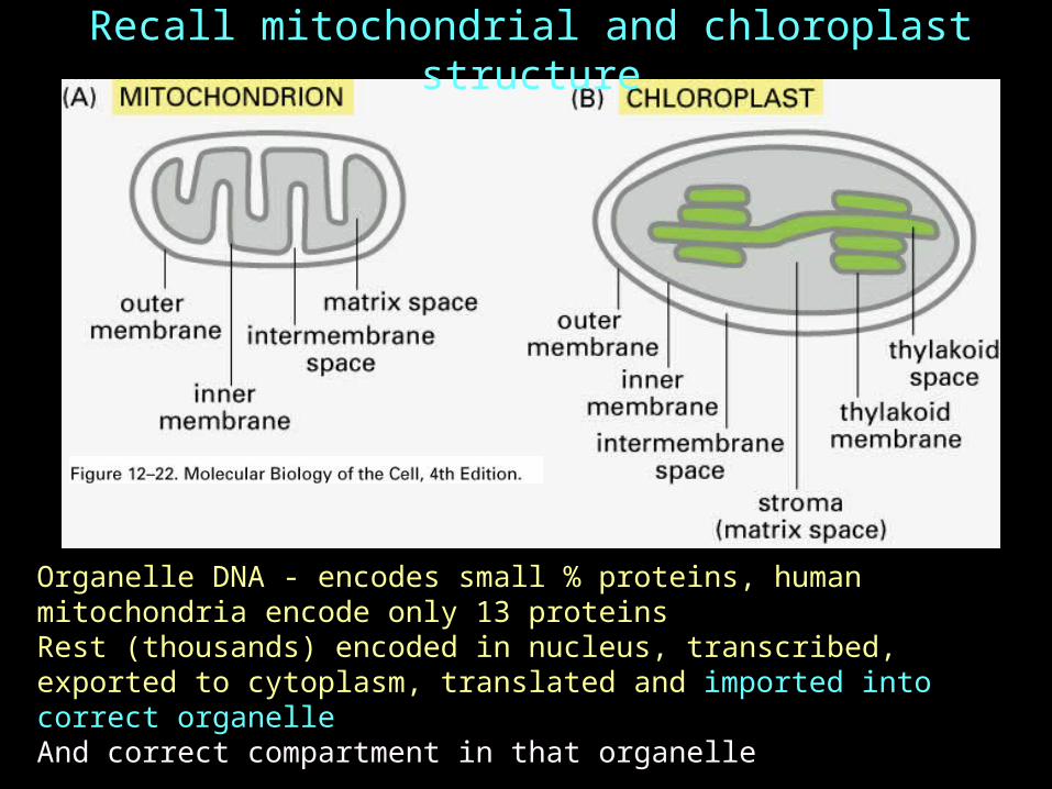
Organelle DNA - encodes small % proteins, human mitochondria encode only 13 proteinsRest (thousands) encoded in nucleus, transcribed, exported to cytoplasm, translated and imported into correct organelleAnd correct compartment in that organelle
Recall mitochondrial and chloroplast structure

Translate mRNA for mitochondrial matrix protein in vitro
Proteins contain N-terminal “signal sequence”
Import into mitochondria is post-translational
Digest with protease
Protein degraded
Add “energized” mitochondria
Protein imported into mitochondria
Imported matrix protein is protected from added protease
Import into mitochondria and chloroplasts is post-translational
Trypsin
Trypsin

Positive charge (red) clustered on one face of helix…
Non-polar aa (green) on the other…
Import is directed by a signal sequence at the N-terminus of mitochondrial proteins
No conserved sequence
Predicted to form “amphipathic” a-helix
Cleaved after protein is imported
MBoC (4) figure 12-23© Garland Publishing

TOMs and TIMs: Import into the mitochondria matrix requires two membrane transporters…
Mitochondrial import signal binds receptor in outer membrane (assoc w “TOM”)
Matrix
Outer membrane
Inner membrane
Cytoplasm
Intermembrane space
Matrix protein w N-terminal signal sequence
Removal of signal sequence
Mature matrix protein
Import receptor
“TOM”
“TIM23”
See ECB figure 15-10
“Contact site” (close apposition of OM & IM)
Transport thru aqueous channels: “TOM” and “TIMs” (Translocaters in Outer/Inner Membrane)
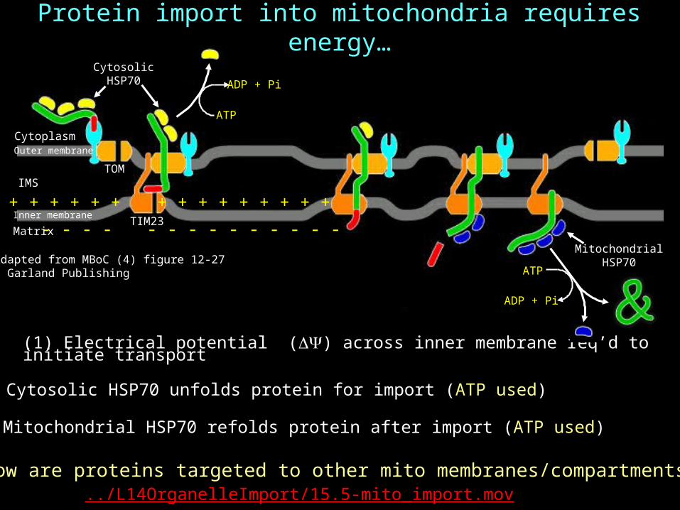
Protein import into mitochondria requires energy…
(1) Electrical potential () across inner membrane req’d to initiate transport
Adapted from MBoC (4) figure 12-27© Garland Publishing
ATP
ADP + Pi
- - - - - - - - - - - - - -
+ + + + + + + + + + + + + + +
Cytoplasm
IMS
Matrix
Outer membrane
Inner membrane
ATP
ADP + Pi
CytosolicHSP70
MitochondrialHSP70
TIM23
TOM
(3) Mitochondrial HSP70 refolds protein after import (ATP used)
(2) Cytosolic HSP70 unfolds protein for import (ATP used)
How are proteins targeted to other mito membranes/compartments?../L14OrganelleImport/15.5-mito_import.mov

How are proteins targeted to mitochondrial membranes and compartments? …the direct route
Cytoplasm
Matrix
Inner membrane
Outer membrane
IMS
TOM
TIM“Stop transfer” signal
Protein in IMSProtein in IM
Cleaved stop transfer(degraded)
Matrix signal (cleaved and degraded)
As before, signal sequence directs import through TOM/TIM23…
Adapted from MBoC (4) figure 12-29
“Stop transfer” signal interrupts translocation through TIM23, releasing protein to inner membrane…
Cleavage of stop transfer signal releases protein to intermembrane space…
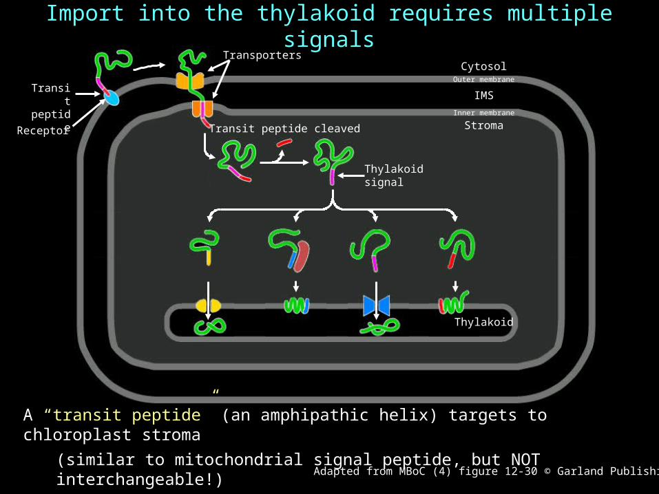
Import into the thylakoid requires multiple signals
A “transit peptide” (an amphipathic helix) targets to chloroplast stroma
(similar to mitochondrial signal peptide, but NOT interchangeable!)
Evidence for four paths to thylakoid Adapted from MBoC (4) figure 12-30 © Garland Publishing
Receptor
Thylakoidsignal
Transporters
Transit peptide cleaved
Transitpeptide
Outer membrane
Inner membrane
Stroma
IMS
Thylakoid
Cytosol

Protein targetingV
esi
cle t
arg
eti
ng
Secretory vesicles
Lysosomes
Endosomes
RetrievalTransport
RER
Golgi
Plasma membrane
See ECB figure 15-5
NLS: (basic)
NES: (L-rich) Signal peptideCytoplasm
Pro
tein
ta
rgeti
ng
Nucleus Mitochondria
Chloroplasts
Additional signals for subcompartments
Next two lectures
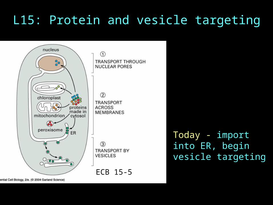
L15: Protein and vesicle targeting
ECB 15-5
Today - import into ER, begin vesicle targeting

GFP-protein in plant cell ER
ER network is extensive
TEM of RER in dog pancreasNote ribosomes on membrane
GFP-protein in plant cell ER
Vesicles derived from ER by biochemical prep are termed microsomes

Some ribosomes bind to ER
What is the evidence for cotranslational transport?
ECB 15-12

Transport of protein into ER is cotranslationalAdd RER microsomes AFTER translation…
Product still ~2kDa larger than in vivo product…Add protease…Product degraded…
Add RER microsomes DURING
translation…
Product processed to mature form…Add protease…Product protected…
INSIDE microsomes!
In vitro product ~2 kDa larger than in vivo product~15-25 addtnl aa at N-terminusAdd protease - product degraded
Translate mRNA in vitro…
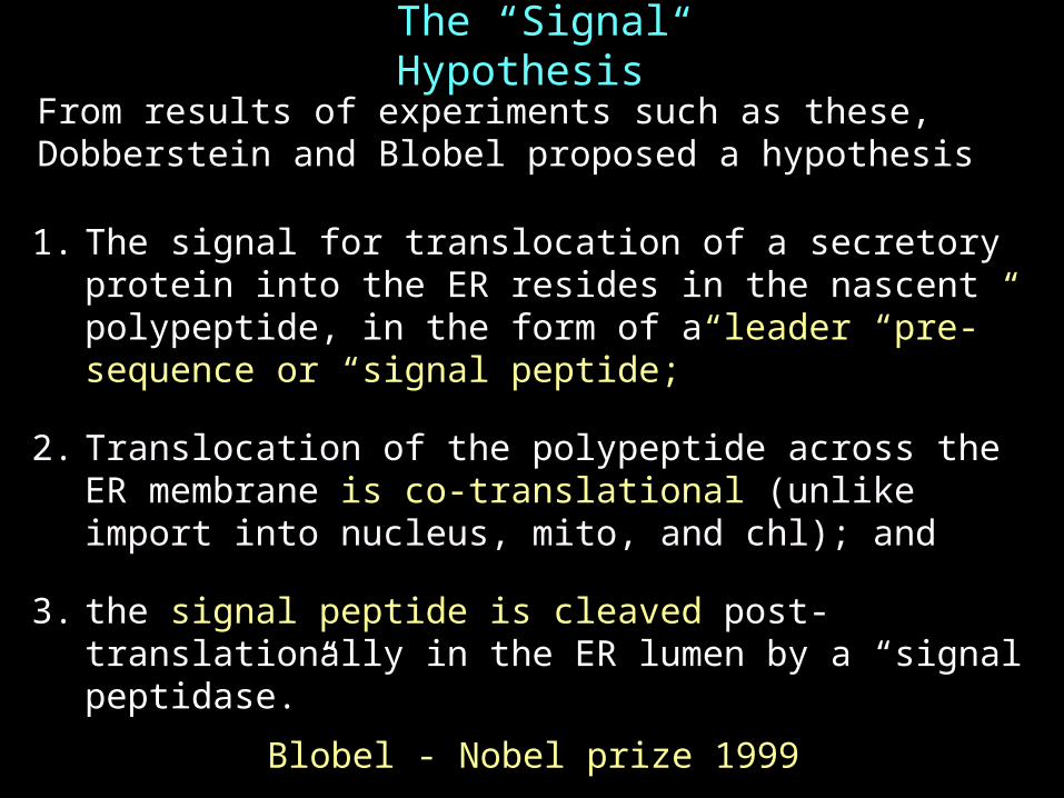
The “Signal Hypothesis”
1. The signal for translocation of a secretory protein into the ER resides in the nascent polypeptide, in the form of a leader “pre-” sequence or “signal peptide;”
2. Translocation of the polypeptide across the ER membrane is co-translational (unlike import into nucleus, mito, and chl); and
3. the signal peptide is cleaved post-translationally in the ER lumen by a “signal peptidase.”
From results of experiments such as these, Dobberstein and Blobel proposed a hypothesis
Blobel - Nobel prize 1999

ER Signal Sequence
No conserved sequence
Signal sequence is 12-25 amino acids Predicted to form-helix with hydrophobic core (yellow aa above)

Signal sequence is both necessary and sufficient for import into ER
Necessary
Sufficient

Requirements for targeting and translocation into the ER
1. “Signal sequence” : hydrophobic a-helix in nascent protein
2. “Signal recognition particle (SRP):” cytoplasmic complex of protein and RNA binds signal sequence
3. “SRP-receptor:” integral ER membrane protein
4. “Translocon:” an aqueous channel through ER membrane (sec61 complex)

Targeting to RER
1. Translation exposes signal sequence outside ribosome
2. SRP -a complex of 300bp RNA and 6 proteins- binds the signal sequence in nascent protein, transiently arrests translation
3. SRP-arrested ribosome binds SRP receptor in ER membrane (targeting)
4. Ribosome and polypeptide handed to a translocation channel (“translocon”). SRP and SRP-R are recycled (requires GTP hydrolysis). Translation resumes and translocation begins
ECB 15-13

Proteins destined for secretion enter ER lumen
Signal peptide targets nascent protein to RER as before
Signal peptide is cleaved by signal peptidase associated with translocation channelTranslation and translocation are completed, releasing completed polypeptide into lumen of RERSignal peptide is degraded What about membrane proteins??
ECB 15-14

As before, signal peptide targets nascent protein to RER
However, “Stop transfer” sequence halts translocation
Membrane proteins contain stop transfer sequence
Protein is released from translocon
Stop transfer sequence acts as transmembrane domain
ECB 15-15

Double- and multipass membrane proteins
Internal signal sequence targets nascent protein to RER…
“Stop transfer” sequence halts translocation and releases protein from translocon… Signal sequence and stop transfer sequence act as transmembrane domains
ECB 15-16

Protein folding in the ER is assisted by “BiP”…“Binding protein” (HSP70 family of ATPases) in ER lumen binds nascent polypeptide as it is being translocated, and assists folding (and translocation?)…
BiP binds nascent protein during translation/translocation…
RER membrane
ER Lumen
Translocon(sec 61 complex)
Signal peptide
Signalpeptidase
“Secreted protein” in
lumen of RER
Adapted from MBoC (4) figure 12-46.See ECB figure 15-14
N
N
C
N
C
Signal peptide
BiP
BiPBiP
BiP
BiP
ADP+Pi
ATP
Release of BiP from folded polypeptide requires energy (ATP)…
Incorrectly folded proteins are held in ER until folded properly, or are targeted for degradation…
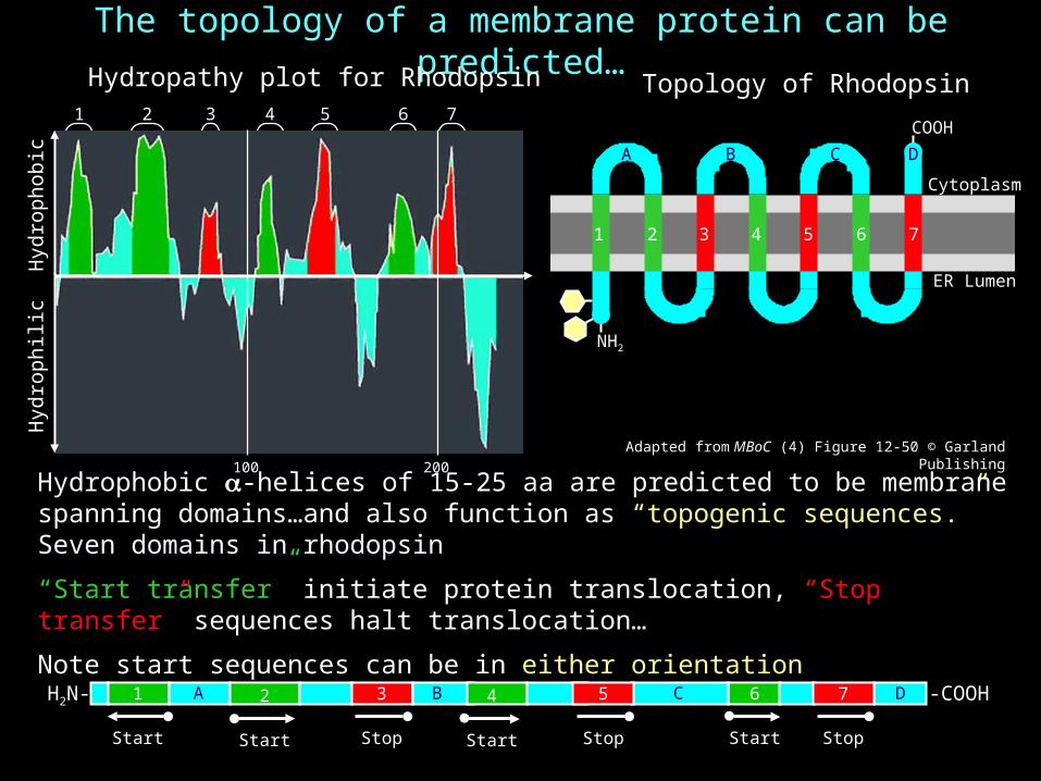
The topology of a membrane protein can be predicted…
Hydrophobic -helices of 15-25 aa are predicted to be membrane spanning domains…and also function as “topogenic sequences.” Seven domains in rhodopsin
“Start transfer” initiate protein translocation, “Stop transfer” sequences halt translocation…
Note start sequences can be in either orientation
Adapted from MBoC (4) Figure 12-50 © Garland Publishing
H2N- -COOH
1 2 3 4 5 6 7
Topology of Rhodopsin
COOH
NH2
ER Lumen
Cytoplasm
1 2 3 4 5 6 7
Hyd
roph
ilic
Hyd
roph
obic
200100
Hydropathy plot for Rhodopsin
Start
6
Stop
71
Start
2
Start Stop
3
Start
4
Stop
5
A B C D
A B C D

Review of the “Signal Hypothesis”
1. The signal for translocation/insertion of a protein into the ER membrane resides in the nascent polypeptide, in the form of a “signal sequence.”
2. Translocation of the polypeptide across the ER membrane is co-translational
3. The signal peptide (of secreted proteins) is cleaved post-translationally in the ER lumen by a “signal peptidase.”
4. Four components: (1) signal sequence, (2) SRP, (3) SRP-R, and (4) translocon
5. Uncleaved signal sequences (and “stop transfer” sequences) function as transmembrane domains in integral membrane proteins…
6. The topology of a protein can be predicted from the “hydropathy” plot of its amino acid sequence… 15.7-ERprotein_trans.mov

Vesicle targeting

Protein and vesicle targetingV
esi
cle t
arg
eti
ng
Secretory vesicles
Lysosomes
Endosomes
RetrievalTransport
RER
Golgi
Plasma membrane
See ECB figure 15-5
NLS: (basic)
NES: (L-rich) Signal peptideCytoplasm
Pro
tein
ta
rgeti
ng
Nucleus Mitochondria
Chloroplasts
Additional signals for subcompartments…
15.1-cell_compartments.mov

Membrane cycling
Secreted proteinsPlasma membrane proteinsExocytosis
(secretion)
endocytosis
ECB15-17
Transport is highly regulated so vesicles carry appropriate cargo for their specific destination

Lumen of organelle is equivalent to outside of cell
xx
xx
x
x
What about membrane protein in ER?
Begin with ER to Golgi

Modification of proteins begins in ER
ECB 15-22
Disulfide bridges
Glycosylation - common in plasma membrane and secreted proteins
Most common glycosylation is addition of a specific oligosaccharide (14mer) to asparagine during translation. Addition is to the NH2 group; N-linked glycoproteins
Addition is done in a single step by transfer from specialized dolichol lipid
This oligo is then extensively modified in diverse ways
Modification begins in ER: Transported to Golgi for more processing
Asn-X-Ser

MBoC (4) figure 13-22 © Garland Publishing
From the ER, proteins are transported to the Golgi
Vesicular-tubular clusters to CGN
RER
Nuclear envelope
Proteins leave the ER in transport vesicles budding from exit sites…
Transport vesicles from ER fuse to form vesicular-tubular clusters…
Vesicular-tubular clusters enter the Golgi by fusing with the cis-Golgi network (CGN)
Glycoproteins are “processed” as they pass thru the Golgi…
Golgi

ECB 15-24
From the ER, proteins are transported to the Golgi
cis-Golgi network (CGN)
cis
trans
medial
Vesicular-tubular clusters in from RER…
Proteins leave the ER in transport vesicles budding from exit sites…
Transport vesicles from ER fuse to form vesicular-tubular clusters…
Vesicular-tubular clusters enter the Golgi by fusing with the cis-Golgi network (CGN)…
Glycoproteins are “processed” as they pass thru the Golgi…
Trans Golgi network (TGN)

The Golgi is biochemically compartmentalized…
Osmium (cis)
Nucleotide diphosphatase (trans)
Acid phosphatase (TGN)
MBoC (4) figure 13-28© Garland Publishing

Glycoproteins are further processed in the Golgi
GlcNAc = N-acetylglucosamine
Mannose
Glucose
Fucose
Galactose, etc.trans
Plasma membrane
Protein synthesis
Golg
i appara
tus cis
Secretory vesicles
ER
medial
Glycosylation at H3N+…
XXNXSXX…COO-
LysosomeConstituitive secretion(Default?)
Regulated secretion
Proteins are sortedin the TGN…Constitutive secretion…Regulated secretion…Lysosome…
TGN
CGNAs protein moves through Golgi, monosaccharides are added or removed in specific Golgi compartmentsRemoval of mannose
Addition of GlcNAc
Addition of galactose
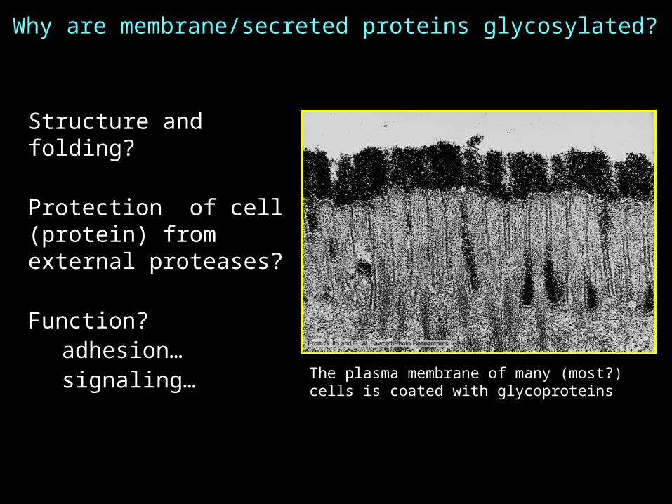
Why are membrane/secreted proteins glycosylated?
Structure and folding?
Protection of cell (protein) from external proteases?
Function?adhesion…signaling…
The plasma membrane of many (most?) cells is coated with glycoproteins

ECB 15-24
Transport through Golgicis-Golgi
network (CGN)
cis
trans-Golgi network (TGN)
trans
medial
Transport vesicles
Cisternal maturation and vesicle transport probably both contribute to membrane flow through Golgi
“Budding”
“Fusion”
2. “Cisternal maturation”
Vesicular-tubular clusters in from RER…
Transport vesicles out
1. Vesicle transport

Next time
Vesicle transport from ER to Golgi
Transport from Golgi Constitutive secretionRegulated secretionTo lysosome