Embed Size (px)
Citation preview
1
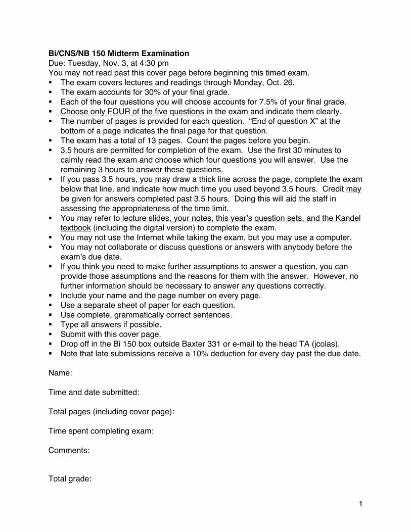
Bi/CNS/NB 150 Midterm Examination Due: Tuesday, Nov. 3, at 4:30 pm You may not read past this cover page before beginning this timed exam. § The exam covers lectures and readings through Monday, Oct. 26. § The exam accounts for 30% of your final grade. § Each of the four questions you will choose accounts for 7.5% of your final grade. § Choose only FOUR of the five questions in the exam and indicate them clearly. § The number of pages is provided for each question. “End of question X” at the
bottom of a page indicates the final page for that question. § The exam has a total of 13 pages. Count the pages before you begin. § 3.5 hours are permitted for completion of the exam. Use the first 30 minutes to
calmly read the exam and choose which four questions you will answer. Use the remaining 3 hours to answer these questions.
§ If you pass 3.5 hours, you may draw a thick line across the page, complete the exam below that line, and indicate how much time you used beyond 3.5 hours. Credit may be given for answers completed past 3.5 hours. Doing this will aid the staff in assessing the appropriateness of the time limit.
§ You may refer to lecture slides, your notes, this year’s question sets, and the Kandel textbook (including the digital version) to complete the exam.
§ You may not use the Internet while taking the exam, but you may use a computer. § You may not collaborate or discuss questions or answers with anybody before the
exam’s due date. § If you think you need to make further assumptions to answer a question, you can
provide those assumptions and the reasons for them with the answer. However, no further information should be necessary to answer any questions correctly.
§ Include your name and the page number on every page. § Use a separate sheet of paper for each question. § Use complete, grammatically correct sentences. § Type all answers if possible. § Submit with this cover page. § Drop off in the Bi 150 box outside Baxter 331 or e-mail to the head TA (jcolas). § Note that late submissions receive a 10% deduction for every day past the due date. Name: Time and date submitted: Total pages (including cover page): Time spent completing exam: Comments: Total grade:
2
Question 1. Action Potentials A. With respect to the Nernst potential of an ion species,
A1 (0.25 pts) Which driving forces (chemical free energies) are balanced at this equilibrium?
The electrical and osmotic driving forces are balanced. OK to say, electrical potential and chemical potential.
A2 (0.25 pts) Which conditions determine these driving forces? The temperature, charge per ion, and the internal:external concentration ratio are all relevant.
B. With respect to the resting potential of the electrical circuit model of a neural membrane, B1 (0.25 pts) Which electrical quantity (or parameter, or property) is conserved to
achieve this steady state? Electric charge. OK to say, inward and outward current are balanced.
B2 (0.25 pts) Which other electrical properties (or parameters, or quantities) determine this conservation?
Each ion’s transmembrane current is the product of its membrane conductance and the difference between its Nernst potential (battery) OK to say conductance and Nernst potential for each ion. The resting potential is the voltage at which the currents across the membrane sum to zero.
C. Many studies of the electrical properties of neurons involve injecting a controlled amount of current into a cell. In a living brain, without electrodes,
C1 (0.25 pts) What naturally causes depolarizations in neurons? Excitatory postsynaptic potentials cause depolarizations. OK to day, pacemaker currents.
C2 (0.25 pts) Where in a cell can this depolarization initiate? EPSPs occur at synapses, which can be located on dendrites, dendritic spines, or the soma.
D. Concerning the integration of electrical signals within neurons, D1 (0.25 pts) Where do action potentials usually initiate? Which underlying parameters
cause this to become the initiation zone? Action potentials usually initiate at the axon hillock. The action potential begins here because of its high density of voltage-gated sodium channels.
D2 (0.25 pts) Is passive spread the only mechanism that spreads depolarization from C2’s answer to D1’s answer? If not, explain.
No. Active propagation, via voltage-gated ion channels, can occur in dendrites. E. Describe the phases of an action potential, using a bulleted list, a timeline, and/or graphs. Make sure to explain:
E1 (0.5 pts) The time course of the opening and closing of sodium channels.
3
Voltage-gated sodium channels open rapidly in response to membrane depolarization, allowing positively-charged sodium ions to enter the cell. They are inactivated via a ball-and-chain mechanism within ~1 ms.
E2 (0.5 pts) The time course of the opening and closing of potassium channels. Voltage-gated potassium channels open as well, allowing positively-charged potassium ions to leave the cell. These channels open and close more gradually than the sodium channels.
E3 (0.5 pts) The all-or-none waveform. A sufficient depolarization leads to a positive feedback loop that results in almost all of the sodium channels opening within a small time window, rapidly depolarizing the membrane. This depolarization results in a similarly stereotyped time course of potassium channels opening, repolarizing, then hyperpolarizing the membrane.
E4 (0.5 pts) The absolute refractory period. The inactivation of the sodium channels makes it impossible to initiate a second action potential less than ~1ms after the first.
E5 (0.5 pts) The relative refractory period. The open potassium channels hyperpolarize the membrane and reduce its resistance, making it more difficult to initiate a second action potential shortly after the first.
F. A neuron has an action potential threshold at -50 mV, achieved by a current pulse of 3 nA. If the neuron doubles in surface area while maintaining its ion channel density,
F1 (0.5 pts) What parameter(s) would change in the equivalent circuit? Would it/they increase or decrease?
The cell’s capacitance, sodium conductance, and potassium conductance would increase.
F2 (0.5 pts) What (qualitative) effect would this have on the cell's threshold for firing action potentials, and why?
This would not affect the threshold potential because it doesn’t alter the osmotic or electrical driving forces on any ion species.
F3 (0.5 pts) What (qualitative) effect would this have on the cell's firing rate in response to a 6 nA, prolonged, constant-amplitude current injection, and why?
This cell would fire more slowly because it would require more current to depolarize efficiently.
G. Return to the “before” conditions of part F. If the density of potassium channels in the neuron’s membrane doubles,
G1 (0.5 pts) What parameter(s) would change in the equivalent circuit? Would it/they increase or decrease?
The membrane K conductance would increase.
4
G2 (0.5 pts) What (qualitative) effect would this have on the cell's threshold for firing action potentials, and why?
This would increase the cell’s threshold potential because more sodium channels would need to be open to overcome the membrane’s increased leakiness and yield action potential (positive feedback) dynamics.
G3 (0.5 pts) What (qualitative) effect would this have on the cell's firing rate in response to a 6 nA, prolonged, constant-amplitude current injection, and why?
The cell would fire more slowly because the 6 nA would depolarize less than before the change.
5
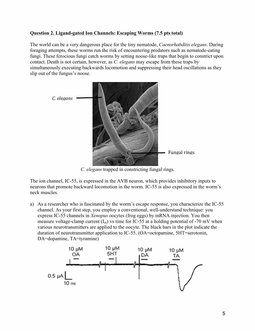
Question 2. Ligand-gated Ion Channels: Escaping Worms (7.5 pts total) The world can be a very dangerous place for the tiny nematode, Caenorhabditis elegans. During foraging attempts, these worms run the risk of encountering predators such as nematode-eating fungi. These ferocious fungi catch worms by setting noose-like traps that begin to constrict upon contact. Death is not certain, however, as C. elegans may escape from these traps by simultaneously executing backwards locomotion and suppressing their head oscillations as they slip out of the fungus’s noose.
C. elegans trapped in constricting fungal rings.
The ion channel, IC-55, is expressed in the AVB neuron, which provides inhibitory inputs to neurons that promote backward locomotion in the worm. IC-55 is also expressed in the worm’s neck muscles. a) As a researcher who is fascinated by the worm’s escape response, you characterize the IC-55
channel. As your first step, you employ a conventional, well-understand technique: you express IC-55 channels in Xenopus oocytes (frog eggs) by mRNA injection. You then measure voltage-clamp current (Im) vs time for IC-55 at a holding potential of -70 mV when various neurotransmitters are applied to the oocyte. The black bars in the plot indicate the duration of neurotransmitter application to IC-55. (OA=octopamine, 5HT=serotonin, DA=dopamine, TA=tyramine)
Fungalrings
C.elegans
6
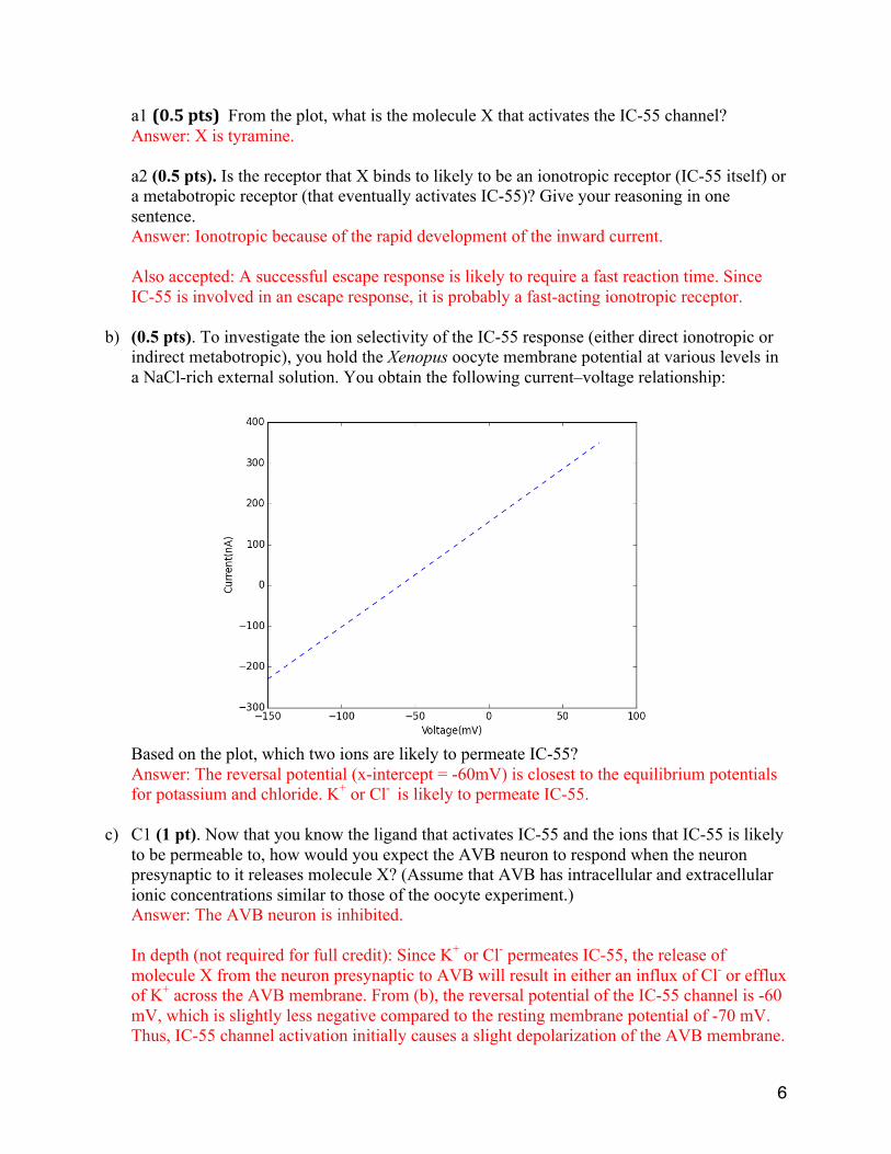
a1 (0.5pts) From the plot, what is the molecule X that activates the IC-55 channel? Answer: X is tyramine. a2 (0.5 pts). Is the receptor that X binds to likely to be an ionotropic receptor (IC-55 itself) or a metabotropic receptor (that eventually activates IC-55)? Give your reasoning in one sentence. Answer: Ionotropic because of the rapid development of the inward current. Also accepted: A successful escape response is likely to require a fast reaction time. Since IC-55 is involved in an escape response, it is probably a fast-acting ionotropic receptor.
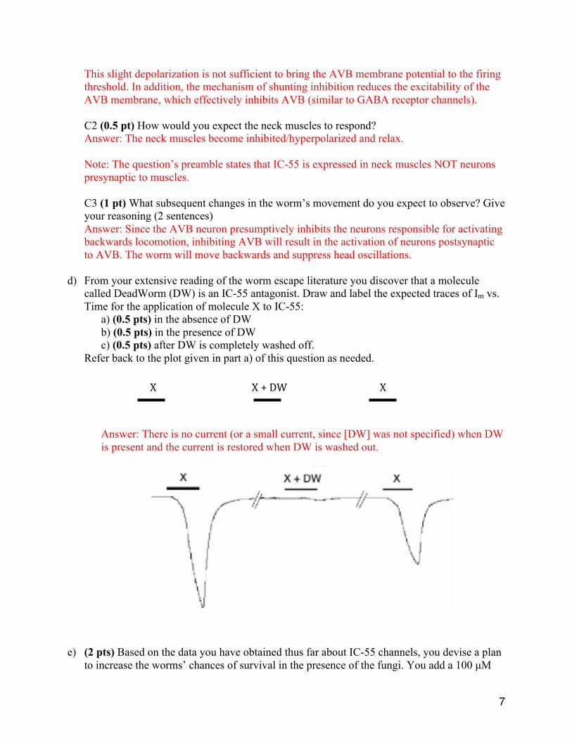
b) (0.5 pts). To investigate the ion selectivity of the IC-55 response (either direct ionotropic or
indirect metabotropic), you hold the Xenopus oocyte membrane potential at various levels in a NaCl-rich external solution. You obtain the following current–voltage relationship:
Based on the plot, which two ions are likely to permeate IC-55? Answer: The reversal potential (x-intercept = -60mV) is closest to the equilibrium potentials for potassium and chloride. K+ or Cl- is likely to permeate IC-55.
c) C1 (1 pt). Now that you know the ligand that activates IC-55 and the ions that IC-55 is likely
to be permeable to, how would you expect the AVB neuron to respond when the neuron presynaptic to it releases molecule X? (Assume that AVB has intracellular and extracellular ionic concentrations similar to those of the oocyte experiment.) Answer: The AVB neuron is inhibited. In depth (not required for full credit): Since K+ or Cl- permeates IC-55, the release of molecule X from the neuron presynaptic to AVB will result in either an influx of Cl- or efflux of K+ across the AVB membrane. From (b), the reversal potential of the IC-55 channel is -60 mV, which is slightly less negative compared to the resting membrane potential of -70 mV. Thus, IC-55 channel activation initially causes a slight depolarization of the AVB membrane.
7
This slight depolarization is not sufficient to bring the AVB membrane potential to the firing threshold. In addition, the mechanism of shunting inhibition reduces the excitability of the AVB membrane, which effectively inhibits AVB (similar to GABA receptor channels). C2 (0.5 pt) How would you expect the neck muscles to respond? Answer: The neck muscles become inhibited/hyperpolarized and relax. Note: The question’s preamble states that IC-55 is expressed in neck muscles NOT neurons presynaptic to muscles. C3 (1 pt) What subsequent changes in the worm’s movement do you expect to observe? Give your reasoning (2 sentences) Answer: Since the AVB neuron presumptively inhibits the neurons responsible for activating backwards locomotion, inhibiting AVB will result in the activation of neurons postsynaptic to AVB. The worm will move backwards and suppress head oscillations.
d) From your extensive reading of the worm escape literature you discover that a molecule
called DeadWorm (DW) is an IC-55 antagonist. Draw and label the expected traces of Im vs. Time for the application of molecule X to IC-55:
a) (0.5 pts) in the absence of DW b) (0.5 pts) in the presence of DW c) (0.5 pts) after DW is completely washed off.
Refer back to the plot given in part a) of this question as needed.
Answer: There is no current (or a small current, since [DW] was not specified) when DW is present and the current is restored when DW is washed out.
e) (2 pts) Based on the data you have obtained thus far about IC-55 channels, you devise a plan
to increase the worms’ chances of survival in the presence of the fungi. You add a 100 µM
X X+DW X
8
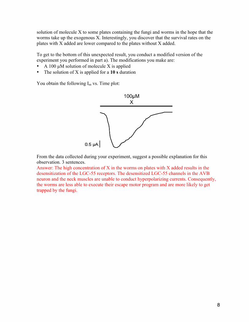
solution of molecule X to some plates containing the fungi and worms in the hope that the worms take up the exogenous X. Interestingly, you discover that the survival rates on the plates with X added are lower compared to the plates without X added. To get to the bottom of this unexpected result, you conduct a modified version of the experiment you performed in part a). The modifications you make are: • A 100 µM solution of molecule X is applied • The solution of X is applied for a 10 s duration You obtain the following Im vs. Time plot:
From the data collected during your experiment, suggest a possible explanation for this observation. 3 sentences. Answer: The high concentration of X in the worms on plates with X added results in the desensitization of the LGC-55 receptors. The desensitized LGC-55 channels in the AVB neuron and the neck muscles are unable to conduct hyperpolarizing currents. Consequently, the worms are less able to execute their escape motor program and are more likely to get trapped by the fungi.
9
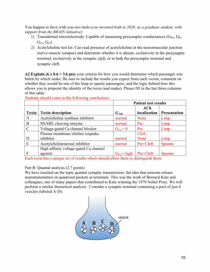
Question 3: Synaptic Transmission (7.5 points total, 3 pages) Part A: Snakes (4.8 points) It is the year 2025. You are on a flight to a conference to present your research as a leading neuroscientist in your field, when suddenly you find that your plane has been filled with many very aggressive, exceedingly venomous snakes. (For a documentary of a highly similar situation, see Snakes on a Plane [2006] http://www.imdb.com/title/tt0417148). Six passengers have been bitten, each by one snake. Here are the “presentations”: three are limp and unresponsive, the others have severe muscle spasms. The crew requires your assistance to determine which toxin is poisoning each. Fortunately, there is an ophidiologist (snake scientist) on board who is familiar with the snakes and their venoms, but cannot distinguish which snake bit which passenger. It is critical to make this distinction because administering the wrong antivenom (antivenin) will very likely be lethal to the victims. The ophidiologist gives you the following information:
Snake Toxin Toxin description Affects step #:
Viperidae voldemortensis A Acetylcholine synthase inhibitor 1 Elapidae basiliskae B SNARE protein cleaving enzyme 3 Viperidae samjacksonis C Voltage-gated Ca2+ channel blocker 4 Elapidae ekans D Plasma membrane choline reuptake inhibitor 6 Pythoninae arbok E Acetylcholinesterase inhibitor 5 Colubridae snakus F Potent voltage-gated Ca2+ channel agonist 4
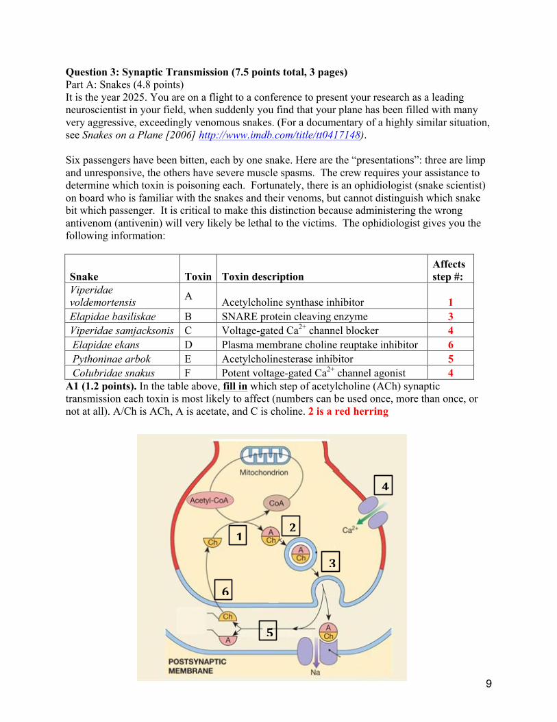
A1 (1.2 points). In the table above, fill in which step of acetylcholine (ACh) synaptic transmission each toxin is most likely to affect (numbers can be used once, more than once, or not at all). A/Ch is ACh, A is acetate, and C is choline. 2 is a red herring
1 2
4
3
6
5
10
You happen to have with you two tools (you invented both in 2020, as a graduate student, with support from the BRAIN initiative):
1) Transdermal microelectrode: Capable of measuring presynaptic conductances (GNa, GK, GCa, GCl)
2) Acetylcholine test kit: Can read presence of acetylcholine at the neuromuscular junction (nerve-muscle synapse) and determine whether it is absent, exclusively in the presynaptic terminal, exclusively in the synaptic cleft, or in both the presynaptic terminal and synaptic cleft.
A2 Explain (6 x 0.6 = 3.6 pts) your criteria for how you would determine which passenger was bitten by which snake. Be sure to include the results you expect from each victim, comment on whether they would be one of the limp or spastic passengers, and the logic behind how this allows you to pinpoint the identity of the toxin (and snake). Please fill in the last three columns of this table. Students should come to the following conclusions: Patient test results
Toxin Toxin description G ion
ACh localization Presentation
A Acetylcholine synthase inhibitor normal None Limp B SNARE cleaving enzyme normal Pre Limp C Voltage-gated Ca channel blocker GCa = 0 Pre Limp
D Plasma membrane choline reuptake inhibitor normal
Cleft / None Limp
E Acetylcholinesterase inhibitor normal Pre+Cleft Spasms
F High affinity voltage-gated Ca channel agonist GCa = high Pre+Cleft Spasms
Each toxin has a unique set of results which should allow them to distinguish them. Part B: Quantal analysis (2.7 points) We have touched on the topic quantal synaptic transmission- the idea that neurons release neurotransmitters in quantized packets at terminals. This was the work of Bernard Katz and colleagues, one of many papers that contributed to Katz winning the 1970 Nobel Prize. We will perform a similar theoretical analysis. Consider a synaptic terminal containing a pool of just 4 vesicles (labeled A-D):
A B C D
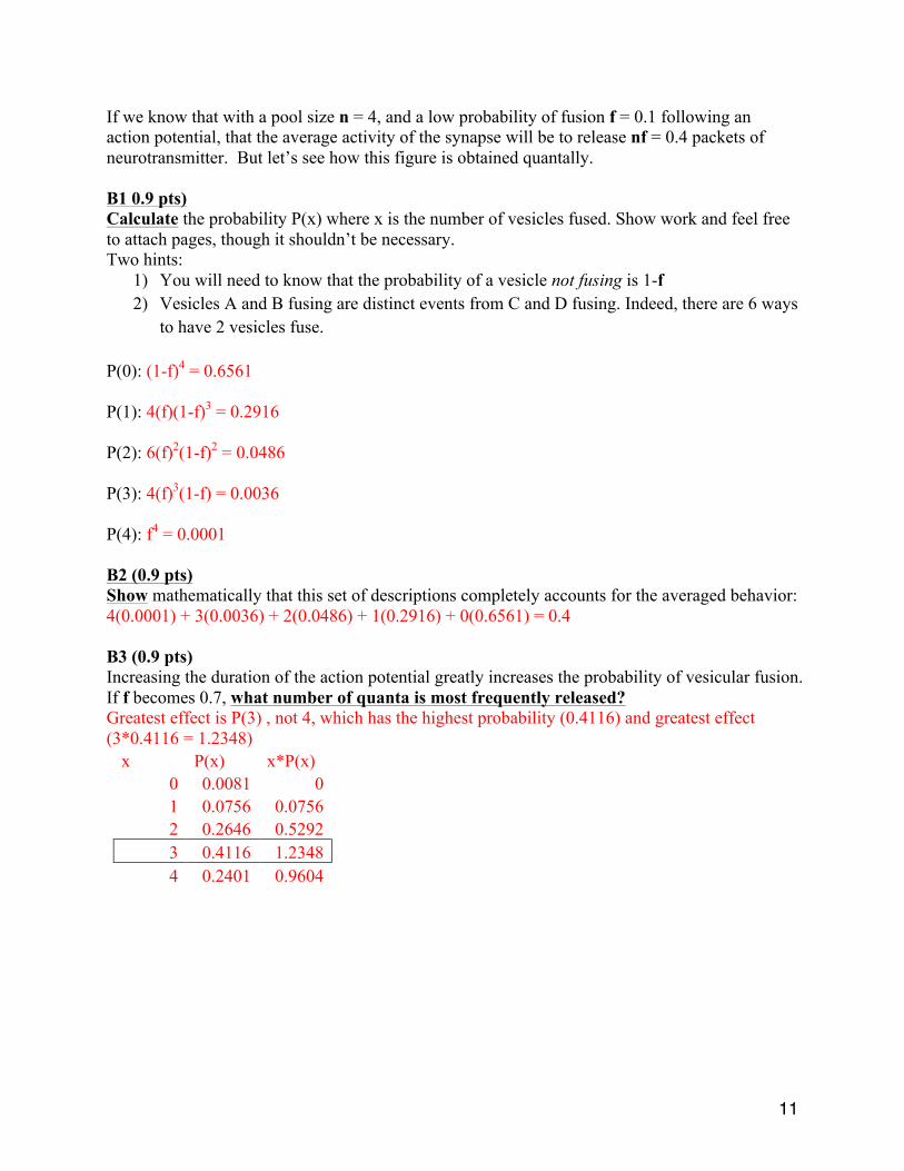
11
If we know that with a pool size n = 4, and a low probability of fusion f = 0.1 following an action potential, that the average activity of the synapse will be to release nf = 0.4 packets of neurotransmitter. But let’s see how this figure is obtained quantally. B1 0.9 pts) Calculate the probability P(x) where x is the number of vesicles fused. Show work and feel free to attach pages, though it shouldn’t be necessary. Two hints:
1) You will need to know that the probability of a vesicle not fusing is 1-f 2) Vesicles A and B fusing are distinct events from C and D fusing. Indeed, there are 6 ways
to have 2 vesicles fuse. P(0): (1-f)4 = 0.6561 P(1): 4(f)(1-f)3 = 0.2916 P(2): 6(f)2(1-f)2 = 0.0486 P(3): 4(f)3(1-f) = 0.0036 P(4): f4 = 0.0001 B2 (0.9 pts) Show mathematically that this set of descriptions completely accounts for the averaged behavior: 4(0.0001) + 3(0.0036) + 2(0.0486) + 1(0.2916) + 0(0.6561) = 0.4 B3 (0.9 pts) Increasing the duration of the action potential greatly increases the probability of vesicular fusion. If f becomes 0.7, what number of quanta is most frequently released? Greatest effect is P(3) , not 4, which has the highest probability (0.4116) and greatest effect (3*0.4116 = 1.2348)
x P(x) x*P(x) 0 0.0081 0 1 0.0756 0.0756 2 0.2646 0.5292 3 0.4116 1.2348 4 0.2401 0.9604
12
Question 4 (7.5 points, 2 pages): Neuroanatomy This year marks the inaugural Bi 150 field trip through spacetime! Caltech engineers have kindly lent us a time machine and C-3PO. You are visiting several major contributors to the history of neuroscience. Each of them is eager to find out what you have learned so far in the course. Please answer the questions below given your current knowledge about the brain to update these folks from the past! 4.A (5 x 0.3 = 1.5 points): Humors Galen of Pergamon (b. 129) is curious about the validity of his now-discredited proposal that nerves convey fluid secreted by the brain and spinal cord to the periphery. 4.A.a. What fluid is the central nervous system in fact suspended in? 4.A.b. What are each of the membranes enveloping the brain and spinal cord called? 4.A.c. Where is this fluid located relative to the aforementioned layers? 4.A.d. Name the general regions inside and outside of the nervous system where this fluid is
located. (You may wish to also draw a diagram and clearly label it.) 4.A.e. Which ion has the highest concentration in this fluid? 4.A.a. The brain and spinal cord are suspended in cerebrospinal fluid (CSF). 4.A.b. The meninges envelop the brain and spinal cord and include the pia mater (closest to
brain), the arachnoid mater, and the dura mater (farthest from brain). 4.A.c. CSF occupies the subarachnoid space between the arachnoid mater and the pia mater. 4.A.d. CSF is also located in ventricles, cisterns, sulci, and the central canal of the spinal cord.
(“Ventricles” sufficient for full credit.) 4.A.e. Na+ ions have the highest concentration in CSF.
13
4.B (5 x 0.3 = 1.5 points): Functional localization Franz Joseph Gall (b. 1758), the founder of phrenology, and Jean Pierre Flourens (b. 1794), one of phrenology’s most outspoken opponents, are debating the extent to which specific functions of the brain can be localized to distinct regions. 4.B.a. Name the four lobes of the brain. 4.B.b. What separates the most rostral lobe from the lobe that is adjacent and caudal to the most
rostral lobe? 4.B.c. Which lobes are the most basic processes underlying vision, audition, somatosensation,
and motor control primarily associated with? 4.B.d. Which lobe is disproportionately large in humans relative to other species? 4.B.e. What is the main feature of human cerebral cortex that complicates estimating its local
surface area via measurements on the skull? 4.B.a. There are frontal, temporal, parietal, and occipital lobes. 4.B.b. The central sulcus separates the frontal and parietal lobes. 4.B.c. Vision, audition, somatosensation, and motor control are associated with the occipital,
temporal, parietal, and frontal lobes, respectively. 4.B.d. Humans have disproportionately large frontal lobes. 4.B.e. Human cerebral cortex is highly gyrencephalic with variable convolutions in the form of
gyri and sulci. (Descriptions such as “wrinkled” sufficient for full credit.) 4.C (5 x 0.3 = 1.5 points): Neuropsychology Paul Pierre Broca (b. 1824) and Carl Wernicke (b. 1848) are proud to announce that they have identified specific areas of the brain concerned with language by studying patients with focal brain lesions that result in particular functional deficits. 4.C.a. If one were to finely slice the entire brain along the horizontal plane, only the rostral-
caudal and medial-lateral axes would be defined for each slice in that plane. Which pairs of cardinal axes are defined for each of the remaining cardinal planes?
4.C.b. Which cardinal plane(s) of the brain exhibit(s) approximate bilateral symmetry? 4.C.c. Which cerebral hemisphere is language primarily associated with for right-handed
people? 4.C.d. Which cerebral hemisphere controls movements of the right hand for left-handed people? 4.C.e. What is the largest structure connecting the two cerebral hemispheres in a human brain? 4.C.a. The dorsal-ventral and medial-lateral axes are defined for the coronal/frontal plane. The
rostral-caudal and dorsal-ventral axes are defined for the sagittal plane. 4.C.b. The horizontal and coronal planes exhibit approximate bilateral symmetry. 4.C.c. Language is primarily associated with the left cerebral hemisphere for right-handed
people. (This is less consistent for left-handed people.) 4.C.d. The right hand is always controlled by the left cerebral hemisphere for both left- and
right-handed people because of contralateral representation. 4.C.e. The corpus callosum is the largest structure connecting the two cerebral hemispheres.
14
4.D (5 x 0.3 = 1.5 points): The neuron doctrine Camillo Golgi (b. 1843) and Santiago Ramón y Cajal (b. 1852), who applied Golgi’s tissue-staining method, wonder about the implications of Ramón y Cajal’s groundbreaking hypothesis that the nervous system is made up of discrete neural cells. 4.D.a. How many neurons, glial cells, and synapses are in an average adult human brain? 4.D.b. Are there more neurons in cerebral cortex or in cerebellar cortex? 4.D.c. How many cell layers does neocortex have? 4.D.d. Are there more synapses in an adult human brain or in a five-year-old human brain? 4.D.e. What is the difference between axons and dendrites? Give both a functional explanation
(i.e., what they do differently) and an anatomical explanation (i.e., how they look different). 4.D.a. There are approximately 85 billion neurons, 85 billion glial cells, and 100 to 500 trillion
synapses. (10^11 and 10^14 sufficient for full credit.) 4.D.b. There are more neurons in cerebellar cortex. 4.D.c. Neocortex has six cell layers. 4.D.d. There are more synapses in a five-year-old human brain as a result of pruning. 4.D.e. Typically, several short dendrites branch out to short distances from the soma of a neuron
to receive incoming signals, whereas a single long, tubular axon extends far from the soma and transmits signals to other neurons.
4.E (5 x 0.3 = 1.5 points): Cytoarchitecture Korbinian Brodmann (b. 1868) is delighted to hear that his system for classifying regions of cerebral cortex based on the structure and organization of cells is still widely accepted and used in the 21st century. 4.E.a. What do gray matter and white matter each primarily consist of? 4.E.b. What do Brodmann areas 1, 2, 3, 4, and 17 have in common in terms of functional roles? 4.E.c. Which of the five aforementioned Brodmann areas exhibit topographic organization? 4.E.d. Which subcortical structure do the five aforementioned Brodmann areas all receive direct
input from? 4.E.e. How much bigger is a human brain than a chimpanzee brain? 4.E.a. Gray matter primarily consists of cell bodies, whereas white matter primarily consists of
myelinated axons. 4.E.b. Brodmann areas (BAs) 1, 2, 3, 4, and 17 all include “primary” cortex that receives or
transmits signals outside of cerebral cortex. BAs 1, 2, and 3 correspond to primary somatosensory cortex. BA 4 corresponds to primary motor cortex. BA 17 corresponds to primary visual cortex.
4.E.c. BAs 1, 2, 3, 4, and 17 are all topographically organized. 4.E.d. BAs 1, 2, 3, 4, and 17 all receive direct input from the thalamus. 4.E.e. A human brain is between three and four times bigger than a chimpanzee brain. (3 or 4
sufficient for full credit.)
15
Question 5. Development. 7.5 points, 2 pages A (9 x 0.3 = 2.7 points) Pair these developmentally important signalling molecules . . . . 1 Cadherin 2 Dystrophin 3 Ephrin 4 Laminin 5 Netrin 6 Neuroligins 7 Neurotrophins 8 Notch 9 Slit . . . with their receptors (not all signals have receptors, and vice-versa) A Cadherin B DCC C Delta D Eph kinase E Integrin F MuSK G Neurexins H Robo I Tyrosine receptor kinase e.g, write “9K” 1A 2,nothing 3D 4E 5B 6G 7I 8C 9H
16
B. (12 x 0.4 = 4.8 points) Give a one-sentence example, during development, of each process: 1 Growth along pioneer axons optic stalk, the rudiment of the neural tube that connects the retina to the diencephalon 2 Growth along glia Cerebellar granule cells along radial glia 3 Gradient sensing Retinotectal maps 4 Matching of intermediate targets at waystations Axons growing from retina to brain 5 Chemoaffinity arising from complementary interactions in molecules encode by two gene families(**not** “chemo-repulsion”) Drosophila Defective proboscis extension response (dpr genes) with 9 Dpr-interacting proteins (DIPs) 6 Transcription factors in collinear gene clusters Hox genes 7 Lateral inhibition sharpening the boundaries among cell fates Notch-delta 8 Limiting neurotrophic factor secreted from a target organ NGF secreted by sympathetic ganglia 9 Switching of a neurotransmitter’s effect, from depolarizing to hyperpolarizing Cl gradients sensed by GABA-A receptors 10 Molecular motors Growth cone extension 11 Crossing the midline Optic chiasm 12 Pruning of excess synapses Nerve-muscle connections