Embed Size (px)
Citation preview

480 Biophysical Journal Volume 99 July 2010 480–488
Bicosomes: Bicelles in Dilute Systems
Gelen Rodrıguez,† Guadalupe Soria,‡ Elisenda Coll,§ Laia Rubio,† Lucyanna Barbosa-Barros,†
Carmen Lopez-Iglesias,§ Anna M. Planas,‡ Joan Estelrich,{ Alfons de la Maza,† and Olga Lopez†*†Departament de Tecnologia Quımica i de Tensioactius, Institut de Quımica Avancada de Catalunya, Consejo Superior de InvestigacionesCientıficas, Barcelona, Spain; ‡Departament d’Isquemia Cerebral i Neurodegeneracio, Institut d’Investigacions Biomediques de Barcelona,Institut d’Investigatigacions Biomediques Agust Pi i Sunyer, Barcelona, Spain; and §Serveis Cientificotecnics and {Departamento deFisicoquımica, Facultad de Farmacia, Universidad de Barcelona, Barcelona, Spain
ABSTRACT Bicelles are discoidal phospholipid nanostructures at high lipid concentrations. Under dilute conditions, bicellesbecome larger and adopt a variety of morphologies. This work proposes a strategy to preserve the discoidal morphology ofbicelles in environments with high water content. Bicelles were formed in concentrated conditions and subsequently encapsu-lated in liposomes. Later dilution of these new structures, called bicosomes, demonstrated that lipid vesicles were able to isolateand protect bicelles entrapped inside them from the medium. Characterization of systems before and after dilution by dynamiclight-scattering spectroscopy and cryo-transmission electron microscopy showed that free bicelles changed in size andmorphology, whereas encapsulated bicelles remained unaltered by the effect of dilution. Free and entrapped bicelles (containingthe paramagnetic contrast agent gadodiamide) were injected into rat brain lateral ventricles. Coronal and sagittal visualizationwas performed by magnetic resonance imaging. Whereas rats injected with free bicelles did not survive the surgery, those in-jected with bicosomes did, and a hyperintensity effect due to gadodiamide was observed in the cerebrospinal fluid. These resultsindicate that bicosomes are a good means of preserving the morphology of bicelles under dilution conditions.
INTRODUCTION
Bicellar systems are composed of new discoidal nanostruc-
tures that consist of a phospholipid with a long hydrocarbon
chain situated in a bilaminar, flat center with a short-chained
phospholipid located at the edges (1). The characteristics of
these bicellar systems, such as their solely lipid content,
organization into a bilayer, and property of aligning in
a magnetic field, have permitted the wide use of these
systems as membrane models in diverse conformational
studies of proteins and membrane peptides (2). In recent
works (3–5), we proposed the use of phospholipid bicelles
for dermatological applications because their small size
allows them to pass through the skin. These studies demon-
strated that bicelles affect the skin barrier differently
depending on different compositional variables, working as
permeabilizing agents of the skin or as reinforcing agents
of the lipid structures of this tissue. In addition to using
bicelles as skin barrier-function modulators, the possibility
of incorporating drugs and other bioactive compounds into
bicelles is being explored. In one study, drugs were incorpo-
rated into bicelles to study drug-membrane interactions (6).
Another recent work focused on the percutaneous absorption
of diclofenac encapsulated in bicelles (7). The incorporation
of this drug results in systems that have smaller diameters
and can maintain structural and chemical stability at least
for 1 week. Therefore, bicelles can be considered good
carriers for skin applications. Their application as carriers
for administration through the systemic route, where the
Submitted December 22, 2009, and accepted for publication March 31,2010.
*Correspondence: [email protected]
Editor: Mark Girvin.
� 2010 by the Biophysical Society
0006-3495/10/07/0480/9 $2.00
water content is high, arises as the next challenge. Depend-
ing on the total lipid concentration, the temperature, and
the molar ratio between long- and short-chain phospholipids,
bicelles can exhibit different morphologies. Under high-
dilution conditions, small discoidal bicelles become large
structures, such as vesicles, lamellar sheets, and rodlike
micelles (4). This behavior could hinder the application of
these systems by specific systemic routes because the prop-
erties of bicellar systems would be lost under diluted condi-
tions, and the possible damage that the aforementioned
structures could cause is not clear. To maintain the size
and shape of the small discoidal bicelles, we propose that
they be encapsulated in lipid vesicles.
Liposomes have been the subject of numerous studies
because of their importance as models for more-complex
biological membranes, their potential use as microencapsula-
tors for drug delivery, and their applications in cosmetics and
clinical use (8–11). These structures, with diameters ranging
from 100 nm to 1 mm, are too large to pass through the skin
for transdermal application. However, they are morphologi-
cally stable under high-dilution conditions and thus are good
carriers for systemic application.
In this work we propose a new nanostructure: bicelles
encapsulated in liposomes. These structures, termed bico-
somes, unite the advantages of disks and vesicles, providing
a good system for application in diluted environments. Char-
acterization of the systems was carried out by means of
dynamic light scattering (DLS) and cryo-transmission elec-
tron microscopy (cryo-TEM). The first technique was used
to determine the average size of the systems, whereas the
second was valuable for characterizing the dimensional
and morphological aspects of the nanostructures, and
doi: 10.1016/j.bpj.2010.03.072
Bicosomes 481
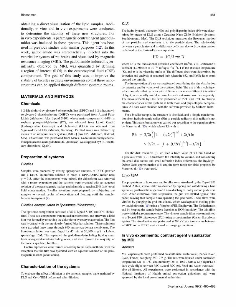
obtaining a direct visualization of the lipid samples. Addi-
tionally, in vitro and in vivo experiments were conducted
to determine the stability of these new structures. For
in vivo experiments, a paramagnetic contrast agent (gadodia-
mide) was included in these systems. This agent has been
used in previous studies with similar purposes (12). In this
work, gadodiamide was stereotactically injected into the
ventricular system of rat brains and visualized by magnetic
resonance imaging (MRI). The gadodiamide-induced hyper-
intensity, observed by MRI, was quantified by defining
a region of interest (ROI) in the cerebrospinal fluid (CSF)
compartment. The goal of this study was to improve the
stability of bicelles in dilute environments so that these nano-
structures can be applied through different systemic routes.
MATERIALS AND METHODS
Chemicals
1,2-Dipalmitoyl-sn-glycero-3-phosphocholine (DPPC) and 1,2-dihexanoyl-
sn-glycero-3-phosphocholine (DHPC) were purchased from Avanti Polar
Lipids (Alabaster, AL). Lipoid S-100, whose main component (>94%) is
soybean phosphatidylcholine (PC), was obtained from Lipoid GmbH
(Ludwigshafen, Germany), and cholesterol (CHO) was obtained from
Sigma-Aldrich-Fluka (Munich, Germany). Purified water was obtained by
means of an ultrapure water system (Milli-Q plus 185; Millipore, Bedford,
MA). Chloroform was purchased from Merck. Gadolinium-diethylenetria-
minepentaacetic acid (gadodiamide, Omniscan) was supplied by GE-Health-
care (Barcelona, Spain).
Preparation of systems
Bicelles
Samples were prepared by mixing appropriate amounts of DPPC powder
and a DHPC chloroform solution to reach a DPPC/DHPC molar ratio
q ¼ 3.5. After the components were mixed, the chloroform was removed
with a rotary evaporator and the systems were hydrated with an aqueous
solution of the paramagnetic marker gadodiamide to reach a 20% (w/v) total
lipid concentration. Bicellar solutions were prepared by subjecting the
samples to several cycles of sonication and freezing until the samples
became transparent (4).
Bicelles encapsulated in liposomes (bicosomes)
The liposome composition consisted of 80% Lipoid S-100 and 20% choles-
terol. These two components were mixed in chloroform, and afterward a lipid
film was formed by removing the chloroform by rotary evaporation. The film
was hydrated with the previously formed bicellar solution. These solutions
were extruded three times through 800-nm polycarbonate membranes. The
liposome solution was centrifuged for 45 min at 20,000 � g in a Labnet
spectrafuge 16M. This separated the gadodiamide-including lipid systems
from non-gadodiamide-including ones, and also formed the majority of
the nonencapsulated bicelles.
Control liposomes were formed according to the same methods, with the
exception that the film was hydrated with an aqueous solution of the para-
magnetic marker gadodiamide.
Characterization of the systems
To evaluate the effect of dilution in the systems, samples were analyzed by
DLS and Cryo-TEM before and after dilution.
DLS
The hydrodynamic diameter (HD) and polydispersity index (PI) were deter-
mined by means of DLS using a Zetasizer Nano ZS90 (Malvern Systems,
Southborough, MA). The DLS technique measures the Brownian motion
of the particles and correlates it to the particle sizes. The relationship
between a particle size and its diffusion coefficient due to Brownian motion
is defined in the Stokes-Einstein equation:
HD ¼ kT=3 ph D
where D is the translational diffusion coefficient (m2/s), k is Boltzmann’s
constant (1.3806503 � 10�23 m2 kg s�2 k�1), T is the absolute temperature
(K), and h is the viscosity (mPa.s). The different sizes were determined by
detection and analysis of scattered light when the 632-nm He/Ne laser beam
crossed the sample.
The interpretation of data was performed considering the size distribution
by intensity and by volume of the scattered light. The use of this technique,
which considers that particles with different sizes scatter different intensities
of light, is especially useful in samples with extensive size heterogeneity.
The measurements by DLS were performed at 25�C and 37�C to observe
the characteristics of the systems at both room and physiological tempera-
tures. All data were obtained with the software provided by Malvern Instru-
ments.
For a bicellar sample, the structure is discoidal, and a simple transforma-
tion from hydrodynamic radius (Rh) to particle radius (r, disk radius) is not
evident. This transformation was carried out according to the equation given
by Mazer et al. (13), which relates Rh with r:
Rh ¼ 3=2r��
1 þ ðt=2rÞ2�1=2þ 2r=t ln
� ½t=2r þ�1 þ ðt=2rÞ2
�1=2� � t=2r��1
For the disk thickness (t), we used a fixed value of 5.4 nm based on
a previous work (4). To transform the intensity to volume, and considering
the small disk radius and small refractive index differences, the Rayleigh-
Debye-Gans approximation (14) and the form factor for disks proposed by
Mazer et al. (13) were used.
Cryo-TEM
The preparations of liposomes and bicelles were visualized by the Cryo-TEM
method. A thin, aqueous film was formed by dipping and withdrawing a bare
specimen grid from the suspension. Glow-discharged, holey carbon grids were
used. After withdrawal from suspension, the grid was blotted against filter
paper, leaving thin sample films spanning the grid holes. These films were
vitrified by plunging the grid into ethane, which was kept at its melting point
by liquid nitrogen (15) using a Vitrobot (FEI, Eindhoven, The Netherlands),
and by keeping the sample before freezing at 100% humidity. The thin films
were vitrified at room temperature. The vitreous sample films were transferred
to a Tecnai F20 microscope (FEI) using a cryotransfert (Gatan, Barcelona,
Spain). The visualization was performed at 200 kV, at a temperature between
�170�C and �175�C, under low-dose imaging conditions.
In vivo experiments: contrast agent visualizationby MRI
Animals
MRI experiments were performed on adult male Wistar rats (Charles River,
Lyon, France) weighing 250–275 g. The rats were housed under controlled
temperature (21 5 1�C) and humidity (55 5 10%), with a 12-h light/12-h
dark cycle (light between 6:00 AM and 6:00 PM). Food and water were avail-
able ad libitum. All experiments were performed in accordance with the
National Institutes of Health animal protection guidelines and were
approved by the local governmental authorities.
Biophysical Journal 99(2) 480–488

TABLE 1 HD and proportion of the particle populations
analyzed by intensity and by volume of light scattered
Intensity Volume
HD (nm) % Intensity HD (nm) % Volume
Bice Peak 1 19.5 91.3 16.3 93.9
Bico Peak 1 489 100 571 100
Bice Dil Peak 1 255 88.4 256 3.6
Peak 2 26.8 11.6 26.6 96.4
Bico Dil Peak 1 800 86.3 850 59.8
Peak 2 72.4 13.7 68.7 40.2
482 Rodrıguez et al.
Stereotaxic injections
The animals were initially anesthetized with 4% isoflurane in O2:N2O (3:7),
which was reduced to 1% isoflurane for maintenance. They were then placed
in a stereotactic apparatus with a flat skull (Stoelting, Wood Dale, IL). The
bregma, the sagittal suture, and the surface of the brain were used as refer-
ences for the anterior-posterior (AP), medio-lateral (ML), and dorso-ventral
(DV) coordinates, respectively. A hole was drilled in the skull for solution
injection into the lateral ventricle at the following stereotaxic coordinates:
AP: �0.04 mm; ML: �0.12 mm; DV: �0.37 mm from the bregma (16).
Fifty microliters of an aqueous solution containing two different prepara-
tions of bicelles mixed with gadodiamide were injected via a 100-mL Ham-
ilton syringe fitted with a 32-G needle at a rate of 2 mL/min. After injection,
the needle was left in place for 5 min to prevent leakage. At the end of the
surgery, the skin was sutured and the animal was moved to the scanner
cradle for MRI. Three solutions containing gadodiamide (in bicelles, bico-
somes, and a control sample of liposomes) were injected. The concentration
of bicelles used for injection was 200 mg/mL. The researcher who conducted
the stereotaxic administration was highly experienced in this procedure.
In vivo MRI
MRI scans were performed under isoflurane anesthesia in a BioSpec 70/30
horizontal animal scanner (Bruker BioSpin, Ettlingen, Germany) equipped
with a 12-cm inner diameter, actively shielded gradient system
(400 mT/m). The coil configuration consisted of a transmit/receive quadra-
ture volume coil. The animals were placed in a supine position in a Plexiglas
holder with a nose cone for administering anesthetic gases, and fixed by
means of a tooth bar, earplugs, and adhesive tape. Tripilot scans were
used to accurately position the animal’s head inside the magnet.
The animal’s head was positioned such that the approximate center of the
brain was located at the magnet’s isocenter. T1-weighted axial and sagittal
images were acquired using a conventional fast low-angle shot imaging
sequence. The scan parameters for the sagittal images were as follows: repe-
tition time (TR) ¼ 350 ms, echo time (TE) ¼ 5.4 ms, two averages, slice
thickness ¼ 1 mm, number of contiguous slices ¼ 15, field of view
(FOV) ¼ 4�4 cm2, and matrix ¼ 256�256�15 pixels, resulting in a spatial
resolution of 0.156�0.156 mm with a 1-mm slice thickness. In addition,
T1 maps with axial orientation were acquired using a rapid acquisition
with relaxation enhancement and variable time of repetition sequence. The
scan parameters were TR ¼ 182.412 ms, 200 ms, 500 ms, 700 ms,
1000 ms, 1400 ms, 2000 ms, 3000 ms, and 6000 ms; TE ¼ 10 ms; slice
thickness ¼ 1 mm; number of contiguous slices ¼ 12; FOV ¼ 3�3 cm2;
and matrix ¼ 128�128�12 pixels, resulting in a spatial resolution of
0.234�0.234 mm with a 1-mm slice thickness. This image acquisition
was performed just after the injection and 4 h, 8 h, and 24 h later.
Image analysis
One ROI was drawn in the CSF compartment of each image to evaluate the
signal enhancement produced by gadodiamide. Signal normalization was
performed by dividing the mean signal intensity in the CSF ROI by the
mean signal in the tongue muscle. Statistical analysis was performed using
Student’s paired t-test, and a value of p < 0.05 was considered significant.
RESULTS
Characterization of systems
Bicellar systems
The HD and the proportion of the particle populations were
obtained by DLS and analyzed by intensity and by volume of
light scattered. The data obtained at room temperature are
shown in Table 1. The HDs for the DPPC/DHPC bicelles
(Bice) were 19.5 nm and 16.3 nm, estimated by intensity
Biophysical Journal 99(2) 480–488
and volume, respectively, with a proportion of the light scat-
tered higher than 90% in both analyses.
The sample containing bicelles was diluted from 20%
(initial concentration) to 0.07% (final concentration) to deter-
mine the effect of dilution on the bicellar structure. When this
sample was diluted (Bice Dil), two populations (one ~26 nm
and one ~255 nm) were observed for both analyses (by inten-
sity and volume). The proportions of light scattered, analyzed
by intensity and volume, were different. This is because large
and small particles contribute differently to the intensity of
scattered light. Large particles scatter light at a higher inten-
sity than small particles (17). For this reason, in systems
that are heterogeneous in size, an analysis by intensity inflates
the proportion of large particles, as shown in Table 1. The
simultaneous analysis by volume indicated a higher propor-
tion of small particles (26.6 nm) than large ones, indicating
the predominant presence of small particles in the system,
although the intensity of light scattered by large particles
was higher than that scattered by the small ones.
The values of PI for the bicellar systems were 0.452 and
0.714 before and after dilution, respectively. These results
indicate that when the sample was diluted, the average size
of the populations increased and the sample became more
heterogeneous.
It is necessary to consider that with this technique, particle
size is approximated to that of a hypothetical hard sphere that
diffuses with the same speed as the particle under examination.
Since the bicellar structure is disk-shaped, the particle size ob-
tained by DLS provides a relative measurement of the structure
dimensions. For this reason, the real disk diameter was calcu-
lated from the light-scattering results using the Mazer equation
as described in Materials and Methods. The diameter obtained
was 23.2 nm, similar to the HD result (19.5 nm; see Table 1). In
addition, the size distribution by intensity was transformed to
the size distribution by volume using the Rayleigh-Debye-
Gans approximation and the form factor for disks, giving
a diameter of 15.2 nm. This value is also on the order of the
HD obtained by volume (16.3 nm; see Table 1). All of the
measurements by DLS were obtained at 25�C and 37�C, and
no modification in size was detected from a temperature effect.
Given that temperature had no effect on these systems, cryo-
TEM experiments were performed only for samples that had
been cryofixed at room temperature. The images obtained offer
a direct visualization of the bicellar systems.

FIGURE 1 Representative micrographs of the samples
before (A) and after (B) dilution. (A) Bice sample: discoidal
structures (bicelles) are shown in all projections, face-on
(white arrow) and edge-on (black arrow). (B) Bice Dil
sample: spherical unilamellar vesicles.
FIGURE 2 Cryo-TEM images of a Bico sample. (A) Bicosomes and
bicelles are shown. (B) Encapsulated bicelles are observed, in their two
projections (white arrows) and nonencapsulated bicelles (black arrows).
(C) Stacks of bicelles adhered in parallel orientation are shown.
Bicosomes 483
Representative micrographs of the samples before (Bice)
and after (Bice Dil) dilution are shown in Fig. 1, A and B,
respectively. The images confirm the results obtained by
DLS, showing that the structures in the Bice samples are
smaller than those in the Bice Dil samples. In addition, the
morphology of the particles changes with the dilution. In
the Bice samples (Fig. 1 A), discoidal bicelles are shown in
all projections, face-on (white arrow) and edge-on (blackarrow), although the most evident is the edge-on projection.
From the structures observed face-on, it may be assumed
that the disks have a roughly circular shape. In addition, the
disks exhibit diameters of ~20 nm, confirming the results ob-
tained from DLS for the disks. In Fig. 1 B (Bice Dil samples),
spherical unilamellar vesicles showing a great variety of sizes
(from 30 to 250 nm) are observed. This agrees with the high PI
(0.714) observed by DLS. Therefore, both the DLS and
microscopy results indicate a transition of bicellar systems
from disks to vesicles caused by the effects of dilution.
Liposomes encapsulating bicelles (bicosomes)
The DLS results for bicosomes (Bico) are shown in Table 1
(values obtained at room temperature). Only one peak,
around 500 nm, was observed in the analyses by intensity
and by volume, and both analyses yielded a proportion of
light scattered of 100%.
When the sample was diluted (Bico Dil), two peaks were
clearly observed (Table 1). The structures became larger,
with diameters of 800 nm (86.3%) and 72.4 nm (13.7%)
by intensity, and 850 nm (59.8%) and 68.7 nm (40.2%) by
volume. The values of PI also increased from 0.259 to
0.612 before and after dilution, respectively. DLS data
acquired at 37�C showed no changes in comparison with
results obtained at room temperature.
To investigate the morphology of these systems, the Bico
and Bico Dil samples were analyzed by cryo-TEM. In this
case, the samples for microscopic observation were also cry-
ofixed only at room temperature. The cryo-TEM micro-
graphs are shown in Figs. 2 and 3. In Fig. 2 (Bico sample),
two kinds of structures are observed: one shows a small
size of ~20 nm, and the other is larger at ~100–500 nm.
Fig. 2 A shows an overview of the Bico sample in which bi-
cosomes and a small number of nonencapsulated bicelles are
seen. Fig. 2 B displays encapsulated bicelles, in their two
projections (white arrows), and nonencapsulated bicelles
(black arrows). Some encapsulated bicelles exhibit an
increase in size (~100 nm) with respect to the nonencapsu-
lated bicelles (20 nm) (Fig. 2 A). In addition, face-to-face
stacking bicelles were detected inside liposomes (Fig. 2 C).
These micrographs report additional information that was
not provided by the DLS results for Bico samples. Only
Biophysical Journal 99(2) 480–488
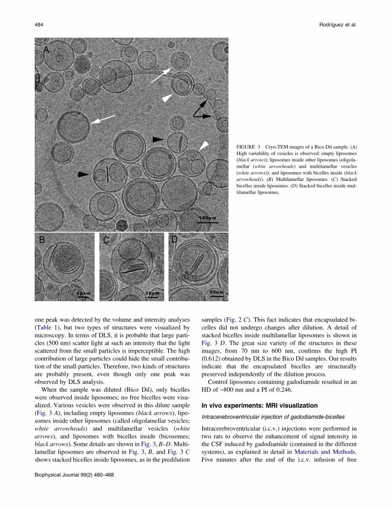
FIGURE 3 Cryo-TEM images of a Bico Dil sample. (A)
High variability of vesicles is observed: empty liposomes
(black arrows); liposomes inside other liposomes (oligola-
mellar (white arrowheads) and multilamellar vesicles
(white arrows)); and liposomes with bicelles inside (black
arrowheads). (B) Multilamellar liposomes. (C) Stacked
bicelles inside liposomes. (D) Stacked bicelles inside mul-
tilamellar liposomes.
484 Rodrıguez et al.
one peak was detected by the volume and intensity analyses
(Table 1), but two types of structures were visualized by
microscopy. In terms of DLS, it is probable that large parti-
cles (500 nm) scatter light at such an intensity that the light
scattered from the small particles is imperceptible. The high
contribution of large particles could hide the small contribu-
tion of the small particles. Therefore, two kinds of structures
are probably present, even though only one peak was
observed by DLS analysis.
When the sample was diluted (Bico Dil), only bicelles
were observed inside liposomes; no free bicelles were visu-
alized. Various vesicles were observed in this dilute sample
(Fig. 3 A), including empty liposomes (black arrows), lipo-
somes inside other liposomes (called oligolamellar vesicles;
white arrowheads) and multilamellar vesicles (whitearrows), and liposomes with bicelles inside (bicosomes;
black arrows). Some details are shown in Fig. 3, B–D. Multi-
lamellar liposomes are observed in Fig. 3, B, and Fig. 3 Cshows stacked bicelles inside liposomes, as in the predilution
Biophysical Journal 99(2) 480–488
samples (Fig. 2 C). This fact indicates that encapsulated bi-
celles did not undergo changes after dilution. A detail of
stacked bicelles inside multilamellar liposomes is shown in
Fig. 3 D. The great size variety of the structures in these
images, from 70 nm to 600 nm, confirms the high PI
(0.612) obtained by DLS in the Bico Dil samples. Our results
indicate that the encapsulated bicelles are structurally
preserved independently of the dilution process.
Control liposomes containing gadodiamide resulted in an
HD of ~800 nm and a PI of 0.246.
In vivo experiments: MRI visualization
Intracerebroventricular injection of gadodiamide-bicelles
Intracerebroventricular (i.c.v.) injections were performed in
two rats to observe the enhancement of signal intensity in
the CSF induced by gadodiamide (contained in the different
systems), as explained in detail in Materials and Methods.
Five minutes after the end of the i.c.v. infusion of free

FIGURE 4 (A) Coronal view of a healthy rat brain. (B) Coronal view of
a rat brain injected with bicelles containing gadodiamide. This rat died after
the injection. The expansion of the ventricular system is noted.
Bicosomes 485
bicelles, both animals died, and MRI was performed post-
mortem. Fig. 4 shows coronal views of the brains from
control (A) and Bice-injected (B) rats. As can be observed
in Fig. 4 B, free bicelles (Bice sample) caused a dramatic
expansion of the whole cerebroventricular system of the rat.
Intracerebroventricular injection of gadodiamide-bicosomes
I.c.v. injections were also performed in rats to observe gado-
diamide enhancement of signal intensity in the CSF by
administration of Bico samples. The rat was injected as
described above. Immediately after (0 h) and 4 h, 8 h, and
24 h later, the animal was scanned. In contrast to the case
with the Bice sample, the animal that received the Bico
sample survived, and this preparation had no apparent toxic
effect. We found an increase in the signal intensity in the
CSF after infusion of the Bico preparation. Fig. 5 A shows
a sagittal view of the brain of a healthy rat (first image)
and a Bico-injected rat 0 h, 4 h, 8 h, and 24 h after the injec-
tion (next four images). The gadodiamide-induced hyperin-
tensity was quantified by drawing one ROI within an
anatomic zone containing CSF at each time point (see
Fig. 5 B) and plotted to observe its time course (Fig. 5 C).
A time-dependent effect was observed, with the maximum
hyperintensity in the scan performed directly after the infu-
sion. A progressive decrease of the signal was observed.
Twenty-four hours after infusion, the intensity was similar
to that found in a control, noninjected animal.
A control experiment with an injection of gadodiamide en-
trapped in liposomes was performed, and the results were
similar to those obtained from the injection of gadodiamide
in the Bico samples.
DISCUSSION
Temperature effect on bicelles
Several factors affect the morphological stability of bicellar
systems (18–20). Temperature and hydration are key param-
eters in determining the formation of a specific structure.
A number of works have reported on the structural transi-
tions of bicellar systems formed by DMPC/DHPC at temper-
ature ranges similar to those used in this work (18,21). These
transitions involved morphological changes in the structures
from disks to cylindrical micelles, perforated lamellar sheets,
and mixed multilamellar vesicles (22). However, the systems
studied here did not exhibit such behavior, and in contrast
showed (by DLS) the same size distribution at 25�C and
37�C. This is because the temperatures under study were
below the DPPC Tm (41�C), and because in bicellar systems,
phase transitions take place from the Tm (23–25). For this
reason, systems containing DMPC (with Tm ¼ 23�C)
showed structural modifications between 25�C and 37�C.
The long-chain phospholipid chosen in this study was
DPPC precisely because the Tm value of this lipid is higher
than physiological temperature. This ensured the structural
and morphological stability of the systems against tempera-
ture effects. In any case, given the interest of these systems
and considering the fluid state exhibited by DMPC at phys-
iological temperature, which promotes increased stratum
corneum permeability (5), other compositions of bicelles
could be considered in future studies.
Dilution effect on bicelles
Another factor that, in general, induces morphological
changes in bicelles is modification of the total lipid
FIGURE 5 (A) Sagittal view of a healthy rat brain (first
image) and a rat injected with bicelles containing gadodia-
mide inside liposomes (Bico sample), scanned at 0 h, 4 h,
8 h and 24 h after the injection. (B) Detail of the ROI
used to monitor the time course of the hyperintensity
induced by the contrast agent. (C) CSF signal intensity
(normalized to muscle intensity) for a control rat (white
bar) and for the rat injected with bicelles containing gado-
diamide inside liposomes (black bars) at different time
points after the injection.
Biophysical Journal 99(2) 480–488

486 Rodrıguez et al.
concentration (20,26). In our work, this modification took
place when the bicellar systems were diluted. Dilution
promoted the growth of structures (Table 1) and the transi-
tion from discoidal bicelles to vesicles (Fig. 1). A recent
study using freeze-fracture electron microscopy also indi-
cated this tendency (4). However, that technique involves
a Pt/C replication process for the samples, which can hinder
visualization of structures smaller than 10 nm. Therefore, we
considered cryo-TEM, which permits direct visualization of
small structures, to be much more appropriate for evaluating
our systems. It is noteworthy that the phase transitions of
discoidal bicelles due to the variation of water content
(or total lipid concentration) are very similar to those
involved in reconstituting the surfactant-lipid micellar
systems in vesicles (27–29). A model for the micelle-to-
vesicle transition proposed by Leng et al. (30) describes
the rapid formation of discoidal aggregates and their growth
and closure to form vesicles. The resemblance between
surfactant-lipid micelles and phospholipid bicelles justifies
their similar behavior. Molecules of DHPC solubilize the
DPPC bilayer, forming discoidal bicelles in a manner similar
to the way in which surfactant molecules solubilize lipid
vesicles and form micelles. In systems containing discoidal
bicelles, DHPC molecules are partitioned between the
discoidal structure (mainly in the edges) and the water
(as monomers). When the water content increases (dilution),
the concentration of DHPC in water decreases, and then
DHPC is removed from the bicelles to let in the water.
This phenomenon and the high hydrophobicity of the
DPPC molecules induce an increase in the molar ratio (q)
of structures, and as a consequence, the disk diameter
increases. High-dilution conditions lead to the fusion and
closure of large bilayered disks, and hence the formation
of vesicles such as those shown in Fig. 1 B.
Relevance of bicosomes
It is reasonable to think that the isolation of bicelles from the
medium would protect these nanostructures from the effect
of posterior dilutions. This is precisely the strategy we
propose here. Bicelles are encapsulated in lipid vesicles or
liposomes, resulting in new nanostructures that we call bico-
somes. Dilution of these systems does not affect the encap-
sulated bicelles (Fig. 3). The exterior lipid membrane ensures
the isolation and stability of the bicelles captured in the inte-
rior; that is, the perfect microenvironment for the discoidal
bicellar system is created inside the lipid vesicles. Liposomes
have the advantage of being stable with temperature changes,
and have a controllable size that is not altered by dilution.
Previous studies have reported the usefulness of these lipid
vesicles as biocompatible and protective structures to encap-
sulate labile molecules, such as proteins, nucleic acids, or
drugs, for pharmaceutical, cosmetic, or chemical applica-
tions (30). Other methods have been used to stabilize the
morphology of discoidal bilayers, such as using bicelles
Biophysical Journal 99(2) 480–488
with charged amphiphiles (31) or disks formed by mixtures
containing polyethylene glycol-lipid conjugates (PEG-
lipids) (32). PEG-lipids have interesting applications, espe-
cially when drugs are included in discoidal bicelles.
However, with the use of PEG-lipids, the properties of
bicelles related to structural versatility, such as the enhancer
effect of the permeability on some physiological barriers,
could be lost. This enhancer effect of bicelles has been
studied for skin purposes and is potentiated by discoidal
structures formed with lipid mixtures in which the alkyl
chains are sufficiently different in length (5,33). Bicelles
formed with PEG-lipids are sterically stabilized, and all
lipids that form these nanostructures have the same or very
similar alkyl chain lengths (32).
The only difference between bicelles that are free in solu-
tion and those trapped in vesicles is an increase in the diam-
eter of the latter and subsequent stacking. The difference in
size, which is demonstrated in Figs. 1 A and 2, is maintained
after the dilution of bicosomes (Fig. 3). This is probably
related to a slight modification in the composition of the
initial bicelles during the formation of the bicosomes. To
form bicosomes, the lipid film, which is composed mainly
of Lipoid phospholipids, is hydrated with the initial solution
of preformed bicelles. At this step, and given the character-
istics of these Lipoid lipids (long alkyl chain), a portion of the
lipids of the film could be incorporated in the bilayer of the
disks. In a similar way, a portion of the lipids of bicelles
could be incorporated in the external membrane of bico-
somes. In water, DHPC exhibits a critical micellar concentra-
tion of 15 mM (34), which is comparable to that of some
mild surfactants, such as octyl glucoside (z18 mM (28)).
Therefore, a certain solubilization of the external membrane
could be suggested. However, the amount of DHPC
incorporated in the external bilayer was not enough to solu-
bilize the lipid membrane, as confirmed by cryo-TEM obser-
vations that were performed 10 days after preparation of the
system. In any case, this lipid redistribution induces an
increase in disk diameter. This increase in size is correlated
with the strong tendency for these structures to stack inside
the vesicles (35).
As mentioned above, the application of discoidal bicelles
for skin purposes is being explored (3,5,33). The water
content of the outermost layer of the skin is ~10–20%.
Such low water content promotes the stability of discoidal
bicelles and the interaction of these nanostructures with the
skin. This interaction promotes effects related to an increased
permeability of the barrier or a reinforcement of the skin
lipids. Similar effects may also be expected in the interaction
of discoidal bicelles with other tissues, although we are
aware that the composition and physical properties of skin
are different from those of other barriers (meaning that other
interactions could occur). Moreover, the water content of the
majority of tissues is usually too high to ensure the stability
of the small disks. Our results confirm that the encapsulation
of discoidal bicelles forming bicosomes preserves the

Bicosomes 487
discoidal structures under dilution. Therefore, we believe
that bicosomes are a good means of introducing bicelles in
biological fluids with high water content, such as blood,
CSF, and saliva. Bicosomes could be used as targeted vehi-
cles to different barriers, such as endothelium (for intrave-
nous administration) and gastric and intestinal mucosas
(for oral administration). A possible interaction mechanism
could be based on an initial interaction of the external Bico
membrane with these barriers. At this point, the incorpora-
tion of PEG-lipids and the addition of antibodies to specific
target sites in this external lipid bilayer seem necessary.
Afterward, encapsulated bicelles could pass through
different pathways (e.g., transcellular or paracellular) or by
endocytosis by the M cells (36,37).
In our in vivo experiments, small discoidal bicelles and
bicosomes (both with gadodiamide) were injected into rats.
Injection of free bicelles killed the animals, whereas injection
of bicosome samples did not. Due to the high water content
of the CSF, both systems underwent a dilution process after
injection, and contact with the CSF induced an increase
in size. Although the size increase by the nanostructures after
injection was not known, we assume that it was in the range
indicated by our DLS results. Large-sized structures should
not be lethal, as control liposomes exhibited sizes of
~800 nm and injection of these samples did not induce death.
Moreover, larger structures have been used for similar
in vivo studies (38). Another factor that allows us to discard
the size as the cause of the animal’s death was the fact that
bicosomes become larger than bicelles after dilution, and
only bicosomes permitted survival. Therefore, the toxicity
of the bicelles may be due to the rapid morphological
changes these structures undergo after they come into
contact with the diluted environment of the CSF. This
sudden and drastic morphological transition could form
a lipid accumulation that obstructs the free flow of CSF
and promotes the expansion of the cerebroventricular
system. This expansion could cause compression of the brain
stem, resulting in death, as previous works have observed
with other methods (39,40). When the Bico sample was
injected, the rat survived because large structures (bico-
somes) that did not change with dilution were introduced
together with a small amount of bicelles. Because only
a small proportion of bicelles underwent a morphological
transition, the lipid mass was not formed, and the obstruction
of flow did not occur.
With regard to bicosomes as paramagnetic contrast agents,
it is noteworthy that these nanostructures behave similarly to
conventional lipid vesicles. Liposomes have been used as
MRI contrast agents in two ways, according to the location
of the paramagnetic complex (in the membrane or encapsu-
lated in the liposome cavity (41)). Incorporation in the
membrane is preferred because it achieves a more enhanced
contrast for conventional liposomes applied intravenously
(42). Other nanostructures based on micelles have also
been used with the same purpose (43). In the work presented
here, gadodiamide-DTPA (a water-soluble substance) was
incorporated inside the vesicles together with the discoidal
bicelles when the bicosomes were formed. This method
showed an appropriate contrast, probably because the
systems were injected intracranially. In addition, the incor-
poration of gadodiamide within vesicles is convenient
because this process avoids the need to conjugate the gado-
diamide to any hydrophobic molecule, so the paramagnetic
probe can be used exactly as it is supplied for perfusion
purposes.
CONCLUSIONS
This work describes a new strategy for stabilizing discoidal
bicelles under diluted conditions. Bicosomes allow bicelles
to reach other tissues, and the bicelles have an enhancer
effect on skin permeability. These nanostructures could
interact with other physiological barriers, and other interac-
tions could occur. The incorporation of PEG-lipids and the
addition of antibodies to specific target sites in the external
lipid membranes is a logical next step. In addition, bico-
somes could be used as stabilizers, since the bicelles trapped
inside can be used to support various hydrophobic drugs,
such as dichlofenac (6,7). In this sense, bicosomes work in
a manner similar to that exhibited by other nanostructures,
such as emulsions and multilamellar liposomes (44,45).
The combination of the well-known characteristics of
liposomes with the versatility and applicability of bicelles
makes bicosomes a unique multifunctional nanostructure.
The authors thank Txema Vicente, Sandalo Roldan-Vargas, and Ramon
Pons for help with the particle size measurements. We also thank Orteve
(Barcelona, Spain) for providing the lipids (Lipoid S-100).
This work was supported by funds from the Comision Interministerial de
Ciencia y Tecnologıa (CTQ 2007-60409) and Generalitat de Catalunya
(GC BIOPOLIM-03086, 2005SR00066).
REFERENCES
1. Sanders, C. R., B. J. Hare, ., J. H. Prestegard. 1994. Magneticallyoriented phospholipid micelles as a tool for the study of membrane asso-ciated molecules. Prog. Nucl. Magn. Reson. Spectrosc. 26:421–444.
2. Sanders, C. R., and J. H. Prestegard. 1990. Magnetically orientablephospholipid bilayers containing small amounts of a bile salt analogue,CHAPSOs. Biophys. J. 586:447–460.
3. Barbosa-Barros, L., C. Barba, ., O. Lopez. 2008. Effect of bicellarsystems on skin properties. Int. J. Pharm. 352:263–272.
4. Barbosa-Barros, L., A. de la Maza, ., O. Lopez. 2008. Penetration andgrowth of DPPC/DHPC bicelles inside the stratum corneum of the skin.Langmuir. 24:5700–5706.
5. Rodrıguez, G., L. Barbosa-Barros, ., O. Lopez. 2009. Conformationalchanges in stratum corneum lipids by effect of bicellar systems.Langmuir. 25:10595–10603.
6. Guo, J., X. Tian, ., A. Makriyannis. 2008. Phospholipid BicelleMembrane Systems for Studying Drug Molecules. Springer, Dordrecht,Netherlands.
7. Rubio, L., C. Alonso, ., O. Lopez. 2010. Bicellar systems for in vitropercutaneous absorption of diclofenac. Int. J. Pharm. 386:108–113.
Biophysical Journal 99(2) 480–488

488 Rodrıguez et al.
8. Teschke, O., and E. F. de Souza. 2002. Liposome structure imaging byatomic force microscopy: verification of improved liposome stabilityduring adsorption of multiple aggregated vesicles. Langmuir.18:6513–6520.
9. Zhao, J. M., Y. E. Har-el, ., P. C. van Zijl. 2008. Size-inducedenhancement of chemical exchange saturation transfer (CEST) contrastin liposomes. J. Am. Chem. Soc. 130:5178–5184.
10. Hironaka, K., Y. Inokuchi, ., H. Takeuchi. 2009. Design and evalua-tion of a liposomal delivery system targeting the posterior segment ofthe eye. J. Control. Release. 136:247–253.
11. Chono, S., R. Fukuchi, ., K. Morimoto. 2009. Aerosolized liposomeswith dipalmitoyl phosphatidylcholine enhance pulmonary insulindelivery. J. Control. Release. 137:104–109.
12. Kobayashi, H., S. Kawamoto, ., P. L. Choyke. 2006. Delivery ofgadolinium-labeled nanoparticles to the sentinel lymph node: compar-ison of the sentinel node visualization and estimations of intra-nodalgadolinium concentration by the magnetic resonance imaging.J. Control. Release. 111:343–351.
13. Mazer, N. A., G. B. Benedek, and M. C. Carey. 1980. Quasielastic light-scattering studies of aqueous biliary lipid systems. Mixed micelleformation in bile salt-lecithin solutions. Biochemistry. 19:601–615.
14. Hofer, M. 1991. European Workshop on Neutron, X-Ray and LightScattering as an Investigative Tool for Colloidal and Polymeric SystemsP. Lindner and T. Zemb, editors. North Holland Delta Series, Amster-dam. 301–324.
15. Honeywell-Nguyen, P. L., P. M. Frederik, ., J. A. Bouwstra. 2002.Transdermal delivery of pergolide from surfactant-based elastic andrigid vesicles: characterization and in vitro transport studies. Pharm.Res. 19:991–997.
16. Paxinos, G., and C. Watson. 2007. The Rat Brain in Stereotaxic Coor-dinates. Elsevier, New York.
17. Barth, H. G. 1984. Modern Methods of Particle Size Analysis. Wiley-Interscience, New York.
18. Sternin, E., D. Nizza, and K. Gawrisch. 2001. Temperature dependenceof DMPC/DHPC mixing in a bicellar solution and its structural implica-tions. Langmuir. 17:2610–2616.
19. Dam, L. V., G. Karlsson, and K. Edwards. 2006. Morphology ofmagnetically aligning DMPC/DHPC aggregates-perforated sheets, notdisks. Langmuir. 28:3280–3285.
20. Struppe, J., and R. R. Vold. 1998. Dilute bicellar solutions for structuralNMR work. J. Magn. Reson. 135:541–546.
21. Hogberg, C. J., and A. P. Lyubartsev. 2006. A molecular dynamicsinvestigation of the influence of hydration and temperature on structuraland dynamical properties of a dimyristoylphosphatidylcholine bilayer.J. Phys. Chem. 110:14326–14336.
22. Harroun, T. A., M. Koslowsky, ., J. Katsaras. 2005. Comprehensiveexamination of mesophases formed by DMPC and DHPC mixtures.Langmuir. 21:5356–5361.
23. Lind, J., J. Nordin, and L. Maler. 2008. Lipid dynamics in fast-tumblingbicelles with varying bilayer thickness: effect of model transmembranepeptides. Biochim. Biophys. Acta. 1778:2526–2534.
24. Sternin, E., T. Zaraiskaya, ., R. M. Epand. 2006. Changes in molec-ular order across the lamellar-to-inverted hexagonal phase transitiondepend on the position of the double-bond in mono-unsaturated phos-pholipid dispersions. Chem. Phys. Lipids. 140:98–108.
25. Loudet, C., S. Manet, ., E. J. Dufourc. 2007. Biphenyl bicelle disksalign perpendicular to magnetic fields on large temperature scales:a study combining synthesis, solid-state NMR, TEM, and SAXS.Biophys. J. 92:3949–3959.
Biophysical Journal 99(2) 480–488
26. Yue, B., C. Y. Huang, ., J. Katsaras. 2005. Highly stable phospholipidunilamellar vesicles from spontaneous vesiculation: a DLS and SANSstudy. J. Phys. Chem. 109:609–616.
27. Goltsov, A. N., and L. I. Barsukov. 2000. Synergetics of the membraneself-assembly: a micelle-to-vesicle transition. J. Biol. Phys. 26:27–41.
28. Lopez, O., M. Cocera, ., A. De la Maza. 2001. Octyl glucoside-medi-ated solubilization and reconstitution of liposomes: structural andkinetic aspects. J. Phys. Chem. 105:9879–9886.
29. Pinaki, R. M., and A. Blume. 2002. Temperature-induced micelle-vesicle transitions in DMPC-SDS and DMPC-DTAB mixtures studiedby calorimetry and dynamic light scattering. J. Phys. Chem.106:10753–10763.
30. Leng, J., S. U. Egelhaaf, and M. E. Cates. 2003. Kinetics of the micelle-to-vesicle transition: aqueous lecithin-bile salt mixtures. Biophys. J.85:1624–1646.
31. Losonczi, J. A., and J. H. Prestegard. 1998. Improved dilute bicellesolutions for high-resolution NMR of biological macromolecules. J.Biomol. NMR. 12:447–451.
32. Johnsson, M., and K. Edwards. 2003. Liposomes, disks, and sphericalmicelles: aggregate structure in mixtures of gel phase phosphatidylcho-lines and poly(ethylene glycol)-phospholipids. Biophys. J.85:3839–3847.
33. Rubio, L., C. Alonso, ., O. Lopez. 2010. Bicellar systems for in vitropercutaneous absorption of diclofenac. Int. J. Pharm. 386:108–113.
34. Martınez-Landeira, P., J. L. Lopez-Fontan, ., F. Sarmiento. 2003.Surface behaviour of C5, C6, C7 and C8 lecithins at the aqueous solu-tion/air interface. Colloids Surf. 216:91–96.
35. Bolze, J., T. Fujisawa, ., A. Naito. 2000. Small angle X-ray scatteringand 31P NMR studies on the phase behavior of phospholipid bilayeredmixed micelles. Chem. Phys. Lett. 329:215–220.
36. Fasano, A. 1998. Innovative strategies for the oral delivery of drugs andpeptides. Trends Biotechnol. 16:152–157.
37. Spellerberg, B., S. Prasad, ., E. Tuomanen. 1995. Penetration of theblood-brain barrier: enhancement of drug delivery and imaging bybacterial glycopeptides. J. Exp. Med. 182:1037–1043.
38. Szoka, Jr., F. C., D. Milholland, and M. Barza. 1987. Effect of lipidcomposition and liposome size on toxicity and in vitro fungicidalactivity of liposome-intercalated amphotericin B. Antimicrob. AgentsChemother. 31:421–429.
39. Kim, D. S., S. Oi, ., J. U. Choi. 1999. A new experimental model ofobstructive hydrocephalus in the rat: the micro-balloon technique.Childs Nerv. Syst. 15:250–255.
40. Slobodian, I., D. Krassioukov-Enns, and M. R. Del Bigio. 2007. Proteinand synthetic polymer injection for induction of obstructive hydroceph-alus in rats. Cerebrospinal Fluid Res. 4:9.
41. Laurent, S., L. Vander Elst, ., R. N. Muller. 2008. Relaxivities of para-magnetic liposomes: on the importance of the chain type and the lengthof the amphiphilic complex. Eur. Biophys. J. 37:1007–1014.
42. Cheng, Z., and A. Tsourkas. 2008. Paramagnetic porous polymersomes.Langmuir. 24:8169–8173.
43. Shiraishi, K., K. Kawano, ., M. Yokoyama. 2009. Preparation andin vivo imaging of PEG-poly(L-lysine)-based polymeric micelle MRIcontrast agents. J. Control. Release. 136:14–20.
44. Washington, C. 1996. Stability of lipid emulsions for drug delivery.Adv. Drug Deliv. Rev. 20:131–145.
45. Gaede, H. C., and K. Gawrisch. 2003. Lateral diffusion rates of lipid,water, and a hydrophobic drug in a multilamellar liposome. Biophys.J. 85:1734–1740.