Embed Size (px)
Citation preview
NATIONAL OPEN UNIVERSITY OF NIGERIA
SCHOOL OF SCIENCE AND TECHNOLOGY
COURSE CODE: BIO 217
COURSE TITLE: GENERAL MICROBIOLOGY
iii
BIO 217
GENERAL MICROBIOLOGY
Course Team Mrs. O. A. F. Ilusanya (Developer/Writer) - Olabisi
Onabanjo University, Ago Iwoye, Ogun State,
Nigeria
NATIONAL OPEN UNIVERSITY OF NIGERIA
COURSE
GUIDE
iv
National Open University of Nigeria
Headquarters
14/16 Ahmadu Bello Way
Victoria Island, Lagos
Abuja Office
5 Dar es Salaam Street
Off Aminu Kano Crescent
Wuse II, Abuja
e-mail: [email protected]
URL: www.nou.edu.ng
Published by
National Open University of Nigeria
Printed 2011
Reprinted 2014
ISBN: 978-058-918-X
All Rights Reserved
v
CONTENTS PAGE
Introduction………………………………………………………. iv
What you will Learn in this Course……………………………… iv
Course Aims……………………………………………………… v
Course Objectives………………………………………………… v
Working through this Course…………………………………….. v
The Course Materials…………………………………………….. vi
Study Unit………………………………………………………… vi
Presentation Schedule……………………………………………. viii
Assessment……………………………………………………….. viii
Tutor-Marked Assignment……………………………………….. viii
Course Marking Scheme…………………………………………. ix
Facilitators/Tutors and Tutorials………………………………… ix
Summary…………………………………………………………. x
BIO 217 GENERAL MICROBIOLOGY
170
INTRODUCTION
General Microbiology is a first semester course. It is a three -credit unit
compulsory course which all students offering Bachelor of Science in
Biology must take.
Microbiology is a branch of biology which involves the study of
microorganisms. Microorganisms can be defined as living organisms
which cannot be seen by the unaided eyes. These organisms include
bacteria, fungi, algae, protozoa, viruses, etc.
Microorganisms are numerous in nature and have some characteristics
which make them ideal specimens for the study of numerous
fundamental like processes which occur at the cellular level in all living
organisms. In microbiology, study of microorganisms is done
extensively by observing their life processes while they are actively
metabolising.
Microorganisms have a wider range of physiological and biochemical
potentials than all other organisms combined. Some are able to utilise
atmospheric nitrogen for the synthesis of proteins and other complex
organic nitrogen compounds.
The study of microorganisms is applicable to all aspects of human
endeavour including: medicine, food, agriculture, conserving human and
animal reaction, combating diseases and used also as biological
weapons. Some organisms are friends (beneficial) while others can be
regarded as foes (harmful) to human beings.
WHAT YOU WILL LEARN IN THIS COURSE
In this course, you have the course units and a course guide. The course
guide will tell you what the course is all about. It is a general overview
of the course materials you will be using and how to use those materials.
It also helps you to allocate the appropriate time to each unit so that you
can successfully complete the course within the stipulated time limit.
The course guide also helps you to know how to go about your Tutor-
Marked Assignment which will form part of your overall assessment at
the end of the course. Also, there will be regular tutorial classes that are
related to this course, where you can interact with your facilitator and
other students.
This course exposes you to microbiology, a very important and
interesting field in biology.
BIO 217 MODULE 5
171
COURSE AIMS
The course aims to give you an understanding of microbiology which is
an important branch of biology.
COURSE OBJECTIVES
To achieve the aim set above, there are objectives. Each unit has a set of
objectives presented at the beginning of the unit. These objectives will
give you what to concentrate/focus on while studying the unit. Please
read the objectives before studying the unit and during your study to
check your progress.
The comprehensive objectives of the course are given below. By the
end of the course, you should be able to:
identify the different components of the microbial world
explain the historical aspects, relevance and scope of
microbiology
explain the general characteristics of the different groups of
microorganisms
describe microbial growth and reproduction and methods of
controlling microbial growth
give a systemic classification of bacteria, fungi, viruses, etc.
explain the causes of microbial variation and hereditary; and
describe some biogeochemical cycles in nature.
WORKING THROUGH THIS COURSE
To successfully complete this course, you are required to read each
study unit, textbooks and other materials provided by the National Open
University of Nigeria.
Reading the referenced materials can also be of great assistance.
Each unit has self assessment exercises which you are advised to do. At
certain periods during the course, you will be required to submit your
assignment for the purpose of assessment.
There will be a final examination at the end of the course. The course
should take you about 17 weeks to complete.
This course guide will provide you with all the components of the
course how to go about studying and how you should allocate your time
to each unit so as to finish on time and successfully.
BIO 217 GENERAL MICROBIOLOGY
172
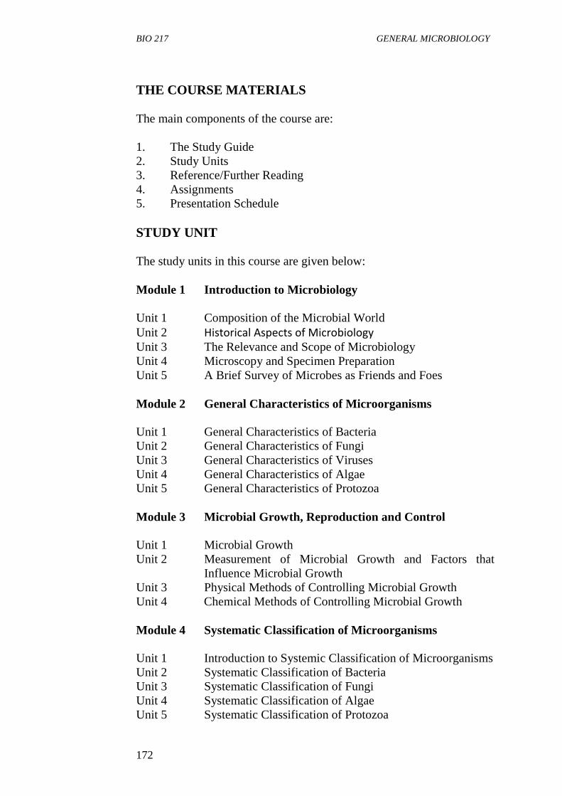
THE COURSE MATERIALS
The main components of the course are:
1. The Study Guide
2. Study Units
3. Reference/Further Reading
4. Assignments
5. Presentation Schedule
STUDY UNIT
The study units in this course are given below:
Module 1 Introduction to Microbiology
Unit 1 Composition of the Microbial World
Unit 2 Historical Aspects of Microbiology
Unit 3 The Relevance and Scope of Microbiology
Unit 4 Microscopy and Specimen Preparation
Unit 5 A Brief Survey of Microbes as Friends and Foes
Module 2 General Characteristics of Microorganisms
Unit 1 General Characteristics of Bacteria
Unit 2 General Characteristics of Fungi
Unit 3 General Characteristics of Viruses
Unit 4 General Characteristics of Algae
Unit 5 General Characteristics of Protozoa
Module 3 Microbial Growth, Reproduction and Control
Unit 1 Microbial Growth
Unit 2 Measurement of Microbial Growth and Factors that
Influence Microbial Growth
Unit 3 Physical Methods of Controlling Microbial Growth
Unit 4 Chemical Methods of Controlling Microbial Growth
Module 4 Systematic Classification of Microorganisms
Unit 1 Introduction to Systemic Classification of Microorganisms
Unit 2 Systematic Classification of Bacteria
Unit 3 Systematic Classification of Fungi
Unit 4 Systematic Classification of Algae
Unit 5 Systematic Classification of Protozoa
BIO 217 MODULE 5
173
Module 5 Microbial Genetics and Biogeochemical Cycling of
Elements
Unit 1 Mechanisms of Genetic Variation and Hereditary
Unit 2 Biogeochemical Cycling of Elements
In module one, unit one deals with the historical aspects of
microbiology, the second unit focuses on the meaning of microbiology,
microorganisms as cells, and the different groups of microorganisms,
and the domains in which they are placed. The third unit focuses on the
relevance and scope of microbiology. The fourth unit focuses on the use
of difference microscopes to study microorganisms while the fifth unit is
a brief survey of microorganisms as friends and foes.
Module two is concerned with the general characteristics of
microorganisms. Units one, two, three, four and five in this module deal
with the characteristics, morphology, distribution and importance of
bacteria, fungi, viruses, algae and protozoa respectively.
In module three, unit one focuses on microbial growth and reproduction,
unit two deals with measurement of microbial growth and factors that
influence microbial growth. Unit three deals with different physical
methods of controlling microbial growth and unit 4 deals with the use of
chemical agents to control microbial growth.
Units one, two, three and four in module 4 deal with the systematic
classification of microorganisms.
In module 5, unit one is on microbial variation and hereditary while unit
two focuses on biogeochemical cycling of nutrients in nature.
Each unit will take a week or two. Lectures will include an introduction,
objectives, reading materials, self assessment exercises, conclusion,
summary, tutor-marked assignments (TMAs), references and other
reading resources.
There are activities related to the lecture in each unit which will help
your progress and comprehension of the unit. You are required to work
on these exercises which together with the TMAs will enable you to
achieve the objectives of each unit.
PRESENTATION SCHEDULE
BIO 217 GENERAL MICROBIOLOGY
174
There is a timetable prepared for the early and timely completion and
submissions of your TMAs as well as attending the tutorial classes. You
are required to submit all your assignments by the stipulated date and
time. Avoid falling behind the schedule time.
ASSESSMENT
There are three aspects to the assessment of this course.
The first one is the self assessment exercises. The second is the tutor-
marked assignments and the third is the written examination or the
examination to be taken at the end of the course.
Do the exercises or activities in the unit by applying the information and
knowledge you acquired during the course. The tutor-marked
assignments must be submitted to your facilitator for formal assessment
in accordance with the deadlines stated in the presentation schedule and
the assignment file.
The work submitted to your tutor for assessment will account for 30% of
your total course work.
At the end of this course, you have to sit for a final or end of course
examination of about a three hour duration which will account for 70%
of your total course mark.
TUTOR-MARKED ASSIGNMENT
This is the continuous assessment component of this course and it
accounts for 30% of the total score. You will be given 4 TMAs by your
facilitator to answer. Three of which must be answered before you are
allowed to sit for the end of course examination.
These answered assignments must be returned to your facilitator.
You are expected to complete the assignments by using the information
and material in your reading references and study units.
Reading and researching into the references will give you a deeper
understanding of the subject.
1. Make sure that each assignment reaches your facilitator on or
before the deadline given in the presentation schedule and
assignment file. If for any reason you are not able to complete
your assignment, make sure you contact your facilitator before
the assignment is due to discuss the possibility of an extension.
BIO 217 MODULE 5
175
Request for extension will not be granted after the due date unless
there in exceptional circumstances.
2. Make sure you revise the whole course content before sitting for
the examination. The self-assessment exercises and TMAs will
be useful for this purposes and if you have any comments please
do before the examination. The end of course examination
covers information from all parts of the course.
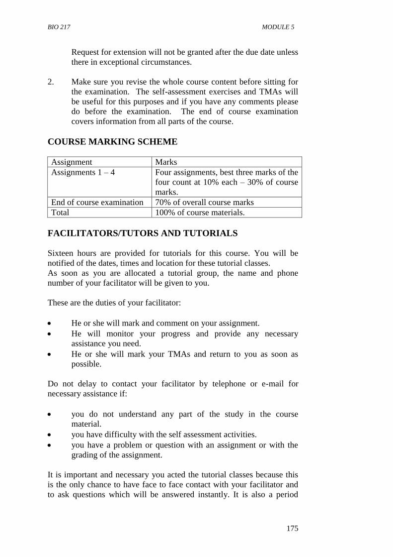
COURSE MARKING SCHEME
Assignment Marks
Assignments 1 – 4 Four assignments, best three marks of the
four count at 10% each – 30% of course
marks.
End of course examination 70% of overall course marks
Total 100% of course materials.
FACILITATORS/TUTORS AND TUTORIALS
Sixteen hours are provided for tutorials for this course. You will be
notified of the dates, times and location for these tutorial classes.
As soon as you are allocated a tutorial group, the name and phone
number of your facilitator will be given to you.
These are the duties of your facilitator:
He or she will mark and comment on your assignment.
He will monitor your progress and provide any necessary
assistance you need.
He or she will mark your TMAs and return to you as soon as
possible.
Do not delay to contact your facilitator by telephone or e-mail for
necessary assistance if:
you do not understand any part of the study in the course
material.
you have difficulty with the self assessment activities.
you have a problem or question with an assignment or with the
grading of the assignment.
It is important and necessary you acted the tutorial classes because this
is the only chance to have face to face contact with your facilitator and
to ask questions which will be answered instantly. It is also a period
BIO 217 GENERAL MICROBIOLOGY
176
where you can point out any problem encountered in the course of your
study.
SUMMARY
General Microbiology is a course that introduces you to the microbial
world around us.
Biology is a field very important to your welfare or well being in both
positive and negative ways. Microorganisms are cellular and acellular
(viruses) entities which are capable of life processes found in plants and
animals.
On completion of this course, you will have an understanding of basic
knowledge of microorganisms, the history of men and women who
contributed to this field of study by their discoveries during their
research works. You also learn the general characteristics of
microorganisms, microbial growth and reproduction, how the
microorganisms are classified or placed in different groups, the
mechanisms of variation and hereditary in microorganisms and the role
of microorganisms in cycling elements in our environments. In addition,
you will be able to answer the following questions:
What is microbiology?
What are microorganisms?
Identify the different groups of microorganisms and their general
characteristics.
Explain the relevance and scope of microbiology.
Differentiate between phyletic classification and phylogenetic
classification.
What are point mutations?
What are frame shift mutations?
Describe the four phases of the growth curve in a closed system.
The list of questions you are expected to answer is not limited to the
above list. Finally, you are expected to apply the knowledge you have
acquired during this course to your practical life.
I believe you will agree with me that microbiology is a very interesting
field of biology with a wide application to life.
I wish you success in this course.
BIO 217 MODULE 5
177
CONTENTS PAGE
Module 1 Introduction to Microbiology……………………. 1
Unit 1 Composition of the Microbial World……………… 1
Unit 2 Historical Aspects of Microbiology……………….. 9
Unit 3 The Relevance and Scope of Microbiology……….. 19
Unit 4 Microscope and Specimen Preparation……………. 24
Unit 5 A Brief Survey of Microbes as Friends and Foes….. 35
Module 2 General Characteristics of Microorganisms…….. 43
Unit 1 General Characteristics of Bacteria………………… 43
Unit 2 General Characteristics of Fungi……………………. 56
Unit 3 General Characteristics of Viruses………………….. 66
Unit 4 General Characteristics of Algae……………………. 75
Unit 5 General Characteristics of Protozoa………………… 81
Module 3 Microbial Growth, Reproduction and Control…… 91
Unit 1 Microbial Growth………………………………… 91
Unit 2 Measurement of Microbial Growth and
Factors that Influence Microbial Growth………… 103
Unit 3 Physical Methods of Controlling Microbial
Growth……………………………………………… 114
Unit 4 Chemical Methods of Controlling Microbial
Growth……………………………………………… 121
Module 4 Systemic Classification of Microorganisms……… 134
Unit 1 Introduction to Systemic Classification of
Microorganisms………………………………….. 134
Unit 2 Systematic Classification of Bacteria…………… 142
Unit 3 Systematic Classification of Fungi………………. 155
Unit 4 Systematic Classification of Algae………………. 160
MAIN
COURSE
BIO 217 GENERAL MICROBIOLOGY
178
Unit 5 Systematic Classification of Protozoa…………… 167
MODULE 5 MICROBIAL GENETICS AND
BIOGEOCHEMICAL CYCLING OF
ELEMENTS
Unit 1 Mechanisms of Genetic Variation and
Hereditary……………………………………. 172
Unit 2 Biogeochemical Cycling of Elements……….. 184
BIO 217 MODULE 5
179
MODULE 1 INTRODUCTION TO MICROBIOLOGY
Unit 1 Composition of the Microbial World
Unit 2 Historical Aspects of Microbiology
Unit 3 The Relevance and Scope of Microbiology
Unit 4 Microscope and Specimen Preparation
Unit 5 A Brief Survey of Microbes as Friends and Foes
UNIT 1 COMPOSITION OF THE MICROBIAL WORLD
CONTENTS
1.0 Introduction
2.0 Objectives
3.0 Main Content
3.1 Microorganisms
3.2 Microorganisms as Cells
3.3 Classification Systems for Microorganisms
3.4 Domain Bacteria
3.5 Domain Archaea
3.6 Domain Eucarya
3.7 Viruses
4.0 Conclusion
5.0 Summary
6.0 Tutor-Marked Assignment
7.0 References/Further Reading
1.0 INTRODUCTION
Microbiology is the study of microorganisms. These are organisms too
small to be seen clearly by the unaided eyes. Microorganisms include
bacteria, fungi, algae, protozoa and entities at the borderline of life that
are called viruses. The cell is the fundamental unit of life. Most
microorganisms are unicellular, in unicellular organisms all the life
processes are performed by a single cell. However, some are
multicellular, having more than one cell. This unit examines the
definition of microbiology, types of microbial cells, the different groups
of microorganisms and the domains in which they are placed and why
viruses are not placed in any of the domains.
BIO 217 GENERAL MICROBIOLOGY
180
2.0 OBJECTIVES
At the end of this unit, you should be able to:
define the term microbiology
list the groups of organisms classified as microorganisms
distinguish between prokaryotic and eukaryotic cells
explain the distribution of microorganisms into domains
state the characteristics of the microorganisms in each domains
state the characteristics of viruses.
3.0 MAIN CONTENT
3.1 Microorganisms
Microorganisms are organisms too small to be seen clearly by the
unaided eyes. They are very small life forms so small that individual
microorganisms cannot be seen without magnification. They include
fungi, bacteria, algae, protozoa and viruses. Some microorganisms
however, like the eukaryotic microorganisms are visible without
magnification. Examples are bread moulds and filamentous algae.
3.2 Microorganisms are Cells
The cell is the fundamental unit of life; a single cell is an entity isolated
from other cells. Two fundamental different types of cells exist among
microorganisms; they are prokaryotic and eukaryotic.
Prokaryotes
These microbial cells lack membrane-bound nucleus and
organelles.
Eukaryotes
Possess a membrane-bound nucleus and organelles.
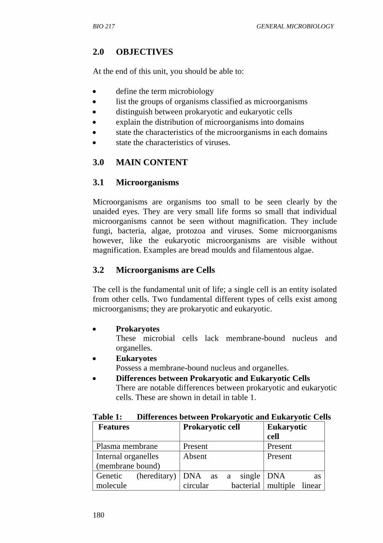
Differences between Prokaryotic and Eukaryotic Cells
There are notable differences between prokaryotic and eukaryotic
cells. These are shown in detail in table 1.
Table 1: Differences between Prokaryotic and Eukaryotic Cells
Features Prokaryotic cell Eukaryotic
cell
Plasma membrane Present Present
Internal organelles
(membrane bound)
Absent Present
Genetic (hereditary)
molecule
DNA as a single
circular bacterial
DNA as
multiple linear
BIO 217 MODULE 5
181
chromosome not
enclosed in a nucleus
chromosomes
enclosed within
a nucleus.
Site of energy(ATP)
generation
Cytoplasm and, in
some cases, plasma
membrane or
photosynthesis
membrane
Cytoplasm and
mitochondria or
chloroplasts.
Source: Atlas et al., (1995)
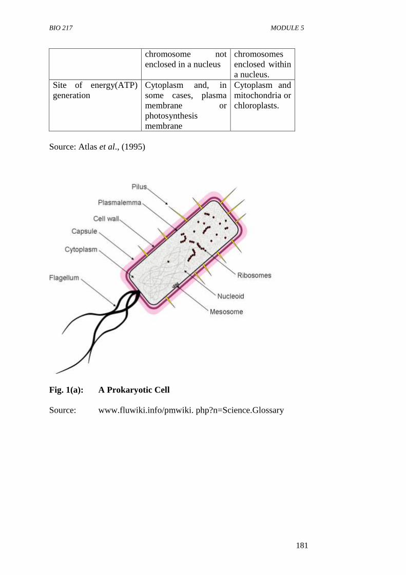
Fig. 1(a): A Prokaryotic Cell
Source: www.fluwiki.info/pmwiki. php?n=Science.Glossary
BIO 217 GENERAL MICROBIOLOGY
182
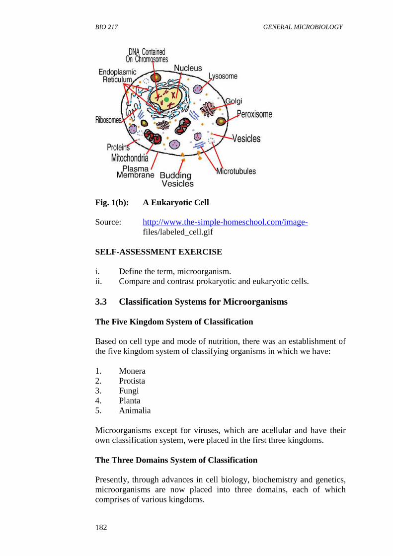
Fig. 1(b): A Eukaryotic Cell
Source: http://www.the-simple-homeschool.com/image-
files/labeled_cell.gif
SELF-ASSESSMENT EXERCISE
i. Define the term, microorganism.
ii. Compare and contrast prokaryotic and eukaryotic cells.
3.3 Classification Systems for Microorganisms
The Five Kingdom System of Classification
Based on cell type and mode of nutrition, there was an establishment of
the five kingdom system of classifying organisms in which we have:
1. Monera
2. Protista
3. Fungi
4. Planta
5. Animalia
Microorganisms except for viruses, which are acellular and have their
own classification system, were placed in the first three kingdoms.
The Three Domains System of Classification
Presently, through advances in cell biology, biochemistry and genetics,
microorganisms are now placed into three domains, each of which
comprises of various kingdoms.
BIO 217 MODULE 5
183
The domains are:
1. Bacteria (prokaryotic – “true bacteria”)
2. Archaea (prokaryotic – “ancient bacteria”)
3. Eucarya (eukaryotic)
3.4 Domain Bacteria
1. They are prokaryotic.
2. They are single celled organisms.
3. They lack membrane bound nucleus and organelles.
4. Most have cell wall that contains peptidoglycan.
5. They are found in the soil, water and air and on other living
organisms.
6. Some are harmful while others are beneficial to man.
3.5 Domain Archaea
1. They were formerly known as archaeobacteria.
2. They are prokaryotic.
3. They are single celled organisms.
4. They lack membrane bound nucleus and organelles.
5. They lack peptidoglycan in their cell walls.
6. They have unique membrane lipids.
7. Some have unusual metabolic characteristics, e.g. methanogens
which generate methane gas.
8. Many are found in extreme environments.
Domain archaea is distinguished from bacteria based upon
1. Differences in ribosomal RNA sequences.
2. The absence of cell wall peptidoglycan.
3. The presence of unique membrane lipids.
SELF-ASSESSMENT EXERCISE
i. List the three domains under which microorganisms are
classified.
ii. List three characteristics each of the following domains:
a. Bacteria
b. Archaea
c. Fungi
BIO 217 GENERAL MICROBIOLOGY
184
3.6 Domain Eucarya
The major groups of microorganism in this domain are protists and
fungi.
Protists
These groups of microorganisms are unicellular algae, protozoa, slime
moulds and water moulds.
Algae
1. They are simple organisms.
2. Mostly unicellular.
3. They are photosynthetic together with cyanobacteria.
4. They produce about 75% of the plant’s oxygen.
5. Commonly found in aquatic environment.
6. They are primary producers in food chains in aquatic habitat.
Protozoa
1. They are unicellular.
2. Eukaryotic organisms and animal like.
3. They are usually motile.
4. Some are free living while some are pathogenic.
Slime Moulds
They are protists which have different forms at different stages of their
life cycles. At a stage they are like protozoa and at another stage like
fungi.
Water Moulds
These are found on the surface of fresh water and moist soils. They feed
on decaying vegetation such as logs and mulch.
Fungi
1. These are microorganisms that range from unicellular forms like
yeasts to moulds and mushrooms which are multicellular with
thread like structures called hyphae.
2. They absorb nutrients from their environments.
3. Many play beneficial roles while others cause diseases in plants,
animals and human.
BIO 217 MODULE 5
185
3.7 Viruses
1. They are acellular entities (non cellular).
2. They lack the fundamental structure of living cell but only carry
out functions of living organisms when in living cells.
3. They are the smallest of all the microorganisms (10,000 smaller
than a typical bacterium).
4. They can only be seen by the electron microscope.
5. They cause many diseases of plants, animals and humans.
6. Entities are not placed in any of the domain but are classified on a
separate system.
They cause many diseases of plants, animals and humans.
4.0 CONCLUSION
Microbiology is the study of microorganisms, most of which are
unicellular while some are multicellular. Presently, they are classified
under three domains. Viruses are classified under a separate system
because they function only as living things when present in living
organisms.
5.0 SUMMARY
In this unit, we have learnt that:
microbiology is the study of microorganisms
microorganisms are organisms too small to be seen with the
unaided eyes
microorganisms include: bacteria, fungi, algae, protozoa and
viruses
microorganisms may be prokaryotic which lack a membrane
bound nucleus or eukaryotic which have a membrane bound
nucleus but undifferentiated tissues
microorganisms are grouped into three domains: bacteria, archaea
and eucarya
the domain bacteria and archaea are simple and prokaryotic
microorganisms. While the domain eucarya consists of the
protists and fungi which are eukaryotic microbes
viruses are acellular entities and are not placed in any of the
domain but are classified on a separate system.
BIO 217 GENERAL MICROBIOLOGY
186
6.0 TUTOR-MARKED ASSIGNMENT
1. Define the term microbiology.
2. Distinguish between a prokaryotic cell and eukaryotic cell.
3. What are the differences between bacteria and archeae?
4. Why are viruses not placed in any of the domains?
7.0 REFERENCES/FURTHER READING
Atlas, R.M. (1995). Microorganisms in Our World. Mosby Year Book.
Inc.
Medigan, M.T. et al. (2009). Brock Biology of Microorganisms. 12th
Edition. Pearson Education Inc.
Pelczar, M.J., Chan, E.C.S. & Krieg, R.N. (2001). Microbiology. 5th
Edition. McGraw-Hill.
Willey, J.M., Sherwood, L.M & Woolverton, C.J. (2008). Microbiology.
7th
Edition. Boston Bur Bridge, IL: McGraw-Hill Higher
Education.
BIO 217 MODULE 5
187
UNIT 2 HISTORICAL ASPECTS OF MICROBIOLOGY
CONTENTS
1.0 Introduction
2.0 Objectives
3.0 Main Content
3.1 The Spontaneous Generation Conflict
3.2 The Recognition of the Role of Microorganisms in
Disease
3.3 The Discovery of Microbial Effects on Organic and
Inorganic Matter
3.4 The Development of Microbiology in this Century
3.5 Era of Molecular Microbiology
4.0 Conclusion
5.0 Summary
6.0 Tutor-Marked Assignment
7.0 References/Further Reading
1.0 INTRODUCTION
The history of microbiology is the story of men and women who
developed a technique, a tool or a concept that was generally adopted in
the studying of microorganisms. It is also the history of events and
metamorphosis of microbiology as a science. In this unit we will be
studying the stages in the development of the science of microbiology,
some early scientists and their contributions to the field of microbiology.
2.0 OBJECTIVES
At the end of this unit, you should be able to:
explain how microorganisms were discovered
discuss the concept of spontaneous generation and the
experiments that were performed to disprove the concept
discuss Koch’s postulate and how they are used to establish a link
between a suspected microorganisms and the disease
discuss the discovery of microbial effect on organic and inorganic
matters
explain development of microbiology in this century
explain the era of molecular biology.
BIO 217 GENERAL MICROBIOLOGY
188
3.0 MAIN CONTENT
Discovery of Microorganisms
The advent of the microscope permitted the studying of microorganisms.
The first microscopes were simple ground glass lenses that magnified
images of previously unseen microorganisms. Among the first to
observe this previously unseen and invisible microbial world were
Robert Hooke and Anthony Van Leeuwenhoek.
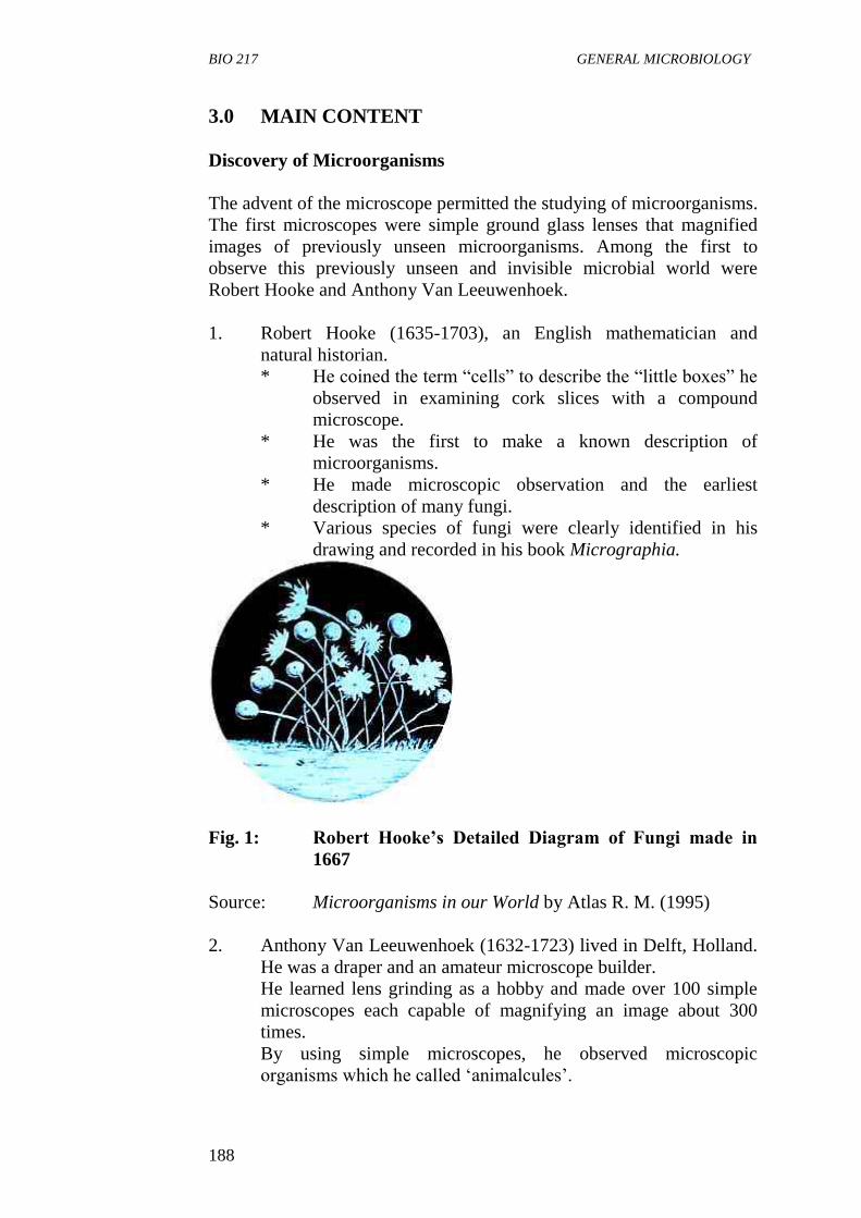
1. Robert Hooke (1635-1703), an English mathematician and
natural historian.
* He coined the term “cells” to describe the “little boxes” he
observed in examining cork slices with a compound
microscope.
* He was the first to make a known description of
microorganisms.
* He made microscopic observation and the earliest
description of many fungi.
* Various species of fungi were clearly identified in his
drawing and recorded in his book Micrographia.
Fig. 1: Robert Hooke’s Detailed Diagram of Fungi made in
1667
Source: Microorganisms in our World by Atlas R. M. (1995)
2. Anthony Van Leeuwenhoek (1632-1723) lived in Delft, Holland.
He was a draper and an amateur microscope builder.
He learned lens grinding as a hobby and made over 100 simple
microscopes each capable of magnifying an image about 300
times.
By using simple microscopes, he observed microscopic
organisms which he called ‘animalcules’.
BIO 217 MODULE 5
189
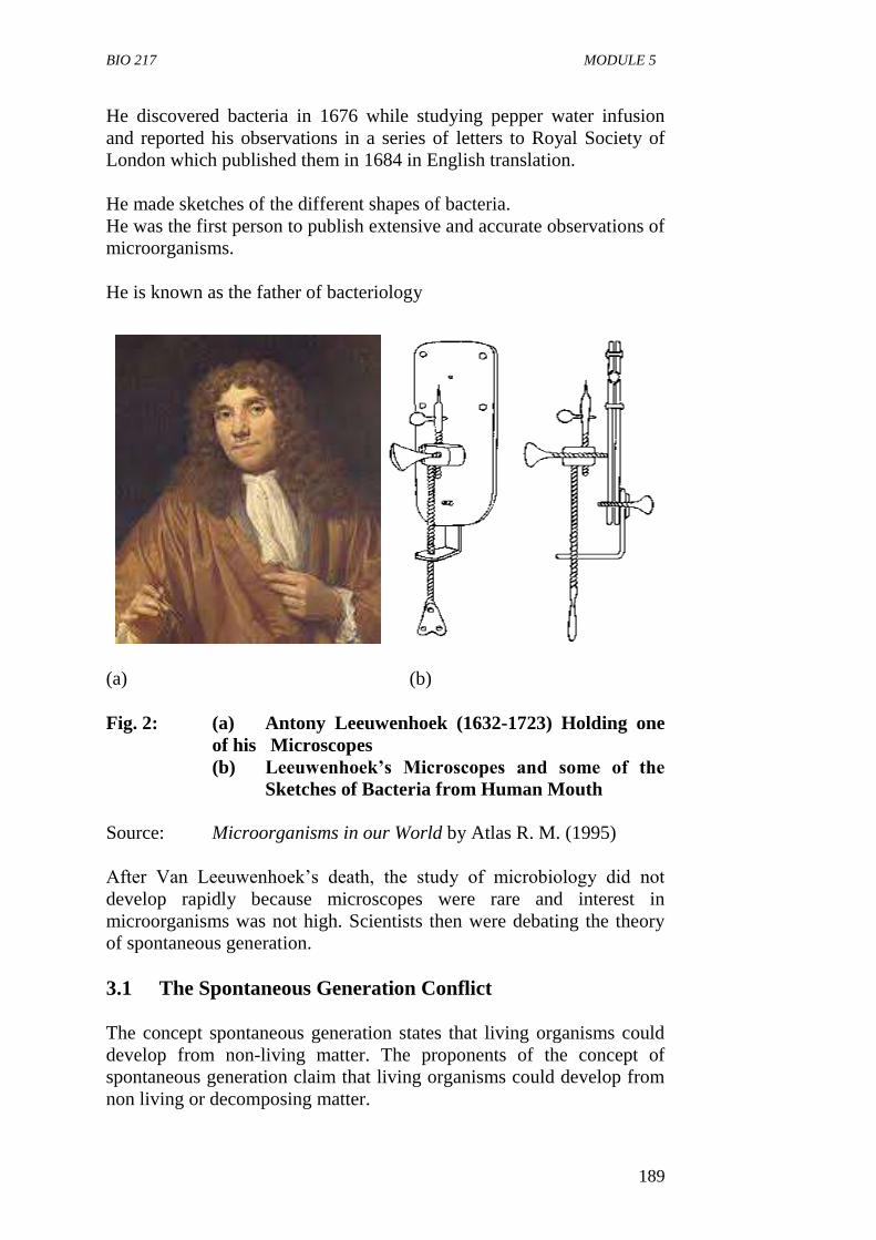
He discovered bacteria in 1676 while studying pepper water infusion
and reported his observations in a series of letters to Royal Society of
London which published them in 1684 in English translation.
He made sketches of the different shapes of bacteria.
He was the first person to publish extensive and accurate observations of
microorganisms.
He is known as the father of bacteriology
(a) (b)
Fig. 2: (a) Antony Leeuwenhoek (1632-1723) Holding one
of his Microscopes
(b) Leeuwenhoek’s Microscopes and some of the
Sketches of Bacteria from Human Mouth
Source: Microorganisms in our World by Atlas R. M. (1995)
After Van Leeuwenhoek’s death, the study of microbiology did not
develop rapidly because microscopes were rare and interest in
microorganisms was not high. Scientists then were debating the theory
of spontaneous generation.
3.1 The Spontaneous Generation Conflict
The concept spontaneous generation states that living organisms could
develop from non-living matter. The proponents of the concept of
spontaneous generation claim that living organisms could develop from
non living or decomposing matter.
BIO 217 GENERAL MICROBIOLOGY
190
1. Francesco Redi (1626-1697) challenged this concept by showing
that maggots on decaying meat came from fly eggs deposited on
the meat, and not from the meat itself.
He carried out a series of experiments on decaying meat
and its ability to produce maggot spontaneously.
He placed meat in three different containers, one was not
covered, and the second was covered with fine gauze to
exclude flies.
Flies laid eggs on the uncovered meat and maggots
developed.
The two other meats did not produce maggots.
Spontaneously, flies were attracted to the gauze-covered
container and laid their eggs on the gauze, these later
produced maggots. Hence, it become evident that the
generation of maggots resulted from the presence of fly
eggs and that meat (a non-living matter) did not
spontaneously generate maggots as previously believed.
2. Louis Jablot (1670) conducted an experiment in which he
divided a hay infusion that had been boiled into two containers: a
heated container that was closed to the air and a heated container
that was freely open to the air. Only the open vessel developed
microorganisms. This further helped to disprove abiogenesis.
3. John Needham (1713-1781) showed that mutton broth boiled in
flasks and then sealed could still develop microorganisms, which
supported the theory of spontaneous generation.
4. Lazzaro Spallanzani (1729-1799) showed that flasks sealed and
then boiled had no growth of microorganisms, and he proposed
that air carried germs to the culture medium. He also commented
that external air might be needed to support the growth of animals
already in the medium. The latter concept was appealing to
supporters of spontaneous generation.
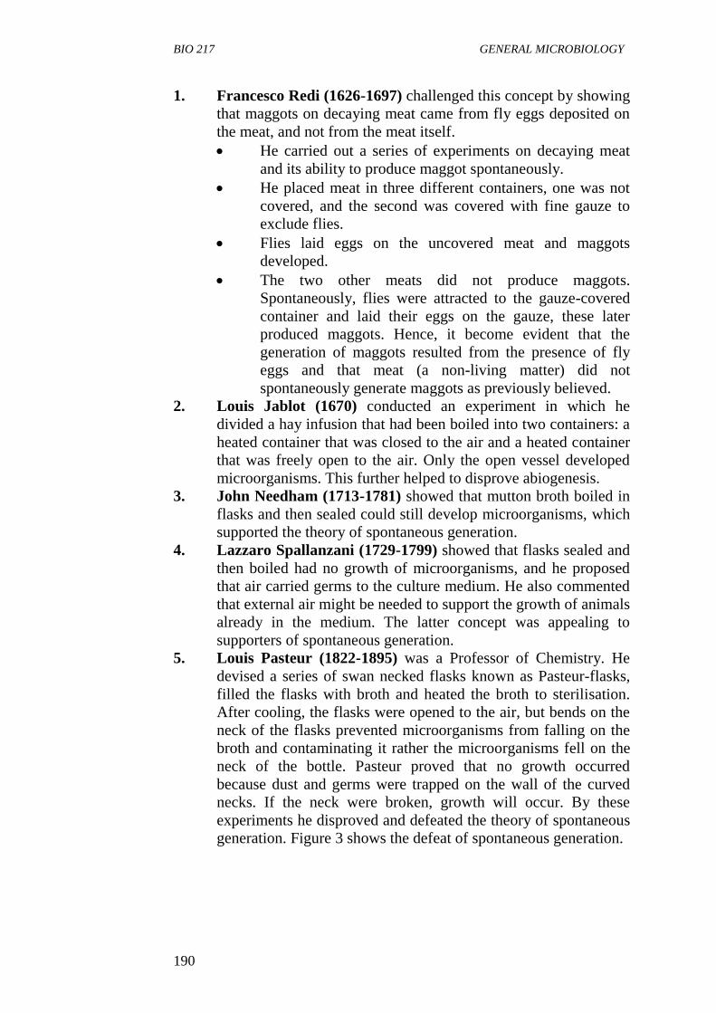
5. Louis Pasteur (1822-1895) was a Professor of Chemistry. He
devised a series of swan necked flasks known as Pasteur-flasks,
filled the flasks with broth and heated the broth to sterilisation.
After cooling, the flasks were opened to the air, but bends on the
neck of the flasks prevented microorganisms from falling on the
broth and contaminating it rather the microorganisms fell on the
neck of the bottle. Pasteur proved that no growth occurred
because dust and germs were trapped on the wall of the curved
necks. If the neck were broken, growth will occur. By these
experiments he disproved and defeated the theory of spontaneous
generation. Figure 3 shows the defeat of spontaneous generation.
BIO 217 MODULE 5
191
Fig. 3: The Defeat of Spontaneous Generation - Pasteur’s
Experiment with the Swan- Necked Bottles
Source: Amoebamike.wordpress.com
Apart from the defeat of the concept of spontaneous generation,
Pasteur’s work led to an effective sterilization method which
involve holding juices and milk at 62.8OC (145
OF) for 30 minutes
known as Pasteurization.
He discovered that alcoholic fermentation was catalyzed by
Living Yeast Cells.
He developed vaccines for the diseases anthrax, fowl cholera and
rabies between 1880 and 1890.
As a result of his research on rabies, he became a legend and the
French government built the Pasteur Institute in Paris in 1888. It
was originally established as a clinical centre for treating rabies,
but is now a major biomedical research centre for antiserum and
vaccine production.
He postulated the Germ Theory of Disease which states that
microorganisms are the cause of infectious diseases.
Pasteur’s work ushered in the Golden Age of Microbiology.
BIO 217 GENERAL MICROBIOLOGY
192
3.2 The Recognition of the Role of Microorganisms in
Disease
1. Agostino Bassi (1773-1856) showed that a silkworm disease was
caused by a fungus.
2. M. J. Berkerley (ca. 1845) demonstrated that the great potato
blight of Ireland was caused by a fungus.
3. Joseph Lister (1872-1912) developed a system of surgery
designed to prevent microorganisms from entering wounds. He
implemented the use of sterile surgical instrument, and used
carbolic acid (phenol) during surgery and on wound dressings.
4. Robert Koch (1843-1910)
Robert Koch was a German physician. He was the first to directly
prove the role of microorganisms in causing diseases. He
established the relationship between Bacillus anthracis and the
disease it causes, anthrax.
Using mice as experimental animals, he demonstrated that when
a small amount of blood from a diseased mouse was injected into
a healthy mouse, the healthy mouse quickly developed anthrax.
From this work he developed Koch’s postulates.
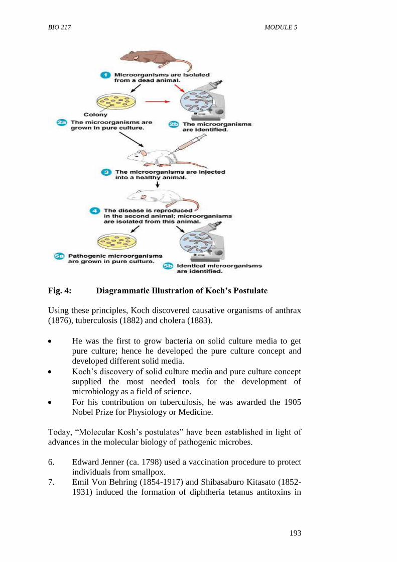
5. Koch’s postulates are
The suspected disease-causing organism should be present in all
cases of the disease and absent from healthy animals.
The suspected organism must be cultivated in a pure
culture away from the animal body.
The isolated organism must cause the disease when
inoculated into a healthy susceptible animal.
The organism must be re-isolated from these experimental
animals and culture again in the laboratory after which it
should still be the same as the original organism.
BIO 217 MODULE 5
193
Fig. 4: Diagrammatic Illustration of Koch’s Postulate
Using these principles, Koch discovered causative organisms of anthrax
(1876), tuberculosis (1882) and cholera (1883).
He was the first to grow bacteria on solid culture media to get
pure culture; hence he developed the pure culture concept and
developed different solid media.
Koch’s discovery of solid culture media and pure culture concept
supplied the most needed tools for the development of
microbiology as a field of science.
For his contribution on tuberculosis, he was awarded the 1905
Nobel Prize for Physiology or Medicine.
Today, “Molecular Kosh’s postulates” have been established in light of
advances in the molecular biology of pathogenic microbes.
6. Edward Jenner (ca. 1798) used a vaccination procedure to protect
individuals from smallpox.
7. Emil Von Behring (1854-1917) and Shibasaburo Kitasato (1852-
1931) induced the formation of diphtheria tetanus antitoxins in
BIO 217 GENERAL MICROBIOLOGY
194
rabbits which were effectively used to treat humans, thus
demonstrating humoral immunity.
3.3 The Discovery of Microbial Effects on Organic and
Inorganic Matter
1. Martinus Beijerinck (1851-1931)
Martinus Beijerinck was a Professor at the Delft
Polytechnic.
He isolated the first pure culture of many soil and aquatic
microorganisms, including sulphate reducing and sulfur
oxidizing bacteria, nitrogen fixing root nodule bacteria.
He described the first virus and the basic principles of
virology.
2. Sergei Winogradsky (1856-1953)
He proposed the concept of chemo-lithotrophy, (the
oxidation of inorganic matter). He worked with soil
bacteria and discovered they could oxidise iron, sulphur
and ammonia to obtain energy. He also studied anaerobic
nitrogen fixation and cellulose decomposition. He
published many scientific papers and a major monograph,
Microbiologic du sol (Soil Microbiology).
Beijerinck and Winogradsky pioneered the use of
enrichment cultures and selective media.
3.4 The Development of Microbiology in this Century
Microbiology established a closer relationship with other disciplines
during the 1940s because of its association with genetics and
biochemistry.
1. George W. Beadle and Edward L. Tatum (ca. 1941) studied the
relationship between genes and enzymes using the bread mould,
Neurospora.
2. Salvadore Lurai and Max Delbruck (ca. 1943) showed that
mutations were spontaneous and not directed by the environment.
3. Oswald T. Avery, Colin M. Mcleod, and Maclyn McCarty (1944)
provided evidence that deoxyribonucleic acid (DNA) was the
genetic material and carried genetic information during
transformation.
BIO 217 MODULE 5
195
3.5 Era of Molecular Microbiology
Began in the 1970s.
Advancement in the knowledge of bacterial physiology,
biochemistry and genetics.
Genetic manipulation which involves the transfer of DNA from
one organism into another or a bacterium and the proteins
encoded by the DNA harvested led to the development of the
field of Biotechnology.
DNA sequencing revealed the phylogenetic (evolutionary)
relationships among bacteria which led to revolutionary new
concepts in microbial systematic.
In 1990s, DNA sequencing gave birth to the field of genomics.
4.0 CONCLUSION
The study and development of microbiology as a field in science became
possible due to the works and contributions of men who discovered
microorganisms, disproved the theories of spontaneous generation and
established the relationship between microorganisms and disease among
other things.
5.0 SUMMARY
Robert Hooke (1635-1703) and Anthony Van Leeuwenhoek
(1632-1723) contributed to the discovery of microorganisms
through the use of microscope.
Experiment by Francesco Redi and others disproved the theory
of spontaneous generation.
Louis Pasteur defeated the theory of spontaneous generation.
Robert Koch developed postulate to establish relationship
between a suspected microorganism and disease.
Serge Winogradsky and Martinus Beijerinck discovered
microbial effect on organic and inorganic matter both of them
pioneered the use of enrichment culture and selective media.
George Beadle and others contributed to the development of
microbiology.
In the twentieth century, era of molecular microbiology began in
the 1970s and led to the field of biotechnology.
In the 1990s, DNA sequencing gave birth to the field of
genomics.
BIO 217 GENERAL MICROBIOLOGY
196
6.0 TUTOR-MARKED ASSIGNMENT
1. Explain Anthony Van Leeuwenhoek contribution to the discovery
of microorganisms.
2. (a) State the concept of spontaneous generation.
(b) Explain the steps involved in using Koch’s postulate to
establish the link between a suspected microorganism and
a disease.
7.0 REFERENCES/FURTHER READING
Atlas, R.M. (1995). Microorganisms in Our World. Mosby Year Book.
Inc.
Medigan, M.T. et al. (2009). Brock Biology of Microorganisms; 12th
Edition, Pearson Education Inc.
Pelczar, M.J., Chan, E.C.S. & Krieg, R.N. (2001). 5th
Edition.
Microbiology; McGraw-Hill.
Willey, J.M., Sherwood, L.M & Woolverton, C.J. (2008). Microbiology.
7th
Edition. Boston Bur Bridge, IL: McGraw-Hill Higher
Education.
amoebamike.wordpress.com
BIO 217 MODULE 5
197
UNIT 3 THE RELEVANCE AND SCOPE OF
MICROBIOLOGY
CONTENTS
1.0 Introduction
2.0 Objectives
3.0 Main Content
3.1 The Basic Aspects of Microbiology
3.2 The Applied Aspects of Microbiology
3.3 The Future of Microbiology
4.0 Conclusion
5.0 Summary
6.0 Tutor-Marked Assignment
7.0 References/Further Reading
1.0 INTRODUCTION
Modern microbiology is a large discipline with different specialised
areas. This is because the entire ecosystem depends on the activities of
microorganisms and microorganisms influence human society in
countless ways. Microbiology has a great impact on medicine,
agriculture, food science, ecology, genetics, biochemistry and other
fields. In this unit, we shall examine the different aspects of
microbiology and their relevance to human life.
2.0 OBJECTIVES
At the end of this unit, you should be able to:
define microbiology
state the two branches of microbiology
identify the different areas of study in basic and applied
microbiology.
3.0 MAIN CONTENT
Main Branches of Microbiology
Microbiology has two main branches:
1. Basic
2. Applied
Both branches intertwine and are complementary to each other.
BIO 217 GENERAL MICROBIOLOGY
198
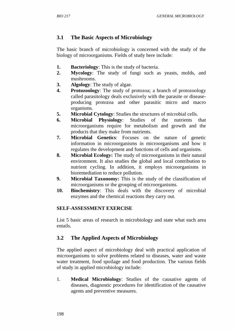
3.1 The Basic Aspects of Microbiology
The basic branch of microbiology is concerned with the study of the
biology of microorganisms. Fields of study here include:
1. Bacteriology: This is the study of bacteria.
2. Mycology: The study of fungi such as yeasts, molds, and
mushrooms.
3. Algology: The study of algae.
4. Protozoology: The study of protozoa; a branch of protozoology
called parasitology deals exclusively with the parasite or disease-
producing protozoa and other parasitic micro and macro
organisms.
5. Microbial Cytology: Studies the structures of microbial cells.
6. Microbial Physiology: Studies of the nutrients that
microorganisms require for metabolism and growth and the
products that they make from nutrients.
7. Microbial Genetics: Focuses on the nature of genetic
information in microorganisms in microorganisms and how it
regulates the development and functions of cells and organisms.
8. Microbial Ecology: The study of microorganisms in their natural
environment. It also studies the global and local contribution to
nutrient cycling. In addition, it employs microorganisms in
bioremediation to reduce pollution.
9. Microbial Taxonomy: This is the study of the classification of
microorganisms or the grouping of microorganisms.
10. Biochemistry: This deals with the discovery of microbial
enzymes and the chemical reactions they carry out.
SELF-ASSESSMENT EXERCISE
List 5 basic areas of research in microbiology and state what each area
entails.
3.2 The Applied Aspects of Microbiology
The applied aspect of microbiology deal with practical application of
microorganisms to solve problems related to diseases, water and waste
water treatment, food spoilage and food production. The various fields
of study in applied microbiology include:
1. Medical Microbiology: Studies of the causative agents of
diseases, diagnostic procedures for identification of the causative
agents and preventive measures.
BIO 217 MODULE 5
199
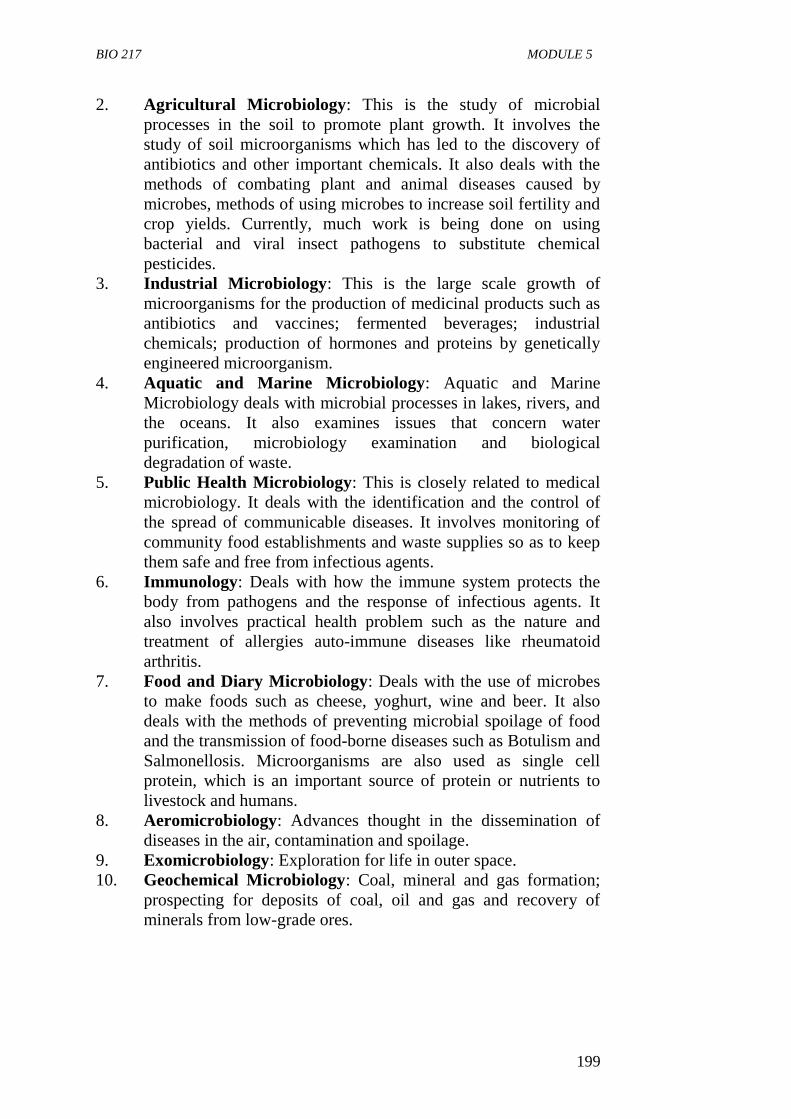
2. Agricultural Microbiology: This is the study of microbial
processes in the soil to promote plant growth. It involves the
study of soil microorganisms which has led to the discovery of
antibiotics and other important chemicals. It also deals with the
methods of combating plant and animal diseases caused by
microbes, methods of using microbes to increase soil fertility and
crop yields. Currently, much work is being done on using
bacterial and viral insect pathogens to substitute chemical
pesticides.
3. Industrial Microbiology: This is the large scale growth of
microorganisms for the production of medicinal products such as
antibiotics and vaccines; fermented beverages; industrial
chemicals; production of hormones and proteins by genetically
engineered microorganism.
4. Aquatic and Marine Microbiology: Aquatic and Marine
Microbiology deals with microbial processes in lakes, rivers, and
the oceans. It also examines issues that concern water
purification, microbiology examination and biological
degradation of waste.
5. Public Health Microbiology: This is closely related to medical
microbiology. It deals with the identification and the control of
the spread of communicable diseases. It involves monitoring of
community food establishments and waste supplies so as to keep
them safe and free from infectious agents.
6. Immunology: Deals with how the immune system protects the
body from pathogens and the response of infectious agents. It
also involves practical health problem such as the nature and
treatment of allergies auto-immune diseases like rheumatoid
arthritis.
7. Food and Diary Microbiology: Deals with the use of microbes
to make foods such as cheese, yoghurt, wine and beer. It also
deals with the methods of preventing microbial spoilage of food
and the transmission of food-borne diseases such as Botulism and
Salmonellosis. Microorganisms are also used as single cell
protein, which is an important source of protein or nutrients to
livestock and humans.
8. Aeromicrobiology: Advances thought in the dissemination of
diseases in the air, contamination and spoilage.
9. Exomicrobiology: Exploration for life in outer space.
10. Geochemical Microbiology: Coal, mineral and gas formation;
prospecting for deposits of coal, oil and gas and recovery of
minerals from low-grade ores.
BIO 217 GENERAL MICROBIOLOGY
200
SELF-ASSESSMENT EXERCISE
State the importance of microbiology in five different fields of human
endeavours.
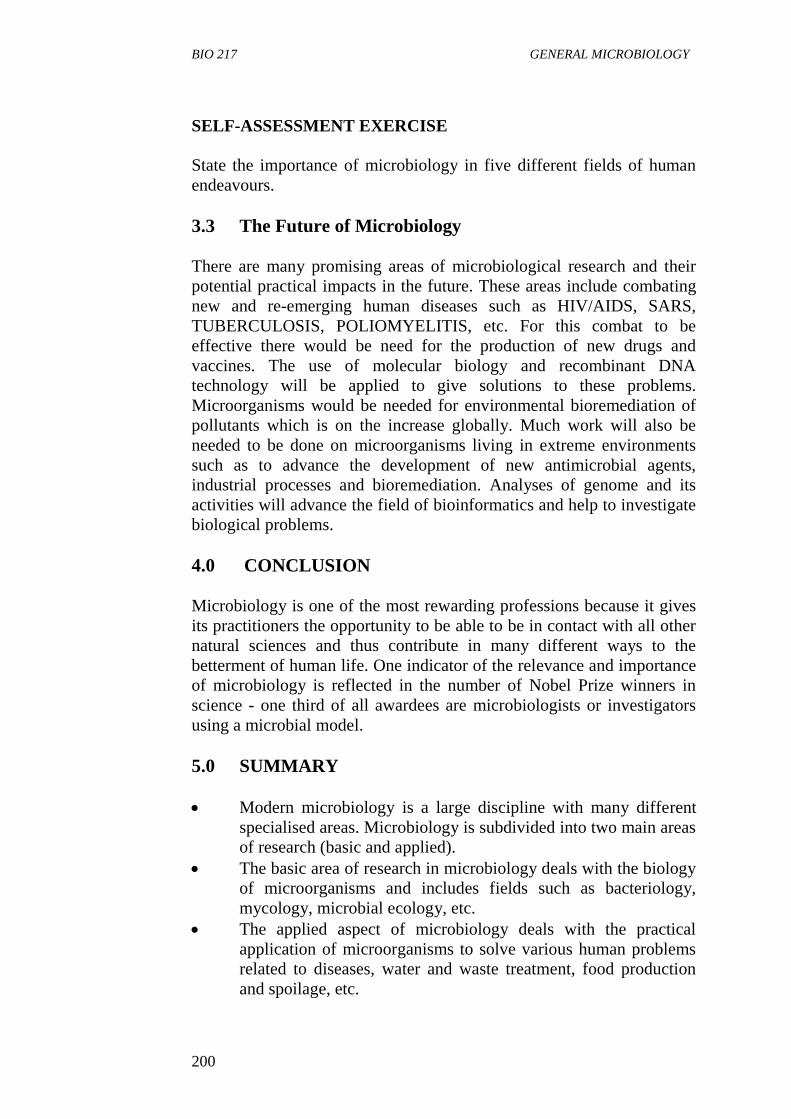
3.3 The Future of Microbiology
There are many promising areas of microbiological research and their
potential practical impacts in the future. These areas include combating
new and re-emerging human diseases such as HIV/AIDS, SARS,
TUBERCULOSIS, POLIOMYELITIS, etc. For this combat to be
effective there would be need for the production of new drugs and
vaccines. The use of molecular biology and recombinant DNA
technology will be applied to give solutions to these problems.
Microorganisms would be needed for environmental bioremediation of
pollutants which is on the increase globally. Much work will also be
needed to be done on microorganisms living in extreme environments
such as to advance the development of new antimicrobial agents,
industrial processes and bioremediation. Analyses of genome and its
activities will advance the field of bioinformatics and help to investigate
biological problems.
4.0 CONCLUSION
Microbiology is one of the most rewarding professions because it gives
its practitioners the opportunity to be able to be in contact with all other
natural sciences and thus contribute in many different ways to the
betterment of human life. One indicator of the relevance and importance
of microbiology is reflected in the number of Nobel Prize winners in
science - one third of all awardees are microbiologists or investigators
using a microbial model.
5.0 SUMMARY
Modern microbiology is a large discipline with many different
specialised areas. Microbiology is subdivided into two main areas
of research (basic and applied).
The basic area of research in microbiology deals with the biology
of microorganisms and includes fields such as bacteriology,
mycology, microbial ecology, etc.
The applied aspect of microbiology deals with the practical
application of microorganisms to solve various human problems
related to diseases, water and waste treatment, food production
and spoilage, etc.
BIO 217 MODULE 5
201
The field of microbiology will be faced with many important
future challenges such as finding new ways to new and re-
emerging diseases, reduced environmental pollution and
investigating biological problems.
6.0 TUTOR-MARKED ASSIGNMENT
1. List the fields of microbiology that deal with the following:
a. Metabolism
b. Enzymology
c. Nucleic Acid and Protein Synthesis
d. Microorganisms in the Natural Environment
e. Microbial Classification
f. Microbial Cell Structure
2. Explain what the field of agricultural microbiology entails.
7.0 REFERENCES/FURTHER READING
Atlas, R.M. (1995). Microorganisms in Our World. Mosby Year Book.
Inc.
Medigan, M.T et al. (2009). Brock Biology of Microorganisms. 12th
Edition. Pearson Education Inc.
Pelczar, M.J., Chan, E.C.S & Krieg, R.N. (2001). Microbiology. 5th
Edition. McGraw-Hill,
Willey, J.M., Sherwood, L.M & Woolverton, C.J. (2008). Microbiology.
(7th
ed.). Boston Bur Bridge, IL: McGraw-Hill Higher Education.
BIO 217 GENERAL MICROBIOLOGY
202
UNIT 4 MICROSCOPE AND SPECIMEN
PREPARATION
CONTENTS
1.0 Introduction
2.0 Objectives
3.0 Main Content
3.1 The Light Microscope
3.1.1 The Bright Field Microscope
3.1.2 The Dark-Field Microscope
3.1.3 The Phase-Contrast Microscope
3.1.4 The Fluorescent Microscope
3.2 Microscope Resolution
3.3 Preparation for Light-Microscope Examination
3.3.1 The Wet Mount or Hanging Drop Technique
3.3.2 Fixed, Stained Smears of Microorganisms
3.3.3 Fixation
3.4 Staining of Specimens
3.5 Electron Microscope
3.5.1 The Transmission Electron Microscope
3.5.2 The Scanning Electron Microscope
4.0 Conclusion
5.0 Summary
6.0 Tutor-Marked Assignment
7.0 References/Further Reading
1.0 INTRODUCTION
Microbiology is the study of organisms too small to be seen distinctly
with the unaided eyes. The nature of this discipline makes the
microscope of crucial importance because the study of microorganisms
is impossible without the microscope. Microscopes provide
magnification which enables us to see microorganisms and study their
structures. The magnification attained by microscopes range from x100
to x400,000 in addition there are different types of microscopes and
many techniques have been developed by which specimens of
microorganisms can be prepared for examination. This unit examines
the different types of microscopes, how the microscopes work and how
specimens are prepared for examination.
BIO 217 MODULE 5
203
2.0 OBJECTIVES
At the end of this unit, you should be able to:
define the term microscope
state the two categories of microscope
describe the bright field microscope
explain the resolving power
describe methods of preparing and staining specimens; and
describe the scanning electron microscope and the transmission
electron microscope.
3.0 MAIN CONTENT
The Microscope
A microscope is an instrument for producing enlarged images of objects
too small to be seen unaided.
Types of Microscopes
Microscopes are of two types:
Light (optical) and electron depending on the principle on which
magnification is done.
3.1 The Light Microscope
This is a type of microscope in which magnification is obtained by a
system of optical lenses using light waves. It includes:
1. Bright Field Microscope
2. Dark Field Microscope
3. Fluorescence Microscope
4. Phase Contract Microscope
Modern microscopes are compound microscopes. That is, the magnified
image formed by the objective lens is further enlarged by one or more
additional lenses.
Most undergraduate students of microbiology perform most of their
examinations with the bright field microscope which is the most widely
used instrument for routine microscopic work. The other types of
microscope are used for special purposes or research investigation.
BIO 217 GENERAL MICROBIOLOGY
204
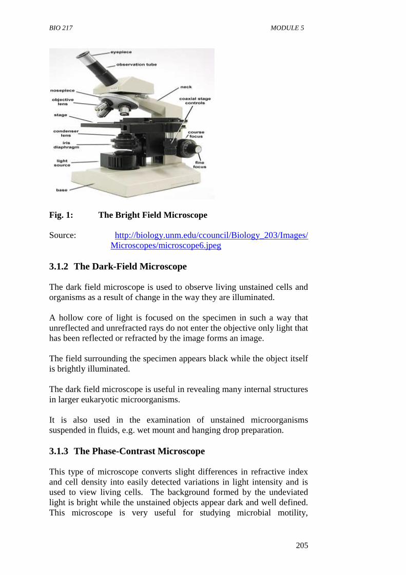
3.1.1 The Bright Field Microscope
The ordinary microscope is called a bright field microscope
because it forms a dark image against a brighter background.
The microscope consists of a sturdy metal body or stand made up
of a base and an arm to which the remaining parts are attached.
A light source, either a mirror or an electric illuminator, is located
at the base.
Two focusing knobs, the fine and coarse adjustment knobs are
located on the arm and can move either the stage or the nose
piece to focus the image.
The stage is positioned about halfway up the arm and hold
microscope slides by slide clips or a mechanical stage clip.
There is a substage condenser mounted within or beneath the
stage which focuses a core of light on the slide.
The upper part of arm of the microscope holds the body assembly
to which a nose piece and one or more eyepieces or ocular lenses
are attached.
Most advanced microscopes have eyepieces for both eyes and are
called binocular microscopes.
The nose piece holds three to five objective lenses of different
magnifying power and is easily rotated to position any objective.
The image you see when viewing a specimen is focused by the
objective and ocular lenses working together.
Light from the specimen which has been illuminated is focused
by the objective lens creating an enlarged image within the
microscope. The ocular lens further magnifies this primary
image.
The total magnification is calculated by multiplying the objective
and eye piece magnification together; e.g. if a 45x objective is
used with a 10x eyepiece, the overall magnification of the
specimen will be 450x.
BIO 217 MODULE 5
205
Fig. 1: The Bright Field Microscope
Source: http://biology.unm.edu/ccouncil/Biology_203/Images/
Microscopes/microscope6.jpeg
3.1.2 The Dark-Field Microscope
The dark field microscope is used to observe living unstained cells and
organisms as a result of change in the way they are illuminated.
A hollow core of light is focused on the specimen in such a way that
unreflected and unrefracted rays do not enter the objective only light that
has been reflected or refracted by the image forms an image.
The field surrounding the specimen appears black while the object itself
is brightly illuminated.
The dark field microscope is useful in revealing many internal structures
in larger eukaryotic microorganisms.
It is also used in the examination of unstained microorganisms
suspended in fluids, e.g. wet mount and hanging drop preparation.
3.1.3 The Phase-Contrast Microscope
This type of microscope converts slight differences in refractive index
and cell density into easily detected variations in light intensity and is
used to view living cells. The background formed by the undeviated
light is bright while the unstained objects appear dark and well defined.
This microscope is very useful for studying microbial motility,
BIO 217 GENERAL MICROBIOLOGY
206
determining the shape of living cells and detecting some bacterial
components such as endospores and inclusion bodies. It is also used in
studying eukaryotes.
3.1.4 The Fluorescent Microscope
This type of microscope exposes a specimen to ultraviolet, violet or blue
light and forms an image of the object with resulting fluorescent light.
The most commonly used fluorescence microscope light is
epifluorescence microscope which is also called incident light or
reflected light microscope. Epifluoresence microscope employs an
objective lens that also acts as a condenser. A mercury vapor arc lamp or
other source produces an intense beam of light that passes through an
exciter filler. The exciter filler transmits on the desired wavelength of
excitation light
The excitation light is directed down the microscope by a speed minor
called the dichromatic minor. This minor reflects light of shorter
wavelength but allows light of longer wavelength to pass through. The
excitation light continues down through the objective lens to specimen
stained with spaced dye molecules called fluorochromes.
3.2 Microscope Resolution
Resolution is the ability of a lens to separate or distinguish between
small objects that are close together, i.e. the microscope must produce a
clear image and not just a magnified one. It is also known as the
resolving power.
Resolution is described mathematically by an equation in the 1870s by
Ernest Abbe, a German physicist. The Abbe equation states that the
minimal distance (d) between two objects that reveal them as separate
entities depends on the wavelength of light () used to illuminate the
specimen and on the numerical aperture of the lens (nsin) which is the
ability of the lens to gather light.
d = 0.5
nsin
as d becomes smaller, the resolution increases and finer details can be
discerned in a specimen; d becomes smaller as the wavelength of light
used decreases and as the numerical aperture (NA) increases.
Hence, the greatest resolution is obtained using a lens with the largest
NA and light with the shortest wavelength.
BIO 217 MODULE 5
207
The relationship between NA and resolution can be expressed as
follows:
d = .
2NA
where d = resolution and = wavelength of light. Using the values 1.3
for NA and 0.5m, the wavelength of green light, for > , resolution can
be calculated as
d = 0.55 = 0.21m.
2 x 1.30
From these calculations, we may conclude that the smallest details that
can be seen by the light microscope are those having dimensions of
approximately 0.2m.
SELF-ASSESSMENT EXERCISE
i. Define the resolving power.
ii. List 5 parts of a light microscope and state the function of each.
3.3 Preparation for Light-Microscope Examination
There are two general methods used for preparing specimens for light-
microscope examination.
i The organisms are suspended in a liquid (the wet-mount or the
hanging drop technique), and
ii The organism is dried fixed and stained before observing under
the microscope.
3.3.1 The wet mount or hanging drop technique
The technique permits examination of organisms in a normal living
condition. A wet mount is made by placing a drop of fluid containing
the organisms on a glass slide and covering the drop with a cover slip.
Petroleum jelly may be used to provide a seal between the slide and
covers slip after which the slide is viewed under the microscope.
This method is desirable because,
it prevents distortion of the morphology of spiral bacteria when
they are stained and dried.
it reveals whether organisms are motile or not.
some cell inclusion bodies are easily observed.
BIO 217 GENERAL MICROBIOLOGY
208
spore formation and germination may also be observed in living
cells.
3.3.2 Fixed, Stained Smears of Microorganisms
These are frequently used for the observation of the morphological
characteristics of bacteria. The procedure makes the cell more clearly
visible, and differences between cells of different species and within the
same species can be demonstrated. The essential steps in this procedure
are:
1. preparation of the film or smear
2. fixation and
3. application of one or more staining solution.
3.3.3 Fixation
Fixation is the process by which the internal and external structures of
cells and microorganisms are preserved and fixed in position. It in-
activates enzymes that might disrupt cell morphology and tough cell
structures so that they do not change during staining and observation. A
microorganism usually is killed and attached firmly to the microscope
slide during fixation.
There are two fundamentally different types of fixation.
1. Heat Fixation: Is routinely used to observe prokaryotes.
Typically, a film of cells (a smear) is gently heated as a slide is
passed through a flame. Heat fixation preserves overall
morphology but not structures within cells.
2. Chemical Fixation: Is used to protect fine cellular sub-structure
and the morphology of larger, more delicate micro organisms.
Chemical fixatives penetrate cells and react with cellular
components, usually proteins and lipids, to render them inactive,
insoluble, and immobile. Common fixative mixtures contain such
components as ethanol, acetic acid, mercuric chloride,
formaldehyde, and glutaraldehyde.
3.4 Staining of Specimens
Although living microorganisms can be directly examined with the light
microscope, they often must be fixed and stained to increase visibility,
accentuate specific morphological features, and preserve them for future
study.
BIO 217 MODULE 5
209
Types of Staining
Simple staining
This is a kind of staining in which a single stain or dye is used.
Basic dyes such as crystal violet, methylene blue, and
carbolfuchsin are used in simple staining to determine the size,
shape and arrangement of prokaryotic acids.
Differential staining
These are staining procedures that make visible the differences
between bacterial cells or part of a bacterial cell. It usually
involves more than one dye used for staining.
Gram staining
The Gram stain was developed in 1884 by the Danish physician
Christian Gram. It is the most widely used differential staining
procedure.
The steps involved are as follows:
i The smear is stained with the crystal violet (which is the primary
stain).
ii This followed by treatment with iodine functioning as a mordant.
iii The smear is decolourised by washing with ethanol or acetone.
iv The smear is counterstained with a simple dye safranin.
Bacteria stained by the Gram stain method fell into two groups:
Gram positive bacteria which retain the crystal violet and appear
deep violet in colour and Gram negative bacteria which, lose the
crystal violet and are counterstained with safranin appear red in
colour.
Acid fast staining
This is another differential staining procedure commonly used to
identify Mycobacterium tuberculosis and Mycobacterium leprae, the
pathogens responsible for tuberculosis and leprosy respectively.
These bacteria have cell walls with high lipid content in particular,
mycolic acid which prevents dye from readily binding to the cells.
In the acid fast staining procedure, the red stain and carbol fuchsin is
used as primary stain; next acid-alcohol is used as a decolouriser. The
acid-alcohol will remove the red stain form bacteria such as Escherichia
coli which the acid fast mycobacteria will remain red.
BIO 217 GENERAL MICROBIOLOGY
210
3.5 Electron Microscope
This type of microscope uses a beam of electron in place of light waves
to produce the image. There are two types:
scanning electron microscope
transmission electron microscope.
3.5.1 The Transmission Electron Microscope
Electron microscopes use a beam of electrons to illuminate and create
magnified images of specimens. Electrons replace light as the
illuminating beam. They can be focused, much as light is in a light
microscope, but their wavelength is around 0.005mm approximately
1000,000 times shorter than that of visible light. Therefore, electron
microscopes have a practical resolution roughly 1,000 times better than
the light microscope, with many electron microscopes point closer than
0.5nm can be distinguished, and the useful magnification is well over
100,000x. In transmission electron microscope, the electron beam is
transmitted through the specimen.
3.5.2 The Scanning Electron Microscope
The scanning electron microscope produces an image from electron
released from atoms on an object’s surface. It has been used to examine
the surfaces of microorganisms in great detail. Many SEM has a
resolution of 7nm or less.
SELF-ASSESSMENT EXERCISE
Differentiate between the transmission electron microscope and
scanning electron microscope.
4.0 CONCLUSION
Scientific observation is an important part of scientific study.
Microbiology as a field of science would not have developed without the
necessary instruments such as the microscope and the methods used for
observing the microorganism.
BIO 217 MODULE 5
211
5.0 SUMMARY
In light microscope, magnification is obtained by a system of optical
lenses using light waves. Many types of light microscopes have been
developed. They include bright fields, dark field, phase contrast and
fluorescence microscope.
Electron microscope uses a beam of electron in place of light
waves to produce the image of an object.
The ordinary compound microscope is called the bright field
microscope because if forms a dark image against a bright
background.
In the bright field microscope which is a compound the primary
image is formed by an objective lens and enlarged by the eye
piece or ocular lens to form the final image.
The dark field microscope uses only refracted light to form an
image and objects glow against a black background.
The dark field microscope is useful in revealing many internal
structures in larger eukaryotic microorganism.
The phase-contrast microscope converts slight differences in
refractive index and cell density into easily detected variations in
light intensity and is used to view living cells, for studying
microbial motility and detecting some bacteria components such
as endspores.
The fluorescent microscope exposes a specimen to ultraviolet,
violet or blue light and forms an image of the object with
resulting fluorescent light.
Two general methods for preparing specimens for light
microscope examination are the wet mount or the hanging drop
technique and the dried fixed stained technique.
A wet mount is made by placing a drop of fluid containing the
organisms on a glass slide and covering the drop with a cover slip
before viewing under the microscope.
Fixation is a process by which the internal and external structures
of cells and microorganisms are preserved and fixed in a position.
It involves preparation of the smear, fixing with heat or chemical
and application of one or more staining solutions.
Electron microscopes use a beam of electrons to illuminate and
create magnified images of specimens.
Simple staining is a kind of staining in which a single stain or dye
such as methylene and crystal violet is used.
Differential staining involves the use of more than one stain or
dye is used to make visible the differences between bacterial cells
or part of a bacterial cell examples are the Gram stain.
BIO 217 GENERAL MICROBIOLOGY
212
6.0 TUTOR-MARKED ASSIGNMENT
1. a What is microscope resolution?
b List the stages involved in preparing a specimen for
observation under the light microscope.
2. a Describe the bright field microscope.
b What is the basic difference between a transmission
electron microscope and a scanning electron microscope?
7.0 REFERENCES/FURTHER READING
Atlas, R.M. (1995). Microorganisms in our World. Mosby Year Book.
Inc.
Medigan, M.T., et al. (2009). Brock Biology of Microorganisms. 12th
Edition. Pearson Education Inc.
Pelczar, M.J., Chan, E.C.S. & Krieg, R.N. (2001). Microbiology. (5th
ed.).McGraw-Hill.
Prescott, Harley & Kleins. Microbiology. (7th
ed.). Boston Bur Bridge,
IL: McGraw-Hill Higher Education.
http://biology.unm.edu/ccouncil/Biology_203/Images/
Microscopes/microscope6.jpeg
BIO 217 MODULE 5
213
UNIT 5 A BRIEF SURVEY OF MICROBES AS FRIENDS
AND FOES
CONTENTS
1.0 Introduction
2.0 Objectives
3.0 Main Content
3.1 Microorganisms and Food Production
3.2 Production of Pharmaceuticals
3.3 Vitamins
3.4 Production of Organic Acids
3.5 Hygiene
3.6 Energy Production
3.7 Useful in the Study of Science
3.8 Recovery of Metals from their Ores
3.9 Microorganisms and Agriculture
3.10 Microorganisms and the Environment
3.11 Sewage Treatment
3.12 Microorganisms as Foes
3.13 Microorganisms as Diseases Agents
3.14 Microorganisms as Agents of Warfare and Terrorism
4.0 Conclusion
5.0 Summary
6.0 Tutor-Marked Assignment
7.0 References/Further Reading
1.0 INTRODUCTION
Microorganisms occur in large numbers of most natural environments
and bring about many changes. Some are desirable and others are
undesirable. Microorganisms affect the well being of people in many
ways. Many are beneficial to man and can be called ‘friends’ while
some are harmful and can be regarded as ‘foes’ to man. The beneficial
impact of microorganisms ranges from the production of goods and
pharmaceutical products, to enhancement of soil fertility, environmental
cleanup while their harmful effect can be seen in their ability to cause
disease in man, animals and plants as well as their usage in biological
warfare. However, there are more species of microorganisms that
perform friendly and beneficial functions than those that harm other
living organisms. This unit gives us a brief survey of microorganisms as
friends and foes.
BIO 217 GENERAL MICROBIOLOGY
214
2.0 OBJECTIVES
At the end of this unit, you should be able to:
explain the different ways in which microorganisms can act as
friends to man
explain ways in which microorganisms can act as foes to man.
3.0 MAIN CONTENT
Microorganisms as Friends
Microorganisms have found application in various aspects of life.
They are useful in food industries to produce many food substances, in
medicine to produce vaccines and antibiotics, in environmental
protection, and in agriculture, to optimise yield.
3.1 Microorganisms and Food Production
Many microorganisms are used to produce many of the foods and
beverages we consume. Microbially-produced food products have
properties that are very different from those of the starting
materials. Most of these food products are produced by
fermentation.
Fermentation is the chemical transformation of organic
compounds carried out by microorganisms and their enzymes. In
industrial fermentation, raw materials (substrate) are converted
by microorganisms in a controlled favourable environment
(created in a fermentor) to form a desired end product substance.
The accumulation of fermentation products such as ethanol and
lactic acid produces characteristic flavours and other desirable
properties in food substances.
Pickles and some sausages are also produced by fermentation
processes.
Microorganisms are used to produce fermented dairy products
such as cheese, yoghurt and acidophilus milk.
They are also used to produce alcoholic beverages such as beer
by conversion of sugar to alcohol and carbon dioxide.
Wine fermented from fruits using yeast strains Saccharomyces
cerevisiae and bread is also produced by using yeasts.
Microorganisms can also be used as direct source of food known
as single cell protein. Various species of yeasts, algae are grown
as single cell protein and use as animal feeds thus helping to meet
the world food needs.
BIO 217 MODULE 5
215
3.2 Production of Pharmaceuticals
Microorganisms are used to produce different pharmaceuticals such as
antibiotics, steroids vitamins, hormones, etc. Antibiotics are microbially
produced substances or substances synthetically derived from natural
sources that inhibit or kill microorganisms, Steroids regulate various
aspects of human metabolisms and are produced by organisms such
Rhizopus nigricans.
Vaccines are produced using microorganisms with the antigenic
properties to elicit a primary immune response; they are used to prevent
many once deadly diseases such as polio, small pox, tuberculosis,
measles, diphtheria and whooping cough.
3.3 Vitamins
Vitamins are essential animal nutritional factors; some vitamins are
produced by microbial fermentation, e.g. Vitamin B12 by Streptomyces,
B12 by Pseudomonas denitrificans and Propionibacterium shermanni.
Riboflavin produced by various species of Clostridium and Ashbya
gossypii.
Human insulin and human growth hormone are produced by genetically
engineered bacteria.
3.4 Production of Organic Acids
Various organic acids are produced by microorganisms examples are:
1. Gluconic acid: used as a pharmaceutical to supply calcium to the
body by several fungi including Penicillium and Aspergillus
species.
Citric acid produced by Aspergillus niger and used as a food
additive especially in the production of soft drinks.
2. Gibberellic acid: a plant hormone is formed by the fungus.
Gibberella fujikuroi. It is used as growth promoting substances
to stimulate plant growth flowering and seed germination.
3. Lactic acid by different lactic acid bacteria for example,
Lactobacillus delbrueckii, lactic and is used in foods as
preservatives, in leather production for deliming hides and in the
textile industry for fabric treatment, plastics making in baking
powders.
BIO 217 GENERAL MICROBIOLOGY
216
3.5 Hygiene
i Hygiene is the avoidance of infection and food spoilage by
eliminating microorganisms from the surrounding.
ii Our knowledge of how disease causing microorganisms spread
has permitted us to reduce the incidence of many diseases. Also
improved sanitation practices have helped to reduce the incidence
of diseases.
iii Microorganisms from the surroundings can be totally removed by
methods such as sterilization or reduced to acceptable levels
using methods such as disinfection and antisepsis. In food
preparation, microbes are reduced to acceptable levels using
methods such as pasteurization, addition of vinegar. While
complete sterility is achieved by autoclaving or irradiation.
3.6 Energy Production
Microorganisms play major roles in energy production. Microbes are
used in fermentation to produce ethanol and in biogas reactions to
produce methane using various forms of agricultural and urban wastes.
Microbial production of synthetic fuels acts as an alternative fuel
resources to petroleum.
The microbial production of ethanol from sugarcane or cornstarch has
become an important source of a valuable fuel, particularly in areas of
the world that have abundant supplies of plant residues such as Brazil
and is becoming popular in the United States.
The bacteria Zymomonas mobilis and Thermoanaerobacter ethanolicus
and different yeast strains are used for product of ethanol.
Methane (natural gas) is produced by methanogenic bacteria is another
important natural renewable energy sources.
Methane can be used for the generation of mechanical, electrical and
heat energy.
3.7 Useful in the Study of Science
Microbes are essential tools in biotechnology, biochemistry, genetics
molecular biology and genomics. Examples are the yeasts
(Saccharomyces cerevisiae) and fission yeast (Shizosaccharomyces
pombe) which are model organisms in science. They can easily be
grown rapidly in large quantities and are easily manipulated.
Biotechnology uses genetic engineering which is the artificial
manipulation of genes and gene products.
BIO 217 MODULE 5
217
Genes from any source can be manipulated and modified using
microorganisms and their enzymes as molecular tools, e.g. human
insulin, a hormone which is very low in people with diabetes is
produced by genetically engineered bacteria into which human genes
have been inserted.
3.8 Recovery of Metals from their Ores
Microorganisms are used to recover metals from their ores by the
process of bioleaching. Bioleaching uses microorganisms to alter the
physical or chemical properties of a metallic ore so that the metal can be
extracted. The bacteria Thiobacillus feroxidans recover copper and
uranium from their ores.
3.9 Microorganisms and Agriculture
Agriculture depends in many ways on the activities of
microorganisms. Microorganisms help in nitrogen fixation used
by plants for growth.
In terrestrial habitats, the microbial fixation of atmospheric
nitrogen is carried out by free living bacteria such as Rhizobium
and Bradyrhizobium living in symbiotic association with plants.
Legumes live in close association with bacteria that form
structures called nodules on their roots.
These bacteria in the root nodules of the legumes, convert
atmospheric N2 fixed into Nitrogen (NH3) that plants use for
growth and eradicates the need for chemical fertilizers.
Microorganisms in the rumen of ruminant animals such as cattle
and sheep also help in the digestion of cellulose present in grasses
on which they feed on.
Microorganisms help in the cycling of nutrients such as carbon,
nitrogen and sulphur which are needed to maintain ecological
balance.
Microorganisms are also used as biological control agents.
Fungi, bacteria and viruses can be used as bioinsecticides or
biospesticides, e.g. Bacillus thuringensis.
Microbial activities in soil and water convert these elements to
forms that are readily assimilated by plants.
3.10 Microorganisms and the Environment
a. Microorganisms can be used to clean up pollution created by
human activities in a process called bioremediation.
BIO 217 GENERAL MICROBIOLOGY
218
b. Pollutants such as pesticides, spilled oil solvents which could
pose human health hazard are degraded to nontoxic substances by
microorganisms.
c. Microorganisms are used to degrade wastes and pollutants so as
to maintain and restore environmental quality.
3.11 Sewage Treatment
Microorganisms are also used in sewage treatment. Specially cultured
microbes are used in the biological treatment of sewage and industrial
waste effluent in a process known as bioaugmentation. These microbes
help to get rid of waste materials which could have accumulated in the
environment.
SELF-ASSESSMENT EXERCISE
i. State the beneficial role of microorganisms in:
a. Agriculture
b. Environmental Protein
c. Food Production.
3.12 Microorganisms as Foes
Microorganism can act as foes to man and other living organisms by
causing diseases and by their usage as biological weapons.
3.13 Microorganisms as Disease Agents
Microbial diseases are still the major cause of death in many developing
countries. Microorganisms cause different diseases in man such as:
AIDS (Acquired Immune Deficiency Syndrome) caused by the
Human Immuno Deficiency Virus (HIV).
Tuberculosis caused by a bacterium, Mycobacterium
tuberculosis.
Cholera caused by a bacteria Vibrio cholera.
Malaria caused by four species of the Protozoa called
Plasmodium transmitted by the female anopheles mosquito.
Other emerging diseases include: bird flu and swine flu.
Microorganisms are also agents of diseases of plants and animals of
agricultural importance. These microbial diseases of plants and animals
BIO 217 MODULE 5
219
cause major economic losses in the agriculture industry and to the
world.
3.14 Microorganisms as Agents of Warfare and Terrorism
Use in Biological Warfare
Biological warfare is also known as germ warfare. It is the use of
pathogens such as viruses, bacteria, or the toxins produced by them as
biological weapons or agents of warfare.
A biological weapon may be used to kill, incapacitate or seriously
impair a person, group of people or even an entire population. It can be
used as a military technique by nations during wars.
There are four kinds of biological warfare agents, bacteria, viruses, fungi
and rickettsiaes. They are living organisms that reproduce with their host
victims who then become contagious with a deadly if weakening
multiple effects.
Toxins on the other hand do not reproduce in the victims but within a
short incubation period (usually with a few hours) kill the victims.
SELF-ASSESSMENT EXERCISE
Explain ways in which microorganism can act as foes to man.
4.0 CONCLUSION
Through the activities of microorganism we have received many
benefits such as production of foods with improved qualities, improved
agricultural crop yields, maintained environmental quality and decrease
the incidence of diseases. However, some microorganisms are still life
threatening and harmful to man.
5.0 SUMMARY
Microorganisms can act as friends or foes to man.
Microorganisms are friends, and useful to man in several ways.
Microorganisms are used in the production of foods such as
breed, beer, wine, dairy products such as cheese, yoghurt, and in
single cell protein.
Microorganisms are used in the production of pharmaceuticals
such as antibiotics, vaccines and steroids.
Microorganisms are used in production of fuels like ethanol and
methane used for energy.
Microorganisms are used in environmental clean-up.
BIO 217 GENERAL MICROBIOLOGY
220
Microorganisms are important in agriculture.
Microorganisms are useful in the study of science.
Microorganisms are used to produce different organic acids such
as lactic acid, and gluconic acid.
Microorganisms are used to maintain hygiene.
Microorganisms also cause diseases of animals and plants.
Microorganisms act as foes by causing diseases such as AIDS,
and tuberculosis in man.
Microorganisms are used as agents of warfare or terrorism.
6.0 TUTOR-MARKED ASSIGNMENT
1. List two fuels produced by microorganisms.
2. Give the name of one bacterium used to recover copper from its
ore.
3. Explain two ways in microorganisms are harmful to man.
7.0 REFERENCES/FURTHER READING
Atlas, R.M. (1995). Microorganisms in Our World. Mosby Year Book.
Inc.
Gray, N. F. (2004). Biological of Waste Water Treatment. Imperial
College Press.
Medigan, M.T., et al. (2009). Brock Biology of Microorganisms. (12th
ed.). Pearson Education Inc.
Pelczar, M.J., Chan, E.C.S. & Krieg, R.N. (2001). Microbiology. (5th
ed.). McGraw-Hill.
Pimental, David (2007). Food, Energy and Society. CRC Press.
Prescott, Harley & Kleins. Microbiology. (7th
ed.). Boston Bur Bridge,
IL: McGraw-Hill Higher Education.
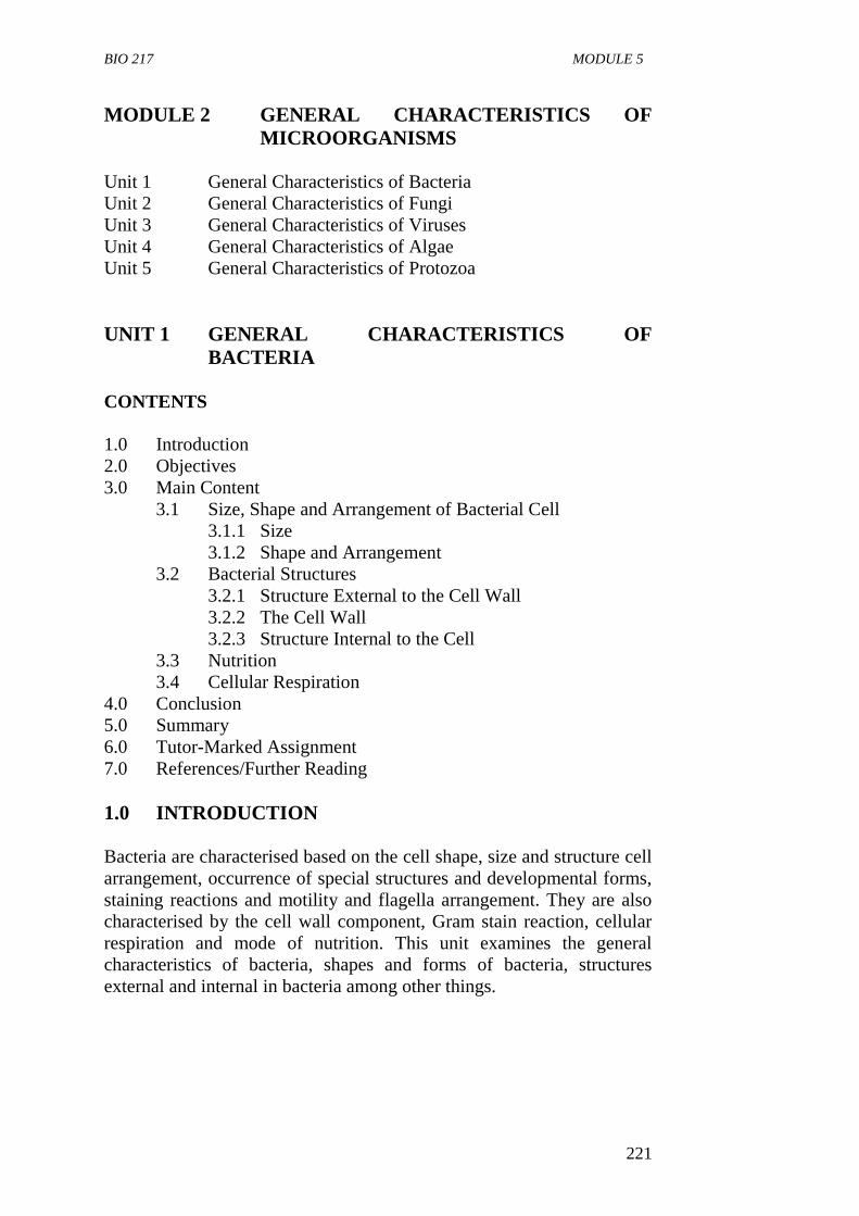
BIO 217 MODULE 5
221
MODULE 2 GENERAL CHARACTERISTICS OF
MICROORGANISMS
Unit 1 General Characteristics of Bacteria
Unit 2 General Characteristics of Fungi
Unit 3 General Characteristics of Viruses
Unit 4 General Characteristics of Algae
Unit 5 General Characteristics of Protozoa
UNIT 1 GENERAL CHARACTERISTICS OF
BACTERIA
CONTENTS
1.0 Introduction
2.0 Objectives
3.0 Main Content
3.1 Size, Shape and Arrangement of Bacterial Cell
3.1.1 Size
3.1.2 Shape and Arrangement
3.2 Bacterial Structures
3.2.1 Structure External to the Cell Wall
3.2.2 The Cell Wall
3.2.3 Structure Internal to the Cell
3.3 Nutrition
3.4 Cellular Respiration
4.0 Conclusion
5.0 Summary
6.0 Tutor-Marked Assignment
7.0 References/Further Reading
1.0 INTRODUCTION
Bacteria are characterised based on the cell shape, size and structure cell
arrangement, occurrence of special structures and developmental forms,
staining reactions and motility and flagella arrangement. They are also
characterised by the cell wall component, Gram stain reaction, cellular
respiration and mode of nutrition. This unit examines the general
characteristics of bacteria, shapes and forms of bacteria, structures
external and internal in bacteria among other things.
BIO 217 GENERAL MICROBIOLOGY
222
2.0 OBJECTIVES
At the end of this unit, you should be able to:
describe the general characteristics of basic bacteria
identify and name the general shapes and forms of bacteria
describe the external and internal structures of bacteria
explain the significance of the cell wall structure and composition
explain the modes of nutrition and energy source in bacteria
explain the modes of cellular respiration in bacteria
explain the modes of reproduction in bacteria.
3.0 MAIN CONTENT
General Characteristics of Microorganism
General characteristics of bacteria:
1. They are prokaryotic
2. They are simplest of all microbial cells
3. Bacteria are single celled organisms
4. They have distinctive cell wall which contain peptidoglycan
5. They are measured in unit called micrometer
6. Bacteria lack a true nucleus but have a region called the nucleroid
region, i.e. DNA is free floating
7. They may have additional DNA called a plasmid
8. Their reproduction is by binary fission
9. They are extremely diverse and numerous in soils and waters.
3.1 Size, Shape and Arrangement of Bacterial Cell
3.1.1 Size
Bacteria are very small, 0.5 to 1.0µm in diameter. Because of their small
size, they have high surface area/volume ratio which results in a high
growth and metabolism rate. No circulatory mechanism is needed for
nutrients taken in because the mass of cell substance to be nourished is
very close to the surface. Examinations of a microbial cell require the
use of a high power microscope usually of about 1,000 diameters.
BIO 217 MODULE 5
223
Fig. 1: A Basic Bacterium Cell
Source: Microorganisms in our World by Atlas (1995)
3.1.2 Shape and Arrangement
The shape of a bacterium is governed by its rigid cell wall which gives it
a definite shape.
Typical shapes of bacteria are:
Cocci (Singular: Coccus), e.g. Staphylococcus
Bacilli (rods) (Singular: rod, bacillus), e.g. Bacillus subtilis
Vibrios (Singular: Vibrio)
Spirilla (Singular: Sprillum)
Spirochaetes (Singular: Spirochaete), e.g. Treponema pallidum
Some species of bacteria are pleomorphic, i.e. they are able to change
their forms especially when grown on artificial media.
1. Cocci: They are round, oval or spherical in diameter
characteristic arrangement when multiplying is based on
arrangement of cells, they are called:
Diplococci: cocci in pairs, e.g. meningococci and
gonococci.
Streptococci cocci in chains.
Staphylococci: cocci in irregular clusters (like a bunch of
grapes).
Tetracocci: cocci in a group of four cells.
Sarcinae: cocci in regular clusters.
2. Bacilli (Rod): These are stick like bacteria with rounded, square,
tapered or swollen ends. They measure 1-10µm in length by 0.3-
1.0µm in width.
BIO 217 GENERAL MICROBIOLOGY
224
Bacilli are not arranged in patterns as complex as cocci. Most
occur singly. Other arrangements are:
Diplobacilli: Rods in pairs.
Streptobacilli: Rods in chains.
Trichomes: Similar to chains but have larger area of
contact between adjacent cells.
Mass together, e.g. Mycobacterium leprae.
Palisade arrangement cells are lined side by side like
matchsticks and at angles to each other like Chinese
lecters, e.g. Corynebacterium diptheriae.
3. Vibrios: These are small slightly curved rods,or comma shaped 3-
4µm in length by 0.5µm in width. Most are motile with a single
flagellum at one end, e.g. Vibrio cholerae.
4. Spirilla: These are helical bacteria, small, regularly coiled, rigid,
organisms measuring 3-4µm in length. Each coil measures about
1µm, e.g. Spirillum minus.
5. Spirochaetes: They are helical, (complete twist), flexible, coiled
organisms, can twist and contort their shapes. Spirochaeters are
divided into three main groups.
Treponemes: Tiny and delicate with regular tight coils,
measuring 6-15µm by 0.2µm in width, e.g. Treponema
pallidum and Treponema pertenue.
Borreliae: Large spirochaetes with irregular open coils 10-
20µm in length by 0.5µm in width, e.g. Borella., duttoni
and Borrelia vinceti.
Leptospires: Tiny spirochaetes with many tightly packed
coils that are difficult to distinguish; 6-20µm in length by
0.1µM in width and have hooked ends, e.g. Leptospira
interrigans.
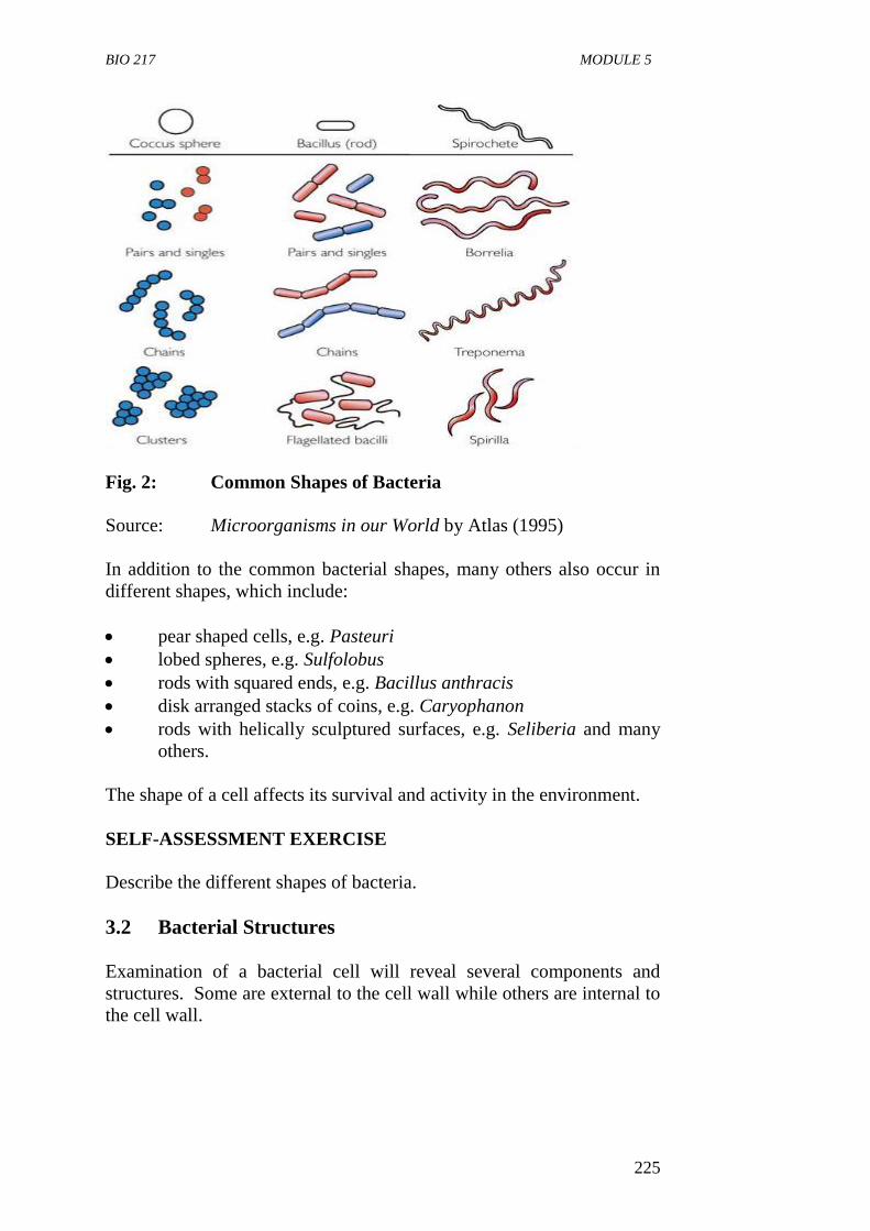
BIO 217 MODULE 5
225
Fig. 2: Common Shapes of Bacteria
Source: Microorganisms in our World by Atlas (1995)
In addition to the common bacterial shapes, many others also occur in
different shapes, which include:
pear shaped cells, e.g. Pasteuri
lobed spheres, e.g. Sulfolobus
rods with squared ends, e.g. Bacillus anthracis
disk arranged stacks of coins, e.g. Caryophanon
rods with helically sculptured surfaces, e.g. Seliberia and many
others.
The shape of a cell affects its survival and activity in the environment.
SELF-ASSESSMENT EXERCISE
Describe the different shapes of bacteria.
3.2 Bacterial Structures
Examination of a bacterial cell will reveal several components and
structures. Some are external to the cell wall while others are internal to
the cell wall.
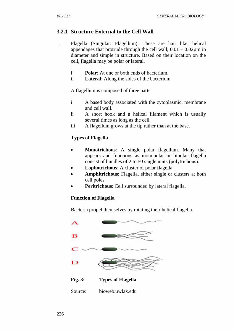
BIO 217 GENERAL MICROBIOLOGY
226
3.2.1 Structure External to the Cell Wall
1. Flagella (Singular: Flagellum): These are hair like, helical
appendages that protrude through the cell wall, 0.01 – 0.02µm in
diameter and simple in structure. Based on their location on the
cell, flagella may be polar or lateral.
i Polar: At one or both ends of bacterium.
ii Lateral: Along the sides of the bacterium.
A flagellum is composed of three parts:
i A based body associated with the cytoplasmic, membrane
and cell wall.
ii A short hook and a helical filament which is usually
several times as long as the cell.
iii A flagellum grows at the tip rather than at the base.
Types of Flagella
Monotrichous: A single polar flagellum. Many that
appears and functions as monopolar or bipolar flagella
consist of bundles of 2 to 50 single units (polytrichous).
Lophotrichous: A cluster of polar flagella.
Amphitrichous: Flagella, either single or clusters at both
cell poles.
Peritrichous: Cell surrounded by lateral flagella.
Function of Flagella
Bacteria propel themselves by rotating their helical flagella.
Fig. 3: Types of Flagella
Source: bioweb.uwlax.edu
BIO 217 MODULE 5
227
2. Pili (Singular: Pilus): They are also called fimbriae. They are
hollow, non-helical filamentous appendages that are thinner,
shorter and more numerous than flagella: long, thin, straight
threads 3-25µm in diameter and 12µm in length. They do not
function in motility since they are found on non-motile and
motile species. Several functions are associated with different
types of pili.
F pilus (Sex pilus) serves as the path of entry of genetic material
during bacterial mating. Some play major role in human infection
by allowing pathogenic bacteria to attach to the epithehal cells
lining the respiratory, intestinal or genitourinary tracts, this
prevents the bacteria from being washed away by the flow of
mucous or body fluids and permits infections to be established.
3. Capsules: This is a viscous substance forming a covering layer
or envelope around the cell wall of some bacteria. Capsules can
be categorised into three based on their visualisation by light
microscope using special staining methods.
If the covering layer can be visualised by light microscope using
special staining methods, it is termed capsule.
Microcapsule: If the layer is too thin to be seen by light
microscope.
Slime: If it is so abundant that many cells are embedded in
a common matrix.
Most bacterial capsules consist of polysaccharides which can be
homopolysaccharides or heteropolysaccharides.
Homopolysaccharides: Capsule made up of/composed of
a single kind of sugar usually synthesized outside the cell
by exocellular enzymes, e.g. glucan (a polymer of glucose)
from sucrose by S. mutans.
Heteropolysaccharides: Composed of several kinds of
sugars.
A few capsules are polypeptide, e.g. Bacillus anthracis has a
capsule made up of a polymer of glutamic acid.
Functions
They may provide protection against temporary drying by
binding water molecules.
BIO 217 GENERAL MICROBIOLOGY
228
They may block attachment of bacteriophages.
They may be antiphagocytuc, i.e. they may inhibit the engulfment
of pathogenic bacteria by white blood cells. Hence contribute to
invasive or infective ability (virulence).
Promote attachment of bacteria to surfaces.
4. Sheaths: Some bacterial species form chains or trichomes
enclosed by a hollow tube called sheaths. These sheaths consist
of a heteropolysaccharides containing glucose, glucuronic acid,
galactose and fucose.
Functions
In a few bacteria, they facilitate moderate change of position.
Sheaths enable individual cells to stay associated in cell colonies.
5. Prosthecae and Stalks
Prosthecae: They are semi rigid extensions of the cell wall and
cytoplasmic membrane and have a diameter less than that of the
cell. Found in some aerobic bacteria from fresh water and marine
environment.
Functions
Increase surface area of the cell for nutrient absorption.
Some have adhesive substances that aid attachment to surfaces.
Stalks: They are non-living ribbon -like or tubular appendages
excreted by some bacterial cells, e.g. found in Gallionella or
Planctomyces.
Functions
They aid in attachment of the cells to surfaces.
SELF-ASSESSMENT EXERCISE
List four different structures external to the cell wall of bacteria and
state one function of each.
3.2.2 The Cell Wall
It is a very rigid structure that gives shape to the cells. It also prevents
the cell from expanding and eventually bursting of uptake of water since
most bacteria live in hypotonic environment (i.e. environments having a
lower osmotic pressure than exists within the bacterial cells). Cell walls
are essential for bacterial growth and division.
BIO 217 MODULE 5
229
Structure and Chemical Composition of the Cell Wall
The cell wall of bacteria is made up of peptidoglycan (sometimes called
Murein). Peptidoglycan is found only in prokaryotes. It is an insoluble,
porous, cross-linked polymer. Peptidoglycan differs in composition and
structure from one species to another but it is basically a polymer of N-
acetyglucosamine, N-acetylmuramie acid, L-alanine, D-alanine, D-
glutamate and adiamino acid.
Bacteria are classified based on differences in the composition of cell
wall. This is determined by the Gram stain technique. Gram stain
identifies bacteria as Gram positive or Gram negative. The Gram stain is
named after Christian Gram, a Danish physician who invented it in
1884.
Gram positive bacteria stained purple whereas Gram negative bacteria
stain pink or red by the Gram stain technique.
The difference is the reaction to the Gram staining technique is due to
the presence of a single 20 to 80µm thick homogenous layer of
peptidoglycan (Murein) which is present in the all wall of Gram positive
bacteria on the other hand, the Gram negative cell is more complex, it
has a 2 to 7µm peptidoglycan layer covered by 7 to 8µm thick outer
layer of lipopolysaccharides (LPS). These LPS are toxic substances
which make Gram negative organisms more harmful than Gram positive
organisms.
3.2.3 Structures Internal to the Cell
1. Cytoplasmic Membrane
This lies immediately beneath the cell wall.
It is approximately 7.5µm (0.0075µm) thick and
composed primarily of phospholipids (20 to 30 percent)
and protein (60 to 70 percent).
It serves as a barrier to most water soluble molecules.
It contains various enzymes involved in respiration, and
metabolism and in synthesis of capsular and cell wall
component.
Proteins are also synthesized in the cytoplasm.
2. Protoplast
A protoplast is the portion of a bacterial, all made up of the
cytplasmic membrane and the cell material bounded by it.
3. The Cytoplasm
This is the cell material bounded by the cytoplasmic membrane
and it may be divided into:
BIO 217 GENERAL MICROBIOLOGY
230
i The cytoplasmic area, granular in appearance and rich in
the macromolecular RNA-protein bodies called
Ribosomes on which proteins are synthesised.
ii The chromatin area rich in DNA and
iii The fluid portion with dissolved substances.
4. Nuclear Material
Unlike eucaryotic cells bacterial cells do not have a distinct
membrane enclosed nucleus but they have an area near the centre
of the cell that is regarded as the nuclear structure, the DNA of
the cell is confined to this area. The DNA is circular and bears
the genes of the cell.
5. Spores and Cysts
Certain bacteria produce spores either within the cells
(endospores) or external to the cell (exospores). The spore is
metabolically dormant form which under appropriate condition
can germinate to form a vegetative cell. Endospores are
extremely resistant to desiccation, staining, disinfecting
chemicals, radiation and heat.
Cysts are also dormant, thick walled desiccation resistant forms
that can germinate also under favourable conditioning.
3.3 Nutrition
The nutrition requirements of bacteria vary widely.
Based on their source of energy, they are classified as:
i Phototrophs: These are bacteria that use light energy as their
energy sources.
ii Chemotrophs: They obtain their energy by oxidizing inorganic or
organic – chemical compounds.
Based on the source of carbon which is the major source of nutrient for
all cells bacteria can be classified as:
Heterotrophs: These are bacteria that derive carbon from
preformed organic nutrients such as sugar or carbohydrate.
Autotrophs: They derive carbon from inorganic sources such as
carbon dioxide.
BIO 217 MODULE 5
231
3.4 Cellular Respiration
Based on whether they need oxygen to survive or not, bacteria may be:
i aerobic or strict aerobes: these require oxygen, e.g. Bacillus
cereus.
ii anaerobic bacteria or strict anaerobes: they cannot tolerate
oxygen, e.g. Clostridium spp.
iii facultative anaerobes: These are generally aerobes but have the
capacity to grow in the absence of oxygen, e.g. Staphylococcus
spp.
3.5 Reproduction
Bacteria reproduce mainly by asexual method which most of the time is
transverse binary fission. This is a process in which a bacterial cell
divides to give two daughter cells after developing a transverse septum
(cross wall).
SELF-ASSESSMENT EXERCISE
Differentiate between Gram positive bacteria and Gram negative
bacteria.
4.0 CONCLUSION
Different types of bacteria differ from one another not only in their
shapes and forms but also in their chemical characteristics, modes of
cellular respiration, nutrition and reproduction.
5.0 SUMMARY
Bacteria are prokaryotic single celled organisms that lack
membrane-bound organelles. Bacteria are very small, with sizes
ranging from 0.5 to 1.0nm in diameter.
The typical shapes of bacteria are Cocci, Bacilli (Rod) Vibrio,
Spirila and Spirochaetes. Some bacteria are pleomorphic.
Structures external to bacterial cell wall include flagella, pili,
capsules, sheaths, prosthecae and stalks.
Flagella are hair-like, helical appendages that protrude through
the cell wall used for locomotion. May be polar or lateral based
on location on the cell.
Types of flagella are Monotrichous, Lophotrichous,
Amphitrichous, Peritrichous.
BIO 217 GENERAL MICROBIOLOGY
232
A capsule is a viscous substance forming a covers layer or
envelope around the cell wall of some bacteria. Capsules act as
protection against drying, bacteriophages and engulfment of
pathogenic bacteria by white blood cells.
The cell wall is rigid structure made up of peptidoglycan that
gives shape to bacterial cells. Bacteria are classified based on
differences in the composition of cell wall as determined by the
Gram stain techniques.
Gram-positive bacteria stained purple while Gram-negative
bacteria stain pink or red by Gram stain technique.
Structures internal to the bacterial cell include cytoplasmic
membrane, protoplast, the cytoplasm, the nuclear material, spores
and cysts.
Based on source of energy bacteria can be classified as
phototrophs and chemotrophs.
Based on source of carbon for nutrition, bacteria can be classified
as heterotrophs and chemotrophs.
Based on oxygen requirement, bacteria are classified as aerobic,
anaerobic and facultative anaerobe.
Bacteria reproduce asexually by transverse binary fission.
Bacteria are very small, simple prokaryotic cells that have
defined shapes and organelles which perform definite functions.
They are capable of independent living and existence.
6.0 TUTOR-MARKED ASSIGNMENT
i. Describe the structure and functions of a flagellum.
ii. State the difference types of bacteria based on their mode of
cellular respiration.
iii. Differentiate between Gram positive and Gram negative bacteria
cell walls.
7.0 REFERENCES/FURTHER READING
Atlas, R.M. (1995). Microorganisms in our World. Mosby Year Book.
Inc.
Medigan, M.T., et al. (2009). Brock Biology of Microorganisms.
(12th
ed.). Pearson Education Inc.
Pelczar, M.J., Chan, E.C.S. and Krieg, R.N. (1993). Microbiology.
(5th
ed.).

BIO 217 MODULE 5
233
McGraw-Hill, “The Prokaryotes: A Review of Classification”.
www.faculty.collegeofthedesert.edu/ admin-/B115 lecture 9-
05STUD-4ppt.
Willey, J.M., Sherwood, L.M & Woolverton, C.J. (2008). Microbiology.
(7th
ed.).Boston Bur Bridge, IL: McGraw-Hill Higher Education.
BIO 217 GENERAL MICROBIOLOGY
234
UNIT 2 GENERAL CHARACTERISTICS OF FUNGI
CONTENTS
1.0 Introduction
2.0 Objectives
3.0 Main Content
3.1 Distinguishing Characteristics of Fungi
3.2 Structure and Forms of Fungi
3.2.1 Yeasts
3.2.2 Molds
3.3 Nutrition and Metabolism
3.4 Reproduction
3.4.1 Asexual Reproduction
3.4.2 Sexual Reproduction
3.5 Physiology
3.6 Importance of Fungi
4.0 Conclusion
5.0 Summary
6.0 Tutor-Marked Assignment
7.0 References/Further Reading
1.0 INTRODUCTION
Fungi are eukaryotic spore bearing organisms that lack chlorophyll and
generally reproduce both sexually and asexually. They are of great
practical and scientific importance. One of the reasons for this is that
many fungi are of microscopic cellular dimensions. Fungi have a
diversity of morphological appearances depending on the species. Fungi
comprise the molds, mushrooms and yeasts. Molds are filamentous and
multicellular while yeasts are unicellular. They are widely distributed
and found wherever moisture is present. They are of great importance to
man in both beneficial and harmful ways. This unit examines the general
characteristics of fungi, the distribution, morphology, nutrition and
reproduction of fungi.
2.0 OBJECTIVES
At the end of this unit, you should be able to:
define a fungus
state the general characteristics of fungi
describe the structure of a yeast
describe the structure of a mold
explain the mode of nutrition in fungi
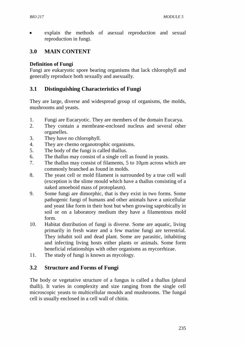
BIO 217 MODULE 5
235
explain the methods of asexual reproduction and sexual
reproduction in fungi.
3.0 MAIN CONTENT
Definition of Fungi
Fungi are eukaryotic spore bearing organisms that lack chlorophyll and
generally reproduce both sexually and asexually.
3.1 Distinguishing Characteristics of Fungi
They are large, diverse and widespread group of organisms, the molds,
mushrooms and yeasts.
1. Fungi are Eucaryotic. They are members of the domain Eucarya.
2. They contain a membrane-enclosed nucleus and several other
organelles.
3. They have no chlorophyll.
4. They are chemo organotrophic organisms.
5. The body of the fungi is called thallus.
6. The thallus may consist of a single cell as found in yeasts.
7. The thallus may consist of filaments, 5 to 10µm across which are
commonly branched as found in molds.
8. The yeast cell or mold filament is surrounded by a true cell wall
(exception is the slime mould which have a thallus consisting of a
naked amoeboid mass of protoplasm).
9. Some fungi are dimorphic, that is they exist in two forms. Some
pathogenic fungi of humans and other animals have a unicellular
and yeast like form in their host but when growing saprobically in
soil or on a laboratory medium they have a filamentous mold
form.
10. Habitat distribution of fungi is diverse. Some are aquatic, living
primarily in fresh water and a few marine fungi are terrestrial.
They inhabit soil and dead plant. Some are parasitic, inhabiting
and infecting living hosts either plants or animals. Some form
beneficial relationships with other organisms as mycorrhizae.
11. The study of fungi is known as mycology.
3.2 Structure and Forms of Fungi
The body or vegetative structure of a fungus is called a thallus (plural
thalli). It varies in complexity and size ranging from the single cell
microscopic yeasts to multicellular moulds and mushrooms. The fungal
cell is usually enclosed in a cell wall of chitin.
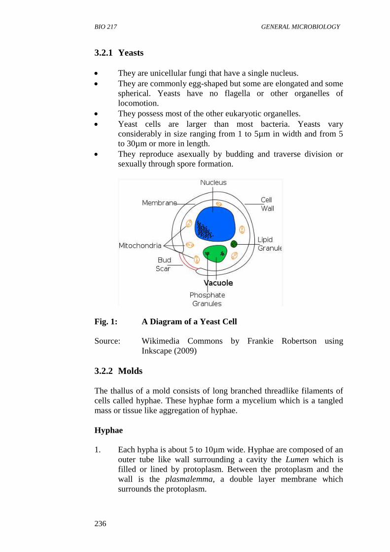
BIO 217 GENERAL MICROBIOLOGY
236
3.2.1 Yeasts
They are unicellular fungi that have a single nucleus.
They are commonly egg-shaped but some are elongated and some
spherical. Yeasts have no flagella or other organelles of
locomotion.
They possess most of the other eukaryotic organelles.
Yeast cells are larger than most bacteria. Yeasts vary
considerably in size ranging from 1 to 5µm in width and from 5
to 30µm or more in length.
They reproduce asexually by budding and traverse division or
sexually through spore formation.
Fig. 1: A Diagram of a Yeast Cell
Source: Wikimedia Commons by Frankie Robertson using
Inkscape (2009)
3.2.2 Molds
The thallus of a mold consists of long branched threadlike filaments of
cells called hyphae. These hyphae form a mycelium which is a tangled
mass or tissue like aggregation of hyphae.
Hyphae
1. Each hypha is about 5 to 10µm wide. Hyphae are composed of an
outer tube like wall surrounding a cavity the Lumen which is
filled or lined by protoplasm. Between the protoplasm and the
wall is the plasmalemma, a double layer membrane which
surrounds the protoplasm.
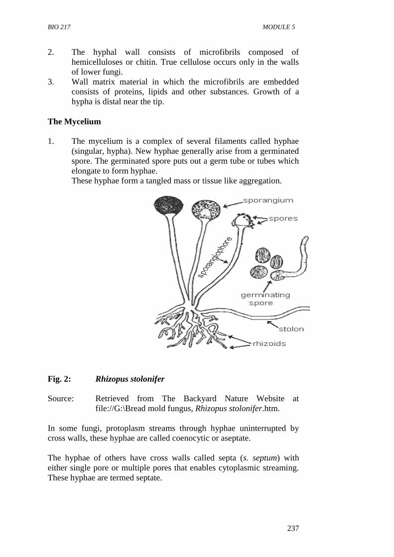
BIO 217 MODULE 5
237
2. The hyphal wall consists of microfibrils composed of
hemicelluloses or chitin. True cellulose occurs only in the walls
of lower fungi.
3. Wall matrix material in which the microfibrils are embedded
consists of proteins, lipids and other substances. Growth of a
hypha is distal near the tip.
The Mycelium
1. The mycelium is a complex of several filaments called hyphae
(singular, hypha). New hyphae generally arise from a germinated
spore. The germinated spore puts out a germ tube or tubes which
elongate to form hyphae.
These hyphae form a tangled mass or tissue like aggregation.
Fig. 2: Rhizopus stolonifer
Source: Retrieved from The Backyard Nature Website at
file://G:\Bread mold fungus, Rhizopus stolonifer.htm.
In some fungi, protoplasm streams through hyphae uninterrupted by
cross walls, these hyphae are called coenocytic or aseptate.
The hyphae of others have cross walls called septa (s. septum) with
either single pore or multiple pores that enables cytoplasmic streaming.
These hyphae are termed septate.
BIO 217 GENERAL MICROBIOLOGY
238
Summarily, hyphae can be said to occur in three forms:
i Nonseptate or coenocytic; such hyphae have no septa.
ii Septate with uninucleate cells.
iii Septate with multinucleate cells. Each cell has more than one
nucleus in each compartment.
SELF-ASSESSMENT EXERCISE
i. What are the major differences between a mold and yeast?
ii. State five general characteristics of fungi.
3.3 Nutrition and Metabolism
Most fungi are saprobes, securing their nutrients from dead organic
matters. They release hydrolytic exo-enzymes that digest external
substrates and absorb the soluble products.
They are also chemoorganoheterotrophs, i.e. they use organic
compounds as a source of carbon, electrons and energy.
Fungi are usually aerobic; however, some yeasts are facultatively
anaerobic and can obtain their energy by fermentation. Obligately
anaerobic fungi are found in the rumen of cattle.
3.4 Reproduction
Reproduction in fungi can either be asexual or sexual.
3.4.1 Asexual Reproduction
Asexual reproduction is a type of reproduction involving only one
parent that produces genetically identical offspring by budding or by the
division of a single cell or the entire organism into two or more parts.
Asexual reproduction, also called somatic or vegetative reproduction is
accomplished in several ways and does not involve the fusion/union of
nuclei, sex cells or sex organs. It may be accomplished by:
fission of somatic cells yielding two similar daughter cells
budding each bud a small outgrowth of the parent cell develops
into a new individual
fragmentation or disjointing of the hyphal cells each fragment
becoming a new organism
spore formation.
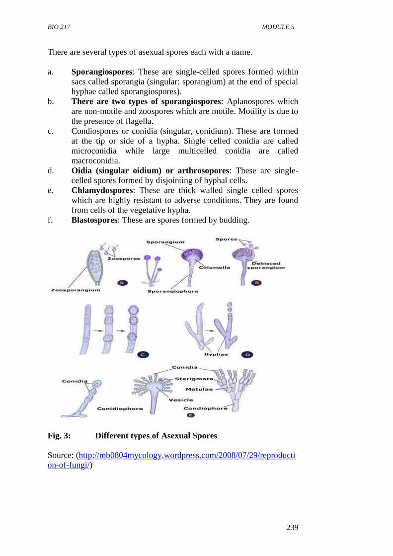
BIO 217 MODULE 5
239
There are several types of asexual spores each with a name.
a. Sporangiospores: These are single-celled spores formed within
sacs called sporangia (singular: sporangium) at the end of special
hyphae called sporangiospores).
b. There are two types of sporangiospores: Aplanospores which
are non-motile and zoospores which are motile. Motility is due to
the presence of flagella.
c. Condiospores or conidia (singular, conidium). These are formed
at the tip or side of a hypha. Single celled conidia are called
microconidia while large multicelled conidia are called
macroconidia.
d. Oidia (singular oidium) or arthrosopores: These are single-
celled spores formed by disjointing of hyphal cells.
e. Chlamydospores: These are thick walled single celled spores
which are highly resistant to adverse conditions. They are found
from cells of the vegetative hypha.
f. Blastospores: These are spores formed by budding.
Fig. 3: Different types of Asexual Spores
Source: (http://mb0804mycology.wordpress.com/2008/07/29/reproducti
on-of-fungi/)
BIO 217 GENERAL MICROBIOLOGY
240
3.4.2 Sexual Reproduction
Sexual reproduction is a type of reproduction in which two parents give
rise to offspring that have unique combinations of genes inherited from
the gametes of the two parents.
It is carried out by fusion of the compatible nuclei of two parent cells.
The process of sexual reproduction begins with the joining of two cells
and fusion of their protoplast (plasmogamy) thus enabling the two
haploid nuclei of two mating types to fuse together (karyogamy) to form
a diploid nucleus. This is followed by meiosis, which again reduces the
number of chromosomes to the haploid number.
The sex organelles of fungi if present are called gametangia. They may
form differentiated sex cells called gametes or may contain instead one
or more gamete nuclei. If the male and female gametangia are
morphologically different, the male gametangium is called the
antheridium (plural antheridia) and the female gamentangium is called
the Oogonium (Oogonia).
Methods of sexual reproduction include:
i Gametic copulation: This is the fusion of naked gametes, one or
both of which are motile.
ii Gamete-gametangial copulation: Two gametangia came into
contact but do not fuse; the male nucleus migrate through a pore
or fertilization to be into the female gamentangium.
iii Gametangial copulation: Two gamentangia or their protoplast
fuse and give rise to a zygote that develops into a resting spore.
iv Somatic copulation: Fusion of somatic or vegetative cells.
v Spermatization: Union of a special male structure called a
spermatium (plural spermatia) with a female receptive structure.
The spermatum empties its content into the female during
plasmogamy.
Sexual spores are produced by the fusion of two nuclei. Examples are:
i Ascospores: These are single-celled spores produced in a sac
called an ascus. There are usually eight ascospores in each ascus.
ii Basidiospore: These are single celled spores borne on a club-
shaped structure called a basidium.
iii Zygospores: These are large thick walled spores formed when
the tips of two sexually compatible hyphae or gametagia fuse
together.
BIO 217 MODULE 5
241
iv Oospores: These are formed with a special female structure, the
oogonium. Fertilization of the eggs or oospheres by the male
gametes formed in an antheridium give rise to oospores.
SELF-ASSESSMENT EXERCISE
Describe sexual reproduction as it occurs in fungi.
3.5 Physiology
Fungi are better able to withstand certain extreme environments
than other microorganisms. They can tolerate more acidic
conditions than other microbes. Some types of yeasts are
facultative; they can grow under both aerobic and anaerobic
conditions. Molds and many types of yeast are usually aerobic
microorganisms.
Fungi grow over a wide range of temperature. The optimum
temperature for most saprobic species is 22 to 300C, while
pathogenic fungi have a higher temperature optimum of 30 to
370C.
Some fungi will grow at or near 00C and thus can cause spoilage
of meat and/or vegetables in cold storage.
3.6 Importance of Fungi
About 90,000 fungal species have been described according to
literature. However, some estimates suggest that 1.5 million
species may exist. Fungi are important to humans in both
beneficial and harmful ways.
Beneficially, fungi act as decomposers. They degrade complex
organic materials in the environment and release simple organic
and inorganic molecules like carbon, nitrogen, phosphorus
needed by other living organisms.
Moulds and yeasts are used in many industrial processes
involving fermentation to produce beer, wine and bread, cheese
soy-sauce, organic acids and many antibiotics.
They are important research tools in the study of fundamental
processes such as cytology, genetics, biochemistry and
microbiology.
On the other hand, fungi cause many diseases of plants, animals
and humans. About 20 new human fungal pathogens are
documented each year.
BIO 217 GENERAL MICROBIOLOGY
242
4.0 CONCLUSION
Fungi are eukaryotic, spore bearing organisms that lack chlorophyll.
They are diverse in distribution, reproduce sexually and asexually. Fungi
are of high importance to man in both beneficially and harmful ways.
7.0 SUMMARY
Fungi are eukaryotic spore bearing organisms that lack
chlorophyll and reproduce both asexually and sexually.
Fungi are widespread in environment and found wherever water
suitable organic nutrients and an appropriate temperature occur.
Habitats of fungi are diverse. Many are terrestrial some are
aquatic and are parasite in living hosts.
The body or vegetative structure of a fungus is called thallus.
Fungi may be grouped into molds or yeasts based on the
development of the thallus.
Yeasts are unicellular fungi that have a single nucleus and
reproduce either asexually by budding or asexually by spore
formation.
A mold is made up of long branched thread-like filament called
hyphae that form a tangled mass called mycelium.
Some fungi are saprophytes and grow best in moist dark habitats.
They are usually aerobic but some types of yeasts are facultative.
Asexual reproduction occurs in fungi by the production of
specific types of spores which are easily dispersed.
Sexual reproduction occurs by the fusion of hyphae or cells of
different mating types.
8.0 TUTOR-MARKED ASSIGNMENT
i. Describe the structure of a mold.
ii. Describe each of the following types of asexual fungal spores:
a. Sporangiospore
b. Conidiospore and
c. Blastospore.
BIO 217 MODULE 5
243
7.0 REFERENCES/FURTHER READING
Atlas, R.M. (1995). Microorganisms in Our World. Mosby Year Book.
Inc.
Medigan, M.T. et al. (2009). Brock Biology of Microorganisms.
(12th
ed.). Pearson Education Inc.
Pelczar, M.J., Chan, E.C.S. & Krieg, R.N. (2001).
(5th
ed.). Microbiology. McGraw-Hill.
Willey, J.M., Sherwood, L.M & Woolverton, C.J. (2008). Microbiology.
(7th
ed.). Boston Bur Bridge, IL: McGraw-Hill Higher Education.
“The Backyard Nature. Bread Mold Fungus,” Rhizopus Stolonifer.htm.
http://mb0804mycology.wordpress.com/2008/07/29/reproduction
-of-fungi
BIO 217 GENERAL MICROBIOLOGY
244
UNIT 3 GENERAL CHARACTERISTICS OF VIRUSES
CONTENTS
1.0 Introduction
2.0 Objectives
3.0 Main Content
3.1 General Characteristics of Viruses
3.2 Virion Size
3.3 The Structure of Viruses
3.4 Viral Genomes
3.5 Capsids Symmetry
3.6 Virus Reproduction
3.7 The Cultivation of Viruses
3.8 Virus Purification and Assay
4.0 Conclusion
5.0 Summary
6.0 Tutor-Marked Assignment
7.0 References/Further Reading
1.0 INTRODUCTION
Viruses are acellular entities. They are genetic elements that cannot
replicate independently of a living cell called the host cell. Viruses have
extracellular forms which enable them to exist outside the host for long
periods. But to multiply, they have to enter a cell in which they can
replicate causing infection. Viruses are the most numerous
microorganisms on earth and infect all types of cellular organisms. The
study of viruses is known as virology. This unit examines the general
characteristics of viruses, their structures, genomes, symmetry,
replication in hosts and purification.
2.0 OBJECTIVES
At the end of this unit, you should be able to:
define the term virus
state the general characteristics of virus
describe the structure of a typical virus particle
explain the symmetry of capids
explain the process of viral replication in susceptible host
state various methods of cutting viruses
state the various methods of virus purification.
BIO 217 MODULE 5
245
3.0 MAIN CONTENT
Definition
Viruses are simple acellular entities that can only reproduce within
living cells.
3.1 General Characteristics of Viruses
i. They are the smallest microorganisms. They range in size from
10 to 400m in diameter and can only be viewed under an
electronmicroscope.
ii. They are acellular, i.e. not cellular and non living.
iii. They only reproduce when present within living cells.
iv. They are infectious agents.
v. A complex virus particle or virion consists of one or more
molecules of DNA or RNA enclosed in a coat of protein.
vi. Viruses can exist in two phases: extracellular and intracellular.
vii. The extracellular phase known as virion possesses few if any
enzymes and cannot reproduce independent of living cells. It is
metabolically inert and does not carry out respiration.
viii. In the intracellular phase, viruses exist primarily as replicating
nucleic acids in the host cells that induce host metabolism to
synthesise virion components which are later released.
Viruses differ from living cells in three ways:
i. They have simple acellular organisation.
ii. The presence of either DNA or RNA but not both in almost all
virions.
iii. They do not have the ability to reproduce independent of cells
and carry out cell division as procaryotes and eukaryotes do.
3.2 Virion Size
Virions range in size from about 10 to 400µm in diameter. The smallest
viruses are a little larger than ribosomes whereas the pox viruses which
include vaccinia are about the same size as the smallest bacteria and can
be seen in the light microscope. Most viruses however, are too small to
be visible in the light microscope and must be viewed with scanning and
transmission electron microscope.
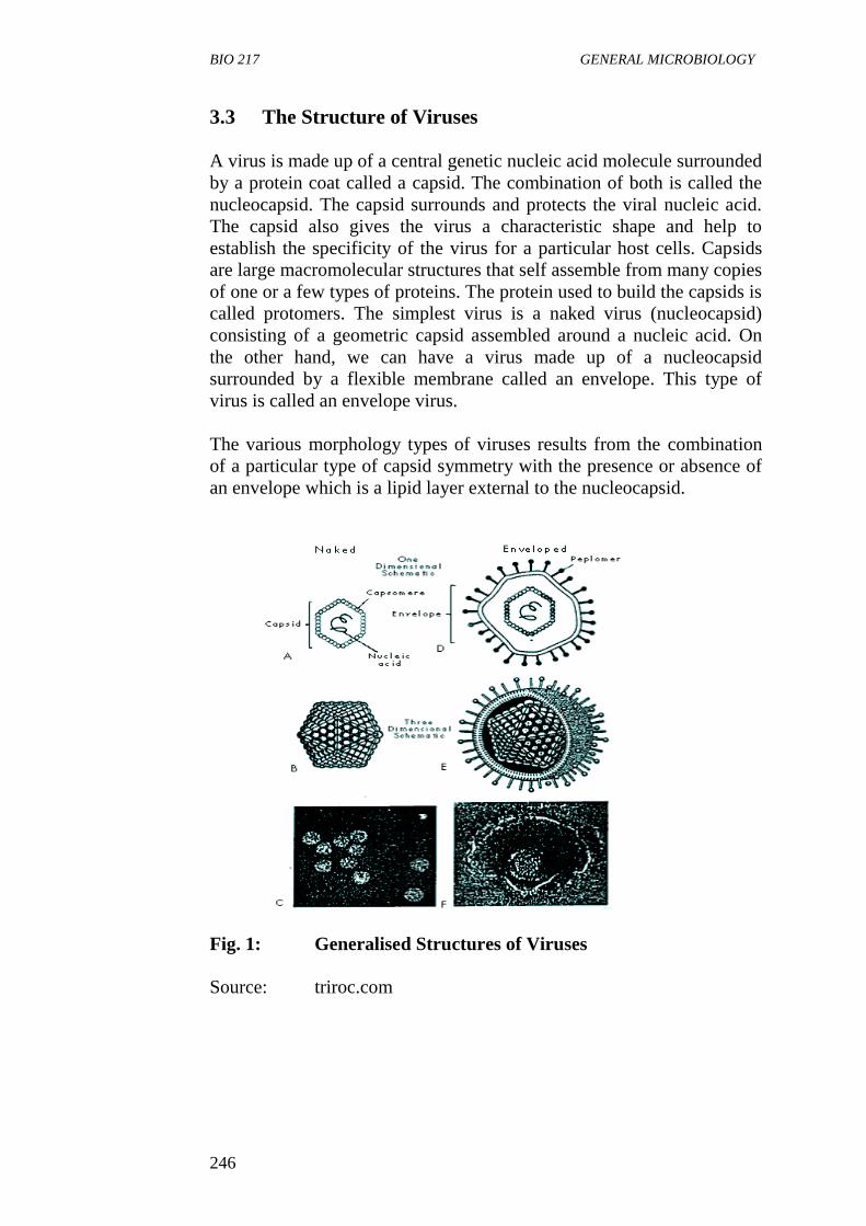
BIO 217 GENERAL MICROBIOLOGY
246
3.3 The Structure of Viruses
A virus is made up of a central genetic nucleic acid molecule surrounded
by a protein coat called a capsid. The combination of both is called the
nucleocapsid. The capsid surrounds and protects the viral nucleic acid.
The capsid also gives the virus a characteristic shape and help to
establish the specificity of the virus for a particular host cells. Capsids
are large macromolecular structures that self assemble from many copies
of one or a few types of proteins. The protein used to build the capsids is
called protomers. The simplest virus is a naked virus (nucleocapsid)
consisting of a geometric capsid assembled around a nucleic acid. On
the other hand, we can have a virus made up of a nucleocapsid
surrounded by a flexible membrane called an envelope. This type of
virus is called an envelope virus.
The various morphology types of viruses results from the combination
of a particular type of capsid symmetry with the presence or absence of
an envelope which is a lipid layer external to the nucleocapsid.
Fig. 1: Generalised Structures of Viruses
Source: triroc.com
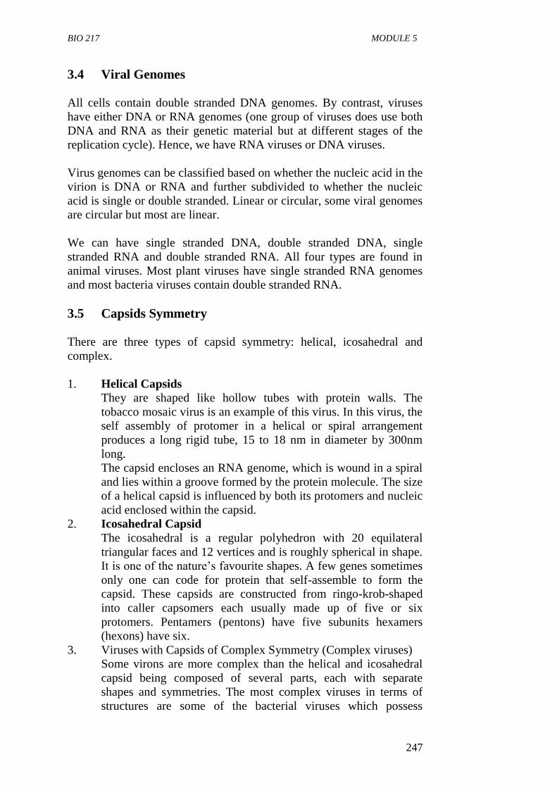
BIO 217 MODULE 5
247
3.4 Viral Genomes
All cells contain double stranded DNA genomes. By contrast, viruses
have either DNA or RNA genomes (one group of viruses does use both
DNA and RNA as their genetic material but at different stages of the
replication cycle). Hence, we have RNA viruses or DNA viruses.
Virus genomes can be classified based on whether the nucleic acid in the
virion is DNA or RNA and further subdivided to whether the nucleic
acid is single or double stranded. Linear or circular, some viral genomes
are circular but most are linear.
We can have single stranded DNA, double stranded DNA, single
stranded RNA and double stranded RNA. All four types are found in
animal viruses. Most plant viruses have single stranded RNA genomes
and most bacteria viruses contain double stranded RNA.
3.5 Capsids Symmetry
There are three types of capsid symmetry: helical, icosahedral and
complex.
1. Helical Capsids
They are shaped like hollow tubes with protein walls. The
tobacco mosaic virus is an example of this virus. In this virus, the
self assembly of protomer in a helical or spiral arrangement
produces a long rigid tube, 15 to 18 nm in diameter by 300nm
long.
The capsid encloses an RNA genome, which is wound in a spiral
and lies within a groove formed by the protein molecule. The size
of a helical capsid is influenced by both its protomers and nucleic
acid enclosed within the capsid.
2. Icosahedral Capsid
The icosahedral is a regular polyhedron with 20 equilateral
triangular faces and 12 vertices and is roughly spherical in shape.
It is one of the nature’s favourite shapes. A few genes sometimes
only one can code for protein that self-assemble to form the
capsid. These capsids are constructed from ringo-krob-shaped
into caller capsomers each usually made up of five or six
protomers. Pentamers (pentons) have five subunits hexamers
(hexons) have six.
3. Viruses with Capsids of Complex Symmetry (Complex viruses)
Some virons are more complex than the helical and icosahedral
capsid being composed of several parts, each with separate
shapes and symmetries. The most complex viruses in terms of
structures are some of the bacterial viruses which possess
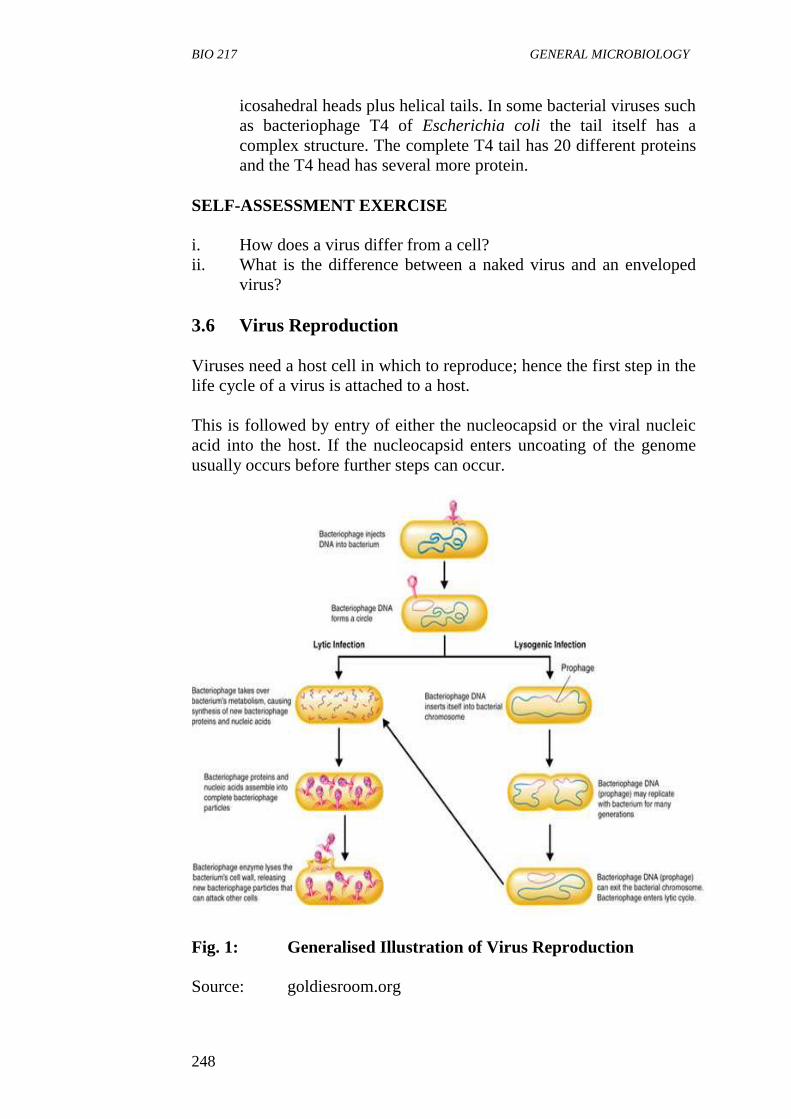
BIO 217 GENERAL MICROBIOLOGY
248
icosahedral heads plus helical tails. In some bacterial viruses such
as bacteriophage T4 of Escherichia coli the tail itself has a
complex structure. The complete T4 tail has 20 different proteins
and the T4 head has several more protein.
SELF-ASSESSMENT EXERCISE
i. How does a virus differ from a cell?
ii. What is the difference between a naked virus and an enveloped
virus?
3.6 Virus Reproduction
Viruses need a host cell in which to reproduce; hence the first step in the
life cycle of a virus is attached to a host.
This is followed by entry of either the nucleocapsid or the viral nucleic
acid into the host. If the nucleocapsid enters uncoating of the genome
usually occurs before further steps can occur.
Fig. 1: Generalised Illustration of Virus Reproduction
Source: goldiesroom.org
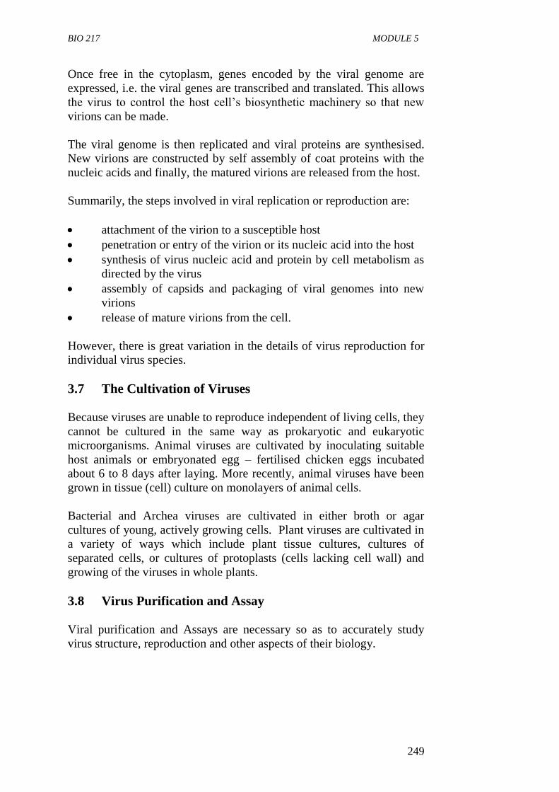
BIO 217 MODULE 5
249
Once free in the cytoplasm, genes encoded by the viral genome are
expressed, i.e. the viral genes are transcribed and translated. This allows
the virus to control the host cell’s biosynthetic machinery so that new
virions can be made.
The viral genome is then replicated and viral proteins are synthesised.
New virions are constructed by self assembly of coat proteins with the
nucleic acids and finally, the matured virions are released from the host.
Summarily, the steps involved in viral replication or reproduction are:
attachment of the virion to a susceptible host
penetration or entry of the virion or its nucleic acid into the host
synthesis of virus nucleic acid and protein by cell metabolism as
directed by the virus
assembly of capsids and packaging of viral genomes into new
virions
release of mature virions from the cell.
However, there is great variation in the details of virus reproduction for
individual virus species.
3.7 The Cultivation of Viruses
Because viruses are unable to reproduce independent of living cells, they
cannot be cultured in the same way as prokaryotic and eukaryotic
microorganisms. Animal viruses are cultivated by inoculating suitable
host animals or embryonated egg – fertilised chicken eggs incubated
about 6 to 8 days after laying. More recently, animal viruses have been
grown in tissue (cell) culture on monolayers of animal cells.
Bacterial and Archea viruses are cultivated in either broth or agar
cultures of young, actively growing cells. Plant viruses are cultivated in
a variety of ways which include plant tissue cultures, cultures of
separated cells, or cultures of protoplasts (cells lacking cell wall) and
growing of the viruses in whole plants.
3.8 Virus Purification and Assay
Viral purification and Assays are necessary so as to accurately study
virus structure, reproduction and other aspects of their biology.
BIO 217 GENERAL MICROBIOLOGY
250
Virus Purification
This involves getting or isolating the viral particle in its pure state,
purification makes use of several virus properties.
Four of the most widely used methods to isolate and purify viruses are:
differential and density gradient centrifugation. This is often used
in the initial purification steps to separate virus particles from
host cells.
precipitation of viruses particles.
denaturation of contaminants.
enzymatic digestion of host cells constituents.
Virus Assays
The quantity of viruses in a sample can be determined either directly by
counting particle numbers using the electron microscope or indirectly by
measurement of an observable effect of the virus using techniques such
as the hemaglutination assay.
SELF-ASSESSMENT EXERCISE
i. Explain how viruses are cultivated in different hosts.
ii. Give the four major approaches by which viruses may be
purified.
4.0 CONCLUSION
It can be seen clearly that viruses are a complex, diverse and fascinating
group of microorganisms, uniquely different from other groups of
microorganisms which are cellular.
7.0 SUMMARY
Viruses are simple acellular entities that can only reproduce
within living cells.
A virus is made up of a central genetic nucleic acid molecule
which could be DNA or RNA surrounded by a protein called
capsid.
Capsids are large macromolecular structures that self assemble
from many copies of one or a few types of proteins.
There are three types of capsids: symmetry helical, icosahedral
and complex.
BIO 217 MODULE 5
251
Viruses range in size from 10 to 400 NM in diameter and can
only be viewed with scanning and transmission electron
microscope.
Viral nucleic acid can either be single stranded or double
stranded DNA or RNA.
Some viral geromes are circular while some are linear.
Virus reproduction can be divided into five steps:
i attachment
ii entry into host
iii synthesis of viral nucleic acid and proteins
iv self assembly of virions and
v release from host.
Viruses are cultivated using tissue cultures, embryonated eggs,
bacterial culture and other living hosts.
Viral purification involves getting the viral particle in its pure
state and involves techniques such as differential and gradient
centrifugation, precipitation, denaturation or digestion of
contaminants.
Virus particles can be counted directly with the transmission
electron microscope or indirectly by the hemagglutination assay.
8.0 TUTOR-MARKED ASSIGNMENT
i. Explain the processes involved in viral replication or virus
reproduction.
ii. Write briefly on viral genomes.
iii. Define the following terms:
a. virus
b. nucleocapsid
c. helical symmetry
d. viral purification.
BIO 217 GENERAL MICROBIOLOGY
252
7.0 REFERENCES/FURTHER READING
Atlas, R.M. (1995). Microorganisms in Our World. Mosby Year Book.
Inc.
Medigan, M.T. et al. (2009). Brock Biology of Microorganisms. (12th
ed.). Pearson Education Inc.
Pelczar, M.J., Chan, E.C.S. & Krieg, R.N. (2001). (5th
ed.).
Microbiology. McGraw-Hill.
Willey, J.M., Sherwood, L.M & Woolverton, C.J. (2008). Microbiology.
(7th
ed.). Boston Bur Bridge, IL: McGraw-Hill Higher Education.
http://www.goldiesroom.org
http://www.triroc.com
BIO 217 MODULE 5
253
UNIT 4 GENERAL CHARACTERISTICS OF ALGAE
CONTENTS
1.0 Introduction
2.0 Objectives
3.0 Main Content
3.1 Occurrence and Distribution of Algae
3.2 Morphology
3.3 Motility
3.4 Reproduction
3.4.1 Asexual Reproduction
3.4.2 Sexual Reproduction
3.5 Biological and Economic Importance of Algae
4.0 Conclusion
5.0 Summary
6.0 Tutor-Marked Assignment
7.0 References/Further Reading
1.0 INTRODUCTION
Algae (singular, alga) are unicellular microorganisms that have
chlorophyll and are photosynthetic. Algae are heterogeneous and range
from microscopic unicellular forms to macroscopic seaweeds. They are
different from green plants due to their simple reproductive structure for
sexual reproduction. Many live in aquatic environments but many also
thrive as subterranean algae. Algae are of great importance to biologist
because single algal cells are complete organisms capable of
photosynthesis and the synthesis of other compounds which constitute
the cell. The study of algae is known as phycology. This unit examines
the general characteristics, the distribution, the morphology and
importance of algae.
2.0 OBJECTIVES
At the end of this unit, you should be able to:
define the term algae
state the general characteristics of algae
describe the general structure of algae with a typical example
draw a well labeled diagram of algae
state the different habitats of algae
explain the methods of reproduction in algae
state the economic importance of algae.
BIO 217 GENERAL MICROBIOLOGY
254
3.0 MAIN CONTENT
General Characteristics of Algae
Algae are eukaryotic microorganisms.
They are photosynthetic microorganisms.
Chlorophyll and other pigments are found in membrane bound
organelles known as chloroplasts. Algae contain a discrete
nucleus. Other inclusions are starch grains, oil droplets and
vacuoles. They contain chlorophyll and utilise light energy to
generate their chemical energy.
They have a wide range of sizes and shapes. Many species occur
as single cells that may be spherical, rod shaped, club-shaped or
spindle-shaped. Others are multicellular and appear in every
conceivable form, shape and degree of complexity.
In most species the cell wall is thin and rigid cell walls of diatoms
are impregnated with silica making them thick and very rigid.
The motile algae such as euglena have flexible cell membrane
called periplasts.
They are also able to produce oxygen from water.
3.1 Occurrence and Distribution
Algae are found in many places on earth. They occur in great abundance
in the ocean, seas, salt lakes, fresh water lakes ponds and streams. Many
are found in damp soil, on rocks, stones and tree barks.
Some are found on plants and animals. Small aquatic forms make up a
large part of the free-floating microscopic life in water called plankton
which is the principal food for aquatic animals including such large ones
as whales. Plankton is generally considered to be composed of both
algae and microscopic animal forms. Phytoplankton is made up of
plants, i.e. algal forms and zooplankton is composed of animal
organisms.
Algae are found where there are sufficient light, moisture and simple
nutrients to sustain them.
Some species of algae grow on the snow and ice of Polar Regions and
mountain peaks, sometimes occurring in such abundance that the
landscape becomes coloured by the red pigments in their cells.
BIO 217 MODULE 5
255
At the other extreme, some algae grow in the hot springs at temperatures
as high as 550C. Some freshwater algae have adapted their metabolism
to the high salt concentration found in the brine lakes of the arid South-
Western United States.
Some algae are adapted to moist soil, the bark of trees and the surface of
rocks, which the algae degrade. The decomposition products are made
available for soil building and enrichment. Algae are often a problem in
water supplies because they produce undesirable taste and odour.
Heavy algal growth may form blankets or mats which interfere with the
use of some natural waters for recreational purposes. These algal mats
may act as barriers to the penetration of oxygen into the water; they
prevent photosynthesis by excluding light from deeper water and this
may cause fish and other marine animal to suffocate.
On the other hand, when dispersed in natural waters, algae increase the
oxygen concentration through photosynthesis. Heavy growth of some
algae reduces hardness of water and removes slats which are the cause
of blackishness. Some algae are endophytic; that is, they are not free-
living but live in other organisms. Such algae are widespread in
protozoa, molluscs, sponges and corals.
3.2 Morphology
Algae have a wide range of sizes and shapes. Many are unicellular and
may be spherical, rod shaped, club shaped or spindle shaped. Others are
multicellular and appear in every conceivable forms, shape and degree
of complexity including membranous colonies, filaments grouped,
singly or in clusters with individual strands which may be branched or
unbranched tubes.
Algal cells are eukaryotic. Most are thin and rigid cell walls; however,
the cell walls of diatoms are impregnated with silica threads which make
them thick and very rigid.
Algae have a discrete nucleus, starch grains on droplets and vacuoles:
chlorophyll and other pigments are found in membrane-bound
organelles known as chloroplasts.
The chloroplast ultra-structure and type of pigment presents in algae are
used for their classification, e.g. green algae, red algae, yellow green
algae, the golden algae, etc.
A very common green alga is spirogyra; a filamentous alga found on the
scum that cover ponds are slow moving water.
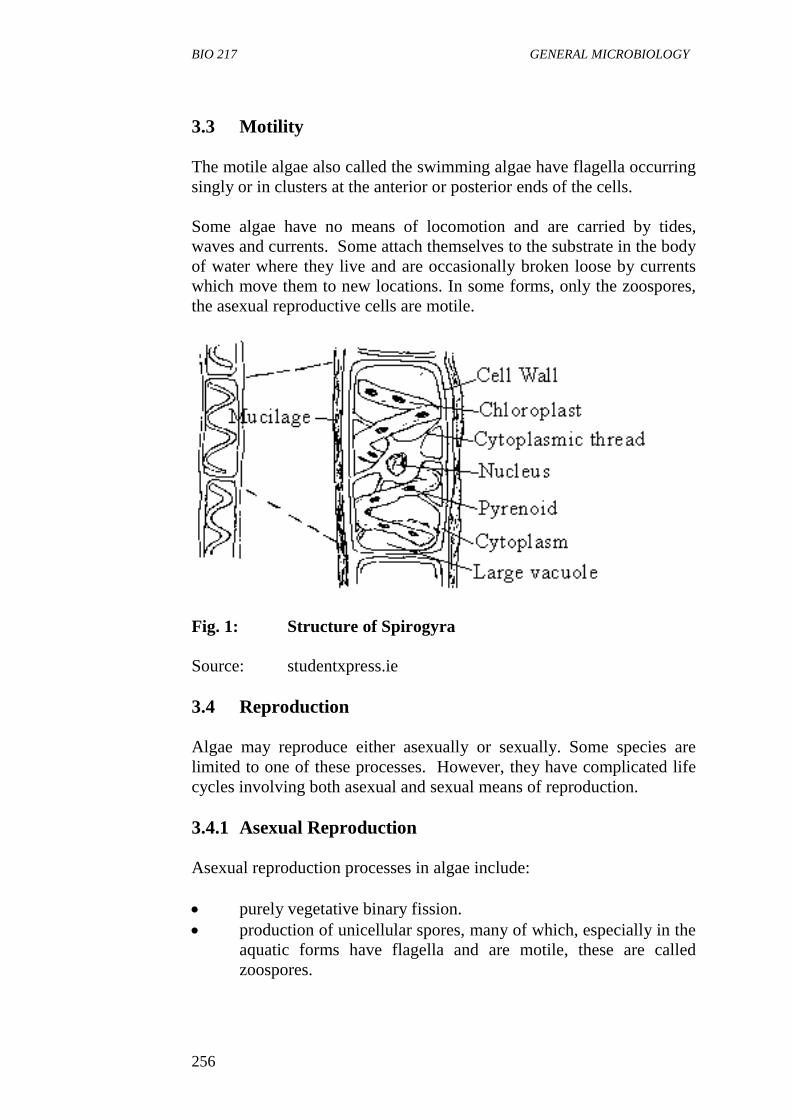
BIO 217 GENERAL MICROBIOLOGY
256
3.3 Motility
The motile algae also called the swimming algae have flagella occurring
singly or in clusters at the anterior or posterior ends of the cells.
Some algae have no means of locomotion and are carried by tides,
waves and currents. Some attach themselves to the substrate in the body
of water where they live and are occasionally broken loose by currents
which move them to new locations. In some forms, only the zoospores,
the asexual reproductive cells are motile.
Fig. 1: Structure of Spirogyra
Source: studentxpress.ie
3.4 Reproduction
Algae may reproduce either asexually or sexually. Some species are
limited to one of these processes. However, they have complicated life
cycles involving both asexual and sexual means of reproduction.
3.4.1 Asexual Reproduction
Asexual reproduction processes in algae include:
purely vegetative binary fission.
production of unicellular spores, many of which, especially in the
aquatic forms have flagella and are motile, these are called
zoospores.
BIO 217 MODULE 5
257
In terrestrial types of algae, non-motile spores or aplanospores are
formed; however, some aplanospores can develop into zoospores.
3.4.2 Sexual Reproduction
All forms of sexual reproduction are found among the algae. In this
processes there is a fusion (conjugation) of sex cells called gametes to
form a zygote. If the gametes are identical, i.e., there is no visible sex
differentiation. The fusion process is called isogamous. However, if two
gametes are different, the process is called heterogamous. In higher
algae, the sex cells are differentiated into male and female.
The female egg cell (ovum) is large and non motile, while the male
gametes (sperm cell) is small and are actively motile. This type of sexual
reproduction is called oogamy.
3.5 Biological and Economic Importance of Algae
1. Algae as Primary Producers
Algae form the base or beginning of most aquatic food chains
because of their photosynthetic activities and are therefore called
primary producers of organic matter.
2. Commercial Product from Algae
Many product of economic value are derived from algal cell
walls. Three of these, agar, alginic acid and carrageenan, are
extracted from the walls of algae. Another, diatomaceous earth, is
composed of millions upon millions of diatom glass walls
deposited over time on either fresh water or the ocean. All three
compounds are used either make gels or to make solution
viscous. Carrageenan has been used as a stabilizer or emulsifier
in foods such as ice cream and other milk products. It is also used
as a binder in toothpaste or in pharmaceutical products, as well as
an agent in ulcer therapy. Carrageenan is also useful as a
finishing compound in the textile and paper industries, as a
thickening agent in sharing creams and lotions and in the soap
industry.
Agar is well known as solidifying agent in the preparation of
microbiological media. It is obtained from red algae. Species of
Gelidium and Gracelaria are used extensively. It is also
important in the food industry when it is valuable in the
manufacture of processed cheese, mayonnaise, pudding, jellies,
baking products and canned goods.
In the pharmaceutical industry, agar can be used as a carrier for a
drug. Lotions and ointments can contain some agar. About 50 per
BIO 217 GENERAL MICROBIOLOGY
258
cent of the ice cream in the U.S. contains alginates which provide
a smooth consistency and eliminate ice crystal formation.
Alginate is also incorporated into cheeses and bakery products,
especially frostings. Other industrial application includes paper
manufacturing, the printing of fabrics and paint thickening.
Diatomaceous earth is used primarily for filters or filter aids. It is
especially suitable because it is not chemically reactive, is not
readily compacted or compressed during use and is available in
many grades.
3. Algae as Food
Many species of algae (mostly red and brown algae) are used as
food in the Far East. Of the red algae are of the most important in
porphyria; it is used as a food in Japan where it is called “mori”
and is usually processed into dried sheets.
4.0 CONCLUSION
Algae are of great and general interest to all biologists because single
algal cells are complete organisms capable of photosynthesis and the
synthesis of a multitude of other compounds which constitute the cell.
7.0 SUMMARY
The algae are photosynthetic eukaryotic microorganisms.
Algae are found in many places on earth including ocean, lakes,
ponds, streams, moist soils, rocks, tree barks, ice and hot springs.
Algae may be unicellular or multicellular, have cell wall, a
discrete nucleus starch grains, oil droplets, vacuoles chlorophyll
and other pigments.
Algae reproduce asexually by binary fission and sexually by
fusion of gametes to form a zygote.
Algae are primary producers in most aquatic food chains.
Commercial products such as agar alginic acid and carrageenan
are derived from algae.
8.0 TUTOR-MARKED ASSIGNMENT
i. State five characteristics of algae.
ii. Discuss the various uses of algae that make the commercially
important.
BIO 217 MODULE 5
259
7.0 REFERENCES/FURTHER READING Atlas, R.M. (1995). Microorganisms in Our World. Mosby Year Book.
Inc.
Medigan, M.T. et al. (2009). Brock Biology of Microorganisms. (12th
ed.). Pearson Education Inc.
Pelczar, M.J., Chan, E.C.S. & Krieg, R.N. (2001). (5th
ed.).
Microbiology. McGraw-Hill.
Willey, J.M., Sherwood, L.M & Woolverton, C.J. (2008). Microbiology.
(7th
ed.). Boston Bur Bridge, IL: McGraw-Hill Higher Education.
Website: http://www.studentxpress.ie
BIO 217 GENERAL MICROBIOLOGY
260
UNIT 5 GENERAL CHARACTERISTICS OF
PROTOZOA
CONTENTS
1.0 Introduction
2.0 Objectives
3.0 Main Content
3.1 Occurrence/Distribution of Protozoa
3.2 Ecology of Protozoa
3.2.1 Free-Living Protozoa
3.2.2 Symbiotic Protozoa
3.3 Morphology of Protozoa
3.3.1 The cytoplasm
3.3.2 Nucleus
3.3.3 Cysts
3.3.4 Locomotory Organelles
3.3.5 Feeding Structure
3.4 Two Examples of Protozoan
3.4.1 Amoeba
3.4.2 Paramecium
3.5 Reproduction of Protozoa
3.5.1 Asexual Reproduction
3.5.2 Sexual Reproduction
3.6 Economic Importance of Protozoa
4.0 Conclusion
5.0 Summary
6.0 Tutor-Marked Assignment
7.0 References/Further Reading
1.0 INTRODUCTION
Protozoa are unicellular, non-photosynthetic eukaryotic organisms. They
are distinguished from other eukaryotic protists by their ability to move
at some stage of their life cycle and by their lack of cell walls. Some
protozoa are free living while some are parasitic. Some protozoa form
colonies, in a colony, the individual cells are joined by cytoplasmic
thread or embedded in a common matrix, hence colonies of protozoa are
essentially a cluster of independent cells. The study of protozoa is
called Protozoology. This unit examines the general characteristics of
protozoa, the morphology, occurrence, reproduction and economic
importance of protozoa.
BIO 217 MODULE 5
261
2.0 OBJECTIVES
At the end of this unit, you should be able to:
define the term, protozoa
state the general characteristics of protozoa
state the places where protozoa are found
describe some intracellular structures of protozoa.
3.0 MAIN CONTENT
General Characteristics of Protozoa
They are unicellular, non-photosynthetic microorganisms.
They are predominantly microscopic in size.
They occur generally as single cells.
They lack cell walls.
They have ability to move at some stages of their life cycle.
Many are motile.
The majority of protozoa are between 5 and 250µm in diameter.
They occur in colonies with each colony having independent
individual cells.
Protozoa may be divided into free-living forms and those living
on or in other organisms.
3.1 Occurrence/Distribution of Protozoa
Protozoa are found in all moist habitats. They are common in the sea, in
soil and freshwater.
Free-living protozoa have even been found in the Polar Regions and at
very high altitudes. Parasitic protozoa may be found in association with
most animal groups. Most protozoa survive dry conditions by the
formation of a resistant cyst or dormant stage.
3.2 Ecology of Protozoa
From the ecological standpoint, protozoa may be divided into free-living
forms and those living on or in other organisms. The latter group is
referred to as the symbiotic protozoa. Some of the symbiotic ones are
parasitic and may cause disease. Others such as those found in the gut of
the termite are beneficial to the host (live in a mutualistic association).
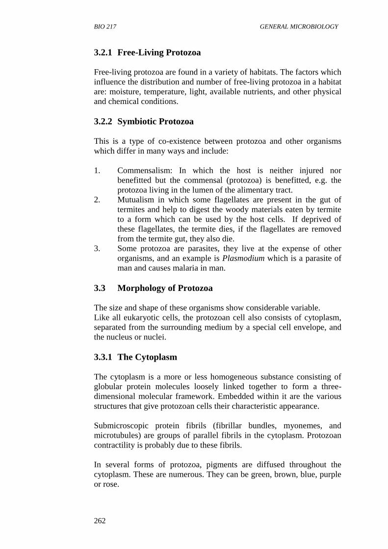
BIO 217 GENERAL MICROBIOLOGY
262
3.2.1 Free-Living Protozoa
Free-living protozoa are found in a variety of habitats. The factors which
influence the distribution and number of free-living protozoa in a habitat
are: moisture, temperature, light, available nutrients, and other physical
and chemical conditions.
3.2.2 Symbiotic Protozoa
This is a type of co-existence between protozoa and other organisms
which differ in many ways and include:
1. Commensalism: In which the host is neither injured nor
benefitted but the commensal (protozoa) is benefitted, e.g. the
protozoa living in the lumen of the alimentary tract.
2. Mutualism in which some flagellates are present in the gut of
termites and help to digest the woody materials eaten by termite
to a form which can be used by the host cells. If deprived of
these flagellates, the termite dies, if the flagellates are removed
from the termite gut, they also die.
3. Some protozoa are parasites, they live at the expense of other
organisms, and an example is Plasmodium which is a parasite of
man and causes malaria in man.
3.3 Morphology of Protozoa
The size and shape of these organisms show considerable variable.
Like all eukaryotic cells, the protozoan cell also consists of cytoplasm,
separated from the surrounding medium by a special cell envelope, and
the nucleus or nuclei.
3.3.1 The Cytoplasm
The cytoplasm is a more or less homogeneous substance consisting of
globular protein molecules loosely linked together to form a three-
dimensional molecular framework. Embedded within it are the various
structures that give protozoan cells their characteristic appearance.
Submicroscopic protein fibrils (fibrillar bundles, myonemes, and
microtubules) are groups of parallel fibrils in the cytoplasm. Protozoan
contractility is probably due to these fibrils.
In several forms of protozoa, pigments are diffused throughout the
cytoplasm. These are numerous. They can be green, brown, blue, purple
or rose.
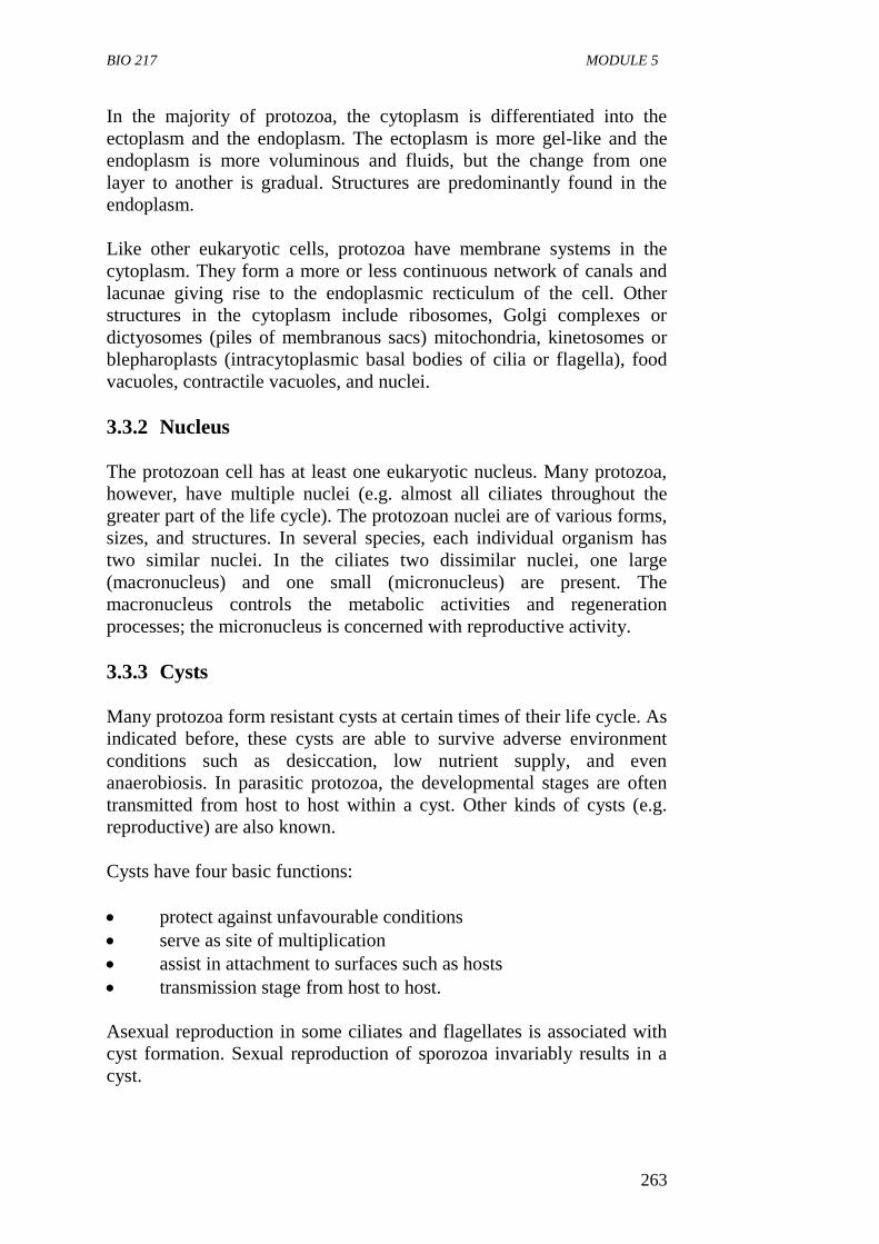
BIO 217 MODULE 5
263
In the majority of protozoa, the cytoplasm is differentiated into the
ectoplasm and the endoplasm. The ectoplasm is more gel-like and the
endoplasm is more voluminous and fluids, but the change from one
layer to another is gradual. Structures are predominantly found in the
endoplasm.
Like other eukaryotic cells, protozoa have membrane systems in the
cytoplasm. They form a more or less continuous network of canals and
lacunae giving rise to the endoplasmic recticulum of the cell. Other
structures in the cytoplasm include ribosomes, Golgi complexes or
dictyosomes (piles of membranous sacs) mitochondria, kinetosomes or
blepharoplasts (intracytoplasmic basal bodies of cilia or flagella), food
vacuoles, contractile vacuoles, and nuclei.
3.3.2 Nucleus
The protozoan cell has at least one eukaryotic nucleus. Many protozoa,
however, have multiple nuclei (e.g. almost all ciliates throughout the
greater part of the life cycle). The protozoan nuclei are of various forms,
sizes, and structures. In several species, each individual organism has
two similar nuclei. In the ciliates two dissimilar nuclei, one large
(macronucleus) and one small (micronucleus) are present. The
macronucleus controls the metabolic activities and regeneration
processes; the micronucleus is concerned with reproductive activity.
3.3.3 Cysts
Many protozoa form resistant cysts at certain times of their life cycle. As
indicated before, these cysts are able to survive adverse environment
conditions such as desiccation, low nutrient supply, and even
anaerobiosis. In parasitic protozoa, the developmental stages are often
transmitted from host to host within a cyst. Other kinds of cysts (e.g.
reproductive) are also known.
Cysts have four basic functions:
protect against unfavourable conditions
serve as site of multiplication
assist in attachment to surfaces such as hosts
transmission stage from host to host.
Asexual reproduction in some ciliates and flagellates is associated with
cyst formation. Sexual reproduction of sporozoa invariably results in a
cyst.

BIO 217 GENERAL MICROBIOLOGY
264
3.3.4 Locomotory Organelles
Protozoa may move by three types of specialised organelles:
pseudopodia, flagella and cilia. In addition, a few protozoa without such
organelles can carry out a gliding movement by body flexion.
1. Pseudopodia
A pseudopodium is a temporary projection of part of the
cytoplasm of those protozoa which do not have a rigid pellicle.
Pseudopodia are therefore characteristic of the amoebas
(sarcodina). These organelles are also used for capturing food
substances.
2. Flagella
The flagellum is an extremely fine filamentous extension of the
cell. As a rule, the number of flagella present in an individual
protozoan varies from one to eight; one or two is the most
frequent number. A flagellum is composed of two parts; an
elastic filament called an axoneme and the contractile
cytoplasmic sheath that surrounds the axoneme.
3. Cilia
1. They are fine and short threadlike extensions from the cell.
2. In addition to their locomotory function, also aid in the
ingestion of food and serve often as a tactile organelle.
They may be uniform in length, or may be of different
lengths depending on their location. Generally, cilia are
arranged in longitudinal, oblique, or spiral rows, inserted
either on the ridges or in the furrows.
3.3.5 Feeding Structure
Food-gathering structures in the protozoa are diverse and range from the
pseudopodia of amoebas through the tentacular feeding tubes of
suctorians to the well-developed “mouths” of many ciliates. Amoebas
gather food by means of pseudopodial engulfment. In ciliates the
cytostome is the actual opening through which food is ingested.
An oral groove is an indentation in the pellicle of certain ciliates. It
guides food toward the cytostome and acts as a concentrating device.
The addition of membranelles to the oral groove makes it a peristome.
Nutrition
1. Nutrition in protozoa is heterotrophic.
2. They obtain cellular energy from organic substances such as
proteins.
3. Protozoa engulf and ingest their food sources.
BIO 217 MODULE 5
265
3.4 Two Examples of Protozoan
3.4.1 Amoeba
Fig. 1a: An Amoeba
Source: www.infovisual.info
3.4.2 Paramecium
Fig. 1b: A Paramecium
Source: www.infovisual.info
BIO 217 GENERAL MICROBIOLOGY
266
3.5 Reproduction of Protozoa
Protozoa general multiply by asexual reproduction. Many protozoa are
able to carry out both asexual and sexual processes. Some parasitic
forms may have an asexual phase in one host and a sexual phase in
another host.
3.5.1 Asexual Reproduction
Asexual reproduction occurs by simple cell division, which can be equal
or unequal – the daughter cells are of equal or unequal sizes,
respectively. If two daughter cells are formed, then the process is called
binary fission. If many daughter cells are formed, it is called multiple
fission. Budding is a variation of unequal cell division.
1. Binary Fission
The simplest form of binary fission is found in the amoebas. The
pseudopodia are withdrawn before the nucleus divides. After the
nucleus divides, the organism elongates and constricts in the
centre in order to form two daughter cells.
2. Multiple Fission
In multiple fission, a single mother (parental) cell divides to form
many daughter (filial) cells. Division is usually preceded by
formation of multiple nuclei within the mother cell, which then
cleaves rapidly to form a corresponding number of daughter cells.
Multiple fission is not as widespread as binary fission but it often
takes place in addition to the latter process. In ciliates and
flagellates, this type of fission is found in relatively few species.
3. Budding
In protozoology it is often used to describe the varied processes
by which sessile protozoa produce motile offspring. That is, the
mother cell remains sessile and releases one or more swarming
daughter cells. The swarmer differs from the parent cell not only
in a lower degree of differentiation but also in the possession of
special locomotor organelles. Some form of budding is found in
all sessile ciliates and is used to disseminate the species while the
mother cell remains in situ.
3.5.2 Sexual Reproduction
Various types of sexual reproduction have been observed among
protozoa. Sexual fusion of two gametes (syngamy or gametogamy)
occurs in various groups of protozoa. They include:
Conjugation, which is generally a temporary union of two
individuals for the purpose of exchanging nuclear material, is a
BIO 217 MODULE 5
267
sexual process found exclusively in the ciliates. After exchange
of nuclei, the conjugants separate and each of them gives rise to
its respective progency by fission or budding. When the gametes
(which develop from trophozoites) are morphologically alike,
they are called isogametes. When they are unlike in morphology
(as well as physiology), they are anisogametes and can be either
microgametes or macrogametes.
3.6 Economic Importance of Protozoa
Protozoa are important links in the food chain of communities in
aquatic environment where they act as primary consumers.
They are used in biological treatment of sewage or industrial
effluents.
Some protozoa cause disease in mammals including man.
They are important research organisms for biologists and
chemists.
4.0 CONCLUSION
Protozoa are eukaryotic heterototrophic microorganisms that are
classified on the basis of morphological characteristics.
7.0 SUMMARY
Protozoa are unicellular non-photosynthetic microorganisms that
lack cell wall and have ability to move at some stages of their
life.
Protozoa are normally found in moist habitats.
Protozoa may be free living found in various habitats or
symbiotic found in co-existing with other organisms.
The size and shape of protozoa vary considerable and the cell
consists of the cytoplasm (separated from the surrounding
medium by an envelope) and the nucleus or nuclei.
Locomotory organelles in protozoa include pseudopodia, cilia
and flagella.
Reproduction in protozoa is mainly asexual, however many are
able to carry out both asexual and sexual reproduction.
Methods of asexual reproduction in protozoa include binary
fission, multiple fission and budding.
Sexual reproduction in protozoa involves the fusion of two
gametes.
Protozoa are primary consumer in food chain, in aquatic
environment. They are used to degrade biological and industrial
effluents. They also cause disease of man and other animals.
BIO 217 GENERAL MICROBIOLOGY
268
8.0 TUTOR-MARKED ASSIGNMENT
i. State five general characteristics of protozoa.
ii. Draw a well labeled diagram of amoeba.
iii. Write on three locomotory organelles in protozoa.
7.0 REFERENCES/FURTHER READING Atlas, R.M. (1995). Microorganisms in Our World. Mosby Year Book.
Inc.
Medigan, M.T. et al. (2009). Brock Biology of Microorganisms. (12th
ed.). Pearson Education Inc.
Pelczar, M.J., Chan, E.C.S. & Krieg, R.N. (2001). (5th
ed.).
Microbiology. McGraw-Hill.
Willey, J.M., Sherwood, L.M & Woolverton, C.J. (2008). Microbiology.
(7th
ed.). Boston Bur Bridge, IL: McGraw-Hill Higher Education.
www.infovisual.info
BIO 217 MODULE 5
269
MODULE 3 MICROBIAL GROWTH, REPRODUCTION
AND CONTROL
Unit 1 Microbial Growth
Unit 2 Measurement of Microbial Growth and Factors that
Influence Microbial Growth
Unit 3 Physical Methods of Controlling Microbial Growth
Unit 4 Chemical Methods of Controlling Microbial Growth
UNIT 1 MICROBIAL GROWTH
CONTENTS
1.0 Introduction
2.0 Objectives
3.0 Main Content
3.1 The Prokaryotic Cell Cycle
3.2 Binary Fission
3.2.1 Stages in Binary Fission
3.2.2 Chromosome Replication and Partitioning
3.2.3 Cytokinesis
3.3 The Growth Curve
3.3.1 Lag Phase
3.3.2 Exponential Phase
3.3.3 The Stationary Phase
3.3.4 Death Phase
3.4 The Mathematics of Growth
3.5 The Continuous Culture of Microorganism
4.0 Conclusion
5.0 Summary
6.0 Tutor-Marked Assignment
7.0 References/Further Reading
1.0 INTRODUCTION
Microbial growth is defined as an increase in the number of cells. A
microbial cell has a lifespan and a species is maintained only as a result
of continued growth of its population. Growth is the ultimate process in
the life of a cell – one cell becoming two and subsequently leading to an
increase in the number in a population of microorganisms. In
microbiology, growth is synonymous to reproduction. This unit
examines the term growth, binary fission, the mode of cell division in
prokaryotic cells, stages in the growth curve and the mathematics of
growth.
BIO 217 GENERAL MICROBIOLOGY
270
2.0 OBJECTIVES
At the end of this unit, you should be able to:
define the term growth
explain the process of a binary fission in a prokaryotic cell
draw and explain the microbial growth curve of microorganisms
in a batch culture
explain the mathematics of growth
describe a continuous culture.
3.0 MAIN CONTENT
Definition of Growth
Growth is defined as an increase in the number of cells in a population
of microorganisms. It is an increase in cellular constituents leading to a
rise in cell number when microorganisms reproduce by processes like
binary fission or budding.
3.1 The Prokaryotic Cell Cycle
A prokaryotic cell cycle is the complete sequence of events from the
formation of a new cell through the next division. Most procaryotes
reproduce by binary fission, budding or fragmentation.
3.2 Binary Fission
Binary fission is a form of asexual reproduction process. In which a
single cell divides into two cells after developing a transverse septum
(crosswall).
Binary fission is a simple type of cell division and the processes
involved are: the cell elongates, replicates its chromosomes and
separates the newly formed DNA molecules so that there is a
chromosome in each half of the cell. A septum is formed at mid cell;
divide the parent cell into two progeny cells and each having its own
chromosome and a copy or complement of other cellular constituents.
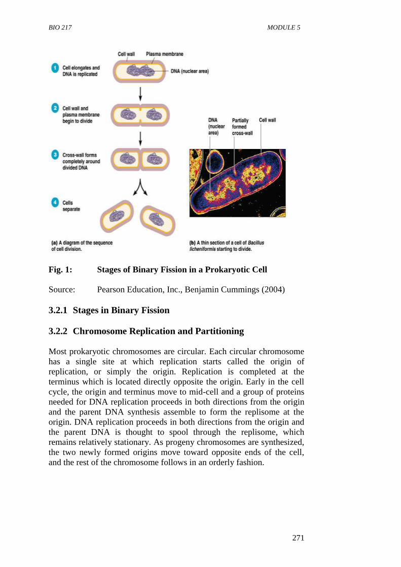
BIO 217 MODULE 5
271
Fig. 1: Stages of Binary Fission in a Prokaryotic Cell
Source: Pearson Education, Inc., Benjamin Cummings (2004)
3.2.1 Stages in Binary Fission
3.2.2 Chromosome Replication and Partitioning
Most prokaryotic chromosomes are circular. Each circular chromosome
has a single site at which replication starts called the origin of
replication, or simply the origin. Replication is completed at the
terminus which is located directly opposite the origin. Early in the cell
cycle, the origin and terminus move to mid-cell and a group of proteins
needed for DNA replication proceeds in both directions from the origin
and the parent DNA synthesis assemble to form the replisome at the
origin. DNA replication proceeds in both directions from the origin and
the parent DNA is thought to spool through the replisome, which
remains relatively stationary. As progeny chromosomes are synthesized,
the two newly formed origins move toward opposite ends of the cell,
and the rest of the chromosome follows in an orderly fashion.
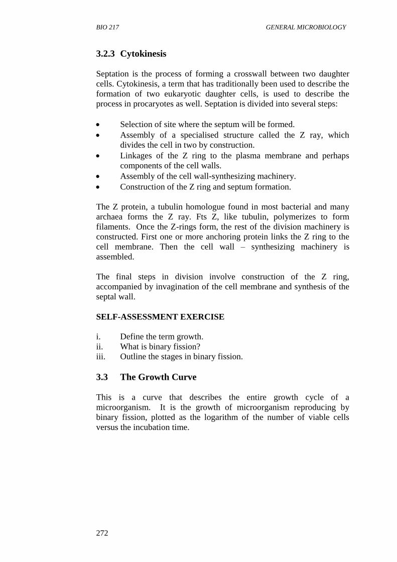
BIO 217 GENERAL MICROBIOLOGY
272
3.2.3 Cytokinesis
Septation is the process of forming a crosswall between two daughter
cells. Cytokinesis, a term that has traditionally been used to describe the
formation of two eukaryotic daughter cells, is used to describe the
process in procaryotes as well. Septation is divided into several steps:
Selection of site where the septum will be formed.
Assembly of a specialised structure called the Z ray, which
divides the cell in two by construction.
Linkages of the Z ring to the plasma membrane and perhaps
components of the cell walls.
Assembly of the cell wall-synthesizing machinery.
Construction of the Z ring and septum formation.
The Z protein, a tubulin homologue found in most bacterial and many
archaea forms the Z ray. Fts Z, like tubulin, polymerizes to form
filaments. Once the Z-rings form, the rest of the division machinery is
constructed. First one or more anchoring protein links the Z ring to the
cell membrane. Then the cell wall – synthesizing machinery is
assembled.
The final steps in division involve construction of the Z ring,
accompanied by invagination of the cell membrane and synthesis of the
septal wall.
SELF-ASSESSMENT EXERCISE
i. Define the term growth.
ii. What is binary fission?
iii. Outline the stages in binary fission.
3.3 The Growth Curve
This is a curve that describes the entire growth cycle of a
microorganism. It is the growth of microorganism reproducing by
binary fission, plotted as the logarithm of the number of viable cells
versus the incubation time.
BIO 217 MODULE 5
273
Stationery phase
Exponential (log)
phase Death
phase
Lag
phase
Time
Fig. 2: Microbial Growth Curve in a Close System
Source: Prescott, Harley and Kleins, Microbiology by Willey et
al., (2008)
When microorganisms are cultivated in liquid medium they are usually
grown in a batch culture or closed system. That is, they are incubated in
a closed culture vessel with a single batch of medium.
The growth curve has four phases: the lag phase, the exponential phase,
the stationary phase and the death phase.
3.3.1 Lag Phase
This is a phase in which there is no increase in the cell number of a
microbial population freshly introduced into a fresh culture medium. In
this phase, there is no cell division and growth. However, the cell is
metabolically active synthesizing new components.
Lag phase before cell division begins may be necessary for these
reasons:
i The cell may be old and depleted of ATP, essential cofactors and
ribosome which the cell synthesises at this phase or stage.
ii The new medium may be different from the one the
microorganism was growing in previously; the cells synthesise
new enzymes to be used in the new medium.
iii The cell is acclimatising to a new environment.
iv The cells may be injured and require time to recover.
Lo
g N
um
ber
of
Via
ble
cel
ls
BIO 217 GENERAL MICROBIOLOGY
274
The period of the lag phase varies depending on the condition of the
microorganisms and the nature of the medium. The phase may be long,
if:
The inoculum is from an old culture or one that has been
refrigerated.
If the new medium is chemically different from the old one from
which the microorganism was taken. However, if a young,
actively growing culture is transferred to a fresh medium of the
same composition, the lag phase will be short or absent.
3.3.2 Exponential Phase
This is also known as the log phase. It is a period in which the
microorganisms are growing and dividing at the maximal rate possible
given their genetic potential, the nature of the medium and the
conditions under which they are growing. The rate of growth is constant
during this phase because the microorganisms are dividing and doubling
in number at regular interval.
It is during this period that the generation time of the organism is
determined.
The log of the number of cells plotted against time results in a straight
line. In this phase, the population is most nearly uniform in terms of
chemical composition of cells, metabolic activity and other
physiological characteristics exponential phase vary among different
bacterial.
The exponential growth is balanced growth this is because cellular
constituents are manufactured at constant rates relative to each other.
Unbalanced growth results if the nutrient level and other environmental
conditions change.
Unbalanced growth is growth during which the rate of synthesis of cell
components vary relative to each other until a new balanced state is
reached. This is observed in two types of experiments (1) shift up and
(2) shift down.
The shift up occurs when a culture of microorganism is transferred from
a nutritionally poor medium to a richer one while the shift down occurs
when a culture of microorganism is transferred from a rich medium to a
poor one.
BIO 217 MODULE 5
275
3.3.3 The Stationary Phase
This is a phase in which population growth ceases and the growth curve
becomes horizontal. In this phase, the total number of viable
microorganisms remains constant. This may result from a balance
between cell division and cell death or the population may simply cease
to divide but remain metabolically active.
Factors responsible for stationary phase when a required nutrient is
exhausted are:
i Nutrient limitation: If an essential nutrient has been used up, e.g.
O2 or carbon source and becomes unavailable to the
microorganisms, population growth will cease.
ii Accumulation of toxic waste products: e.g. Streptococci can
produce too much lactic acid and other organic acids from sugar
fermentation that their medium becomes acidic and growth is
inhibited.
iii When a critical population level has been reached: This will
prevent further dividing and doubling of the cells/when physical
conditions do not permit a further increase in population size.
Once the stationery growth phase is reached, there is no further net
increase in bacterial cell numbers.
3.3.4 Death Phase
This is a phase in which the number of viable cells begins to decline.
During this phase, the number of living cells decreases because the rate
of cell death exceeds the rate of new cell formation. The depletion of
essential nutrients and the accumulation of laboratory products such as
acids contribute to the death rate.
3.4 The Mathematics of Growth
During the exponential phase each microorganism is dividing at constant
interval which means the population will double in number during a
specific length of time called the generation time. One cell divides to
produce two cells. Thus, if we start with a single bacterium, the increase
in population is by geometric progression:
1 2 22 2
3 2
4 2
5 …2n
Where n= the number of generations. Each succeeding generation,
assuming no cell death, doubles the population. The total population N
at the end of a given time period would be expectedly:
BIO 217 GENERAL MICROBIOLOGY
276
N=1 X 2n ………….Eq. (1)
However, under practical conditions, the number of bacteria no
inoculated at time zero is not 1 but more likely several thousand, so the
formula now becomes:
N = N0 X 2n ………….Eq. (2)
Where N = total population at the end of given time period.
Solving equation (2) for n, we have:
log10N=log10N0 + nlog102
n=log10N-log10N0
log102 ………….Eq. (3)
N0= Initial population at a given time
n= the number of generation.
If we now substitute the value of Log102, which is 0.301, in the above
equation,
n = log10N-log10N0
0.301
n = 3.3 (log10N-log10N0)
………….Eq. (4)
Thus, using equation (4), we can calculate the number of generations
that have taken place, provided we know the initial population and the
population after the growth has occurred.
The generation time g (the time required for the population to double)
can be determined from the number of generations n that occur in a
particular time interval t.
Using equation 4 for n, the generation time can be calculated by the
formula.
g = t = t
n 3.3 (log10N – log 10N0) ..………….Eq. (5)
The growth rate R is the number of generations per hours) and it is the
reciprocal of the generation time (g). It also the slope of straight n
obtained when the log number of cells is plotted against the time.
BIO 217 MODULE 5
277
Hence we have: R = 3.3 log10N – log10 No
t
SELF-ASSESSMENT EXERCISE
i. In what phase of the growth curve are cells dividing in a regular
and orderly process?
ii. Why do cells enter the stationery phase?
3.5 The Continuous Culture of Microorganism
A continuous culture is an open culture. The continuous culture vessel
maintains a constant volume to which fresh medium is added at a
constant rate and an equal volume of spent culture medium is removed
at the same rate.
Once such a system is in equilibrium the chemostat volume, cell number
and nutrient status remain constant and the system is said to be in a
steady state.
Two major types of continuous culture system commonly used are:
i chemostats and
ii turbidostats.
The chemostat: A chemostat is a device in which a liquid medium is
continuously fed into the bacteria culture. It is an apparatus designed to
permit the growth of bacterial cultures at controlled rates. A chemostat
is constructed so that sterile medium is fed into the culture vessel at the
same rate as the spent media containing microorganisms is removed.
The culture medium for a chemostat possesses an essential nutrient in
limiting quantities. Hence, the growth rate is determined by the rate at
which new medium is fed into the growth chamber and the final cell
density depends on the concentration of the limiting nutrients.
The rate of nutrient exchange is expressed as the dilution rate (D), the
rate at which medium flows through the culture vessel relative to the
vessel volume, where f is the flow rate (ml/hr) and V is the vessel
volume (ml)
D= f
v
Both the microbial population and generation time are related to the
dilution rate. The generation time decreases as the dilution rate increase,
while the microbial population density remains unchanged over a wide
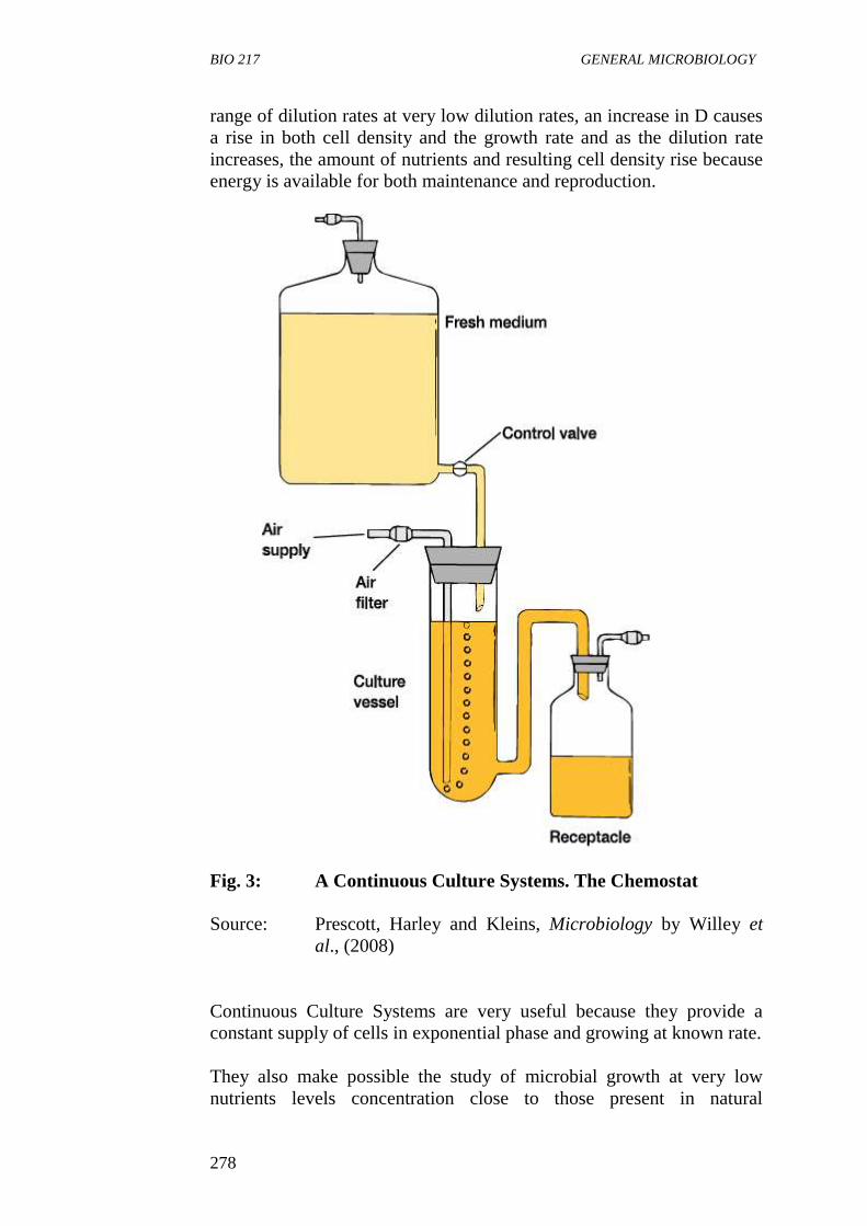
BIO 217 GENERAL MICROBIOLOGY
278
range of dilution rates at very low dilution rates, an increase in D causes
a rise in both cell density and the growth rate and as the dilution rate
increases, the amount of nutrients and resulting cell density rise because
energy is available for both maintenance and reproduction.
Fig. 3: A Continuous Culture Systems. The Chemostat
Source: Prescott, Harley and Kleins, Microbiology by Willey et
al., (2008)
Continuous Culture Systems are very useful because they provide a
constant supply of cells in exponential phase and growing at known rate.
They also make possible the study of microbial growth at very low
nutrients levels concentration close to those present in natural
BIO 217 MODULE 5
279
environments. These are useful for research in many areas. Continuous
systems are also used in food and industrial microbiology.
4.0 CONCLUSION
Growth is the ultimate process in the life of a cell. Microbial cells have a
finite lifespan. And a species is maintained only as a result of continued
growth of its population.
5.0 SUMMARY
Growth is an increase in the number of cell in a population.
Most procaryotes reproduce by binary fission, a process in which
the cell elongates and the chromosome is replicated and
segregates to opposite poles of the cell prior to the formation of a
septrum, which divides the cell into two progeny cells
Two overlapping pathways function during the procaryotic cell
cycle: the pathway for chromosome replication and segregation
and the pathway for septrum formation, both are complex and
poorly understood. The partitioning of the progeny chromosomes
may involve homologues of eucaryotic cytoskeletal proteins.
When microorganisms are grown in a closed system or batch
culture, the resulting growth curve usually has four phases; the
lag, exponential or log, stationery, and death phases.
In the exponential phase, the population number of cells
undergoing binary fission doubles at a constant interval called the
doubling or generation time. The mean growth rate constant (k)
is the reciprocal of the generation time.
Exponential growth is balance growth; cell components are
synthesized at constant rates relative to one another. Changes in
culture conditions (e.g. in shift-up and shift-down experiments)
lead to unbalanced growth. A portion of the available nutrients is
used to supply maintenance energy.
Microorganisms can be grown in an open system in which
nutrients are constantly provided and wastes removed.
A continuous culture system is an open system that can maintain
a microbial population in the log phase. There are two types of
these systems: chemostats and turbidostats.
The chemostat is a continuous device in which a liquid medium is
continuously fed into the bacteria culture.
BIO 217 GENERAL MICROBIOLOGY
280
6.0 TUTOR-MARKED ASSIGNMENT
i. Describe the four phases of the growth curve in a closed system.
ii. Define the terms (i) generation time and (ii) mean growth rate.
iii. During log-phase growth of a bacterial culture, a sample is taken
at 8.00 a.m. and found to contain 1,000 cells per milliliter. A
second sample is taken at 5.54 p.m. and is found to contain
1,000,000 cells per milliliters. What is the generation time in an
hour?
7.0 REFERENCES/FURTHER READING
Medigan, M.T., et al. (2009). Brock Biology of Microorganisms. 12th
Edition. Pearson Education Inc.
Pelczar, M.J., Chan, E.C.S. & Krieg, R.N. (1993). Microbiology. 5th
Edition. McGraw-Hill.
Willey, J.M., Sherwood, L.M & Woolverton, C.J. (2008). Microbiology.
7th
Edition. Boston Bur Bridge, IL: McGraw-Hill Higher
Education.
BIO 217 MODULE 5
281
UNIT 2 MEASUREMENT OF MICROBIAL GROWTH
AND FACTORS THAT INFLUENCE
MICROBIAL GROWTH
CONTENTS
1.0 Introduction
2.0 Objectives
3.0 Main Content
3.1 Measurement of Total Cell Number
3.1.1 Direct Count Methods
3.1.2 Viable Counting Methods
3.1.3 Most Probable Number (MPN) Technique
3.2 Measurement of Cell Mass
3.2.1 Determination of Microbial Dry Weight
3.2.2 Spectrophotometry
3.3 Factors Influencing Microbial Growth
3.3.1 Temperature
3.3.2 Oxygen Concentration
3.3.3 pH or Acidity
3.3.4 Solute and Water Activity
4.0 Conclusion
5.0 Summary
6.0 Tutor-Marked Assignment
7.0 References/Further Reading
1.0 INTRODUCTION
Measurement of microbial growth helps to determine growth rate and
generation time. Population growth is measured by following changes
in cell number and cell mass; this is because growth leads to increase in
both. Also, there are different factors that affect the growth rate of
microorganisms. This unit examines different methods of measurement
growth and factors affecting microbial growth.
2.0 OBJECTIVES
At the end of this unit, you should be able to:
describe the methods of measuring total sum number of
microorganisms
describe the methods of counting viable cells of microorganisms
describe the methods of measuring cells mass of microorganisms
state the advantage and disadvantages of the difference methods
of measuring microbial growth
BIO 217 GENERAL MICROBIOLOGY
282
explain how different environmental factors affect microbial
growth.
3.0 MAIN CONTENT
Measurement of Microbial Growth
Population growth is measured by following changes in the number of
cells or changes in the level of some cellular components. This could be
done by measuring the total cell number or by measuring the cell mass.
3.1 Measurement of Total Cell Number
The total number of microbial cells can be achieved by using direct
count methods.
3.1.1 Direct Count Methods
Bacteria, microorganisms can be enumerated by direct counting
procedure using:
i Special counting chambers such as hemocytometer and Petroff-
itausser chamber can be employed to determine the number of
bacteria. These chambers are ruled with squares of a know area
and are constructed in such a way that a film of liquid of known
depth can be introduced between the slide and the cover slips,
and viewed under the microscope. Consequently, the volume of
the sample overtyping each square is known. These specially
designed slides have chambers of known depth with an etched
grid on the chamber bottom. The number of microorganism in a
sample can be calculated by taking into account the chambers
volume and any sample dilution required.
Advantages
i Using a counting chamber is easy, inexpensive, and quick.
ii It gives information about the size and morphology of
microorganism.
Disadvantages
i. The microbial population must be fairly large for accuracy
because it involves sampling a small volume.
Prokaryotes are more easily counted in these chambers if they are
stained or when a phase contrast or fluorescence microscope is
used.
BIO 217 MODULE 5
283
ii. Large microorganisms such as protists and yeast can be directly
counted using electronic chambers such as the coulter counter
and the flow cytometer. The microbial suspension is forced
through a small hole or orifice in the coulter chamber. An
electrical current flow through the hole and electrodes placed on
both sides of the orifice measure its electrical resistance. Each
time a microbial cell passes through the orifice, electrical
resistance increases or the conductivity drops and the cell is
counted.
The coulter counter gives accurate results with larger cells and is
extensively used in hospital laboratories to count red and white
blood cells.
iii. The Membrane Filter Technique
The number of bacterial in aquatic sample can be determined
from direct counts after the bacteria have been trapped on special
membrane filters such as a nitrocellulose 0.2 µM pore size filter
or a black polycarbonate membrane filter. The sample is first
filtered through the membrane after which the bacterial are
stained with a fluorescent dye such as acridine orange or the
DNA stain DAPI and observed under the microscope.
The stained cells are easily observed against the black background of the
membrane filter and can be counted when viewed under the microscope.
The disadvantage of this method is that it does not distinguish between
dead cells and live cells.
3.1.2 Viable Counting Methods
These methods involve plating serial dilutions of a suspension of
microorganisms unto a suitable solid growth medium and after a period
of incubation (in which single cells multiply to form visible colonies)
the number of colonies are counted or enumerated. These methods are
referred to as viable counting methods because they count only those
cells that are alive and able to reproduce. Two commonly used methods
are the spread plate technique and the pour plate technique.
i In the spread plate technique, the suspension sample is spread
over the surface of an agar plate containing growth nutrients
while in the pour plate technique; the suspension is mixed with
the agar while it is still in the liquid state and poured into the
plate.
The plates are incubated to allow the organisms to grow and form
colonies. It is assumed that each colony arises from an individual
bacterial cell. By counting the number of colonies formed the
BIO 217 GENERAL MICROBIOLOGY
284
colony forming units (CFUs) and taking account of the dilution
factors, the concentration of bacteria in the original can be
calculated.
Two or three plates of the same sample are counted to determine
the viable number of bacteria. Countable plates are those having
between 30 to 300 colonies. Less than 30 colonies is not
acceptable for statistical reasons and more than 300 colonies are
likely to produce colonies too close to distinguish as individual
CFUs such samples are noted as TNTC (too numerous to count).
ii The membrane filter technique is another method that can be
used for viable counts of bacteria. The sample is poured through
the filter which traps bacteria. The filter is then placed on an agar
medium or a pad soaked with liquid media and incubated until
each cell forms a separate colony. A colony count gives the
number of microorganisms in the filtered sampled and special
media can be used selectively for specific microorganisms.
Advantages of Viable Counts Methods or Plating Techniques
i. They are simple technique sensitive.
ii. Widely used for viable counts of bacteria and other
microorganisms in samples of food, water and soil.
Disadvantages or Limitation
i. It is selective.
ii. The nature of the growth medium and the incubation condition
determine which bacteria can be grown and counted.
iii. Sometimes, cells are viable but not culturable.
The viable plate count technique is selective because no one
combination of media allows the growth of all types of bacteria.
3.1.3 Most Probable Number (MPN) Technique
Most probable number is a statistical method based on probability
theory. In this method, multiple serial dilutions are performed to reach a
point of extinction. The point of extinction is the dilution level at which
no single cell is deposited into one or more multiple tubes.
A criterion such as development of cloudiness or turbidity or gas
production is established for indicating whether a particular dilution
contains bacteria.
BIO 217 MODULE 5
285
The pattern of positive and negative test results are then used to estimate
the concentration of bacteria in the original sample by comparing the
observed pattern of result with a table of statistical probabilities for
obtaining those results.
SELF-ASSESSMENT EXERCISE
Briefly describe methods by which total number of microbial cells can
be measured.
3.2 Measurement of Cell Mass
Techniques for measuring changes in cell mass can be used to measure
growth of microorganisms. They include:
3.2.1 Determination of Microbial Dry Weight
Cells growing in liquid medium are collected by centrifugation, washed,
dried in an oven and weighed. This is a useful technique for measuring
growth of filamentous fungi; however, it is time consuming and not very
sensitive.
3.2.2 Spectrophotometry
This method depends on the fact that microbial cells scatter light that
strikes them because microbial cells in a population are of roughly
constant size; the amount of scattering is directly proportioned to the
biomass of cell present and indirectly related to cell number. When the
concentration reaches about 10 million cells (107) per ml, the medium
appears slightly cloudy or turbid. Further increase in concentration result
in greater turbidity and less light is transmitted through the medium. The
extent of light scattering can be measured by a spectrophotometer and is
almost linearly related to cell concentration at low absorbance level. If
the amount of a substance in each cell is constant, the total quantity of
the cell constituted is directly related to the total microbial cell mass.
3.4 Factors Influencing Microbial Growth
The rate of microbial growth and death are greatly influenced by
environmental factors or parameters.
Some environmental conditions favour rapid microbial growth
while others do not permit bacterial reproduction.
Understanding the influence of environmental factors on
microorganisms helps in the control of microbial growth and the
study of ecological distribution of microorganisms.
BIO 217 GENERAL MICROBIOLOGY
286
These factors include: temperature, solute, and water activity, pH,
oxygen level, pressure, radiation.
The growth and activities of microorganisms are greatly affected by
physical and chemical state of the environment. The four key factors are
temperature, pH, water and availability.
SELF-ASSESSMENT EXERCISE
Briefly describe methods by which the total number of microbial cells
can be measured.
3.3.1 Temperature
Temperature is the most important factor affecting the growth and
survival of microorganism. At either too cold or too hot temperature,
microorganisms will not be able to grow and may even die.
For every microorganism, there is a minimum temperature below which
growth is not possible, an optimum temperature at which growth is most
rapid and a maximum temperature above which growth is not possible.
These three temperatures are called the cardinal temperature and are
characteristics for any given microorganism.
The Temperature Classes of Microorganisms
Microorganisms can be placed in five classes based on their temperature
for growth:
i Psychrophiles: These are organisms that grow well at 0ºC and
have an optimum growth temperature of 150C or lower. The
maximum is around 100C. They are readily isolated from arctic
and antartic habitat, many psychrophiles are found in the ocean.
ii Psychrotrophs or Facultative Psychrophiles: These organisms can
grow at 0ºC to 7ºC even though they have optima between 20ºC
and 30ºC and maxima at about 35ºC. These organisms are the
major causes of spoilage of refrigerated food.
iii Mesophiles: These are microorganism with growth optima
around 20ºC to 45ºC. They often have temperature minimum of
15ºC to 20ºC. Their maximum is about 45ºC or lower. Examples
of this group of microorganisms are the human pathogens
because of their environment the body is fairly constant 37ºC
iv Thermophiles: These microorganisms can grow at temperature of
55ºC or higher. Their growth minimum is usually around 45ºC
and they often have optima between 55ºC and 60ºC. Majority of
thermophiles are prokaryotes although a few photosynthetic
BIO 217 MODULE 5
287
protists and fungi are thermophiles. These organisms flourish in
many habitats including compost, heating hay stacks, hot water
line and hot spring.
v Extreme Thermophiles or Hyperthermophiles: These are
prokaryotes that have growth optima between 80ºC and 113ºC.
They do not grow well below 55ºC. Pyrococcus abyss and
Pyrodictum are example of marine hyperthermophiles found in
hot areas of the seafloor.
3.3.2 Oxygen Concentration
The importance of oxygen to the growth of an organism correlate with
its metabolism especially with the processes it uses to conserve the
energy supplied by its source. Based on the ability to grow in the
presence or absent of oxygen. Microorganisms are classified as:
i Aerobes: These are organism that able to grow in the presence of
atmospheric oxygen.
ii Anerobic: They grow in the absence of atmospheric oxygen
iii Facultative: these are organism that do not require oxygen for
growth but grow better in its presence.
iv Aerotolrant Anaerobe: These are not dependent on oxygen. They
grow equal whether oxygen is present or absent, e.g. Enterococus
faccalis.
v Strick or Obligate Anaerobe: They do not tolerate oxygen at all
and due to its presence, e.g. Clostridium pasteurianum.
vi Microaerophile: These organisms are damaged by normal
atmospheric level of oxygen (20%) and require O2 level below
the range of 2 to 10% for growth, e.g. Campylobacter.
A microbial group may show more than one type of relationship to
oxygen (O2). A type is found among the prokaryotes and protozoa.
Fungi are normally aerobic but a few species particularly among the
yeasts are facultative anaerobes. Photosynthetic protists are almost
always obligate aerobes.
3.3.3 pH or Acidity
pH is a measure of the hydrogen ion activity of a solution and is defined
as the negative logarithms of the hydrogen concentration (expressed in
terms of molarity).
pH = -log(H+) = log(1/(H
+))
BIO 217 GENERAL MICROBIOLOGY
288
The pH scale extends from ph 0.0 to ph 14 and each pH unit represents a
tenfold change in hydrogen ion concentration. Based on pH growth
range and pH growth optimum we have the following group of
organisms.
i Acidophiles: They have their growth optimum between ph 0 and
5.5.
ii Neutrophiles: Growth optimum between 5.5 and 8.0.
iii Alkalophiles: Growth optimum between 8.0 and 11.5.
Extreme alkalophiles have growth optima at pH 10 or higher. In general,
different microbe groups have characteristics pH preference. Most
bacteria and protists are neurophiles. Most fungi are acidophiles (pH
between 4 and 6). Photosynthetic protist also favours slight acidity.
Many archaea are acidophiles.
3.3.4 Solute and Water Activity
This is the ability of a microorganism to grow over a wide range of
water activity or osmotic concentration. Selectively permeable plasma
membrane separates microorganisms from their environment; hence
they are affected by changes in the osmotic concentration of their
surroundings. If a microorganism is placed in hypotonic solution (one
with a lower osmotic concentration) water will enter the cell and cause it
to burst if nothing is done to prevent it. On the other hand, if a
microorganism is placed in hypertonic solution (one with a higher
osmotic concentration) water will flow out of the cell.
In microbes that have cell walls (i.e. most prokaryotes, fungi and algae),
the membrane shrinks away from the cell wall - a process called
plasmolysis. The amount of water activity actually available to
microorganism is expressed in terms of water activity (aw). Some
microbes are adapted to extreme hypertonic environment.
In microbes that have cell wall (i.e. most prokaryotes, fungi and algae)
the membrane shrinks away from the cell wall by a process called
plasmolysis. Some organisms are adapted to extreme hypertonic
environment. Microorganisms usually have a specific requirement for
NaCl in addition to growing optimally at the water activity of seawater
such organism are called halophiles. Halophiles grow optimally in the
presence of NaCl or other salt at a concentration above 0.2M.
Most microorganisms are unable to cope with environment of very low
water activity and either die or become dehydrated and dormant under
such condition. Halotolerant organisms can tolerate some reduction in
BIO 217 MODULE 5
289
the aw of their environment but grow best in the absence of the added
solute. By contrast, some organisms thrive and indeed require low water
activity for growth.
Organisms able to live in environments high in sugar as a solute are
called Osmophiles and those able to grow in very dry environments
(made dry by lack of water rather than by dissolved solute) are called
xerophiles.
Radiation
Sunlight is the major source of radiation on the earth. It includes visible
light, ultraviolet (UV) radiation, infrared ray, and radio waves. Visible
light is a most conspicuous and important aspect of our environment.
Most life is dependent on the ability of photosynthetic organisms to trap
the light energy of the sun. Most forms of electromagnetic radiation are
very harmful to microorganisms. This is particularly true of ionizing
radiation, radiation of very short wavelength and high energy, which can
cause atoms to lose electrons (ionize). Two major forms of ionizing
radiation are:
i X -rays, which are artificially produced, and
ii Gamma rays which are emitted during radioisotope decay.
Low level of ionization will produce mutation and may indirectly result
in death, whereas higher levels are directly lethal. Ultraviolet (UV)
radiation can kill all kinds of microorganism due to its short wavelength
(approximately from 10-400nm) and high energy the most effectively
absorbed by DNA.
SELF-ASSESSMENT EXERCISE
Briefly describe the five classes of microorganisms based on
temperature.
4.0 CONCLUSION
The growth of microorganisms is greatly affected by the chemical and
physical nature of their environment. An understanding of the factors
that influence microbial growth aids ecological distribution of
microorganisms.
BIO 217 GENERAL MICROBIOLOGY
290
9.0 SUMMARY
Microbial population can be counted directly with counting
chambers, electronic counters, or fluorescence microscope.
Viable counting techniques such as the spread plate, the pour
plate, or the membrane filter can be employed.
Most bacterial, photosynthetic protists, and fungi have rigid cell
walls and are hypertonic to the habitat because of solutes such as
amino acids, polyols, and potassium ions. The amount of water
actually available to microorganism is expressed in terms of the
water activity (aw).
Each species of microorganism has an optimum pH for growth
and can be classified as an acidophile, neutrophile, or alkalophile.
Microorganisms can alter the pH of their surroundings, and mos
culture media must be buffered to stabilise the pH.
Microorganisms have distinct temperature ranges for growth with
minima, maxima, and optima – the cardinal temperatures. These
ranges are determined by the effects of temperature on the rates
of catalysis, protein denaturation, and membrane disruption.
There are five major classes of microorganisms with respect to
temperature preferences: (1) psychrophiles (2) facultative
psychrophiles or psychrotrophs, (3) mesophiles, (4) thermophiles,
and (5) hyperthermophiles.
Microorganisms can be placed into at least five different
categories based on their response to the presence of O2: obligate
aerobes, facultative anaerobes, aerotolerant anaerobes, strict or
obligate anaerobes, and microaerophiles.
Most deep-sea microorganisms are barotolerant, but some are
barophilic and require high pressure for optimal growth.
High-energy or short-wavelength radiation harms organisms in
several ways ionizing radiation. X-rays and gamma rays – ionizes
molecules and destroys DNA and other cell components.
Ultraviolent (UV) radiation induces the formation of thymine
dimmers and strand breaks in DNA.
10.0 TUTOR-MARKED ASSIGNMENT
i. What are the advantages and disadvantages of the viable plate
count method?
ii. What are cardinal temperatures?
iii. Describe the types of oxygen relationship seen in microorganism.
BIO 217 MODULE 5
291
7.0 REFERENCES/FURTHER READING
Atlas, R.M. (1995). Microorganisms in Our World. Mosby Year Book.
Inc.
Medigan, M.T. et al. (2009). Brock Biology of Microorganisms.
(12th
ed.). Pearson Education Inc.
Pelczar, M.J., Chan, E.C.S. & Krieg, R.N. (2001). (5th
ed.).
Microbiology. McGraw-Hill.
Willey, J.M., Sherwood, L.M & Woolverton, C.J. (2008). Microbiology.
(7th
ed.). Boston Bur Bridge, IL: McGraw-Hill Higher Education.
BIO 217 GENERAL MICROBIOLOGY
292
UNIT 3 PHYSICAL METHODS OF CONTROLLING
MICROBIAL GROWTH (STERILISATION)
CONTENTS
1.0 Introduction
2.0 Objectives
3.0 Main Content
3.1 The Pattern of Microbial Death
3.2 Heat Sterilisation
3.2.1 Measuring Heat Sterilisation
3.2.2 Moist Heat Sterilisation
3.2.3 Dry Heat Sterilisation
3.3 Filter Sterilisation/Filtration
3.4 Radiation
3.4.1 Ionising Radiation
3.4.2 Ultraviolet (UV) Radiation
4.0 Conclusion
5.0 Summary
6.0 Tutor-Marked Assignment
7.0 References/Further Reading
1.0 INTRODUCTION
Most microorganisms are beneficial to human beings; however, many
have undesirable consequences. They cause food spoilage and many
diseases to plants, animal and human. To control and inhibit the growth
and activities of harmful microorganisms which are capable of causing
diseases and contaminating water, food and substances used. This unit
examines the various frequently employed physical methods of
sterilisation which are: heat, filtration and radiation. Different
sterilisation techniques such as heat, radiation are used which totally
remove microorganisms from an object or habitat.
2.0 OBJECTIVES
At the end of this unit, you should be able to:
define and explain the term sterilisation
explain the use of heat sterilisation to control microbial growth
describe the use of various filters to sterilise liquid substances
explain the use of radiation for sterilisation.
BIO 217 MODULE 5
293
3.0 MAIN CONTENT
Definition of Sterilisation
Sterilisation is the process by which all living cells, spores and acellular
entities (e.g.) viruses, viroids and prions) are either destroyed or
removed from an object or habitat.
It can also be defined as the killing or removal of all viable organisms
within a growth medium.
A sterile object is totally free of viable microorganisms, spores and other
infectious agents.
3.1 The Pattern of Microbial Death
Microbial population death is exponential or logarithmic, meaning the
population will be reduced by the same fraction at constant interval. If
the logarithm of the population remaining is plotted against the time of
exposure to the agent, a straight-line plot will result. A bacterium is
often defined as dead if it is not viable and does not grow and reproduce
when inoculated into a culture medium that normally supports its
growth.
3.2 Heat Sterilisation
The most common sterilisation method used for controlling and
destroying microbial growth is the use of heat.
Heat can kill microorganisms by denaturing the enzymes which
prevent them from multiplying.
At temperature exceeding the maximal growth temperature, the
death rate exceeds the growth rate.
Factors that determine the effectiveness of heat sterilization
include the temperature and duration of the heat treatment, and
whether the heat is moist or dry
3.2.1 Measuring Heat Sterilisation
All microorganisms have a maximum growth temperature beyond which
viability decreases. Viability is lost because at very high temperatures
most macromolecules lose structure and function, a process called
denaturation. The effectiveness of heat as a sterilant is measured by the
time required for a tenfold reduction in the viability of a microbial
population at a given temperature. This is the decimal reduction time or
D. The time and temperature, therefore, must be adjusted to achieve
sterilisation for each specific set of condition. In addition, the type of
heat is also important. Moist heat has been observed to possess better
BIO 217 GENERAL MICROBIOLOGY
294
penetrating power than dry heat and, at a given temperature, produces a
faster reduction in the number of living organisms.
The determination of a decimal reduction time requires a large number
of viable count measurements. An easier way to characterise the heat
sensitivity of an organism is to measure the thermal death point i.e. the
time it takes to kill all cells at a given temperature. Thermal death point
(TDP) is the lowest temperature required to kill all the microorganisms
in a liquid suspension in 10 minutes. Heat is the most widely applicable
and effective agent for killing microorganisms and also the most
economical and easily controlled. To determine the thermal death time,
samples of a cell suspension are heated for different times, mixed with a
culture medium and incubated. If all the cells have been killed, no
growth is observed in the incubated samples. The thermal death time
depends on the size of the population tested; a longer time is required to
kill all cells in a large population than in a small one.
3.2.2 Moist Heat Sterilisation
Moist heat readily kills viruses, bacteria and fungi. It kills by degrading
nucleic acids and by denaturing enzymes and other essential proteins.
Exposure of microorganisms to boiling water will destroy vegetable
cells and eukaryotic spores. However, this temperature will not kill
bacterial endospores.
In order to destroy bacterial endospores moist heat sterilization above
1000C using saturated steam under pressure is done. Steam sterilization
is carried out with an autoclave.
The Autoclave
The autoclave is a sealed heating device that allows the entrance of
steam under pressure. The killing of heat-resistant endospores requires
heating at temperatures above 1000C (the boiling point of water at
normal atmospheric pressure). This is accomplished by applying steam
under pressure at a temperature of 1210C.
The materials to be sterilised are placed in a chamber and the chamber is
sealed. Steam is transferred from a jacket into the chamber forcing out
all the air. The steam is held in the chamber for 15 minutes at 1210C and
then vented from the chamber.
If an object being sterilised is bulky, heat transfer to the interior is
retarded and the total heating time must be extended to ensure that the
entire object is at 1210C for 10-15 minutes. Extended times are also
required when large volumes of liquid are being autoclaved because
large volumes take longer time to reach sterilisation temperatures. It
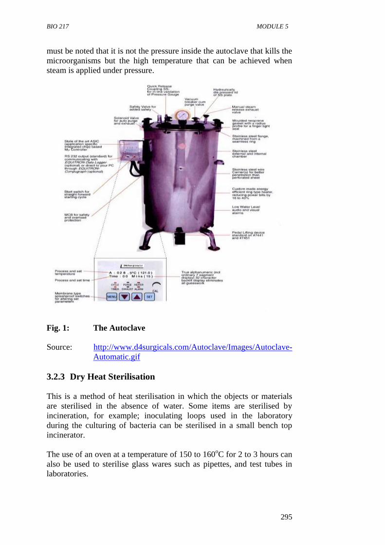
BIO 217 MODULE 5
295
must be noted that it is not the pressure inside the autoclave that kills the
microorganisms but the high temperature that can be achieved when
steam is applied under pressure.
Fig. 1: The Autoclave
Source: http://www.d4surgicals.com/Autoclave/Images/Autoclave-
Automatic.gif
3.2.3 Dry Heat Sterilisation
This is a method of heat sterilisation in which the objects or materials
are sterilised in the absence of water. Some items are sterilised by
incineration, for example; inoculating loops used in the laboratory
during the culturing of bacteria can be sterilised in a small bench top
incinerator.
The use of an oven at a temperature of 150 to 160oC for 2 to 3 hours can
also be used to sterilise glass wares such as pipettes, and test tubes in
laboratories.
BIO 217 GENERAL MICROBIOLOGY
296
Dry heat kills the microorganisms by the oxidation of the cell
constituents and denaturation of proteins.
This method of sterilisation has some definite advantages, it does not
corrode glassware and metal instruments as moist heat does and it can
be used to sterilise powders, oils and other similar items. However, it is
very slow and less effective than moist heat. For example, the spores of
Clostridium botulinum the organism that causes botulism are killed in 5
minutes at 1210C by moist heat but only after two hours at 160
0C with
dry heat.
SELF-ASSESSMENT EXERCISE
i. Define the term sterilisation
ii. Explain the use of moist heat for sterilisation.
3.3 Filter Sterilisation/Filtration
1. Filtration is a method that accomplishes decontamination and
even sterilisation.
2. Heat-sensitive liquids and gases are sterilised by the use of
filtration method.
3. The liquid or gas is passed through a filter, a device with pores
too small for the passage of microorganisms, but large enough to
allow the passage of the liquid or gas. The selection of filters for
sterilisation must account for the size range of the contaminants
to be excluded.
4. Some microbial cells are greater than 10 µm in diameter, and the
smallest bacteria are less than 0.3 µm in diameter. Rather than
directly destroy the contaminating microorganism the filters
simply removes them.
5. There are two types of filters:
a Depth Filters
b Membrane Filters.
Depth Filters
This is made up of fibrous or granular materials that have been bonded
into a thick layer filled with twisting channels of small diameters.
They can also be made of diatomaceous earth, unglazed porcelain,
asbestos, bromosilicates and other similar materials.
BIO 217 MODULE 5
297
The solution containing microorganisms is sucked through the thick
layer of the fibrous material under vacuum and microbial cells are
removed by physical screening or entrapment and by adsorption to the
surface of the filter material.
They are used for the filter sterilisation of air in industrial processes. In
the home, the filter is used in forced air heating and cooling systems is a
simple depth filter, designed to trap particulate matter such as dust,
spores, and allergens.
Depth filters are important for bio-safety applications. For example,
manipulation of cell cultures, microbial cultures, and growth media
require that contamination of both the operator and the experimental
materials are minimal. These operations can be efficiently performed in
a biological safety cabinet with airflow, both in and out of the cabinet,
directed through a depth filter called a HEPA filter or high-efficiency
particulate air filter. A typical HEPA-Filter is a single sheet of
borosilicate (glass) fibers that has been treated with a water-repellant
binder. It removes 0.3 µM test particles with an efficiency of at least
99.97%; they thus effectively remove both small and large particles,
including most microorganisms, from the airstreams.
Membrane Filters
Membrane filters are the most common type of filters used for
liquid sterilization in the microbiology laboratory.
They are porous membrane, a little over 0.1mm thick, made of
cellulose acetate, cellulose nitrate polycarbonate, polyvinylidene
fluoride and other synthetic materials.
Membranes with pores about 0.2µM in diameter are used to
remove most vegetative cells but not viruses from solutions
ranging in volume from 1ml to many litres.
The membrane is held in a special holder and is often preceded
by depth filter to remove larger particles that may clog the
membrane filter.
It differs from the depth filter because it functions more like a
sieve and trapping particles on the filter surface. About 80-85%
of the membrane surface area consists of open pores. The
porosity provides for a relatively high fluid flow rate.
The solution to be sterilised is forced through the filter with a
vacuum or with pressure from a syringe, peristaltic pump and
collected in a previously sterilised container.
They are used to sterilise pharmaceutical ophthalmic solutions,
culture media, oils, antibiotics and other heat sensitive solutions.
BIO 217 GENERAL MICROBIOLOGY
298
Air can also be sterilised by filtration. Examples are surgical
masks and cotton plugs on culture vessels that let air in but keep
microorganisms out.
SELF-ASSESSMENT EXERCISE
Explain the use of membrane filter for sterilisation.
3.4 Radiation
Many forms of electromagnetic radiation are very harmful to
microorganisms. Microwaves, ultraviolet (UV) radiation, X-rays,
gamma rays (Y-rays) and electrons can effectively reduce microbial
growth if applied in the proper close.
3.4.1 Ionising Radiation
Ionising radiation is a form of radiation which has very short wave
length and high energy, which can cause atoms to lose electrons
(ionize). Two major forms of ionising radiation are:
i X-rays (short wavelength of 10-3
to 102 nanometers) which are
artificially produced.
ii Gamma rays (short wavelength of 10-3
to 10-1
nanometer) which
are emitted during radioisotope decay.
Low levels of doses of Radiation will produce mutation in the
microorganisms which may indirectly lead to the death of
microorganisms. In cases where large doses or high levels of these
radiation are used the microorganisms are directly and instantly killed.
Ionising radiation can be used to sterilise items.
Gamma and X-radiation have high penetrating power and are able to kill
microorganisms by inducing or forming toxic free radicals (ions) viruses
and other microorganisms are inactivated by exposure to ionising
radiation. Ionising radiations are used to pasteurise or sterilise products;
e.g. most commercially produced disposables and plastic petri dishes are
sterilised by exposure to gamma rays.
3.4.2 Ultraviolet (UV) Radiation
These are radiation of short wavelength (from 10 to 400µm) and
high energy.
The most lethal UV radiation has a wavelength of 260µm, the
wavelength mostly absorbed by DNA.
BIO 217 MODULE 5
299
The primary mechanism of UV damage is the formation of
thymine dimmers in DNA.
Two adjacent thymines in a DNA strand are covalently joined to
inhibit DNA replication and function.
Ultraviolet (UV) radiation can kill all kinds of microorganisms
especially microorganisms on or near the surface of clear solution
exposure to ultraviolet rays can be used to maintain sterility of
surfaces such as bench tops in laboratories and hospitals.
Radiation is currently used for sterilisation and decontamination in the
medical supplies and food industries. In the U.S. for example, the Food
and Drug Administration has approved the use of radiation for
sterilisation of such diverse items as surgical supplies, disposable
labware, drugs, and even tissue grafts. However, because of the costs
and hazards associated with radiation equipment, this type of
sterilisation is limited to large industrial applications or specialised
facilities. The use of radiation sterilisation practice has not been readily
accepted in some countries because of fears of possible radioactive
contamination, alteration in nutritional value, production of toxic or
carcinogenic products, and perceived “off” tastes in irradiated food.
SELF-ASSESSMENT EXERCISE
Why is ionising radiation more effect than ultraviolet radiation for
sterilisation of food products?
4.0 CONCLUSION
Different methods of sterilisation are used to sterilise different types of
objects and to make them free of viable microorganism spores and other
infectious objects.
9.0 SUMMARY
Sterilisation is the process by which all living cells, spores and
cellular entities (viruses) are either completely destroyed or
removed from an object or habitat.
Physical methods frequently used to sterilise are heat, low
temperature, filtration and radiation.
Heat sterilisation is used to kill all microorganisms in a sample.
It may be moist or dry. Moist heat sterilisation involves the use
of autoclave.
Autoclaving involves using steam under pressure at a temperature
of 1210C for 15 minutes.
Dry heat involves sterilising objects at a higher temperature, in
the absence of water and for a longer exposure time.
BIO 217 GENERAL MICROBIOLOGY
300
The use of oven at a temperature of 1600C to 170
0C for 2 to 3
hours to sterilise glasswares and the use of incinerator are
examples of dry heat sterilisation.
Microorganisms can be effectively removed by filtration with
either depth filters or membrane filters.
Radiation of short wavelength or high energy ionising and
ultraviolet radiation can be used to sterilise objects.
10.0 TUTOR-MARKED ASSIGNMENT
1. In a tabular form, give physical or chemical agents that should be
best used for the sterilization of the following items:
a. Glass pipettes
b. Nutrient agar
c. Antibiotic solution
d. Interior of a biological safety cabinet
e. Package of plastic Petri dishes
f. Water
2. Explain the difference types of radiation that are destructive to
microorganisms.
7.0 REFERENCES/FURTHER READING Atlas, R.M. (1995). Microorganisms in Our World. Mosby Year Book.
Inc.
Medigan, M.T. et al. (2009). Brock Biology of Microorganisms.
(12th
ed.). Pearson Education Inc.
Pelczar, M.J., Chan, E.C.S. & Krieg, R.N. (2001). (5th
ed.).
Microbiology. McGraw-Hill.
Willey, J.M., Sherwood, L.M & Woolverton, C.J. (2008). Microbiology.
(7th
ed.). Boston Bur Bridge, IL: McGraw-Hill Higher Education.
http://www.d4surgicals.com/Autoclave/Images/Autoclave-
Automatic.gif.
BIO 217 MODULE 5
301
UNIT 4 CHEMICAL METHODS OF CONTROLLING
MICROBIAL GROWTH (DISINFECTION)
CONTENTS
1.0 Introduction
2.0 Objectives
3.0 Main Content
3.1 Antimicrobial Agents
3.2 Characteristics of an Ideal Antimicrobial Agent
3.3 Factors for the Selection of a Chemical Agent
3.4 Major Groups of Chemical Antimicrobial Agents
3.4.1 Phenolics
3.4.2 Alcohols
3.4.3 Halogens
3.4.4 Heavy Metals and their Compounds
3.4.5 Quaternary Ammonium Compounds (Detergents)
3.4.6 Aldehyde
3.5 Sterilizing Gases
3.5.1 Ethylene Oxide (EtO)
3.5.2 Betapropiolactore (BPL)
3.5.3 Vaporised Hydrogen Peroxide
4.0 Conclusion
5.0 Summary
6.0 Tutor-Marked Assignment
7.0 References/Further Reading
1.0 INTRODUCTION
A large number of chemical compounds have the ability to inhibit the
growth and metabolism of microorganisms or to kill them. They are
called antimicrobial agents. These different chemicals are available for
use as disinfectants and each has its own characteristics and mode of
action, advantages and disadvantages. This unit examines the term
disinfection, the characteristics of an ideal disinfectant and different
chemical groups used as disinfectants.
2.0 OBJECTIVES
At the end of this unit, you should be able to:
define the term disinfection
state the characteristics of an ideal disinfectant
list different chemicals that can be used as disinfectants
explain the use of different chemical as disinfection agents.
BIO 217 GENERAL MICROBIOLOGY
302
3.0 MAIN CONTENT
Definition of Disinfection
Disinfection is the killing, inhibition or removal of organisms that
may be capable of causing diseases.
It is the process of destroying infectious agents.
Disinfectants are antimicrobial agents, usually chemicals, used to
carry out disinfection; they are normally used on an inanimate
object, e.g. disinfection may not lead to the total removal of
microorganisms because viable spores and a few microorganisms
may remain.
Antimicrobial bleach (sodium hypochlorite) solution, for
example, is a disinfectant used to clean and disinfects food
preparation areas.
3.1 Antimicrobial Agents
An antimicrobial agent is a natural or synthetic chemical that kills or
inhibits the growth of microorganisms. Agents that kill organisms are
called -cidal agents, with a prefix indicating the type of microorganisms
killed. Thus, they are called bactericidal, fungicidal and viricidal agents
because they kill bacteria, fungi and viruses, respectively. Agents that do
not kill but only inhibit growth are called - static agents. These include
bacteriostatic, fungistatic and viristatic compounds.
3.2 Characteristics of an Ideal Antimicrobial Agent
or Disinfectant
i. It should have a broad spectrum of antimicrobial activity i.e. it
must be effective against a wide range of infectious agents such
as gram positive and Gram negative bacteria, acid fast bacteria,
bacterial endospores, fungi and viruses.
ii. It must be active even at low concentration.
iii. It must be active in the presence of organic matter.
iv. Non-toxicity to human and other animals. It should be toxic to
the infectious agent.
v. It must be non-corroding and non-staining.
vi. It must be stable upon storage.
vii. Odourless or with pleasant smell.
viii. It must be soluble in water and lipids for proper penetration of
microorganisms.
ix. It must be uniform in composition so that active ingredients are
present in each application.
x. It must have a low surface tension so as to penetrate cracks in
surfaces.
BIO 217 MODULE 5
303
xi. It must be readily available.
xii. It must be relatively inexpensive.
3.3 Factors for the Selection of a Chemical Agent
The major factors that need to be considered in the process of selecting
the most appropriate chemical agent for a specific practical application
are:
1. The nature of the material to be treated: e.g. a chemical agent
used to disinfect contaminated utensils might be quite
unsatisfactory for application to the skin.
2. Types of microorganisms: Chemical agents are not all equally
effective against bacteria, fungi, viruses, and other
microorganisms. Spores are more resistant than vegetative cells.
Differences exist between Gram-positive and Gram-negative
bacteria.
3. Environmental condition: Such as temperature, pH, time,
concentration, and presence of extraneous organic materials, may
all have a bearing on the rate and efficiency of antimicrobial
action.
SELF-ASSESSMENT EXERCISE
What is a disinfectant?
3.4 Major Groups of Chemical Antimicrobial Agents
Phenol and phenolic compounds
Alcohols
Halogens
Heavy metals and their compounds
Dyes
Detergents
Quaternary ammonium compounds
Aldehydes
Gaseous agents
3.4.1 Phenolics
Phenol is also known as carbolic acid and is the oldest recognised
disinfectant.
Phenol (Carbolic acid) was the first widely used antiseptic and
disinfectant. In 1867, Joseph Lister employed it to reduce risk of
infection during surgery.
BIO 217 GENERAL MICROBIOLOGY
304
The mode of the Action of phenol is by disrupting plasma
membranes; inactivate enzymes and denaturing proteins of
microorganism.
They are stable when heated or dried and retain their activity in
the presence of organic material. It is used for disinfection of
hospital floors and walls.
Examples of phenol and phenolics (phenol derivatives) are
cresols, xylenols and orthophel/phenols which are used as
disinfectants in laboratories and hospitals.
Advantage
They are effective against microbial agents of tuberculosis, effective in
the presence of organic material and remain on surface long after
application.
Disadvantage
They have a disagreeable odour and can cause skin irritation.
3.4.2 Alcohols
Alcohols are widely used as disinfectants and antiseptic. They are
bactericidal and fungicidal but not sporicidal. Some viruses are also
destroyed by alcohols.
Two most popular alcohols germicides are ethanol and isopropanol
usually used at 70 to 80% concentration. Isopropanol has the highest
bactericidal activity and is the most widely used. They act by denaturing
proteins and by dissolving membrane lipids and acting as a dehydrating
agent. 10-15 minutes soaking a thermometer in alcohol is sufficient to
disinfect the thermometer
3.4.3 Halogens
A halogen is any of the five elements in group VIIA of the periodic
table. They are fluorine, chlorine, bromine, iodine and astatine and are
effective microbial elements widely used as disinfectants. The halogens
iodine and chlorine are important antimicrobial agents.
Chlorine
Chlorine is the usual disinfectant for municipal water supplies
and swimming pools. It is also used in diary and food industries.
Various forms of chlorine are used for disinfection. It may be
applied as chlorine gas, sodium hypochlorite (bleach) or calcium
hypochlorite. All yield hypochlorous acid (HCLO) followed by
BIO 217 MODULE 5
305
atomic oxygen. It causes oxidation of cellular materials,
destruction of vegetative bacteria and fungi by disrupting
membranes and inactivating enzymes but does not destroy spores.
The germicidal action of chlorine is based on the formation of
hypochlorites when it is added to water. HA releases an active
form of death of almost all microorganisms occur within 30
minutes of use.
Chlorine is also an excellent disinfectant for individual use. Small
quantities of drinking water can be disinfected with halozone
tablets. This tablet (parasulfoen dichloramido benzoic acid)
slowly releases chlorite when added to water and disinfects it in
about 30 minutes. It is frequently used by campers lacking access
to uncontaminated drinking water.
Chlorine is an effective disinfectant because it is inexpensive,
effective and easy to employ.
It is commonly used in disinfecting and deodorizing most houses.
Food processing plants and restaurants also use calcium and
sodium hypochlorite solution to disinfect utensils.
Hypochlorite is used in hospital to disinfect rooms, surfaces and
non-surgical instruments. It used on a consumable product such
as drinking water, their concentration must be reduced before
product is consumed. The germicidal action of chlorine is based
on the formation of hypochlorous acid when it is added to water.
Hypochlorous acid releases an active form of oxygen that reacts
with cellular biochemical.
Chlorine forms condensed into liquids is widely used for
disinfection and is the standard treatment for disinfecting
drinking water in many communities.
It is also used to disinfect effluents from sewage treatment plants
to minimise the spread of pathogenic microorganisms. It is also
used to disinfect swimming pools. A residual chlorine level of
0.5mg/l will achieve control of microbial population and prevent
the multiplication of pathogens in swimming pools. Such levels
are harmless to human tissues. Usually, commercial forms of
sodium or calcium hypochlorite contain 5.25% chlorine. These
need to be diluted ten times to achieve the forms used for
disinfection in homes and swimming pools. The commercial
types are Chlorox, JIK, Parazone, etc. The powdered form used
in swimming pools and water treatment plants is known as HTH
(commercially).
3.4.4 Heavy Metals and their Compounds
Heavy metals such mercury, silver, zinc, copper and arsenic are
used as germicides. For example, copper sulphate is an effective
algicide in lakes and swimming pools.
BIO 217 GENERAL MICROBIOLOGY
306
They act by combining with proteins and inactivating them. They
may also precipitate cell proteins.
1% of Silver nitrate is a solution added to the eyes of infants to
prevent ophthalmic gonorrhea.
3.4.5 Quaternary Ammonium Compounds (Detergents)
These are detergents that have antimicrobial activity.
Detergents are organic cleaning agents that are amphipathic,
having both polar hydrophilic and non-polar hydrophobic
components.
They act by disrupting microbial membrane and by denaturing
proteins.
If the detergents are electrically charged, they are termed ionic.
Anionic (negatively charged) detergents are only mildly
bactericidal and are used as laundry detergents to remove soil and
debris. They also reduce number of microorganisms associated
with the item being washed.
Cationic (positively charged) detergents are highly bactericidal,
i.e. they kill bacteria. They are effective against Staphylococcus
and various viruses.
Advantages
They are stable, non-toxic not inactivated by hard water and soap.
Cationic detergents are used as disinfectant for food utensils and small
instruments.
3.4.6 Aldehydes
Formaldehyde and glutaraldehyde are useful for disinfection.
They are highly reactive molecules that combine with nucleic
acid and proteins and inactive them. They are sporicidal and can
be used as chemical sterilants.
Formaldehyde is usually dissolved in water or alcohol before use.
A 2% buffered solution of glutaraldehyde is an effective
disinfectant. It is less irritating than formaldehyde and is used to
disinfect hospitals and laboratory equipment. Glutaraldehyde
usually disinfects objects within minutes but may require as long
as 12 hours to destroy all spores.
SELF-ASSESSMENT EXERCISE
List five groups of chemical antimicrobial agents and explain their uses.
BIO 217 MODULE 5
307
3.5 Sterilising Gases
Gases such as ethylene oxide gas are used to sterilise heat sensitive
items such as disposable plastic Petri dishes, syringes, heart lung
machines components, sutures and catheters.
3.5.1 Ethylene Oxide (EtO)
This is both microbicidal and sporicidal and kills by combining with cell
proteins. Sterilisation is carried out in a special ethylene oxide steriliser,
very much resembling an autoclave in appearance, that control the EtO
concentration, temperature, and humidity. Because pure EtO is
explosive, it is usually mixed with either CO2 or
dichlorodifluoromethane. The ethylene oxide concentration, humidity
and temperature influence the rate of sterilisation. A clean object can be
sterilised if treated for 5 to 8 hours at 40 to 50% and the EtO
concentration at 700mg/litre. Extensive aeration of the sterilized
material is necessary to remove residual EtO because it is very toxic.
3.5.2 Betapropiolactone (BPL)
This is occasionally employed as a sterilising gas. In the liquid form it
has been used to sterilise vaccines and sera. BPL decomposes to an
inactive form after several hours and is therefore not as difficult to
eliminate as EtO. It also destroys microorganisms more readily than
ethylene oxides but does not penetrate materials well. It may be
carcinogenic. For these reasons, BPL has not been used as extensively as
EtO.
3.5.3 Vaporised Hydrogen Peroxide
This can be used to decontaminate biological safety cabinets, operating
rooms and other large facilities. These systems introduce vapourised
hydrogen peroxide (H2O2) into the enclosure for some time, depending
on the size of the enclosure and material within. Hydrogen peroxide is
toxic and kills a wide variety of microorganisms. However, during the
course of the decontamination process, it breaks down to H2O and
oxygen, both of which are harmless. Other advantages of these systems
are that they can be used at a wide range of temperatures (4 to 80oC) and
they do not damage most materials.
BIO 217 GENERAL MICROBIOLOGY
308
4.0 CONCLUSION
Many different chemicals are available for use as disinfectants and each
has its own advantages and disadvantages. In selecting an agent, it is
important to keep in mind the characteristics of a desirable disinfectant.
9.0 SUMMARY
Chemical agents are usually used as disinfectants.
Characteristics of an ideal disinfectant include broad spectrum
antimicrobial activity, stability, non-toxicity to humans, non-
conoding and non-staining, easy penetration no odour or pleasant
odour among others.
Phenolics are used as disinfectants in hospitals and laboratories.
They act by denaturing proteins, disrupting cell membranes and
inactivating enzymes of microorganisms.
Halogens (Chlorine and iodine) kill microorganising by oxidizing
cellular constituents.
Chlorine is used to disinfect municipal water supply and
swimming pools.
The germicidal action of chlorine is based on the formation of
hypochlorous acid when it is added to water.
Alcohols are the most effective and most used agents for
sterilisation and disinfectant.
Aldehydes such as formaldehyde and glutaraldehyde can sterilise
as well as disinfect because they kill spores.
Cationic detergents are used as disinfectants antiseptic; they
disrupt the membrane and denature proteins of microorganisms.
Ethylene oxide gas is use to sterilise heat sensitive materials like
disposable plastic Petri dishes.
10.0 TUTOR-MARKED ASSIGNMENT
1. List five characteristics of ideal antimicrobial agents.
2. Explain the use of chlorine as a disinfectant.
7.0 REFERENCES/FURTHER READING
Atlas, R.M. (1995). Microorganisms in Our World. Mosby Year Book.
Inc.
Medigan, M.T. et al. (2009). Brock Biology of Microorganisms.
(12th
ed.). Pearson Education Inc.

BIO 217 MODULE 5
309
Pelczar, M.J., Chan, E.C.S. & Krieg, R.N. (2001). (5th
ed.).
Microbiology. McGraw-Hill.
Willey, J.M., Sherwood, L.M & Woolverton, C.J. (2008). Microbiology.
(7th
ed.). Boston Bur Bridge, IL: McGraw-Hill Higher Education.
BIO 217 GENERAL MICROBIOLOGY
310
MODULE 4 SYSTEMIC CLASSIFICATION OF
MICROORGANISMS
Unit 1 Introduction to Systemic Classification of Microorganisms
Unit 2 Systematic Classification of Bacteria
Unit 3 Systematic Classification of Fungi
Unit 4 Systematic Classification of Algae
Unit 5 Systematic Classification of Protozoa
UNIT 1 INTRODUCTION TO SYSTEMIC
CLASSIFICATION OF MICROORGANISMS
CONTENTS
1.0 Introduction
2.0 Objectives
3.0 Main Content
3.1 The Polyphase Taxonomy
3.2 Methods of Classification
3.3 Taxonomic Ranks
3.4 Technique for Determining Microbial Taxonomy and
Phylogeny
3.4.1 Classical Characteristics
3.4.2 Molecular Characteristics
4.0 Conclusion
5.0 Summary
6.0 Tutor-Marked Assignment
7.0 References/Further Reading
1.0 INTRODUCTION
In order to make sense of the diversity of organisms, it is necessary to
group similar organisms together and organise these groups in a non-
overlapping hierarchical arrangement. One of the tools needed to
perform this grouping is a reliable classification method. The Swedish
botanist Carl von Linne or Carolus Linnaeus, as he is often called,
developed the first natural classification based largely on anatomical
characteristics in the middle of the eighteenth century. The term
systematic is often used for taxonomy. However, many taxonomists
define systemics in more general terms as “the scientific study of
organisms with the ultimate objective of characterizing and arranging
them in an orderly manner.” Any study of the nature of organisms, when
the knowledge gained is used in taxonomy, is a part of systematics.
Thus, systematics encompasses discipline such as morphology, ecology,
epidemiology, biochemistry, molecular biology and physiology. This
BIO 217 MODULE 5
311
unit examines the definition of terms in systematic classification of
microorganisms, methods used to classify microorganisms, taxonomic
ranks and different characteristics used in classifying microorganisms.
2.0 OBJECTIVES
At the end of this unit, you should be able to:
define the following terms: taxonomy, nomenclature,
classification, identification and systemics
explain the different methods that have been used in classification
explain the different taxonomy ranks
explain the different characteristics used in classification and
identification of microorganisms.
3.0 MAIN CONTENT
Definition of Terms
Taxonomy is defined as the science of biological classification.
In a broader sense, it consists of three separate but interrelated
parts: classification, nomenclature, and identification.
Classification – This is the organisation of organisms into
progressively more inclusive groups on the basis of either
phenotypic similarity or evolutionary relationship. It is the
arrangement of organisms into groups called taxa based on
mutual similarity.
Nomenclature – This is the branch of taxonomy concerned with
the assignment of giving names to taxonomic groups in
agreement with published rules.
Identification – This is the practical side of taxonomy, the process
of determining if a particular isolate belongs to a recognised
taxon.
Systematics - The term systematics is often used for taxonomy. It
is the scientific study of organisms with the ultimate objective of
characterising and arranging them in an orderly manner.
In practice, the determination of the genes and species of a newly
discovered prokaryote is based on polyphasic taxonomy. This approach
includes phylogenetic phenotypic and genotypic features.
3.1 The Polyphase Taxonomy
The polyphasic approach to taxonomy uses three kinds of methods:
phenotypic, genotypic and phylogenetic for the identification and
description of bacteria. Phenotypic analysis examines the
BIO 217 GENERAL MICROBIOLOGY
312
morphological, metabolic, physiological and chemical characteristics of
the organisms. Genotypic analyses consider comparative aspects of cells
at the level of the genome.
These two kinds of analyses group organisms on the basis of
similarities. They are complemented by phylogenetic analysis which
places organisms in a framework of evolutionary relationships. The
habitat and ecology of the organisms is also used in polyphasic
taxonomy.
3.2 Methods of Classification
Phenotypic classification: This is the grouping of
microorganisms together based on the mutual similarity of the
phenotypic characteristics. This classification system succeeded
in bringing order to biological diversity and classified the
function of morphological structures. Organisms sharing many
characteristics make up a single group or taxon.
Phylogenetic classification: Compares organisms on the basis of
evolutionary relationships. The term phylogeny refers to the
evolutionary development of species. This method is restricted
because of lack of good fossil records.
Genotypic classification: Compares the genetic similarity
between organisms’ individual genes or whole genomes can be
compared.
Numeric taxonomy: This is grouping of taxonomic units into
taxa on the basis of their character state by numerical methods.
Information about the properties of organisms is converted into a
form suitable for numerical analysis and they are compared by
means of a computer. The resulting classification is based on
general similarity as judged by comparison of many
characteristics each given equal weight. The result of numerical
taxonomic analysis is often summarised with a threadlike
diagram called a dendogram.
3.3 Taxonomic Ranks
The classification of microbes involves placing them within hierarchical
taxonomic levels. Microbes in each level or rank share a common set of
specific features. The highest rank is the domain, and all procaryotes
belong to either the Bacteria or the Archaea. Within each domain, each
microbe is assigned (in descending order) to a phylum, class, order,
family, genus and species.
The basic taxonomic group in microbial taxonomy is the species.
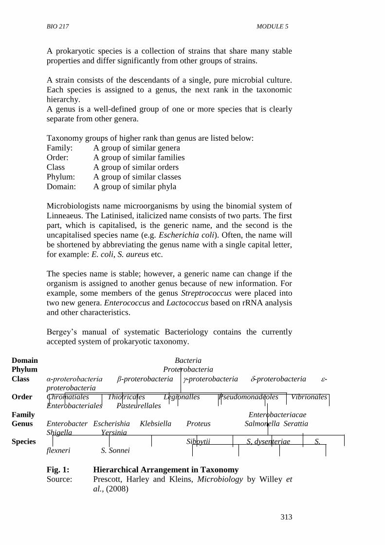
BIO 217 MODULE 5
313
A prokaryotic species is a collection of strains that share many stable
properties and differ significantly from other groups of strains.
A strain consists of the descendants of a single, pure microbial culture.
Each species is assigned to a genus, the next rank in the taxonomic
hierarchy.
A genus is a well-defined group of one or more species that is clearly
separate from other genera.
Taxonomy groups of higher rank than genus are listed below:
Family: A group of similar genera
Order: A group of similar families
Class A group of similar orders
Phylum: A group of similar classes
Domain: A group of similar phyla
Microbiologists name microorganisms by using the binomial system of
Linneaeus. The Latinised, italicized name consists of two parts. The first
part, which is capitalised, is the generic name, and the second is the
uncapitalised species name (e.g. Escherichia coli). Often, the name will
be shortened by abbreviating the genus name with a single capital letter,
for example: E. coli, S. aureus etc.
The species name is stable; however, a generic name can change if the
organism is assigned to another genus because of new information. For
example, some members of the genus Streptrococcus were placed into
two new genera. Enterococcus and Lactococcus based on rRNA analysis
and other characteristics.
Bergey’s manual of systematic Bacteriology contains the currently
accepted system of prokaryotic taxonomy.
Domain Bacteria
Phylum Proterobacteria
Class α-proterobacteria β-proterobacteria -proterobacteria -proterobacteria -
proterobacteria
Order Chromatiales Thiotricales Legionalles Pseudomonadeoles Vibrionales
Enterobacteriales Pasteurellales
Family Enterobacteriacae
Genus Enterobacter Escherishia Klebsiella Proteus Salmonella Serattia
Shigella Yersinia
Species Siboytii S, dysenteriae S.
flexneri S. Sonnei
Fig. 1: Hierarchical Arrangement in Taxonomy
Source: Prescott, Harley and Kleins, Microbiology by Willey et
al., (2008)
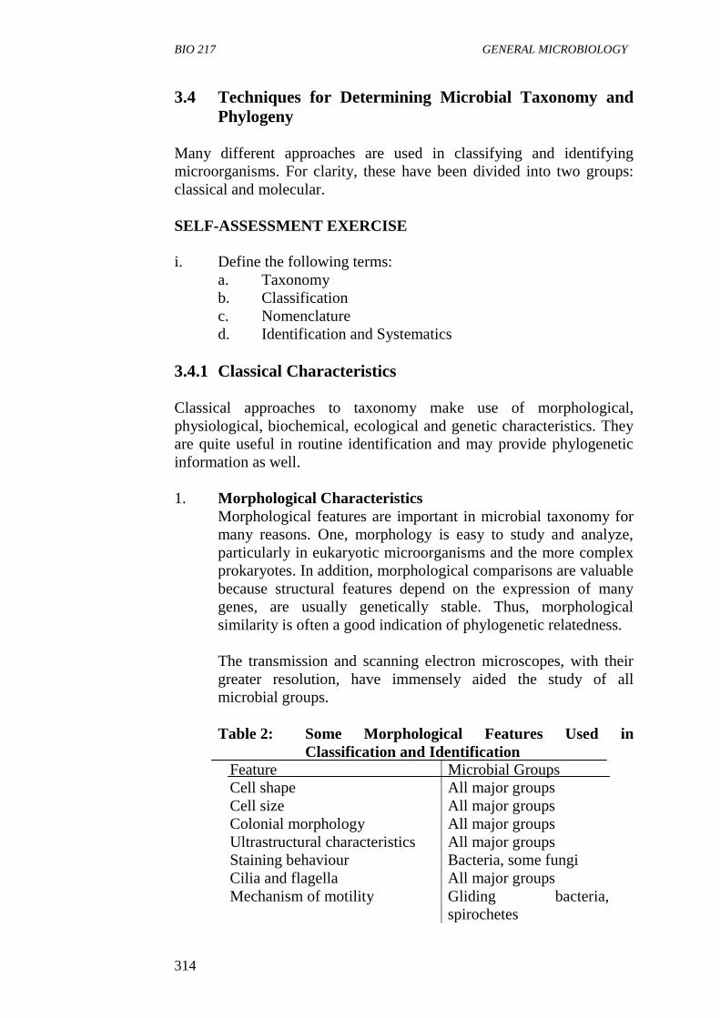
BIO 217 GENERAL MICROBIOLOGY
314
3.4 Techniques for Determining Microbial Taxonomy and
Phylogeny
Many different approaches are used in classifying and identifying
microorganisms. For clarity, these have been divided into two groups:
classical and molecular.
SELF-ASSESSMENT EXERCISE
i. Define the following terms:
a. Taxonomy
b. Classification
c. Nomenclature
d. Identification and Systematics
3.4.1 Classical Characteristics
Classical approaches to taxonomy make use of morphological,
physiological, biochemical, ecological and genetic characteristics. They
are quite useful in routine identification and may provide phylogenetic
information as well.
1. Morphological Characteristics
Morphological features are important in microbial taxonomy for
many reasons. One, morphology is easy to study and analyze,
particularly in eukaryotic microorganisms and the more complex
prokaryotes. In addition, morphological comparisons are valuable
because structural features depend on the expression of many
genes, are usually genetically stable. Thus, morphological
similarity is often a good indication of phylogenetic relatedness.
The transmission and scanning electron microscopes, with their
greater resolution, have immensely aided the study of all
microbial groups.
Table 2: Some Morphological Features Used in
Classification and Identification
Feature Microbial Groups
Cell shape All major groups
Cell size All major groups
Colonial morphology All major groups
Ultrastructural characteristics All major groups
Staining behaviour Bacteria, some fungi
Cilia and flagella All major groups
Mechanism of motility Gliding bacteria,
spirochetes
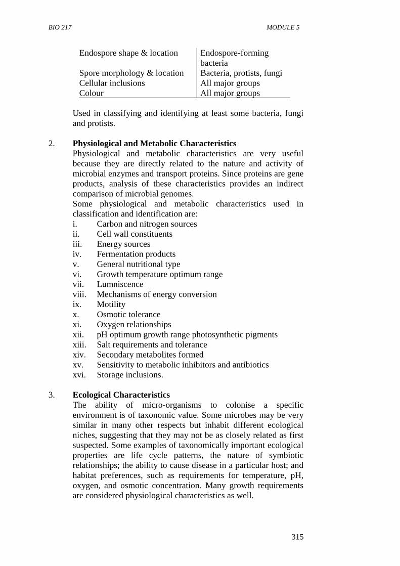
BIO 217 MODULE 5
315
Endospore shape & location Endospore-forming
bacteria
Spore morphology & location Bacteria, protists, fungi
Cellular inclusions All major groups
Colour All major groups
Used in classifying and identifying at least some bacteria, fungi
and protists.
2. Physiological and Metabolic Characteristics
Physiological and metabolic characteristics are very useful
because they are directly related to the nature and activity of
microbial enzymes and transport proteins. Since proteins are gene
products, analysis of these characteristics provides an indirect
comparison of microbial genomes.
Some physiological and metabolic characteristics used in
classification and identification are:
i. Carbon and nitrogen sources
ii. Cell wall constituents
iii. Energy sources
iv. Fermentation products
v. General nutritional type
vi. Growth temperature optimum range
vii. Lumniscence
viii. Mechanisms of energy conversion
ix. Motility
x. Osmotic tolerance
xi. Oxygen relationships
xii. pH optimum growth range photosynthetic pigments
xiii. Salt requirements and tolerance
xiv. Secondary metabolites formed
xv. Sensitivity to metabolic inhibitors and antibiotics
xvi. Storage inclusions.
3. Ecological Characteristics
The ability of micro-organisms to colonise a specific
environment is of taxonomic value. Some microbes may be very
similar in many other respects but inhabit different ecological
niches, suggesting that they may not be as closely related as first
suspected. Some examples of taxonomically important ecological
properties are life cycle patterns, the nature of symbiotic
relationships; the ability to cause disease in a particular host; and
habitat preferences, such as requirements for temperature, pH,
oxygen, and osmotic concentration. Many growth requirements
are considered physiological characteristics as well.
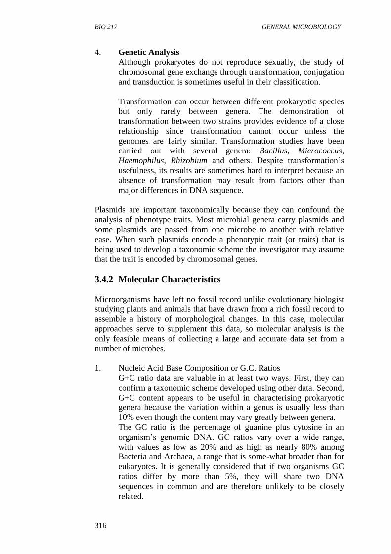
BIO 217 GENERAL MICROBIOLOGY
316
4. Genetic Analysis
Although prokaryotes do not reproduce sexually, the study of
chromosomal gene exchange through transformation, conjugation
and transduction is sometimes useful in their classification.
Transformation can occur between different prokaryotic species
but only rarely between genera. The demonstration of
transformation between two strains provides evidence of a close
relationship since transformation cannot occur unless the
genomes are fairly similar. Transformation studies have been
carried out with several genera: Bacillus, Micrococcus,
Haemophilus, Rhizobium and others. Despite transformation’s
usefulness, its results are sometimes hard to interpret because an
absence of transformation may result from factors other than
major differences in DNA sequence.
Plasmids are important taxonomically because they can confound the
analysis of phenotype traits. Most microbial genera carry plasmids and
some plasmids are passed from one microbe to another with relative
ease. When such plasmids encode a phenotypic trait (or traits) that is
being used to develop a taxonomic scheme the investigator may assume
that the trait is encoded by chromosomal genes.
3.4.2 Molecular Characteristics
Microorganisms have left no fossil record unlike evolutionary biologist
studying plants and animals that have drawn from a rich fossil record to
assemble a history of morphological changes. In this case, molecular
approaches serve to supplement this data, so molecular analysis is the
only feasible means of collecting a large and accurate data set from a
number of microbes.
1. Nucleic Acid Base Composition or G.C. Ratios
G+C ratio data are valuable in at least two ways. First, they can
confirm a taxonomic scheme developed using other data. Second,
G+C content appears to be useful in characterising prokaryotic
genera because the variation within a genus is usually less than
10% even though the content may vary greatly between genera.
The GC ratio is the percentage of guanine plus cytosine in an
organism’s genomic DNA. GC ratios vary over a wide range,
with values as low as 20% and as high as nearly 80% among
Bacteria and Archaea, a range that is some-what broader than for
eukaryotes. It is generally considered that if two organisms GC
ratios differ by more than 5%, they will share two DNA
sequences in common and are therefore unlikely to be closely
related.
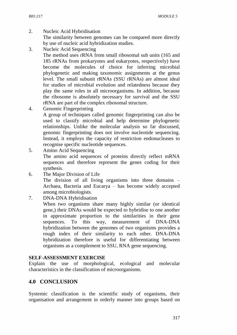
BIO 217 MODULE 5
317
2. Nucleic Acid Hybridisation
The similarity between genomes can be compared more directly
by use of nucleic acid hybridization studies.
3. Nucleic Acid Sequencing
The method uses rRNA from small ribosomal sub units (165 and
185 rRNAs from prokaryotes and eukaryotes, respectively) have
become the molecules of choice for inferring microbial
phylogenetic and making taxonomic assignments at the genus
level. The small subunit rRNAs (SSU rRNAs) are almost ideal
for studies of microbial evolution and relatedness because they
play the same roles in all microorganisms. In addition, because
the ribosome is absolutely necessary for survival and the SSU
rRNA are part of the complex ribosomal structure.
4. Genomic Fingerprinting
A group of techniques called genomic fingerprinting can also be
used to classify microbial and help determine phylogenetic
relationships. Unlike the molecular analysis so far discussed,
genomic fingerprinting does not involve nucleotide sequencing.
Instead, it employs the capacity of restriction endonucleases to
recognise specific nucleotide sequences.
5. Amino Acid Sequencing
The amino acid sequences of proteins directly reflect mRNA
sequences and therefore represent the genes coding for their
synthesis.
6. The Major Division of Life
The division of all living organisms into three domains –
Archaea, Bacteria and Eucarya – has become widely accepted
among microbiologists.
7. DNA-DNA Hybridisation
When two organisms share many highly similar (or identical
gene,) their DNAs would be expected to hybridise to one another
in approximate proportion to the similarities in their gene
sequences. To this way, measurement of DNA-DNA
hybridization between the genomes of two organisms provides a
rough index of their similarity to each other. DNA-DNA
hybridization therefore is useful for differentiating between
organisms as a complement to SSU, RNA gene sequencing.
SELF-ASSESSMENT EXERCISE
Explain the use of morphological, ecological and molecular
characteristics in the classification of microorganisms.
4.0 CONCLUSION
Systemic classification is the scientific study of organisms, their
organisation and arrangement in orderly manner into groups based on
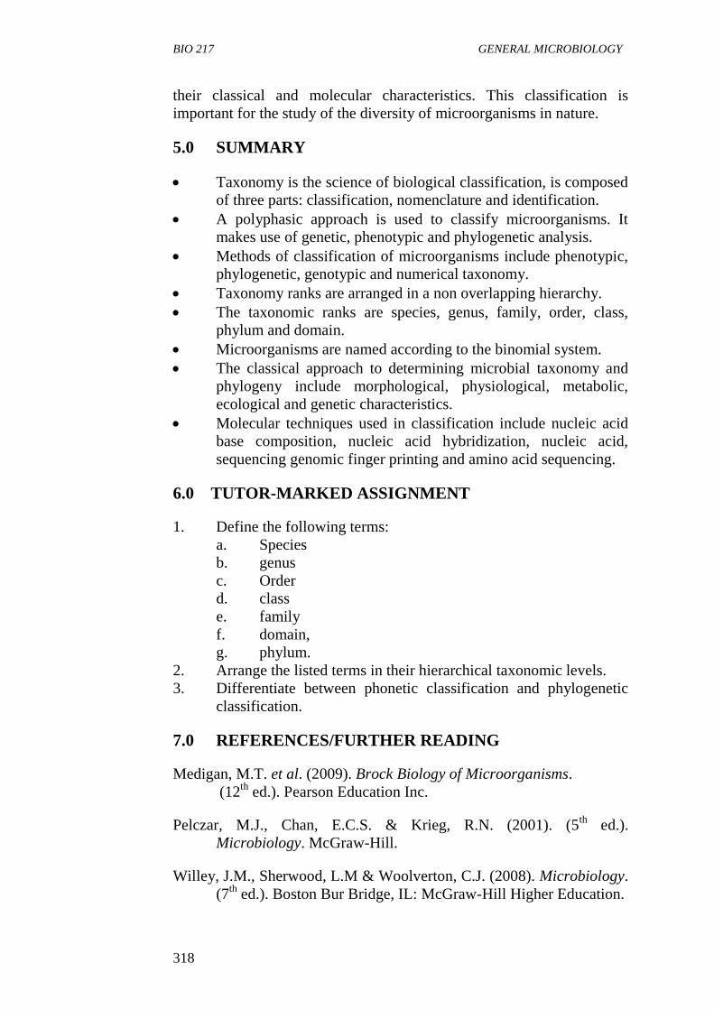
BIO 217 GENERAL MICROBIOLOGY
318
their classical and molecular characteristics. This classification is
important for the study of the diversity of microorganisms in nature.
5.0 SUMMARY
Taxonomy is the science of biological classification, is composed
of three parts: classification, nomenclature and identification.
A polyphasic approach is used to classify microorganisms. It
makes use of genetic, phenotypic and phylogenetic analysis.
Methods of classification of microorganisms include phenotypic,
phylogenetic, genotypic and numerical taxonomy.
Taxonomy ranks are arranged in a non overlapping hierarchy.
The taxonomic ranks are species, genus, family, order, class,
phylum and domain.
Microorganisms are named according to the binomial system.
The classical approach to determining microbial taxonomy and
phylogeny include morphological, physiological, metabolic,
ecological and genetic characteristics.
Molecular techniques used in classification include nucleic acid
base composition, nucleic acid hybridization, nucleic acid,
sequencing genomic finger printing and amino acid sequencing.
6.0 TUTOR-MARKED ASSIGNMENT
1. Define the following terms:
a. Species
b. genus
c. Order
d. class
e. family
f. domain,
g. phylum.
2. Arrange the listed terms in their hierarchical taxonomic levels.
3. Differentiate between phonetic classification and phylogenetic
classification.
7.0 REFERENCES/FURTHER READING
Medigan, M.T. et al. (2009). Brock Biology of Microorganisms.
(12th
ed.). Pearson Education Inc.
Pelczar, M.J., Chan, E.C.S. & Krieg, R.N. (2001). (5th
ed.).
Microbiology. McGraw-Hill.
Willey, J.M., Sherwood, L.M & Woolverton, C.J. (2008). Microbiology.
(7th
ed.). Boston Bur Bridge, IL: McGraw-Hill Higher Education.
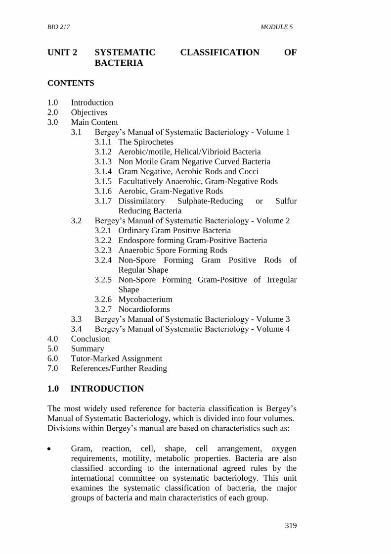
BIO 217 MODULE 5
319
UNIT 2 SYSTEMATIC CLASSIFICATION OF
BACTERIA
CONTENTS
1.0 Introduction
2.0 Objectives
3.0 Main Content
3.1 Bergey’s Manual of Systematic Bacteriology - Volume 1
3.1.1 The Spirochetes
3.1.2 Aerobic/motile, Helical/Vibrioid Bacteria
3.1.3 Non Motile Gram Negative Curved Bacteria
3.1.4 Gram Negative, Aerobic Rods and Cocci
3.1.5 Facultatively Anaerobic, Gram-Negative Rods
3.1.6 Aerobic, Gram-Negative Rods
3.1.7 Dissimilatory Sulphate-Reducing or Sulfur
Reducing Bacteria
3.2 Bergey’s Manual of Systematic Bacteriology - Volume 2
3.2.1 Ordinary Gram Positive Bacteria
3.2.2 Endospore forming Gram-Positive Bacteria
3.2.3 Anaerobic Spore Forming Rods
3.2.4 Non-Spore Forming Gram Positive Rods of
Regular Shape
3.2.5 Non-Spore Forming Gram-Positive of Irregular
Shape
3.2.6 Mycobacterium
3.2.7 Nocardioforms
3.3 Bergey’s Manual of Systematic Bacteriology - Volume 3
3.4 Bergey’s Manual of Systematic Bacteriology - Volume 4
4.0 Conclusion
5.0 Summary
6.0 Tutor-Marked Assignment
7.0 References/Further Reading
1.0 INTRODUCTION
The most widely used reference for bacteria classification is Bergey’s
Manual of Systematic Bacteriology, which is divided into four volumes.
Divisions within Bergey’s manual are based on characteristics such as:
Gram, reaction, cell, shape, cell arrangement, oxygen
requirements, motility, metabolic properties. Bacteria are also
classified according to the international agreed rules by the
international committee on systematic bacteriology. This unit
examines the systematic classification of bacteria, the major
groups of bacteria and main characteristics of each group.
BIO 217 GENERAL MICROBIOLOGY
320
2.0 OBJECTIVES
At the end of this unit, you should be able to:
identify the major groups of bacteria
state the major characteristics which set each group apart from
others
identify important genera within the appropriate group.
3.0 MAIN CONTENT
3.1 Bergey’s Manual of Systematic Bacteriology - Volume 1
Ordinary Gram Negative Bacteria
Bergey’s Manual of Systematic Bacteriology, Volume 1 is made up of
the ordinary Gram negative chemoheterotrophic bacteria. Many of
which have clinical, industrial or agricultural importance. They include:
3.1.1 The Spirochetes
These bacteria have helical shapes. They have the ability to twist and
contract their shape (they are flexible). There is presence of a special
kind of flagella termed periplasmic flagella or axial fibrils which may be
more than one. They are so thin they cannot be easily seen in light
microscope. When gram stained, dark field microscope is used to
visualise these organisms, they are Gram negative bacteria. Many
spirochetes are human pathogens.
Important genera
Treponema: causes syphilis and yaws
Borrelia: causes lymes diseases
Leptospira: causes fever, liver and kidney damages
3.1.2 Aerobic/motile, Helical/Vibrioid Bacteria
They are Gram negative bacteria. The cells are rigid and range from
vibrioid (having less than one tumor twist) to helical. They swim by
means of polar flagella.
They are aerobic or microaerophilic. Most are harmless saprobes and
occur in soil, fresh water or marine environment but a few are parasitic
and pathogenic for human animals and other bacteria.
BIO 217 MODULE 5
321
Important Genera
Spirillum - adapted to low concentration of organic matter
Azospirillum - associated with plant roots, nitrogen fixer
important to agriculture.
Campylobater- a food poisoning bacteria
Bdellovibrio - Predator on bacteria.
3.1.3 Non Motile Gram Negative Curved Bacteria
These bacteria have rigid cell that are curved to various degrees forming
coil, helical spirals and sometime ring, they are not motile. They occur
mainly in soil, fresh water and marine environment.
3.1.4 Gram Negative, Aerobic Rods and Cocci
This group of bacteria forms one of the largest and most diverse group
of bacteria. They are straight or slightly curved rods, some are cocci.
They are a strictly respiratory type of metabolism:
Many industrially, medical, and environmentally important bacteria
habitats include soil, water, animal parasites.
Important Genera
a. Pseudomonas-Opportunistic infections in burns. Aerobic motile
with rods with polar flagella. Many may synthesize a yellow
green-pigment that fluoresces under UV light. Are resistant to
many chemicals and antibiotics.
b. Legionella - Legionnaire’s Disease, fastidiuos organisms found in
many environments.
c. Neisseria - STD mostly of humans and animals
d. Brucella - Obligate intracellular parasites
e. Bordetella - Whooping and kernel coughs
f. Francisella - Tularemia in rabbits, require cysteine.
g. Rhizobium - A nitrogen-fixing soil bacterium
h. Agrobacterium - Used to introduce DNA into plants.
3.1.5 Facultatively Anaerobic, Gram-Negative Rods
Many important pathogens, oxidase and a requirement for organic
Habitats include soil, plants, and animals, respiratory and intestinal
tracts
Many in this group known as ‘enterics’ found in human intestine)
Important genera: (inhabit intestine of animals) bulky dysentery
Escherichia, Salmonella, Shigella, Klebsiella, Yersina, Vibrio,
BIO 217 GENERAL MICROBIOLOGY
322
Haemophilus, Gardnerella, Pasteurella, Proteus, Seratia
3.1.6 Aerobic, Gram-Negative Rods
Straight, curved and helical rods. They are rigid. They are obligately
anaerobic (cannot live in pressure of O2)
Habitats: Mostly in intestinal traits, some in mouth and genital trait and
some of the most common organism in the intestine.
Important genera:
Bacteriodes
Fusobacterium
Leptotrichia
3.1.7 Dissimilatory Sulphate-Reducing or Sulfur - Reducing
Bacteria
They have a Gram-negative cell wall.
They are found in anaerobic sediments; reduce oxidised forms of
sulphur to H2S. Dissimilation = nutrition not assimilated but rather
excreted. Occur in mud, fresh water, marine or brackish environments.
Important genera: Desulfovibrio – vibroid or helical cells.
Desulfococcus
SELF-ASSESSMENT EXERCISE
In what way does spirochete differ from other bacteria?
3.2 Bergey’s Manual of Systematic Bacteriology - Volume 2
3.2.1 Ordinary Gram- Positive Bacteria
Bergey’s Manual of Systematic Bacteriology, Volume 2 is made up of
the ordinary gram positive chemoheterotrophic bacteria,many of which
have clinical or industrial or agricultural importance. They are:
a. Gram-Positive Cocci. Aerobic/Facultatively Anaerobic Cocci
They possess cytochrome.
They are able to respire with oxygen.
Some can also obtain energy under anaerobic conditions by
fermentation.
Members are placed in two families.
Deinococcaceae and Micrococcaceae.
BIO 217 MODULE 5
323
Important genera
1. Micrococcus
aerobes or facultative anaerobes that form irregular
clusters by dividing in two or more planes.
2. Streptococcus
aerotolerant anaerobes that obtain energy from
fermenting sugars to lactic acid
form chains by dividing in one or two planes
lack catalase
can cause extensive tissue destruction by the release
of enzymes that degrade fibrin.
3. Staphylococcus
common human pathogen
responsible for skin abscesses or boils
will tolerate and grow in high salt concentrations
will rapidly develop antibiotic resistance.
4. Peptococcus
obligate anaerobes lack both catalase and enzymes
to ferment lactic acid
form pairs or irregular clusters
can cause many infections.
5. Peptostreptococcus
b Aerotolerant Fermentative Cocci
They do not have cytochromes.
They have only a fermentative type of metabolism and do not
respire yet they can grow anaerobically or aerobically.
The cells are arranged in pairs, chains or tetrads.
Some representative genera include Streptococcus. Leuconostoc
and Pediococcus.
c Anaerobic Gram-Positive Cocci
These cocci have a fermentative type of metabolism, cells in
clusters, tetrads, short or long chains.
3.2.2 Endospore Forming Gram-Positive Bacteria
Most are rod shaped but some are cocci.
Majority are gram-positive but a few species stain gram-negative.
Motility if present is by means of peritrichous flagella.
Some genera included in the group are:
Aerobic/Facultatively Anaerobic spore forming Rods and Cocci.
These groups are rod and cocci in shape.
BIO 217 GENERAL MICROBIOLOGY
324
Two genera found in the group are Bacillus and Sporosarcina.
Bacillus. They are rod shaped bacteria.
May form exocellular enzymes that hydrolyse proteins and
complex polysaccharides.
They form endospores which are heat resistant. They are
harmless saprobes found in soil, fresh water or sea water.
Examples include B. subtilis, B. cereus and B. thuringiensis.
Sporosarcina: They are cocci in shape and arranged in tetrads or
cubical packets of eight cells. They are widely distributed in
fertile soil.
3.2.3 Anaerobic Spore forming Rods
They have a fermentative type of metabolism. They are widely
distributed in soil, in marine and fresh water anaerobic sediments. They
include Clostridium and Desulfotomaculum.
3.2.4 Non-Spore Forming Gram Positive Rods of Regular
Shape
They are short or long rods in shape.
The group is made of harmless saprobes.
Some are parasitic organisms.
Important Genera
1. Lactobacillus
(a) foods and cheeses.
2. Listeria
(a) infection of brain and its membranes, will damage
fetus.
3. Erysipelothrix
(a) red sores in human.
3.2.5 Non-Spore Forming Gram-Positive of Irregular Shape
This group contains a heterogenous variety of bacteria.
The few common features are
Straight to slightly curved rods.
1. May be aerobic or facultatively anaerobic.
Some examples of the genera in the group include
Corynebacterium, Arthrobacter, Brevibacterium,
Micrococcus and Cellulomonas.
Corynebacterium: This genus contains rod shaped cells
which are pleomorphic and frequently exhibit club-shaped
swellings and a palisade arrangement.
BIO 217 MODULE 5
325
They may be saprobes, pathogens of human and or animals as
well as plant pathogens. An example is C.diptheriae which
causes diphtheria in humans, C. sepedonicum which causes ring
rot of potatoes.
2. Aerobic/Facultatively Anaerobic Branched Filamentous Rods.
The bacteria of this group form colonies which at first are
microscopic in size (microcolonies) and contain branched
filamentous cells. As the colonies develop to macroscopic size
many of the cells become diphtheroid (like caryobacteria) or
cocci in shape. Examples include Agromyces and Arachnia
3. Anaerobic Non-filamentous or Filamentous
The organisms are either anaerobes or if facultatively anaerobic
are preferentially anaerobic. Two of the genera in this group are
Propionbacterium and Actinomyces. They are differentiated by
their morphology and by their fermentation end products as
determined by gas chromatography.
Important genera
1. Corynebacterium
(a) can cause diphtheria.
2. Propionibacterium: anaerobic, causes acne
3. Eubacterium
4. Actinomyces:
(a) branching filamentous soil microbes.
3.2.6 Mycobacterium
They are aerobic bacteria. Their cell walls contain large amounts of
lipids. They are made up of a single genus Mycobacterium which are
slightly curved or straight rods that may show branching.
They are acid fast.
Some are saprophytes, e.g. M. phlei
While some are pathogens, e.g. M.tuberculosis.
3.2.7 Nocardioforms
They are aerobic bacteria that produce a substrate mycelium i.e. a mat of
branching hyphae formed under the surface of the agar medium.
Important genera
Nocardia: pulmonary nocardiosis
Some pathogens.
BIO 217 GENERAL MICROBIOLOGY
326
SELF-ASSESSMENT EXERCISE
i. List the group of bacteria placed in Bergey’s Manual of
Systematic Bacteriology Volume II.
ii. Differentiate between Staphylococcus and Streptococcus.
3.3 Bacteria with Unusual Properties
The organisms in this volume have unusual properties which are quite
different from those in volumes one and two. The anoxygenic
phototrophs can be divided into two major groups based on their
pigmentation purple bacteria and green bacteria. They occur in
anaerobic fresh water or marine environment. They may also occur
beneath the surface of shallow aquatic environments rich in organic
matter such as stagnant ponds and ditches.
Anoxygenic Phototropic Bacteria
They belong to the order Rhodospirillales. They are Gram-negative and
capable of carrying out photolithothrophic or photoorganotrophic type
of metabolism. They contain bacteriochlorophyll. Also present in their
cells are various water-insoluble carotenoid pigments which can also
trap or absorb light energy and transmit it to the bacteriochlorophyll.
The anoxygenic bacteria grow phototrophically only under anaerobic
conditions and are incapable of forming O2 because they possess only
photosystems.
Oxygenic Phototrophic Bacteria
They are bacteria that contain chlorophyll. They can use light as an
energy source and evolve O2 in a manner similar to that of green plants.
The group include: the Cyanobacteria (blue-green algae).
Gliding, Fruiting Bacteria
Gram-negative, non-phototrophic bacteria
They lack flagella; yet can glide across solid surfaces.
They have a complex life cycle in which the cells swarm together in
masses and form fruiting bodies.
The fruiting bodies contain myxo-spores which are shorter and thicker
than the vegetative cells.
BIO 217 MODULE 5
327
They are found in surface layers of soil, compost, manure, rotting wood
and animal dung. Constituent genera include Stigmatella Chondromyes.
They are aerobic or microaerophilic organisms.
They live in soil or water.
Gliding, Non-fruiting Bacteria
Gram-negative non-phototrophic rods, filaments or multicellular
trichomes that glide across solid surfaces: fruiting bodies are not
produced.
Examples of organisms include Cytophaga, Flexibacter or Vitreoscilla
Beggiotoa, Simonsiella,Saprospira and Thiothrix.
Sheathed Bacteria
Gram-negative non-phototrophic bacteria that form an external sheath
that covers the chain or trichomes.
They inhabit fresh water of marine environments. Among the genera
included in this group are Sphaerotilus, Leptothrix, etc.
Budding and/or Appendaged Bacteria
They are Gram-negative non-phototrophic bacteria that reproduce
asymmetrically by budding and or form prosthecea or stalks (Non-
living ribbon-like or tubular appendages that are excreted by the cell).
The organisms range from aerobic to microaerophilic to facultatively
anaerobic. Examples include Hyphomicrobium Ancalomicrobium,
Caulobacter.
Chemolithotrophic Bacteria
Gram-negative non-phototrophic bacteria that obtain energy for carbon
dioxide fixation from the oxidation of ammonia, nitrite, reduced sulfur
compounds or ferrous iron.
Examples of families in this group are the nitrifying bacteria, the sulfur
metabolizing bacteria and the Siderocapsaceae.
Many of these organisms are found in the soil, fresh water and marine
environments.
BIO 217 GENERAL MICROBIOLOGY
328
Archaeobacteria
Gram-positive or Gram-negative that are phylogenetically distinct from
eubacteria; some produce methane gas; some require unusually high
level of NaCl for growth: others are distinguished by their ability to
grow at a low pH and a high temperature. Three main categories of
archaeobacteria recognised are the methanogens the red extreme
halophiles, and the thermo-acidophiles.
SELF-ASSESSMENT EXERCISE
List the major group of organism in volume 3 and state the
characteristics of each.
3.4 Bergey Manual of Systematic Bacteriology - Volume 4
Gram-positive filamentous bacteria of complex morphology
The organisms in volume IV are aerobic gram positive bacteria which
form structures such as mycelium of filamentous hyphae and asexual
spores as found in microscopic eukaryotic fungi and have different cell
types of cellwalls based on amino acid and sugar composition.
Filamentous bacteria that divide in more than one plane
The hyphae divide not only transversely but also longitudinally to
produce cluster or packet of cells or spore; cell-wall type iii; soil
organisms, animal pathogens, and symbiotic nitrogen-fixers are
represented. The three genera present in this group are
Geodermatophilus, Dermatophilus and Frankia.
Filamentous bacteria that form true sporangia
Harmless soil and water organism whose hyphae divide in a single
plane; the spores are formed within special sacs; cell-wall type ii or iii
A good example of a genus in this division is Actinoplanes.
Streptomyces and related genera
The hyphae divide in a single plane; long chain of conidiospore are
formed at the tips of sporogenic hyphae; the organism are mainly
harmless soil organism that are noted for production of antibiotics; a few
are human or plant pathogen; cell-wall type.
Several genera in this group include Streptroverticullium,
Actinopycnidium, Actinosporangium but the most popular of these
genera is Streptomyces.
BIO 217 MODULE 5
329
Additional Filamentous Bacteria Having Uncertain Taxonomic
Placement
The taxonome placement of the bacteria of this heterogeneous group is
not yet agreed upon because many of the organisms have unusual and
striking morphological or physiological characteristics such as:
(i) The extreme halophilism exhibited by Actinopolyspora.
(ii) Formation of heat resistant spores by Thermoactinomyces and
other unusual properties.
A heterogenic collection of organism whose relationship to the major
groups of gram-positive filamentous bacteria is not yet agreed upon;
some have remarkable morphological or physiological properties; a few
organisms are pathogenic for humans; the cell-wall type vary.
4.0 CONCLUSION
Division within Bergey’s Manual of Systematic Bacteriology Volumes
I-IV is based on characteristics such as gram reaction, cell shape, cell
arrangement, oxygen requirement motility and metabolic properties.
11.0 SUMMARY
The Bergey’s Manual of Systematic Bacteriology Volume 1-IV is
as reference for the classification of bacteria.
Division is based on characteristics such as Gram Stain reaction,
cell shape, oxygen requirement and other properties.
Bergey’s Manual Volume 1 is made up of ordinary Gram
negative bacteria. They include: the spirochetes which have
helical shapes, flexible with periplasmic flagella. Important
genera include Treponema and Leptospira Aerobic, motile helical
or vibriod bacteria with rigid cells and vibriod or helical in shape
with polar flagella. Important genera are Spirillum and
Azospirillum.
Non-motile Gram negative curved bacteria have rigid cell that are
curved, they occur in soil, fresh water and marine environment.
Gram negative aerobic rods and cocci are straight or slightly
curved rods; they include Pseudomonas, Nisseria and other
genera.
Other gram-negative organisms in this group include facultatively
anaerobic Gram-negative rods, Aerobic Gram-negative rods, and
dissimilatory sulphate-reducing bacteria
BIO 217 GENERAL MICROBIOLOGY
330
Bergey’s Manual of Systematic Bacteriology Volume 2 is made
of ordinary Gram positive bacteria among which are
Aerobic/facultatively anaerobic cocci.
Aerotolerant fermentative cocci, Anaerobic Gram positive, cocci.
Endospore forming gram positive bacteria of regular and
irregular shapes and Mycobacterium.
Bergey’s Manual of Systematic Bacteriology – Volume 3 is made
up bacteria of unusual properties among which are Anoxygenic
Phototropic Bacteria, Oxygenic Phototropic Bacteria, Gliding
fruiting Bacteria, Gliding Non-fruiting Bacteria Sheathed
Bacteria and Building and unappendaged Bacteria.
Volume IV of Bergey’s Manual of Systematic Bacteriology is
made up of gram positive bacteria of complex morphology which
include filamentous bacteria that divide in more than one plane,
filamentous bacteria that form true sporangia, Streptomyces and
related genera and filamentous bacteria having uncertain
taxonomic placement.
12.0 TUTOR-MARKED ASSIGNMENT
i. What genera of gram-negative bacteria are associated with plants
as nitrogen fixers?
ii. Write on systematic classification of Gram-positive cocci.
7.0 REFERENCES/FURTHER READING
“Bergey’s Manual of Systematic Bacteriology.”
Medigan, M.T. et al. (2009). Brock Biology of Microorganisms. (12th
ed.). Pearson Education Inc.
Pelczar, M.J., Chan, E.C.S. & Krieg, R.N. (2001). (5th
ed.).
Microbiology. McGraw-Hill.
Willey, J.M., Sherwood, L.M & Woolverton, C.J. (2008). Microbiology.
(7th
ed.). Boston Bur Bridge, IL: McGraw-Hill Higher Education.
BIO 217 MODULE 5
331
UNIT 3 SYSTEMATIC CLASSIFICATION OF FUNGI
CONTENTS
1.0 Introduction
2.0 Objectives
3.0 Main Content
3.1 Zygomycota
3.2 Ascomycota
3.3 Basidiomycetes
3.4 Glomeromycota
3.5 Microsporidea
3.6 Uredinomycetes and Ustilaginomycetes
4.0 Conclusion
5.0 Summary
6.0 Tutor-Marked Assignment
7.0 References/Further Reading
1.0 INTRODUCTION
The classification of fungi like that of bacteria is described mainly for
practical application but it also bears some relation to phylogenic
consideration. The nomenclature is binomial, with a generic and specific
name (e.g. Aspergillus niger). Species are collected in genera, families
(suffice-acae), families in order (suffix ales), and order in class (suffix
mycetes). Sequence analysis of 185-RNA and certain protein – coding
gene has shown that the fungi comprises a monophyletic group with
eitht subdivisions. Four of these subdivisions, the Chytridiomycetes,
Zygomycota, Ascomycota and Basidomycota have been recognised as
separate groups for some time. The other four – Uredinomycetes,
Ustilaginomycetes, Glomeromycota and Microsporidia have been
proposed recently as separate groups. This unit examines the systematic
classification of fungi.
2.0 OBJECTIVES
At the end of this unit, you should be able to:
identify the different divisions of fungi
state the characteristics of each division of fungi and
state the mode of reproduction of each division of fungi.
BIO 217 GENERAL MICROBIOLOGY
332
3.0 MAIN CONTENT
Chytridiomycetes or Chytrids
Chytridiomycetes or chytrids are the earliest and the simplest group of
fungi. They are unique among fungi because they produce motile
zoospore with a single posterior whiplash flagellum. The cell wall is
made of chitin. Some exist as single cell while other form colonies with
hyphae. Some are free living and found living on plant or animal matter
in fresh water, mud or soil. Others are parasitic and infect aquatic plant
and animal including insects. They display a variety of life cycles
involving both asexual and sexual reproduction. Sexual reproduction
usually involves production of sporangiospores from sporagia.
Based on zoospore morphology, the orders with the Chytridiomycetes
include the Blastocladiales, Monoblepharidales, Neocallimasticales,
Spizellomycetales and the Chytridiales.
3.1 Zygomycota
The Zygomycota are made of fungi called Zygomycetes. They are
commonly found in soil and on decaying plant materials. All are
multinucleate (coenocytic). Asexual spores develop in sporangia at the
tip of aerial hypae and are usually dispersed by wind. Sexual
reproduction produces tough, thick walled zygotes called zygospores
that can remain dormant when the environment is too harsh for growth
of the fungus. The bread mould Rhizopus stolonifer is very common
member of this division, the fungus grows on moist, carbohydrate-rich
foods such as bread and vegetables.
SELF-ASSESSMENT EXERCISE
What feature of Zygomycetes gives this group its name?
3.2 Ascomycota
Members of this group are called Ascomycetes, commonly known as sac
fungi. These ascomycetes are large and highly diverse groups of fungi
ranging from single-celled species such as yeast (Saccharomyces) to
species that are filamentous like Neurospora.
The ascomycetes derived their names from the production of asci
(singular, ascus) cells in which two haploid nuclei from different mating
types come together and fuse, forming a diploid nucleus that undergoes
meiosis to form haploid ascopores.
BIO 217 MODULE 5
333
Asexual reproduction in ascomycetes is by the production of conidia
formed at the tip of specialised hyphae called conidiospore, an example
of fungi in this group is yeast. Cell division in Saccharomyces cerevisiae
(yeast) occurs by budding. During the budding process, a new cell forms
as a small outgrowth of the old cell, the bud gradually enlarges and
separates from the parent cell. Sexual reproduction also involves ascus
formation with each ascus usually bearing eight haploid ascospores.
3.3 Basidiomycota
The Basidiomycota include the Basidiomycetes, commonly known as
club fungi. Examples include jellyfungi, puffballs, toadstools and
mushrooms. They are named for their characteristic structure cell, the
basidium, which is involved. A basidium is produced at the tip of
hyphae and is normally club shaped. Two or more basidiospores are
produced by the basidium and basidia may be held within fruiting
bodies called basidiocarps. The basidiomycetes affect humans in many
ways. Most are saprobes that decompose plant debris, e.g. cellulose and
lignin, e.g. Polyporus squamosus. Some are used as food, e.g. the
mushroom. Agaricus campestris while some like Crytococcus,
Neoformans are important human and animal pathogens.
3.4 Glomeromycota
The glomeromycetes are a relatively small group of fungi with major
ecological importance. Only about 160 species of glomeromycetes are
currently known. All known species of glomeromycetes form
endomycorrhizae, also called arbuscular mycorrhizae with the roots of
herbaceous plants. They aid the plant acquisition of materials from the
soil. They produce only asexually and are mostly coenocytic in their
morphology. There is mutualistic relationship between the fungus, the
fungus help protect the plant from stress and deliver soil nutrients to the
plant which in turn provides carbohydrates to the fungi.
3.5 Microsporidea
These are tiny (2-5µm), unicellular parasite of animals and protists.
They have been considered protists and are sometime cited as such.
Molecular analysis of ribosomal RNA and specific protein such as α-
and β-tubulin shows that they are most closely related to fungi.
However, unlike fungi, they lack mitochondria, peroxisomes and
centriols. They are obligate parasites that infect insects, fish and human,
in particular they infect immunosuppressed individuals such as those
with HIV/AIDS; an example is Enterocystozoa spp which causes
diarrhea and pneumonia. It reproduces asexually by spore formation.
BIO 217 GENERAL MICROBIOLOGY
334
3.6 Uredinomycetes and Ustilaginomycetes
Both the uredinomycetes and the ustilaginomycetes include plant
pathogens causing rust and smuts. Some uredinomycetes include human
pathogens. They are often considered basidomycota. Both unlike the
basidomycota, they do not form large basidocarps instead small basidia
arise from hyphae at the surface of the host plant. The hyphae grow
either intracellular or extracellularly in the plant tissue. A good example
is Ustilago maydis, a common corn pathogen that causes smuts.
SELF-ASSESSMENT EXERCISE
State the role of arbuscular mycorrhizae on plants.
4.0 CONCLUSION
Systematic classification of fungi is based on sequence analyses of 185r
RNA and some protein coding genes and on the characteristics of the
sexual spores and fruiting bodies present during sexual reproduction.
11.0 SUMMARY
Sequence analyses of 185r RNA and some protein-coding genes show
that the fungi comprise a monophyletic group with eight subdivisions.
Chytridiomycetes are the earliest and simplest group of fungi.
They produce motile spores, some are saprobic while some are
parasites.
Zygomycetes are coenocytic (multinucleate) saprobic. They
reproduce asexually by spore formation and sexually by fusion of
two gametangia to form a zygospore, an example is the bread
mould Rhizopus stolonifer.
Ascomycota are known as sac shaped reproductive structure
called an ascus. Asexual reproduction occurs by conidia
production while sexual reproduction occurs by fusion of
gametes.
Basidiomycota are the club fungi. They are named after their
basidium which carries 4 basidiospores. Some are saprobes,
while some are pathogens of human and animals.
Glomeromycota form endomycorrhizae with plants roots. They
help to increase nutrient intake in the plants.
Microsporidia are still sometimes considered as protists but
molecular analysis has shown they are most closely related to
fungi. They lack mitochondria, centrioles and peroxisomes. They
are usually pathogens to insect and those with compromised
immunity.
BIO 217 MODULE 5
335
Uredinomycetes and Ustilaginomycetes are often considered
basidiomycetes but they do not form basidiocarp.
12.0 TUTOR-MARKED ASSIGNMENT
1. What are chytridiomycetes? How do they differ from other fungi?
2. In what structure are the basidiospores formed?
7.0 REFERENCES/FURTHER READING
Atlas, R.M. (1995). Microorganisms in Our World. Mosby Year Book.
Inc.
Medigan, M.T. et al. (2009). Brock Biology of Microorganisms. (12th
ed.). Pearson Education Inc.
Pelczar, M.J., Chan, E.C.S. & Krieg, R.N. (2001). (5th
ed.).
Microbiology. McGraw-Hill.
Willey, J.M., Sherwood, L.M & Woolverton, C.J. (2008). Microbiology.
(7th
ed.). Boston Bur Bridge, IL: McGraw-Hill Higher Education.
BIO 217 GENERAL MICROBIOLOGY
336
UNIT 4 SYSTEMATIC CLASSIFICATION OF ALGAE
CONTENTS
1.0 Introduction
2.0 Objectives
3.0 Main Content
3.1 Rhodophycophyta
3.2 Xanthophycophyta
3.3 Chrysophycophyta
3.4 Phaeoophycophyta
3.5 Bacillariophycophyta
3.6 Euglenophycophyta
3.7 Chlorophycophyta
3.8 Cryptophycophyta
3.9 Pyrrophycophyta
4.0 Conclusion
5.0 Summary
6.0 Tutor-Marked Assignment
7.0 References/Further Reading
1.0 INTRODUCTION
Algae are generally classified on the basis of the following
characteristics:
nature and properties of pigment
chemistry of reserve food products or assimilatory products of
photosynthesis
type and number, insertion (point of attachment), and
morphology of flagella
chemistry and physiological features of cell walls
morphological characteristics of cells and thalli
life history, reproductive structures and method of reproduction.
This unit examines the systemic classification of algae and the
characteristics of the major groups of algae.
2.0 OBJECTIVES
At the end of this unit, you should be able to:
identify the major groups of algae
state the types of pigment present in each group of algae
state the major characteristics of each group of algae
state the mode of reproduction of each group of algae.
BIO 217 MODULE 5
337
3.0 MAIN CONTENT
3.1 Rhodophycophyta
The Rhodophycophyta, or red algae, are marine forms found in the
warmer seas and oceans, but some grow in colder water as well as in
fresh water. Most red algae grow in the subtidal (submerged) zone, only
a few being able to survive desiccation or exposure. Some species
deposit upon their surfaces lime from seawater; ultimately this results in
deposition of lime in the ocean and plays a part in the formation of algal
reefs. They range from unicellular to multicellular. They are
photosynthetic and contain chlorophyll A. Their chloroplast lack
chlorophyll B and contain phycobiliproteins. The major light-harvesting
pigments of the cyanobacteria, the reddish colour of red algae results
from phycoerythrin, an accessory pigment that masks the green colour
of chlorophyll. And example is Gelidium from which agar is made,
Polysiphonia and Chondrus crispus.
3.2 Xanthophycophyta - The Yellow-Green Algae
These yellow-green algae were once classified with the green algae.
However, their pale green or yellow-green colouration indicates that
they have a unique group of pigments. They are found more frequently
in temperate regions in freshwater and marine habitats, as well as on and
in soil. Xanthophytes exist as single cell colonies, and as both branched
and unbranched filaments. Motile genera are not common, although,
some reproduce asexually by motile reproductive cells (zoospores).
Flagella are of unequal lengths.
The xanthophyte walls are typically of cellulose and pectin. The cellular
storage product is an oil or (a branched glucan) chrysolaminarin.
Vaucheria, the water felt, is a well-known member of this division and
is widely distributed on moist soil and in both quiet and rapidly flowing
water. Both freshwater and marine species are known. Zoospores are
formed singly in terminal sporangia in asexual reproduction.
3.3 Chrysophycophyta – The Golden Algae
Species of Chrysophycophyta are predominantly flagellates; some are
amoeboid, with pseudopodia extensions of the protoplasm. The naked
amoeboid forms can ingest particulate food with the pseudopodia. Non
motile coccoid and filamentous forms are also included in the division.
The chrysophycophyta differ from the green algae in the nature of their
pigments, in storing reserved food as oil or chrysolaminarin rather than
BIO 217 GENERAL MICROBIOLOGY
338
as starch, and in their frequent incorporation of silica. Most forms are
unicellular, but some form colonies. Their characteristic colour is due to
the marking of their cholorophyll by brown pigments. Reproduction is
commonly asexual (binary fission) but occasionally isogamous. An
example is Ochromonus.
SELF-ASSESSMENT EXERCISE
What are the major characteristics of the Crysophycophyta?
3.4 Phaeoophycophyta – The Brown Algae
These algae are multicellular and contain a brown pigment which gives
them their characteristic colour and common name of brown algae, or
brown seaweeds. Nearly all are marine dwelling and most frequently,
found in the cool ocean waters. They are structurally quite complex, and
some – the kelps – are large, the individual plants reaching a length of
several hundred feet. Many have holdfasts; and some have air bladders,
which give them buoyancy. They reproduce asexually by zoospores and
sexually by isogamy and heterogamy. This group includes algae used in
commerce, such as the many varieties of kelp. They are used as food for
humans, other animals and fish.
3.5 Bacillariophycophyta – The Diatoms
Members of this group are the diatoms, they are found in both fresh and
salt water and in moist soil. They are abundant in cold waters. Diatoms
are the most plentiful form of plankton in the Arctic. The thousands of
species diatoms provide an ever present and abundant food supply for
aquatic animals. Diatoms are unicellular, colonial, or filamentous and
occur in a wide variety of shapes. Each cell has a single prominent
nucleus which is massive and ribbon-like, or smaller lens-like, plastids.
They produce shells (cell walls) containing silica, some of which are
very beautiful. Shells of diatoms are called frustules. Deposits of these
shells resulting from centuries of growth are called diatomite or
diatomaceous earth.
3.6 Euglenophycophyta – The Euglenoids
They are unicellular organisms and they are actively motile by means of
flagella. They reproduce by cell division. Of particular interest is the
genus Euglena, which is representative of a group designated as animals
by some zoologists but as plants by many botanists. Euglena is widely
distributed and occurs in soil as well as in water, where it often forms a
variety film or bloom.
BIO 217 MODULE 5
339
The Euglena is not rigid; it is pliable. There is no cell wall containing
cellulose. The outer membrane is an organised periplast. An anterior
“gullete” is present even though, no food is ingested through it. Certain
species develop a prominent stigma or red eyespot. Contractile vacuoles
and fibrils are also present in the cell. All these are animal attributes. On
the other hand, the organism carries out photosynthesis. A few types can
even ingest particulate food through transient openings adjacent to the
gullet. Reproduction is by longitudinal binary fission. Dormant cysts are
formed by all types.
3.7 Chlorophycophyta – The Green Algae
Members of this large and diverse group of organisms, called green
algae, are principally freshwater species. They are also found in
seawater, and many of them are terrestrial. The cells of the
chlorophycophyta have a well-defined nucleus and usually, a cell wall,
and the chlorophyll pigments are in chloroplasts, as in higher plants. The
majority of green algae contain one chloroplast per cell. It may be
laminate, cup-shaped, or reticulate. The chloroplasts also often contain
dense regions called pyrenoids, on which surface starch granules are
formed. Food reserves are stored as starch, a product of photosynthesis.
They bear chloroplasts containing chlorophyll A and B giving them their
characteristic green colour.
There are many single-celled forms and many colonial types of green
algae. Many unicellular green algae are motile by flagella action.
Colonial types occur as spheres, filaments, or plates. Some species have
special structures called holdfasts, which anchor them to submerged
objects or aquatic plants. An example is Chlamydomonas.
Chlamydomonas is considered a typical green algae. It is a typical
unicellular, motile, green alga and is widely distributed in soils and
freshwater. It varies from 3 to 29m in common forms and is motile
except during cell division. Motility is by means of two flagella. Each
cell has one nucleus and a single large chloroplast that in most species is
cup-shaped, although in some, it may be star-shaped or layered. The cell
wall contains cellulose; in many species, an external gelatinous layer is
also present. There is some evidence that the red eyespot or stigma in
the chloroplast is the site of light perception.
In addition to motile unicellular algae like Chlamydomonas, other, non-
motile unicellular green algae are widely distributed. One of the most
important of these is Chlorella, which has served usefully as a tool in
many investigations on photosynthesis and supplemental food supply.
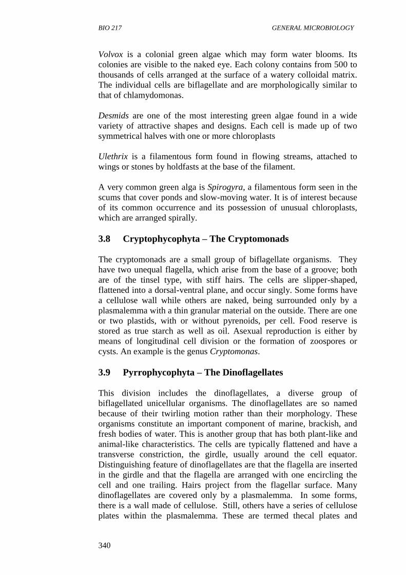
BIO 217 GENERAL MICROBIOLOGY
340
Volvox is a colonial green algae which may form water blooms. Its
colonies are visible to the naked eye. Each colony contains from 500 to
thousands of cells arranged at the surface of a watery colloidal matrix.
The individual cells are biflagellate and are morphologically similar to
that of chlamydomonas.
Desmids are one of the most interesting green algae found in a wide
variety of attractive shapes and designs. Each cell is made up of two
symmetrical halves with one or more chloroplasts
Ulethrix is a filamentous form found in flowing streams, attached to
wings or stones by holdfasts at the base of the filament.
A very common green alga is Spirogyra, a filamentous form seen in the
scums that cover ponds and slow-moving water. It is of interest because
of its common occurrence and its possession of unusual chloroplasts,
which are arranged spirally.
3.8 Cryptophycophyta – The Cryptomonads
The cryptomonads are a small group of biflagellate organisms. They
have two unequal flagella, which arise from the base of a groove; both
are of the tinsel type, with stiff hairs. The cells are slipper-shaped,
flattened into a dorsal-ventral plane, and occur singly. Some forms have
a cellulose wall while others are naked, being surrounded only by a
plasmalemma with a thin granular material on the outside. There are one
or two plastids, with or without pyrenoids, per cell. Food reserve is
stored as true starch as well as oil. Asexual reproduction is either by
means of longitudinal cell division or the formation of zoospores or
cysts. An example is the genus Cryptomonas.
3.9 Pyrrophycophyta – The Dinoflagellates
This division includes the dinoflagellates, a diverse group of
biflagellated unicellular organisms. The dinoflagellates are so named
because of their twirling motion rather than their morphology. These
organisms constitute an important component of marine, brackish, and
fresh bodies of water. This is another group that has both plant-like and
animal-like characteristics. The cells are typically flattened and have a
transverse constriction, the girdle, usually around the cell equator.
Distinguishing feature of dinoflagellates are that the flagella are inserted
in the girdle and that the flagella are arranged with one encircling the
cell and one trailing. Hairs project from the flagellar surface. Many
dinoflagellates are covered only by a plasmalemma. In some forms,
there is a wall made of cellulose. Still, others have a series of cellulose
plates within the plasmalemma. These are termed thecal plates and
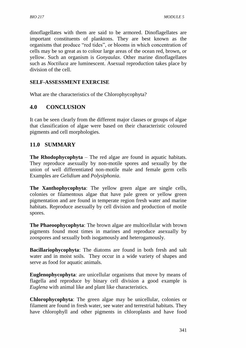
BIO 217 MODULE 5
341
dinoflagellates with them are said to be armored. Dinoflagellates are
important constituents of planktons. They are best known as the
organisms that produce “red tides”, or blooms in which concentration of
cells may be so great as to colour large areas of the ocean red, brown, or
yellow. Such an organism is Gonyaulax. Other marine dinoflagellates
such as Noctiluca are luminescent. Asexual reproduction takes place by
division of the cell.
SELF-ASSESSMENT EXERCISE
What are the characteristics of the Chlorophycophyta?
4.0 CONCLUSION
It can be seen clearly from the different major classes or groups of algae
that classification of algae were based on their characteristic coloured
pigments and cell morphologies.
11.0 SUMMARY
The Rhodophycophyta – The red algae are found in aquatic habitats.
They reproduce asexually by non-motile spores and sexually by the
union of well differentiated non-motile male and female germ cells
Examples are Gelidium and Polysiphonia.
The Xanthophycophyta: The yellow green algae are single cells,
colonies or filamentous algae that have pale green or yellow green
pigmentation and are found in temperate region fresh water and marine
habitats. Reproduce asexually by cell division and production of motile
spores.
The Phaeoophycophyta: The brown algae are multicellular with brown
pigments found most times in marines and reproduce asexually by
zoospores and sexually both isogamously and heterogamously.
Bacillariophycophyta: The diatoms are found in both fresh and salt
water and in moist soils. They occur in a wide variety of shapes and
serve as food for aquatic animals.
Euglenophycophyta: are unicellular organisms that move by means of
flagella and reproduce by binary cell division a good example is
Euglena with animal like and plant like characteristics.
Chlorophycophyta: The green algae may be unicellular, colonies or
filament are found in fresh water, see water and terrestrial habitats. They
have chlorophyll and other pigments in chloroplasts and have food
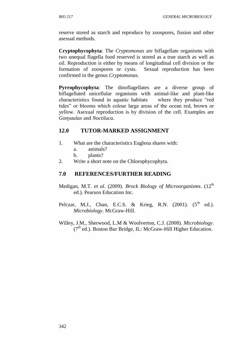
BIO 217 GENERAL MICROBIOLOGY
342
reserve stored as starch and reproduce by zoospores, fission and other
asexual methods.
Cryptophycophyta: The Cryptomonas are biflagellate organisms with
two unequal flagella food reserved is stored as a true starch as well as
oil. Reproduction is either by means of longitudinal cell division or the
formation of zoospores or cysts. Sexual reproduction has been
confirmed in the genus Cryptomonas.
Pyrrophycophyta: The dinoflagellates are a diverse group of
biflagellated unicellular organisms with animal-like and plant-like
characteristics found in aquatic habitats where they produce “red
tides” or blooms which colour large areas of the ocean red, brown or
yellow. Asexual reproduction is by division of the cell. Examples are
Gonyaulax and Noctiluca.
12.0 TUTOR-MARKED ASSIGNMENT
1. What are the characteristics Euglena shares with:
a. animals?
b. plants?
2. Write a short note on the Chlorophycophyta.
7.0 REFERENCES/FURTHER READING
Medigan, M.T. et al. (2009). Brock Biology of Microorganisms. (12th
ed.). Pearson Education Inc.
Pelczar, M.J., Chan, E.C.S. & Krieg, R.N. (2001). (5th
ed.).
Microbiology. McGraw-Hill.
Willey, J.M., Sherwood, L.M & Woolverton, C.J. (2008). Microbiology.
(7th
ed.). Boston Bur Bridge, IL: McGraw-Hill Higher Education.
BIO 217 MODULE 5
343
UNIT 5 SYSTEMATIC CLASSIFICATION OF
PROTOZOA
CONTENTS
1.0 Introduction
2.0 Objectives
3.0 Main Content
3.1 The Flagellates (subphylum Mastigophorea)
3.2 The Zooflagellates (Class Zoomastigophorea)
3.3 The Amoebas (Subphylum Sarcodina)
3.4 The Sporozoa (Phylum Apicomplexa)
3.5 The Ciliates (Phylum Ciliophora)
3.6 Other Ciliated Protozoa
4.0 Conclusion
5.0 Summary
6.0 Tutor-Marked Assignment
7.0 References/Further Reading
1.0 INTRODUCTION
Traditionally, the protozoa are grouped together based on general
similarities: they lack cell walls, they are colourless and motile, they
exhibit a wide range of morphologies and inhabit many different kinds
of habitat, and they play major roles in human society and health. The
different forms of protozoa have been grouped together not because they
are all related in an evolutionary way, but simply for convenience. The
old classification schemes of protozoa were based primarily on
organelles of locomotion. The major groups are called Phyla (singular,
phylum). This unit determines the systematic classification of protozoa
and different phyla in which they were placed.
2.0 OBJECTIVES
At the end of this unit, you should be able to:
identify the major phyla of protozoa
state the characteristics of each phylum
differentiate between different phyla; and
list some species found in each group or phyla.
BIO 217 GENERAL MICROBIOLOGY
344
3.0 MAIN CONTENT
3.1 The Flagellates (Subphylum Mastigophorea)
These protozoa are conventionally divided into two groups: The plant-
like forms (class Phytomastigophorea, the phytoflagellates) and the
animal-like forms (class Zoomastigophorea, the zooflagellates). Plant-
like protozoa usually contain green or yellow chloroplasts as well as
flagella and are photosynthetic. The zooflagellates have no chlorophyll
and are heterotrophic. All members have one or more flagella. Some
have pseudopodia. Asexual reproduction occurs by longitudinal binary
fission. A form of multiple fission takes place in some organisms.
Encystment is common but sexual reproduction is not.
These organisms are considered as algae by some biologists. Since the
zooflagellates have no chlorophyll, they must obtain nutrition
heterotrophically. All members of this group have one or more flagella;
some members are capable of forming pseudopodia.
3.2 The Zooflagellates (Class Zoomastigophorea)
The Choanoflagellates (order Choanoflagellida) are distinctive in that
they are either stalked or embedded in jelly, and each cell has a thin
transparent collar that encircles a single flagellum. The collar functions
as a food catching device.
Organisms in the order Kinetoplastida are grouped together because of
the presence of a kinetoplast (an extra nuclear region of DNA associated
with the mitochondrion). The single mitochondrion itself is extensive,
traversing the length of the body as single tube, hoop or network of
branching tubes. One or two flagella may be present; if there are two,
one is either trailing free or attached to the body, with undulating
membranes occurring in some cases.
3.3 The Amoebas (Subphylum Sarcodina)
Structure
Amoebas get their name from the Greek word “amoibe”,
meaning “change” because their shapes are constantly changing.
A typical example is Amoeba proteus.
Amoebas are composed of protoplasm differentiated into a cell
membrane, cytoplasm and a nucleus. The cytoplasm shows
granules as well as vacuoles containing food, wastes, water, and
possibly gases. The outer membrane is selective and permits the
passage of certain soluble nutrients into the cell and waste
materials out of the cell. Solid food is ingested with the help of
BIO 217 MODULE 5
345
pseudopodia. The nucleus functions in reproduction, metabolism
and the transmission of hereditary characteristics.
Amoeba reacts to various physical and chemical stimuli in their
surroundings. This is an irritability response which is at least
superficially analogous to responses of higher organisms to their
environment.
Nutrition of Amoeba
Uses pseudopodia to capture its food.
Reproduction – amoeba is asexually binary fission.
3.4 The Sporozoa (Phylum Apicomplexa)
They are in constant motion. They move by sending out portions of their
bodies in one direction which the whole body follows. They use
pseudopodia to capture food. Reproduction is asexually by binary
fission. Some have the ability of encysting in unfavourable condition.
Most are free living, some are saprophytic; however, one species
Entamoeba histolytica causes amoebic dysentery in man. All sporozoa
are parasitic for one or more animal species. Adult forms have no
organs of motility but all are probably motile by gliding at one stage of
their life cycle. They cannot engulf solid particles, but feed on the host’s
cells or body fluids.
Many have complicated life cycles, certain stages of which may occur in
one host and other stages in a different host. They all produce spores at
some time in their life history. Their life cycles exhibit an alternation of
generations of sexual and asexual forms, such that the intermediate host
usually harbours the asexual forms and the final host, the sexual forms.
Sometimes, humans serve as hosts to both forms.
Toxoplasmosis and malaria are the major human diseases caused by
sporozoa. Malaria is caused by Plasmodium asporozoa which infect the
liver and red blood cells.
3.5 The Ciliates (Phylum Ciliophora)
The ciliates are protozoa with cilia for locomotion. Common examples
of the ciliated protozoa are included in the genus Paramecium, found in
freshwater ponds and lakes where adequate food supplies exist.
BIO 217 GENERAL MICROBIOLOGY
346
Structure/Morphology
Paramecium
Paramecium moves rapidly by rhythmic beating of the cilia.
Nutrition: Paramecium takes in food through a fixed cytostoma at
the base of the gullet.
Excretion is through the contractile vacuole.
Reproduction – Reproduce asexually by binary fission
conjugation may also occur.
Paramecia are microscopic, some, however, are just barely visible to the
unaided eye. The outer layer of the cell is less flexible than the outer
membrane of the amoeba, and the interior is composed of semifluid,
granular protoplasm containing nuclei and vacuoles of several kinds.
Paramecia are easily distinguished by their characteristic shape, which
has been likened to that of a slipper. The anterior (front) end of the cell
is rounded, and the posterior (rear) end is slightly pointed. The entire
cell is covered with hundreds of short hair-like projections called cilia,
which are the organs of locomotion and also serve to direct food into the
cytosome.
3.6 Other Ciliated Protozoa
The ciliated protozoa are represented by many forms other than the
paramecia. Colpoda is a common freshwater genus. The genus
Didinium lives on a diet of paramecia, which are captured by a special
structure and swallowed whole. The genus Stentor comprises large
cone-shaped protozoa that move about freely but attach to some object
by a tapered lower end while feeding.
4.0 CONCLUSION
Classification in protozoa is by the use of general characteristics and not
as a result of evolution.
5.0 SUMMARY
Phytomastigophora are divided into two groups. The plant like
forms class Phytoflagellates which have chlorophyll and flagella
forms, class Zoomastigophora which have no chlorophyll and are
heterotrophic.
The amoebae (subphylum Sarcodina) are constantly changing
their shapes and composed of protoplasm differentiated into a cell
membrane cytoplasm and a nucleus and feed with the help of the
pseudopodia.
BIO 217 MODULE 5
347
The sporozoa (phylum Apicomplexes) are parasitic for one or
more animal species. They feed by the use of pseudopodia and
reproduce asexually by binary fission.
The ciliates (phylum Ciliophora): The ciliates are protozoa with
cilia for locomotion.
Other ciliates include Colpodia and the genera Didinium and the
genus Stentor.
6.0 TUTOR-MARKED ASSIGNMENT
1. State the two groups of flagellates.
2. Describe the structure of paramecium.
7.0 REFERENCES/FURTHER READING
Atlas, R.M. (1995). Microorganisms in Our World. Mosby Year Book.
Inc.
Medigan, M.T. et al. (2009). Brock Biology of Microorganisms. (12th
ed.). Pearson Education Inc.
Pelczar, M.J., Chan, E.C.S. & Krieg, R.N. (2001). (5th
ed.).
Microbiology. McGraw-Hill.
Willey, J.M., Sherwood, L.M & Woolverton, C.J. (2008). Microbiology.
(7th
ed.). Boston Bur Bridge, IL: McGraw-Hill Higher Education.
BIO 217 GENERAL MICROBIOLOGY
348
MODULE 5 MICROBIAL GENETICS AND
BIOGEOCHEMICAL CYCLING OF
ELEMENTS
Unit 1 Mechanisms of Genetic Variation and Hereditary
Unit 2 Biogeochemical Cycling of Elements
UNIT 1 MECHANISMS OF GENETIC VARIATION
AND HEREDITARY
CONTENTS
1.0 Introduction
2.0 Objectives
3.0 Main Content
3.1 Mutation
3.1.1 Spontaneous Mutation
3.1.2 Induced Mutation
3.2 Types of Mutation
3.3 Genetic Recombination
3.4 Mechanism of Recombination
3.4.1 Conjugation
3.4.2 Transformation
3.4.3 Transduction
4.0 Conclusion
5.0 Summary
6.0 Tutor-Marked Assignment
7.0 References/Further Reading
1.0 INTRODUCTION
Each living organism resembles its ancestors in most of its characters.
The maintenance of specific properties, that is, the constancy of
characters over generations is called heredity.
Genetics is the study of inheritance (heredity) and the variability of the
characteristics of an organism. Inheritance exacts transmission of
genetic information from parents to their progeny (offspring).
Deoxyribonucleic acid (DNA) is the chemical substance responsible for
hereditary in all cells because it beams the genetic information. Each
genetic character can be assigned to a gene which carries the
information. Microorganisms are capable of transmitting genetic
information from generation to generation with great accuracy. (This
unit examines the causes of variation in microorganisms). Variability or
variation of the inherited characteristics occurs or alters as a result of
BIO 217 MODULE 5
349
change in the genetic makeup of a cell or in environmental conditions.
Variation in microorganisms takes place by mutation. Recombination
and gene transfer.
2.0 OBJECTIVES
At the end of this unit, you should be able to:
define the term genetic variation
define mutation.
3.0 MAIN CONTENT
Genetic Variation
This is changes in or of a gene which leads to a loss of the enzymes or to
the production of an altered enzymes, hence, to recognizable changes in
the hereditary character.
Genetic variation in bacteria can take place by:
mutation
gene transfer or recombination.
3.1 Mutation
Mutation can be defined as a change in the nucleotide sequence of DNA.
Mutation can involve the addition, deletion or substitution of
nucleotides. These changes in the nucleotides are stable and heritable
and are passed down from one generation to the next. Mutation
introduces genetic variation among organisms.
A mutant is a strain of any cell or virus carrying a change in the
nucleotide sequence.
A mutant is different from its parent strain in:
i Genotype: The nucleotide sequence of the genome.
ii The Phenotype: The observable property of the mutant in the
altered phenotype is called a mutant phenotype.
The strain isolated originally from nature is called the wild type strain.
Mutation can occur spontaneously or induced under the influence of
external agents (mutagens).
BIO 217 GENERAL MICROBIOLOGY
350
3.1.1 Spontaneous Mutation
This is mutation that occurs without exposure to external agents or any
known mutagenic treatment. It occurs at a fairly constant frequency in a
particular organism, one per 106 to 10
10 in a population derived from a
single bacterium. It may result from errors in DNA replication or from
the action of mobile genetic molecules called transposons.
Mutation arising from error in DNA replication can be:
Transition Mutation: This is the substitution of a purine for
another purine (A or G) or one pyrimidine for another pyrimidine
(C or T) that lead to a stable alteration of the nucleotide sequence.
This type of mutation is relatively common.
Transversion Mutation: This is a mutation in which a purine is
substited for a pyrimidine or pyrimidine for a purine. This is
rarer due to the steric problems of pairing purines with purines
and pyrimidine with pyrimidine. Both Transition and
Transversion mutations are types of base substitution in point
mutation.
Mutations as result of lesions on DNA which result in apurine
sites, apyrimidinic sites, oxidation of DNA.
Mutation as a result of the insertion of DNA segments into genes.
Insertion usually inactivates genes. They are caused by
movement of insertion sequences and transpons.
3.1.2 Induced Mutation
This is mutation caused by external agent (mutagens), which may be
physical or chemical agents.
1. Physical Agents: Physical agents include ultraviolet (UV) light,
raising radiation, visible light and heat. Ionizing radiation (e.g. X-
rays) can break both single and double strand DNA. The
frequency of mutation is greatly increased at a temperature of
37oC and above.
2. Chemical Agent: There are three main types of mutagenic
chemicals.
(i) Base analogs: These are chemicals structurally similar to
normal DNA bases and can be substituted for them during
DNA replication. These compounds exhibit based pairing
properties different from the bases they replace and
eventually cause a stable mutation. An example: 5-
bromouracil (5-BU) an analog of thymine.
BIO 217 MODULE 5
351
(ii) DNA Modifying Agents: These chemicals react
chemically with DNA. They change a base structure and
alter its base pairing characteristics. Examples are Methyl-
nitrosoguanidine and Hydroxylamine.
(iii) Intercalating Agents: These are chemicals with flat
molecules that can intercalate (slip in) between bases pairs
in the central stack of the DNA helix. They include single
nucleotide pair insertion and deletions. Examples are
acridine such as proflavin and acridine orange.
3.2 Types of Mutation
Two common types of mutation are: point mutation and frame shift
mutation.
1. Point Mutations
Point mutations occur as a result of the substitution of one
nucleotide for another in the specific nucleotide sequence of a
gene or defined as change in only one base pair.
Point mutations in protein – coding genes can affect protein
structure and are named according to if and how they change the
encoded protein.
The most common type of point mutations are:
(i) Silent mutation: Change the nucleotide sequence of a
codon but do not change the amino acid encoded by that
codon.
(ii) Missence Mutations: This involves a single base
substitution that changes a codon for one amino acid into a
codon for another.
(iii) Nonsense Mutation: This causes the early termination of
translation and therefore results in a shortened
polypeptide. They are called nonsense mutation because
they convert a sense codon to a non-sense or stop code.
2. Frame Shift Mutations
These mutations result form an addition or loss of one or more
nucleotides in a gene and are termed insertion or deletion
mutations respectively.
This results in a shift or the reading frame.
Frame shift mutations usually are very harmful and yield mutant
phenotypes resulting from the synthesis of non-functional protein.
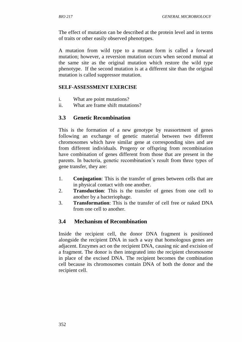
BIO 217 GENERAL MICROBIOLOGY
352
The effect of mutation can be described at the protein level and in terms
of traits or other easily observed phenotypes.
A mutation from wild type to a mutant form is called a forward
mutation; however, a reversion mutation occurs when second mutual at
the same site as the original mutation which restore the wild type
phenotype. If the second mutation is at a different site than the original
mutation is called suppressor mutation.
SELF-ASSESSMENT EXERCISE
i. What are point mutations?
ii. What are frame shift mutations?
3.3 Genetic Recombination
This is the formation of a new genotype by reassortment of genes
following an exchange of genetic material between two different
chromosomes which have similar gene at corresponding sites and are
from different individuals. Progeny or offspring from recombination
have combination of genes different from those that are present in the
parents. In bacteria, genetic recombination’s result from three types of
gene transfer, they are:
1. Conjugation: This is the transfer of genes between cells that are
in physical contact with one another.
2. Transduction: This is the transfer of genes from one cell to
another by a bacteriophage.
3. Transformation: This is the transfer of cell free or naked DNA
from one cell to another.
3.4 Mechanism of Recombination
Inside the recipient cell, the donor DNA fragment is positioned
alongside the recipient DNA in such a way that homologous genes are
adjacent. Enzymes act on the recipient DNA, causing nic and excision of
a fragment. The donor is then integrated into the recipient chromosome
in place of the excised DNA. The recipient becomes the combination
cell because its chromosomes contain DNA of both the donor and the
recipient cell.
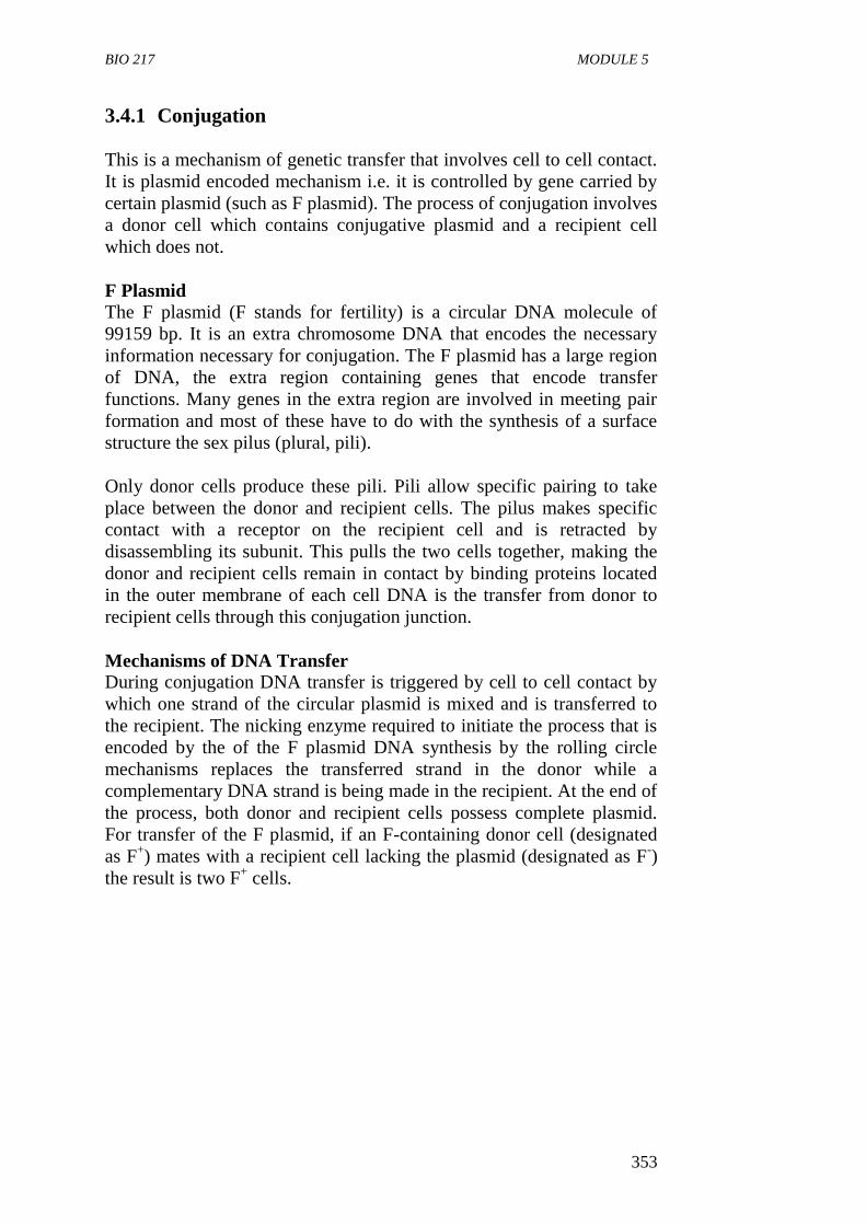
BIO 217 MODULE 5
353
3.4.1 Conjugation
This is a mechanism of genetic transfer that involves cell to cell contact.
It is plasmid encoded mechanism i.e. it is controlled by gene carried by
certain plasmid (such as F plasmid). The process of conjugation involves
a donor cell which contains conjugative plasmid and a recipient cell
which does not.
F Plasmid
The F plasmid (F stands for fertility) is a circular DNA molecule of
99159 bp. It is an extra chromosome DNA that encodes the necessary
information necessary for conjugation. The F plasmid has a large region
of DNA, the extra region containing genes that encode transfer
functions. Many genes in the extra region are involved in meeting pair
formation and most of these have to do with the synthesis of a surface
structure the sex pilus (plural, pili).
Only donor cells produce these pili. Pili allow specific pairing to take
place between the donor and recipient cells. The pilus makes specific
contact with a receptor on the recipient cell and is retracted by
disassembling its subunit. This pulls the two cells together, making the
donor and recipient cells remain in contact by binding proteins located
in the outer membrane of each cell DNA is the transfer from donor to
recipient cells through this conjugation junction.
Mechanisms of DNA Transfer
During conjugation DNA transfer is triggered by cell to cell contact by
which one strand of the circular plasmid is mixed and is transferred to
the recipient. The nicking enzyme required to initiate the process that is
encoded by the of the F plasmid DNA synthesis by the rolling circle
mechanisms replaces the transferred strand in the donor while a
complementary DNA strand is being made in the recipient. At the end of
the process, both donor and recipient cells possess complete plasmid.
For transfer of the F plasmid, if an F-containing donor cell (designated
as F+) mates with a recipient cell lacking the plasmid (designated as F
-)
the result is two F+ cells.
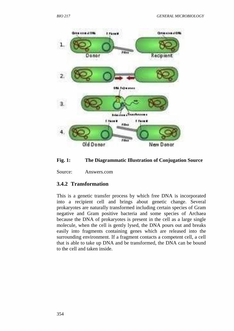
BIO 217 GENERAL MICROBIOLOGY
354
Fig. 1: The Diagrammatic Illustration of Conjugation Source
Source: Answers.com
3.4.2 Transformation
This is a genetic transfer process by which free DNA is incorporated
into a recipient cell and brings about genetic change. Several
prokaryotes are naturally transformed including certain species of Gram
negative and Gram positive bacteria and some species of Archaea
because the DNA of prokaryotes is present in the cell as a large single
molecule, when the cell is gently lysed, the DNA pours out and breaks
easily into fragments containing genes which are released into the
surrounding environment. If a fragment contacts a competent cell, a cell
that is able to take up DNA and be transformed, the DNA can be bound
to the cell and taken inside.
BIO 217 MODULE 5
355
Fig. 2: The Diagrammatic Illustration of Transformation in Cells
Source: harunyahya.com
Competency for transformation is a complex phenomenon and is
dependent on several conditions such as stage of growth. Natural
transformation has been discovered so far in certain genera including
Streptococcus, Bacillus, Acinectobacter, Azobacter, Helicobacter and
Pseudomonas. Gene transfer by this in process occurs in soil and aquatic
environment.
3.4.3 Transduction
This is a mechanism of genetic transfer in which a bacterial virus
(bacteriophage) transfers DNA from one cell to another. Virus can
transfer the host genes in two ways:
i generalised transduction and
ii specialised transduction
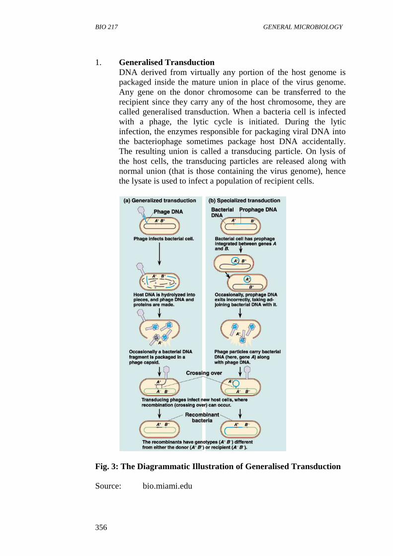
BIO 217 GENERAL MICROBIOLOGY
356
1. Generalised Transduction
DNA derived from virtually any portion of the host genome is
packaged inside the mature union in place of the virus genome.
Any gene on the donor chromosome can be transferred to the
recipient since they carry any of the host chromosome, they are
called generalised transduction. When a bacteria cell is infected
with a phage, the lytic cycle is initiated. During the lytic
infection, the enzymes responsible for packaging viral DNA into
the bacteriophage sometimes package host DNA accidentally.
The resulting union is called a transducing particle. On lysis of
the host cells, the transducing particles are released along with
normal union (that is those containing the virus genome), hence
the lysate is used to infect a population of recipient cells.
Fig. 3: The Diagrammatic Illustration of Generalised Transduction
Source: bio.miami.edu
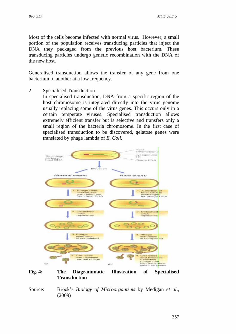
BIO 217 MODULE 5
357
Most of the cells become infected with normal virus. However, a small
portion of the population receives transducing particles that inject the
DNA they packaged from the previous host bacterium. These
transducing particles undergo genetic recombination with the DNA of
the new host.
Generalised transduction allows the transfer of any gene from one
bacterium to another at a low frequency.
2. Specialised Transduction
In specialised transduction, DNA from a specific region of the
host chromosome is integrated directly into the virus genome
usually replacing some of the virus genes. This occurs only in a
certain temperate viruses. Specialised transduction allows
extremely efficient transfer but is selective and transfers only a
small region of the bacteria chromosome. In the first case of
specialised transduction to be discovered, gelatose genes were
translated by phage lambda of E. Coli.
Fig. 4: The Diagrammatic Illustration of Specialised
Transduction
Source: Brock’s Biology of Microorganisms by Medigan et al.,
(2009)
BIO 217 GENERAL MICROBIOLOGY
358
When lambda lysogenizes a host cell, phage genomes are integrated into
the host DNA at a specific site. The region in which lambda integrates in
the E. Coli chromosome is next to the cluster of the gene that encode the
enzyme for galactose utilisation. After insertion, viral DNA replication
is under control of bacterial host chromosome. Upon induction, the viral
DNA separates from the host DNA by a process that is reverse of
integration.
The phenotype of microorganisms can be affected in various ways:
i Morphological Mutations: Changes in the microorganism
colonial or cellular morphology of the microorganisms.
ii Lethal Mutation: Resulting in the death of the microorganism.
iii Biochemical Mutation: Causing a change in the biochemistry of
the cell.
3.4.4 Mutation Detection involves
i. Visual Observation
ii. Replace Plating Technique.
4.0 CONCLUSION
Microbial variation can arise as a result of mutations which can occur
spontaneous or induced by physical and chemical changes. Mutation
leads to changes in protein structure and function which in turn alter the
phenotype of an organism. This results in the organism (mutant) being
different from the one found originally in nature (the wild type).
5.0 SUMMARY
Mutation is a stable heritable change in the nucleotide sequence
of a DNA molecule.
Mutation can be spontaneous or induced.
Spontaneous mutation can arise from replication errors
(transition, transversion, addition and deletion of nucleotide)
from DNA lesions and from insertion of DNA segments and
transpons.
Induced mutation can be from physical and chemical agents
called mutagens.
Physical agents, of mutation include ultraviolet rays, ionizing
rays, radiation, light, heat, etc.
Chemical agents of mutation are base analogs, intercalating
agents and DNA modifying agents.
BIO 217 MODULE 5
359
Main types of mutation are point mutation and frame shift
mutation.
Mutations are recognised when they cause a change from the
normal wild type phenotype. A mutant phenotype can be restored
to wild type by either reversion or suppressor mutations.
Some major types of mutations categorised based on the effect on
phenotypes are morphological, lethal and biochemical.
Replace plating can be used for detection and isolating mutants.
Amen test is used to screen for mutagens and potential
carcinogens.
6.0 TUTOR-MARKED ASSIGNMENT
1. What is mutation?
2. List four ways in which spontaneous mutation might arise.
3. List three methods of recombination in a prokaryotic cell.
4. Explain the mechanism of conjugation in bacteria.
7.0 REFERENCES/FURTHER READING
Atlas, R.M. (1995). Microorganisms in Our World. Mosby Year Book.
Inc.
Medigan, M.T. et al. (2009). Brock Biology of Microorganisms. (12th
ed.). Pearson Education Inc.
Pelczar, M.J., Chan, E.C.S. & Krieg, R.N. (2001). (5th
ed.).
Microbiology. McGraw-Hill.
Willey, J.M., Sherwood, L.M & Woolverton, C.J. (2008). Microbiology.
(7th
ed.). Boston Bur Bridge, IL: McGraw-Hill Higher Education.
BIO 217 GENERAL MICROBIOLOGY
360
UNIT 2 BIOGEOCHEMICAL CYCLING OF
ELEMENTS
CONTENTS
1.0 Introduction
2.0 Objectives
3.0 Main Content
3.1 Peculiar Features of Biogeochemical Cycles
3.2 Carbon Cycle
3.3 Nitrogen Cycle
3.3.1 Nitrogen Fixation
3.3.2 Nitrification
3.3.3 Denitrification
3.3.4 Ammonification
3.4 Sulphur Cycle
3.5 The Phosphorus Cycle
4.0 Conclusion
5.0 Summary
6.0 Tutor-Marked Assignment
7.0 References/Further Reading
1.0 INTRODUCTION
Life on earth would not be possible without microbes. Soil
microorganisms serve as biogeochemical agents for the conversion of
complex organic compounds into simple inorganic compounds or into
their constituents’ elements. The overall process is called
Mineralisation. Biogeochemical cycling refers to the biological and
chemical processes that elements such as carbon, nitrogen, sulfur, iron
and magnesium undergo during microbial metabolism. This conversion
of complex organic compounds into inorganic compounds or elements
provides for the continuity of elements (or their compounds) as nutrients
for plants and animals including man. This unit examines the
biogeochemical cycles of elements such as carbon, sulfur, nitrogen and
phosphorous.
2.0 OBJECTIVES
At the end of this unit, you should be able to:
define biogeochemical cycling
state the features of biogeochemical cycles
describe the carbon cycle
describe the sulphur cycle
BIO 217 MODULE 5
361
describe the phosphorus cycle.
3.0 MAIN CONTENT
Biogeochemical Cycling
Biogeochemical cycling is the movement of materials via biochemical
reactions through biospheres. The biosphere is that portion of the earth
and its atmosphere in which living organisms occur. The activities of
microorganisms within the biosphere have a direct impact on the quality
of human life. Microorganisms are especially important in recycling
materials.
The metabolism carried out by microorganisms often transfers materials
from one place to another. Changes in the chemical forms of various
elements can lead to the physical movement of materials. Sometimes
causing transfer between the atmosphere (air) hydrosphere (water) and
lithosphere (land).
Biogeochemical cycling also refers to the biological and chemical
processes that elements such as carbon, nitrogen, sulfur, iron and
magnesium undergo during microbial metabolism.
It can also be defined as cyclical path that elements take as they flow
through living (biotic) and non-living (abiotic) components of the
ecosystem. These cycles are important because a fixed and limited
amount of the elements that make up living cells exists on earth and in
the atmosphere. Thus in order for an ecosystem to maintain and sustain
its characteristics and life forms elements must continuously be
recycled. For example, if the organic carbon that animals use as an
energy source and exhale as carbon dioxide (CO2) were not eventually
converted back to an organic form we would run out of organic carbon
to build cells.
Elements involved in the biogeochemical cycles are used for three
general purposes.
(i) Biomass Production
In biomass production, the element transferred (e.g. N, C, etc) is
incorporated into cell materials for example, all organisms
require nitrogen to produce amino acids, hence plants and many
prokaryotes assimilates nitrogen by incorporating ammonia
(NH3) to synthesise the amino acid glutamate. Animals cannot
incorporate ammonia instead require amino acids in their diets.
Some prokaryotes can reduce atmosphere nitrogen to form
ammonia, which can then be incorporated into cellular material.
BIO 217 GENERAL MICROBIOLOGY
362
(ii) Energy Source
A reduced form of the element is used to generate energy in form
of ATP. For example reduced carbon compounds such as lipids
and amino acids are used as energy source by heterotrophs.
Chemolitrophs can use reduced inorganic molecules such as
hydrogen sulfide (H2S) ammonia (NH3) and Hydrogen gas.
(iii) Terminal Electron Acceptor i.e. Carbohydrate Oxidised to
CO2 Electrons from the energy source are transferred to an oxidised
form of the element during respiration, this reduces the terminal
electron acceptor in aerobic conditions, O2 is used as a terminal
electron acceptor. In anaerobic conditions, some prokaryotes can
use nitrate, sulphate (SO4), and carbon dioxide (CO2) as terminal
electron acceptors.
Global climate change, temperature precipitation and stability of
ecosystem are all dependent on biogeochemical cycles.
3.1 Peculiar Features of Biogeochemical Cycles
Elements required are in five forms and mostly from non-living
reservoir in the atmosphere. They can also be gotten from
sedimentary rock or water.
The elements go in cycle and are always free in inorganic state in
abiotic environment and when needed in biotic environment, they
are turned to organic state. They can be combined with other
elements.
The recycling of these elements maintains a necessary balance of
nutrient and they are maintained throughout.
The cycles (biogeochemical) are complex and they involve the
activity of producers, consumers and decomposers.
All organisms participate directly in recycling by removing,
adding or altering nutrients. Microorganisms are noted for
metabolic conversion especially in changing some elements from
one nutritional form to another.
The total turnover rate of element is most rapid in atmosphere
and lowest in sedimentary rocks.
SELF-ASSESSMENT EXERCISE
i. What is biogeochemical cycle of elements?
ii. State three features of biogeochemical cycles.
BIO 217 MODULE 5
363
3.2 Carbon Cycle
Carbon is a very important element as it makes up organic matter which
is a part of all life. Carbon follows a certain route on earth called the
CARBON CYCLE.
The carbon cycle primarily involves the transfer of carbon dioxide and
organic carbon between the atmosphere where carbon occurs principally
as inorganic CO2 and the hydrosphere and lithosphere which contain
varying concentrations of organic and inorganic compounds.
The carbon cycle begins with carbon fixation, which is the conversion of
CO2 to organic matter. Plants are thought of as the principal CO2 fixing
organisms but at least half of the carbon on earth is fixed by microbes;
particularly marine photosynthetic prokaryotes and protists.
Cyanobacteria such as Prochlorococcus and Synechococcus are
involved in carbon fixation using energy from sunlight.
Chemolithoautotrophic microorganisms such as Thiobacillus and
Beggiatoa also fix CO2 into organic matter while metabolising
compounds such as H2S for energy.
Once carbon is fixed into organic compounds, the next stage in the cycle
involves its transfer from population to population within the biological
community, supporting the growth of a variety of heterotrophic
organisms, i.e. heterotrophs such as animals and protozoa that eat
autotrophs and may in turn be eaten by other animals. Hence, they
acquire organic carbon to build biomass and to oxidize to gain energy.
Decomposers use the remains of primary producers and consumers for
the same purposes.
The respiratory and fermentative metabolism of heterotrophic organisms
returns inorganic carbon dioxide to the atmosphere completing the
carbon cycle. When plants and animals die, these organic compounds
are decomposed by bacteria and fungi and during decomposition the
organic compounds are oxidised and CO2 is returned to the cycle.
Carbon is stored in rocks such as limestone (CaCO3) and is dissolved as
carbonate ions (CO3) in oceans. Vast deposits of fossil organic matter
exist in the form of fossil fuel such as coal and petroleum. Burning
these fuel releases CO2 resulting in an increased amount of CO2 in the
atmosphere.
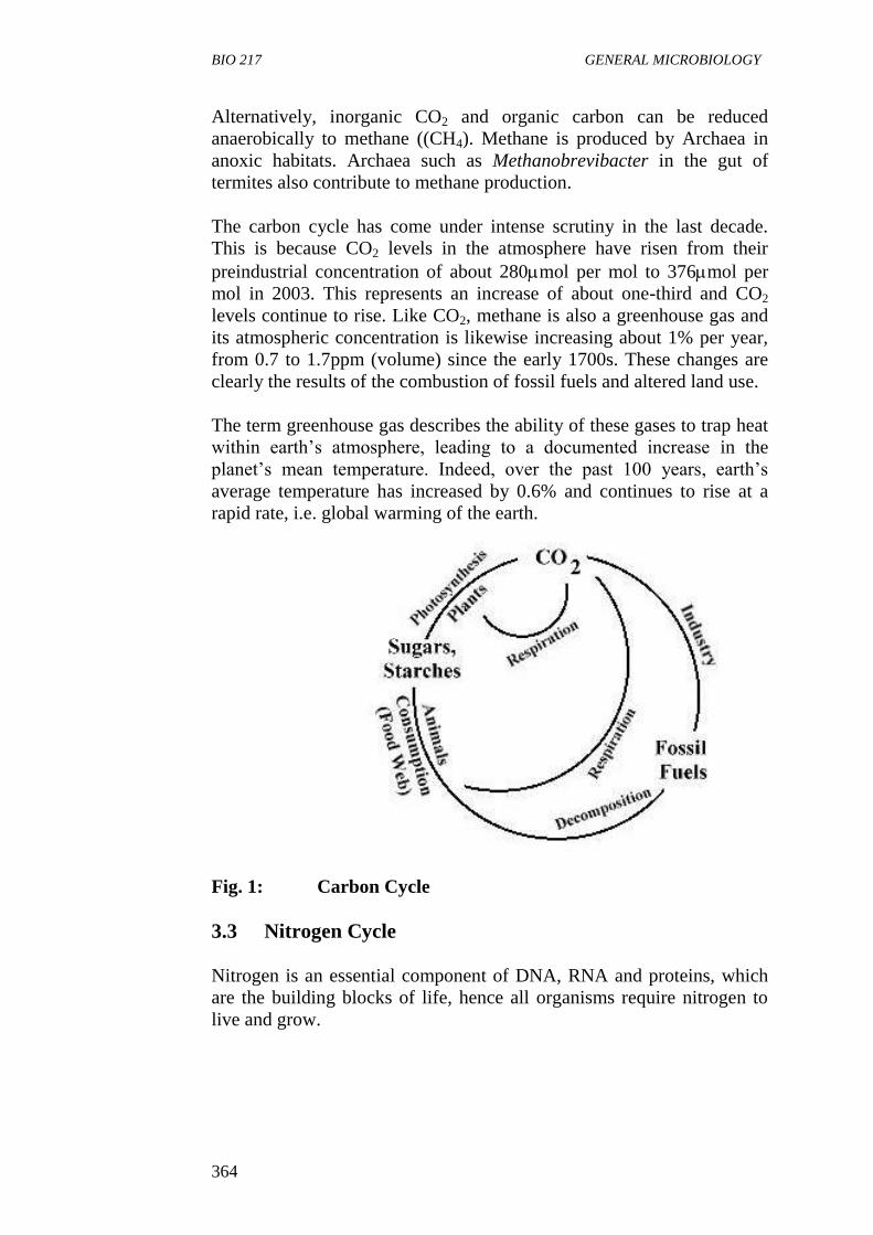
BIO 217 GENERAL MICROBIOLOGY
364
Alternatively, inorganic CO2 and organic carbon can be reduced
anaerobically to methane ((CH4). Methane is produced by Archaea in
anoxic habitats. Archaea such as Methanobrevibacter in the gut of
termites also contribute to methane production.
The carbon cycle has come under intense scrutiny in the last decade.
This is because CO2 levels in the atmosphere have risen from their
preindustrial concentration of about 280mol per mol to 376mol per
mol in 2003. This represents an increase of about one-third and CO2
levels continue to rise. Like CO2, methane is also a greenhouse gas and
its atmospheric concentration is likewise increasing about 1% per year,
from 0.7 to 1.7ppm (volume) since the early 1700s. These changes are
clearly the results of the combustion of fossil fuels and altered land use.
The term greenhouse gas describes the ability of these gases to trap heat
within earth’s atmosphere, leading to a documented increase in the
planet’s mean temperature. Indeed, over the past 100 years, earth’s
average temperature has increased by 0.6% and continues to rise at a
rapid rate, i.e. global warming of the earth.
Fig. 1: Carbon Cycle
3.3 Nitrogen Cycle
Nitrogen is an essential component of DNA, RNA and proteins, which
are the building blocks of life, hence all organisms require nitrogen to
live and grow.
BIO 217 MODULE 5
365
Nitrogen, the most abundant substance in the atmosphere or air (almost
80%) is not directly useable by most organisms. This is because the
strong triple bond between the nitrogen atoms in the molecule makes it
relatively inert. Only a few bacteria are able to utilise nitrogen directly.
For plants and animals to be able to use N2, N2 gas must first be
converted to more chemically available forms such as ammonium
(NH4+), Nitrate (NO3
-1), (NO2
-1) and organic nitrogen containing
compounds such as amino acids and proteins. Microorganisms also are
able to utilise these other forms of nitrogen mentioned above.
The conversion of nitrogen compounds primarily by microorganisms
changes the oxidation states of nitrogenous compounds and establishes a
nitrogen cycle.
Three processes carried out by microorganisms are critical in the
nitrogen cycle. They are:
1. nitrogen fixation
2. nitrification and
3. denitrification.
3.3.1 Nitrogen Fixation
This is strictly a bacterial process in which molecular nitrogen is
converted to ammonium ion and it is the only naturally occurring
process that makes nitrogen available to living organisms. It is carried
out by a few bacteria that have a nitrogenase enzymes system.
This process brings nitrogen from the atmosphere to the hydrosphere
and lithosphere. Because plants depend on the availability of nitrogen
for growth, microbial metabolism of nitrogen containing compounds has
a dramatic impact on agricultural productivity. Plants and animals rely
entirely on the fixed forms of nitrogen (such as ammonium and nitrate
ions) provided by bacterial nitrogen fixation. Nitrogen fixation involves
two types of bacteria or microorganisms).
1. Free Living Nitrogen Fixing Bacteria
These bacteria are found in high numbers or concentration in the
rhizosphere (the region where the soil and roots make contacts).
In aquatic environment, blue green alga is an important free-
living nitrogen fixer. Trichodesmium fix nitrogen aerobically
while free-living anaerobes such as members of the genus
Clostridium fix nitrogen anaerobically.
BIO 217 GENERAL MICROBIOLOGY
366
2. Symbiotic Nitrogen Fixing Bacteria
Members of the genera Rhizobium, Bradyrhizobium and others
are important nitrogen fixing bacteria. They play an important
role in plant growth for crop production. These bacteria live in
association with leguminous plants. The bacteria invade the root
cells to form nodules, receives carbohydrate and a favourable
environment from their host plants in exchange for the nitrogen
they fix. However, other bacterial symbiotics fix nitrogen, for
example the Actinomycete Frankia fixes nitrogen while
colonizing many type of woody shrub. The heterocystous
cyanobacterium Anabaenea fixes nitrogen when in association
with the water fern Azolla.
The product of nitrogen fixation is ammonia (NH3). It is immediately
incorporated into amino N-atoms and eventually as amino acids. The
nitrogenase enzymes are very sensitive to oxygen and must be protected
from oxidizing effects.
The nitrogen cycle continues with the degradation of these molecules
into ammonium (NH4+) within mixed assemblages of microbes
Rhizobium and Bradyrhizobium species generally exhibits rates of
nitrogen fixation that are two or three times higher than these
accompanied by free nitrogen fixing bacteria.
3.3.2 Nitrification
This is a process carried out by chemolithotrophic bacteria which
convert ammonium ions to nitrate (NO3-) ions. It is a two step process
whereby ammonium ion is first oxidised to nitrite (NO2-) which is then
oxidised to nitrate.
First step NH4+ NO2
-
Ammonium ion Nitrite ion
Second step NO2- NO3
-
Nitrite ion Nitrate ion
Bacteria of the genera Nitrosomonas and Nitrosococcus play important
role in the first step. Nitrobacter and related Chemolithotrophic bacteria
carry out the second step. In addition Nitrosomonas eutropha has been
found to oxidise ammonium ion anaerobically to nitrite oxide (N.O)
using nitrogen dioxide (NO2) as an acceptor in a denitrification related
reaction.
BIO 217 MODULE 5
367
Genera of Nitrification Bacteria
Genus Converts Habitat
Nitrosomonas Ammonia to nitrite Soil, freshwater marine
Nitrosospira Ammonia to nitrite Soils
Nitrosococcus Ammonia to nitrite Soils, freshwater marine
Nitrosotobus Ammonia to nitrite Soils
Nitrobacter Nitrite to nitrate Soils, freshwater marine
Nitrospira Nitrite to nitrate Marine
Nitrococcus Nitrite to nitrate Marine
The production of nitrate is important because it can be reduced and
incorporated into organic nitrogen. This process is known as
assimilatory nitrate reduction (i.e. the use of nitrate as a source of
organic nitrogen is an example of assimilatory reduction) because
assimilatory reduction of nitrate sometimes accumulates a transient
intermediate. Alternatively, some microorganisms use nitrate as a
terminal electron acceptor during anaerobic respiration this is a form of
dissimilatory reduction.
3.3.3 Denitrification
This is a process in which nitrate is removed from the ecosystem and
returned to the atmosphere as dinitrogen gas (N2) through a series of
reactions. Denitrification leads to the return or loss of nitrogen to the
atmosphere as nitrogen gas. Through this process, oxidised forms of
nitrogen such as nitrate and nitrite are converted to dinitrogen (N2) and
to a lesser extent, nitrous oxide gas.
Denitrification is an anaerobic process carried out by denitrifying
bacteria which convert nitrate to dinitrogen in the following sequence:
NO3- NO2
- NO N2O N2
Nitrate Nitrite Nitric Nitrous Nitrogen
Ion ion oxide oxide gas
Nitric oxide and nitrous oxide are both environmentally important gases.
N2O is an important Greenhouse gas contributing to global climate
change while nitric oxide contributes to smog. Once converted to
dinitrogen nitrogen is unlikely to be reconverted to a biologically
available form because it is a gas and is rapidly lost to the atmosphere.
Denitrification is the only nitrogen transformation that removes nitrogen
from ecosystem (essentially irreversibly). Denitrification is a form of
dissimilatory reduction. Finally, nitrate can be transformed to ammonia
in dissimilatory reduction by a variety of bacteria, including Geobacter,
Metallireducens, Desulfovibro spp and Clostridium spp. Other sp
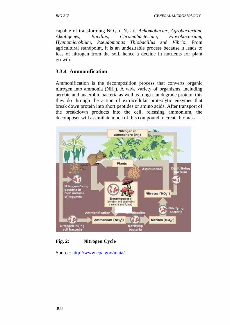
BIO 217 GENERAL MICROBIOLOGY
368
capable of transforming NO3 to N2 are Achomobacter, Agrobacterium,
Alkaligenes, Bacillus, Chromobacterium, Flavobacterium,
Hypnomicrobium, Pseudomonas Thiobacillus and Vibrio. From
agricultural standpoint, it is an undesirable process because it leads to
loss of nitrogen from the soil, hence a decline in nutrients for plant
growth.
3.3.4 Ammonification
Ammonification is the decomposition process that converts organic
nitrogen into ammonia (NH3). A wide variety of organisms, including
aerobic and anaerobic bacteria as well as fungi can degrade protein, this
they do through the action of extracellular proteolytic enzymes that
break down protein into short peptides or amino acids. After transport of
the breakdown products into the cell, releasing ammonium, the
decomposer will assimilate much of this compound to create biomass.
Fig. 2: Nitrogen Cycle
Source: http://www.epa.gov/maia/
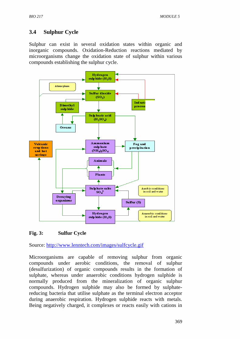
BIO 217 MODULE 5
369
3.4 Sulphur Cycle
Sulphur can exist in several oxidation states within organic and
inorganic compounds. Oxidation-Reduction reactions mediated by
microorganisms change the oxidation state of sulphur within various
compounds establishing the sulphur cycle.
Fig. 3: Sulfur Cycle
Source: http://www.lenntech.com/images/sulfcycle.gif
Microorganisms are capable of removing sulphur from organic
compounds under aerobic conditions, the removal of sulphur
(desulfurization) of organic compounds results in the formation of
sulphate, whereas under anaerobic conditions hydrogen sulphide is
normally produced from the mineralization of organic sulphur
compounds. Hydrogen sulphide may also be formed by sulphate-
reducing bacteria that utilise sulphate as the terminal electron acceptor
during anaerobic respiration. Hydrogen sulphide reacts with metals.
Being negatively charged, it complexes or reacts easily with cations in
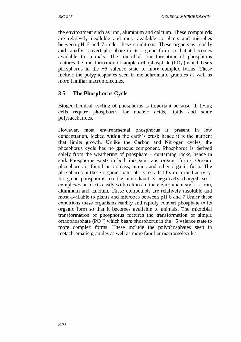
BIO 217 GENERAL MICROBIOLOGY
370
the environment such as iron, aluminum and calcium. These compounds
are relatively insoluble and most available to plants and microbes
between pH 6 and 7 under these conditions. These organisms readily
and rapidly convert phosphate to its organic form so that it becomes
available to animals. The microbial transformation of phosphorus
features the transformation of simple orthophosphate (PO4-) which bears
phosphorus in the +5 valence state to more complex forms. These
include the polyphosphates seen in metachromatic granules as well as
more familiar macromolecules.
3.5 The Phosphorus Cycle
Biogeochemical cycling of phosphorus is important because all living
cells require phosphorus for nucleic acids, lipids and some
polysaccharides.
However, most environmental phosphorus is present in low
concentration, locked within the earth’s crust; hence it is the nutrient
that limits growth. Unlike the Carbon and Nitrogen cycles, the
phosphorus cycle has no gaseous component. Phosphorus is derived
solely from the weathering of phosphate – containing rocks, hence in
soil. Phosphorus exists in both inorganic and organic forms. Organic
phosphorus is found in biomass, humus and other organic form. The
phosphorus in these organic materials is recycled by microbial activity.
Inorganic phosphorus, on the other hand is negatively charged, so it
complexes or reacts easily with cations in the environment such as iron,
aluminum and calcium. These compounds are relatively insoluble and
most available to plants and microbes between pH 6 and 7.Under these
conditions these organisms readily and rapidly convert phosphate to its
organic form so that it becomes available to animals. The microbial
transformation of phosphorus features the transformation of simple
orthophosphate (PO4-) which bears phosphorus in the +5 valence state to
more complex forms. These include the polyphosphates seen in
metachromatic granules as well as more familiar macromolecules.
BIO 217 MODULE 5
371
Fig. 4: Phosphorus Cycle
Source:
http://www.starsandseas.com/SAS%20Ecology/SAS%20chemcycles/cy
cle_phosphorus.htm
SELF-ASSESSMENT EXERCISE
i. Define the following terms
a. Nitrogen Fixation
b. Nitrification
c. Denitrification
d. Ammonification
4.0 CONCLUSION
Microorganisms in the course of their growth and metabolism interact
with each other to cycle nutrients such as carbon, sulphur and
phosphorus. The nutrient cycling called biogeochemical cycling of
elements involves both biological and chemical processes and is of
global importance.
13.0 SUMMARY
Biogeochemical cycling of elements is the movement of
materials via biochemical reactions through biospheres.
It also refers to the biological and chemical processes that
elements such as carbon nitrogen and sulfur undergo during
microbial metabolism.
Elements involved in biogeochemical cycles are used for three
general purposes which are: (i) Biomass Production (ii) Energy
Source and (iii) Terminal Electron Acceptor.
BIO 217 GENERAL MICROBIOLOGY
372
The carbon cycle primarily involves the transfer of carbondioxide
and organic carbon between the atmosphere where it occurs as
principally as inorganic CO2 and the hydrosphere and tithosphere
which contain varying concentration of organic and inorganic
compounds.
The first step in carbon cycle is carbon fixation which is the
conversion of CO2 to organic matter by organisms such as
Cyanobacteria.
The second stage is the transfer of the fixed carbon from
population to population within the biological community.
The respiratory and fermentative metabolism of heterotrophic
organisms returns inorganic carbon dioxide to the atmosphere.
Decomposition of organic compounds such as plants and animals
return CO2 to the cycle.
Carbon is stored in rocks such as limestones (CaCO3) and is
dissolved as carbonate ions (CO3) in oceans.
Burning of fuels such as coal and petroleum releases carbon
dioxide to the atmosphere.
Nitrogen is an essential compound of DNA, RNA and proteins
which are the building blocks of life.
Atmospheric Nitrogen is not directly useable by most organisms
but has to be converted to stable organic forms such as
ammonium and nitrates.
Three processes carried out by microorganisms in the nitrogen
cycle are nitrogen fixation, nitrification and denitrification.
Nitrogen fixation is strictly a bacterial process in which molecule
nitrogen is converted to ammonium ion by a few bacteria that
have a nitrogenase enzyme system.
Nitrogen fixation is carried out by free-living nitrogen fixing
bacteria such as Azobacter, Cyanobacteria and Clostridium found
in the rhizosphere of plant.
Symbiotic nitrogen fixing bacteria such as Rhizobium and
Bradyrhizobium which in symbiotic association with leguminous
plants help fix nitrogen in soil while Cyanobacterium, Anabaenea
fix nitrogen when in association with the water fern.
Nitrification is carried out by chemolithotropic bacteria which
convert ammonium ions to nitrate (NO3) ions in two steps.
Nitrosomonas and Nitrosospira convert ammonia to nitrite while
Nitrococcus and Nitrobacter convert nitrite to nitrate.
Denitrification converts nitrate to dinitrogen.
Ammonification is the decomposition process that converts
organic nitrogen to ammonia (NH3).
Oxidation – Reduction reactions mediated by microorganisms
change the oxidation states of sulphur with in various compounds
establishing the sulphur cycle.
BIO 217 MODULE 5
373
Biogeochemical cycling of phosphorus is important because all
living cells require phosphorus for nucleic acids, lipids and some
polysaccharides.
The phosphorus cycle has no gaseous state and phosphorus is
derived solely from weathering phosphate containing rocks and
organic phosphorus in biomass humus and other organic forms.
14.0 TUTOR-MARKED ASSIGNMENT
1. a (i) State three uses of biogeochemical cycles.
(ii) State specific examples of microorganisms.
b Explain the role of symbiotic nitrogen fixing bacteria in
biological nitrogen fixation in agricultural soils.
c) Outline the steps involved in nitrification.
7.0 REFERENCES/FURTHER READING
Atlas, R.M. (1995). Microorganisms in Our World. Mosby Year Book.
Inc.
Medigan, M.T. et al. (2009). Brock Biology of Microorganisms. (12th
ed.). Pearson Education Inc.
Pelczar, M.J., Chan, E.C.S. & Krieg, R.N. (2001). (5th
ed.).
Microbiology. McGraw-Hill.
Willey, J.M., Sherwood, L.M & Woolverton, C.J. (2008). Microbiology.
(7th
ed.). Boston Bur Bridge, IL: McGraw-Hill Higher Education.
http://www.epa.gov/maia/
http://www.starsandseas.com/SAS%20Ecology/SAS%20chemcycles/cy
cle_phosphorus.htm
http://www.lenntech.com/images/sulfcycle.gif