Embed Size (px)
Citation preview

Bio 230 - Microbiology - Spring 2010 Study Guide 16
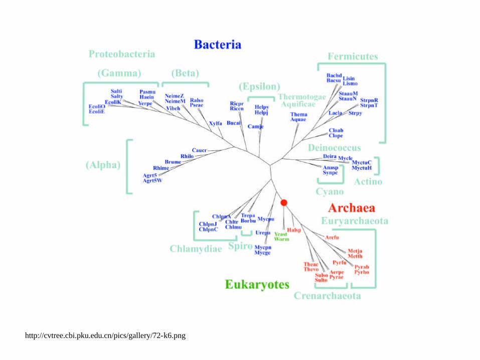
http://cvtree.cbi.pku.edu.cn/pics/gallery/72-k6.png

Alpha-Proteobacteria
http://www.life.umd.edu/classroom/bsci424/BSCI223WebSiteFiles/AlphaProteobacteria.gif
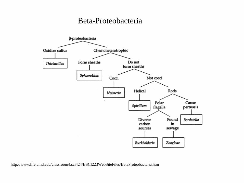
http://www.life.umd.edu/classroom/bsci424/BSCI223WebSiteFiles/BetaProteobacteria.htm
Beta-Proteobacteria
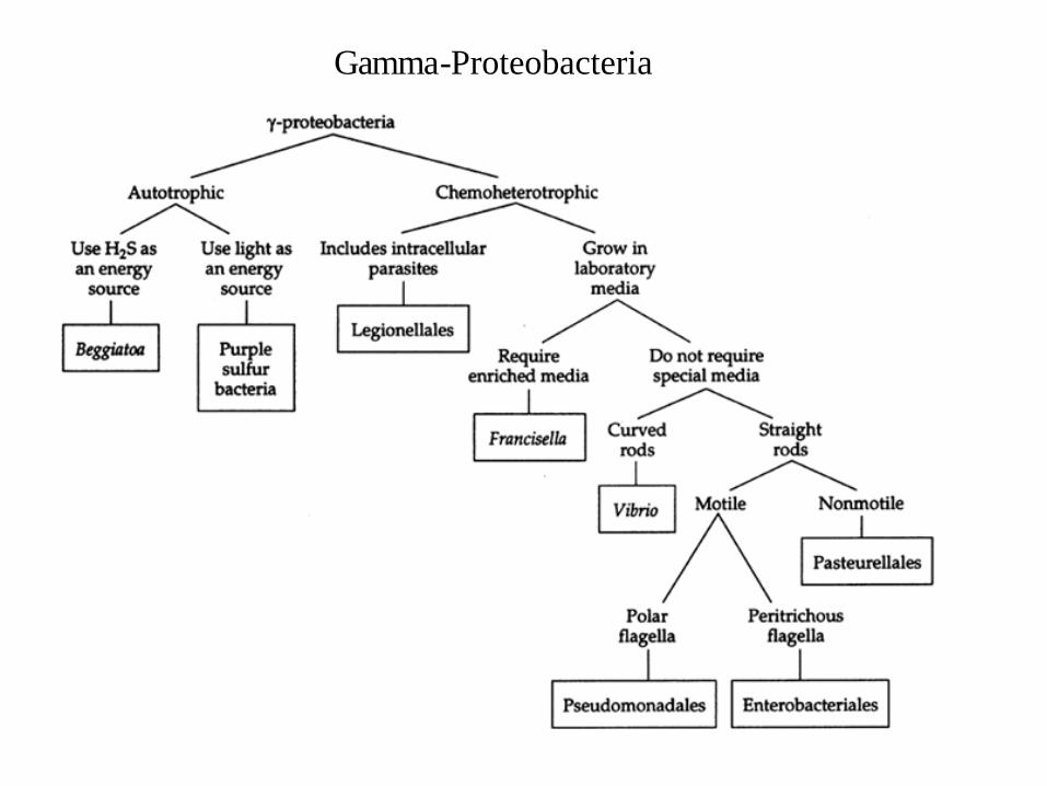
Gamma-Proteobacteria
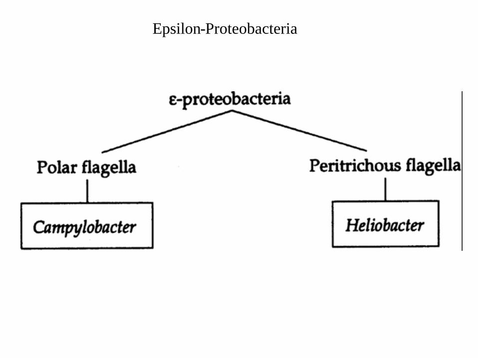
Epsilon-Proteobacteria
MAT (methionine adenosyltransferase E.C.2.5.1.6.) is a housekeeping metabolic enzyme that catalyzes the synthesis of SAM (S-adenosylmethionine) from ATP and methionine.
MAT Database
http://www2.iib.uam.es/gfsanchez/MAT/Main.htm

http://commtechlab.msu.edu/sites/DLC-ME/curious/caOc96LC.gif
http://www.bbc.co.uk/radio4/science/media/bdellovibrio-bacteria.jpg
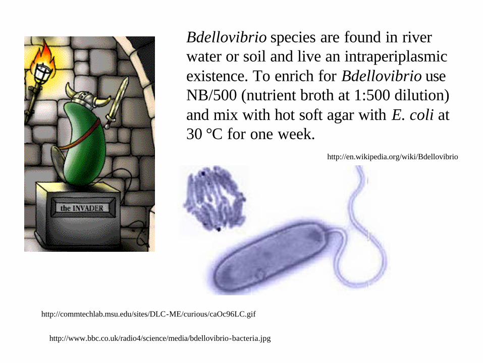
Bdellovibrio species are found in river water or soil and live an intraperiplasmic existence. To enrich for Bdellovibrio use NB/500 (nutrient broth at 1:500 dilution) and mix with hot soft agar with E. coli at 30 °C for one week.
http://en.wikipedia.org/wiki/Bdellovibrio

https://www.cals.ufl.edu/SOS/ppprez/Pathogens%20as%20Prey%20Presentation.ppt.
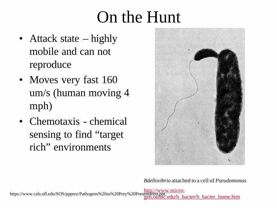
On the Hunt• Attack state – highly
mobile and can not reproduce
• Moves very fast 160 um/s (human moving 4 mph)
• Chemotaxis - chemical sensing to find “target rich” environments
Bdellovibrio attached to a cell of Pseudomonas
http://www.micro-gen.ouhsc.edu/b_bacter/b_bacter_home.htmhttps://www.cals.ufl.edu/SOS/ppprez/Pathogens%20as%20Prey%20Presentation.ppt.

Chemotaxis• Bdellovibrio genome contains 18 known
chemotaxis genes and 1 known aerotaxis genes• At least one gene shown to directly contribute to
predation efficiency• Actual chemical molecules and signals used are
unknown• Chemotaxis likely used to find environments
favorable to prey species• Quorum sensing signals are not utilized
https://www.cals.ufl.edu/SOS/ppprez/Pathogens%20as%20Prey%20Presentation.ppt.

Attacking Prey• Target of attack on prey outer cell membrane
is unknown• No known mechanisms of resistance in prey• Likely target is either non-specific or highly
conserved receptor necessary for cell viability
• Subject of intense research as exact target has been naturally selected as a favorable strategy and may lead to development of novel antibiotics
https://www.cals.ufl.edu/SOS/ppprez/Pathogens%20as%20Prey%20Presentation.ppt.

Entry and Attachment• Bdellovibrio uses flagella to make physical
contact, grabs on with pili• Attachment is initially reversible, becomes
permanent after initial “recognition” step• May have preferred prey – attachment rates
differ in mixed populations• Enters through hole induced in outer
membrane by peptidoglycan hydrolysishttps://www.cals.ufl.edu/SOS/ppprez/Pathogens%20as%20Prey%20Presentation.ppt.
Formation of the Bdelloplast• Bdellovibrio restabilizes prey outer
membrane to contain nutrients and protect against dehydration
• Bdellovibrio attaches to the inner membrane causing the cytoplasm to round up into the osmotically stable Bdelloplast
• Exact mechanism of Bdelloplast formation is unknown but prey membrane maintains structural integrity
• Bdellovibrio begins to extract and digest prey cellular components
https://www.cals.ufl.edu/SOS/ppprez/Pathogens%20as%20Prey%20Presentation.ppt.

Prey Utilization• Nearly all of the prey bacteria is utilized• Prey components are broken down via
hydrolysis and used to replicate additional Bdellovibrio or metabolized for energy
• About 50% of prey nucleic acid utilized for de novo synthesis
• Ribose from other 50% metabolized for energy
• Final rupture of cell wall degrades peptidoglycans, liberating sugars and amino acids that can be used by Bdellovibrio
https://www.cals.ufl.edu/SOS/ppprez/Pathogens%20as%20Prey%20Presentation.ppt.
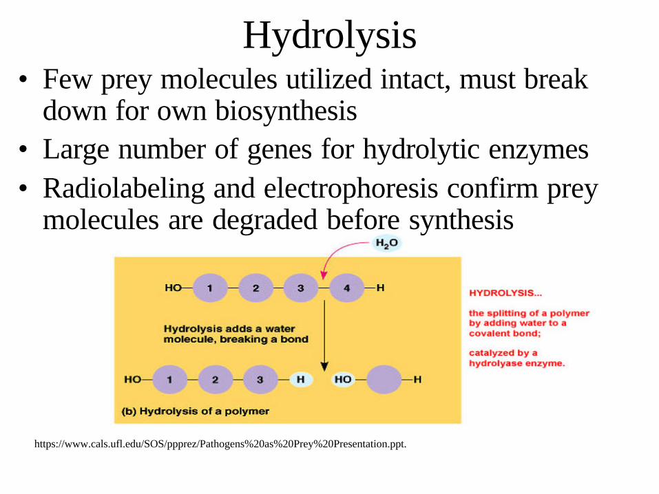
Hydrolysis• Few prey molecules utilized intact, must break
down for own biosynthesis• Large number of genes for hydrolytic enzymes• Radiolabeling and electrophoresis confirm prey
molecules are degraded before synthesis
https://www.cals.ufl.edu/SOS/ppprez/Pathogens%20as%20Prey%20Presentation.ppt.

Synthesis• Sophisticated coordination by predator
regulators ensures smooth pathway from degradation to synthesis
• Hydrolytic degradation products incorporated into ongoing synthesis
• Regulators are unknown• Bdellovibrio genome contains many
synthesis genes, including 3 for ATP alone
https://www.cals.ufl.edu/SOS/ppprez/Pathogens%20as%20Prey%20Presentation.ppt.

Cell Membrane Construction• It is unclear if Bdellovibrio scavenges large
membrane molecules• Current evidence suggests Bdellovibrio
synthesizes its own membrane macromolecules
• Bdellovibrio Lipid A has a unique structure• Outer Membrane Protein (OMP)
scavenging confirmed by SDS-PAGE, refuted by mass spectroscopy
https://www.cals.ufl.edu/SOS/ppprez/Pathogens%20as%20Prey%20Presentation.ppt.

Nucleic Acid Synthesis• Prey nucleic acids initially cuts with endonucleases• Molecules then taken apart one at a time with
exonucleases• To replicate offspring, Bdellovibrio must
synthesize much more nucleic than contained within prey – E coli prey cell contains 4M bp– Bdellovibrio produced 4 offspring synthesizes
approximately 15M bp
https://www.cals.ufl.edu/SOS/ppprez/Pathogens%20as%20Prey%20Presentation.ppt.

Protein Synthesis
• Bdelloplast can not replicate full set of amino acids
• This suggests Bdellovibrio is only capable of protein synthesis with access to prey amino acids
https://www.cals.ufl.edu/SOS/ppprez/Pathogens%20as%20Prey%20Presentation.ppt.

Reproduction• # of offspring limited by raw
material available from prey• As Bdelloplast grows it
elongates into a filamentous structure containing all replicated chromosomes
• As Bdellovibrio senses the prey has become exhausted the Bdelloplast is organized and septates into the individual progeny
• The remaining prey membrane is lysed and progeny are set free
Starr and Baigent,, JOURNAL OF BACTERIOLOGY, May, 1966 Vol. 91, No. 5
https://www.cals.ufl.edu/SOS/ppprez/Pathogens%20as%20Prey%20Presentation.ppt.

http://upload.wikimedia.org/wikipedia/commons/8/86/Rickettsia_rickettsii.jpg
Rickettsia rickettsi
Rickettsia is a genus of motile, Gram-negative, non-sporeforming, highly pleomorphic bacteria that can present as cocci (0.1 µm in diameter), rods (1-4 µm long) or thread-like (10 µm long). Obligate intracellular parasites, the Rickettsia depend on entry, growth, and replication within the cytoplasm of eukaryotic host cells (typically endothelial cells).

Certain segments of Rickettsial genomes resemble that of mitochondria.The deciphered genome of R. prowazekii is 1,111,523 bp long and contains 834 protein-coding genes.Unlike free-living bacteria, it contains no genes for anaerobic glycolysis or genes involved in the biosynthesis and regulation of amino acids and nucleosides. In this regard it is similar to mitochondrial genomes; in both cases, nuclear (host) resources are used. ATP production in Rickettsia is the same as that in mitochondria. In fact, of all the microbes known, the Rickettsia is probably the closest "relative" (in phylogenetic sense) to the mitochondria. Unlike the latter, the genome of R. prowazekii, however, contains a complete set of genes encoding for the tricarboxylic acid cycle and the respiratory chain complex.
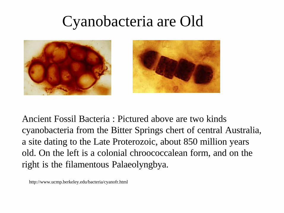
Ancient Fossil Bacteria : Pictured above are two kinds cyanobacteria from the Bitter Springs chert of central Australia, a site dating to the Late Proterozoic, about 850 million years old. On the left is a colonial chroococcalean form, and on the right is the filamentous Palaeolyngbya.
http://www.ucmp.berkeley.edu/bacteria/cyanofr.html
Cyanobacteria are Old
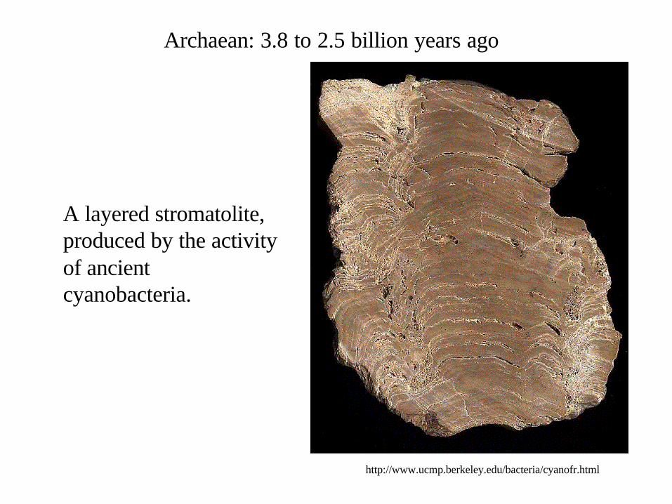
A layered stromatolite, produced by the activity of ancient cyanobacteria.
Archaean: 3.8 to 2.5 billion years ago
http://www.ucmp.berkeley.edu/bacteria/cyanofr.html
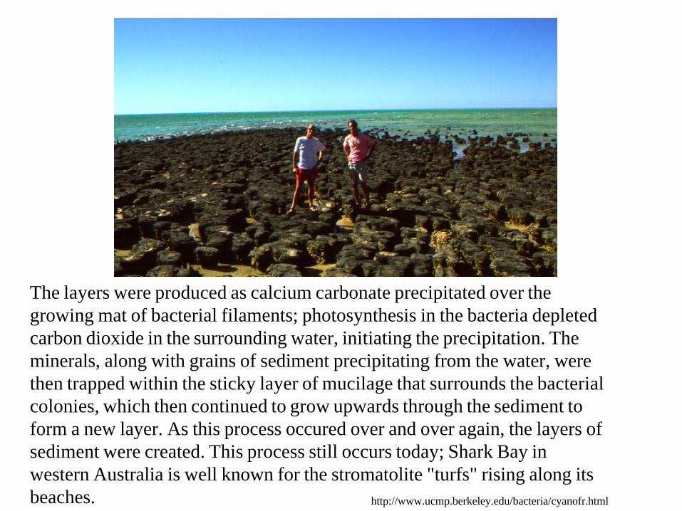
The layers were produced as calcium carbonate precipitated over the growing mat of bacterial filaments; photosynthesis in the bacteria depleted carbon dioxide in the surrounding water, initiating the precipitation. The minerals, along with grains of sediment precipitating from the water, were then trapped within the sticky layer of mucilage that surrounds the bacterial colonies, which then continued to grow upwards through the sediment to form a new layer. As this process occured over and over again, the layers of sediment were created. This process still occurs today; Shark Bay in western Australia is well known for the stromatolite "turfs" rising along its beaches. http://www.ucmp.berkeley.edu/bacteria/cyanofr.html

The Cyanobacteria are morphologically an extremely diverse group.
So much so that without an electron microscope, the botanists thought it was their domain, rather than that of the microbiologists
http://www.keweenawalgae.mtu.edu/ALGAL_PAGES/cyanobacteria.htm

http://www.bact.wisc.edu/Microtextbook/images/book_4/chapter_2/2-53.jpg
http://www.keweenawalgae.mtu.edu/ALGAL_IMAGES/cyanobacteria/Anabaena_jason_dbtow17_2016.jpg
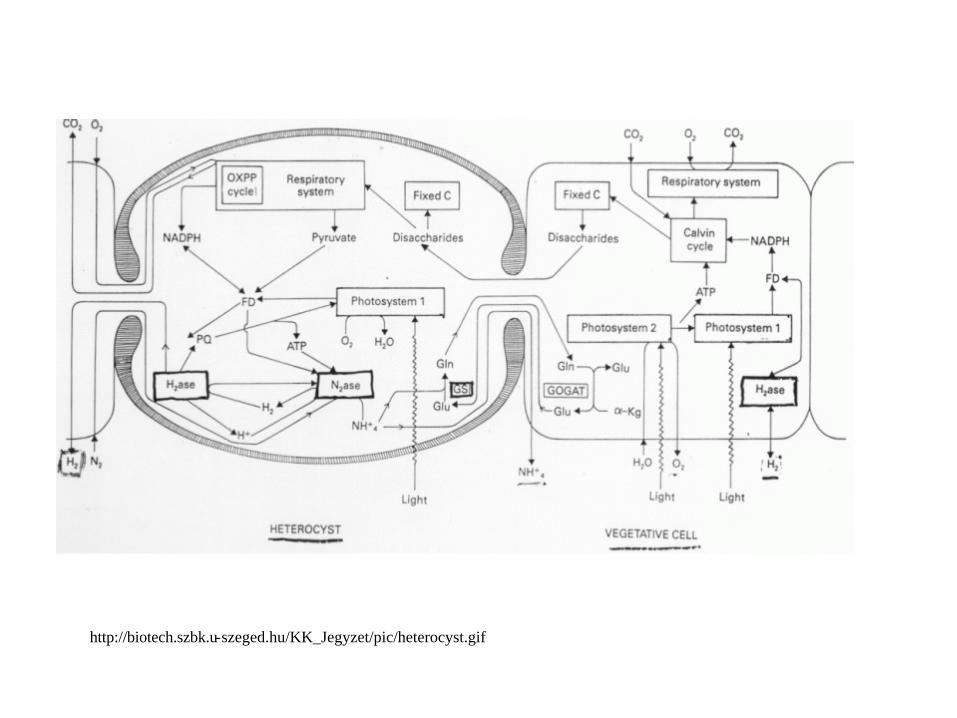
http://biotech.szbk.u-szeged.hu/KK_Jegyzet/pic/heterocyst.gif

Anabaena filaments which have been genetically engineered so that the heterocysts are expressing a fluorescent protein:
http://www.mun.ca/biochem/courses/3107/Topics/Site_specific_Recomb.html
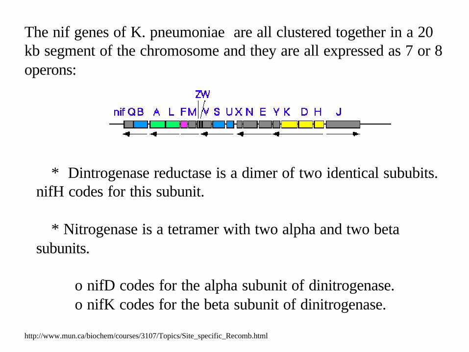
The nif genes of K. pneumoniae are all clustered together in a 20 kb segment of the chromosome and they are all expressed as 7 or 8 operons:
* Dintrogenase reductase is a dimer of two identical sububits. nifH codes for this subunit.
* Nitrogenase is a tetramer with two alpha and two beta subunits.
o nifD codes for the alpha subunit of dinitrogenase.o nifK codes for the beta subunit of dinitrogenase.
http://www.mun.ca/biochem/courses/3107/Topics/Site_specific_Recomb.html

In Anabaena sp. PCC 7120, the nif genes were originally mapped using vegetative cell DNA. A surprise finding was the fact that the genes were not tightly clustered and, worse, the nifK gene was located 11 kb away from nifD as shown in the following map:
This was troubling because NifK and NifD are required in equimolar amounts in the cell and there was no obvious way to regulate how this would be so.
http://www.mun.ca/biochem/courses/3107/Topics/Site_specific_Recomb.html
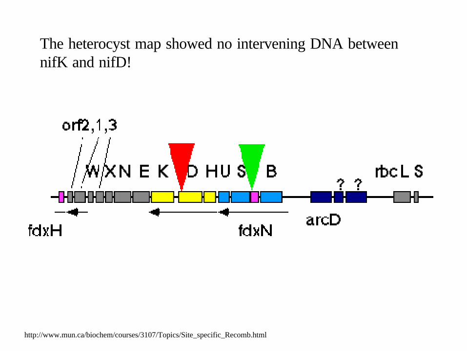
The heterocyst map showed no intervening DNA between nifK and nifD!
http://www.mun.ca/biochem/courses/3107/Topics/Site_specific_Recomb.html

Further research proved what was going on. Heterocyst differentiation actually required a site-specific recombination event to excise this intervening element. The recombination is catalyzed by XisA which acts at two directly repeating 11 bp sequences within the nifD gene (shown as large red triangles). Since the initial discovery of this DNA rearrangement, two further DNA rearrangements have been characterized:
* a 55 kb element that interrupts the fdxN gene is removed during heterocyst differentiation. This recombination is catalyzed by the xisF gene which acts at two directly repeating 5 bp sequences within the fdxN gene (shown as green triangles above). As a result, the nifB-fdxN-nifS-nifU operon can then be expressed properly.
* a 10.6 kb element is excised from the hupL gene, which codes for the large subunit of the enzyme hydrogenase, by site-specific recombination between 16 bp direct repeats that flank the element late during heterocyst differentiation. This recombination is catalyzed by the xisC gene which is also located at one end of the element. This rearrangement permits a proper HupL protein to be synthesized.
In all three rearrangements, the enzyme that catalyzes the removal of the element is encoded adjacent to one of the junctions within the element that is removed. XisA and XisC are site-specific tyrosine recombinases - they are related to each other but distantly related to other integrases.
http://www.mun.ca/biochem/courses/3107/Topics/Site_specific_Recomb.html
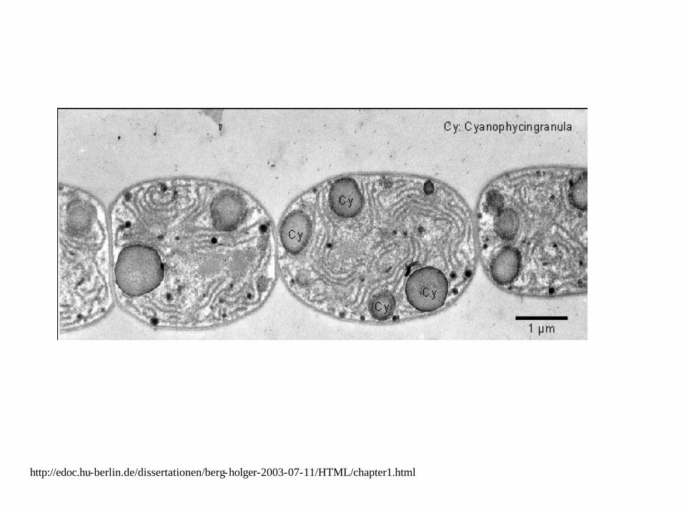
http://edoc.hu-berlin.de/dissertationen/berg-holger-2003-07-11/HTML/chapter1.html

http://edoc.hu-berlin.de/dissertationen/berg-holger-2003-07-11/HTML/chapter1.html

http://edoc.hu-berlin.de/dissertationen/berg-holger-2003-07-11/HTML/chapter1.html
Cyanobacteria can play a in important role in defining water quality
Geosmin
Toxins
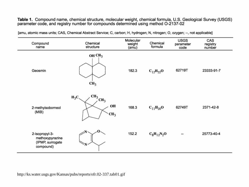
http://ks.water.usgs.gov/Kansas/pubs/reports/ofr.02-337.tab01.gif
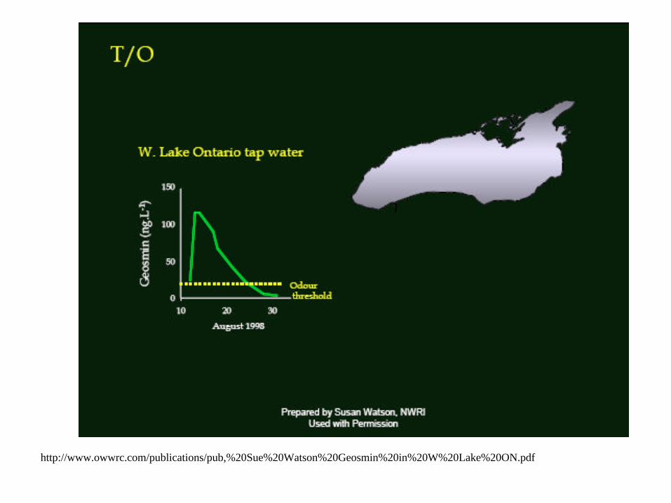
http://www.owwrc.com/publications/pub,%20Sue%20Watson%20Geosmin%20in%20W%20Lake%20ON.pdf
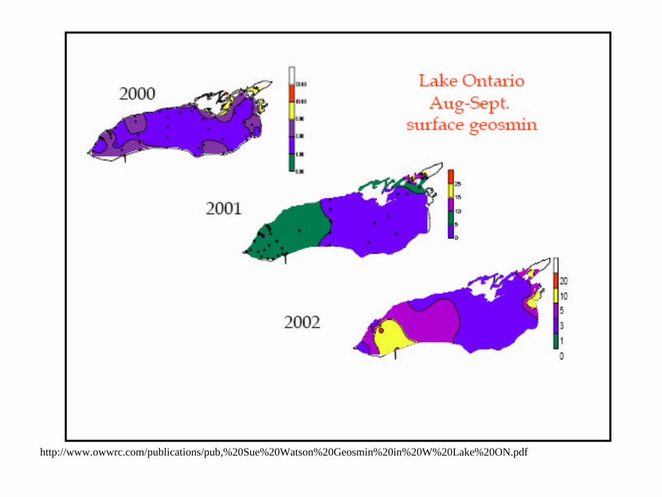
http://www.owwrc.com/publications/pub,%20Sue%20Watson%20Geosmin%20in%20W%20Lake%20ON.pdf
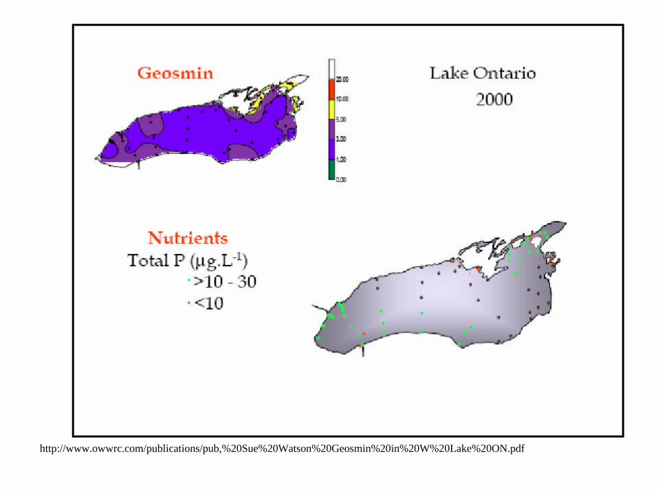
http://www.owwrc.com/publications/pub,%20Sue%20Watson%20Geosmin%20in%20W%20Lake%20ON.pdf

Microcystis aeruginosa
http://www.cyanobacteria-platform.com/main.html
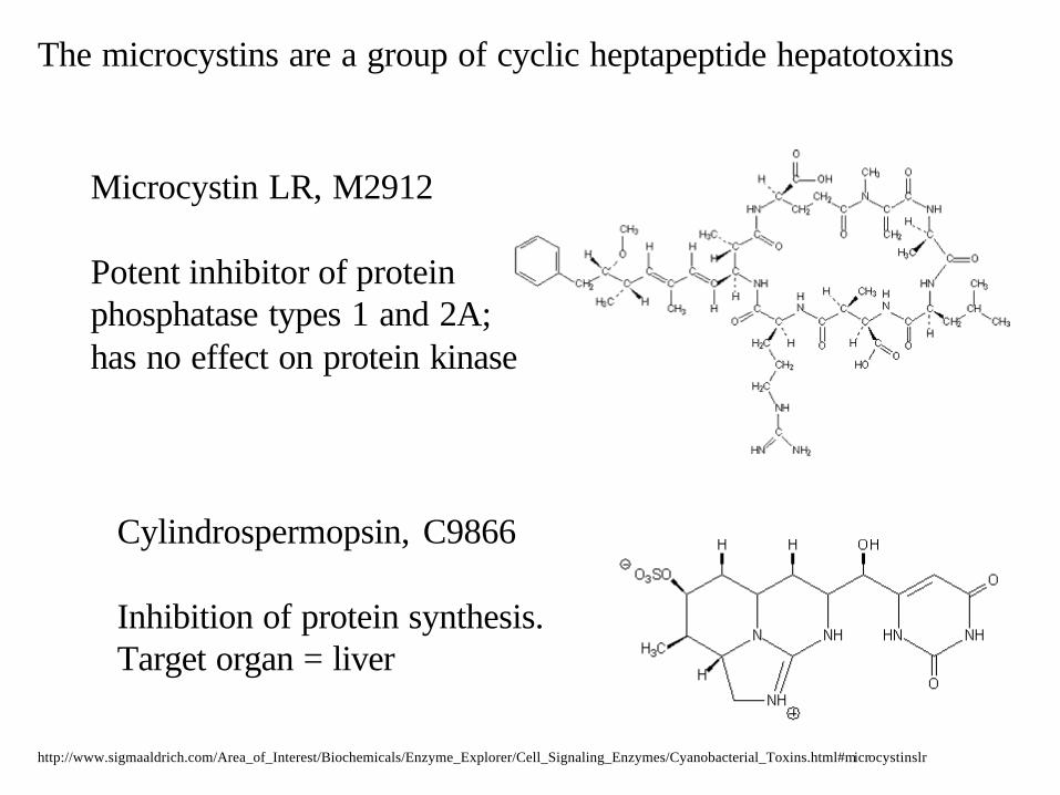
Microcystin LR, M2912
Potent inhibitor of protein phosphatase types 1 and 2A; has no effect on protein kinase
The microcystins are a group of cyclic heptapeptide hepatotoxins
Cylindrospermopsin, C9866
Inhibition of protein synthesis.Target organ = liver
http://www.sigmaaldrich.com/Area_of_Interest/Biochemicals/Enzyme_Explorer/Cell_Signaling_Enzymes/Cyanobacterial_Toxins.html#microcystinslr

http://ejournal.sinica.edu.tw/bbas/content/2000/3/130401.JPG

The End


![Induction of Interleukin 2 Production but not Methionine ...[CANCER RESEARCH 52, 3361-3366, June 15, 1992] Induction of Interleukin 2 Production but not Methionine Adenosyltransferase](https://img.pdfslide.net/doc/110x75/611ace0a32e6d405ac64c0d3/induction-of-interleukin-2-production-but-not-methionine-cancer-research-52.jpg)