Embed Size (px)
Citation preview

Three-Dimensional Structure of ATP:Corrinoid Adenosyltransferase fromSalmonella typhimuriumin Its Free State, Complexed with MgATP, or Complexed
with Hydroxycobalamin and MgATP†,‡
Cary B. Bauer,§ Maris V. Fonseca,| Hazel M. Holden,§ James B. Thoden,§ Thomas B. Thompson,§
Jorge C. Escalante-Semerena,*,| and Ivan Rayment*,§
Department of Biochemistry, UniVersity of Wisconsin, Madison, Wisconsin 53706, and Department of Bacteriology,UniVersity of Wisconsin, Madison, Wisconsin 53706
ReceiVed September 12, 2000; ReVised Manuscript ReceiVed NoVember 6, 2000
ABSTRACT: In Salmonella typhimurium, formation of the cobalt-carbon bond in the biosynthetic pathwayfor adenosylcobalamin is catalyzed by the product of thecobA gene which encodes a protein of 196amino acid residues. This enzyme is an ATP:co(I)rrinoid adenosyltransferase which transfers an adenosylmoiety from MgATP to a broad range of co(I)rrinoid substrates that are believed to include cobinamide,its precursor cobyric acid and probably others as yet unidentified, and hydroxocobalamin. Three X-raystructures of CobA are reported here: its substrate-free form, a complex of CobA with MgATP, and aternary complex of CobA with MgATP and hydroxycobalamin to 2.1, 1.8, and 2.1 Å resolution,respectively. These structures show that the enzyme is a homodimer. In the apo structure, the polypeptidechain extends from Arg28 to Lys181 and consists of anR/â structure built from a six-stranded parallelâ-sheet with strand order 324516. The topology of this fold is very similar to that seen in RecA protein,helicase domain, F1ATPase, and adenosylcobinamide kinase/adenosylcobinamide guanylyltransferase wherea P-loop is located at the end of the first strand. Strikingly, the nucleotide in the MgATP‚CobA complexbinds to the P-loop of CobA in the opposite orientation compared to all the other nucleotide hydrolases.That is, theγ-phosphate binds at the location normally occupied by theR-phosphate. The unusual orientationof the nucleotide arises because this enzyme transfers an adenosyl group rather than theγ-phosphate. Inthe ternary complex, the binding site for hydroxycobalamin is located in a shallow bowl-shaped depressionat the C-terminal end of theâ-sheet of one subunit; however, the active site is capped by the N-terminalhelix from the symmetry-related subunit that now extends from Gln7 to Ala24. The lower ligand of cobalaminis well-ordered and interacts mostly with the N-terminal helix of the symmetry-related subunit. Interestingly,there are few interactions between the protein and the polar side chains of the corrin ring which accountsfor the broad specificity of this enzyme. The corrin ring is oriented such that the cobalt atom is located∼6.1 Å from C5′ of the ribose and is beyond the range of nucleophilic attack. This suggests that aconformational change occurs in the ternary complex when Co(III) is reduced to Co(I).
Cobalamin is the largest cofactor utilized in biologicalsystems and consists of a highly decorated corrin ring at thecenter of which lies a cobalt atom (Figure 1). The cobaltatom is hexacoordinate where four of the positions are filledby nitrogen atoms belonging to the tetrapyrrole and the othertwo positions are occupied by the upper and lower ligands.
The lower ligand is usually a 5,6-dimethylbenzimidazolemoiety that is covalently attached to the corrin ring via anucleotide loop. The upper ligand is variable; however, thebiologically functional forms of cobalamin have either anadenosyl or methyl group covalently attached to the cobalt.The unusual chemistry associated with cobalamin-dependentenzymes is the result of a labile single cobalt-carbon bondbetween the cobalt atom and the upper ligand.
The carbon-metal bond is unique in biological systems,and as a consequence, considerable effort has been devotedtoward understanding the cobalamin biosynthetic pathwayand the enzymes that utilize this cofactor. The identity ofthe chemical steps and the enzymes involved in the aerobicpathway were first elucidated forPseudomonas denitrificans(1). Likewise, there is considerable knowledge of theanaerobic pathway inSalmonella typhimurium(2). To date,25 gene products have been identified for the synthesis ofadenosylcobalamin; however, the identity of the enzymesinvolved in the synthesis of the lower ligand base 5,6-dimethylbenzimidazole remains unknown. There is still
† This research was supported in part by NIH Grants AR35186 andGM58281 to I.R. and GM40313 to J.C.E.-S. M.V.F. was supported byMARC Fellowship GM17528. Use of the Argonne National LaboratoryStructural Biology Center beamlines at the Advanced Photon Sourcewas supported by the U.S. Department of Energy, Office of BasicEnergy Research, under Contract W-31-109-ENG-38.
‡ The X-ray coordinates and structure factors have been depositedin the Protein Data Bank under file names 1G5R, 1G5T, and 1G64for the apo structure, the MgATP complex, and the ternary complex,respectively.
* To whom correspondence should be addressed. I.R.: Departmentof Biochemistry, 433 Babcock Dr., Madison, WI 53706; phone,(608) 262-0437; fax, (608) 262-1319; e-mail, Ivan•[email protected]. J.C.E.-S.: Department of Bacteriology, Fred Hall,Linden Drive, Madison, WI 53706; e-mail, [email protected].
§ Department of Biochemistry, University of Wisconsin.| Department of Bacteriology, University of Wisconsin.
361Biochemistry2001,40, 361-374
10.1021/bi002145o CCC: $20.00 © 2001 American Chemical SocietyPublished on Web 12/14/2000

considerably more to be learned about the organization,structure, and function of the proteins that are responsiblefor this remarkable feat, especially since there appear to besignificant differences between the aerobic and anaerobicbiosynthetic pathways. One of the variables appears to bethe timing of cobalt insertion and the formation of thecobalt-carbon bond (3, 4). This is especially intriguingbecause it involves the formation of the molecular featurethat is ultimately responsible for the final function ofcobalamin.
In P. denitrificans, cobalt is inserted into the corrinoid inthe biosynthetic pathway after synthesis of hydrogenobyrinicacid a,c-diamide by the enzyme cobaltochelatase (5). There-after, adenosylation occurs to yield adenosylcobyrinic acida,c-diamide (6). In contrast, insertion of cobalt and adeno-sylation is thought to occur much earlier in the anaerobicbiosynthetic pathway ofS. typhimurium, perhaps as early ascobalt-precorrin 3 (M. V. Fonseca and J. C. Escalante-Semerena, unpublished results). This suggests that thesubstrate specificity of the intervening biosynthetic enzymesin the aerobic and anaerobic pathways must be different toaccommodate the absence or presence of the adenosyl group.
In bothP. denitrificansandS. typhimurium, adenosylationof the central cobalt atom occurs in a three-step process:(1) the cobalt is reduced from Co(III) to Co(II) in a one-electron transfer, (2) Co(II) is reduced to Co(I) in a secondsingle-electron transfer to generate a very powerful nucleo-phile, and finally, (3) the Co(I) conducts a nucleophilic attackon the adenosyl moiety of ATP to leave the cobalt atom ina Co(III) state. Several corrinoid reductase activities havebeen reported, some of which have been isolated from avariety of organisms (7-11); however, the identity of thegenes encoding these enzymes is still unknown. There isgood evidence that flavin nucleotides are involved in thereduction of the cobalt since cell-free extracts from bothClostridium tetanomorphumandProprionibacterium freun-denreichii require these cofactors for function (11, 12).Likewise, inEscherichia coli, a flavodoxin has been shown
to catalyze the reduction of cobalamin when bound tomethionine synthase (13, 14); however, a recent study ofthe adenosylation reaction inS. typhimuriumhas demon-strated that reduction from Co(III) to Co(II) can be ac-complished by the dihydroflavins alone without an enzymaticpartner (15). Regardless of the manner in which the cobaltatom is reduced, it seems highly likely that the final reductionfrom Co(II) to Co(I) must occur on the enzyme responsiblefor the adenosyl transfer because of the reactivity of Co(I).
The enzyme responsible for the adenosylation reaction isthe product of genecobOin P. denitrificansandcobAin S.typhimurium (6, 16). Both of these enzymes have beenpurified and partially characterized (6, 17). In the case ofCobO fromP. denitrificans, the enzyme shows specificityfor cobyrinic acid a,c-diamide and the corrinoids that occurlater in the biosynthetic pathway. Studies on CobA fromS.typhimuriumsuggest broader specificity for this enzyme sincethe cobalt ion is inserted earlier than inP. denitrificans;however, the exact point of adenosylation is unknown. Broadspecificity for CobA might be of some evolutionary advan-tage since it allows corrinoids that have lost their upper ligandto be recycled and it facilitates entry of exogenous corrinoidsinto the biosynthetic pathway. Interestingly, CobA is ableto transfer a variety of nucleosides to the cobalt, includingCTP, UTP, and GTP in decreasing order of preference (17).
Nucleophilic attack of Co(I) on the ribosyl moiety of thenucleotide is believed to heterolytically displace triphosphate.Earlier studies on the transferase activity inC. tetanomor-phum demonstrated that the product is triphosphate (18),while in Pr. freundenreichii, the orthophosphate and pyro-phosphate were reported to be the product of the reactioncatalyzed by the adenosyltransferase enzyme (12); however,neither protein was characterized in detail. In the case ofthe enzyme fromP. denitrificans, the exact nature of theproduct is still unknown, while for CobA fromS. typhimu-rium, unpublished findings indicate that the product islikewise triphosphate (M. V. Fonseca and J. C. Escalante-Semerena, unpublished results).
The reaction catalyzed by this enzyme poses severalinteresting questions with respect to the substrate specificityand the oxidation state of the substrate. First, how does theprotein coordinate the reduction of Co(II) to Co(I) and thetransferase activity? Second, what is the relationship betweenthe nucleotide and the cobalt prior to nucleophilic attack sincethere is good evidence that the nucleotide must bind beforethe corrinoid (17)? Third, what features of the enzyme allowit to accommodate corrinoids that differ widely in thesubstitution of their corrin rings? To address these questions,a structural and biochemical investigation of CobA fromS.typhimurium was initiated. Here we report the X-raystructures of ATP:corrinoid adenosyltransferase fromS.typhimurium(CobA) in its substrate-free state (apo), com-plexed with MgATP, and in a ternary complex with CobA,hydroxycobalamin, and MgATP determined to 2.1, 1.8, and2.1 Å resolution, respectively.
MATERIALS AND METHODS
Purification and OVerproduction of the ATP:CorrinoidAdenosyltransferase (CobA).Overproduction and purificationof the CobA enzyme were performed as previously describedwith some modifications (17), including monitoring via
FIGURE 1: Chemical structure of 5′-deoxyadenosylcobalamin. InS. typhimurium, the lower ligand is 5,6-dimethylbenzimidazole (45).
362 Biochemistry, Vol. 40, No. 2, 2001 Bauer et al.
SDS-PAGE1 (19) and employing the in vitro corrinoidadenosylation assay.
Cells of strain JE2884 [metE205 ara-9DEL902(cobA-trp)/pGP1-2, pCOBA15] were grown in 6 L of Luria-Bertani(LB) broth containing ampicillin (50µg/mL), kanamycin (50µg/mL), and 22 mM glucose at 30°C with shaking untilthey reached anA650 of ∼0.7. Cultures were shifted to 42°C for 30 min and then to 37°C for 1.5 h to allow synthesisof CobA. The resultant cells were pelleted by centrifugationin a Sorvall centrifuge (DuPont Instruments, Wilmington,DE) at 10000g utilizing a Sorvall GSA rotor.
These cells were resuspended in 150 mL of 50 mM Tris-HCl buffer (pH 8.0) at 4°C which contained 5 mM DTT(buffer A) and 16µg/mL protease inhibitor and phenyl-methanesulfonyl fluoride. The cell suspension was lysed bysonication with a Sonic Dismembrator (model 550 FisherScientific) for 7.5 min (large tip, setting of 6, 50% duty).Cell-free extracts were obtained by centrifugation at 40000gfor 2 h on aSorvall TYPE-SS34 rotor (Sorvall Instruments,Newtown, CT). Approximately 6.5 mg of protein wasobtained per milliliter of cell-free extract.
Precipitation of proteins in the cell-free extract wasperformed at 4°C using finely ground Ultrapure ammoniumsulfate (ICN Biochemicals, Aurora, OH). Ammonium sulfatewas added to the cell-free extract to 25% saturation. Theprotein solution was centrifuged at 10000g for 10 min toremove precipitated material from the first step. Followingcentrifugation, ammonium sulfate was added to 45% satura-tion. The precipitated protein was collected by centrifugationas described above.
Hydrophobic Interaction Chromatography.The proteinpellet from the previous step was resuspended in 70 mL ofbuffer A containing 20% saturation ammonium sulfate. Theprotein solution was loaded onto a Phenyl-Sepharose (SigmaChemical Co., St. Louis, MO) column (2.5 cm× 14 cm, 70mL bed volume) equilibrated with buffer A containing 20%ammonium sulfate. The column was developed at a flowrate of 54 mL/h. Protein was loaded at a concentration of10 mg/mL of resin. The column was washed with 250 mLof the loading buffer followed by a 350 mL reverse lineargradient (from 20 to 0%) of ammonium sulfate in buffer A.CobA eluted at the end of the gradient.
Dye-Ligand Affinity Chromatography.Fractions from thePhenyl-Sepharose step containing CobA were pooled. Potas-sium chloride (KCl) was added to the pool of fractions to afinal concentration of 0.2 M. A Cibacron Blue 3GA Type3000 (Sigma) column (1.5 cm× 11 cm, 20 mL bed volume)was equilibrated with buffer A containing 0.2 M KCl anddeveloped at a flow rate of 60 mL/h. Approximately 6.8 mgof protein/mL of resin was loaded. After the column hadbeen washed with 60 mL of equilibration buffer, a 100 mLlinear gradient of from 0.2 to 2 M KCl was used to developthe column. CobA was∼95% homogeneous as judged bySDS-PAGE. Fractions containing homogeneous CobA werepooled and concentrated with a Centriprep 10 device(Amicon, Inc., Beverly, MA). This protocol yielded between
30 and 50 mg of homogeneous CobA from 6 L of cells,depending on the level of overexpression.
Purification and OVerproduction of Selenomethionine-CobA (SeMet-CobA).SeMet-CobA was also overproducedfrom strain JE2884. Cells of strain JE2884 were grown onVogel-Bonner minimal medium (20) supplemented withglucose (35 mM). All amino acids, except methionine, wereadded to a final concentration of 5 mM. Additionally, themedium was supplemented with 5 mM adenine and SeMet(5 mM). SeMet-CobA was purified according to theprotocol described for the wild-type enzyme (see above). Allbuffers used in the SeMet-CobA purification were spargedwith oxygen-free nitrogen 30-45 min before use. Fractionscontaining purified SeMet-CobA were pooled and kept at4 °C under a nitrogen atmosphere in the presence of 5 mMDTT. Approximately 30 mg of homogeneous SeMet-CobAwas obtained from this procedure.
Apo-CobA Crystallization X-ray Data Collection andStructure Determination. Crystals of CobA (both wild typeand SeMet) were grown by microbatch at 4°C from 13%methyl ether-polyethylene glycol 350 and 100 mM NaCl in50 mM MES (pH 5.5). The final protein concentration ineach droplet was approximately 5 mg/mL. Small hexagonalplate-shaped crystals appeared spontaneously in 3-4 daysand achieved maximum dimensions 0.1 mm× 0.4 mm×0.4 mm over a period of several weeks.
Prior to X-ray data collection, crystals of the seleno-methionine-substituted apo-CobA were transferred to asynthetic mother liquor composed of 25% methyl ether-polyethylene glycol 350 and 250 mM NaCl in 50 mM MES(pH 5.5) and allowed to equilibrate at 4°C overnight.Crystals were then transferred incrementally to a cryopro-tectant solution consisting of 25% ethylene glycol, 20%methyl ether-polyethylene glycol 350, and 300 mM NaCl,in 50 mM MES (pH 5.5). The crystals were transferredsuccessively to droplets with 30, 50, and 100% cryopro-tectant/synthetic mother liquor ratios and allowed to standfor 1 min. A single crystal was picked up in a loop of surgicalsuture and flash-frozen in a stream of cold liquid nitrogengas. Apo-CobA crystallizes in the hexagonal space groupP6122 with one subunit in the asymmetric unit and thefollowing unit cell dimensions:a ) b ) 49.4 Å andc )250.2 Å.
Multiple anomalous dispersion (MAD) data were collectedat four wavelengths on a single frozen crystal at beamline19-ID of the Structural Biology Center at the AdvancedPhoton Source in Argonne, IL. For each wavelength, 100images were recorded on a 3× 3 tiled CCD detector with1° oscillations. An additional 100, 1° images were thenrecorded with aφ value that differed by 180° from theoriginal images (inverse beam method). The diffraction datawere processed and scaled with the HKL2000 software suite(21). The Friedel differences in the reference data set (peak)were externally locally scaled to remove systematic errorswith the program BIGSCALE (G. Wesenberg and I. Ray-ment, unpublished data). The other three data sets were thenlocally scaled to the reference data set. Data collection andprocessing statistics are presented in Table 1.
The positions of five of the seven selenium atoms werelocated with automated Patterson methods utilized by theprogram SOLVE (22, 23). Phases were then calculated fromthe MAD data sets with the program CNS (24). Density
1 Abbreviations: DTT, dithiothreitol; EDTA, ethylenediaminetet-raacetic acid; rms, root-mean-square; AdoCbl, adenosylcobalamin;AdoCbi, adenosylcobinamide; OH-CBL, hydroxycobalamin; DMB, 5,6-dimethylbenzimidazole; MES, 2-(N-morpholino)ethanesulfonic acid;SDS-PAGE, sodium dodecyl sulfate-polyacrylamide gel electro-phoresis.
Structure of ATP:Corrinoidadenosyl Transferase Biochemistry, Vol. 40, No. 2, 2001363

modification (solvent flipping) was performed with CNS(24). This procedure yielded an electron density map thatshowed continuous electron density over a large region ofthe unit cell. Residues Arg28-Lys181 were built into theelectron density with the program TURBO FRODO (25) andrefined with TNT (26). The missing residues at the N- andC-termini could not be located in the electron density map.Analysis of the backbone dihedral angles with PROCHECKrevealed that for the apo-CobA, 95.5% of the residuesconformed to the most favorable regions and the other 4.5%conformed to other additionally allowed regions (27). Thefinal R-factor was 17.8% for X-ray data between 30 and 2.1Å resolution with anR-free of 24.7%. The final refinementstatistics are presented in Table 2.
MgATP‚CobA Structure Determination. Crystals of apo-CobA described above were transferred to a solution thatcontained 25% methyl ether-polyethylene glycol 350, 250mM NaCl in 50 mM MES, 1 mM MgATP, and 1 mM OH-CBL at pH 5.5. The crystals were then frozen as beforeexcept for the inclusion of MgATP and OH-CBL in thesolutions. The X-ray data were recorded at a single wave-length at beamline 19-ID of the Structural Biology Centerat the Advanced Photon Source in Argonne, IL. One hundredeighty images were recorded on a 3× 3 tiled CCD detectorwith 1° oscillations. The diffraction data were processed andscaled with the HKL2000 software suite (21). The structurewas determined by molecular replacement starting from the
apo structure and refined with TNT (26). Electron densitywas clearly visible for residues Glu27-Ala183 together withthe MgATP ligand. Although the density for the triphosphatemoiety is well-defined, the adenine group appears to adoptmultiple conformations within the nucleotide binding site.There was no evidence for hydroxycobalamin even thoughit was included in the transfer solutions. Analysis of thebackbone dihedral angles with PROCHECK revealed thatfor the MgATP‚CobA complex, 94.7% of the residuesconformed to the most favorable regions, whereas the other5.3% conformed to other additionally allowed regions (27).The final R-factor was 19.2% for X-ray data between 30.0and 1.8 Å resolution with anR-free of 26.7%. The finalrefinement statistics are presented in Table 2.
Hydroxycobalamin-CobA Crystallization. Prior to crystal-lization, dilute CobA (2 mg/mL) was mixed with 2 mM ATP,2 mM MgCl2, and 2 mM hydroxycobalamin (OH-CBL). Theprotein solution was allowed to stand on ice for 30 min andthen concentrated to 10 mg/mL. Crystals of the OH-CBL‚CobA complex were grown from microbatch at 4°C froma range of 5-9% polyethylene glycol 3350 with 300 mMNaCl in imidazole buffer at pH 7.0. Small rod-shaped crystalsappeared overnight. A large number of crystals appeared ineach well and stopped growing after a period of 2-3 daysregardless of the polyethylene glycol concentration present.To combat this problem, each well was fed with 4µL of afresh polyethylene glycol/CobA solution (at the original
Table 1: X-ray Data Collection Statistics
apo-CobA OH-CBL‚CobA
SeMet MgATP‚CobA SeMet wild-type
remote 1 peak edge remote 2 nativea wild-type peak edge remote native
wavelength (Å) 0.94290 0.97939 0.97960 1.0205 0.97939 0.7009 0.97923 0.97947 0.98325 0.908X-ray source APS 19-ID APS 19-ID APS 19-ID APS 19-ID APS 19-ID APS 19-ID APS 19-ID APS 19-ID APS 19-ID SSRL 7-1resolution (Å) 2.6 2.6 2.6 2.6 2.19
(2.18-2.10)1.8 (1.86-1.8) 2.5 2.5 2.5 2.05
(2.12-2.05)no. of unique
reflections6977 6998 6968 6255 10494 (698) 17014 (1357) 18240 18212 18157 29124
(2803)redundancy 3.4 3.3 3.3 3.3 6.0 (2.5) 8.3 (4.7) 5.8 5.8 5.6 4.6 (3.9)completeness
(%)98.3 (98.4) 98.2 (97.8) 98.3 (98.4) 98.2 (98.3) 90.7 (63.2) 93.3 (63.2) 98.2 (98.4) 98.3 (99.4) 97.3 (98.2) 98.1 (97.1)
avgerageI/σ 35.6 52.5 51.9 64.7 35.9 (7.2) 31.6 (3.0) 28.5 26.7 25.7 24.3 (3.6)R-mergeb 0.054 0.057 0.041 0.033 8.2 (17.7) 6.1 (31.1) 0.085 0.096 0.073 5.5 (28.1)
a Numbers in parentheses represent the statistics for the highest-resolution shell.b R-merge) (∑|Ihkl-I|/(∑Ihkl) where the average intensity I istaken over all symmetry equivalent measurements and Ihkl is the measured intensity for a given reflection.
Table 2: Refinement Statisticsa
apo-CobA apo-Coba‚MgATP OH-CBL‚CobA
resolution limits (Å) 30.0-2.1 30-1.8 30-2.1R-factorb 17.8 19.2 20.6R-free 24.7 26.7 28.8no. of reflections (working set) 10482 14575 29095no. of reflections (test set) 1033 1435 2943no. of protein atoms 1216 1212 2774no. of solvent atoms 137 164 447no. solvent molecules 136 H2O, 1 Cl- 132 H2O, 1 ATP, 1 Mg2+ 1 OH-CBL, 2 Mg2+, 2 ATP, 292 H2Omultiple conformations Asn97, Glu141 Gln110, MgATP Glu141
averageB value for main chain atoms 20.0 26.3 35.3averageB value for all protein atoms 24.2 30.6 39.1averageB value for solvent atoms 45.4 50.1 46.0weighted rms deviations from ideality
bond lengths (Å) 0.017 0.015 0.015bond angles (deg) 2.58 2.73 2.60planarity (trigonal) (Å) 0.007 0.003 0.008planarity (others) (Å) 0.009 0.007 0.013torsional angles (deg) 16.6 16.8 17.7
a TNT refinement.b R-factor ) Σ||Fo| - k|Fc||/Σ|Fo| × 100.
364 Biochemistry, Vol. 40, No. 2, 2001 Bauer et al.

polyethylene glycol concentration) every other day for aperiod of 2 weeks. With this method, crystals achievedmaximum dimensions of 0.1 mm× 0.1 mm× 0.5 mm.
Hydroxycobalamin-CobA Data Collection and StructureDetermination.Prior to X-ray data collection, crystals of theOH-CBL‚CobA complex were transferred to a syntheticmother liquor containing 18% polyethylene glycol 3350, 300mM NaCl, and 0.5 mM OH-CBL in 50 mM imidazole (pH7.0) and allowed to equilibrate overnight at 4°C. Acryoprotectant solution containing 18% polyethylene glycol3350, 20% ethylene glycol, 300 mM NaCl, and 1 mM OH-CBL in 50 mM imidazole at pH 7.0 was prepared. Thecrystals were first transferred into a 1:1 mixture of syntheticmother liquor and cryoprotectant and allowed to stand for 5min. Subsequently, they were transferred to a droplet ofstraight cryoprotectant solution. A single crystal was pickedup in a loop of surgical suture and flash-frozen in a streamof cold liquid nitrogen gas. Crystals of CobA were shownto belong to either the space groupP43212 or P41212 byprecession photography with two subunits per asymmetricunit and the following unit cell dimensions:a ) b ) 79.4Å and c ) 141.7 Å.
Multiple anomalous dispersion (MAD) data were collectedat four wavelengths on a single frozen crystal at beamline19-ID of the Structural Biology Center at the AdvancedPhoton Source in Argonne, IL. For each wavelength, 180images were recorded on a 3× 3 tiled CCD detector with1° oscillations. The diffraction data were processed andscaled using the HKL2000 software suite (21). The Friedeldifferences in the reference data set (peak) were externallylocally scaled to remove systematic errors with the programBIGSCALE (I. Rayment and G. Wesenberg, unpublishedresults). The other three data sets were then locally scaledto the reference data set. X-ray data collection and processingstatistics are presented in Table 1.
The positions of the selenium atoms could not be readilylocated by any programs utilizing automated Pattersonmethods (SOLVE or CNS), by direct methods (SHELXS-97 or Shake-n-Bake), or by manual examination of anoma-lous Patterson maps. It was eventually deduced that a 2-foldnoncrystallographic axis was positioned in the unit cell suchthat it lay parallel to a crystallographic 4-fold axis. The resultof this noncrystallographic symmetry was that many of thePatterson vectors overlap on the Harker section, resulting inone large peak. This interpretation was later shown to becorrect once the positions of the selenium atoms were known.
The positions of five of seven selenium atoms wereultimately located by molecular replacement utilizing the apostructure as a phasing model. These positions were deducedfrom appropriate anomalous dispersion difference maps andrefined with the program CNS (24). The phases wereimproved by extensive cyclic solvent flattening and 2-foldaveraging with the program DM (28, 29), which yielded anelectron density map that showed continuous electron densityfor a CobA dimer, as well as unambiguous density forhydroxycobalamin and MgATP molecules in one-half of thedimer. The temperature factors for the hydroxycobalaminare very similar to those of the protein, which suggests fulloccupancy for this ligand. No cobalamin density wasobserved in the opposite half of the dimer. Residues Arg28-Tyr196 for monomer A and residues Gln7-Tyr196 for mono-mer B were built into the electron density with TURBO
FRODO (25). At this point, the refinement was continuedagainst a higher-resolution data set recorded at the StanfordSynchrotron Radiation Laboratory. At this stage, MgATPwas built into both subunits together with one molecule ofhydroxycobalamin. The structure was refined with TNT(24, 26). Analysis of the backbone dihedral angles withPROCHECK revealed that for the OH-CBL‚CobA complex,90.1% of the residues conformed to the most favorableregions and the other 9.1% conformed to other additionallyallowed regions (27). The finalR-factor was 20.6% for X-raydata between 30.0 and 2.1 Å resolution with anR-free of28.8%. Refinement statistics are presented in Table 2. TheX-ray coordinates and structure factos have been depositedin the Protein Data Bank under file names 1G5R, 1G5T,and 1G64 for the apo-CobA, MgATP‚CobA, and OH-CBL‚CobA complex, respectively.
RESULTS AND DISCUSSION
Structure of Apo-CobA. The final model of apo-CobAcontains 154 amino acid residues out of 196 expected fromthe amino acid sequence together with 136 water molecules.The polypeptide chain extends without breaks from Arg28
to Lys181. A representative section of the electron density isshown in Figure 2a. In the apo structure, the N- andC-terminal regions of the polypeptide chain are disordered;however, as noted later, these are well-defined in the presenceof both MgATP and hydroxycobalamin. A ribbon represen-tation of apo-CobA is displayed in Figure 3a. The coredomain of the protein, which extends from Arg28 to Ala183,consists of anR/â structure that is centered on a six-strandedparallel â-sheet with strand order 324516. The strands areconnected by fiveR-helices that lie on both sides of the sheetas expected from the strand order. The topology of this foldis very similar to that seen in RecA protein, F1ATPase,helicase core domain, and adenosylcobinamide kinase/adenosylcobinamide guanylyltransferase (30-33), all ofwhich belong to the P-loop-containing family of nucleotidehydrolases as shown in Figure 4. Consistent with thisclassification, CobA contains a P-loop motif (GNGKGKT)defined by residues Gly36-Thr42 (16, 34). In this case,however, the motif contains only seven residues rather thanthe eight or nine typically observed in other members of thefamily (35, 36). In CobA, the P-loop resides at the end ofthe N-terminal strand in the sequence and is flanked by onlyone other strand at the edge of the sheet (strand 6 in thesequence). As shown later, the manner in which ATP iscoordinated by the P-loop is unique to CobA, differing fromall other members of the nucleotide hydrolase superfamily.Even so, the topological similarity of CobA to other membersof this group suggests that this enzyme evolved from aprimordial kinase or nucleotide hydrolase.
The arrangement of secondary structural elements in CobAserves to generate a large bowl-shaped cavity that lies at theC-terminal ends of theâ-strands. This depression is formedby the loops that connect theâ-strands and their succeedingR-helices. The P-loop lies on one side of this large cavityand serves to identify the location of the active site.
Tertiary Structure. CobA is a homodimer, where the twosubunits are related by a crystallographic 2-fold in the apostructure (Figure 3b). The total surface area buried by dimerformation is ∼2870 Å2 as determined with the program
Structure of ATP:Corrinoidadenosyl Transferase Biochemistry, Vol. 40, No. 2, 2001365
AREAIMOL in the CCP4 program package (28). Thissubstantial subunit interface is dominated by an antiparallelinteraction formed between the last strand (â6) of the sheetof one subunit (including its C-terminal extension) and thesymmetry-related motif (Figure 3b). It is noteworthy that theC-terminal extensions make extensive contacts at the subunit-subunit interface. The symmetry-related interactions betweentheR-helices that follow the P-loop also contribute substan-tially to the buried surface area.
Structure of the MgATP‚CobA Complex. The location ofthe MgATP binding site in CobA was identified by soakingthe apo crystals in MgATP. Except for the region ofpolypeptide chain immediately surrounding the nucleotidesubstrate, the structural changes were quite small. The rmsdifference between the apo structure and the MgATPcomplex was 0.16 Å for 149 structurally equivalentR-car-bons as determined with the program ALIGN (37). As shownin Figure 5a, the electron density corresponding to the threephosphates of the nucleotide is unambiguous. In contrast,however, the ribose and adenine moieties appear to adoptmultiple conformations (A and B) and are less well definedin this complex. As expected, the phosphates are coordinatedby the P-loop, but remarkably, the orientation of thetriphosphate moiety is opposite to that observed in all othernucleotide-dependent enzymes that utilize this motif. Thereare comparatively few interactions between the protein andthe adenosine moiety for either conformation in this complex(Figure 5a). Orientation A for the ATP is the same as thatobserved in the hydroxycobalamin-CobA complex describedlater, whereas conformation B appears to reflect the greaterstructural freedom of the nucleotide in the absence of thecorrinoid. A detailed description of the protein-MgATPinteractions is deferred until the discussion of the ternarycomplex. It is noteworthy, however, that the unusualconformation of MgATP is seen in the absence of thecorrinoid.
Figure 5b shows the P-loop from theâ-subunit ofF1ATPase (31) with bound AMP-PNP superimposed ontoCobA with ATP in conformation A. As can be seen, thethree phosphates and their associated magnesium ions lienestled against the P-loop and occupy similar locations,
except that theR-phosphate of ATP in CobA lies in the sameposition as theγ-phosphate of AMP-PNP in F1ATPase (andall other P-loop-containing enzymes). As a consequence, theadenosine of ATP in CobA extends across the end of theP-loop, whereas in F1ATPase, the adenosine group liesagainst theR-helix that succeeds the P-loop. The comparisonof CobA with F1ATPase suggests why the P-loop in CobAis one residue shorter than the stereotypical motif. Removalof one amino acid residue flattens the loop and eliminates asteric clash that would otherwise occur.
It is highly unlikely that CobA could have evolved froma nucleotide hydrolase without changing the coordination ofthe nucleotide in a fundamental way for the following reason.In other hydrolases, theγ-phosphate is in a position to betransferred to either water or some another functional group(the aminopropanol group of adenosylcobinamide in CobU,for example). Typically, theγ-phosphate is somewhatexposed to a cavity that contains the attacking nucleophile.Conversely, in most enzymes that utilize a P-loop, the 5′-carbon of the ribose is buried in a nucleoside binding pocket.This simply could not work for the reaction catalyzed byCobA. In CobA, where the adenosyl group is transferred,inverse use of the P-loop leaves the 5′-carbon of the riboseexposed for nucleophilic attack. Clearly, this enzyme couldnot function as an adenosyltransferase if the nucleotide werebound in a conventional manner.
The conformation of ATP observed in CobA is alsosomewhat unusual. Rather than the extended conformationas seen in F1ATPase, myosin, and the G-proteins (31, 36),the nucleotide is folded back onto itself. The adenosineexhibits a C2′-endo conformation for the ribose in conjunc-tion with an anti conformation for the adenine which areboth common. In contrast, the torsional angle about the C4′-C5′ bond places the triphosphate moiety over the endo faceof the sugar. The conformation of the sugar reflects theanticipated geometry for nucleophilic attack of the cobalt(I)of the corrinoid on the 5′-carbon of the sugar moiety asshown by the structure of the hydroxycobalamin‚MgATP‚CobA complex described below.
Structure of the OH-CBL‚CobA Complex. The asymmetricunit of the ternary complex contains one dimer of CobA,
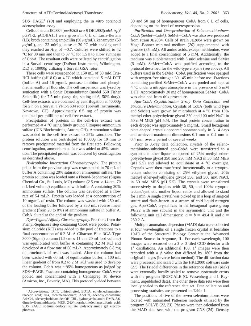
FIGURE 2: Stereoview of representative electron density corresponding to the apo-CobA structure. The map was calculated with coefficientsof the form 2Fo - Fc. This figure was prepared with the programs Molscript and Bobscript (46, 47).
366 Biochemistry, Vol. 40, No. 2, 2001 Bauer et al.

two molecules of MgATP, and one molecule of hydroxy-cobalamin. The electron densities for the hydroxycobalaminand its associated MgATP are unambiguous and clearlydefine the orientations of the corrinoid, its nucleotide loop,and lower ligand as shown in Figure 6. The final model forthe dimer of the OH-CBL‚CobA complex contains 359amino acid residues out of the 392 that are expected. Thepolypeptide chain extends without breaks from Arg28 toTyr196 for subunit A and from Gln7 to Tyr196 for subunit B.A total of 447 solvent molecules were included in the model.Although there are two active sites per dimer, only one isoccupied by hydroxycobalamin in the crystal. This is
probably an artifact of crystallization since the enzymekinetics do not suggest anticooperativity (17). The active sitethat is missing the hydroxycobalamin does contain MgATP.This adopts a conformation similar to that seen in the activesite that binds hydroxycobalamin and to that of conformationA of the MgATP‚CobA complex. The following discussionwill focus on the active site that displays the ternary complex.
As is evident from Figure 6, coordination of hydroxyco-balamin is accompanied by the ordering of amino acidresidues that were disordered in the apo and MgATP‚CobAstructures. The binding site for hydroxycobalamin is formedmostly from subunit A which forms a shallow bowl-shaped
FIGURE 3: Ribbon representations of apo-CobA. A stereoview of one subunit of CobA where the P-loop is highlighted in yellow and thestrands are numbered according to their order in the sequence is shown in panel a. A ribbon representation of the two crystallographicallyrelated subunits of CobA viewed approximately down the 2-fold axis of symmetry is displayed in panel b. The N- and C-terminal residuesare disordered in the apo structure. The final amino acid residues of the C-terminus observed in the apo structure form a prominent interactionwith those from the neighboring subunit. This figure was prepared with the programs Molscript and Bobscript (46, 47).
Structure of ATP:Corrinoidadenosyl Transferase Biochemistry, Vol. 40, No. 2, 2001367

depression at the C-terminal end of theâ-sheet; however,the active site is capped by the N-terminal helix from thesymmetry-related subunit (B) that now extends from Gln7
to Ala24. The corresponding amino acid residues in subunitA are disordered due to the absence of hydroxycobalaminin subunit B. As depicted in Figure 6, the corrin ring islocated above the nucleotide where the cobalt atom isseparated by a distance of∼6.1 Å from C5′ of the ribose.
MgATP Binding Site.A detailed view of the ternarycomplex is shown in Figure 7. As can be seen, the nucleotideis almost completely enclosed on one side by the proteinand on the other side by the corrin ring. From the arrange-ment of MgATP and hydroxycobalamin, it is clear that thenucleotide must bind before the corrinoid as suggested fromthe kinetic behavior of CobA (17).
The electron density shows a well-defined magnesium ionthat is octahedrally coordinated by two nonbridging phos-phate oxygens from theR- andâ-phosphates, the side chainhydroxyl of Thr42, a carboxylate oxygen of Glu128, and twowater molecules. This coordination is very similar to thatobserved in subunit F of F1ATPase, except for the carboxy-late ligand. The latter ligand, if it exists in F1ATPase, resideson a structurally different secondary structural element.
There are extensive contacts between the P-loop and thetriphosphate moiety of ATP, as would be expected (Figures7 and 8). All of the amino acid residues, except for Gly36
and Gly38, of the P-loop [defined by residues Gly36-Thr42
(GNGKGKT)] are involved in coordinating theR-, â-, andγ-phosphates. Common to all proteins that utilize P-loops,there is extensive hydrogen bonding between the main chainamide hydrogens and the phosphoryl oxygens. As discussedabove, the ATP is coordinated in the reverse manner to thatobserved in all other P-loop proteins. This has an interestingeffect on the distribution of ligands since there is now anadditional negative charge in the position normally occupiedby theR-phosphate. The extra negative charge is neutralizedby the inclusion of Arg51′ (from the 2-fold related subunit)and Thr43 in the coordination sphere of theγ-phosphate. Theequivalent ligands are absent in other nucleotide bindingproteins. Additional compensation for the negative chargeon theR-phosphate is provided by positioning this group atthe N-terminal end of theR-helix that follows the P-loopand allows the peptidic NH groups of Gly40 and Thr43 tocontribute to the coordination sphere. It is noteworthy thatthere are very few differences in the positions of the sidechains that coordinate MgATP between the apo structure andMgATP complex, except for Lys39 which rotates away fromthe location that becomes occupied by the ribose.
In contrast to the triphosphate moiety of ATP, there arecomparatively few hydrogen bonding interactions betweenthe adenosine component and the protein. Only two hydrogenbonds are observed between Asp195 and O3′ of the riboseand between the carbonyl oxygen of Asn37 and the aminogroup of the adenine. This would account for the observedspecificity of CobA toward nucleotide (17). Biochemicalstudies demonstrate that CobA exhibits 37% of the activitytoward GTP relative to ATP, whereas the activities towardCTP and UTP were 98 and 88%, respectively. If GTP wereto bind in a manner similar to that observed for ATP, it wouldintroduce a carbonyl oxygen in proximity to Asp195, whichwould be energetically unfavorable. Conversely, the pyri-midine rings of CTP or UTP might occupy the void left bythe purine ring without introducing any unfavorable contactsand hence would not affect the kinetics under the conditionsutilized for the assays.
Hydroxycobalamin Binding Site.Hydroxycobalamin bindson top of a bowl-shaped cavity at the C-terminal end of theâ-strands that encloses MgATP (Figure 9). The corrin ringis roughly perpendicular to the plane of theâ-sheet in sucha way that the most of the amide groups that decorate thecorrin ring are exposed to solvent, whereas the hydrophobiccomponents are occluded by hydrophobic side chains. Thecorrinoid is abutted on two sides by loops that extend fromâ-strands 3, 4, and 6 and enclosed by the N-terminalR-helixfrom Gln7′ to Gln25′ of the adjacent subunit. Interestingly,the N-terminal R-helix interacts predominantly with thenucleotide loop of the hydroxycobalamin where it providesa salt bridge between Arg16′ and a phosphate oxygen of thenucleotide loop together with a limited number of hydro-phobic interactions from Thr13′, Val17′, and Val21′. Thisarrangement leaves the hydrophilic components of the corrinring exposed to solvent.
The lower ligand of hydroxycobalamin, DMB, is clearlycoordinated to the cobalt ion of the corrin ring where theCo-N bond distance is 2.2 Å. Ligand-on coordination was
FIGURE 4: Topological drawings for (a) CobA (ATP:corrinoidadenosyl transferase), (b) CobU (adenosylcobinamide kinase/adenosylcobinamide phosphate guanylyltransferase), and (c) theRecA protein. The figure was prepared from coordinates from PDBentries 1G64, 1CBU, and 2REB (30, 33).
368 Biochemistry, Vol. 40, No. 2, 2001 Bauer et al.

unexpected because of the ability of CobA to adenosylateboth cob(I)inamide, which lacks the nucleotide loop, and cob-(I)alamin. It was also unexpected from the precedenceestablished by the mode of binding of cobalamin observedin methionine synthase and methylmalonyl-CoA mutase sinceboth of these show replacement of the lower ligand ofcobalamin with a histidine side chain (38, 39). Coordinationof the cobalt by the lower ligand is utilized in somecobalamin-dependent enzymes, as observed in diol dehy-dratase (40). In CobA, it appears that the broad substratespecificity for the lower ligand is provided by conformationalflexibility in the N-terminalR-helix.
Strikingly, there are very few hydrogen bonds betweenhydroxycobalamin and the protein. This absence of specific
interactions between the hydrophilic components of thecorrinoid is consistent with the anticipated broad specificityof this enzyme. As noted earlier, it is unknown for CobA atwhich point in the biosynthetic pathway the corrin ringbecomes adenosylated; however, the orthologous enzymefrom P. denitrificansis able to adenosylate cobyrinic acidand all of the subsequent intermediates in the biosyntheticpathway. It is expected that CobA fromS. typhimuriumwillhave even broader specificity since in this organism thecobalt is inserted considerably sooner. There are considerabledifferences between cobyrinic acid and hydroxycobalaminthat potentially could have a profound influence on ligandbinding, for example, amidation of six carboxylate groups.Thus, the corrin binding site must be able to accommodate
FIGURE 5: Electron density for MgATP and its orientation relative to the P-loop in F1ATPase. Panel a shows a stereoview of the electrondensity for the two orientations of MgATP in CobA calculated with coefficients of the formFo - Fc where the substrate was omitted fromthe phase calculation. Panel b displays a part of theâ-subunit of F1ATPase together with its MgAMP-PNP superimposed onto the MgATPof CobA. F1ATPase and MgAMP-PNP are depicted in cyan and blue, respectively, whereas CobA and MgATP are colored in red andyellow, respectively. The superposition was done with the program ALIGN (37) modified at the University of Wisconsin by G. Wesenbergto allow selection of the segments used for the alignment (available on request). The figure was prepared from coordinates from PDBentries 1COW (31) and 1G5T. This figure was prepared with the programs Molscript and Bobscript (46, 47).
Structure of ATP:Corrinoidadenosyl Transferase Biochemistry, Vol. 40, No. 2, 2001369

a wide range of substrates that differ greatly in ionic chargeand state of assembly of the nucleotide loop. Exposure ofthe amide groups to solvent and the avoidance of anyhydrogen bonding contacts would appear to be prerequisitesfor a binding site that can recognize such a diverse group ofmolecules. Likewise, the sparse interactions between the
aminopropanol arm and nucleotide loop and the proteinwould also appear to be consistent with this structural theme.Again, the coordination of the corrinoid by CobA differsconsiderably from that seen in cobalamin-dependent enzymessuch as methionine synthase, methylmalonyl-CoA mutase,and diol dehydratase where there are numerous specific
FIGURE 6: Ternary complex of CobA, MgATP, and hydroxycobalamin. A stereoview of the electron density for the hydroxycobalamin andMgATP is displayed in panel a. A ribbon representation of the OH-CBL‚CobA complex viewed approximately down the noncrystallographic2-fold axis is shown in panel b. The single hydroxycobalamin molecule observed in the lattice is coordinated mostly by subunit A. However,the N-terminus of subunit B is ordered in this complex and caps the active site where it interacts with the nucleotide arm and lower ligand.The electron density map was calculated with coefficients of the formFo - Fc where the ligands were omitted from the phase calculation.This figure was prepared with the programs Molscript and Bobscript (46, 47).
370 Biochemistry, Vol. 40, No. 2, 2001 Bauer et al.
interactions between the side groups of the corrinoid andthe protein (38-40).
The N-terminal helix is very poorly conserved in CobAhomologues and may be missing in thermophiles such asPyrococcus horikoshii. The variability of this component ofthe structure may play an important role in defining the rangeof substrates that can be accommodated by a specific enzyme.Likewise, it implies that this helix might be unnecessary forbinding some substrates.
It appears that the coordination of the corrin ring is drivenby hydrophobic interactions. As shown in Figure 9b, thehydrophobic components of the corrin ring are adjacent to agroup of hydrophobic amino acid side chains. This includesTrp93, Tyr131, Tyr135, and Phe184. There is also a hydrophobiccomponent to the interaction between the N-terminalR-helixon the adjacent subunit and the nucleotide loop of hydroxy-cobalamin.
Catalytic and EVolutionary Considerations.Surprisingly,there are no significant hydrogen bonding contacts betweenthe corrinoid and MgATP. This most likely arises becausethe corrin ring is somewhat distant from MgATP. Indeed,C5′ of the ribose is located∼6.1 Å from the cobalt which istoo far for nucleophilic attack. Although the Co(I) intermedi-ate is highly reactive, it still must be reasonably close toC5′ of the ribose for transfer to occur. This suggests thatreduction of the corrinoid to Co(I) is accompanied by astructural rearrangement that brings C5′ of the ribose andcobalt closer together. Although not evident in the electrondensity, it is anticipated that the coordination sphere of thecobalt in the present structure should be completed by a watermolecule since most Co(III) complexes are hexacoordinate.Indeed, there is ample room for a water molecule betweenC5′ of the ribose and the cobalt ion. Removal of this watermolecule from the coordination sphere (if present) must occurprior to reduction to Co(I) because, otherwise, this highlyreactive nucleophile might react with water rather than C5′of the ribose. This suggests that the rearrangement of theternary complex occurs after reduction of Co(III) to Co(II).Structural studies of the nucleotide hydrolases in the RecAfamily suggest that the strand between the P-loop and Walker
B motif may be involved in sensing the state of the nucleotide(41); however, in CobA, these residues are not in contactwith the MgATP. Thus, it is difficult to project how aconformational change is accommodated on the basis oforthologous structures.
As noted previously, the topology of CobA belongs to theP-loop-containing family of nucleotide hydrolases. Withinthis superfamily, its overall fold is remarkably similar to thatof adenosylcobinamide kinase/adenosylcobinamide guany-lyltransferase (CobU inS. typhimurium), RecA protein, andF1ATPase (Figure 4). CobU is the enzyme that is responsiblefor phosphorylating the aminopropanol arm of adenosylco-binamide and, in a subsequent step, activating the cobinamideby the addition of GMP to form adenosylcobinamide-GDP(42). This group of proteins shares no measurable level ofamino acid sequence similarity, except for the P-loop.Remarkably, the same protein fold, which is fairly unusualwithin the structural database, is found in two enzymes(CobA and CobU) that are separated by only a few steps inthe cobalamin biosynthetic pathway.
Beyond their common folds, the structures of CobA andCobU are quite different. For example, CobA is a dimer,whereas CobU is a trimer. Furthermore, although thecorrinoid binding site for CobU is unknown, it seems likelythat CobU binds adenosylcobinamide with great specificityin a large groove between protein subunits that lies ap-proximately parallel to theâ-sheet in a way that presentsthe aminopropanol arm of the cobinamide to theγ-phosphateof the ATP. This is in contrast to CobA where the corrinring lies perpendicular to theâ-sheet and presents the centerof the corrin ring to C5′ of the ribose held in place by theP-loop. Additionally, CobU is believed to utilize the P-loopin a conventional manner for its kinase reaction. However,CobU is an unusual enzyme since it is a small protein thatfunctions as both a kinase and guanylyltransferase where thetransferase activity proceeds through a covalent guanylyl-histidine intermediate (43). Interestingly, there is considerableevidence that the kinase and transferase active sites overlapsuch that in the transferase reaction the GTP binds acrossthe P-loop in the reverse order as seen in CobA (44). Thus,
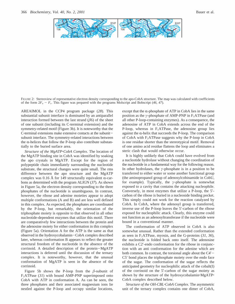
FIGURE 7: Closeup stereoview of the MgATP binding site. All of the side chains that are involved in coordinating the nucleotide arelabeled. For clarity, the bonds between the main chain atoms in the P-loop are displayed in cyan. The relationship between C5′ of the riboseand the cobalt of the corrin ring is also visible. This figure was prepared with the programs Molscript and Bobscript (46, 47).
Structure of ATP:Corrinoidadenosyl Transferase Biochemistry, Vol. 40, No. 2, 2001371

CobA and CobU appear to share the common feature ofutilizing their respective P-loops in a novel way to bind theirnucleotide substrates.
It has been postulated that CobU arose from a primordialkinase and evolved the ability to function as a transferasebecause of the disorder-order transition or substrate-inducedactive site that occurs when the guanylyl nucleotide binds(44). The same suggestion can now be applied to CobA.Perhaps it also originated as a kinase that possessed thenormal use of the P-loop. The evolutionary significance ofthe structural similarity between CobA and CobU is un-
known, but provides another intriguing example of themanner in which protein folds have been retooled duringevolution.
CONCLUSIONS
The structure described here for the ternary complex ofCobA, MgATP, and hydroxycobalamin answers many of thequestions that instigated this structural study. It provides anexplanation for the kinetic order of substrate binding andthe broad specificity of the enzyme. Most likely, this complex
FIGURE 8: Schematic representation of the hydrogen bonding pattern of the protein, MgATP, and hydroxycobalamin. The conformation ofthe ligands has been distorted from that observed in the ternary complex for clarity. Arg16′ and Arg51′ are shaded in gray and indicate sidechains from the 2-fold related subunit. This figure was prepared with the programs Molscript and Bobscript (46, 47).
372 Biochemistry, Vol. 40, No. 2, 2001 Bauer et al.

represents the initial structural state in the adenosylationprocess since it is likely that the reduction to cobalt(I) occurson the enzyme. The long distance of 6.1 Å between the cobaltof the corrin ring and C5′ of the ribose, however, suggeststhat a structural rearrangement must occur during reductionof the cobalt(III) to cobalt(I) to facilitate formation of thecobalt-carbon bond. Certainly, water has to be eliminatedfrom the proximity of the cobalt to increase the efficiencyof the reaction. The nature of this rearrangement is unknownand awaits further structural and spectroscopic study.
Until recently, little was known regarding the manner inwhich the cobalt was reduced from Co(III) to Co(I). Thereduction occurs in two single-electron transfers. Initially,it was believed that a reductase was necessary for each step,but it has now been reported that endogenous concentrationsof dihydroflavins in solution may be sufficient for the
reduction from Co(III) to Co(II) (15). The second electrontransfer cannot be accomplished by dihydroflavins alone andrequires a protein to mediate this process. This raises thequestion of whether there is a specific binding site for a Co-(II) reductase. Given the variety of corrinoids that can beadenosylated by this enzyme and its orthologues in otherorganisms, it is expected that the protein involved in theelectron transfer process will utilize some structural com-ponent of the enzyme rather than recognizing the substrate.Examination of the structure reveals the presence of anexposed hydrophobic patch consisting of Trp69, Pro70, Met192,and Tyr196. It remains to be proven whether these residuesinfluence the adenosylation reaction. These questions arepresently being addressed by additional biochemical andX-ray structural studies.
FIGURE 9: Closeup stereoviews of the corrin binding site. A side view to show the capping of the corrin binding pocket by the N-terminalR-helix from the symmetry-related subunit is depicted in panel a. A top view to show the arrangement of hydrophobic ligands in proximityto the hydrophobic components of the corrin ring is shown in panel b. All of the ligands that are involved in coordinating the corrinoid arelabeled. This figure was prepared with the programs Molscript and Bobscript (46, 47).
Structure of ATP:Corrinoidadenosyl Transferase Biochemistry, Vol. 40, No. 2, 2001373

ACKNOWLEDGMENT
We gratefully acknowledge the superb technical assistancefrom the following staff members at the Structural BiologyCenter at the Advanced Photon Source in Argonne, IL: Dr.Frank Rotella, Dr. Norma E. C. Duke, Dr. Ruslan Sanishvili,and Dr. Jack Lazarz. We also thank Dr. S.-J. Suh for theinitial sample of CobA used in preliminary crystallizationexperiments. We thank the reviewers for their insightfulcomments on the manuscript.
REFERENCES
1. Blanche, F., Cameron, B., Crouzet, J., Debussche, L., Thibaut,D., Vuilhorgne, M., Leeper, F. J., and Battersby, A. R. (1995)Angew. Chem., Int. Ed. 34, 383-411.
2. Rondon, M. R., Trzebiatowski, J. R., and Escalante-Semerena,J. C. (1997)Prog. Nucleic Acid Res. Mol. Biol. 56, 347-384.
3. Raux, E., Thermes, C., Heathcote, P., Rambach, A., andWarren, M. J. (1997)J. Bacteriol. 179, 3202-3212.
4. Santander, P. J., Roessner, C. A., Stolowich, N. J., Holderman,M. T., and Scott, A. I. (1997)Chem. Biol. 4, 659-666.
5. Debussche, L., Couder, M., Thibaut, D., Cameron, B., Crouzet,J., and Blanche, F. (1992)J. Bacteriol. 174, 7445-7451.
6. Debussche, L., Couder, M., Thibaut, D., Cameron, B., Crouzet,J., and Blanche, F. (1991)J. Bacteriol. 173, 6300-6302.
7. Pezacka, E. H. (1993)Biochim. Biophys. Acta 1157, 167-177.
8. Blanche, F., Maton, L., Debussche, L., and Thibaut, D. (1992)J. Bacteriol. 174, 7452-7454.
9. Watanabe, F., and Nakano, Y. (1997)Methods Enzymol. 281,289-295.
10. Weissbach, H., Brot, N., and Lovenberg, W. (1966)J. Biol.Chem. 241, 317-321.
11. Walker, G. A., Murphy, S., and Huennekens, F. M. (1969)Arch. Biochem. Biophys. 134, 95-102.
12. Brady, R. O., Castanera, E. G., and Barker, H. A. (1962)J.Biol. Chem. 223, 2325-2332.
13. Hoover, D. M., Jarrett, J. T., Sands, R. H., Dunham, W. R.,Ludwig, M. L., and Matthews, R. G. (1997)Biochemistry 36,127-138.
14. Jarrett, J. T., Hoover, D. M., Ludwig, M. L., and Matthews,R. G. (1998)Biochemistry 37, 12649-12658.
15. Fonseca, M. V., and Escalante-Semerena, J. C. (2000)J.Bacteriol. 182, 4304-4309.
16. Suh, S.-J., and Escalante-Semerena, J. C. (1993)Gene 129,93-97.
17. Suh, S.-J., and Escalante-Semerena, J. C. (1995)J. Bacteriol.177, 921-925.
18. Peterkofsky, A., and Weissbach, H. (1963)J. Biol. Chem. 238,1491-1497.
19. Laemmli, U. K. (1970)Nature 227, 680-685.20. Vogel, H. J., and Bonner, D. M. (1956)J. Biol. Chem. 218,
97-106.21. Otwinowski, Z., and Minor, W. (1997) inMethods in
Enzymology(Carter, C. W. J., Sweet, R. M., Abelson, J. N.,and Simon, M. I., Eds.) pp 307-326, Academic Press, NewYork.
22. Terwilliger, T. C., and Berendzen, J. (1999)Acta Crystallogr.D55, 849-861.
23. Terwilliger, T. C. (1997) inMethods in Enzymology(Carter,C. W. J., Sweet, R. M., Abelson, J. N., and Simon, M. I.,Eds.) pp 530-537, Academic Press, New York.
24. Brunger, A. T., Adams, P. D., Clore, G. M., DeLano, W. L.,Gros, P., Grosse-Kunstleve, R. W., Jiang, J. S., Kuszewski,J., Nilges, M., Pannu, N. S., Read, R. J., Rice, L. M.,Simonson, T., and Warren, G. L. (1998)Acta Crystallogr. D54,905-921.
25. Roussel, A., and Cambillau, C. (1991) inSilicon GraphicsGeometry Partners Directory, Silicon Graphics.
26. Tronrud, D. E. (1997)Methods Enzymol. 277, 306-319.27. Laskowski, R. A., MacArthur, M. W., Moss, D. S., and
Thornton, J. M. (1993)J. Appl. Crystallogr. 26, 283-291.28. Collaborative Computational Project No. 4 (1994)Acta
Crystallogr. D50, 760-763.29. Cowtan, K., and Main, P. (1998)Acta Crystallogr. D54, 487-
493.30. Story, R. M., Weber, I. T., and Steitz, T. A. (1992)Nature
355, 318-325.31. Abrahams, J. P., Leslie, A. G. W., Lutter, R., and Walker, J.
E. (1994)Nature 370, 621-628.32. Subramanya, H. S., Bird, L. E., Brannigan, J. A., and Wigley,
D. B. (1996)Nature 384, 379-383.33. Thompson, T. B., Thomas, M. G., Escalante-Semerena, J. C.,
and Rayment, I. (1998)Biochemistry 37, 7686-7695.34. Walker, J. E., Saraste, M., Runswick, M. J., and Gay, N. J.
(1982)EMBO J. 1, 945-951.35. Saraste, M., Sibbald, P. R., and Wittinghofer, A. (1990)Trends
Biol. Sci. 15, 430-434.36. Smith, C. A., and Rayment, I. (1996)Biophys. J. 70, 1590-
1602.37. Cohen, G. H. (1997)J. Appl. Crystallogr. 30, 1160-1161.38. Drennan, C. L., Huang, S., Drummond, J. T., Matthews, R.
G., and Ludwig, M. L. (1994)Science 266, 1669-1674.39. Mancia, F., Keep, N. H., Nakagawa, A., Leadlay, P. F.,
McSweeney, S., Rasmussen, B., Bo¨secke, P., Diat, O., andEvans, P. R. (1996)Structure 4, 339-350.
40. Shibata, N., Masuda, J., Tobimatsu, T., Toraya, T., Suto, K.,Morimoto, Y., and Yasuoka, N. (1999)Struct. Folding Des.7, 997-1008.
41. Lenzen, C. U., Steinmann, D., Whiteheart, S. W., and Weis,W. I. (1998)Cell 94, 525-536.
42. O’Toole, G. A., and Escalante-Semerena, J. C. (1995)J. Biol.Chem. 270, 23560-23569.
43. Thomas, M. G., Thompson, T. B., Rayment, I., and Escalante-Semerena, J. C. (2000)J. Biol. Chem. 275, 27576-27586.
44. Thompson, T. B., Thomas, M. G., Escalante-Semerena, J. C.,and Rayment, I. (1999)Biochemistry 38, 12995-13005.
45. Johnson, M. G., and Escalante-Semerena, J. C. (1992)J. Biol.Chem. 267, 13302-13305.
46. Kraulis, P. J. (1991)J. Appl. Crystallogr. 24, 946-950.47. Esnouf, R. M. (1999)Acta Crystallogr. D55, 938-940.
BI002145O
374 Biochemistry, Vol. 40, No. 2, 2001 Bauer et al.


![Induction of Interleukin 2 Production but not Methionine ...[CANCER RESEARCH 52, 3361-3366, June 15, 1992] Induction of Interleukin 2 Production but not Methionine Adenosyltransferase](https://img.pdfslide.net/doc/110x75/611ace0a32e6d405ac64c0d3/induction-of-interleukin-2-production-but-not-methionine-cancer-research-52.jpg)