Embed Size (px)
Citation preview
Copyright © 2009 Thorne Research, Inc. All Rights Reserved. No Reprint Without Written Permission.
Alternative Medicine Review Volume 14, Number 3 2009
Page 226
EnvironmentalMedicineReview Article
Page 226
AbstractPlant-derived polyphenols are increasingly receiving attention as dietary supplements for the homeostatic management of inflammation, to support detoxification, and for anticancer, weight loss, and other benefits. Their pro-homeostatic effects on genes, transcription factors, enzymes, and cell signaling pathways are being intensively explored, but the poor bioavailability of some polyphenols likely contributes to poor clinical trial outcomes. This review covers four polyphenol preparations with poor bioavailability and their complexation into phytosomes to bypass this problem. Silybin and the other silymarin flavonolignans from milk thistle conserve tissue glutathione, are liver-protective, and have anticancer potential. Curcumin and its related diphenolic curcuminoids have potent antioxidant, anti-inflammatory, and anti-carcinogenic properties. The green tea flavan-3-ol catechins have antioxidant, anti-inflammatory, cardio- and neuro-protective effects, and anti-carcinogenic benefits, with fat oxidation effects coupled to weight loss. The complex grape seed proanthocyanidin mix (including catechin and epicatechin monomers and oligomers) counters oxidative stress and protects the circulatory system. For each of these preparations, conversion into phytosomes has improved efficacy without compromising safety. The phytosome technology creates intermolecular bonding between individual polyphenol molecules and one or more molecules of the phospholipid, phosphatidylcholine (PC). Molecular imaging suggests that PC molecule(s) enwrap each polyphenol; upon
oral intake the amphipathic PC molecules likely “usher” the polyphenol through the intestinal epithelial cell outer membrane, subsequently accessing the bloodstream. PC itself has proven clinical efficacy that contributes to phytosome in vivo actions. As a molecular delivery vehicle, phytosome technology substantially improves the clinical applicabilities of polyphenols and other poorly absorbed plant medicinals.(Altern Med Rev 2009;14(3):226-246)
IntroductionMedicinal nutrients derived from plants have
been used for health maintenance and disease manage-ment since the dawn of history. One class of phytomedi-cines currently receiving increased scrutiny is the poly-phenols. These number in the thousands and include, but are not limited to, the various flavonoid subclasses. But many polyphenols are very poorly absorbed when taken orally, posing the greatest obstacle to routine clini-cal application.1 Where possible, the conversion of poly-phenols to phytosome forms improves oral bioavailabil-ity without compromising safety. This review focuses on the four most widely studied polyphenol phytosome preparations for anti-inflammatory, anti-neoplastic, de-toxification, and weight loss applications:
Bioavailability and Activity of Phytosome Complexes from Botanical
Polyphenols: The Silymarin, Curcumin, Green Tea, and Grape Seed Extracts
Parris M. Kidd, PhD
Parris M. Kidd, PhD – Cell biology; University of California, Berkeley; contributing editor, Alternative Medicine Review; health educator; biomedical consultant to the dietary supplement industryCorrespondence address: 10379 Wolf Drive, Grass Valley, CA 95949Email: [email protected]
Copyright © 2009 Thorne Research, Inc. All Rights Reserved. No Reprint Without Written Permission.
Alternative Medicine Review Volume 14, Number 3 2009
Phytosomes
Page 227
X silymarin flavonolignans, most specifically silybin (silibinin, silymarin I), which enhance systemic antioxidant status, are liver protectants, support liver detoxification, and have anticancer potential
X curcuminoid polyphenols, which have potent antioxidant, anti-inflammatory, and anticancer properties
X green tea flavan-3-ol catechins, which have antioxidant, anti-inflammatory, and anticancer benefits, and have demonstrated fat oxidation effects coupled to weight loss
X grape seed catechin and epicatechin complex, including monomeric and oligomeric proanthocyanidins, which counters oxidative stress, protects circulation, and has anti-inflammatory and anticancer effects
Background to Phytosome TechnologyPhytosome technology emerged in 1989.2
Based on a histochemical observation that certain poly-phenols had strong bonding affinity for phospholipids in their intact plant tissue, a group of Italian researchers focused on polyphenol preparations known to be poorly bioavailable when taken orally. These were typically mix-tures of polyphenols extracted from single plant species, and their conversion into phytosome forms markedly increased their bioavailability.3 To make phytosomes, the polyphenol mix is chemically reacted with a phos-pholipid preparation, consisting mainly of phosphati-dylcholine (PC), which is also the major phospholipid of living tissues. The resulting phytosome molecular complex is tested for bioavailability and efficacy, usually in direct comparison to its non-phytosome form.
For the four phytosome preparations under review, the findings from systematic bioavailability comparisons show that when administered orally, phy-tosome complexation enhances the blood levels of poly-phenol constituents by factors of at least 2-6 times.3-7 Phytosome technology has proven to be a breakthrough for the clinical applicability of botanical polyphenols, since improved bioavailability generally results in enhanced efficacy.
The flavonoids and other polyphenol mol-ecules are multi-ring compounds generally too large to be absorbed by simple diffusion,1 nor are they subject to active intestinal uptake as occurs with some vitamins and minerals. They also tend to be poorly soluble in water or lipids. PC by contrast is an amphipathic mol-ecule, having a positively charged headgroup and two neutrally charged tailgroups,8 a rare molecular charac-teristic that renders PC miscible in both water and lipid environments. By complexing a polyphenol with PC to make a phytosome, the polyphenol comes to share some of PC’s versatile solubility properties.
Direct demonstration of the phytosome action is not yet feasible, but from what is known of these mo-lecular constituents, it is inferred that the water-miscible PC molecules enhance the dispersion of the poorly wa-ter-soluble polyphenol molecules into the water-soluble environment of the gastrointestinal lumen. PC ostensi-bly further enhances transfer from the lumen into the lipid-soluble environment of the outer cell membrane of the epithelial absorptive cells (enterocytes). The entero-cyte outer membrane has a lipid molecular bilayer that consists largely of PC. It is feasible that the PC of the phytosome merges into this PC domain of the entero-cyte membrane, carrying the polyphenol with it and so “ushering” the polyphenol into the cell.2-4
Phytosomes Differ from LiposomesTo appreciate the uniqueness of phytosomes it
is necessary to differentiate them from liposomes. The unit phytosome is a molecular-level association involv-ing as few as two molecules (one PC plus one polyphe-nol). The unit liposome is an aggregate of hundreds of phospholipid molecules into a spherule, within which other molecules are compartmentalized but not specifi-cally bonded. Whereas, the liposome concept remains unproven as an oral delivery vehicle, the phytosome is known to dramatically enhance oral delivery.
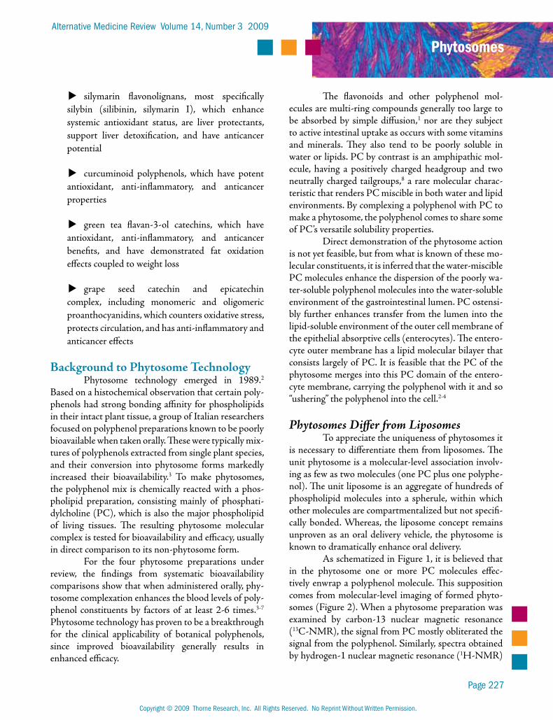
As schematized in Figure 1, it is believed that in the phytosome one or more PC molecules effec-tively enwrap a polyphenol molecule. This supposition comes from molecular-level imaging of formed phyto-somes (Figure 2). When a phytosome preparation was examined by carbon-13 nuclear magnetic resonance (13C-NMR), the signal from PC mostly obliterated the signal from the polyphenol. Similarly, spectra obtained by hydrogen-1 nuclear magnetic resonance (1H-NMR)
Copyright © 2009 Thorne Research, Inc. All Rights Reserved. No Reprint Without Written Permission.
Alternative Medicine Review Volume 14, Number 3 2009
Review Article
Page 228
show a similar obliteration of the catechin pattern by the PC.3 Note in Figure 2 that in the blue phytosome spectrum, the spectrum from the polyphenol (red) is eclipsed by the orange spec-trum from phosphatidylcholine. This is consistent with a physical enwrapment of the polyphenol by the PC molecule, as configured in Figure 1.
Using a different phyto-some model, that of glycyrrhetinic acid with PC, an infrared spectrum analysis yielded similar findings to the 13C- and 1H-NMR analyses of (+)-catechin with PC.3 The most reasonable interpretation from these molecular imaging spectra is that the polyphenol molecule is being shielded from the view of the imaging probes. A corollary of these results is that in the unit phytosome, the polyphenol (or other phytomedicinal molecule) is held close to the PC molecule(s) by some form of quasi-stable bonding. In effect, the phytosome is a hybrid
molecule that resembles PC in being sub-stantially lipid-soluble and water-soluble. Once this hybrid molecule enters the intestinal tract, its largely amphipathic character facilitates its transition from the water-soluble environment of the in-testinal lumen to the lipid-soluble envi-ronment of the enterocyte cell membrane.
Phosphatidylcholine Itself Has Clinical Efficacy
Depending on the proportions of PC contained, and the doses ingested, phytosome preparations can deliver clini-cally significant amounts of PC. This is an important aspect of phytosome activ-ity because PC itself has important clini-cal applications.8 PC, the most significant dietary source of the essential nutrient choline, is an orthomolecule ubiquitous in known life forms.8 Besides being an
Figure 1. Schematic of the Phytosome Molecular Complex3
Figure 2. Molecular Imaging (13C-NMR) of PC, the Polyphenol (+)-Catechin, and the Phytosome Combination
180 160 140 120 100 80 60 40 20 0PPM
Distearoyl-phosphatidylcholinein C6D6
(+)-Catechinin DMSO-D6
1-1 Molar complexin C6D6
Top spectrum (orange) represents distearoyl-phosphatidylcholine (PC). Middle spectrum (red) represents (+)-catechin, a flavan-3-ol. Bottom spectrum (blue) represents a 1:1 molar complex between PC and (+)-catechin. From: Indena SpA, Italy
Copyright © 2009 Thorne Research, Inc. All Rights Reserved. No Reprint Without Written Permission.
Alternative Medicine Review Volume 14, Number 3 2009
Phytosomes
Page 229
important emulsifier in the lungs, gastrointestinal tract, and bile, PC is the principal molecular building block for circulating lipoproteins and for cell membranes.9 The cell’s network of membranes provides the main locale for energetic transformations, manages the vast majority of life processes, and directs the cell’s overall metabolism.10
The First Phytosomes: Milk Thistle Flavonoids
The first commercial phytosome prepara-tion was based on the flavonolignan silybin, the major constituent of silymarin, a flavonol complex extracted from the milk thistle fruit (Silybum marianum, family Asteraceae/Compositae). This phytosome preparation was initially christened IDB 1016 or Silipide3,11,12 and subsequently recast as Siliphos® Phytosome™.3 Silybin-phosphatidylcholine is clinically validated for its anti-oxidant, anti-inflammatory, and liver detoxification benefits, as reviewed in 2005 in this journal.4 Therefore, this section primarily updates its status from research published during the intervening period.
Silymarin BackgroundThe fruit of the milk thistle plant has been a
liver support remedy for 2,000 years.13 The active con-stituents of silymarin include predominantly silybin (Figure 3), followed by silydianin, silychristin, and isosi-lybin. These are lignan derivatives of flavonols (flavono-lignans), likely produced within the plant by enzymatic combination of a flavonol with a coniferyl alcohol.
Silybin is an effective an-tioxidant, conserving glutathione (GSH) in liver cells while stabiliz-ing the liver cell membranes against oxidative attack.13 Its antioxidant potency is bolstered by its effec-tive chelation of iron. In fact, in a human clinical trial silybin even lowered serum ferritin.14 Silybin is a proven liver protectant; in animal experiments it blocked the oxida-tive toxicities of acetaminophen, al-cohol, carbon tetrachloride, and the mushroom toxins phalloidin and alpha-amanitin.3,4 These findings correlate with decades of clinical
observations that silybin improves survival for humans exposed to deathcap mushrooms (Amanita species).15
Absorption and Tissue Fate of Silybin Phytosome
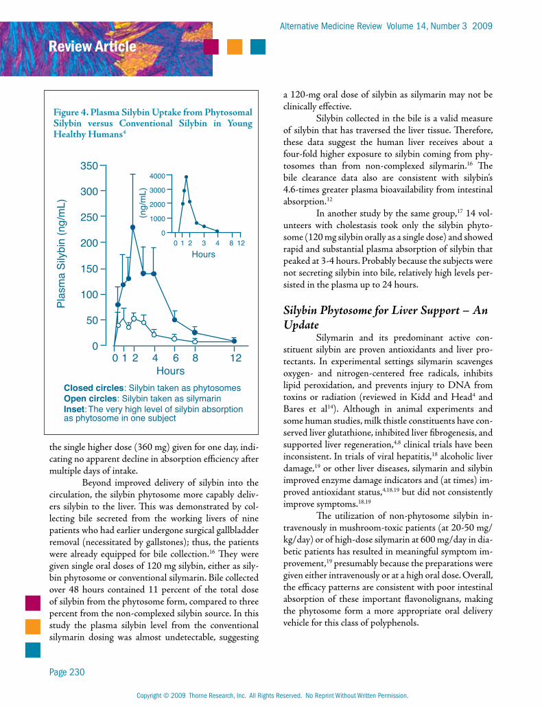
The animal and human pharmacokinetics of the silybin phytosome complex have been reviewed in depth.4 With respect to bioavailability, it is the most thoroughly researched of the existing phytosome prepa-rations. For equal quantities of silybin taken by mouth, the phytosome form achieves markedly higher plasma levels of silybin than does the conventional, non-phyto-some form (Figure 4).
The comparative uptakes of silybin from the phytosome form versus the non-phytosome form were investigated in two human studies. In the first, young healthy subjects (ages 16-26, n=8) took single 360-mg doses of silybin by mouth, either as the phytosome or as conventional silybin.12 After eight hours the plasma silybin level achieved from the phytosome was almost three times that of the non-complexed silybin (Figure 4). By measuring the total area under the curve (AUC), it was determined that silybin is absorbed 4.6-times better from its phytosome than its conventional form.
The second human pharmacokinetic study was conducted with the same healthy young volunteers.12 In this study, rather than a single dose of 360 mg, the silybin dose was 240 mg twice daily (120 mg every 12 hours) for eight days. This pattern of daily intake achieved high plasma concentrations and high total ab-sorption on the eighth day, matching those attained by
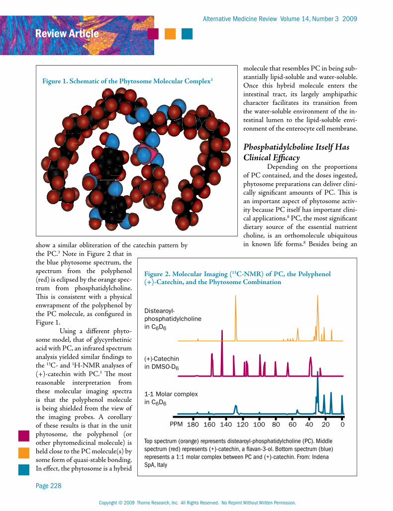
Figure 3. Structure of Silybin, Primary Flavonolignan of Silymarin and the Siliphos Phytosome Complex4
HO
OH O
OHOH
O
O
O
H
H
H
H
CH2OH
OCH3
Copyright © 2009 Thorne Research, Inc. All Rights Reserved. No Reprint Without Written Permission.
Alternative Medicine Review Volume 14, Number 3 2009
Review Article
Page 230
the single higher dose (360 mg) given for one day, indi-cating no apparent decline in absorption efficiency after multiple days of intake.
Beyond improved delivery of silybin into the circulation, the silybin phytosome more capably deliv-ers silybin to the liver. This was demonstrated by col-lecting bile secreted from the working livers of nine patients who had earlier undergone surgical gallbladder removal (necessitated by gallstones); thus, the patients were already equipped for bile collection.16 They were given single oral doses of 120 mg silybin, either as sily-bin phytosome or conventional silymarin. Bile collected over 48 hours contained 11 percent of the total dose of silybin from the phytosome form, compared to three percent from the non-complexed silybin source. In this study the plasma silybin level from the conventional silymarin dosing was almost undetectable, suggesting
a 120-mg oral dose of silybin as silymarin may not be clinically effective.
Silybin collected in the bile is a valid measure of silybin that has traversed the liver tissue. Therefore, these data suggest the human liver receives about a four-fold higher exposure to silybin coming from phy-tosomes than from non-complexed silymarin.16 The bile clearance data also are consistent with silybin’s 4.6-times greater plasma bioavailability from intestinal absorption.12
In another study by the same group,17 14 vol-unteers with cholestasis took only the silybin phyto-some (120 mg silybin orally as a single dose) and showed rapid and substantial plasma absorption of silybin that peaked at 3-4 hours. Probably because the subjects were not secreting silybin into bile, relatively high levels per-sisted in the plasma up to 24 hours.
Silybin Phytosome for Liver Support – An Update
Silymarin and its predominant active con-stituent silybin are proven antioxidants and liver pro-tectants. In experimental settings silymarin scavenges oxygen- and nitrogen-centered free radicals, inhibits lipid peroxidation, and prevents injury to DNA from toxins or radiation (reviewed in Kidd and Head4 and Bares et al14). Although in animal experiments and some human studies, milk thistle constituents have con-served liver glutathione, inhibited liver fibrogenesis, and supported liver regeneration,4,8 clinical trials have been inconsistent. In trials of viral hepatitis,18 alcoholic liver damage,19 or other liver diseases, silymarin and silybin improved enzyme damage indicators and (at times) im-proved antioxidant status,4,18,19 but did not consistently improve symptoms.18,19
The utilization of non-phytosome silybin in-travenously in mushroom-toxic patients (at 20-50 mg/kg/day) or of high-dose silymarin at 600 mg/day in dia-betic patients has resulted in meaningful symptom im-provement,19 presumably because the preparations were given either intravenously or at a high oral dose. Overall, the efficacy patterns are consistent with poor intestinal absorption of these important flavonolignans, making the phytosome form a more appropriate oral delivery vehicle for this class of polyphenols.
Figure 4. Plasma Silybin Uptake from Phytosomal Silybin versus Conventional Silybin in Young Healthy Humans4
350
300
250
200
150
100
50
00 1 2 4 6 8 12
4000
3000
2000
1000
00 1 2 3 4 8 12
Plas
ma
Sily
bin
(ng/
mL)
Closed circles: Silybin taken as phytosomesOpen circles: Silybin taken as silymarinInset: The very high level of silybin absorptionas phytosome in one subject
Hours
(ng/
mL)
Hours
Copyright © 2009 Thorne Research, Inc. All Rights Reserved. No Reprint Without Written Permission.
Alternative Medicine Review Volume 14, Number 3 2009
Phytosomes
Page 231
The silybin phytosome complex has better re-sults for lowering liver enzymes, albeit in relatively small clinical trials.4,20-25 The phytosome form has produced degrees of symptomatic improvement in clinical trials of liver cirrhosis and alcoholic, iatrogenic, and viral hep-atitis (types A, B, and C).20-25 Taken altogether, the trial data suggest that liver damage indicators in patients with acute or chronic hepatitis B and/or C will respond to 800-1,600 mg/day of Siliphos (providing 240-480 mg/day silybin) over 7-120 days.21,22,24,25
Findings from animal research suggest the si-lybin phytosome has further potential for clinical ap-plication; for example, an early-generation silymarin phytosome protected rat fetuses against ethanol toxic-ity better than non-phytosome silymarin.26 It also pro-tected broiler chicks against the toxic effects of aflatoxin B1.27 Furthermore, silybin’s iron-chelating property in vitro also may apply in vivo. In a recent clinical trial of chronic hepatitis C patients, the silybin phytosome sig-nificantly lowered serum ferritin levels, particularly in patients with advanced liver fibrosis.14
Silymarin and silybin are well tolerated. Silybin intakes up to 1,080 mg/day as phytosome are well toler-ated even by patients with compensated cirrhosis.14 A 2008 trial that utilized the silybin phytosome for pros-tate cancer determined that up to 13,000 mg/day of the phytosome (providing about 3,900 mg/day of silybin) is well tolerated by patients with advanced cancer.28
Future Directions: Anti-Inflammatory and Anticancer Potential of Siliphos
Mechanistically, the anti-inflammatory and an-ticancer effects of silybin and the other flavonolignans are related to the potent inhibition of nuclear factor-kappaB (NF-kB). This transcription factor is linked with numerous genes that regulate inflammation, im-mune function, stress response, cell differentiation, apoptosis, and cell survival, and is critically involved in the processes of development and progression of can-cers.29 Silybin is a potent inhibitor of NF-kB activation, as induced by a variety of anti-inflammatory agents.30
Manna et al tested silybin in a number of in vitro human cell experimental systems and found it regulated NF-kB 100 times better than aspirin.30 Fur-thermore, NF-kB is itself regulated by several kinase enzymes that belong to the mitogen-activated protein kinase (MAPK) family and by the C-Jun N-terminal
kinase ( JNK). The Manna study found silybin also blocked these kinases without posing a threat to cell survival.30 Currently (mid-2009), at least 11 clinical tri-als are in progress that are utilizing silybin, silymarin, or Siliphos for liver protection and other therapeutic ap-plications.31
There is other substantial laboratory evidence that the milk thistle flavonolignans have anticancer po-tential. Ramasamy and Agarwal32 reviewed the consid-erable in vitro and in vivo evidence that silybin alone or as a phytosome has anti-proliferative, anti-angiogenic, and anti-metastatic effects. Well tolerated even at very high doses,28 silybin and/or silibin-PC complex are worthy of further exploration as cancer therapeutics. A phase II randomized trial is underway in children and young adults with acute lymphoblastic leukemia.33 This trial is designed to assess silymarin for its liver-protec-tive effects against chemotherapy-induced toxicity.
The Curcumin Polyphenol Complex and Curcumin Phytosome
Curcumin polyphenols are responsible for the yellow color of turmeric and curry. They are the main polyphenols in the rhizome (underground stem) of the turmeric plant (Curcuma longa, family Zingiberaceae). Currently the curcumins are commanding intense re-search effort, with more than 2,400 articles published between 1999 and 2009.
Although the curcumins are collectively en-dowed with potent antioxidant, anti-inflammatory, and anticancer properties, clinical research and application is limited by poor oral bioavailability. A recently devel-oped curcumin phytosome may remove this limitation on the clinical efficacy of the curcumin complex.
Chemistry and Current Clinical StatusCommercial curcumin is prepared from tur-
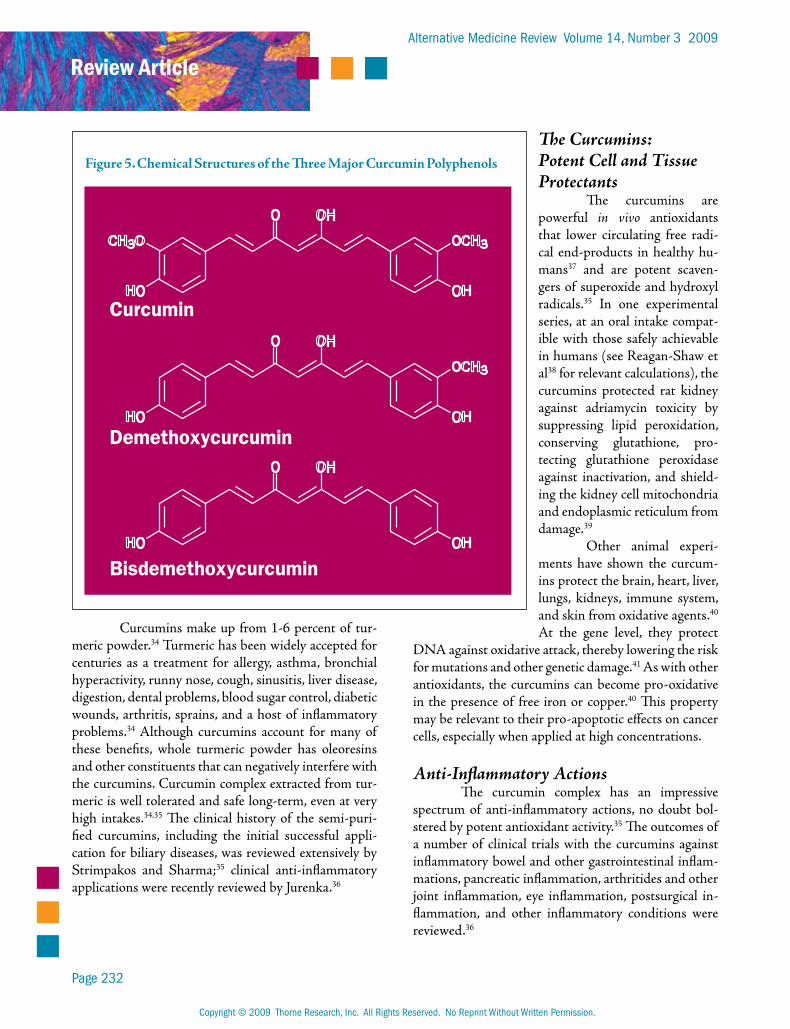
meric powder and contains primarily three curcumin polyphenols (Figure 5). Curcumin (diferuloylmethane; curcumin I) predominates, followed by demethoxy-curcumin (curcumin II) and bisdemethoxycurcumin (curcumin III), with other natural derivatives consti-tuting a maximum three percent of the powder.34 This trio is commonly referred to as “the curcumin complex” or simply “the curcumins.” Pure curcumin I is rare and references to “curcumin” usually refer to the curcumin complex.
Copyright © 2009 Thorne Research, Inc. All Rights Reserved. No Reprint Without Written Permission.
Alternative Medicine Review Volume 14, Number 3 2009
Review Article
Page 232
Curcumins make up from 1-6 percent of tur-meric powder.34 Turmeric has been widely accepted for centuries as a treatment for allergy, asthma, bronchial hyperactivity, runny nose, cough, sinusitis, liver disease, digestion, dental problems, blood sugar control, diabetic wounds, arthritis, sprains, and a host of inflammatory problems.34 Although curcumins account for many of these benefits, whole turmeric powder has oleoresins and other constituents that can negatively interfere with the curcumins. Curcumin complex extracted from tur-meric is well tolerated and safe long-term, even at very high intakes.34,35 The clinical history of the semi-puri-fied curcumins, including the initial successful appli-cation for biliary diseases, was reviewed extensively by Strimpakos and Sharma;35 clinical anti-inflammatory applications were recently reviewed by Jurenka.36
The Curcumins: Potent Cell and Tissue Protectants
The curcumins are powerful in vivo antioxidants that lower circulating free radi-cal end-products in healthy hu-mans37 and are potent scaven-gers of superoxide and hydroxyl radicals.35 In one experimental series, at an oral intake compat-ible with those safely achievable in humans (see Reagan-Shaw et al38 for relevant calculations), the curcumins protected rat kidney against adriamycin toxicity by suppressing lipid peroxidation, conserving glutathione, pro-tecting glutathione peroxidase against inactivation, and shield-ing the kidney cell mitochondria and endoplasmic reticulum from damage.39
Other animal experi-ments have shown the curcum-ins protect the brain, heart, liver, lungs, kidneys, immune system, and skin from oxidative agents.40 At the gene level, they protect
DNA against oxidative attack, thereby lowering the risk for mutations and other genetic damage.41 As with other antioxidants, the curcumins can become pro-oxidative in the presence of free iron or copper.40 This property may be relevant to their pro-apoptotic effects on cancer cells, especially when applied at high concentrations.
Anti-Inflammatory ActionsThe curcumin complex has an impressive
spectrum of anti-inflammatory actions, no doubt bol-stered by potent antioxidant activity.35 The outcomes of a number of clinical trials with the curcumins against inflammatory bowel and other gastrointestinal inflam-mations, pancreatic inflammation, arthritides and other joint inflammation, eye inflammation, postsurgical in-flammation, and other inflammatory conditions were reviewed.36
Figure 5. Chemical Structures of the Three Major Curcumin Polyphenols
O OH
OCH3
OH
CH3O
HO
O OH
OCH3
OHHO
O OH
OHHO
Curcumin
Demethoxycurcumin
Bisdemethoxycurcumin
Copyright © 2009 Thorne Research, Inc. All Rights Reserved. No Reprint Without Written Permission.
Alternative Medicine Review Volume 14, Number 3 2009
Phytosomes
Page 233
The positive clinical findings with the curcum-ins as anti-inflammatories are supported by a large body of experimental work. As examples, the curcumins po-tently inhibit carrageenan-induced paw edema in mice, acute lung injury by cyclophosphamide in rats, two forms of experimental arthritis in rats, chemically-in-duced ulcerative colitis in mice, and pancreatitis in two rat models.40
Nutrigenomic and other cutting-edge gene probe techniques have made it possible to correlate multiple, interlocking gene actions with higher-level processes such as inflammation. The curcumin complex influences many genes involved with the initiation and regulation of inflammation:
X Curcumins can down-regulate NF-kB, the nuclear transcription factor and critical upstream regulator of genes that control acute and chronic inflammation cascades, among others.29 The curcumins can inhibit NF-kB activation as induced by various known pro-inflammatory agents.42
X Perhaps via NF-kB, the curcumins down-regulate other pro-inflammatory enzymes such as lipoxygenases43 and inducible nitric oxide synthase (iNOS).40
X The curcumins inhibit other transcription factor products such as signal transducer and activator of transcription (STAT), peroxisome proliferator-activated receptor-g (PPAR-g), and β-catenin.44
X Cyclooxygenase-2 (COX-2) is the inducible form of COX that predominates at inflammatory sites and also likely plays a critical role in tumor promotion. Curcumin inhibits COX-2 activation by pro-inflammatory agents.43
X Activator protein 1 (AP-1) and JNK also can be antagonized by the curcumins.44,45
Cognitive Effects of CurcuminInflammation is implicated in diverse neuro-
degenerative disorders.45-47 The pathways involved in neuro- inflammation have been explored in some detail in Alzheimer’s disease (AD). Among the known molecu-lar mediators are reactive oxygen species (ROS), reactive nitric oxide species generated by iNOS, lipid peroxida-tion products, and the genes NF-kB and phosphorylated JNK.45 The curcumin complex can block these media-tors at effective concentrations that range between 1 and 2 micromolar and are attainable in humans.43,45,48
The curcumin complex has been subjected to two randomized, controlled trials for AD, both of which failed to produce clinical benefit. In the first trial, the curcumins did not reach the brain; sophisticated assays found no free curcumin in the plasma and mere nanomo-lar levels of curcumin glucuronide, the major curcumin metabolite.49 With the second trial, poor bioavailability may have again compromised success. Among the pa-tients receiving the highest curcumin dose (4 g/day), only 2/12 had greater than trace levels of curcumins in the blood after six months of dosing.50 There is evidence, however, the curcumins do oppose brain inflammation and can enhance amyloid removal.
In AD brains the microglia, macrophage-like cells resident in the brain tissue, seem unable to dispose of beta-amyloid like healthy brains.51 They also display surface markers of inflammation and a pro-inflamma tory balance of gene expression. In addition, macrophages and monocytes cross the blood-brain barrier and enter the brain, but then fail to ingest and destroy brain beta-amyloid. Like the resident microglia, these migratory im-mune cells over-express proinflammatory enzymes such as COX-2 and iNOS in the brain, both linked to AD progression.51 When AD cells are immersed in bisdeme-thoxycurcumin at a 0.1 micromolar concentration, their phagocytic activity is boosted and they are able to dis-pose of beta-amyloid.52
There is epidemiological evidence the curcum-ins protect against premature neurodegeneration. A 2006 study of elderly Singaporeans found those who consumed curry “occasionally” and “often or very often” scored significantly better on the Mini-Mental State Examination (MMSE) compared to those who “never or rarely” consumed curry.53 This affords hope that with improved bioavailability the curcumins could be effective neuroprotective supplements.
Copyright © 2009 Thorne Research, Inc. All Rights Reserved. No Reprint Without Written Permission.
Alternative Medicine Review Volume 14, Number 3 2009
Review Article
Page 234
Promise for Cancer Chemoprevention and Treatment
The curcumins have shown efficacy against all stages of cancer – initiation, promotion, proliferation, and metastasis.35,36,40,54 They have shown chemopreven-tive effects in animal models of colon, duodenal, esoph-ageal, stomach, and oral carcinogenesis.40 They also block cancer promotion by phorbol esters and other experimental agents in animals, and can interfere with angiogenesis and metastasis.34,40,55 They induce apopto-sis in cancer cells in culture, while leaving normal cells unaffected.40,54
One of the first human trials looked at chemo-prevention. Patients (n=25) with pre-malignant lesions were biopsied at baseline, started on curcumin complex, followed for three months, then biopsied again.56 The curcumin dose began at 500 mg/day, then was escalated stepwise from 500 mg/day to 8,000 mg/day by three months. There was histological improvement of pre-cancerous lesions in cases of cervical neoplasia (1 of 4 patients), stomach metaplasia (1 of 6 patients), bladder dysplasia (1 of 2 patients), oral leukoplakia (2 of 7 pa-tients), and Bowen’s disease of the skin (2 of 6 patients). The two patients who attained the highest intakes of 8,000 mg/day reached 1.77±1.87 micromolar serum levels, which is within an order of magnitude of concen-trations effective against cancer in animals.40
Concerning treatment of established cancer, poor bioavailability may have confounded clinical suc-cess, although this may not be the limiting factor in colorectal cancer. As reported in Garcea et al,41 daily doses of 1,800 or 3,600 mg/day for seven days resulted in plasma levels of curcumin below the limit of quanti-tation (that is, below 3 nmol/L), but did deliver quan-tifiable levels to the colorectal tissues – 7-20 nmol/g tissue. These amounts were sufficient to partially sup-press DNA damage. Malignant colorectal tissue upon baseline biopsy had 2.5 times greater DNA damage than normal colorectal tissue (measured as DNA ad-duct M1G). However, in those patients who received 3,600 mg/day, these adducts in the malignant tissue fell significantly – by about half – into the range of normal colorectal tissue (p<0.05).
In a phase II trial of curcumin for advanced pancreatic cancer, two of 21 patients showed slowing of disease progression, although free curcumin was almost undetectable in the plasma.57 This trial also sampled
for the cancer-related factors NF-kB and phosphory-lated signal transducer and activator of transcription 3 (pSTAT-3) at baseline and day 8. Both were signifi-cantly elevated in the cancer patients at baseline com-pared to the healthy control subjects, but by day 8 both were reduced toward non-cancer levels. This 10-percent disease response rate would likely be improved by uti-lizing curcumin preparations that would produce mi-cromolar levels in the plasma.
Studied in vitro without the limitations of bio-availability, the curcumins influence a number of genes implicated in carcinogenesis and the subsequent stages of cancer. These include “master” genes that up-regulate the production of detoxification enzymes (e.g., cyto-chrome p450 enzymes) and down-regulate NF-kB and AP-1, both of which can promote inflammation and cancer cell survival.29,36,54 The evidence suggests as these higher-level genes become influenced by the curcumins, they in turn initiate a downstream cascade of genes that manage apoptosis, the cell cycle and cell proliferation, and various growth factors involved with new blood ves-sel formation; for example, vascular endothelial growth factor (VEGF).54
The problems with curcumin bioavailability seemingly have not dampened researchers’ enthusiasm to pursue anticancer benefits. Several cancer trials are completed but not yet published; of the minimum 16 clinical trials with curcumins that are currently active, 10 are on cancer (including three on cancer preven-tion).58 The cancer types being studied include colon and rectal, other intestinal, pancreatic, multiple myelo-ma, and osteosarcoma. The cancer trials completed to date have demonstrated some potential of the curcum-ins against cancer, despite known poor bioavailability, so the trial outcomes from using better-absorbed forms of curcumin ought to be more impressive. The advent of a phytosomal curcumin offers an optimistic future for clinical curcumin trials.
Phytosomes Markedly Improve the Bioavailability of the Curcumins
As most recently reviewed by Jurenka36 and by Villegas et al,54 conventional curcumin preparations are poorly absorbed. Human chronic pharmacokinetic studies indicate free curcumin remains at or below 25 nanomolar in plasma over an oral daily dosage range of 3.6-12 g curcumin complex taken for a week or longer.54
Copyright © 2009 Thorne Research, Inc. All Rights Reserved. No Reprint Without Written Permission.
Alternative Medicine Review Volume 14, Number 3 2009
Phytosomes
Page 235
This poor average bioavailability is compounded by great variability among subjects.59
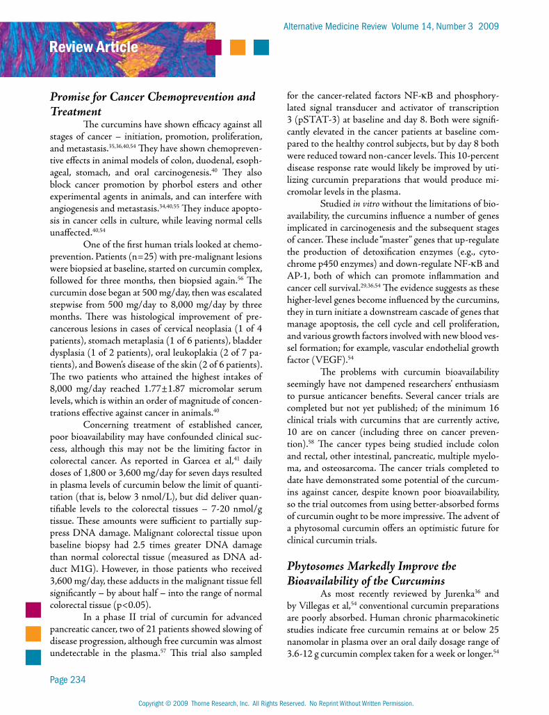
The bioavailability of the curcumin phytosome preparation (Meriva®, from Indena SpA) has been test-ed in rats against Indena’s equivalent non-phytosome curcumin extract (Figure 6).6 The rats were fasted over-night, then received by mouth either the phytosomal or the non-phytosomal preparation. Blood samples were obtained at 15, 30, 60, or 120 minutes after oral gavage. Curcumin complex was administered by oral gavage at 360 mg/kg body weight, in either phytosome or non-phytosome form. Values are the means (n=3) and the
bars represent standard deviations (SD). An asterisk indicates the values at that time point were significantly divergent from each other (p<0.01).
Curcumin I was identified in the rat blood samples along with its metabolites curcumin glucuro-nide, curcumin sulfate, tetrahydrocurcumin, and hexa-hydrocurcumin.6 The phytosome preparation displayed superior curcumin plasma absorption from the first 30 minutes. As shown qualitatively in Figure 6, for the pe-riod 0-120 minutes the AUC values were 5.6 times bet-ter for the curcumin phytosome compared to the non-phytosome preparation (actual data 26.7 mcg/min/mL versus 4.8 mcg/min/mL). In this study, the liver also accumulated significantly more curcumin I from the phytosome compared to the non-phytosome.6
The primary problems limiting bioavailability of conventional curcumin complex are poor solubility in water, molecular instability including rapid hydro-lytic degradation, and rapid molecular modification by conjugation, mostly in the liver, to the glucuronide and sulfate forms that are rapidly excreted. These have un-known bioactivity and unlikely cross the blood-brain barrier. Conversion of the curcumins into phytosomes addresses these issues.
As with other phytosome preparations, the curcumin molecule’s limited solubility should be miti-gated by the presence of PC. The physico-chemical en-wrapment of the polyphenol molecule by the larger PC molecule may well decrease its molecular instability to improve its effective half-life.
Data from a small, single-dose human study with Meriva, not yet published, suggests the relative plasma absorption of curcuminoids in phytosome form is significantly greater than curcuminoids in non-phyto-some form.7 Larger absorption trials are pending.
The Green Tea Catechins and their Phytosome Complex
Green tea, a product of the plant Camellia sinensis (family Theaceae), contains polyphenols, spe-cifically catechins of the flavan-3-ol class and their gal-late derivatives. These green tea catechins (GTC) have diverse benefits. They are potent antioxidants and anti-inflammatories,60 provide cardiovascular-61 and neuro-protectant62 effects, and accelerate fat oxidation with its consequent potential for weight loss.63,64 They are being
Figure 6. Plasma Curcumin I in Rats from Curcumin Phytosome (solid line) or Non-complexed Curcumin (broken line)
•Plas
ma
curc
umin
con
cent
ratio
n(n
g/m
L)
* *
••
•
•
16
12
8
4
0
Time after administration (min)0 15 30 60 120
From: Marczylo T, Verschoyle R, Cooke D, et al. Comparison of systemic availability of curcumin with that of curcumin formulated with phosphatidylcholine. Cancer Chemother Pharmacol 2007;60:171-177.
Plasma levels of curcumin I in rats that had received curcumin as phytosome (solid line) or conventional curcumin complex (broken line). Administration was by oral gavage at 360 mg curcumin complex per kg body weight. Values are the means (n=3) and the bars represent standard deviations (SD). An asterisk indicates the values at that time point were signifi-cantly divergent from each other (p<0.01).
Copyright © 2009 Thorne Research, Inc. All Rights Reserved. No Reprint Without Written Permission.
Alternative Medicine Review Volume 14, Number 3 2009
Review Article
Page 236
actively investigated as anticancer agents. To date, effi-cacy in controlled clinical trials has been inconsistent, possibly due to poor bioavailability.
Chemistry and Clinical Status The polyphenols most consistently found
in green tea extracts are, in decreasing order of concentration:65,66
X (-)-epigallocatechin-3-gallate (EGCG) X (-)-epigallocatechin (EGC) X (-)-epicatechin-3-gallate (ECG) X (-)-epicatechin (EC)
with small amounts of: X (-)-epigallocatechin-3
(3’methyl)-gallate (EGCMG) X (+)-catechin (C) X (+)-gallocatechin-3-gallate (GTG)
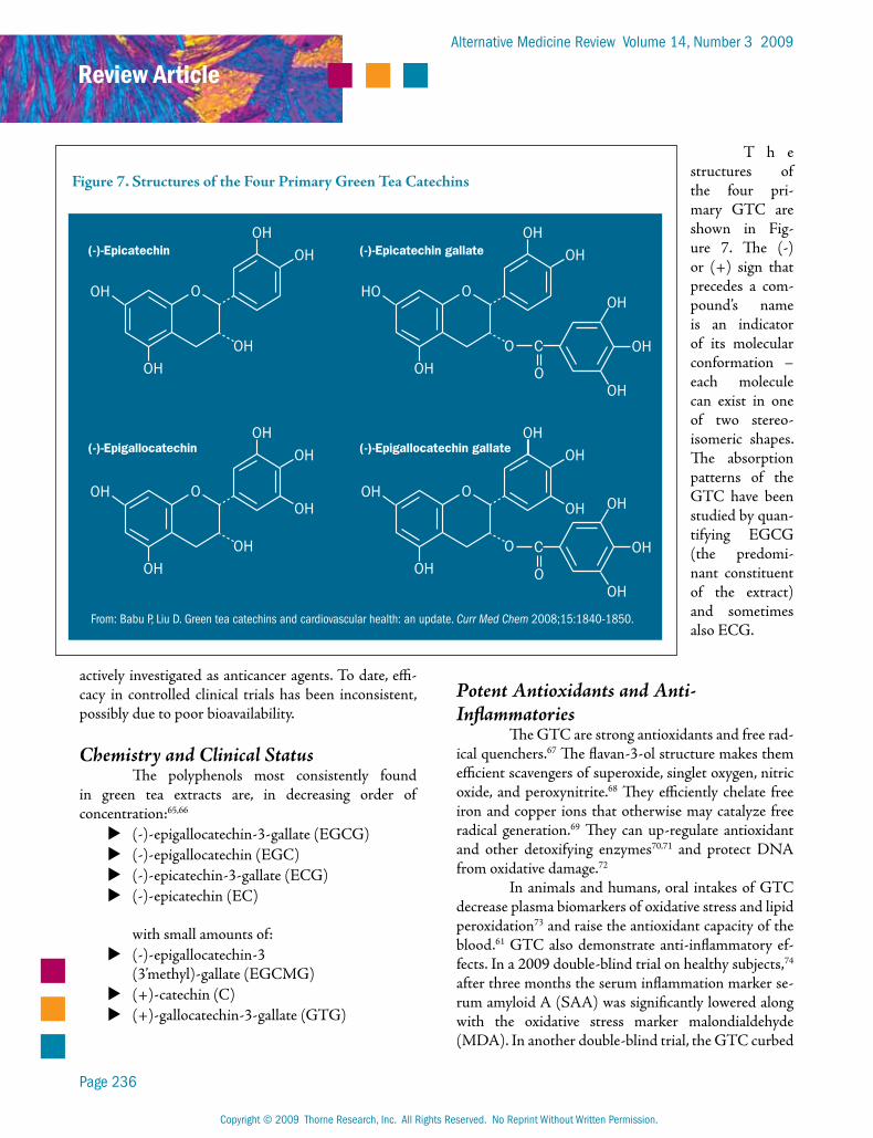
T h e structures of the four pri-mary GTC are shown in Fig-ure 7. The (-) or (+) sign that precedes a com-pound’s name is an indicator of its molecular conformation – each molecule can exist in one of two stereo-isomeric shapes. The absorption patterns of the GTC have been studied by quan-tifying EGCG (the predomi-nant constituent of the extract) and sometimes also ECG.
Potent Antioxidants and Anti-Inflammatories
The GTC are strong antioxidants and free rad-ical quenchers.67 The flavan-3-ol structure makes them efficient scavengers of superoxide, singlet oxygen, nitric oxide, and peroxynitrite.68 They efficiently chelate free iron and copper ions that otherwise may catalyze free radical generation.69 They can up-regulate antioxidant and other detoxifying enzymes70,71 and protect DNA from oxidative damage.72
In animals and humans, oral intakes of GTC decrease plasma biomarkers of oxidative stress and lipid peroxidation73 and raise the antioxidant capacity of the blood.61 GTC also demonstrate anti-inflammatory ef-fects. In a 2009 double-blind trial on healthy subjects,74 after three months the serum inflammation marker se-rum amyloid A (SAA) was significantly lowered along with the oxidative stress marker malondialdehyde (MDA). In another double-blind trial, the GTC curbed
Figure 7. Structures of the Four Primary Green Tea Catechins
OH
OHOH
OHOH
O
OH
OHOH
OHOH
OOH
HO
OHO
OHOH
OOH
OH
OHC
O
OH
OHO
OHOH
OOH OH
OH
OHC
O
(-)-Epicatechin
(-)-Epigallocatechin
(-)-Epicatechin gallate
(-)-Epigallocatechin gallate
From: Babu P, Liu D. Green tea catechins and cardiovascular health: an update. Curr Med Chem 2008;15:1840-1850.
Copyright © 2009 Thorne Research, Inc. All Rights Reserved. No Reprint Without Written Permission.
Alternative Medicine Review Volume 14, Number 3 2009
Phytosomes
Page 237
gum inflammation;75 they also have potent antibacterial action that could further contribute to gum anti-plaque resistance.76
Like other flavonoids, the green tea catechins can down-regulate the nuclear transcription factors NF-kB and AP-1, either of which may promote chronic inflammation and carcinogenesis when abnormally acti-vated.67
Cardiovascular ProtectionGreen tea catechins consumed as whole green
tea have been extensively studied for cardiovascular (CV) protective effects in prospective trials, epidemio-logical studies, animal models, and in vitro.61,67,74,77 Most clinical studies have shown some benefit, although some have not.67,74 Clinical inconsistency may be relat-ed to the obvious confounding variables associated with drinking tea. For example, there is no general agreement as to what quantities of which substances constitute a cup of tea.74 Research designs are trending toward us-ing standardized GTC preparations, often formulated around EGCG content.
The conclusions from epidemiological studies on green tea consumption and cardiovascular health are generally favorable.67 The larger the population studied, the more reliable the outcome. The largest one to date, conducted on more than 40,000 Japanese, found elevat-ed green tea consumption was associated with reduced CV and all-cause mortality.78 This study also found an inverse association of GTC intakes with stroke risk, al-though only in females.
Prospective clinical trials on GTC for CV pro-tection have been few. The trials on blood lipids and dia-betes management have not produced consistent find-ings, but a 2009 double-blind trial on relatively healthy subjects74 found systolic blood pressure was significant-ly lowered by 5 mm Hg after three weeks and remained lowered after three months; total and LDL cholesterol were lowered by 9 mg/dL. The oxidative stress marker MDA and the inflammatory marker SAA were also sig-nificantly lowered. The test preparation was a standard-ized GTC mixture (decaffeinated, 400 mg/day) encap-sulated together with theanine, an anti-anxiety nutrient normally present in green tea (200 mg/day). According to the researchers, theanine has the potential for such benefits, so this study’s positive outcome cannot be fully credited to the GTC.74
There is other evidence green tea catechins have endothelial protective benefits. In an open trial, chronic smokers (n=20) ingesting green tea powder (8 g/day) for two weeks demonstrated significantly im-proved endothelial function (p<0.001) measured as flow-mediated, endothelium-dependent vasodilation (FMD).79 Pure EGCG (300 mg) also acutely improved FMD in patients with coronary heart disease.80
Numerous pro-homeostatic CV actions have been experimentally identified for GTC, EGCG in par-ticular, but most occur at too high a concentration to have human clinical relevance. One exception may be VEGF, which normally stimulates blood vessel endo-thelial cells to divide in tumor angiogenesis and can pro-mote inflammation in the atherosclerotic vessel wall.66,81 VEGF activity can be partially blocked at EGCG con-centrations of 5-50 micromolar,82 a range achievable with the green tea phytosome preparation (see the Bio-availability section).
Neuroprotection: Parkinson’s Disease, Stroke, Dementia
Green tea catechins are receiving intense scru-tiny as multifunctional compounds for neuroprotec-tion. The radical scavenging, iron chelating, gene mod-ulating, and cell signaling activities, together with the ability to cross the blood-brain barrier, render GTC potentially clinically significant central nervous system protectants.62
Although no prospective clinical trials are avail-able, the evidence favoring GTC for neuroprotection continues to gain favor. Findings from two epidemio-logical studies suggest GTC consumption may reduce the risk for Parkinson’s disease.83,84 Various GTC have been found to protect against nerve cell death from the Parkinsonian trigger methylphenyltetrahydropyridine (MPTP) in animal models.62,85,86 The presence of un-bound, redox-active iron is a well-established feature of Parkinson’s disease87 and GTC (especially EGCG) have potent iron-chelating properties (see Mandel et al62 for an in-depth discussion). EGCG also potently inhibits the enzyme catechol-O-methyltransferase (COMT) and therefore could help conserve synaptic dopamine in the Parkinson’s brain.88
Copyright © 2009 Thorne Research, Inc. All Rights Reserved. No Reprint Without Written Permission.
Alternative Medicine Review Volume 14, Number 3 2009
Review Article
Page 238
GTC have demonstrated potent central ner-vous system (CNS) protection in animal models of stroke (see Bastianetto et al85 for references). They re-duce hippocampal damage and brain edema caused by experimental cerebral ischemia in gerbils;89 in the in-farcted area EGCG helps keep the cells viable by pre-serving mitochondrial integrity.90 EGCG’s protective actions extend to the cell membrane level, where it acti-vates protein kinase C enzymes that are survival factors for the nerve cells.62,91 EGCG even can bond directly with phospholipids of cell membranes to activate pro-tein kinase C and probably also other key membrane signaling pathways.92
In animal models of Alzheimer’s disease, some GTC specifically bind with, and help clear, amyloid-beta from the brain.85,93 This property was further ex-plored with cells in culture. In hippocampal cells from rat brain, GTC inhibited the formation of amyloid-beta fibrils implicated in neuron death.85 Although a GTC complex was protective in the concentration range of 7-22 micromolar, EGCG alone was more effective, being significantly protective at 5-10 micromolar con-centration. These are ranges attainable in the plasma by oral dosing with phytosomal GTC, as reviewed in detail below.
Some researchers contend amyloid is merely a symptom and that oxidative stress could be a more proximate cause of neural cell death, with the amyloid fibrils being merely oxidation end-products.94 The po-tent antioxidant power of EGCG and other GTC could contribute to their neuroprotective benefits, but this mechanism does not fully fit the observed effects, and other mechanisms are also thought to be involved, for example, higher-level actions of EGCG, including the down-regulation of pro-apoptotic genes (e.g., bax, bad, fas and caspases 1 and 6), and the up-regulation of ki-nase enzymes such as protein kinase C.95
Although GTC undergo significant metabo-lism and p450 conjugation after absorption, animal studies demonstrate GTC cross the blood-brain bar-rier.96,97 GTC, and especially EGCG, have the potential to be effective CNS chemoprotectants.85
Green Tea and EGCG Anticancer StatusConsiderable in vitro, animal model data, and
other experimental findings on GTC strongly sup-port their further investigation as cancer chemopre-
ventives.66 Experimentally, GTC and more specifically EGCG have been used to induce apoptosis and cell cycle arrest in many cancer cell types (including lung, colon, pancreas, skin, and prostate) without affecting normal cells.98 This process is essential to normal func-tioning during health and serves also to protect against neoplastic-cancer transformation by eliminating geneti-cally damaged cells or others that escape normal growth regulation.
EGCG taken orally (200 mg/day for three months) was reported effective for cancer prevention in a trial on women with pre-neoplastic, human papilloma virus-infected cervical lesions.99 Six out of 10 women re-sponded to 200 mg/day EGCG after 8-12 weeks. GTC showed anti-neoplastic effects in men with androgen-independent prostate carcinoma.100 In another EGCG trial, stages I and II breast cancer patients experienced a lower recurrence rate and longer disease-free period.101
GTC and especially EGCG also have shown potential for combination chemoprevention of cancer. As one example, they showed synergy with sulindac against intestinal neoplasias.102
Somewhat similar to curcumin, GTC and es-pecially EGCG have multiple effects on cell signaling pathways rooted in the outer cell membranes and impli-cated in human cancer. Judging from combined in vivo and in vitro data, it seems clear EGCG inhibits NF-kB, a major transcription factor sensitive to oxidative stress and centrally involved in the management of growth, immunity, inflammation, and cell death. EGCG also in-hibits the MAPK cascade and in a lower concentration range (5-20 micromolar). Some MAPK components cross-react (“crosstalk”) with NF-kB and are implicated in cell proliferation, differentiation, and apoptosis.66 Deregulation of the MAPK pathway has been observed in a number of human cancers, at stages of proliferation through to metastasis.66
EGCG also may combat cancer angiogenesis by inhibiting the binding of VEGF to its receptors in the blood vessel endothelia.82 Many of the experimental findings with GTC/EGCG against cancer have ques-tionable relevance to human cancers since they were conducted with GTC or EGCG at concentrations not routinely attainable by oral intakes.103 Some of these putative anticancer mechanisms would become more relevant if the GTC/EGCG absorption could be im-proved by less than one order of magnitude.
Copyright © 2009 Thorne Research, Inc. All Rights Reserved. No Reprint Without Written Permission.
Alternative Medicine Review Volume 14, Number 3 2009
Review Article
Page 239
Given the difficulties with bioavailability, clini-cal studies with oral GTC benefit from the use of ab-sorption biomarkers. The two most prevalent GTC, namely EGCG and ECG, were validated as biomarkers for long-term GTC dosing by Wang et al.104 Most of the in vitro and animal studies used GTC and/or EGCG concentrations of 10 micromolar or higher, but conven-tional doses to humans fall short of this level (usually peaking at or below 2 micromolar). The advent of the green tea phytosome offers possibilities for bridging this gap.
Green Tea Phytosome Enhances EGCG Bioavailability
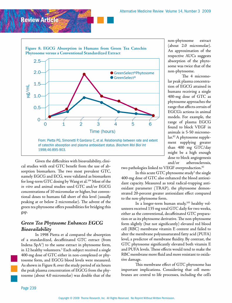
In 1998 Pietta et al compared the absorption of a standardized, decaffeinated GTC extract (from Indena SpA3) to the same extract in phytosome form, in 12 healthy volunteers.5 Each subject received a single 400-mg dose of GTC either in non-complexed or phy-tosome form, and EGCG blood levels were measured. As shown in Figure 8, over the study period of six hours the peak plasma concentration of EGCG from the phy-tosome (about 4.0 micromolar) was double that of the
non-phytosome extract (about 2.0 micromolar). An approximation of the respective AUCs suggests absorption of the phyto-some was twice that of the non-phytosome.
The 4 micromo-lar peak plasma concentra-tion of EGCG attained in humans receiving a single 400-mg dose of GTC as phytosome approaches the range that affects certain of EGCG’s actions in animal models. For example, the range of plasma EGCG found to block VEGF in animals is 5-50 micromo-lar.82 A phytosome supple-ment supplying greater than 400 mg GTC/day might be a high enough dose to block angiogenesis and/or atherosclerosis,
two pathologies linked to VEGF overproduction.82
In this acute GTC phytosome study5 the single 400-mg dose of GTC also enhanced the blood antioxi-dant capacity. Measured as total radical-trapping anti-oxidant parameter (TRAP), the phytosome demon-strated 20-percent greater antioxidant effect compared to the non-phytosome form.
In a longer-term human study,105 healthy vol-unteers received 135 mg total GTC daily for two weeks, either as the conventional, decaffeinated GTC prepara-tion or as its phytosome derivative. The non-phytosome form slightly (but not significantly) elevated red blood cell (RBC) membrane vitamin E content and failed to alter the membrane polyunsaturated fatty acid (PUFA) level, a predictor of membrane fluidity. By contrast, the GTC phytosome significantly elevated both vitamin E and PUFA levels. These effects would tend to make the RBC membrane more fluid and more resistant to oxida-tive damage.
This membrane effect of GTC phytosome has important implications. Considering that cell mem-branes are central to life processes, including the cell’s
Figure 8. EGCG Absorption in Humans from Green Tea Catechin Phytosome versus a Conventional Standardized Extract
2.5
2.0
1.5
1.0
0.5
0
Time (hours)
0 1 2 3 4 5 6
μg/
mL
GreenSelect®PhytosomeGreenSelect®
From: Pietta PG, Simonetti P, Gardana C, et al. Relationship between rate and extent of catechin absorption and plasma antioxidant status. Biochem Mol Biol Int 1998;46:895-903.
Copyright © 2009 Thorne Research, Inc. All Rights Reserved. No Reprint Without Written Permission.
Alternative Medicine Review Volume 14, Number 3 2009
Review Article
Page 240
energy flow,10 the GTC by protecting cell membranes are contributing to increased cell membrane efficiency and improved energy utilization. This rationale could help explain the clinical observations that GTC sig-nificantly increase energy expenditure in humans and thereby promote fat oxidation.
Green Tea and Green Tea Phytosome for Weight Loss
Results from weight loss trials with the con-ventional GTC extract have been inconsistent, with poor bioavailability a likely complicating factor. In a recent double-blind trial conducted with 60 obese sub-jects, GTC (750 mg/day for 90 days) resulted in sig-nificantly decreased weight by 5.1 kg over the placebo group after eight weeks (p<0.05) and by 3.3 kg after 12 weeks (p<0.05). At the eighth week, resting energy ex-penditure was significantly higher in the green tea group (p<0.001) as was respiratory quotient (p<0.05).106
In a 2008 double-blind trial conducted in Ja-pan,63 non-obese patients with type 2 diabetes ingested green tea beverages daily for 12 weeks. They received either 583 mg/day GTC (n=23, the test group) or 96 mg/day GTC (n=20, the control group). The mean BMIs at baseline were 25.6 and 24.0 for test and control groups, respectively. Average energy intake was 1,680 kcal during the study period, and did not differ sig-nificantly between groups. At week 12, the GTC group had a significantly greater decrease in waist circumfer-ence (an average 3.3 cm loss, versus a 0.1 cm gain for the control group, p<0.01 for the intergroup difference). Waist-to-hip ratio also was significantly decreased in the GTC group compared to the control group (p<0.05 intergroup).
In this trial,63 serum insulin was found slightly but significantly increased in the GTC group at 12 weeks and slightly decreased in the control group (p<0.05 in-tergroup). From subgroup analysis it emerged that the significant insulin increase was confined to patients tak-ing insulinotropic agents throughout the trial. In this group, the glycation biomarker hemoglobin A1C was significantly decreased from baseline at week 12. Alto-gether, the findings from this comprehensive trial sug-gest GTC can facilitate weight loss and perhaps also a modest improvement of endogenous insulin produc-tion.
The green tea phytosome complex has also been shown to facilitate weight loss. In an Italian tri-al,64 100 overweight subjects, 20-40 percent over ideal weight, were placed on a hypocaloric diet (1,850 and 1,350 calories daily for men and women, respectively). The test group (n=50) received 300 mg/day green tea phytosome (Greenselect®; approximating 100 mg GTC); the control group (n=50) received only the hy-pocaloric diet. At 90 days, the phytosome group had ex-perienced significant weight loss (a mean 14.0 kg) while the control group had lost an average of 5 kg (not sta-tistically significant). More trials are needed to confirm this initial finding, but the green tea phytosome may have the potential to be safe and effective for weight loss in subjects who are markedly overweight.
Green Tea and EGCG Trials in ProgressGTC and EGCG are currently receiving at-
tention in terms of clinical trial research. As of mid-2009, there were at least 38 clinical trials in progress.31 Of these, 24 were on cancer, including eight for cancer prevention. The cancer types being tested are prostate, bladder, ovarian, breast, lung, cervical, skin, breast, esophageal, chronic lymphocytic leukemia, follicular lymphoma, and a category “all solid tumors.”
Other disease trials under development with GTC/EGCG in mid-2009 include Parkinson’s dis-ease, multiple sclerosis, ulcerative colitis, fat oxidation and weight loss, diabetes, cystic fibrosis, cardiovascular protection, osteoporosis, skin damage, immunity, ocular hypertension, antiviral effects, and oral hygiene.31
As anticancer therapeutics, and for neuropro-tective, cardiovascular, and systemic anti-inflammatory support, GTC have considerable clinical potential. They operate through multiple mechanistic pathways that include but eclipse their antioxidant powers. Some of these pathways are likely to become functionally rel-evant for humans as oral bioavailability is improved. The phytosome preparation minimally doubles GTC bioavailability and also provides the therapeutic benefit of PC.
Grape Seed Polyphenols and their Phytosome Complex
Grape seed extract is a concentrated source of polyphenols, more specifically polyhydroxylated flavan-3-ols. The grape seed polyphenols resemble the
Copyright © 2009 Thorne Research, Inc. All Rights Reserved. No Reprint Without Written Permission.
Alternative Medicine Review Volume 14, Number 3 2009
Phytosomes
Page 241
catechins of green tea in basic molecular structure (fla-van-3-ols and their gallates), except grape seed constitu-ents reach larger molecular sizes. Having a complicated molecular profile, they comprise (+)-catechin, (-)-epi-catechin, and polymers of these along with their gallate derivatives. Technically these are “oligomeric proantho-cyanidins” (sometimes termed “oligomeric procyani-dins”). For convenience they will be referred to as grape seed polyphenols or GSP.
Complex Preparations, Potent ProtectantsCommercial preparations of GSP vary greatly
in chemical profiles, making study interpretation chal-lenging. One important aspect is the higher molecular weight polyphenols tend to be very poorly bioavail-able.107 Thus, preparations skewed toward lower mo-lecular weight compositions are likely to be more effica-cious.
The Leucoselect® preparation is the only GSP complex available in phytosome form.3 Leucoselect’s molecular profile is:108
X Fraction 1. (+)-catechin, (-)-epicatechin totaling 15 percent by weight
X Fraction 2. (-)-epicatechin gallate, dimers, trimers, tetramers and their gallates, 80 percent
X Fraction 3. Mixed pentamers, hexamers, heptamers, and their gallates, five percent
These molecular identities and distributions are verified using high performance liquid chromatog-raphy with ultraviolet light detection (HPLC-UV) and other sophisticated analytical methodologies.108 The high content of the smaller molecules (fractions 1 and 2) is crucial to the biological activity of the extract, and the Leucoselect production process concentrates these two fractions at the expense of fraction 3.108
Experimental studies verify the antioxidant capacities of GSP. These polyphenols are scavengers of superoxide, hydroxyl, peroxyl, and peroxynitrite radi-cals;109-112 they chelate redox-active metals;110,111 and they can protect cell membranes against oxidative at-tack.112,113 In RBCs subjected to oxidative stress, GSP markedly decreased membrane lipid peroxidation, recy-cled oxidized tocopherol in the membrane, and delayed hemolysis.113,114 Conversion of the Leucoselect GSP preparation to phytosomes preserves these capacities and improves their delivery to the tissues.
The Leucoselect Grape Seed PhytosomeThe Leucoselect phytosome is prepared at a ra-
tio of one part GSP to three parts PC by weight.108 It has been validated by in vitro, animal, and clinical trials, pri-marily for cardiovascular protection. Direct data on ab-sorption are not yet available, perhaps because of the limi-tations to quantifying these complex mixtures in plasma.
The Leucoselect GSP in phytosome form have demonstrated potent antioxidant effects. In a 2002 study, 10 healthy subjects received 110 mg/day of Leucose-lect GSP as phytosomes for 30 days, then RBCs were sampled and analyzed.104 Levels of RBC-membrane alpha-tocopherol rose significantly over baseline (59.5%, p<0.001); the RBC-membrane fatty acid composition shifted to a slightly higher level of polyunsaturates. Oxi-dized lymphocyte DNA was markedly decreased (68%, p<0.05).
The Leucoselect phytosome also can protect against intensified oxidative stress in vivo. The elevation of blood lipids that typically occurs following a meal is often accompanied by an elevation of oxidative end-products and is a suspected atherosclerosis risk factor.115 In an open trial,116 eight healthy volunteers consumed a fatty meal rich in oxidized and oxidizable lipids without (control) or with 300 mg Leucoselect GSP as phyto-some. Plasma lipid peroxides did not increase after con-sumption of the meal when grape seed phytosome was included. Conversely, adding the phytosome to the meal significantly increased plasma antioxidant capacity fol-lowing the meal, while the controls showed no increase. The researchers concluded the Leucoselect phytosome minimized post-meal oxidative stress by decreasing pro-oxidant and increasing antioxidant levels in plasma.
Cigarette smoke is a well-proven source of oxi-dative stress. Each puff of cigarette smoke contains tril-lions of carbon- and oxygen-centered oxidative species. In a randomized, double-blind, crossover trial, male heavy smokers over the age of 50 (n=24) received Leucoselect phytosome at 300 mg/day of GSP or a placebo for four weeks.117 The supplement significantly improved low-density lipoprotein (LDL) cholesterol resistance to oxida-tion. Thiobarbituric acid reactive substances (TBARS), an index of lipid peroxidation and oxidative stress, was significantly reduced in the LDL (p<0.05 versus placebo); and the lag phase of LDL time to oxidation, a measure of LDL antioxidant resistance, was prolonged in compari-son with placebo (p<0.005).
Copyright © 2009 Thorne Research, Inc. All Rights Reserved. No Reprint Without Written Permission.
Alternative Medicine Review Volume 14, Number 3 2009
Review Article
Page 242
Another double-blind trial involved type 2 diabetics (n=24) who received either 300 mg/day Leucoselect phytosome or a placebo for four weeks.108 Diabetes generally elevates oxidative stress. The GSP phytosome significantly reduced urinary excretion of a free radical end-product (F2 isoprostane) compared to placebo (p<0.04).
In experiments with rabbits, Leucoselect phy-tosome protected the heart from experimental isch-emia-reperfusion injury, improving myocardial pump performance and increasing the release of protective prostacyclin into circulation.111 Leucoselect also can bind to the luminal surfaces of arterial endothelial cells, protecting them from oxidative stress.113 This suggests that along with silymarin, the curcumins, and the green tea catechins, GSP phytosome can benefit cell-level ho-meostasis via cell membrane signaling pathways.
Grape Seed Polyphenols for Inflammation and Cancer
Although clinical research on GSP for inflam-mation and cancer is not as advanced compared to sily-marin, the curcumins, and green tea catechins, to date there is abundant animal and in vitro evidence suggest-ing GSP also have efficacy for these applications.
When mice of the SKH-1 hairless strain are exposed to high levels of ultraviolet radiation B (UVB), the skin tissue becomes depleted of antioxidants and inflammation is triggered. Initiation of inflammation is linked with activation of the NF-kB and the MAPK cell signaling cascades. Long-term activation of these pathways is also causally linked to skin carcinogenesis (reviewed in Sharma118). GSP added to the mouse diet blocked UVB destruction of glutathione, other anti-oxidants, and antioxidant enzymes in the skin, as well as lipid peroxidation and protein oxidation. GSP was found to down-regulate the NF-kB and MAPK path-ways, along with related downstream pathways such as cyclin D1, iNOS, and COX also linked to inflamma-tion.
When this SKH-1 strain was set up for skin cancer induction by UVB (“photocarcinogenesis”), GSP effectively blocked the process.119 As little as 0.2-percent GSP by weight added to the diet resulted in significantly fewer mice with tumors at week 15 (p<0.05). Number of tumors per mouse was also reduced (p<0.05), as was
tumor size (p<0.001), compared to the control group fed an unsupplemented diet.
Numerous other animal and cell culture stud-ies have been conducted with GSP for inflammation and cancer, as recently summarized and discussed in Nandakumar et al.120 Various GSP preparations have shown potential to counter inflammation and block cancer initiation, promotion, tumor proliferation, an-giogenesis, and metastasis. As with other poorly bio-available polyphenols, utilizing GSP in phytosome form holds obvious promise for enhancing its clinical efficacy.
ConclusionThe substantial data available on certain poly-
phenols as phytosome complexes makes a strong case for integrating these safe and well-tolerated therapies into clinical practice. Attributes include:
X markedly enhanced intestinal absorption and tissue delivery of specific polyphenols
X significantly improved efficacy compared to the chemically equivalent non-phytosome form
X no compromise in safety, in contrast to technologies that synthesize or otherwise chemically modify the parent polyphenol molecule and consequently destroy its natural configuration
X additional benefit from PC, the phytosome nutrient co-constituent
The availability of phytosome preparations of silybin and its allied flavonolignans, the curcumin di-phenolic complex, the green tea catechins, and the grape seed polyphenols should give further impetus to clinical research into these diverse nutrient classes. Extending beyond the established antioxidant capabilities of these molecules are their capacities to interact with cell mem-brane signaling molecular complexes, diffusible tran-scription factors inside cells, and intranuclear receptor complexes. Using them in phytosome form for oral dos-ing, with the assistance of the cell membrane molecule PC, vastly improves the chances to target the cell and tissue sites where phytosomes might provide the most clinical benefit.
Copyright © 2009 Thorne Research, Inc. All Rights Reserved. No Reprint Without Written Permission.
Alternative Medicine Review Volume 14, Number 3 2009
Phytosomes
Page 243
Phytosome technology is not limited to poly-phenols; in theory any nutrient molecule is eligible for conversion. Surprisingly, despite proven superior perfor-mance and steadily expanding clinical repertoires, phy-tosome preparations remain under-utilized. Clinicians and other researchers would do well to consider using phytosome preparations for applications that have been earlier explored with conventional polyphenol prepara-tions, and for new applications still being contemplated.
References1. Manach C, Scalbert A, Morand C, et al. Polyphenols:
food sources and bioavailability. Am J Clin Nutr 2004;79:727-747.
2. Bombardelli E, Curri SB, Della Loggia R, et al. Complexes between phospholipids and vegetal derivatives of biological interest. Fitoterapia 1989;60:1-9.
3. www.indena.com/pdf/ephytosome.pdf [Accessed June 20, 2009]
4. Kidd P, Head K. A review of the bioavailability and clinical efficacy of milk thistle phytosome: a silybin-phosphatidylcholine complex (Siliphos®). Altern Med Rev 2005;10:193-203.
5. Pietta P, Simonetti P, Gardana C, et al. Relationship between rate and extent of catechin absorption and plasma antioxidant status. Biochem Mol Biol Int 1998;46:895-903.
6. Marczylo TH, Verschoyle RD, Cooke DN, et al. Comparison of systemic availability of curcumin with that of curcumin formulated with phosphatidylcholine. Cancer Chemother Pharmacol 2007;60:171-177.
7. Indena SpA. Human pharmacokinetic study with curcumin. Unpublished.
8. Kidd PM. Phosphatidylcholine. In: Czap K, Miller AL, Head KA, et al, eds. Alternative Medicine Review Monographs Volume One. Dover, ID: Thorne Research, Inc.; 2002:310-315.
9. Alberts B, Johnson A, Lewis J, et al. Molecular Biology of the Cell. 4th ed. New York, NY: Garland Science; 2002.
10. Hulbert AJ, Else PL. Membranes and the setting of energy demand. J Exp Biol 2005;208:1593-1599.
11. Malandrino S, Pifferi G. IdB-1016. Drugs Future 1990;15:226-227.
12. Barzaghi N, Crema F, Gatti G, et al. Pharmacokinetic studies on IdB 1016, a silybin-phosphatidylcholine complex, in healthy human subjects. Eur J Drug Metab Pharmacokinet 1990;15:333-338.
13. Kidd PM. Glutathione. In: Czap K, Miller AL, Head KA, et al, eds. Alternative Medicine Review Monographs Volume One. Dover, ID: Thorne Research, Inc.; 2002:184-192.
14. Bares JM, Berger J, Nelson JE, et al. Silybin treatment is associated with reduction in serum ferritin in patients with chronic hepatitis C. J Clin Gastroenterol 2008;42:937-944.
15. Enjalbert F, Rapior S, Nouguier-Soule J, et al. Treatment of amatoxin poisoning: 20-year retrospective analysis. J Toxicol Clin Toxicol 2002;40:715-757.
16. Schandalik R, Gatti G, Perucca E. Pharmacokinetics of silybin in bile following administration of silipide and silymarin in cholecystectomy patients. Arzneimittelforschung 1992;42:964-968.
17. Schandalik R, Perucca E. Pharmacokinetics of silybin following oral administration of silipide in patients with extrahepatic biliary obstruction. Drugs Exp Clin Res 1994;20:37-42.
18. Mayer KE, Myers RP, Lee SS. Silymarin treatment of viral hepatitis: a systematic review. J Viral Hepat 2005;12:559-567.
19. Pradhan SC, Girish C. Hepatoprotective herbal drug, silymarin from experimental pharmacology to clinical medicine. Indian J Med Res 2006;124:491-504.
20. Marena C, Lampertico M. Preliminary clinical development of silipide: a new complex of silybin in toxic liver disorders. Planta Med 1991;57:A124-A125.
21. Marcelli R, Bizzoni P, Conte D, et al. Randomized controlled study of the efficacy and tolerability of a short course of IdB 1016 in the treatment of chronic persistent hepatitis. Eur Bull Drug Res 1992;1:131-135.
22. Buzzelli G, Moscarella S, Giusti A, et al. A pilot study on the liver protective effect of silybin-phosphatidylcholine complex (IdB1016) in chronic active hepatitis. Int J Clin Pharmacol Ther Toxicol 1993;31:456-460.
23. Moscarella S, Giusti A, Marra F, et al. Therapeutic and antilipoperoxidant effects of silybin-phosphatidylcholine complex in chronic liver disease: preliminary results. Curr Ther Res 1993;53:98-102.
24. Vailati A, Aristia L, Sozze E, et al. Randomized open study of the dose-effect relationship of a short course of IdB 1016 in patients with viral or alcoholic hepatitis. Fitoterapia 1993;94:219-228.
25. Buzzelli G, Moscarella S, Barbagli S, et al. Therapeutic effect of silipide in patients with chronic hepatitis C non-responders (NRs) to interferon (IFN) treatment. J Hepatol 1994;21:17.
26. Busby A, La Grange L, Edwards J, King J. The use of a silymarin/phospholipid compound as a fetoprotectant from ethanol-induced behavioral deficits. J Herb Pharmacother 2002;2:39-47.
27. Tedesco D, Steidler S, Galletti S, et al. Efficacy of silymarin-phospholipid complex in reducing the toxicity of aflatoxin B1 in broiler chicks. Poult Sci 2004;83:1839-1843.
28. Flaig TW, Gustafson DL, Su LJ, et al. A phase I and pharmacokinetic study of silybin-phytosome in prostate cancer patients. Invest New Drugs 2007;25:139-146.
29. Sarkar FH, Li Y, Wang Z, Kong D. NF-kappa B signaling pathway and its therapeutic implications in human diseases. Int Rev Immunol 2008;27:293-319.
30. Manna SK, Mudkopadhyay A, Van NT, Aggarwal BB. Silymarin suppresses TNF-induced activation of NF-kappaB, c-Jun N-terminal kinase, and apoptosis. J Immunol 1999;163:6800-6809.
Copyright © 2009 Thorne Research, Inc. All Rights Reserved. No Reprint Without Written Permission.
Alternative Medicine Review Volume 14, Number 3 2009
Review Article
Page 244
31. www.clinicaltrials.gov/ct2/results?term=silybin; www.clinicaltrials.gov/ct2/results?term=silymarin; www.clinicaltrials.gov/ct2/results?term=siliphos [Accessed July 1, 2009]
32. Ramasamy K, Agarwal R. Multitargeted therapy of cancer by silymarin. Cancer Lett 2008;269:352-362.
33. www.clinicaltrials.gov/ct2/show/NCT00055718?term=silymarin&rank=5; www.cancer.gov/clinicaltrials/ft-CPMC-IRB-14117 [Accessed July 1, 2009]
34. Goel A, Kunnumakkara AB, Aggarwal BB. Curcumin as “Curecumin”: from kitchen to clinic. Biochem Pharmacol 2008;75:787-809.
35. Strimpakos AS, Sharma RA. Curcumin: preventive and therapeutic properties in laboratory studies and clinical trials. Antioxid Redox Signal 2008;10:511-545.
36. Jurenka JS. Anti-inflammatory properties of curcumin, a major constituent of Curcuma longa: a review of preclinical and clinical research. Altern Med Rev 2009;14:141-153.
37. Soni KB, Kuttan R. Effect of oral curcumin administration on serum peroxides and cholesterol levels in human volunteers. Indian J Physiol Pharmacol 1992;36:273-275.
38. Reagan-Shaw S, Nihal M, Ahmad N. Dose translation from animal to human studies revisited. FASEB J 2008;22:659-661.
39. Venkatesan N, Punithavathi D, Arumugam V. Curcumin prevents adriamycin nephrotoxicity in rats. Br J Pharmacol 2000;129:231-234.
40. Hatcher H, Planalp R, Cho J, et al. Curcumin: from ancient medicine to current clinical trials. Cell Mol Life Sci 2008;65:1631-1652.
41. Garcea G, Berry DP, Jones DJ, et al. Consumption of the putative chemopreventive agent curcumin by cancer patients: assessment of curcumin levels in the colorectum and their pharmacodynamic consequences. Cancer Epidemiol Biomarkers Prev 2005;14:120-125.
42. Singh S, Aggarwal BB. Activation of transcription factor NF-kappa B is suppressed by curcumin (diferuloylmethane) [corrected]. J Biol Chem 1995;270:24995-25000.
43. Huang MT, Lysz T, Ferraro T, et al. Inhibitory effects of curcumin on in vitro lipoxygenase and cyclooxygenase activities in mouse epidermis. Cancer Res 1991;51:813-819.
44. Shishodia S, Singh T, Chaturvedi MM. Modulation of transcription factors by curcumin. Adv Exp Med Biol 2007;595:127-148.
45. Begum AN, Jones MR, Lim GP, et al. Curcumin structure-function, bioavailability, and efficacy in models of neuroinflammation and Alzheimer’s disease. J Pharmacol Exp Ther 2008;326:196-208.
46. Kidd PM. Integrated brain restoration after ischemic stroke – medical management, risk factors, nutrients, and other interventions for managing inflammation and enhancing brain plasticity. Altern Med Rev 2009;14:14-35.
47. Kidd PM. Alzheimer’s disease, amnestic mild cognitive impairment, and age-associated memory impairment: current understanding and progress toward integrative prevention. Altern Med Rev 2008;13:85-115.
48. Pendurthi UR, Williams JT, Rao LV. Inhibition of tissue factor gene activation in cultured endothelial cells by curcumin. Suppression of activation of transcription factors Egr-1, AP-1, and NF-kappa B. Arterioscler Thromb Vasc Biol 1997;17:3406-3413.
49. Baum L, Lam CW, Cheung SK, et al. Six-month randomized, placebo-controlled, double-blind, pilot clinical trial of curcumin in patients with Alzheimer disease. J Clin Psychopharmacol 2008;28:110-113.
50. Ringman J, Cummings J. Pilot Alzheimer’s disease curcumin clinical trial. University of California Los Angeles; 2008: unpublished.
51. Fiala M, Liu PT, Espinosa-Jeffrey A, et al. Innate immunity and transcription of MGAT-III and Toll-like receptors in Alzheimer’s disease patients are improved by bisdemethoxycurcumin. Proc Natl Acad Sci U S A 2007;104:12849-12854.
52. Zhang L, Fiala M, Cashman J, et al. Curcuminoids enhance amyloid-beta uptake by macrophages of Alzheimer’s disease patients. J Alzheimers Dis 2006;10:1-7.
53. Ng TP, Chiam PC, Lee T, et al. Curry consumption and cognitive function in the elderly. Am J Epidemiol 2006;164:898-906.
54. Villegas I, Sanchez-Fidalgo S, Alarcon de la Lastra C. New mechanisms and therapeutic potential of curcumin for colorectal cancer. Mol Nutr Food Res 2008;52:1040-1061.
55. Menon LG, Kuttan R, Kuttan G. Inhibition of lung metastasis in mice induced by B16F10 melanoma cells by polyphenolic compounds. Cancer Lett 1995;95:221-225.
56. Cheng AL, Hsu CH, Lin JK, et al. Phase I clinical trial of curcumin, a chemopreventive agent, in patients with high-risk or pre-malignant lesions. Anticancer Res 2001;21:2895-2900.
57. Dhillon N, Aggarwal BB, Newman RA, et al. Phase II trial of curcumin in patients with advanced pancreatic cancer. Clin Cancer Res 2008;14:4491-4499.
58. www.clinicaltrials.gov/ct2/results?term=curcumin [Accessed July 3, 2009]
59. Vareed SK, Kakarala M, Ruffin MT, et al. Pharmacokinetics of curcumin conjugate metabolites in healthy human subjects. Cancer Epidemiol Biomarkers Prev 2008;17:1411-1417.
60. Coyle CH, Philips BJ, Morrisroe SN, et al. Antioxidant effects of green tea and its polyphenols on bladder cells. Life Sci 2008;83:12-18.
61. Kuriyama S. The relation between green tea consumption and cardiovascular disease as evidenced by epidemiological studies. J Nutr 2008;138:1548S-1553S.
62. Mandel SA, Avramovich-Tirosh Y, Reznichenko L, et al. Multifunctional activities of green tea catechins in neuroprotection. Modulation of cell survival genes, iron-dependent oxidative stress and PKC signaling pathway. Neurosignals 2005;14:46-60.
63. Nagao T, Meguro S, Hase T, et al. A catechin-rich beverage improves obesity and blood glucose control in patients with Type 2 diabetes. Obesity (Silver Spring) 2009;17:310-317.
Copyright © 2009 Thorne Research, Inc. All Rights Reserved. No Reprint Without Written Permission.
Alternative Medicine Review Volume 14, Number 3 2009
Phytosomes
Page 245
64. Di Pierro F, Menghi AB, Barreca A, et al. Greenselect® phytosome as an adjunct to a low-calorie diet for treatment of obesity: a clinical trial. Altern Med Rev 2009;14:154-160.
65. Beltz LA, Bayer DK, Moss AL, Simet IM. Mechanisms of cancer prevention by green and black tea polyphenols. Anticancer Agents Med Chem 2006;6:389-406.
66. Khan N, Afaq F, Saleem M, et al. Targeting multiple signaling pathways by green tea polyphenol (-)-epigallocatechin-3-gallate. Cancer Res 2006;66:2500-2505.
67. Zhao B, Guo Q, Xin W. Free radical scavenging by green tea polyphenols. Methods Enzymol 2001;335:217-231.
68. Babu PV, Liu D. Green tea catechins and cardiovascular health: an update. Curr Med Chem 2008;15:1840-1850.
69. Wang CZ, Mehendale SR, Yuan CS. Commonly used antioxidant botanicals: active constituents and their potential role in cardiovascular illness. Am J Chin Med 2007;35:543-558.
70. Khan SG, Katiyar SK, Agarwal R, Mukhtar H. Enhancement of antioxidant and phase II enzymes by oral feeding of green tea polyphenols in drinking water to SKH-1 hairless mice: possible role in cancer chemoprevention. Cancer Res 1992;52:4050-4052.
71. Chou FP, Chu YC, Hsu JD, et al. Specific induction of glutathione S-transferase GSTM2 subunit expression by epigallocatechin gallate in rat liver. Biochem Pharmacol 2000;60:643-650.
72. Luo H, Tang L, Tang M, et al. Phase IIa chemoprevention trial of green tea polyphenols in high-risk individuals of liver cancer: modulation of urinary excretion of green tea polyphenols and 8-hydroxydeoxyguanosine. Carcinogenesis 2006;27:262-268.
73. Khan N, Mukhtar H. Tea polyphenols for health promotion. Life Sci 2007;81:519-533.
74. Nantz MP, Rowe CA, Bukowski JF, Percival SS. Standardized capsule of Camellia sinensis lowers cardiovascular risk factors in a randomized, double-blind, placebo-controlled study. Nutrition 2009;25:147-154.
75. Krahwinkel T, Willershausen B. The effect of sugar-free green tea chew candies on the degree of inflammation of the gingiva. Eur J Med Res 2000;5:463-467.
76. Sakanaka S, Okubo T, Akachi S, et al. In: Yamamoto T, Juneja L, eds. Chemistry and Applications of Green Tea. New York, NY: CRC Press; 1997:145-149.
77. Zaveri NT. Green tea and its polyphenolic catechins: medicinal uses in cancer and noncancer applications. Life Sci 2006;78:2073-2080.
78. Kuriyama S, Shimazu T, Ohmori K, et al. Green tea consumption and mortality due to cardiovascular disease, cancer, and all causes in Japan: the Ohsaki study. JAMA 2006;296:1255-1265.
79. Kim W, Jeong MH, Cho SH, et al. Effect of green tea consumption on endothelial function and circulating endothelial progenitor cells in chronic smokers. Circ J 2006;70:1052-1057.
80. Widlansky ME, Hamburg NM, Anter E, et al. Acute EGCG supplementation reverses endothelial dysfunction in patients with coronary heart disease. J Am Coll Nutr 2007;26:95-102.
81. Dulak J. Nutraceuticals as anti-angiogenic agents: hopes and reality. J Physiol Pharmacol 2005;56:51-67.
82. Neuhaus T, Pabst S, Stier S, et al. Inhibition of the vascular-endothelial growth factor-induced intracellular signaling and mitogenesis of human endothelial cells by epigallocatechin-3-gallate. Eur J Pharmacol 2004;483:223-227.
83. Checkoway H, Powers K, Smith-Weller T, et al. Parkinson’s disease risks associated with cigarette smoking, alcohol consumption, and caffeine intake. Am J Epidemiol 2002;155:732-738.
84. Tan EK, Tan C, Fook-Chong SM, et al. Dose-dependent protective effect of coffee, tea, and smoking in Parkinson’s disease: a study in ethnic Chinese. J Neurol Sci 2003;216:163-167.
85. Bastianetto S, Yao ZX, Papadopoulos V, Quirion R. Neuroprotective effects of green and black teas and their catechin gallate esters against beta-amyloid-induced toxicity. Eur J Neurosci 2006;23:55-64.
86. Levites Y, Weinreb O, Maor G, et al. Green tea polyphenol (-)-epigallocatechin-3-gallate prevents N-methyl-4-phenyl-1,2,3,6-tetrahydropyridine-induced dopaminergic neurodegeneration. J Neurochem 2001;78:1073-1082.
87. Zecca L, Youdim MB, Riederer P, et al. Iron, brain ageing, and neurodegenerative disorders. Nat Rev Neurosci 2004;5:863-873.
88. Lu H, Meng X, Yang CS. Enzymology of methylation of tea catechins and inhibition of catechin-O-methyltransferase by (-)-epigallocatechin gallate. Drug Metab Dispos 2003;31:572-579.
89. Lee H, Bae JH, Lee SR. Protective effect of green tea polyphenol EGCG against neuronal damage and brain edema after unilateral cerebral ischemia in gerbils. J Neurosci Res 2004;77:892-900.
90. Sutherland BA, Shaw OM, Clarkson AN, et al. Neuroprotective effects of (-)-epigallocatechin gallate after hypoxia-ischemia-induced brain damage: novel mechanisms of action. FASEB J 2005;19:258-260.
91. Kim SY, Ahn BH, Kim J, et al. Phospholipase C, protein kinase C, Ca2+/calmodulin-dependent protein kinase II, and redox state are involved in epigallocatechin gallate-induced phospholipase D activation in human astroglioma cells. Eur J Biochem 2004;271:3470-3480.
92. Kumazawa S, Kajiya K, Naito A, et al. Direct evidence of interaction of a green tea polyphenol, epigallocatechin gallate, with lipid bilayers by solid-state Nuclear Magnetic Resonance. Biosci Biotechnol Biochem 2004;68:1743-1747.
Copyright © 2009 Thorne Research, Inc. All Rights Reserved. No Reprint Without Written Permission.
Alternative Medicine Review Volume 14, Number 3 2009
Review Article
Page 246
93. Choi YT, Jung CH, Lee SR, et al. The green tea polyphenol (-)-epigallocatechin gallate attenuates beta-amyloid-induced neurotoxicity in cultured hippocampal neurons. Life Sci 2001;70:603-614.
94. Shurkin J. Alternative ideas about Alzheimer’s. Sci Am Mind 2009;July-Aug:10-11.
95. Levites Y, Amit T, Youdim MB, Mandel S. Involvement of protein kinase C activation and cell survival/cell cycle genes in green tea polyphenol (-)-epigallocatechin 3-gallate neuroprotective action. J Biol Chem 2002;277:30574-30580.
96. Lee SY, Kim CY, Lee JJ, et al. Effects of delayed administration of (-)-epigallocatechin gallate, a green tea polyphenol on the changes in polyamine levels and neuronal damage after transient forebrain ischemia in gerbils. Brain Res Bull 2003;61:399-406.
97. Suganuma M, Okabe S, Oniyama M, et al. Wide distribution of [3H](-)-epigallocatechin gallate, a cancer preventive tea polyphenol, in mouse tissue. Carcinogenesis 1998;19:1771-1776.
98. Ahmad N, Feyes DK, Nieminen AL, et al. Green tea constituent epigallocatechin-3-gallate and induction of apoptosis and cell cycle arrest in human carcinoma cells. J Natl Cancer Inst 1997;89:1881-1886.
99. Ahn WS, Yoo J, Huh SW, et al. Protective effects of green tea extract (Polyphenon E and EGCG) on human cervical lesions. Eur J Cancer Prev 2003;12:383-390.
100. Jatoi A, Ellison N, Burch PA, et al. A phase II trial of green tea in the treatment of patients with androgen independent metastatic prostate carcinoma. Cancer 2003;97:1442-1446.
101. Fujiki H. Two stages of cancer prevention with green tea. J Cancer Res Clin Oncol 1999;125:589-597.
102. Suganuma M, Ohkura Y, Okabe S, Fujiki H. Combination cancer chemoprevention with green tea extract and sulindac shown in intestinal tumor formation in Min mice. J Cancer Res Clin Oncol 2001;127:69-72.
103. Dashwood RH. Frontiers in polyphenols and cancer prevention. J Nutr 2007;137:267S-269S.
104. Wang JS, Luo H, Wang P, et al. Validation of green tea polyphenol biomarkers in a Phase II human intervention trial. Food Chem Toxicol 2008;46:232-240.
105. Simonetti P, Pietta PG, Brusamolino A, et al. Antioxidant activity of green tea: “in vivo” study. 16th International Congress of Nutrition; Montreal, Canada: July/August 1997.
106. Auvichayapat P, Prapochanung M, Tunkamnerdthai O, et al. Effectiveness of green tea on weight reduction in obese Thais: a randomized, controlled trial. Physiol Behav 2008;93:486-491.
107. Donovan JL, Manach C, Rios L, et al. Procyanidins are not bioavailable in rats fed a single meal containing a grapeseed extract or the procyanidin dimer B3. Br J Nutr 2002;87:299-306.
108. www.indena.it/pdf/leucoselect.pdf [Accessed June 5, 2009]
109. Maffei Facino R, Carini M, Aldini G, et al. Free radicals scavenging action and anti-enzyme activities of procyanidines from Vitis vinifera. A mechanism for their capillary protective action. Arzneimittelforschung 1994;44:592-601.
110. Maffei Facino R, Carini M, Aldini G, et al. Procyanidines from Vitis vinifera seeds protect rabbit heart from ischemia/reperfusion injury: antioxidant intervention and/or iron and copper sequestering ability. Planta Med 1996;62:495-502.
111. Carini M, Stefani R, Aldini G, et al. Procyanidins from Vitis vinifera seeds inhibit the respiratory burst of activated human neutrophils after lysosomal enzyme release. Planta Med 2001;67:714-717.
112. Aldini G, Carini M, Piccoli A, et al. Procyanidins from grape seeds protect endothelial cells from peroxynitrite damage and enhance endothelium-dependent relaxation in human artery: new evidences for cardio-protection. Life Sci 2003;73:2883-2898.
113. Maffei Facino R, Carini M, Aldini G, et al. Sparing effect of procyanidins from Vitis vinifera on vitamin E: in vitro studies. Planta Med 1998;64:343-347.
114. Simonetti P, Ciappellano S, Gardana C, et al. Procyanidins from Vitis vinifera seeds: in vivo effects on oxidative stress. J Agric Food Chem 2002;50:6217-6221.
115. Ursini F, Zamburlini A, Cazzolato G, et al. Postprandial plasma lipid hydroperoxides: a possible link between diet and atherosclerosis. Free Radic Biol Med 1998;25:250-252.
116. Natella F, Belelli F, Gentili V, et al. Grape seed proanthocyanidins prevent plasma postprandial oxidative stress in humans. J Agric Food Chem 2002;50:7720-7725.
117. Vigna GB, Costantini F, Aldini G, et al. Effect of a standardized grape seed extract on low-density lipoprotein susceptibility to oxidation in heavy smokers. Metabolism 2003;52:1250-1257.
118. Sharma SD, Meeran SM, Katiyar SK. Dietary grape seed proanthocyanidins inhibit UVB-induced oxidative stress and activation of mitogen-activated protein kinases and nuclear factor-kappaB signaling in in vivo SKH-1 hairless mice. Mol Cancer Ther 2007;6:995-1005.
119. Katiyar SK. Grape seed proanthocyanidines and skin cancer prevention: inhibition of oxidative stress and protection of immune system. Mol Nutr Food Res 2008;52:S71-S76.
120. Nandakumar V, Singh T, Katiyar SK. Multi-targeted prevention and therapy of cancer by proanthocyanidins. Cancer Lett 2008;269:378-387.