Embed Size (px)
Citation preview

RESEARCH ARTICLE
Biochemical and molecular characterization
of the isocitrate dehydrogenase with dual
coenzyme specificity from the obligate
methylotroph Methylobacillus Flagellatus
Anastasia Y. Romkina☯, Michael Y. Kiriukhin*☯
Ajinomoto-Genetika Research Institute, Moscow, Russia
☯ These authors contributed equally to this work.
Abstract
The isocitrate dehydrogenase (MfIDH) with unique double coenzyme specificity from Methylo-
bacillus flagellatus was purified and characterized, and its gene was cloned and overexpressed
in E. coli as a fused protein. This enzyme is homodimeric,—with a subunit molecular mass of
45 kDa and a specific activity of 182 U mg -1 with NAD+ and 63 U mg -1 with NADP+. The MfIDH
activity was dependent on divalent cations and Mn2+ enhanced the activity the most effectively.
MfIDH exhibited a cofactor-dependent pH-activity profile. The optimum pH values were 8.5
(NAD+) and 6.0 (NADP+).The Km values for NAD+ and NADP+ were 113 μM and 184 μM
respectively, while the Km values for DL-isocitrate were 9.0 μM (NAD+), 8.0 μM (NADP+). The
MfIDH specificity (kcat/Km) was only 5-times higher for NAD+ than for NADP+. The purified
MfIDH displayed maximal activity at 60˚C. Heat-inactivation studies showed that the MfIDH
was remarkably thermostable, retaining full activity at 50˚C and losting ca. 50% of its activity
after one hour of incubation at 75˚C. The enzyme was insensitive to the presence of intermedi-
ate metabolites, with the exception of 2 mM ATP, which caused 50% inhibition of NADP+-linked
activity. The indispensability of the N6 amino group of NAD(P)+ in its binding to MfIDH was dem-
onstrated. MfIDH showed high sequence similarity with bacterial NAD(P)+-dependent type I iso-
citrate dehydrogenases (IDHs) rather than with eukaryotic NAD+-dependent IDHs. The unique
double coenzyme specificity of MfIDH potentially resulted from the Lys340, Ile341 and Ala347
residues in the coenzyme-binding site of the enzyme. The discovery of a type I IDH with double
coenzyme specificity elucidates the evolution of this subfamily IDHs and may provide funda-
mental information for engineering enzymes with desired properties.
Introduction
Isocitrate dehydrogenase (IDH) is a key enzyme in the tricarboxylic acid (TCA) cycle that cata-
lyzes the oxidative decarboxylation of isocitrate, which is accompanied by the reduction of
NAD(P)+ to NAD(P)H, to yield α-ketoglutarate that is used for biosynthesis. This enzyme
belongs to the large and ancient β-decarboxylating dehydrogenase superfamily and plays cen-
tral roles in energy metabolism, glutamate/amino acid biosynthesis and vitamin production
PLOS ONE | https://doi.org/10.1371/journal.pone.0176056 April 19, 2017 1 / 15
a1111111111
a1111111111
a1111111111
a1111111111
a1111111111
OPENACCESS
Citation: Romkina AY, Kiriukhin MY (2017)
Biochemical and molecular characterization of the
isocitrate dehydrogenase with dual coenzyme
specificity from the obligate methylotroph
Methylobacillus Flagellatus. PLoS ONE 12(4):
e0176056. https://doi.org/10.1371/journal.
pone.0176056
Editor: Eugene A. Permyakov, Russian Academy of
Medical Sciences, RUSSIAN FEDERATION
Received: January 23, 2017
Accepted: April 4, 2017
Published: April 19, 2017
Copyright: © 2017 Romkina, Kiriukhin. This is an
open access article distributed under the terms of
the Creative Commons Attribution License, which
permits unrestricted use, distribution, and
reproduction in any medium, provided the original
author and source are credited.
Data Availability Statement: All relevant data are
within the paper and its Supporting Information
files.
Funding: Ajinomoto-Genetika Research Institute,
Moscow, Russia is a research center of Ajinomoto
Co., Inc. Ajinomoto Co., Inc provided support in the
form of salaries for authors AYR MYK and did not
have any additional role in the study design, data
collection and analysis, decision to publish, or
preparation of the manuscript.

[1,2]. Due to their central role in metabolism, IDHs are distributed throughout Archaea, Bac-
teria, and Eukarya [3]. Two types of IDHs are distributed based on their coenzymes: NAD+-
specific IDH (EC 1.1.1.41, NAD-IDH) and NADP+-specific IDH (EC 1.1.1.42, NADP-IDH).
Three types of IDHs can be distinguished by other criteria: type I IDHs (NAD+ and NADP+),
type II homodimeric IDHs (NADP+-specific) and monomeric IDHs (NADP+-specific).
Recently, novel type II homodimeric NAD-IDHs from O. lucimarinus, Micromonas sp. and C.
litoralis,- and novel monomeric NAD-IDHs from Campylobacter sp. were discovered [3,4].
Prokaryotes usually have one IDH, whose dependence on NADP+ or NAD+ is correlated with
the presence or absence of a glyoxylate bypass in the organism; however, some organisms,
such as M. tuberculosis, P. psychrophila or Vibrio sp., have two structurally different isozymes
[1,5–7]. For example, both C. psychrerythraea and X. campestris have one homodimeric type I
IDH and one monomeric IDH with different biochemical properties [8–10]. Most prokaryotic
IDHs that have been investigated are NADP+-dependent and homodimeric [11–17]. A few
NADP+-dependent monomeric IDHs [9,18–20] and homotetrameric IDH from T. maritima[17] have also been characterized. Recently, numerous prokaryotic and archaeal homodimeric
NAD+-IDHs and, a few monomeric NAD+-IDHs, have been reported [21–27]. However,
NAD+-dependency is relatively rare in prokaryotic IDHs, and true double coenzyme specific-
ity has never been reported. One common feature shared by these prokaryotes is that they lack
a complete TCA cycle due to the absence of an α-ketoglutarate dehydrogenase [24]. The exact
functions of prokaryotic NAD+-IDHs are still unclear.
Insufficient data exist regarding IDHs from methylotrophic bacteria. Lloyd and Weitzman,
demonstrated that an IDH from the obligate methylotroph M. methylotrophusis is NAD+-
linked [28]. Two isozymes, one that is NAD+-dependent and one that is NADP+-dependent,
were partially purified from the obligate methylotroph Pseudomonas W6 [29]. The facultative
methylotroph P. oleovorans possessed only an NAD+-specific IDH [30].
Methylobacillus flagellatus is an obligate methylotroph with the 2-keto-3-deoxy-6-phospho-
gluconate aldolase/transaldolase variant of the ribulose monophosphate (RuMP) pathway of
formaldehyde fixation [31]. This organism has an incomplete TCA cycle, and lacks - α-keto-
glutarate dehydrogenase [32]. Thus, the IDH of M. flagellatus provides α-ketoglutarate, which
participates in NH4+ fixation following the reaction catalysed by NADP-dependent glutamate
dehydrogenase. In M. flagellatum this enzyme as well as citrate synthase are not regulated by
intermediary metabolites [33–35]. NH4+ assimilation might be regulated only at the level of
the reactions in the RuMP cycle or by the modulation of IDH activity [35]. Although only one
gene encoding an IDH was found in the genome of M. flagellatus, we demonstrated that
NAD+-IDH activity is up-regulated and NADP+-IDH activity is down-regulated in the N-lim-
ited cultures [33].
In this paper, we report the purification, cloning, heterologous expression, and characteri-
zation of the IDH from M. flagellatus (MfIDH). We provide experimental evidence that dem-
onstrates that MfIDH is an enzyme with bona fide double cofactor specificity and its catalytic
efficiency with NAD+ (kcat/Km) is comparable to the efficiency of prokaryotic NADP+-IDHs.
This detailed enzymatic characterization of MfIDH adds a new and interesting member to the
IDH family.
Materials and methods
Bacterial strains and growth conditions
Methylobacillus flagellatus ATCC51484 was obtained from the laboratory collection and was
aerobically grown at 42˚C in minimal medium, containing 2% (v/v) methanol [31]. The geno-
mic DNA was isolated with a Promega Wizard kit.
Isocitrate dehydrogenase from Methylobacillus Flagellatus
PLOS ONE | https://doi.org/10.1371/journal.pone.0176056 April 19, 2017 2 / 15
Competing interests: The authors have declared
that no competing interests exist. We state that
author’s commercial affiliations do not alter our
adherence to PLOS ONE policies on sharing data
and materials.

Preparation of cell-free extracts and purification of the native enzyme
All procedures were carried out at 4˚C. Cells were harvested by centrifugation, washed with an
equal volume of 50 mM potassium phosphate buffer (PPB), pH 7.5, and centrifuged again. The
pellet was resuspended in an appropriate amount of the same buffer and disrupted by sonica-
tion. The cell debris was removed by centrifuging the sample for 30 min at 10 000×g. The
supernatant was heat treated for 30 min at 55˚C and rapidly cooled. The precipitated protein
was removed by centrifugation. Solid (NH4)2SO4 was added to the heat-treated extract until
the solution reached 50% saturation. The protein that precipitated was removed by centrifuga-
tion and discarded. The solution concentration of (NH4)2SO4 was increased to 80% saturation,
and the resulting protein precipitate was collected by centrifugation and redissolved in a mini-
mal volume of 50 mM Tris-HCl buffer, pH 7.0 (buffer A). This enzyme solution was loaded
onto an S-200 Sephacryl column (90×2.6 cm), that was equilibrated with buffer A. Active frac-
tions, wich were eluted with buffer A, were pooled and loaded onto a Red Sepharose CL-6B
(Pharmacia Biotech) column (15×1.6 cm), that had been previously equilibrated with buffer A.
The enzyme was eluted with a linear KCl gradient (0–1 M during 15 column volumes). The
active fractions were pooled and concentrated, and their buffer was changed to a 25 mM his-
tidine-HCl buffer, pH 6.4 (buffer B) by using a Vivaspin 20 centrifugal concentrator. The
enzyme solution was loaded onto a Polybuffer exchanger PBE94 (Pharmacia Biotech) column
(30×1.0 cm), that had been previously equilibrated with buffer B. The column was washed
with buffer B (one column volume) and then it contents were eluted with 11 column volumes
of Polybuffer 74, pH 5.0. The active fractions were pooled and concentrated, and their buffer
was changed to 25 mM Tris-HCl, pH 7.2, by using a centrifugal concentrator. The results of a
typical purification procedure are reported in S1 Table.
Plasmid construction
Based on the genome sequence of M. flagellatus ATCC51484 (GenBank accession no.
CP000284) [32], two specific primers were designed to amplify the complete IDH gene: sense
primer
50-GCGCGCCATGGGCAGCAGCCATCATCATCATCATCACAGCAGCGGCATGTCTACAAAAATCAAAGTACCCACTACTG-30 (NcoI site underlined) and antisense primer
50-CCATTGGATCCTGACATGTGCTTGACGATCTCCGCACCGAATTCTGCACTGC-30
(BamHI site underlined). The expression vector pET-15b was used for the heterologous
expression of MfIDH. The PCR product containing the IDH gene was purified, digested and
ligated into the NcoI/BamHI-digested multiple cloning site of pET-15b; in this manner, the
plasmid region encoding the thrombin recognition site was eliminated, creating the recombi-
nant plasmid pET-MfIDH. The presence of the MfIDH gene with a 6x His-tag coding
sequence directly downstream of the start codon was confirmed by sequencing.
Overexpression and purification
The E. coli BL21 (DE3) strain harboring the pET-MfIDH plasmid was cultured overnight in
Luria–Bertani (LB) medium supplemented with 150 μg/mL of ampicillin at 37˚C. The cells
were then inoculated into 100 mL of fresh LB (with the same antibiotic) to a final OD600nm of
up of 0.1 and grown until the cell density reached an OD600nm of 0.5–0.6. At this time, IPTG
was added to the culture at a final concentration of 1 mM; the incubation continued for 3
more hours. The cells were harvested and resuspended in sonication buffer. The cell debris
was then removed by centrifuging the sample at 12 000× g for 15 min at 4˚C. The recombinant
MfIDH with the 6x-His -tag on its N-terminus was purified using Ni-NTA Affinity Resin
(Clontech, La Jolla, CA) according to the manufacturer’s instructions. The protein fractions
Isocitrate dehydrogenase from Methylobacillus Flagellatus
PLOS ONE | https://doi.org/10.1371/journal.pone.0176056 April 19, 2017 3 / 15

were eluted with an imidazole gradient from 30 mM (in the binding buffer) to 500 mM (in the
elution buffer). The fraction containing the recombinant MfIDH was dissolved in a buffer
with 50 mM Tris-HCl (pH 7.5) and 10% glycerol. The purity of the recombinant enzyme was
analyzed and confirmed using 12% SDS-PAGE. For the Western blot analysis, the SDS-PAGE
gels were transferred to a nitrocellulose membrane by electroblotting. The membrane was
blocked for 1 h at room temperature with 5% skim milk in a buffer containing 50 mM Tris-
HCl (pH 7.5), 150 mM NaCl, and 0.2% Tween-20. His-tagged polyclonal antibody (Thermo
Fisher Scientific, USA) and alkaline phosphatase-conjugated goat anti-rabbit IgG (Promega,
USA) were applied to the blot, at the appropriate dilution. The chemiluminescence signal was
visualized by exposing the blots to X-ray film.
Measurement of enzyme activity
The IDH activity was routinely measured by monitoring the reduction of NAD+ (or
NADP+) at 340 nm. The reaction mixtures were incubated at 42˚C and contained 100 mM
Tris-HCl buffer (pH 7.5), 2 mM MnCl2, 5.0 mM DL-isocitrate, 0.4 mM NAD+ or NADP+
and the enzyme, with a total volume of 1.0 mL. After determing their pH optima, NAD+-
IDH activity was measured in 100 mM Tris-HCl buffer (pH 8.5), and NADP+-IDH activity
was measured in 100 mM Bis-Tris-HCl buffer (pH 6.0). The increase in NAD(P)H concen-
tration was determined by monitoring the absorbance at 340 nm with a thermostated Shi-
madzu-1800 UV-Vis spectrophotometer (Shimadzu Corp, Japan) and converting the
absorbance to concentration using a molar extinction coefficient of 6.22 mM-1 cm-1. One
unit (U) of activity was defined as 1 μmol of NAD(P)H formed per min. The protein concen-
trations were determined using the Bio-Rad protein assay kit (Bio-Rad, USA) with bovine
serum albumin as the standard. All the reported values are the means of at least three inde-
pendent experiments.
Characterization of the native and recombinant MfIDHs
The molecular mass of the native and recombinant MfIDH was estimated using gel filtration
chromatography with a HiLoad 10/300 Superdex 200 column (GE Healthcare), equilibrated
with 0.05 M PPB (pH 7.0) containing 0.15 M NaCl and 0.01% sodium azide. The protein stan-
dards used to calibrate the molecular weights determined from the gel were carbonic anhy-
drase (29 kDa), albumin (66 kDa), alcohol dehydrogenase (150 kDa), β-amylase (200 kDa),
apoferritin (443 kDa) and thyroglobulin (669 kDa).
The effects of pH and temperature on the native MfIDH activity were determined in the
presence of Mn2+. To obtain its pH profile, the enzyme’s activity was assayed in 100 mM buffer
(Bis-Tris-HCl, pH 5.0–7.0, Tris–HCl, pH 7.5–9.0 or 2-amino-2-methyl-1,3-propanediol, pH
9.5–10.5). The effect of the temperature on the activity was determined for temperatures of up
to 65˚C. The influence of temperature on protein stability was investigated by incubating the
pure enzyme (0.07 mg/ml) in 50 mM PPB at different temperatures for 60 min. Next, the ali-
quots were immediately cooled on ice and then their residual activity was assayed. The kinetic
parameters for the native MfIDH were determined by measuring the activity of native MfIDH
at various concentrations of one substrate (isocitrate and NADP+ or NAD+) while at saturating
concentrations of the other substrate. The apparent kinetic parameters were derived from a
double-reciprocal Lineweaver-Burk plot.
The effects of different metal ions (2 mM MnCl2, 2 mM MgCl2, 2 mM CaCl2, 2 mM
CuSO4, and 2 mM ZnSO4), metabolites or cofactor analogs on the native/recombinant activity
were determined using the standard assay protocol.
Isocitrate dehydrogenase from Methylobacillus Flagellatus
PLOS ONE | https://doi.org/10.1371/journal.pone.0176056 April 19, 2017 4 / 15
Polyacrylamide gel-electrophoresis
12% SDS-PAGE and non-denaturing gradient polyacrylamide (4–20%, w/v) electrophoresis
were carried out as described elsewhere. The IDH activity after electrophoresis was measured
by incubating the gel slices in a solution that, stains based on the enzyme activity: 100 mM
Tris-HCl buffer (pH 7.5), 2 mM MnCl2, 5.0 mM DL-isocitrate, 2.0 mM NAD+ or NADP+, 1.0
mM Nitroblue tetrazolium, and 0.5 mM phenazine methosulfate in 100 mM Tris-HCl buffer,
pH 7.5. Isoelectric focusing experiments were performed in a horizontal slab gel. Pharmalyte
3–10 (GE Healthcare) was used to obtain a pH gradient.
Structure-based protein sequence alignment
X-ray crystal structures of A. thiooxidans NAD-IDH (AtIDH, 2D4V), E. coli NADP-IDH
(EcIDH, 9ICD) and B. subtilis NADP-IDH (BsIDH, 1HQS) were downloaded from the PDB
database (http://www.rcsb.org/pdb/home/home.do). The homology models of Z. mobilisNAD-IDH (ZmIDH) and M.flagellatus IDH (MfIDH) were generated using the SWISS-MO-
DEL modeling server (http://swissmodel.expasy.org). The structure based amino acid
sequence alignment was made using the CLUSTALX program (ftp://ftp.ebi.ac.uk/pub/
software/clustalw2) and the ESPRIPT 3.0 web tool (http://espript.ibcp.fr/ESPript/ESPript/)
[36,37].
Results and discussion
Enzyme purification and characteristics
The native and the recombinant 6x-His-tagged MfIDHs have essentially the same biochemical
characteristics; the native enzyme was used in this study unless otherwise noted. A 232-fold
purification of native MfIDH was achieved with a 46% recovery. Polyacrylamide gel electro-
phoresis in denaturing and non-denaturing conditions showed that the native and recombi-
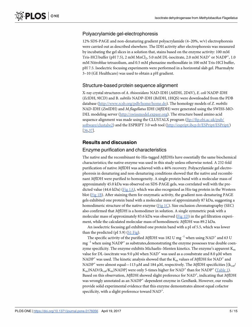
nant MfIDH were purified to homogeneity. A single protein band with a molecular mass of
approximately 45.0 kDa was observed on SDS-PAGE gels, was correlated well with the pre-
dicted value (44.6 kDa) (Fig 1A), which was also recognized as His-tag protein in the Western
blot (Fig 1B). After staining them for enzymatic activity, the gradient non-denaturing PAGE
gels exhibited one protein band with a molecular mass of approximately 87 kDa, suggesting a
homodimeric structure of the native enzyme (Fig 1C). Size exclusion chromatography (SEC)
also confirmed that MfIDH is a homodimer in solution. A single symmetric peak with a
molecular mass of approximately 83.6 kDa was observed (Fig 1D) in the gel filtration experi-
ment, while the calculated molecular mass of homodimeric MfIDH was 89.2 kDa.
An isoelectric focusing gel exhibited one protein band with a pI of 5.5, which was lower
than the predicted (pI 5.9) (S1 Fig).
The specific activity of the purified MfIDH was 182 U mg -1 when using NAD+ and 63 U
mg -1 when using NADP+ as substrates,demonstrating the enzyme possesses true double coen-
zyme specificity. The enzyme exhibits Michaelis–Menten kinetics. The enzyme’s apparent Km
value for DL-isocitrate was 9.0 μM when NAD+ was used as a cosubstrate and 8.0 μM when
NADP+ was used. The kinetic analysis showed that the Km values of MfIDH for NAD+ and
NADP+ were almost equal—113 μM and 184 μM, respectively. The MfIDH specificities [(kcat/
Km)NAD/(kcat/Km)NADP] were only 5-times higher for NAD+ than for NADP+ (Table 1).
Based on this observation, MfIDH showed slight preference for NAD+, indicating that MfIDH
was wrongly annotated as an NADP+-dependent enzyme in GenBank. However, our results
provide solid experimental evidence that this enzyme demonstrates almost equal cofactor
specificity, with a slight preference toward NAD+.
Isocitrate dehydrogenase from Methylobacillus Flagellatus
PLOS ONE | https://doi.org/10.1371/journal.pone.0176056 April 19, 2017 5 / 15
Fig 1. Overexpression, purification and oligomeric state determination of the recombinant MfIDH. (a) The protein purity was
determined using 12% SDS-PAGE. M, protein marker; lane 1, crude extracts of cells harboring plasmid pET-MfIDH after induction with IPTG;
lane 2, purified protein. (b) Detection of MfIDH by Western blot using the anti-6×His antibody as a probe. Lane 1, negative control, crude
extracts of cells harboring pET-15b(+) with IPTG induction; lane 2, purified protein. (c) Gradient non-denaturing PAGE. M, protein marker; lane
1, purified native MfIDH; lane 2, purified recombinant MfIDH; Zymogram assay of the purified proteins. Staining for the NADP+-dependent
activity: lane 3, native MfIDH, lane 4, recombinant MfIDH. Staining for the NAD+-dependent activity: lane 5, native MfIDH, lane 6, recombinant
MfIDH. (d) Molecular mass determination using gel filtration chromatography. The flow rate was 0.5 mL min-1, and the proteins were detected
by monitoring their absorbance at 280 nm. The molecular mass standard curve is inset. The measurement of the recombinant MfIDH is
represented as a dark dot (●). The standard proteins are represented as open circles (�) and are carbonic anhydrase (29 kDa), albumin (66
kDa), alcohol dehydrogenase (150 kDa), β-amylase (200 kDa), apoferritin (443 kDa) and thyroglobulin (669 kDa). The Ve of the recombinant
MfIDH is 13.36 mL.
https://doi.org/10.1371/journal.pone.0176056.g001
Table 1. Kinetic parameters on the activity of MfIDH.
NAD+ NADP+
Km (μM) kcat (s-1) kcat/Km (μM-1s-1) Km (μM) kcat (s-1) kcat/Km (μM-1s-1) Specificity (kcat/Km) NAD/(kcat/Km) NADP
113 166 1.5 184 56 0.3 5
https://doi.org/10.1371/journal.pone.0176056.t001
Isocitrate dehydrogenase from Methylobacillus Flagellatus
PLOS ONE | https://doi.org/10.1371/journal.pone.0176056 April 19, 2017 6 / 15

According to Zhu et al, NAD+ usage is an ancestral trait and NADP+ dependency by pro-
karyotic IDHs emerged near the time that eukaryotic mitochondria first appeared, (some 3.5
billion years ago). The switch of the coenzyme specificity of prokaryotic IDH from NAD+ to
NADP+ is an ancient adaptation to the anabolic demand for NADPH during growth on ace-
tate [1]. The aerobic Gram-negative bacterium M. flagellatus which has an uncoupled TCA
cycle contains an IDH that is specific for both NAD+ and NADP+, which provides flexibility to
use either available cofactor and generate NADH or NADPH.
The Km value of MfIDH for NAD+ (113 μM) is higher than that determined for P. furiosusNAD+-IDH (68 μM) [23], but lower than those of Z. mobilis NAD+-IDH (245 μM), S. suisNAD+-IDH (233 μM), A. thiooxidans NAD+-IDH (184 μM), S.mutans NAD+-IDH (154 μM),
and M. capsulatus NAD+-IDH (122 μM) [16].
The Km value of MfIDH for NADP+ (184 μM) is much higher than those of most homodi-
meric or monomeric IDHs, such as B. subtilis NADP+-IDH (15 μM) [38], E. coli NADP+-IDH
(17 μM) [39], P. nautica NADP+-IDH (25 μM), and S. diastaticus NADP+-IDH (8.5 μM) [18],
but is in the range of those of H. volcanii NADP+-IDH (101 μM) [40] and H. pylori NADP+-
IDH (176 μM) [15]. The Km value of MfIDH for DL-isocitrate (8–9 μM) is within the range
observed for many characterized IDHs [18].
Although the NAD+-linked MfIDH activity has a lower cofactor affinity than its NADP+-
dependent counterparts, its catalytic efficiency (1.5 μM-1 s-1) is very close to those of the IDHs
from E. coli IDH (4.7 μM-1s-1) and B. longum (1.87 μM-1s-1), and is higher than those of the
NAD+-linked IDHs from Z.mobilis IDH (0.46 μM-1s-1) and A. thiooxidans IDH (0.25 μM-1s-1)
[11,26]. In contrast, the catalytic efficiency of the NADP+-linked activity of MfIDH (0.3 μM-1s-1)
is much lower and comparable with the efficiency of NAD+-dependent homodimeric IDHs.
Sequence analysis
The IDH gene inM.flagellatus (MfIDH) is 1242 bp in length and encodes a polypeptide of 413
amino acids. The overall GC content is approximately 56.17% (genome 55.7%), which is similar
to those of the chromosomes ofMethylophilaceae species (37–57%) [41]. The search for regions
that are identical to theMfIDH gene indicated that the highest identity values were with IDHs
from the following organisms: Methylobacillus glycogenes (96%),Methylovorus glucosotrophus(91%), Methylotenera mobilis (87%), Candidatus Methylopumilus turicensis (86%), Methyloteneraversatilis (86%), andMethylophilus methylotrophus (86%). The amino acid identities of MfIDH
with typical homodimeric NADP+-IDHs from E. coli and B. subtilis, and with NAD+-IDHs from
A. thiooxidans and Z.mobilis were 66, 62, 58 and 56%, respectively. The 3D-structure ofMfIDH
was modeled using the AtIDH (2D4V) structure as a template. A secondary- structure-based
alignment revealed that most structural elements that are, involved in the binding of the substrate
and coenzyme are highly conserved within prokaryotic homodimeric type I IDHs (Fig 2).
The interactions between the 2’-phosphate of NADP+ and the amino acid residues Lys344,
Tyr345 and Val351 in EcIDH, and Lys350, Tyr351 and Val357 in BsIDH have been declared
the determinants of NADP+ cofactor specificity [1,42]. The possibility of switching the cofactor
preference was shown experimentally by replacement of the original motif Lys350, Tyr351 and
Val357 in NADP+-IDH of E.coli with the mutated motif Asp350, Ile351 and Ala357 in engi-
neered NAD+-IDH of E.coli [42]. According to Dean and Golding, Imada et al; substitution of
the Lys with an Asp, results in the formation of double hydrogen bonds with the 20- and 30-
hydroxyl groups of the adenosine ribose of NAD+ and the repelling of the negatively charged
2’-phosphate of NADP+ through electrostatic repulsion, which together cause the NAD+ cofac-
tor specificity of AtIDH (Asp357, Ile358 and Ala364) and ZmIDH (Asp348, Ile349 and Ala355)
[42,43].
Isocitrate dehydrogenase from Methylobacillus Flagellatus
PLOS ONE | https://doi.org/10.1371/journal.pone.0176056 April 19, 2017 7 / 15

Furthermore, the amino acid residues Asp328, Ile329 and Ala335 have been declared the
determinants of NAD cofactor specificity in NAD-IDH from Pyrococcus furiosus [23]. The
site-directed mutagenesis experiment that replaced Asp328 with Lys328 in the cofactor dis-
crimination site of the NAD+-IDH from P. furiosus; led to a significant reduction in Km for
NADP (~27fold), whereas the Km for NAD was unaltered and the specificity for NADP was
increased five-fold compared with the wild-type enzyme. This motif–Lys328, Ile329 and
Ala335 results in a double coenzyme specificity of chimeric P.furiosus IDH. The introduction
of the double replacement of Asp-328–Lys/Ile-329–Tyr (motif Lys328, Tyr329 and Ala347) has
not changed the efficiency of NADP-IDH, but rather slightly increased both Km and Kcat for
NADP. The kcat was unaltered compared with the single-mutated enzyme [23]. The structure-
based alignment revealed that there is the same motif—Lys340, Ile341 and Ala347 in naturally
occurred MfIDH. Thus, signature residues involved in substrate discrimination in MfIDH
Fig 2. Structure-based sequence alignment of MfIDH with other dimeric IDHs. High-resolution crystal
structures of the A. thiooxidans NAD-IDH (AtIDH, 2D4V), B. subtilis NADP-IDH (BsIDH, 1HQS) and E. coli
NADP-IDH (EcIDH, 9ICD) were downloaded from the PDB database. The MfIDH model of the IDH from M.
flagellatus and the ZmIDH model of the NAD-IDH from Z. mobilis were generated using the SWISS-MODEL
modeling server with AtIDH structure as the template. The secondary structure of MfIDH is depicted above
the alignment. The completely conserved amino acids are highlighted as shaded red boxes. The conserved
residues involved in cofactor- (●) and substrate-binding (▲) are indicated, respectively. The conserved
phosphorylation sites are indicated (■). The lysine residues that may be acetylated in MfIDH and EcIDH are
highlighted with shades light-blue and pink boxes, respectively. The major cofactor specificity determinants
are highlighted with shaded yellow boxes and indicated with stars ($). The alignment was drawn with
ESPRIPT 3.0.
https://doi.org/10.1371/journal.pone.0176056.g002
Isocitrate dehydrogenase from Methylobacillus Flagellatus
PLOS ONE | https://doi.org/10.1371/journal.pone.0176056 April 19, 2017 8 / 15

appeared to be Lys340, Ile341 and Ala347 (Fig 2). The ability to use efficiently both NAD+ and
NADP+ as cofactors is caused by the presence of these three key amino acids in the protein
structure of MfIDH. The MfIDH was incorrectly annotated as NADP+ specific isocitrate dehy-
drogenase. We suggest that MfIDH can be annotated as a homodimeric type I isocitrate dehy-
drogenase with dual coenzyme specificity.
Post-translational modifications are one of the most efficient biological mechanisms for
regulating enzyme activity and cellular physiology. The activity of EcIDH is regulated by an
IDH-kinase/phosphatase (aceK) that responds to changes in the metabolic environment [44].
Although phosphorylation sites are conserved in EcIDH (Ser113), AtIDH (Ser113), BsIDH
(Ser104), ZmIDH (Ser102) and MfIDH (Ser114), no corresponding IDH-kinase/phosphatase
gene was found in the genome of M. flagellatus.It was recently found that lysine acetylation [45–47] as well as succinylation [48,49] activi-
ties are abundant in E. coli and might be involved in modifying or regulating the activities of
enzymes involved in the synthesis of building blocks in response to environmental changes
and critical metabolic processes. Six acetylation sites were found in EcIDH [47]. Zhang et al,
by mimic mutagenesis demonstrated that both Lys100 and Lys242 are important for the activ-
ity of EcIDH and that lysine succinylation is likely to inhibit or abolish its enzymatic function
[48]. Analog sites corresponding to the lysine acetylation sites of EcIDH (Lys142, Lys177,
Lys230, Lys242, Lys265, Lys378) [47] are also found in MfIDH (Lys143, Lys231, Lys243,
Lys266). Analogues lysine succinylation sites of EcIDH (Lys100, Lys186, Lys199, Lys230,
Lys235, Lys242, Lys387) [49] were also conserved in MfIDH (Lys101, Lys187, Lys200, Lys231,
Lys236, Lys243, Lys383) (Fig 2), although there is no evidence that MfIDH can be regulated by
acetylation or succinylation in vivo.
Effects of pH and temperature
The effects of the pH on the MfIDH activity were determined for the NAD+- and NADP+-
linked reactions in the presence of Mn2+. Surprisingly, MfIDH exhibited a strict cofactor-
dependent pH-activity profile, which has never been described in the literatur. The results dem-
onstrate that the optimum pH is 8.5 with NAD+ and 6.0 with NADP+ (Fig 3A). For NAD+-
linked activity, this value is similar to those of the Z. mobilis (pH 8.5) [26] and A. thiooxidansNAD+-IDHs (pH 8.5) [21], but is lower than that of the H. thermophilus NAD+-IDH (pH 10.5)
[24]. For the NADP+-linked activity, this pH value is rather similar to that of the IDH from the
acidophilic fungus A. niger (pH 6.0–8.0) [50]. The temperature for maximum activity MfIDH is
approximately 60˚C, which is similar to those of the B. longum IDH (60˚C) [51] and L. interro-gan IDH (60˚C) [12], but higher than that of the E. coli IDH (50˚C) [22] (Fig 3B). Heat-inactiva-
tion studies revealed that the MfIDH is remarkably thermostable, retaining its full activity at
50˚C and losing ca. 50% of its activity after one hour of incubation at 75˚C (Fig 3C). Thus, the
thermostability of MfIDH is closer to that of IDHs from thermophiles rather than mesophiles
[52]. The increased thermostability of MfIDH may be explained by its possessing twofold fewer
Cys residues than EcIDH does (0.70 and 1.40%, respectively); having fewer Cys residues is a
common trend for thermophilic proteins [22]. The aromatic cluster in the clasp domain has
previously been observed in the IDHs of hyperthermophilic A. fulgidus and A. pernix, and is
believed to stabilize the interface [22]. The aromatic cluster of the A. fulgidus IDH contains
Phe179, which is substituted by the nonpolar residue Met in typical mesophilic IDHs, e.g.,
Met183 (AtIDH), Met183 (EcIDH) and Met172 (ZmIDH). Interestingly, MfIDH has a polar res-
idue (Gly184) at the same position (Fig 2), which is typical for methylotrophic IDHs, e.g., M.
glycogenes, M. glucosotrophus, M. mobilis, M. versatilis and others. The role of Gly in the clasp
stabilization at elevated temperature requires further investigation.
Isocitrate dehydrogenase from Methylobacillus Flagellatus
PLOS ONE | https://doi.org/10.1371/journal.pone.0176056 April 19, 2017 9 / 15

Effects of metal ions on MfIDH activity
The effects of different cations on the MfIDH activity were studied, and the results indicate
that MfIDH retaines ca. 18% of its activity even without the addition of divalent ions (Table 2).
This behavior is unusual for most IDHs, whose actvities entirely depend on the binding of a
divalent cation [3]. Mn2+ was found to be the ion that most effectively enhances the enzyme’s
activity, although Mg2+ can act as a significant substitute by providing up to 58–75% of the
enzyme’s maximal activity. Whereas the MfIDH activity is completely inhibited by Ca2+ and
Cu2+, it is entirely restored by the addition of 2 mM of Mn2+ (data not shown). Although most
IDHs are strongly inhibited by Zn2+, we observed a very interesting effect that Zn2+ addition
has on MfIDH activity. NAD+-linked activity at pH 8.5 was strongly inhibited by Zn2+ but par-
tially restored at pH 6.0.
In contrast, the NADP+-linked activity at pH 8.5 was fully activated by the presence of Zn2+
but decreased by half at pH 6.0, similar to the NAD+-linked activity. Thus, the pH optimum of
Fig 3. Effects of pH and temperature on the activity of MfIDH. (a) The effects of pH on the NAD+-dependent (●) and NADP+-dependent (�) activities of
MfIDH from pH 5.0 to 10.0 in the presence of Mn2+. (b) The effects of temperature on NAD+-dependent (●) and NADP+-dependent (�) activities of MfIDH
from 45 to 65˚C. (c) Heat-inactivation profiles of NAD+-dependent (●) and NADP+-dependent (�) activities of MfIDH incubated at 50 to 80˚C. The
incubation time is 60 min.
https://doi.org/10.1371/journal.pone.0176056.g003
Table 2. Effect of metal ions on the activity of MfIDH.
Metal ions Relative activity (%)
NAD+ NADP+
None 18.0 ± 3.0 17.0 ± 3.5
Mn2+ 100.0 ± 2.9* 100.0 ± 3.7*
Mg2+ 75.0 ± 1.5 58.0 ± 2.0
Ca2+ 0 0
Cu2+ (pH 8.5) 10.0 ± 3.5 8.0 ± 2.5
Cu2+ (pH 6.0) 0 0
Zn2+ (pH 8.5) 3.5 ± 2.5 100.0 ± 3.0
Zn2+ (pH 7.0) 25.0 ± 2.0 25.0 ± 2.5
Zn2+ (pH 6.0) 50.0 ± 1.5 50.0 ± 2.7
Activity of pure MfIDH was determined with 2 mM metal ions in the standard reaction mixture at pH optimum,
unless otherwise specified.
* A 100% activity corresponds to 182 U mg -1 with NAD+ and 63 U mg -1 with NADP+.
https://doi.org/10.1371/journal.pone.0176056.t002
Isocitrate dehydrogenase from Methylobacillus Flagellatus
PLOS ONE | https://doi.org/10.1371/journal.pone.0176056 April 19, 2017 10 / 15

the NADP+-linked MfIDH activity drastically changed from pH 6.0 (Mn2+) to pH 8.5 (Zn2+).
Interaction with Zn2+ can modulate the MfIDH activity in an interesting manner. Because all
the metal binding sites are highly conserved in MfIDH (Fig 2), there is no plausible explana-
tion for these phenomena.
Effects of analogous cofactors on the MfIDH activity
The effects of different cofactor analogs on the MfIDH activity were examined (Table 3). More
than half of the NAD+- or NADP+-linked activity was retained when the amide group of the
nicotinamide ring was replaced by the acetyl group in 3-acetylpyridine adenine dinucleotide
(phosphate); thus, the amide group is not indispensable for binding. In contrast, the substitu-
tion of the N6 amino group of the adenine ring with the oxo-group in nicotinamide hypoxan-
thine dinucleotide (phosphate) completely abolished NAD(P)+ binding. To our knowledge,
this is the first report that demonstrates the indispensability of the amino group of adenine in
cofactor recognition. Imada et al. thoroughly studied amino acid residues that are involved in
the recognition of the adenine and nicotinamide rings of the cofactor [43]. The adenine N6
atom is hydrogen-bonded with the carbonyl oxygen of Asn-348 and has amino–aromatic
hydrogen-bond interactions with the imidazole ring of His-335, which are conserved interac-
tions in the type I IDHs (Fig 2).
Substrate specificity and inhibition
No appreciable effect on the activity of MfIDH was observed upon addition of the following
compounds (at final concentrations of 5 mM, unless noted) to the reaction mixture: glutamate,
glutamine, α-ketoglutarate, oxaloacetate, cis-aconitate, citrate, pyruvate, malate, fumarate,
succinate, ADP (2 mM), AMP (2 mM), CoA, AcCoA, NADH, and NADPH (at a final concen-
tration of 0.2 mM). ATP (2 mM) caused 50% inhibition of only the NADP+-linked MfIDH
activity. Thus, MfIDH activity is not regulated at the metabolic level, as has been demonstrated
for IDHs from organisms with a complete TCA cycle.
Conclusions
The isocitrate dehydrogenase from M. flagellatus was purified, overexpressed and character-
ized in the present study. Our data reveal that MfIDH exhibits unique double coenzyme speci-
ficity toward both NAD+ and NADP+ cofactors, and its activity is dependent on divalent
cations. MfIDH exhibits a strict cofactor-dependent pH-activity profile. Our study also shows
Table 3. Effect of cofactor analogous on the activity of MfIDH.
Cofactor Relative activity (%)
NAD+ 100.0 ± 2.5*
APAD+ 55.0 ± 2.5
NHD+ 4.0 ± 1.2
NADP+ 100.0 ± 2.8*
APADP+ 60.0 ± 2.5
NHDP+ 2.0 ± 1.9
APAD(P)+, 3-Acetylpyridine adenine dinucleotide (Phosphate); NHD(P)+, Nicotinamide hypoxanthine
dinucleotide (Phosphate). Activity of pure MfIDH was determined with 0.4 mM cofactor analogous in the
standard reaction mixture at pH optimum.
* A 100% activity corresponds to 182 U mg -1 with NAD+ and 63 U mg -1 with NADP+.
https://doi.org/10.1371/journal.pone.0176056.t003
Isocitrate dehydrogenase from Methylobacillus Flagellatus
PLOS ONE | https://doi.org/10.1371/journal.pone.0176056 April 19, 2017 11 / 15

that MfIDH is remarkably thermostable and is not regulated at the metabolic level. We suggest
the major amino acids in the protein structure of MfIDH that determine the double cofactor
specificity. The enzymatic characterization of MfIDH can enrich our knowledge of type I
IDHs and might be useful for the engineering of IDHs with desirable specificities.
Supporting information
S1 Fig. Isoelectric focusing of the native MfIDH. The determination of the isoelectric point
of the native MfIDH. M, pI markers; lane 1, purified protein.
(TIFF)
S1 Table. Summary of the purification of the native MfIDH.
(DOCX)
Acknowledgments
We thank Smirnov SV for helping with the purification and protein mass determination of the
recombinant MfIDH.
Author Contributions
Conceptualization: AYR MYK.
Data curation: AYR MYK.
Formal analysis: AYR MYK.
Investigation: AYR MYK.
Methodology: AYR MYK.
Project administration: AYR MYK.
Resources: AYR MYK.
Software: AYR MYK.
Supervision: MYK.
Validation: AYR MYK.
Visualization: AYR MYK.
Writing – original draft: AYR MYK.
Writing – review & editing: AYR MYK.
References1. Zhu G, Golding GB, Dean AM. The selective cause of an ancient adaptation. Science. 2005; 307:
1279–1282. https://doi.org/10.1126/science.1106974 PMID: 15653464
2. Shimizu T, Yin L, Yoshida A, Yokooji Y, Hachisuka S, Sato T, et al. Structure and function of an ances-
tral-type β-decarboxylating dehydrogenase from Thermococcus kodakarensis. Biochem J. 2016; 105–
122. https://doi.org/10.1042/BCJ20160699 PMID: 27831491
3. Wang P, Lv C, Zhu G. Novel type II and monomeric NAD+ specific isocitrate dehydrogenases: phyloge-
netic affinity, enzymatic characterization, and evolutionary implication. Nat Sci reports. 2015; 5: 1–11.
4. Wu M-C, Tian C-Q, Cheng H-M, Xu L, Wang P, Zhu G-P. A novel type II NAD+-specific isocitrate dehy-
drogenase from the marine bacterium Congregibacter litoralis KT71. PLoS One. 2015; 10: e0125229.
https://doi.org/10.1371/journal.pone.0125229 PMID: 25942017
Isocitrate dehydrogenase from Methylobacillus Flagellatus
PLOS ONE | https://doi.org/10.1371/journal.pone.0176056 April 19, 2017 12 / 15

5. Banerjee S, Nandyala A, Podili R, Katoch VM, Hasnain SE. Comparison of Mycobacterium tuberculosis
isocitrate dehydrogenases (ICD-1 and ICD-2) reveals differences in coenzyme affinity, oligomeric state,
pH tolerance and phylogenetic affiliation. BMC Biochem. 2005; 6: 20. https://doi.org/10.1186/1471-
2091-6-20 PMID: 16194279
6. Ishii A, Suzuki M, Sahara T, Takada Y, Sasaki S, Fukunaga N. Genes encoding two isocitrate dehydro-
genase isozymes of a psychrophilic bacterium, Vibrio Genes Encoding Two Isocitrate Dehydrogenase
Isozymes of a Psychrophilic Bacterium, Vibrio sp. Strain ABE-1. J Bacteriol. 1993; 175: 6873–6880.
PMID: 8226630
7. Matsuo S, Shirai H, Takada Y. Isocitrate dehydrogenase isozymes from a psychrotrophic bacterium,
Pseudomonas psychrophila. Arch Microbiol. 2010; 192: 639–650. https://doi.org/10.1007/s00203-010-
0595-3 PMID: 20549192
8. Suzuki K, Takada Y. Characterization of NADP +-dependent isocitrate dehydrogenase isozymes from a
psychrophilic bacterium, Colwellia psychrerythraea strain 34H. Biosci Biotechnol Biochem. 2016; 80:
1492–1498. https://doi.org/10.1080/09168451.2016.1165602 PMID: 27033696
9. Maki S, Yoneta M, Takada Y. Two isocitrate dehydrogenases from a psychrophilic bacterium, Colwellia
psychrerythraea. Extremophiles. 2006; 10: 237–249. https://doi.org/10.1007/s00792-005-0493-9
PMID: 16418792
10. Lv C, Wang P, Wang W, Su R, Ge Y, Zhu Y, et al. Two isocitrate dehydrogenases from a plant pathogen
Xanthomonas campestris pv. campestris 8004. Bioinformatic analysis, enzymatic characterization, and
implication in virulence. J Basic Microbiol. 2016; 56: 975–985. https://doi.org/10.1002/jobm.201500648
PMID: 27282849
11. Huang SP, Cheng HM, Wang P, Zhu GP. Biochemical characterization and complete conversion of
coenzyme specificity of isocitrate dehydrogenase from Bifidobacterium longum. Int J Mol Sci. 2016; 17:
1–2.
12. Zhao X, Wang P, Zhu G. Enzymatic Characterization of a Type II Isocitrate Dehydrogenase from Patho-
genic Leptospira interrogans serovar Lai Strain 56601. Appl Biochem Biotechnol. 2014; 487–496.
https://doi.org/10.1007/s12010-013-0521-7 PMID: 24092452
13. Jin MM, Wang P, Li X, Zhao XY, Xu L, Song P, et al. Biochemical characterization of NADP+-dependent
isocitrate dehydrogenase from Microcystis aeruginosa PCC7806. Mol Biol Rep. 2013; 40: 2995–3002.
https://doi.org/10.1007/s11033-012-2371-8 PMID: 23264072
14. Prasad UV, Vasu D, Kumar YN, Kumar PS, Yeswanth S, Swarupa V, et al. Cloning, Expression and
Characterization of NADP-Dependent Isocitrate Dehydrogenase from Staphylococcus aureus. Appl
Biochem Biotechnol. 2013; 169: 862–869. https://doi.org/10.1007/s12010-012-0027-8 PMID:
23288593
15. Huang D, Liu J, Shen G. Cloning, expression, and enzymatic characterization of isocitrate dehydroge-
nase from Helicobacter pylori. Protein J. 2009; 28: 443–447. https://doi.org/10.1007/s10930-009-9212-
1 PMID: 19921412
16. Stokke R, Madern D, Fedøy AE, Karlsen S, Birkeland NK, Steen IH. Biochemical characterization of iso-
citrate dehydrogenase from Methylococcus capsulatus reveals a unique NAD+-dependent homotetra-
meric enzyme. Arch Microbiol. 2007; 187: 361–370. https://doi.org/10.1007/s00203-006-0200-y PMID:
17160675
17. Steen IH, Madern D, Karlstrom M, Lien T, Ladenstein R, Birkeland NK. Comparison of Isocitrate Dehy-
drogenase from Three Hyperthermophiles Reveals Differences in Thermostability, Cofactor Specificity,
Oligomeric State, and Phylogenetic Affiliation. J Biol Chem. 2001; 276: 43924–43931. https://doi.org/
10.1074/jbc.M105999200 PMID: 11533060
18. Zhang BB, Wang P, Wang A, Wang WC, Tang WG, Zhu GP. Expression and characterization of a
novel isocitrate dehydrogenase from Streptomyces diastaticus No. 7 strain M1033. Mol Biol Rep. 2013;
40: 1615–1623. https://doi.org/10.1007/s11033-012-2210-y PMID: 23073782
19. Wang A, Cao ZY, Wang P, Liu AM, Pan W, Wang J, et al. Heteroexpression and characterization of a
monomeric isocitrate dehydrogenase from the multicellular prokaryote Streptomyces avermitilis MA-
4680. Mol Biol Rep. 2011; 38: 3717–3724. https://doi.org/10.1007/s11033-010-0486-3 PMID:
21104016
20. Kanao T, Kawamura M, Fukui T, Atomi H, Imanaka T. Characterization of isocitrate dehydrogenase
from the green sulfur bacterium Chlorobium limicola: A carbon dioxide-fixing enzyme in the reductive tri-
carboxylic acid cycle. Eur J Biochem. 2002; 269: 1926–1931. PMID: 11952794
21. Inoue H, Tamura T, Ehara N, Nishito A. Biochemical and molecular characterization of the NAD+-
dependent isocitrate dehydrogenase from the chemolithotroph Acidithiobacillus thiooxidans. FEMS
Microbiol Lett. 2002; 214:127–232. PMID: 12204383
22. Stokke R, Karlstrom M, Yang N, Leiros I, Ladenstein R, Birkeland NK, et al. Thermal stability of isoci-
trate dehydrogenase from Archaeoglobus fulgidus studied by crystal structure analysis and engineering
Isocitrate dehydrogenase from Methylobacillus Flagellatus
PLOS ONE | https://doi.org/10.1371/journal.pone.0176056 April 19, 2017 13 / 15

of chimers. Extremophiles. 2007; 11: 481–493. https://doi.org/10.1007/s00792-006-0060-z PMID:
17401542
23. Steen I, Lien T, Madsen M, Birkeland NK. Identification of cofactor discrimination sites in NAD-isocitrate
dehydrogenase from Pyrococcus furiosus. Arch Microbiol. 2002; 178: 297–300. https://doi.org/10.1007/
s00203-002-0439-x PMID: 12209263
24. Aoshima M, Ishii M, Igarashi Y. A novel biotin protein required for reductive carboxylation of 2-oxogluta-
rate by isocitrate dehydrogenase in Hydrogenobacter thermophilus TK-6. Mol Microbiol. 2004; 51: 791–
798. PMID: 14731279
25. Wang P, Jin M, Su R, Song P, Wang M, Zhu G. Enzymatic characterization of isocitrate dehydrogenase
from an emerging zoonotic pathogen Streptococcus suis. Biochimie. Elsevier Masson SAS; 2011; 93:
1470–1475. https://doi.org/10.1016/j.biochi.2011.04.021 PMID: 21586311
26. Wang P, Jin M, Zhu G. Biochemical and molecular characterization of NAD+-dependent isocitrate dehy-
drogenase from the ethanologenic bacterium Zymomonas mobilis. FEMS Microbiol Lett. 2012; 327:
134–41. https://doi.org/10.1111/j.1574-6968.2011.02467.x PMID: 22117777
27. Wang P, Song P, Jin M, Zhu G. Isocitrate Dehydrogenase from Streptococcus mutans: Biochemical
Properties and Evaluation of a Putative Phosphorylation Site at Ser102. PLoS One. 2013; 8: 1–8.
28. Lloyd AJ, Weitzman PDJ. Purification and characterization of NAD-linked isocitrate dehydrogenase
from Methylophilus methylotrophus. Biochemical Society Transactions. 1988; 16: 871–872.
29. Hofmann KH, Babel W. Regulation of NAD+- and NADP+-linked isocitrate dehydrogenase in the obli-
gate methylotrophic bacterium Pseudomonas W6. Zeitschrift fur Allg Mikrobiol. 1980; 20: 399–404.
30. Anthony C. The biochemistry of methylotrophs. London: Academic. 1983.
31. Methylobacillus flagellatus Govorukhina et al. ATCC® 51484TM [Internet]. [cited 18 Jan 2017]. Avail-
able: https://www.lgcstandards-atcc.org/products/all/51484.aspx?geo_country=ru#history
32. Chistoserdova L, Lapidus A, Han C, Goodwin L, Saunders L, Brettin T, et al. Genome of Methylobacillus
flagellatus, molecular basis for obligate methylotrophy, and polyphyletic origin of methylotrophy. J Bac-
teriol. 2007; 189: 4020–4027. https://doi.org/10.1128/JB.00045-07 PMID: 17416667
33. Baev M V., Kiriukhin MY, Tsygankov YD. Regulation of ammonia assimilation in an obligate methylo-
troph Methylobacillus flagellatum under steady-state and transient growth conditions. Antonie van
Leeuwenhoek, Int J Gen Mol Microbiol. 1997; 71: 353–361.
34. Kiriukhin MY, Detkov SY, Baev M V, Tsygankov YD. NADP + -dependent glutamate dehydrogenase
from the obligate methylotroph Methylobacillus flagellatum. FEMS Microbiol Lett. 1992; 93: 155–160.
35. Kiriukhin MY, Detkov SY, Baev MV, Tsygankov YD. Citrate synthase from the obligate methylotroph
Methylobacillus flagellatum. FEMS Microbiol Lett. 1993; 113: 101–105.
36. Gouet P, Courcelle E, Stuart DI, Metoz F. ESPript: analysis of multiple sequence alignments in Post-
Script. Bioinformatics. 1999; 15: 305–8. PMID: 10320398
37. Larkin MA, Blackshields G, Brown NP, Chenna R, Mcgettigan PA, McWilliam H, et al. Clustal W and
Clustal X version 2.0. Bioinformatics. 2007; 23: 2947–2948. https://doi.org/10.1093/bioinformatics/
btm404 PMID: 17846036
38. Singh SK, Miller SP, Dean A, Banaszak LJ, Laporte DC. Bacillus subtilis isocitrate dehydrogenase. A
substrate analogue for Escherichia coli isocitrate dehydrogenase kinase/phosphatase. J Biol Chem.
2002; 277: 7567–7573. https://doi.org/10.1074/jbc.M107908200 PMID: 11751849
39. Chen R, Yang H. A highly specific monomeric isocitrate dehydrogenase from Corynebacterium glutami-
cum. Arch Biochem Biophys. 2000; 383: 238–245. https://doi.org/10.1006/abbi.2000.2082 PMID:
11185559
40. Rodrıguez-Arnedo A, Camacho M, Llorca F, Bonete MJ. Complete reversal of coenzyme specificity of
isocitrate dehydrogenase from Haloferax volcanii. Protein J. 2005; 24: 259–266. https://doi.org/10.
1007/s10930-005-6746-8 PMID: 16284723
41. Lapidus A, Clum A, LaButti K, Kaluzhnaya MG, Lim S, Beck DAC, et al. Genomes of three methylo-
trophs from a single niche reveal the genetic and metabolic divergence of the methylophilaceae. J Bac-
teriol. 2011; 193: 3757–3764. https://doi.org/10.1128/JB.00404-11 PMID: 21622745
42. Dean AM, Golding GB. Protein engineering reveals ancient adaptive replacements in isocitrate dehy-
drogenase. Proc Natl Acad Sci U S A. 1997; 94: 3104–3109. PMID: 9096353
43. Imada K, Tamura T, Takenaka R, Kobayashi I, Namba K, Inagaki K. Structure and quantum chemical
analysis of NAD+-dependent isocitrate dehydrogenase: hydride transfer and co-factor specificity. Pro-
teins. 2008; 70: 63–71. https://doi.org/10.1002/prot.21486 PMID: 17634983
44. el-Mansi EM. Control of metabolic interconversion of isocitrate dehydrogenase between the catalytically
active and inactive forms in Escherichia coli. FEMS Microbiol Lett. 1998; 166: 333–9. PMID: 9770290
Isocitrate dehydrogenase from Methylobacillus Flagellatus
PLOS ONE | https://doi.org/10.1371/journal.pone.0176056 April 19, 2017 14 / 15

45. Zhang K, Zheng S, Yang JS, Chen Y, Cheng Z. Comprehensive profiling of protein lysine acetylation in
Escherichia coli. J Proteome Res. 2013; 12: 844–51. https://doi.org/10.1021/pr300912q PMID:
23294111
46. Zhang J, Sprung R, Pei J, Tan X, Kim S, Zhu H, et al. Lysine acetylation is a highly abundant and evolu-
tionarily conserved modification in Escherichia coli. Mol Cell Proteomics. American Society for Bio-
chemistry and Molecular Biology; 2009; 8: 215–25. https://doi.org/10.1074/mcp.M800187-MCP200
PMID: 18723842
47. Yu BJ, Kim JA, Moon JH, Ryu SE, Pan J-G. The diversity of lysine-acetylated proteins in Escherichia
coli. J Microbiol Biotechnol. 2008; 18: 1529–36. PMID: 18852508
48. Zhang Z, Tan M, Xie Z, Dai L, Chen Y, Zhao Y. Identification of lysine succinylation as a new post-trans-
lational modification. Nat Chem Biol. 2011; 7: 58–63. https://doi.org/10.1038/nchembio.495 PMID:
21151122
49. Weinert BT, Scholz C, Wagner SA, Iesmantavicius V, Su D, Daniel JA, et al. Lysine succinylation is a
frequently occurring modification in prokaryotes and eukaryotes and extensively overlaps with acetyla-
tion. Cell Rep. 2013; 4: 842–851. https://doi.org/10.1016/j.celrep.2013.07.024 PMID: 23954790
50. Meixner-Monori B, Kubicek CP, Harrer W, Schreferl G, Rohr M. NADP-specific isocitrate dehydroge-
nase from the citric acid-accumulating fungus Aspergillus niger. Biochem J. 1986; 236: 549–57. PMID:
3753466
51. Zhang G, Gao B, Adeolu M, Khadka B, Gupta RS. Phylogenomic Analyses and Comparative Studies
on Genomes of the Bifidobacteriales: Identification of Molecular Signatures Specific for the Order Bifido-
bacteriales and Its Different Subclades. Front Microbiol. 2016; 7: 1–17.
52. Karlstrom M, Steen IH, Madern D, Fedoy AE, Birkeland NK, Ladenstein R. The crystal structure of a
hyperthermostable subfamily II isocitrate dehydrogenase from Thermotoga maritima. FEBS J. 2006;
273: 2851–2868. https://doi.org/10.1111/j.1742-4658.2006.05298.x PMID: 16759231
Isocitrate dehydrogenase from Methylobacillus Flagellatus
PLOS ONE | https://doi.org/10.1371/journal.pone.0176056 April 19, 2017 15 / 15