Embed Size (px)
Citation preview
Chapter 10
© 2012 Butnaru et al., licensee InTech. This is an open access chapter distributed under the terms of the Creative Commons Attribution License (http://creativecommons.org/licenses/by/3.0), which permits unrestricted use, distribution, and reproduction in any medium, provided the original work is properly cited.
Biocompatibility and Biological Performance of the Improved Polyurethane Membranes for Medical Applications
Maria Butnaru, Ovidiu Bredetean, Doina Macocinschi, Cristina Daniela Dimitriu, Laura Knieling and Valeria Harabagiu
Additional information is available at the end of the chapter
http://dx.doi.org/10.5772/34653
1. Introduction
Polyurethanes (PUs) are one of the most “pluripotent” synthetic polymer classes used in
medical applications. Due to their structural versatility, they have been widely discussed as
materials appropriate for biomedical applications (Abd El-Rehim & El-Amaouty, 2004;
Guelcher et. al., 2007; Guelcher, 2008; Kavlock et. al, 2007; J.S. Lee et. al., 2001; Lelah &
Cooper, 1987; Siepe et. al., 2007). Up to now, new PUs have been synthesized that possess
good mechanical properties. Most of them are considered biocompatible on account of in
vitro cytotoxicity evaluation.
However, it is well known that structural and mechanical adaptability of PUs is not always
accompanied by cell and tissue biocompatibility. Therefore, numerous data in the literature
are focused on biocompatibilization or functionalization of PUs (Yao, 2008; Sartori, 2008,
Huang & Xu, 2010). Some promising methods for the improvement of biological response of
PUs are conjugation, blending or coating with natural polymers. Thus, polysaccharides as
chitosan, cellulose and their derivatives (Raschip, 2009; Zia, 2009; Zuo, 2009), proteins and
glycoproteins as collagen, fibrin, fibronectin (R. Chen et. al., 2010; Sartori et. al., 2008),
proteoglycans and glycosaminoglycans (Gong et. al., 2010) and other molecules (Hwang &
Meyerhoff, 2008; Hsu et. al., 2004; Makala et. al., 2006; Song et. al., 2005; Verma & Marsden,
2005) are employed successfully for PUs modification. Owing its specific properties,
hydroxypropylcellulose (HPC) is already used as binder, thickener, lubricating material
(artificial tears) and emulsion stabilizer in pharmaceutical and food industry. Moreover,
HPC may provide interactions through its hydroxyl radicals, being an excellent compound
for copolymerization in scaffolds for tissue engineering and in drug delivery systems
(Berthier et. al., 2011; D. Chen & Sun, 2000; Gutowska et. al., 2001; Raschip et. al., 2009;
Polyurethane 202
Valenta & Auner, 2004). In previous studies we found that when added to PU structure,
HPC improves hydrophilicity and mechanical properties of PUs by increasing the elasticity
of the resulted materials (Macocinschi et. al., 2009).
Considering the reviewed concept of biocompatibility as “the ability to exist in contact with
tissues of the human body without causing an unacceptable degree of harm to the body”
(Williams, 2008), our interdisciplinary work was focused on the synthesis of PU-based
materials with improved ability to long-time functional integration. PU/HPC membranes
were prepared by blending method. HPC was chosen due to its physical-chemical
properties, its demonstrated biocompatibility and accessibility. The aim of the chapter is to
highlight the most important criteria, able to predict the behaviour of material-tissue
interfaces and the long-term material-tissue integration, in order to select most suitable
compositions and morphologies for specific medical application. Thus, surface zeta (ζ)
potential, wettability (as contact angle measurement and water uptake), pH modification
after long time hydration and autoclaving, protein adsorption at protein physiological
concentration and some relevant elements of bulk and surface morphology are treated as
screening criteria for suitable membrane choice in the first part of the chapter. Biological
performance evaluation, such as oxidative stress action, thrombogenicity and in vivo
behaviour of PU/HPC membranes are further discussed.
2. Materials and methods
2.1. Preparation of polymer samples
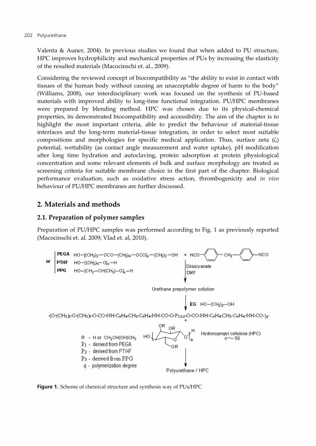
Preparation of PU/HPC samples was performed according to Fig. 1 as previously reported
(Macocinschi et. al. 2009; Vlad et. al, 2010).
Figure 1. Scheme of chemical structure and synthesis way of PUs/HPC
Biocompatibility and Biological Performance of the Improved Polyurethane Membranes for Medical Applications 203
Briefly, isocyanate terminated urethane prepolymers were first synthesized by the
polyaddition reactions between 4',4’-diphenylmethane diisocyanate (MDI) and macrodiols in
N,N-dimethylformamide (DMF) as solvent. Poly(ethylene adipate)diol (PEGA, Mn = 2000
g/mol), polytetrahydrofuran (PTHF, Mn = 2000 g/mol) or poly(propylene)glycol (PPG, Mn =
2000 g/mol) were used as macrodiols. The urethane prepolymers were treated in a subsequent
step with ethylene glycol (EG) as chain extender. Finally, HPC (average weight molecular
weight Mw = 95 000 g/mol) was added to PU solutions to obtain the following compositions
for all PU/HPC samples: macrodiol/MDI/EG/HPC = 52.24 /36.57/7.27/3.92 (weight ratios). As
the molar ratio between isocyanate groups in MDI and the sum of hydroxylic groups in
macrodiol and EG was 1.02, the excess of isocyanate groups linked to PU prepolymers were
available to bind a part of HPC chains. Membranes with about 1 mm thickness were prepared
by pouring PU/HPC DMF solutions in distilled water, at 40 oC. The formed films were then
dried under vacuum for several days and kept in distilled water for solvent removing.
To half of PUs with PEGA macrodiol in the soft segment no HPC was added to obtain PU-
PEGA reference sample. HPC containing samples based on PEGA, PTHF and PPG
macrodiols were codified as PU-PEGA/HPC; PU-PTHF/HPC and PU-PPG/HPC,
respectively.
2.2. ζ potential determination
ζ potential of the PU membranes was measured by streaming potential method using a
commercial electrokinetic analyzer SurPASS, (Anton Paar GmbH, Graz, Austria). For each
sample, ζ potential has been measured in 0.1 M NaCl solution at physiological 7.4 pH value,
a 300 mbar electrolyte pressure and a 80 ml/min flow rate. For statistical reasons, four
streaming potentials were measured. The mean value of these data was used for potential
calculation by Fairbrother–Mastin equation, considering also the effect of surface
conductivity (Luxbacher, 2006)
2.3. Wettability
Wettability of the PU membranes was determined by measuring the surface contact angle
and water uptake. For surface contact angle, uniform drops of the tested liquid (double-
distilled water) with a volume of 2 μl were deposited on the film surface and the contact
angles were measured after 30 s, using a video-based optical contact angle measuring device
equipped with a Hamilton syringe in a temperature-controlled environmental chamber. All
measurements were performed at room temperature of 25 C. Repeated measurements of a
given contact angle were all within the range of ± 3 degrees. Water uptake was calculated as
the ratio between fully hydrated and dried sample weights.
2.4. Material extraction in a simulated biological microenvironment
Material extraction in a simulated biological microenvironment was done for long period of
time (over 2 months) in Hank’s Balanced Salt Solution (HBSS) without Ca2+ and Mg2+, with
Polyurethane 204
glucose, and phenol red as pH indicator. For extraction experiments, 0.2 g of each
membrane, cut in very small pieces (see Fig. 2), were incubated in 2 ml of HBSS solution at
37oC. pH variation was monitored daily, based on phenol red indicator colour and
measured after 1, 2, 3, 30 and 60 days of incubation using Mettler Toledo SevenGo SG2ELK
pH-meter.
2.5. Scanning Electron Microscopy (SEM)
SEM analysis of PU/HPC membrane cross-sections was performed using a VEGA TESCAN
microscope, in high vacuum mode, at an acceleration voltage of 30 kV.
2.6. Protein adsorption
Amount of protein adsorption on membrane surfaces was measured in three different
conditions: (a) on individual protein solutions of fibrinogen (FB) at 3 mg/ml (95% clotable
from Sigma-Aldrich) and serum albumin (SA) at 45 mg/ml (bovine SA (BSA) from Sigma-
Aldrich); (b) FB and BSA mixed solutions of physiological concentrations (3 mg/ml for BSA
and 45 mg/ml for FB); (c) complex protein conditions (platelet poor blood plasma (PPP)).
Prior adsorption experiment, the PU/HPC films were brought to equilibrium with
phosphate buffer saline (PBS) up to reaching maximum hydration, for about 72 h. Briefly,
PU/HPC hydrated membranes with 0.5 cm x 0.5 cm surface area were covered with 0.25 ml
of one of the protein solutions or with blood plasma and kept at 37 oC for 30 min. FB and
BSA concentration in incubated medium was determined before and after incubation. A
turbidimetric method based on the formation of an insoluble complex with Na2SO4 was
used for FB determination. The method based on antigen–antibody reaction was performed
for SA measuring, using a Dialab kit, Austria. FB and SA reaction products were assessed on
a Piccos 05 UV–VIS spectrophotometer at λ = 530 nm for FB and λ = 340 nm for SA. The
adsorbed amount of proteins was calculated with the following relation:
2 ( )Adsorbedproetin(mg/cm )
Co Ce V
S
(1)
where Co and Ce are the initial and post-incubation concentrations of protein solution
(mg/ml), V is the incubated volume of the protein solution (ml) and S is the surface of the
incubated PU/HPC sample
2.7. Total Antioxidant Status (TAS)
TAS was measured in blood plasma obtained by human blood centrifugation at 1000 G for
20 min. PU samples were incubated in blood plasma for 1, 2 and 3 days at 37 oC and mild
orbital shacking. The TAS measurement was made by standard protocol provided by
Randox TAS kit. Thus, 2,2’-azino-di-[3-ethylbenzthiazoline sulphonate] (ABTS)® was
incubated with a peroxidase (metmyoglobine) and H2O2 to produce the ABTS®+ radical
cations having a stable blue-green colour that was measured at 600 nm on a
Biocompatibility and Biological Performance of the Improved Polyurethane Membranes for Medical Applications 205
spectrophotometer mentioned in the previous section. By adding blood plasma containing
antioxidants a suppression of this colour to a degree which is proportional to their
concentration is observed. Control serum (“standard” provided by the determination kit)
was used for data validation. TAS values were calculated based on the measured
absorbance in the standard, blood plasma sample and blank (buffer provided by the kit)
before and after H2O2 adding. The absorbance differences (ΔA) between measurement
before and after H2O2 adding for standard, sample or blank solutions were used for
calculation of TAS concentration according to relations 2 and 3:
concentrationof standard
FactorA blank- Astandard
(2)
TASmMol/L=Factor ΔAblank-ΔAsample (3)
2.8. Haemocompatibility testing
Haemocompatibility of membrane surface was evaluated by haemolysis and coagulation
tests. All tests were performed on well swollen PU samples in PBS. Haemolysis was
determined using 0.25 ml of blood (human blood from healthy voluntary donors, collected
on 3.8 % sodium citrate solution as anticoagulant in 9:1 v/v ratio) that was incubated with 1
cm2 surface area PU samples for 30 min at 37 oC. Haemoglobin released from lysed
erythrocytes was measured by spectrophotometric method at λ = 545. Prothrombin time was
measured after 1 hour incubation of polymer sample in blood plasma. Standard laboratory
method was applied using PT kit (Biodevice, Italy) and ACL 100 coagulometer. Blood
plasma was obtained by blood centrifugation at 1000 G for 10 min.
Platelet adhesion on material surface was determined based on number of platelet counted in
0.1 ml platelet rich blood plasma (PRP), before and after membrane (0.5 cm x 0.5 cm)
incubation for 1 hour at 37 oC. PRP was obtained by blood centrifugation at 400 G for 20
min. Improved Neubauer haemocytometer was used for platelet counting. Clot weight test
was performed by adding 0.2 ml of human blood upon well swollen samples with 1cm2
surface area. The thrombus formation was started by adding 0.05 ml CaCl2 solution (0.025
mol/l). Each formed thrombus was weighed and compared with control. Collagen film was
used as positive pro-coagulant control and normal blood plasma without polymer sample as
negative control.
2.9. In vivo biocompatibility
Subcutaneous implantation experiment was performed on Wistar 200 g weight male rats.
Testing protocol was designed according to ISO 10993-2 (Animal Welfare Requirements)
and the guidelines of Council for International Organizations of Medical Sciences (CIOMS).
The pieces of autoclaved purified or unpurified membranes (0.5 x 0.5 cm size) were
implanted under both sites (right and left) of dorso-lateral skin. Material purification was
performed by immersion in sterile distilled water for 1 week and equilibration in
Polyurethane 206
physiological salted sterile solution for 24 hours before subcutaneous implantation. All
surgical procedures were done under thiopental anaesthesia, using a dosage of 35 mg/kg
body weight. Lots of six animals for each material were taken in each experiment. The
period of 10 or 30 days was chosen for material examination. Explanted samples together
with surrounding tissue were fixed in 10% formaldehyde solution embedded in paraffin
wax, sliced in 15 μm pieces and stained using Hematoxylin – Eosin (HE) method for cell
examination and Masson’s trichrome for collagen fibres.
3. Results and discussions
3.1. Primary screening criteria for the appropriate selection of PU/HPC
membranes for medical usage
Biocompatibility of PUs, seen in terms of specific application, is a result of a “bio-
appropriate” expression of surface and bulk properties achieved by synthesis and scaffold
fabrication methods. Thus, surface ζ potential and surface wettability are important
characteristics responsible for specific tissue-material interaction mechanisms, starting with
protein adsorption that can be influenced in turn by specific physiological/pathological
tissue environment.
3.1.1. Surface ζ potential and wettability
Surface charge plays an important and active role in tissue-material interaction and must be
considered in accordance with the targeted application. The importance of surface charge on
cell adhesion, biofilm formation or thrombogenesis was demonstrated (Cai et. al., 2006;
Colman & Schmaier, 1997; Kang et. al., 2006; Khorasani et. al., 2006). These phenomena are a
consequence of adsorptive behavior of proteins on charged surface rather than the effect of
electrostatic interactions with cells (Keselowsky et. al., 2003; Wilson et. al., 2005). Many data
refer to the effect of surface charge on biological phenomena (Jelinek et. al., 2010; Kang et.
al., 2006). However, there are not many data reporting surface charge and its clear relevance
for biocompatibility of PU-based membranes. Moreover, it is difficult to estimate the electro-
kinetic properties of such surfaces, mainly due to the complexity of the chemical
composition but also due to membrane variable porosity and swelling behavior that can
influence surface charge values (Yaroshchuk & Luxbacher, 2010). ). Surface ζ potential of
material is a property that reflects surface charge. Some reported data have shown that
poly(ether-urethane)s exhibit a very negative (– 25 mV) ζ potential, while poly(ester-
urethane)s are less negative (- 12 mV). Contradictory data were published on the beneficial
effect of positively (Khorasani et. al., 2006) or negatively charged surfaces (Sanders et. al.,
2005) on cells attachment and proliferation.
Thus, this section is aimed to predict the influence of surface potential and wettability on the
biocompatibility and biological performances of PU-based samples. Table 1 shows
hydrophilic/hydrophobic properties and ζ potential of examined PU-based samples.
Biocompatibility and Biological Performance of the Improved Polyurethane Membranes for Medical Applications 207
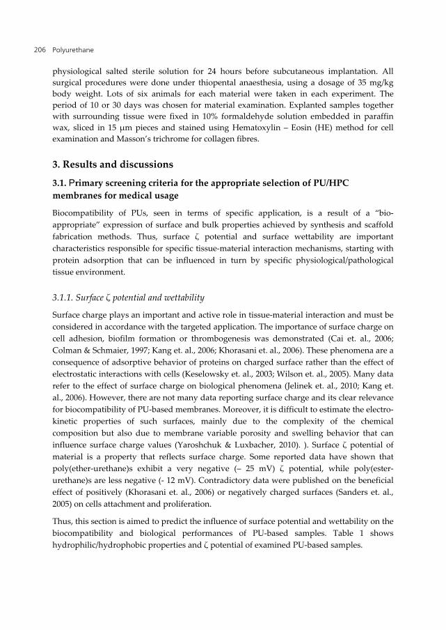
Material
samples
Contact angle
WU (%) ζ (mV) First immersion Second immersion θadv(o) θrec(o) H(%) θadv(o) θrec(o) H(%)
PU-PEGA 85.3±1.1 54.3±0.6 36.3 51.0±0.5 54.1±0.6 5.6 141±10 - 4.31
PU-PEGA/HPC 84.8±1.1 44.2±0.5 47.9 52.6±0.5 43.7±0.5 16.9 140±4 + 3.14
PU-PTHF/HPC 77.4±1.1 42.9±0.5 44.5 31.6±0.4 42.3±0.4 25.2 167±3 + 0.78
PU-PPG/HPC 85.6±1.1 44.8±0.5 47.7 60.3±0.6 44.1±0.5 27.0 92±6 + 4.85
Table 1. Dynamic contact angle values (θ) in contact with water, hysteresis (H) resulted from advanced
(adv) and receded (rec) contact angles, water uptake (WU) (Macoconschi et. al., 2009) and ζ potential of
the PU samples
As one can see from Table 1, PU-PEGA has a slightly negative ζ potential, probably due to
the presence of carboxylic groups resulted by the hydrolysis of residual isocyanate groups
during membrane precipitation in water. After blending with HPC, the residual isocyanated
groups linked to PU prepolymer are reacted with the hydroxyl groups of HPC and all
PU/HPC membranes showed a slightly positive surface. The most hydrophilic sample (PU-
PTHF/HPC) exhibited the most neutral ζ potential. This observation is in accordance to
other data that report dependence of surface charge on water swelling capacity (Aranberri-
Askargorta et. al., 2003).
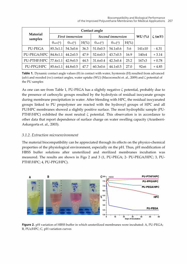
3.1.2. Extraction microenvironment
The material biocompatibility can be appreciated through its effects on the physico-chemical
properties of the physiological environment, especially on the pH. Thus, pH modification of
HBSS buffer solutions after unsterilized and sterilized membranes incubation was
measured. The results are shown in Figs 2 and 3 (1, PU-PEGA; 2- PU-PEGA/HPC; 3, PU-
PTHF/HPC; 4, PU-PPG/HPC).
Figure 2. pH variation of HBSS buffer in which unsterilized membranes were incubated: A, PU-PEGA;
B, PUs/HPC; C, pH variation curves
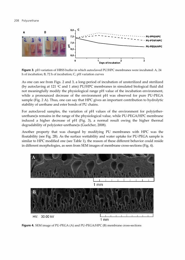
Polyurethane 208
Figure 3. pH variation of HBSS buffer in which autoclaved PU/HPC membranes were incubated: A, 24
h of incubation; B, 72 h of incubation; C, pH variation curves
As one can see from Figs. 2 and 3, a long-period of incubation of unsterilized and sterilized
(by autoclaving at 121 oC and 1 atm) PU/HPC membranes in simulated biological fluid did
not meaningfully modify the physiological range pH value of the incubation environment,
while a pronounced decrease of the environment pH was observed for pure PU-PEGA
sample (Fig. 2 A). Thus, one can say that HPC gives an important contribution to hydrolytic
stability of urethane and ester bonds of PU chains.
For autoclaved samples, the variation of pH values of the environment for poly(ether-
urethane)s remains in the range of the physiological value, while PU-PEGA/HPC membrane
induced a higher decrease of pH (Fig. 3), a normal result owing the higher thermal
degradability of poly(ester-urethane)s (Guelcher, 2008).
Another property that was changed by modifying PU membranes with HPC was the
floatability (see Fig. 2B). As the surface wettability and water uptake for PU-PEGA sample is
similar to HPC modified one (see Table 1), the reason of these different behavior could reside
in different morphologies, as seen from SEM images of membrane cross-sections (Fig. 4).
Figure 4. SEM image of PU-PEGA (A) and PU-PEGA/HPC (B) membrane cross-sections
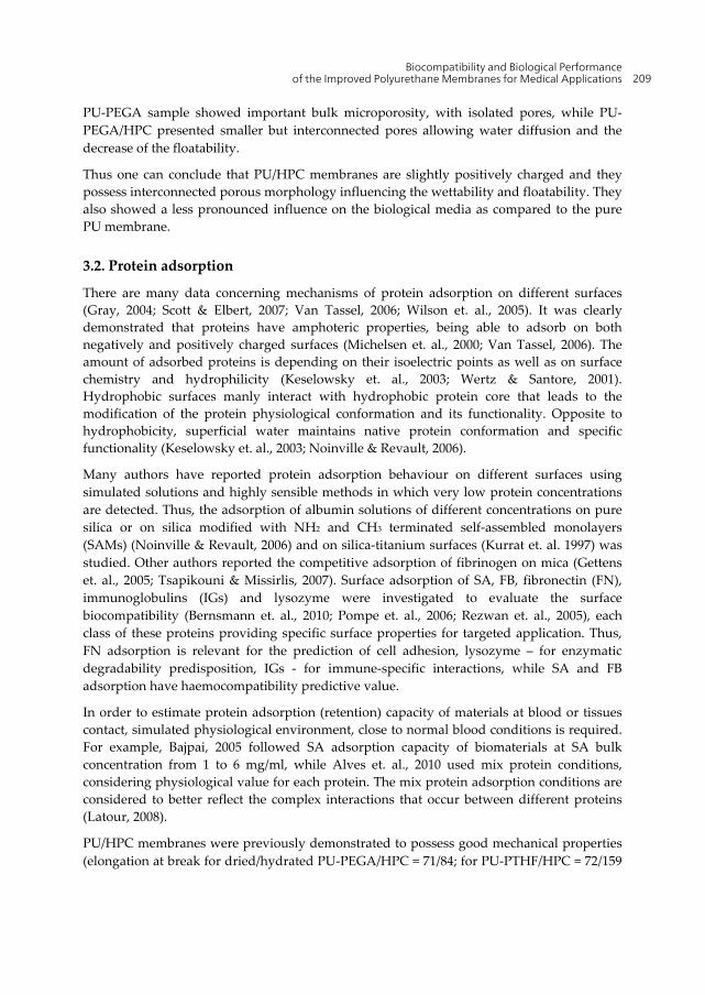
Biocompatibility and Biological Performance of the Improved Polyurethane Membranes for Medical Applications 209
PU-PEGA sample showed important bulk microporosity, with isolated pores, while PU-
PEGA/HPC presented smaller but interconnected pores allowing water diffusion and the
decrease of the floatability.
Thus one can conclude that PU/HPC membranes are slightly positively charged and they
possess interconnected porous morphology influencing the wettability and floatability. They
also showed a less pronounced influence on the biological media as compared to the pure
PU membrane.
3.2. Protein adsorption
There are many data concerning mechanisms of protein adsorption on different surfaces
(Gray, 2004; Scott & Elbert, 2007; Van Tassel, 2006; Wilson et. al., 2005). It was clearly
demonstrated that proteins have amphoteric properties, being able to adsorb on both
negatively and positively charged surfaces (Michelsen et. al., 2000; Van Tassel, 2006). The
amount of adsorbed proteins is depending on their isoelectric points as well as on surface
chemistry and hydrophilicity (Keselowsky et. al., 2003; Wertz & Santore, 2001).
Hydrophobic surfaces manly interact with hydrophobic protein core that leads to the
modification of the protein physiological conformation and its functionality. Opposite to
hydrophobicity, superficial water maintains native protein conformation and specific
functionality (Keselowsky et. al., 2003; Noinville & Revault, 2006).
Many authors have reported protein adsorption behaviour on different surfaces using
simulated solutions and highly sensible methods in which very low protein concentrations
are detected. Thus, the adsorption of albumin solutions of different concentrations on pure
silica or on silica modified with NH2 and CH3 terminated self-assembled monolayers
(SAMs) (Noinville & Revault, 2006) and on silica-titanium surfaces (Kurrat et. al. 1997) was
studied. Other authors reported the competitive adsorption of fibrinogen on mica (Gettens
et. al., 2005; Tsapikouni & Missirlis, 2007). Surface adsorption of SA, FB, fibronectin (FN),
immunoglobulins (IGs) and lysozyme were investigated to evaluate the surface
biocompatibility (Bernsmann et. al., 2010; Pompe et. al., 2006; Rezwan et. al., 2005), each
class of these proteins providing specific surface properties for targeted application. Thus,
FN adsorption is relevant for the prediction of cell adhesion, lysozyme – for enzymatic
degradability predisposition, IGs - for immune-specific interactions, while SA and FB
adsorption have haemocompatibility predictive value.
In order to estimate protein adsorption (retention) capacity of materials at blood or tissues
contact, simulated physiological environment, close to normal blood conditions is required.
For example, Bajpai, 2005 followed SA adsorption capacity of biomaterials at SA bulk
concentration from 1 to 6 mg/ml, while Alves et. al., 2010 used mix protein conditions,
considering physiological value for each protein. The mix protein adsorption conditions are
considered to better reflect the complex interactions that occur between different proteins
(Latour, 2008).
PU/HPC membranes were previously demonstrated to possess good mechanical properties
(elongation at break for dried/hydrated PU-PEGA/HPC = 71/84; for PU-PTHF/HPC = 72/159
Polyurethane 210
and for PU-PPG/HPC = 53/55), appropriate for cardio-vascular applications (Macocinschi et.
al., 2009). The physisorption of SA and FB is further highlighted as screening criteria for
biocompatibility and, more specifically, haemocompatibility. Very short characteristics of
SA and FB, important for protein-material interaction are given below.
SA is a protein belonging to the so called “soft” class of proteins, with a molar mass of about
65 kD for BSA and 67 kD for human SA (HSA). This protein represents about 60% of the
blood proteins. Normal blood concentration of HSA is 35 – 50 mg/ml. This protein is
involved in many physiological phenomena as carrier protein for fatty acids, metals,
cholesterol, bile pigment, hormones and drugs. SA is also characterised by antioxidant
properties (Bourdon et. al. 1999; Kouoh et. al., 1999; H. Lee et. al., 2000) that is higher in
alkaline pH, up to 8 (H. Lee et. al., 2000). SA is preponderantly negatively charged, its
isoelectric point being close to 4.8 (Carter & Ho 1994; Noinville & Revault, 2006).
Approximately 67% of the secondary SA structure is represented by the α-helix. It was
demonstrated that the stability of SA secondary structure strictly depends on pH (Freeman,
2006) that influence the protein conformation. Thus, at pH = 5, SA takes almost spherical,
native, unfolded shape that forms a thick layer on the adsorptive surfaces. At pH = 7 (close
to physiological pH), due to molecular spreading, SA forms an extended contact area with
adsorptive surfaces. This behavior can be influenced by surface charge, surface functionality
and functionality distribution, surface morphology or wettability conditions (Wilson et. al.,
2005). The role of adsorbed SA on biomaterial biocompatibility is still ambiguously
described in the literature. While some authors have demonstrated biocompatibility
improvement of material with increased adsorption of SA (Eberhart et.al. 1987; Marconi et.
al., 1996; Randrasana et. al., 1994), others demonstrated a better biocompatibility of SA-
resistant surfaces (Ostuni et. al., 2001; Wan et. al., 2006).
FB is a high molecular weight (340 kD) complex glycoprotein that has 2 molecular
domains, each of them consisting of three polypeptide chains called Aα, Bβ and γ.
Molecular updated analysis of FB can be found in recent reports (Cardinali et.al, 2010). FB
is an important factor of haemostasis. Through fibrin network formation as first cell
scaffold, FB is involved in wound healing and tissue regeneration. Its normal blood
concentration varies from 2 to 4 mg/ml. In inflammations or in other pathological statuses
- as cardiovascular diseases - FB can reach up to 7 mg/ml, therefore adsorption properties
of biomaterials for this protein should be carefully analysed, especially for those targeted
for blood contact applications.
The results obtained in adsorption experiments of SA and FB from both individual and
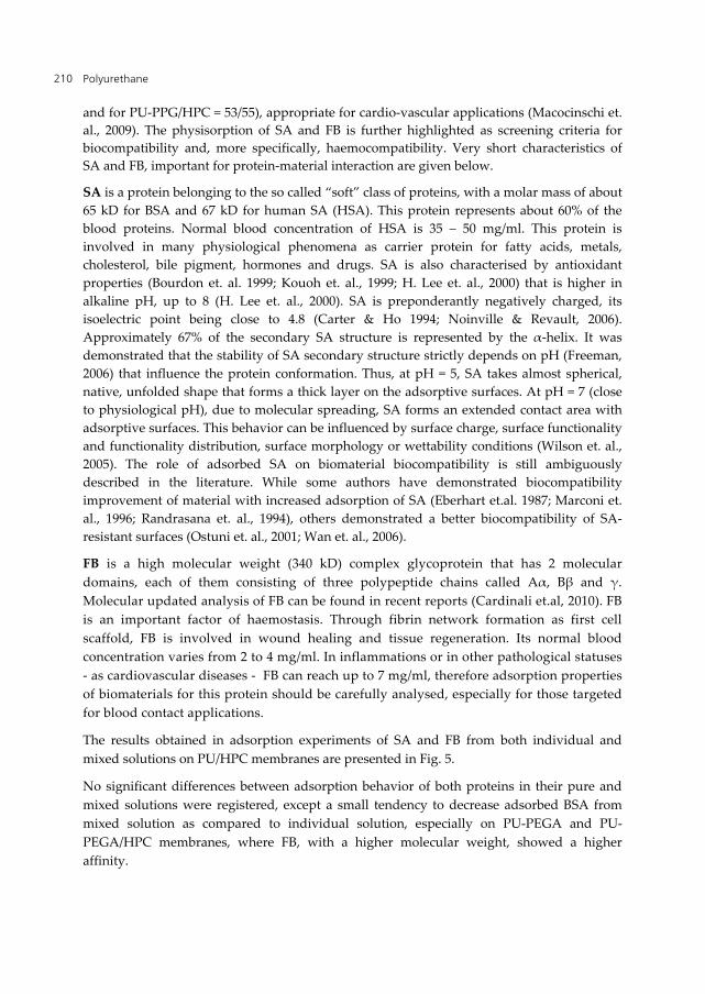
mixed solutions on PU/HPC membranes are presented in Fig. 5.
No significant differences between adsorption behavior of both proteins in their pure and
mixed solutions were registered, except a small tendency to decrease adsorbed BSA from
mixed solution as compared to individual solution, especially on PU-PEGA and PU-
PEGA/HPC membranes, where FB, with a higher molecular weight, showed a higher
affinity.
Biocompatibility and Biological Performance of the Improved Polyurethane Membranes for Medical Applications 211
Figure 5. Amount of adsorbed BSA (left) and FB (right) from individual protein solutions and in co-
adsorptive environment (mixed protein solution) of physiological concentrations, i.e., 3 mg/ml for FB
and 45 mg/ml for BSA
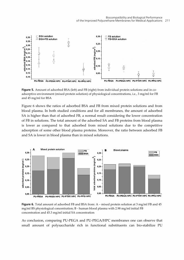
Figure 6 shows the ratios of adsorbed BSA and FB from mixed protein solutions and from
blood plasma. In both studied conditions and for all membranes, the amount of adsorbed
SA is higher than that of adsorbed FB, a normal result considering the lower concentration
of FB in solutions. The total amount of the adsorbed SA and FB proteins from blood plasma
is lower as compared to that adsorbed from mixed solutions due to the competitive
adsorption of some other blood plasma proteins. Moreover, the ratio between adsorbed FB
and SA is lower in blood plasma than in mixed solutions.
Figure 6. Total amount of adsorbed FB and BSA from: A – mixed protein solution at 3 mg/ml FB and 45
mg/ml BS physiological concentration; B - human blood plasma with 2.98 mg/ml initial FB
concentration and 45.3 mg/ml initial SA concentration
As conclusion, comparing PU-PEGA and PU-PEGA/HPC membranes one can observe that
small amount of polysaccharide rich in functional substituents can bio-stabilize PU
Polyurethane 212
structures and improve their resistance for autoclaving procedures as important step in
ready to use biomaterials preparation. From all the data presented in this section, one can
say that the more hydrophilic PU-PTHF/HPC membrane could be the most appropriate for
biomedical applications.
3.3. In vitro and in vivo performances of PU/HPC membranes
The biocompatibility of PUs are widely discussed and questioned, mostly in the past. In the
last two decades new generation of PUs that combine mechanical advantages with the
biological performances emerged (Gisselfa et. al., 2002; Jordan & Chaikof et. al., 2007; Jun et.
al., 2005; Kavlock et. al., 2007; Parveen et. al., 2008). For many years it has been considered
that PUs biocompatibility is spotless due to their products of degradation, e.g., aromatic
polyamines. As it is well known for the most part of biocompatible materials, the life time of
their in vitro functionality is quite short. This is a consequence of their intrinsic physico-
chemical properties, on one hand, and of the tissue action on the material, on the other hand
(Anderson, 2001; Guelcher, 2008; Shen & Horbett, 2001).
3.3.1. Oxidative in vitro behavior
Oxidative degradation of PUs caused by hydrolytic or enzymatic mechanism was
intensively discussed (Christenson et. al., 2004; Guelcher, 2008; Gary & Howard, 2002;
Sutherland et. al., 1993). First of all, PUs designed for tissue-contact devices undergo
hydrolytic degradation as a result of watering with physiological solutions. This process has
an impact especially on poly(ester-urethane)s that can generate hydroxy-acids, being
susceptible to induce reactive oxygen species (ROS) production following the material-
tissues interaction. By means of this mechanism, PUs can be implied in the sustained
oxidative degradation and a wide range of pathological states.
As it is well known, ROS can trigger subtle mechanisms responsible for diseases generation
through the peroxidation of cell membrane lipids and DNA damage (Marnett, 2002; Tribble
et. al., 1987; Yagi, 1987). The most susceptible organs to oxidative aggression are the heart,
vessels, lung, gut, liver, brain and nerves (Ames et. al., 1993; Förstermann, 2008; Paradis et.
al., 1997; Rahman et. al., 2002; Sayre et. al., 1997).
In a normal body state, ROS appear constantly as a result of some biological errors or as a
consequence of some short living reactive intermediate products generated by the cell
aerobic metabolism. Endogenous enzymatic and nonenzymatic pathways are responsible
for the formation of free radicals. These pathways are balanced by two endogenous
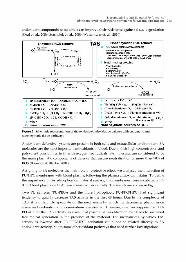
antioxidant pathways, which form the TAS (see fig. 7).
While some harmful material characteristics can be marked as cytotoxic or proinflammatory
by standard testing, others, such as oxidative stress (that causes long-time material failure),
are undetectable by using short period testing. Thus, well known biocompatible materials
were found to display surface alteration or cracking after long-time implantation. Adding
Biocompatibility and Biological Performance of the Improved Polyurethane Membranes for Medical Applications 213
antioxidant compounds to materials can improve their resistance against tissue degradation
(Oral et. al., 2006; Stachelek et. al., 2006; Wattamwar et. al., 2010).
Figure 7. Schematic representation of the oxidative/antioxidative balance with enzymatic and
nonenzymatic tissue pathways
Antioxidant defensive systems are present in both cells and extracellular environment. SA
molecules are the most important antioxidants in blood. Due to their high concentration and
polyvalent possibilities to fit with oxygen free radicals, SA molecules are considered to be
the main plasmatic components of defence that assure neutralisation of more than 70% of
ROS (Bourdon & Blache, 2001).
Assigning to SA molecules the main role in protective effect, we analysed the interaction of
PU/HPC membranes with blood plasma, following the plasma antioxidant status. To define
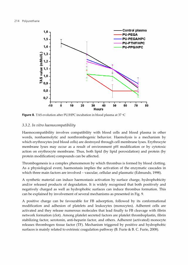
the importance of SA adsorption on material surface, the membranes were incubated at 37 oC in blood plasma and TAS was measured periodically. The results are shown in Fig. 8.
Two PU samples (PU-PEGA and the more hydrophobic PU-PPG/HPC) had significant
tendency to quickly decrease TAS activity in the first 48 hours. Due to the complexity of
TAS, it is difficult to speculate on the mechanism by which the decreasing phenomenon
arises and certainly more examinations are needed. However, one can suppose that PU-
PEGA alter the TAS activity as a result of plasma pH modification that leads to sustained
free radical generation in the presence of the material. The mechanisms by which TAS
activity is lowered after PU-PPG/HPC incubation could not be related directly to SA
antioxidant activity, but to some other oxidant pathways that need further investigations.
Polyurethane 214
Figure 8. TAS evolution after PU/HPC incubation in blood plasma at 37 oC
3.3.2. In vitro haemocompatibility
Haemocompatibility involves compatibility with blood cells and blood plasma in other
words, nonhaemolytic and nonthrombogenic behavior. Haemolysis is a mechanism by
which erythrocytes (red blood cells) are destroyed through cell membrane lyses. Erythrocyte
membrane lyses may occur as a result of environment pH modification or by cytotoxic
action on erythrocyte membrane. Thus, both lipid (by lipid peroxidation) and protein (by
protein modification) compounds can be affected.
Thrombogenesis is a complex phenomenon by which thrombus is formed by blood clotting.
As a physiological event, haemostasis implies the activation of the enzymatic cascades in
which three main factors are involved – vascular, cellular and plasmatic (Edmunds, 1998).
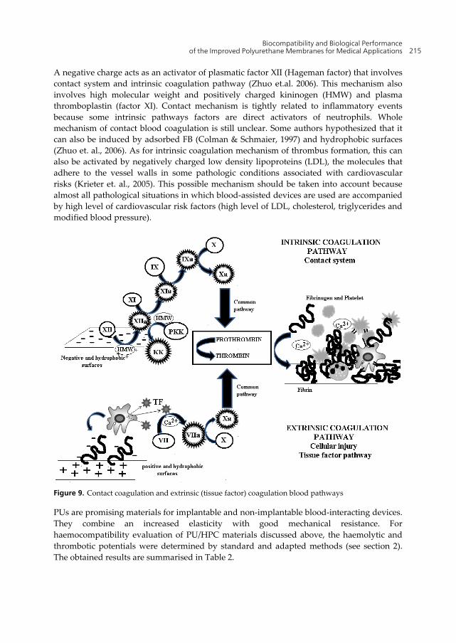
A synthetic material can induce haemostasis activation by surface charge, hydrophobicity
and/or released products of degradation. It is widely recognised that both positively and
negatively charged as well as hydrophobic surfaces can induce thrombus formation. This
can be explained by involvement of several mechanisms as presented in Fig. 9.
A positive charge can be favourable for FB adsorption, followed by its conformational
modification and adhesion of platelets and leukocytes (monocytes). Adherent cells are
activated and they release numerous molecules that lead finally to FB cleavage with fibrin
network formation (clot). Among platelet secreted factors are platelet thromboplastin, fibrin
stabilizing factor, serotonin, anti-heparin factor, and others. Adherent (activated) monocyte
releases thrombogen tissue factor (TF). Mechanism triggered by positive and hydrophobic
surfaces is mainly related to extrinsic coagulation pathway (B. Furie & B. C. Furie, 2008).
Biocompatibility and Biological Performance of the Improved Polyurethane Membranes for Medical Applications 215
A negative charge acts as an activator of plasmatic factor XII (Hageman factor) that involves
contact system and intrinsic coagulation pathway (Zhuo et.al. 2006). This mechanism also
involves high molecular weight and positively charged kininogen (HMW) and plasma
thromboplastin (factor XI). Contact mechanism is tightly related to inflammatory events
because some intrinsic pathways factors are direct activators of neutrophils. Whole
mechanism of contact blood coagulation is still unclear. Some authors hypothesized that it
can also be induced by adsorbed FB (Colman & Schmaier, 1997) and hydrophobic surfaces
(Zhuo et. al., 2006). As for intrinsic coagulation mechanism of thrombus formation, this can
also be activated by negatively charged low density lipoproteins (LDL), the molecules that
adhere to the vessel walls in some pathologic conditions associated with cardiovascular
risks (Krieter et. al., 2005). This possible mechanism should be taken into account because
almost all pathological situations in which blood-assisted devices are used are accompanied
by high level of cardiovascular risk factors (high level of LDL, cholesterol, triglycerides and
modified blood pressure).
Figure 9. Contact coagulation and extrinsic (tissue factor) coagulation blood pathways
PUs are promising materials for implantable and non-implantable blood-interacting devices.
They combine an increased elasticity with good mechanical resistance. For
haemocompatibility evaluation of PU/HPC materials discussed above, the haemolytic and
thrombotic potentials were determined by standard and adapted methods (see section 2).
The obtained results are summarised in Table 2.
Polyurethane 216
All studied membranes showed a low haemolytic activity, lower for PU/HPC than for pure
PU-PEGA sample.
Material
samples
Haemolytic
potential Thrombotic potential
Released Hb
(%)1 FB (mg/ml)2 PT (s)3
Adhered platelet
(cells x 105/mm2)
% blood
clot amount4
PU-PEGA 6,7±0,2 2,79±0,04 11,06±0,4 1,40±0,08 40%
PU-PEGA/HPC 5,2±0,1 2,87±0,04 10,9±0,09 0,82±0,05 29%
PU-PTHF/HPC 4,2±0,2 2,90±0,01 10,9±0,09 0,86±0,05 15%
PU-PPG/HPC 5,5±0,1 2,77±0,07 10,9±0,07 1,25±0,09 89%
Table 2. Haemolytic and thrombotic potential of the PU/HPC samples: Hb-haemoglobin; FB-
fibrinogen; PT- prothrombin time
As for thrombotic action, a correlation between adsorbed FB, platelet adhesion and amount
of formed clot was registered, while no significant variation was recorded for PT. This latter
parameter was kept within the normal limits (see footnote 3).
The judgement strictly based on the haemocompatibility results permits to state that all
examined materials have an acceptable thrombotic potential (referring to physiological
requirements). Considering clot amount and all the other characteristics discussed above, it is
obvious that PU-PEGA and PU-PPG/HPC are not suitable for long-time functional integration.
3.3.3. In vivo biocompatibility and performance
The technological progress achieved in the last decades in apparently unrelated areas
(biomaterials, biotechnology, cell and molecular biology, tissue engineering, and polymer
science) has generated a boost in the development and use of devices for medical and/or other
type of applications (e.g. artificial organs, biosensors, catheters, heart valves) (Shastri, 2003). In
spite of real improvement of this sort of devices there are still some important problems to face
since implanted medical devices usually reveal different degree of loss of functionality over
time after insertion (Göpferich, 1996). Tissue or blood-device interface interactions or a lack of
biocompatibility resulting from the normal homeostatic response of the body to the
implantation injury, determining an inadequate in vivo functionality and longevity, remains a
serious concern (Callahan & Natale, 2008; Fujimoto et. al., 2007; Morais et. al., 2010).
In order to protect the body from the foreign object, under normal physiological conditions,
the body reacts by several nonspecific mechanisms (immune and inflammatory cells
recruitment), usually termed foreign body reaction (FBR) (Anderson, 2001). There is an
imperative call for knowing the degree to which the pathophysiological conditions are
1 Percentage of released Hb over negative control 2 FB concentration remained in blood plasma after incubation. FB control was 2,98 ± 0,04 mg/ml 3 Physiological normal value according to related laboratory are between 8,3 s and 11,3 s 4 Percentage of blood clotting over negative control (blood without incubated material)
Biocompatibility and Biological Performance of the Improved Polyurethane Membranes for Medical Applications 217
created, the homeostatic mechanisms are disturbed, and the resolution of the inflammatory
response (simple put, the measure of the host reaction). All of these will finally establish the
effective compatibility of a specific device. In the same time, understanding these reactions
(the implant versus the host and the host versus the implanted device) will reduce health
problems to the beneficiary of the device and device malfunction. Usually, for practical
reasons, the homeostatic mechanisms are separately assessed even if it is well known that
they are profoundly interrelated (Sieminski & Gooch, 2000).
The first event after a device/material insertion is that the body generates quickly a sort of
“interface” via nonspecific adsorption of plasma/tissue soluble proteins on the implant
surface (Shen & Horbett, 2001). There are some well identified elements that determine the
FBR strength: device material composition, surface chemistry, size and shape, porosity,
degradation, velocity as well as the place of device insertion (Ratner & Bryant, 2004)
As presented shortly below, tissue injury associated with device implantation, initiates a
complex set of events (nonspecific inflammatory reaction and wound healing responses)
that will bring about a FBR (Wahl et. al., 1989). The stages of inflammatory responses are
well studied and can be separate in acute and chronic inflammatory periods.
The initial phase, acute stage, starts quickly in matter of hours, lasts for several days (up to
14 days) and is underlined by rapid device interface generation and typical for this phase,
different degree of neutrophil leucocytes responses (Jiang et. al., 2007). The main result of
this stage is the building of temporary interface material-tissue, the cleaning-up of the injury
place and the vasodilation that bring more blood in the affected area.
The acute inflammatory reaction typically decline in maximum 14 days with a
“biocompatible” material. Some local conditions (extent of surgical injury, body reactivity)
or properties of the implanted device can trigger a chronic inflammatory evolution
(Kirkpatrick et al., 1998).
Numerous blood and tissue proteins such as cytokines (e.g. tumor necrosis factor (TNF),
interleukins (IL-6, IL-8), matrix metalloproteases (MMP-1, MMP-3), granulocyte-
macrophage growth factors (GM-CSF)) are released, and leukocytes adhere to the
endothelium of the blood vessels and infiltrate the injury site. These proteins are strong
calling factors for monocytes, cells which will migrate to the site of inflammation where they
will differentiate into macrophage. If inflammatory stimuli persist, the conditions that can
lead to chronic inflammation are created. Cell population of this stage of inflammatory
reaction is usually characterized by the presence of monocytes, macrophages, and
lymphocytes (Bhardwaj et al., 2010). Also, in this step it can be noticed that the proliferation
of blood vessels (angiogenesis), and connective tissue occurs that participate in remodelling
of the affected area. The formation of blood vessels is crucial for wound healing, supplying
necessary factors for tissues reconstruction. In the end, the granulomatous tissue is replaced
by an extracellular matrix (ECM) that acts not only as a physical scaffold but also as an
essential modulator of the biological processes, including differentiation, development,
regeneration, repair, as well as tumour progression. The end phase of the FBR draws in
wrapping the implant by a collagenic fibrous capsule that limits the implant and therefore
Polyurethane 218
prevents it from interacting with the surrounding tissue. The main tissue events of the
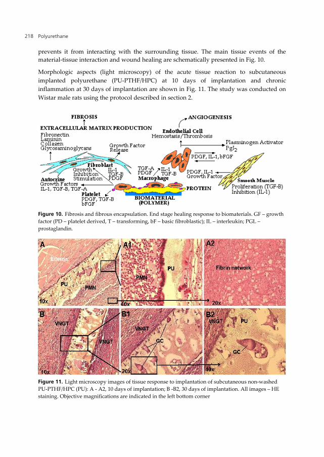
material-tissue interaction and wound healing are schematically presented in Fig. 10.
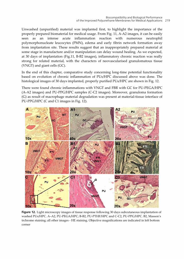
Morphologic aspects (light microscopy) of the acute tissue reaction to subcutaneous
implanted polyurethane (PU-PTHF/HPC) at 10 days of implantation and chronic
inflammation at 30 days of implantation are shown in Fig. 11. The study was conducted on
Wistar male rats using the protocol described in section 2.
Figure 10. Fibrosis and fibrous encapsulation. End stage healing response to biomaterials. GF – growth
factor (PD – platelet derived, T – transforming, bF – basic fibroblastic); IL – interleukin; PGL –
prostaglandin.
Figure 11. Light microscopy images of tissue response to implantation of subcutaneous non-washed
PU-PTHF/HPC (PU): A - A2, 10 days of implantation; B -B2, 30 days of implantation. All images – HE
staining. Objective magnifications are indicated in the left bottom corner
Biocompatibility and Biological Performance of the Improved Polyurethane Membranes for Medical Applications 219
Unwashed (unpurified) material was implanted first, to highlight the importance of the
properly prepared biomaterial for medical usage. From Fig. 11, A-A2 images, it can be easily
seen as an intense acute inflammation reaction with numerous neutrophil
polymorphonucleate leucocytes (PMN), edema and early fibrin network formation away
from implantation site. These results suggest that an inappropriately prepared material at
some stage in manufacture and/or manipulation can delay wound healing. As we expected,
at 30 days of implantation (Fig.11, B-B2 images), inflammatory chronic reaction was really
strong for related material, with the characters of neovascularised granulomatous tissue
(VNGT) and giant cells (GC).
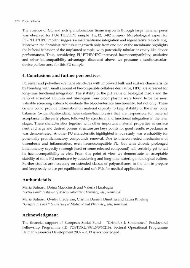
In the end of this chapter, comparative study concerning long-time potential functionality
based on evolution of chronic inflammation of PUs/HPC discussed above was done. The
histological images of 30 days implanted, properly purified PUs/HPC are shown in Fig. 12.
There were found chronic inflammations with VNGT and FBR with GC for PU-PEGA/HPC
(A-A2 images) and PU-PPG/HPC samples (C-C2 images). Moreover, granuloma formation
(G) as result of macrophage material degradation was present at material-tissue interface of
PU-PPG/HPC (C and C1 images in Fig. 12).
Figure 12. Light microscopy images of tissue response following 30 days subcutaneous implantation of
washed PUs/HPC. A–A2, PU-PEGA/HPC; B-B2, PU-PTHF/HPC and C-C2, PU-PPG/HPC. B2, Masson’s
trchrome staining; all other images - HE staining. Objective magnifications are indicated in left bottom
corner
Polyurethane 220
The absence of GC and rich granulomatous tissue ingrowth through large material pores
was observed for PU-PTHF/HPC sample (Fig.12, B-B2 images). Morphological aspect for
PU-PTHF/HPC implant suggests a material-tissue integration and regenerative remodelling.
Moreover, the fibroblast-rich tissue ingrowth only from one side of the membrane highlights
the bifacial behavior of the implanted sample, with potentially tubular or cavity-like device
performances. Thus, considering PU-PTHF/HPC increased haemocompatibility, oxidative
and other biocompatibility advantages discussed above, we presume a cardiovascular-
device performance for this PU sample.
4. Conclusions and further perspectives
Polyester and polyether urethane structures with improved bulk and surface characteristics
by blending with small amount of biocompatible cellulose derivative, HPC, are screened for
long-time functional integration. The stability of the pH value of biological media and the
ratio of adsorbed albumin and fibrinogen from blood plasma were found to be the most
valuable screening criteria to evaluate the blood-interface functionality, but not only. These
criteria could provide information on material capacity to keep stability of the main body
balances (oxidant/antioxidant, haemostasis/haemolysis) that are responsible for material
acceptance in the early phase, followed by structural and functional integration in the later
stages. These characteristics together with other important material properties as surface
neutral charge and desired porous structure are keys points for good results expectance as
was demonstrated. Another PU characteristic highlighted in our study was washability for
potentially proinflammatory compounds removal. Due to interconnected mechanisms of
thrombosis and inflammation, even haemocompatible PU, but with chronic prolonged
inflammatory capacity (through itself or some released compound) will certainly get to fail
its haemocompatibility in vivo. From this point of view we demonstrate an acceptable
stability of some PU membrane by autoclaving and long-time watering in biological buffers.
Further studies are necessary on extended classes of polyurethanes in the aim to prepare
and keep ready to use pre-equilibrated and safe PUs for medical applications.
Author details
Maria Butnaru, Doina Macocinsch and Valeria Harabagiu
”Petru Poni” Institute of Macromolecular Chemistry, Iasi, Romania
Maria Butnaru, Ovidiu Bredetean, Cristina Daniela Dimitriu and Laura Knieling
”Grigore T. Popa “ University of Medicine and Pharmacy, Iasi, Romania
Acknowledgment
The financial support of European Social Fund – “Cristofor I. Simionescu” Posdoctoral
Fellowship Programme (ID POSTDRU/89/1.5/S/55216), Sectoral Operational Programme
Human Resources Development 2007 – 2013 is acknowledged.
Biocompatibility and Biological Performance of the Improved Polyurethane Membranes for Medical Applications 221
5. References
Abd El-Rehim, H.A.A., & El-Arnaouty, M.B. (2004). Properties and Biocompatibility of
Polypropylene Graft Copolymer Films. Journal of Biomedical Materials Research Part B:
Applied Biomaterials, Vol. 68B, No.2, (February 2004), pp.209-215, ISSN 1552-4973
Alves, C.M., Reis, R.L., & Hunt, J.A. (2010). The Competitive Adsorption of Human Proteins
Onto Natural-Based Biomaterials. Journal of the Royal Society, Interface / The Royal Society,
Vol. 50, No. 7, (February 2010), pp. 1367-1377, ISSN 1742-5689
Ames, B.N., Shigenaga, M.K., & Hagen, T.M. (1993). Oxidants, Antioxidants and The
Degenerative Diseases of Aging. Proceedings of the National Academy of Sciences of the
United States of America, Vol. 90, No. 17, (September 1993), pp. 7915-7922, ISSN 0027-
8424
Anderson, J.M. (2001). Biological Responses to Materials. Annual Review of Materials Research,
Vol. 31, No. 1, (May 2001), pp. 81–110, ISSN 1531-7331
Aranberri-Askargorta, I., Lampke, T., & Bismarck, A. (2003). Wetting Behavior of Flax Fibers
as Reinforcement for Polypropylene. Journal of Colloid and Interface Science, Vol.263,
No.2, (July 2003), pp.580–589, ISSN 0021-9797
Bajpai, A.K. (2005). Blood Protein Adsorption Onto a Polymeric Biomaterial of Polyethylene
Glycol and Poly[(2-Hydroxyethyl Methacrylate)-Coacrylonitrile] and Evaluation of In
Vitro Blood Compatibility. Polymers International, Vol. 54, No. 2, (February 2005), pp.
304-315, ISSN 0959-8103
Bernsmann, F., Frisch, B., Ringwald, C., & Ball, V. (2010). Protein Adsorption on Dopamine–
Melanin Films: Role of Electrostatic Interactions Inferred from f-Potential
Measurements Versus Chemisorptions. Journal of Colloid and Interface Science, Vol. 344,
No. 1, (April 2010), pp. 54–60, ISSN 0021-9797
Berthier, D.L., Herrmann, A., & Ouali, L. (2011). Synthesis of Hydroxypropyl Cellulose
Derivatives Modified with Amphiphilic Diblock Copolymer Side-Chains for The Slow
Release of Volatile Molecules. Polymer Chemistry, Vol. 2, No. 9, (May 2011), pp. 2093-
2101, ISSN 1759-9954
Bhardwaj, U., Radhacirsshana, S., Papadimitrakopoulos, F., Burgess, D.J. (2010). PLGA/PVA
Hydrogel Composites for Long-Term Inflammation Control Following Subcutaneous
Implantation. International journal of pharmaceutics, Vol. 484, No. 1-2, (January 2010), pp.
78–86, ISSN 0378-5173
Bourdon, E., & Blache, D. (2001). The Importance of Proteins in Defense Against Oxidation.
Antioxidants & Redox Signaling, Vol. 3, No. 2, (April 2001), pp. 293-311, ISSN 1523-0864
Bourdon, E., Loreau, N., & Blanche, D. (1999). Glucose and Free Radicals Impair the
Antioxidant Properties of Serum Albumin, The FASEB Journal, Vol. 13, No. 2, (February
1999), pp. 233-234, ISSN 0892-6638
Cai, K., Frant, M., Bossert, J., Hildebrand, G., Liefeith, K., & Jandt, K.D. (2006). Surface
Functionalized Titanium Thin Films: Zeta-potential, Protein Adsorption and Cell
Proliferation. Colloids and Surfaces B: Biointerfaces, Vol. 50, No.1, (June 2006), pp. 1–8,
ISSN 0927-7765
Polyurethane 222
Callahan, T.D. 4th, & Natale, A. (2008). Catheter Ablation of Atrial Fibrillation. The Medical
Clinics of North America, Vol. 92, No.1, (Janury 2008), pp.179–201, ISSN 0025-7125
Cardinali, B., Profumo, A., Aprile, A., Byron, O., Morris, G., Harding, S.E., Stafford, W.F., &
Rocco, M. (2010). Hydrodynamic and Mass Spectrometry Analysis of Nearly-Intact
Human Fibrinogen, Chicken Fibrinogen, and of a Substantially Monodisperse Human
Fibrinogen Fragment X. Archives of Biochemistry and Biophysics, Vol. 493, No. 2, (January
2010), pp. 157–168, ISSN 0003-9861
Carter, D.C. & Ho, J.X. (1994). Structure of Serum Albumin. Advances in Protein Chemistry
Vol. 45, (1994), pp.155–203, ISSN 0065-3233
Chen, D., & Sun, B. (2000). New Tissue Engineering Material Copolymers of Derivatives of
Cellulose and Lactide: Their Synthesis and Characterization. Materials Science and
Engineering: C, Vol. 11, No. 1, (June 2000), pp. 57-60, ISSN 0928-4931
Chen, R., Huang, C., Ke, Q., He, C., Wang, H., & Mo, X. (2010). Preparation and
Characterization of Coaxial Electrospun Thermoplastic Polyurethane/Collagen
Compound Nanofibers for Tissue Engineering Applications. Colloids and Surfaces B:
Biointerfaces, Vol. 79, No. 2, (September 2010), pp. 315–325, ISSN 0927-7765
Christenson, E.M., Anderson, J.M., & Hiltner A. (2004). Oxidative Mechanisms of
Poly(carbonate urethane) and Poly(ether urethane) Biodegradation: In Vivo and In
Vitro Correlations. Journal of Biomedical Materials Research, Vol. 70A, No. 2, (August
2004), pp. 245–255, ISSN 0021-9304
Colman, R.W., & Schmaier, A.H. (1997). Contact System: A Vascular Biology Modulator
With Anticoagulant, Profibrinolytic, Antiadhesive, and Proinflammatory Attributes.
Blood, Vol. 90, No. 10, (November 1997), pp 3819-3843, ISSN 0006-4971
Eberhart, R.C., Munro, M.S., Wiliams, G.B., Kulkarni, P.V., Shannon, W.A., Brink, B.E., &
Fry, W.J. (1987). Albumin Adsorption and Retention on C18-alkyl-derivatized
Polyurethane Vascular Grafts. Artificial Organs, Vol. 11, No. 5, (October 1987), pp. 375-
382, ISSN 0160-564X
Edmunds, L.H.Jr . (1998). Inflammatory Response to Cardiopulmonary Bypass. The Annals of
Thoracic Surgery, Vol. 66, No. 5, (November 1998), pp.12-16, ISSN 0003-4975
Förstermann, I. (2008). Oxidative Stress in Vascular Disease: Causes, Defense Mechanisms
and Potential Therapies. Nature Clinical Practice. Cardiovascular Medicine, Vol. 5, No. 6,
(June 2008), pp. 338-349, ISSN 1743-4297
Freeman, N. (2006). Analysis of the Structure at the Interface, In: Proteins at Liquid – Solid
Interfaces (Principle and practice), P. Dejardin, (Ed.), pp. 75-104, Springer, ISBN-10 3-540-
32657-X, Berlin, Germany
Fujimoto, K.L., Guan, J., Oshima, H., Sakai, T., & Wagner, W.R. (2007). In Vivo Evaluation of
a Porous, Elastic, Biodegradable Patch for Reconstructive Cardiac Procedures. The
Annals of Thoracic Surgery, Vol. 83, No. 2, (February 2007), pp. 648 –54, ISSN 0003-4975
Furie, B., & Furie, B.C. (2008). Mechanisms of Thrombus Formation. The New England Journal
of Medicine, Vol. 359, (August 2008), pp. 938-949, ISSN 0028-4793
Gary, T. & Howard, G.T. (2002). Biodegradation of Polyurethane: A Review. International
Biodeterioration & Biodegradation, Vol. 49, No. 4 (June 2002), pp. 245 – 252, ISSN 0964-
8305.
Biocompatibility and Biological Performance of the Improved Polyurethane Membranes for Medical Applications 223
Gettens, R.T., Bai, Z., & Gilbert, J.L. (2005). Quantification of The Kinetics and
Thermodynamics of Protein Adsorption Using Atomic Force Microscopy. Journal of
Biomedical Materials Research. Part A, Vol. 72, No.3, (March 2005), pp. 246-257, ISSN 1549-
3296
Gisselfa, K., Edberg, B., & Flodin, P. (2002). Synthesis and Properties of Degradable
Poly(urethane urea)s To Be Used for Ligament Reconstructions. Biomacromolecules, Vol.
3, No. 5, (September-October 2002), pp. 951-958, ISSN 1525-7797
Gong, F., Lu, Y., Guo, H., Cheng, S., & Gao, Y. (2010). Hyaluronan Immobilized
Polyurethane as a Blood Contacting Material. International Journal of Polymer Science,
Vol. 2010, Article ID 807935, (March 2010), pp.1-8, ISSN 1687-9422
Göpferich, A. (1996). Polymer Degradation and Erosion: Mechanisms and Applications.
European Journal of Pharmaceutics and Biopharmaceutics, Vol. 42, No. 1, (1996), pp. 1–11,
ISSN 0939-6411
Gray, J.J. (2004). The Interaction of Proteins With Solid Surfaces. Current Opinion in
Structural Biology, Vol. 14, No.1, (February 2004), pp. 110–115, ISSN 0959-440X
Guelcher, S., Srinivasan, A., Hafeman, A., Gallagher, K., Doctor, J., Khetan, S., Mcbride, S., &
Hollinger, J. (2007). Synthesis, In Vitro Degradation, and Mechanical Properties of Two-
Component Poly(Ester Urethane)Urea Scaffolds: Effects of Water and Polyol
Composition. Tissue Engineering, Vol.13, No. 9, (September 2007), pp. 2321-2333, ISSN
1076-3279
Guelcher, S.A. (2008). Biodegradable Poliurethanes: Syntesis and Applications in
Regenerative Medicine. Tissue Engineering, Vol. 14, No.1, (March 2008), pp. 3-17, ISSN
2152-4947
Gutowska, A., Jeong, B., & Jasionowski, M. (2001). Injectable Gels for Tissue Engineering.
The Anatomical Record, Vol. 263, No.4, (August 2001), pp. 342–349, ISSN 0003-276X
Hsu, H.S., Kao, Y.C., & Lin, Y.C. (2004). Enhanced Biocompatibility in Biostable
Poly(carbonate)urethane. Macromolecular Bioscience, Vol.4, No. 4, (April 2004), pp. 464–
470, ISSN 1616-5187
Huang, J., & Xu, W. (2010). Zwitterionic Monomer Graft Copolymerization Onto
Polyurethane Surface Through a PEG Spacer. Applied Surface Science, Vol. 256, No. 12,
(April 2010), pp. 3921–3927, ISSN 0169-4332
Hwang, S., & Meyerhoff, M.E. (2008). Polyurethane With Tethered Copper(II)ecyclen
Complex: Preparation, Characterization and Catalytic Generation of Nitric Oxide from
S-nitrosothiols. Biomaterials, Vol. 29, No. 16, (June 2008), pp. 2443-2452, ISSN 1552-4973
Jelinek, M., Kocourek, T., Remsa, J., Mikšovský, J., Zemek, J., Smetana, K. Jr., Dvořánková,
B., & Luxbacher, T. (2010). Diamond/Graphite Content and Biocompatibility of DLC
Films Fabricated by PLD. Applied Physics A, Materials Science & Processing, Vol. 110,
No.4, (June 2010), pp.579-583, ISSN 0947-8396
Jiang, W.W., Su, S.H., Eberhart, R.C., & Tang, L. (2007). Phagocyte Responses to Degradable
Polymers. Journal of Biomedical Materials Research. Part A, Vol. 82, No. 2, (August 2007),
pp. 492–497, ISSN 1549-3296
Jordan, S.W. & Chaikof, E.L. (2007). Novel Thromboresistant Materials. Journal of Vascular
Surgery, Vol. 45, Suppl A, (June 2007), pp. 104A-115A, ISSN 0741-5214
Polyurethane 224
Jun, H.W., Taite, L.J., & West, J.L. (2005). Nitric Oxide-Producing Polyurethanes.
Biomacromolecules, Vol. 6, No. 2, (March-April 2005), pp. 838-844, ISSN 1525-7797
Kang, S., Hoek, E.M.V., Choi, H., & Shin, H. (2006). Effect of Membrane Surface Properties
During the Fast Evaluation of Cell Attachment. Separation Science and Technology, Vol.
41, No. 7, (2006), pp. 1475–1487, ISSN 0149-6395
Kavlock, K.D., Pechar, T.W., Hollinger, J.O., Guelcher, S.A., & Goldstein, A.S. (2007).
Synthesis and Characterization of Segmented Poly(esterurethane urea) Elastomers for
Bone Tissue Engineering. Acta Biomaterialia, Vol. 3, No.4, (July 2007), pp. 475–484, ISSN
1742-7061
Keselowsky, B.G., Collard, D.M., & García, A.J. (2003). Surface Chemistry Modulates
Fibronectin Conformation and Directs Integrin Binding and Specificity to Control Cell
Adhesion. Journal of Biomedical Materials Research Part A, Vol. 66A, No. 2, (August 2003),
pp. 247–259, ISSN 1549-3296
Khorasani, M.T., MoemenBellah, S., Mirzadeh, H., & Sadatnia, B. (2006). Effect of Surface
Charge and Hydrophobicity of Polyurethanes and Silicone Rubbers on L929 Cells
Response. Colloids and Surfaces B: Biointerfaces, Vol. 51, No. 2, (August 2006), pp.112–119,
ISSN 0927-7765
Kirkpatrick, C.J., Bittinger, F., Wagner, M., Köhler, H., van Kooten, T.G., Klein, C.L., & Otto,
M. (1998). Current trends in Biocompatibility Testing. Proceedings of The Institution of
Mechanical Engineers. Part H, Journal of Engineering in Medicine, Vol. 212, No. 2, (1998),
pp. 75–84, ISSN 0954-4119
Kouoh, F., Gressier, B., Luyckx, M., Brunet, C., Dine T., Cazin M., & Cazin, J.C. (1999).
Antioxidant Properties of Albumin: Effect on Oxidative Metabolism of Human
Neutrophil Granulocytes. II Farmaco, Vol. 54, No. 10, (October 1999), pp. 695-699, ISSN
0014-827X
Krieter, D.H., Steinke, J., Kerkhoff, M., Fink, E., Lemke, H.D., Zingler, C., Müller, G.A., &
Schuff-Werner, P. (2005). Contact Activation in Low-Density Lipoprotein Apheresis
Systems. Artificial Organs, Vol. 29, No.1, (January 2005), pp. 47-52, ISSN 0160-564X
Kurrat, R., Prenosil, J. E., & Ramsden, J. J. (1997). Kinetics of Human and Bovine Serum
Albumin Adsorption at Silica–Titania Surfaces. Journal of Colloid and Interface science,
Vol. 185, No. 1 (January 1997), pp. 1-8, ISSN 0021-9797
Latour, R.A.Jr. (2008) Biomaterials: Protein–Surface Interactions. In: Encyclopedia of
Biomaterials and Biomedical Engineering, Vol. 1, Wnek G.E., Bowlin G.L. (Ed.), pp. 270-
285, Informa Healthcare, ISBN-10 1-4200-7953-0, New York, USA.
Lee, H., Cha, M.K., Kim, I.H. (2000). Activation of Thiol-Dependent Antioxidant Activity of
Human Serum Albumin by Alkaline pH is Due to the B-like Conformational Change.
Archives of Biochemistry and Biophysics, Vol. 380, No. 2, (August 2000), pp. 309-318, ISSN
0570-6963
Lee, J.S., Cho, Y.S, Lee, J.W, Kim, H.J., Pyun, D.J., Park, M.H., Yoon, T.R., Lee, H.J. &
Kuroyanagy, Y., (2001). Preparation of Wound Dressing Using Hydrogel Polyurethane
Foam. Trends in Biomaterials & Artificial Organs, Vol. 15, No. 1, (July 2001), pp. 4-6, ISSN
0971-1198
Biocompatibility and Biological Performance of the Improved Polyurethane Membranes for Medical Applications 225
Lelah, M.D., & Cooper, J.L. (1987) Polyurethanes in Medicine, CRC Press, ISBN 0849363071,
Boca Raton, Florida, U.S.A.
Luxbacher, T. (2006). Electrokinetic Characterization of Flat Sheet Membranes by Streaming
Current Measurement. Desalination, Vol. 199, (March 2006), pp. 376–377, ISSN 0011-9164
Macocinschi, D., Filip, D., Vlad, S., Cristea, M., & Butnaru, M. (2009). Segmented
Biopolyurethanes for Medical Applications. Journal of Materials Science: Materials in
Medicine, Vol. 20, No. 8, (August 2009), pp. 1659–1668, ISSN 0957-4530
Makala, U., Wood, L., Ohmanb, D.E., & Wynnea, K.J. (2006). Polyurethane Biocidal
Polymeric Surface Modifiers. Biomaterials, Vol. 27, No. 8, (March 2006), pp. 1316–1326,
ISSN 1552-4973
Marconi, W., Galloppa, A., Martellini, A., & Piozzi, A. (1996). New Polyurethane
Compositions Able to Bond High Amounts of Both Albumin and Heparin. II:
Copolymers and Polymer Blends. Biomaterials, Vol. 17, No. 18, (September 1996),
pp.1795-1802, ISSN 0142-9612
Marnett, L.J. (2002). Oxy radicals, Lipid Peroxidation and DNA Damage. Toxicology, Vol.
181-182, (December 2002), pp. 219-222, ISSN 0300-483X
Michelsen, A.E., Santi, C., Holme, R., Lord, S.T., Simpson-Haidaris, P.J., Solum, N.O.,
Pedersen, T.M., & Brosstad, F. (2000). The Charge-Heterogeneity of Human Fibrinogen
as Investigated by 2D Electrophoresis. Thrombosis Research, Vol. 100, No.6, (December
2000), pp. 529-535, ISSN 0049-3848
Morais, J.M., Papadimitrakopoulos, F., & Burgess, D.J. (2010). Biomaterials/Tissue
Interactions: Possible Solutions to Overcome Foreign Body Response, The AAPS Journal,
June, Vol. 12, No. 2, (June 2010), pp. 188-196, ISSN 1550-7416
Noinville, S., & Revault, M. (2006). Conformations of Proteins Adsorbed at Liquid–Solid
Interfaces. In Proteins at Liquid – Solid Interfaces (Principle and Practice), P. Dejardin, (Ed.),
pp. 119-150, Springer, ISBN-10 3-540-32657-X, Berlin, Germany
Oral, E., Rowella, S.L., & Muratoglu, O.K. (2006). The effect of α-Tocopherol on The
Oxidation and Free Radical Decay in Irradiated UHMWPE. Biomaterials, Vol. 27, No. 32,
(November 2006), pp. 5580–5587, ISSN 0142-9612
Ostuni, E., Chapman, R.G., Holmlin, R.E., Takayama, S., & Whitesides, G.M. (2001). A
Survey of Structure –Property Relationships of Surfaces that Resist the Adsorption of
Protein. Langmuir: the ACS Journal of Surfaces and Colloids, Vol. 17, No. 18, (September
2001), pp. 5605–5620, ISSN 0743-7463
Paradis, V., Kollinger, M., Fabre, M., Holstege, A., Poynard, T., & Bedossa, P. (1997). In Situ
Detection of Lipid Peroxidation by-Products in Chronic Liver Diseases. Hepatology, Vol.
26, No. 1, (July 1997), pp. 135–142, ISSN 0270-9139
Parveen, N., Khan, A.A., Baskar, S., Habeeb, M.A., Babu, R., Abraham, S., Yoshioka, H.,
Mori, Y., & Mohammed, H.C. (2008). Intraperitoneal Transplantation of Hepatocytes
Embedded in Thermoreversible Gelation Polymer (Mebiol Gel) in Acute Liver Failure
Rat Model. Hepatitis Monthly, Vol. 8, No.4, (2008), pp. 275-280, ISSN 1735-143X
Pompe, T., Renner, L., & Werner, C. (2006). Fibronectin at Polymer Surfaces with Graduated
Characteristics. In Proteins at Liquid – Solid Interfaces (Principle and Practice), P. Dejardin,
(Ed), pp. 175-198, Springer, ISBN-10 3-540-32657-X, Berlin, Germany
Polyurethane 226
Rahman, I., van Schadewijk, A.A.M., Crowther, A.J., Hiemstra, P.S., Stolk, J., MacNee, W., &
De Boer, W.I. (2002). 4-Hydroxy-2-Nonenal, a Specific Lipid Peroxidation Product, Is
Elevated in Lungs of Patients with Chronic Obstructive Pulmonary Disease. American
Journal of Respiratory and Critical Care Medicine, Vol. 166, No. 4, (August 2002), pp.
490-495, ISSN 1073-449X
Randrasana, S., Baquey, C.H., Delmond, B., Daudé, G., & Filliatre, C. (1994). Polyurethanes
Grafted by Pendent Groups With Different Sizes and Functionality. Clinical Materials,
Vol. 15, No. 4, (1994), pp. 287-292, ISSN 0267-6605
Raschip, I.E., Vasile, C., & Macocinschi, D. (2009). Compatibility and Biocompatibility Study
of New HPC/PU Blends. Polymers International, Vol. 58, No.1, (January 2009), pp. 4–16,
ISSN 0959-8103
Ratner, B.D., & Bryant, S.J. (2004). Biomaterials: Where We Have Been and Where We Are
Going. AnnualReview of Biomedical Engineering, Vol. 6, (2004), pp. 41–75, ISSN 1523-9829
Rezwan, K., Meiera, L.P., & Gauckler, L.J. (2005). Lysozyme and Bovine Serum Albumin
Adsorption on Uncoated Silica and AlOOH-Coated Silica Particles: The Influence of
Positively and Negatively Charged Oxide Surface Coatings. Biomaterials, Vol. 26, No. 21
(July 2005), pp. 4351–4357, ISSN 0142-9612
Sanders, J.E., Lamont, S.E., Karchin, A., Golledge, S.L., & Ratner, B.D. (2005). Fibro-Porous
Meshes Made From Polyurethane Micro-Fibers: Effects of Surface Charge on Tissue
Response. Biomaterials, Vol. 26, No. 7, (March 2005), pp. 813–818, ISSN 0142-9612
Sartori, S., Rechichi, A., Vozzi, G., D’Acunto, M., Heine, E., Giusti, P., & Ciardelli, G. (2008).
Surface Modification of A Synthetic Polyurethane By Plasma Glow Discharge:
Preparation and Characterization of Bioactive Monolayers. Reactive & Functional
Polymers, Vol. 68, No. 3 (March 2008), pp. 809–821, ISSN 1381-5148
Sayre, L.M., Zelasko, D.A., Harris, P.L., Perry, G., Alomon, R.G., & Smith, M.A. (1997). 4-
Hydroxynonenal-Derived Advanced Lipid Peroxidation End Products Are Increased in
Alzheimer's Disease. Journal of Neurochemistry, Vol. 68, No. 5, (May 1997), pp. 2092–
2097, ISSN 0022-3042
Scott, E.A., & Elbert, D.L. (2007). Mass Spectrometric Mapping of Fibrinogen Conformations
at Poly(Ethylene Terephthalate) Interfaces. Biomaterials, Vol. 28, No. 27, (September
2007), pp. 3904–3917, ISSN 0142-9612
Shastri, V.P. (2003). Non-Degradable Biocompatible Polymers In Medicine: Past, Present and
Future. Current Pharmaceutical Biotechnology, Vol. 4, No. 5, (October 2003), pp. 331–337,
ISSN 1389-2010
Shen, M., & Horbett, T.A. (2001). The Effects of Surface Chemistry and Adsorbed Proteins on
Monocyte /Macrophage Adhesion to Chemically Modified Polystyrene Surfaces. Journal
of Biomedical Materials Research, Vol. 57, No. 3, (December 2001), pp. 336–345, ISSN 0021-
9304
Sieminski, A.L., & Gooch, K.J. (2000). Biomaterial–Microvasculature Interactions.
Biomaterials, Vol. 21, No. 22, (November 2000), pp. 2232–2241, ISSN 0142-9612
Siepe, M., Giraud, M.N., Liljensten, E., Nydegger, U., Menasche, P., Carrel, T., & Tevaearai,
HT. (2007). Construction of Skeletal Myoblast-Based Polyurethane Scaffolds for
Biocompatibility and Biological Performance of the Improved Polyurethane Membranes for Medical Applications 227
Myocardial Repair. Artificial Organs, Vol. 31, No.6, (June 2007), pp. 425–433, ISSN 0160-
564X
Song, M., Xia, H.S., Yao, K.J., & Hourston, D.J. (2005). A Study on Phase Morphology and
Surface Properties of Polyurethane/Organoclay Nanocomposite. European Polymer
Journal, Vol. 41, No. 2, (April 2005), pp. 259–266, ISSN 0014-3057
Stachelek, S.J., Alferiev, I., Choi, H., Chan, C.W., Zubiate, B., Sacks, Composto, M.R., Chen,
I.W., & Levy, R.J. (2006). Prevention of Oxidative Degradation of Polyurethane by
Covalent Attachment of Di-Tert-Butylphenol Residues. Journal of Biomedical Materials
Research. Part A, Vol. 78, No. 4, (September 2006), pp. 653-661, ISSN 1549-3296
Sutherland, K., Mahoney, J.R., Coury, A.J, & Eatonil, J.W. (1993). Degradation of
Biomaterials by Phagocyte-Derived Oxidants. The Journal of Clinical Investigation, Vol.
92, No.5, (November 1993), pp. 2360-2367, ISSN 0021-9738
Tribble, D.L., Aw, T.Y., & Jones, D.P. (1987). The Pathophysiological Significance of Lipid
Peroxidation in Oxidative Cell Injury. Hepatology, Vol. 7, No. 2, (March-April 1987), pp.
377–386, ISSN 0270-9139
Tsapikouni, T.S., & Missirlis, Y.F. (2007). pH and Ionic Strength Effect on Single Fibrinogen
Molecule Adsorption on Mica Studied With AFM. Colloids and Surfaces B: Biointerfaces,
Vol. 57, No. 1, (May 2007), pp. 89–96, ISSN 0927-7765
Valenta, C., Auner, B.G. (2004). The Use of Polymers for Dermal and Transdermal Delivery.
European Journal of Pharmaceutics and Biopharmaceutics,Vol. 58, No. 2, (September 2004),
pp. 279-289, ISSN 0939-6411
Van Tassel, P.R. (2006). Protein Adsorption Kinetics: Influence of Substrate Electric
Potential. In Proteins at Liquid – Solid Interfaces (Principle and Practice), P. Dejardin, (Ed.),
pp. 1-22 , Springer, ISBN-10 3-540-32657-X, Berlin, Germany
Verma, S., & Marsden, P.A. (2005). Nitric Oxide-Eluting Polyurethanes -- Vascular Grafts of
the Future? The New England Journal of Medicine, Vol. 353, No. 7, (August 2005), pp. 730-
731, ISSN 0028-4793
Vlad, S., Butnaru, M., Filip, D., Macocinschi, D., Nistor, A., Gradinaru L.M., & Ciobanu, C.
(2010). Polyetherurethane Membranes Modified with Renewable Resource as a
Potential Candidate for Biomedical Applications. Digest Journal of Nanomaterials and
Biostructures, Vol. 5, No. 4, (October-December 2010), pp. 1089-1100, ISSN 1842 - 3582
Wahl, S.M., Wong, H., & McCartney-Francis, N. (1989). Role of Growth Factors in
Inflammation and Repair. Journal of Cellular Biochemistry, Vol. 40, No. 2, (June 1989), pp.
193–199, ISSN 0730-2312
Wan, L.S., Xu, Z.K., & Huang, X.J. (2006). Approaches to Protein Resistance on The
Polyacrylonitrile-Based Membrane Surface: An Overview. In: Proteins at Liquid – Solid
Interfaces (Principle and Practice), P. Dejardin, (Ed.), pp. 245-270, Springer, ISBN-10 3-540-
32657-X, Berlin, Germany
Wattamwar, P.P., Mo, Y., Wan, W., Palli, R., Zhang, Q., & Dziubla, T.D. (2010). Antioxidant
Activity of Degradable Polymer Poly(trolox ester) to Suppress Oxidative Stress Injury in
The Cells. Advanced Functional Materials, Vol. 20, No.1, (January 2010), pp. 147–154, ISSN
1616-301X
Polyurethane 228
Wertz, C.F., & Santore, M.M. (2001). Effect of Surface Hydrophobicity on Adsorption and
Relaxation Kinetics of Albumin and Fibrinogen: Single-Species and Competitive
Behavior. Langmuir: The ACS Journal of Surfaces and Colloids, Vol. 17, No. 10, (2001), pp.
3006-3016, ISSN 0743-7463
Williams, D.F. (2008). On the Mechanisms of Biocompatibility. Biomaterials, Vol. 29, No. 20
(July 2008), pp. 2941–2953, ISSN 0142-9612
Wilson, C.J., Clegg, R.E., Leavesley, D.I., & Pearcy, M.J. (2005). Mediation of Biomaterial–
Cell Interactions by Adsorbed. Proteins: A Review. Tissue Engineering,Vol. 11, No. 1/2,
(January-February 2005), pp.1-18, ISSN 1076-3279
Yagi, K. (1987). Lipid Peroxides and Human Diseases. Chemistry and Physics of Lipids,
Vol. 45, No. 2-4, (November-December 1987), pp. 337-351, ISSN 0009-3084
Yao, C., Li, X., Neohb, K.G, Shib, Z., & Kangb, E.T. (2008). Surface Modification and
Antibacterial Activity of Electrospun Polyurethane Fibrous Membranes with
Quaternary Ammonium Moieties. Journal of Membrane Science, Vol. 320, No. 1-2, (July
2008), pp. 259–267, ISSN 0376-7388
Yaroshchuk, A., & Luxbacher, T. (2010). Interpretation of Electrokinetic Measurements with
Porous Films: Role of Electric Conductance and Streaming Current within Porous
Structure. Langmuir: the ACS Journal of Surfaces and Colloids, Vol. 26, No. 13, (July 2010),
pp. 10882–10889, ISSN 0743-7463
Zhuo, R, Siedlecki, C.A., & Vogler, E.A. (2006). Autoactivation of Blood Factor XII at
Hydrophilic and Hydrophobic surfaces. Biomaterials, Vol. 27, No. 24, (April 2006), pp.
4325-4332, ISSN 0142-9612
Zia, K.M., Barikanib, M., Zuberc, M., Bhattia, I.A., & Barmarb, M. (2009). Surface
Characteristics of Polyurethane Elastomers Based on Chitin/1,4-Butane Diol Blends.
International Journal of Biological Macromolecules, Vol. 44, No.2, (March 2009), pp. 182–
185, ISSN 0141-8130
Zuo, D.Y., Tao, Y.Z., Chen, Y.B., & Xu, W.L. (2009). Preparation and Characterization of
Blend Membranes of Polyurethane and Superfine Chitosan Powder. Polymer Bulletin,
Vol. 62, No.5, (January 2009), pp. 713–725, ISSN 0170-0839