Embed Size (px)
Citation preview

THE PUBLISHING HOUSE MEDICINE OF THE ROMANIAN ACADEMY Research article
BIOCOMPATIBILITY TESTING IN CELL CULTURE OF FOUR CE MENTING MATERIALS FOR DISJUNCTION DEVICES, USED IN ORTHODON TICS
RUXANDRA IOANA BARTOK 1, OLGA SORITAU2, RADU STANCIU3, DRAGOŞ STANCIU3, ANCA TEMELCEA3, CONSTANTIN MARIAN VARLAN 1, BOGDAN DIMITRIU4, ANCA DUMITRIU5 and IOANA SUCIU4
1Department of Odontotherapy, Faculty of Dental Medicine, UMF „Carol Davila”, Romania 2Laboratory of Radiobiology and Tumoral Biology, Oncologic Institute „Prof. I.Chiricuta”, Cluj-Napoca, Romania
3Department of Orthodontics and Dento-Facial Orthopedics, Faculty of Dental Medicine, UMF „Carol Davila”, Romania 4Department of Endodontics, Faculty of Dental Medicine, UMF „Carol Davila”, Romania
5Department of Parodontology, Faculty of Dental Medicine, UMF „Carol Davila”, Romania Corresponding author: Ruxandra Ioana BARTOK, E-mail: [email protected]
Received January 13, 2011
The purpose of this study was to compare fibroblasts and human osteoblast response to contact with four cementing materials for disjunction devices used in orthodontics to track their potential effects on tissues.The materials tested were Adhesor (Spofa Dental), Ketac Cem (3M ESPE), Fuji Ortho (GC) and Transbond Plus (3M Unitek). Cell attachment and cell viabillity test was performed with Alamar blue. Testing was conducted on separate components of materials, effect of substrate to differentiate itself from the effect eluent. Eluent substrates were compared with control cells grown on plastic plates. More features were tracked such as cell attachment, proliferation and their morphology using microscopy and immunofluorescence.
Key words: human cell line, cytotoxicity, cementing materials.
INTRODUCTION
Throughout the years, biomaterials and their effects on the oral structures have been tested to prove their performance for successful treatment. In this perspective, glass ionomer cements have been used to improve retention of orthodontic bands, because of the properties of this material such as adherence to enamel/metal, fluoride release and antimicrobial effects. In the same time, the resin modified glass ionomer cements have significantly greater bond strength compared with conventional glass ionomer cements.
Because orthodontic bands are usually fixed with a thin layer of cementing materials, those properties have been various developed and tested. Little research is available regarding long term effect of orthodontic cementing materials on the oral mucosa. That is why we tried to test the biocompatibility of the following orthodontic cementing materials, to evaluate the biologic Proc. Rom. Acad., Series B, 2012, 1, p. 27–36
response and the irritant potential of these cementing agent: Adhesor (zinc phosphate cement), Ketac Cem (conventional glass ionomer cement), Fuji Ortho (resin modified glass ionomer cement), Transbond (compomer).
Conventional glass ionomer cements (GIC) set through an acid-base reaction between an ion-leachable glass and a polyacid and hasn't got a strong bonding strenght. The resin modified glass ionomer cement (RMGIC) has a good bonding strenght and sets through an acid-base reaction and polymerization; the polyacid-modified composite resin contain both composites and glass ionomers, undergo setting by polymerization.
MATERIALS AND METHODS
Cementing materials were tested using two cell lines: HFL cells (Human Lung Fibroblast) and human osteoblast grown on plastic substrate.
Ruxandra I. Bartok et al. 28
The cementing materials tested were: I: Adhesor, Spofa Dental II: Ketac Cem, 3M ESPE III: Fuji Ortho, GC IV: Transbond Plus, 3M Unitek. Substrates were prepared separating the substrate itself
(powder) of the eluent, to test the effect of each component separately. Dilution of substrates was performed in sterile double distilled water, for 5-7 days at 37°C. These dilutions was applied to 50µl/well in 96-well plates. Eluent were also diluted in sterile distilled water, of 1:10, 10µl / well. The plates were sterilized by exposure to UV for 3 hours, in niche sterile air laminar flow. For chamber slides same procedure was used, with application of 100µl substrate / well.
Human fibroblasts and osteoblasts, stored in nitrogen at -196°C, were quickly defrost in 7ml of medium preheated to 37° and centrifuge at 1000 rpm for 5 min. Growing medium is DMEM / HAM F-12 with 10% fetal serum, 100U/ml Penicillin, 100µg/ml Streptomycin, 2 mM L-Glutamine, 1% NEA. Cells were seeded in 25 cm2 flasks Cole.
After 3 days of cultivation, after the cells have adapted to new environmental conditions, fibroblasts and osteoblasts
cells were detached from plates by treating with 0.25% trypsin EDTA for 5 min.Trypsin was then inactivated by adding 5 ml of medium with serum and cells were centrifuged for 5 min at 1000 rpm.
The supernatant was discarded and counting of cells was initiated using Thoma counting chamber. Cell suspension was adjusted so that in each well with substrate 20 000 cells to be seeded. Control consisted of cells grown without substrate, the same conditions.
For immunocytochemical stain 40 000 cells / well were seeded in chamber slides (Chamber slides with 4 wells). Images were taken in microscopy in phase contrast at 24 hours and 4 days. After 5 days of culture samples were fixed with 4% paraformaldehyde.
Alamar Blue test is used primarily as a test of cell viability. Resazurin is a non-fluorescent dye is converted to resorufin (fluorescent red) through a mechanism of reduction in metabolically active cells. Fluorescence intensity is directly proportional to the number of viable cells. 2×10 5 cells / well were suspended in 200µl complete medium and seeded on the surface of the materials tested, sterilized and placed on 96-well plates. Each determination was performed in triplicate.
Table 1
Obl 1 hour (Eluent)
Dunnett's Multiple Comparison Test Mean Diff. q
Significant? P <0.05? Summary 95% CI of diff
I vs. II -1621 0.09446 No DK -51040 To 47790
I vs. III -38,120 2221 No DK -87530 To 11300
I vs. CTRL -75,880 4422 Yes ** -125,300 To -26,460
Table 2
Hfl 1 hour (Eluent)
Dunnett's Multiple Comparison Test Mean Diff. q
Significant? P <0.05? Summary 95% CI of diff
I vs. II -145.3 0.06523 No DK -6561 To 6271
I vs. III -14,850 6665 Yes *** -21270 To -8434
I vs. CTRL -71,200 31.96 Yes *** -77620 To -64790
Table 3
Obl 5 days (Eluent)
Dunnett's Multiple Comparison Test
Significant? P <0.05? Summary 95% CI of diff
I vs. II No DK -387600 To 384500
I vs. III No DK -586700 To 185400
I vs. CTRL Yes * -901,200 To -129,100
Table 4
HFL 5 days (Eluent)
Dunnett's Multiple Comparison Test Mean Diff. q
Significant? P <0.05? Summary 95% CI of diff
I vs. II -5509 0.7938 No DK -25490 To 14470
I vs. III -30,450 4387 Yes ** -50430 To -10460
I vs. CTRL -391,100 56.36 Yes *** -411,100 To -371,100
Biocompatibility testing of four cementing materials 29
Table 5
OBL 1 hour (Substrate)
Dunn's Multiple Comparison Test Difference in rank sum Significant? P <0.05? Summary I vs. II 1000 No DK I vs. III -6000 No DK I vs. IV -5667 No DK I vs. CTRL -9333 No DK II vs. III -7000 No DK II vs. IV -6667 No DK II vs. CTRL -10.33 Yes * III vs. IV 0.3333 No DK III vs. CTRL -3333 No DK IV vs. CTRL -3667 No DK
Table 6
HFL 1 hour (Substrate)
Dunnett's Multiple Comparison Test Mean Diff. q
Significant? P <0.05? Summary 95% CI of diff
I vs. II -15,840 0.2796 No DK -179600 To 147900 I vs. III -558,200 9854 Yes *** -722,000 To -394,500 I vs. IV -333,800 5892 Yes *** -497,500 To -170,000 I vs. CTRL -362,500 6399 Yes *** -526,300 To -198,800
Table 7
Obl 24 hours (Substrate)
Dunnett's Multiple Comparison Test Mean Diff. q
Significant? P <0.05? Summary 95% CI of diff
I vs. II -54,240 2165 No DK -126,700 To 18,190 I vs. III -214,800 8571 Yes *** -287,200 To -142,400 I vs. IV -121,200 4835 Yes ** -193,600 To -48,740 I vs. CTRL -154,100 6148 Yes *** -226,500 To -81,630
Table 8
HFL 24 hours (Substrate)
Dunnett's Multiple Comparison Test Mean Diff. q
Significant? P <0.05? Summary 95% CI of diff
I vs. II -117,600 2311 No DK -264,600 To 29,490 I vs. III -365,500 7185 Yes *** -512,600 To -218,500 I vs. IV -168,400 3310 Yes * -315,500 To -21,340 I vs. CTRL -306,800 6029 Yes *** -453,800 To -159,700
Table 9
Obl 5 days (Substrate)
Dunnett's Multiple Comparison Test q
Significant? P <0.05? Summary 95% CI of diff
I vs. II 0.4953 No DK -167700 To 237000 I vs. III 3306 Yes * -433,800 To -29,070 I vs. IV 3057 Yes * -416,400 To -11,690 I vs. CTRL 5786 Yes *** -607,400 To -202,700
Table 10
HFL 5 days (Substrate)
Dunnett's Multiple Comparison Test Mean Diff. q
Significant? P <0.05? Summary 95% CI of diff
I vs. II -15,840 0.2587 No DK -201000 To 169300 I vs. III -558,200 9115 Yes *** -743,300 To -373,100 I vs. IV -333,800 5450 Yes ** -518,900 To -148,700 I vs. CTRL -362,500 5920 Yes ** -547,600 To -177,400
Ruxandra I. Bartok et al. 30
After 2 hours, 24 hours, 3 days and 5 days of cultivation to 10µl of Alamar blue ×10 in 100µl culture medium / well was added. The plates were incubated for 1 hour at 37°C in dark. Medium was transferred to another plate for reading, and staining intensity was measured in absorption using a BioTek Synergy 2 plate reader at 570nm, reference to 600 nm. Statistical analysis was performed with a soft results Praph Pad Prism 5, One-way ANOVA test using Dunnett Multiple Comparison Test, by setting p <0.05.
Statistical analysis was performed using GraphPad Prism 5 statistics a software program (La Jolla, CA, USA), which was compared to the control group response cells from samples grown on substrates or in the presence of eluent, and the samples studied to each other, using the One-way ANOVA, Dunnett's Multiple Comparison Test (with statistical significance set at p <0.05. such intervals occur in graphs with p value * symbol, as follows: p <0.001 = ***, p- 0.001 to 0.01 + **, p-0 .01 to 0, 05 = *). The analysis results showed a statistical difference to the control substrate OBL I and compared to controls, sample III and IV of HFL after one hour of cell seeding (Tables 5-10). This statistical difference was maintained at 24 hours and 5 days of cultivation, cell response to this eluent showed their high toxicity from the first hour of cultivation. Significant statistical differences were also observed for the eluent material I, compared to controls in all determinations and to the eluent material for HFL III at a time and 24 hours for both cell types (Tables 1-4).
Immunocytochemical stain protocol: Expression of proteins on the surface of fibroblasts and
osteoblasts and F actin was highlighted by labeling cells with fluorochrome labeled monoclonal antibodies, with visualization by fluorescence microscopy. After 5 days of cultivation, cells in chamber-slides were washed 3X with PBS 9 (Phosphate Buffer Saline) and fixed with 4% paraformaldehyde (PFA). For F actin staining was applied to a phase of cell membrane permeability by 0.1% Triton X100 for 15 min at room temperature, followed by three washes with PBS. To block nonspecific binding of monoclonal antibodies, cells were
exposed 20 min to a blocking solution: 10% BSA (bovine serum albumin) in PBS. Osteoblastic cells were labeled with osteopontin (OP) and fibroblasts with CD 90 and incubated overnight at 4°C, followed by three washes with PBS. Fluorochrome labeled secondary antibodies used were goat antimouse IgG1 FITC type, which were incubated for 45 min at room temperature in dark, followed by washing 3X with PBS. For visualization of actin cytoskeleton fibers F, faloidin marked with TRITC was used. Mounting medium in fluorescent dye DAPI stain was used to highlight nuclei. DAPI staining of nuclei is done when installing the blade slide using mounting medium containing DAPI dye. Imunomarcate cells were visualized with a microscope stage with epifluorescence inverted, Zeiss Axiovert, each fluorochrome using the appropriate filter: 488 nm for FITC, TRITC and 346 to 546nm for DAPI. Taking pictures was performed with a CCD camera AxioCam MRC and analyzed with image analysis software Axiovision Rel 4.6.
RESULTS AND DISCUSSION
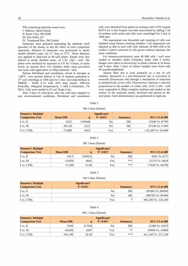
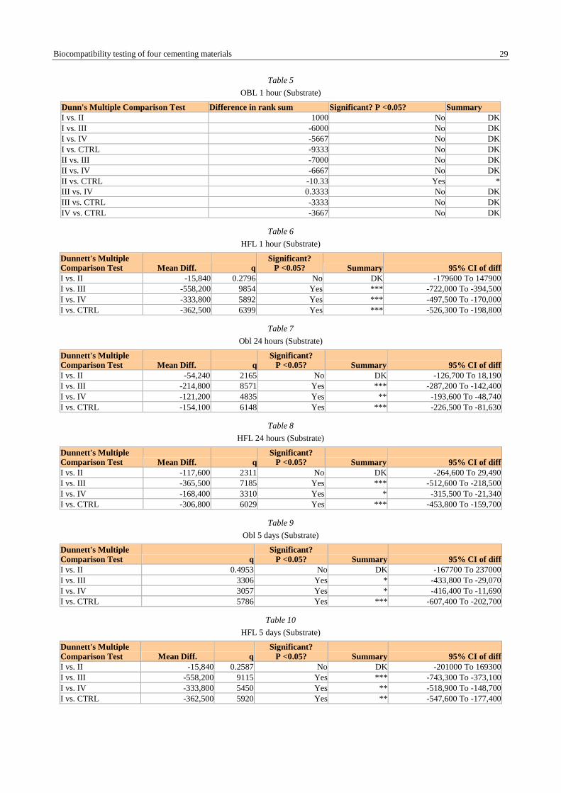
Effects of orthodontic devices cementing materials on the cells were visualized in microscopy (phase inverted Zeiss Axiovert with epifluorescence D1), 24 hours and 4 days of cultivation. Actual substrates (powders for Adhesor, Ketac Cem and Fuji Ortho) or full substrate (for Transbond Plus) showed an acceptable biocompatibility for substrates III and IV compared with control cells, grown on plastic (Fig. 1 and Fig.2).
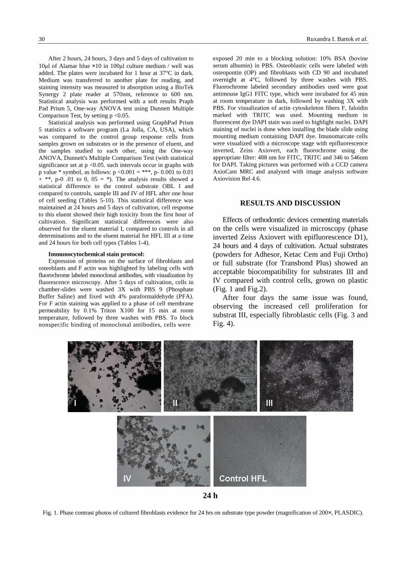
After four days the same issue was found, observing the increased cell proliferation for substrat III, especially fibroblastic cells (Fig. 3 and Fig. 4).
Fig. 1. Phase contrast photos of cultured fibroblasts evidence for 24 hrs on substrate type powder (magnification of 200×, PLASDIC).
24 h
Biocompatibility testing of four cementing materials 31
Fig. 2. Phase contrast photos of cultured osteoblast samples for 24 hrs on type substrate powder (magnification of 200×, PLASDIC).
Fig. 3. Phase contrast photos of cultured fibroblasts samples for 4 days on substrate type powder (magnification of 200×, PLASDIC).
24 h
4 days
Ruxandra I. Bartok et al. 32
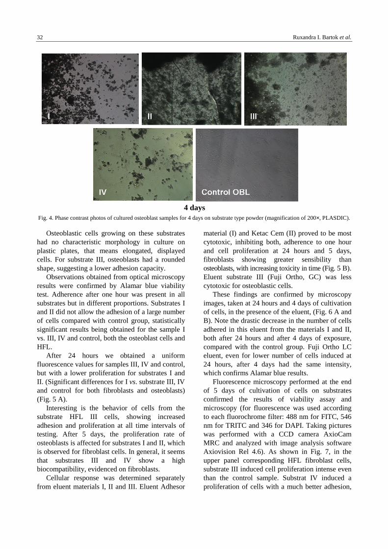
Fig. 4. Phase contrast photos of cultured osteoblast samples for 4 days on substrate type powder (magnification of 200×, PLASDIC).
Osteoblastic cells growing on these substrates had no characteristic morphology in culture on plastic plates, that means elongated, displayed cells. For substrate III, osteoblasts had a rounded shape, suggesting a lower adhesion capacity.
Observations obtained from optical microscopy results were confirmed by Alamar blue viability test. Adherence after one hour was present in all substrates but in different proportions. Substrates I and II did not allow the adhesion of a large number of cells compared with control group, statistically significant results being obtained for the sample I vs. III, IV and control, both the osteoblast cells and HFL.
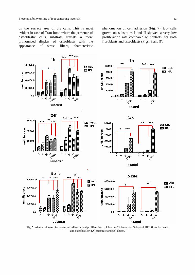
After 24 hours we obtained a uniform fluorescence values for samples III, IV and control, but with a lower proliferation for substrates I and II. (Significant differences for I vs. substrate III, IV and control for both fibroblasts and osteoblasts) (Fig. 5 A).
Interesting is the behavior of cells from the substrate HFL III cells, showing increased adhesion and proliferation at all time intervals of testing. After 5 days, the proliferation rate of osteoblasts is affected for substrates I and II, which is observed for fibroblast cells. In general, it seems that substrates III and IV show a high biocompatibility, evidenced on fibroblasts.
Cellular response was determined separately from eluent materials I, II and III. Eluent Adhesor
material (I) and Ketac Cem (II) proved to be most cytotoxic, inhibiting both, adherence to one hour and cell proliferation at 24 hours and 5 days, fibroblasts showing greater sensibility than osteoblasts, with increasing toxicity in time (Fig. 5 B). Eluent substrate III (Fuji Ortho, GC) was less cytotoxic for osteoblastic cells.
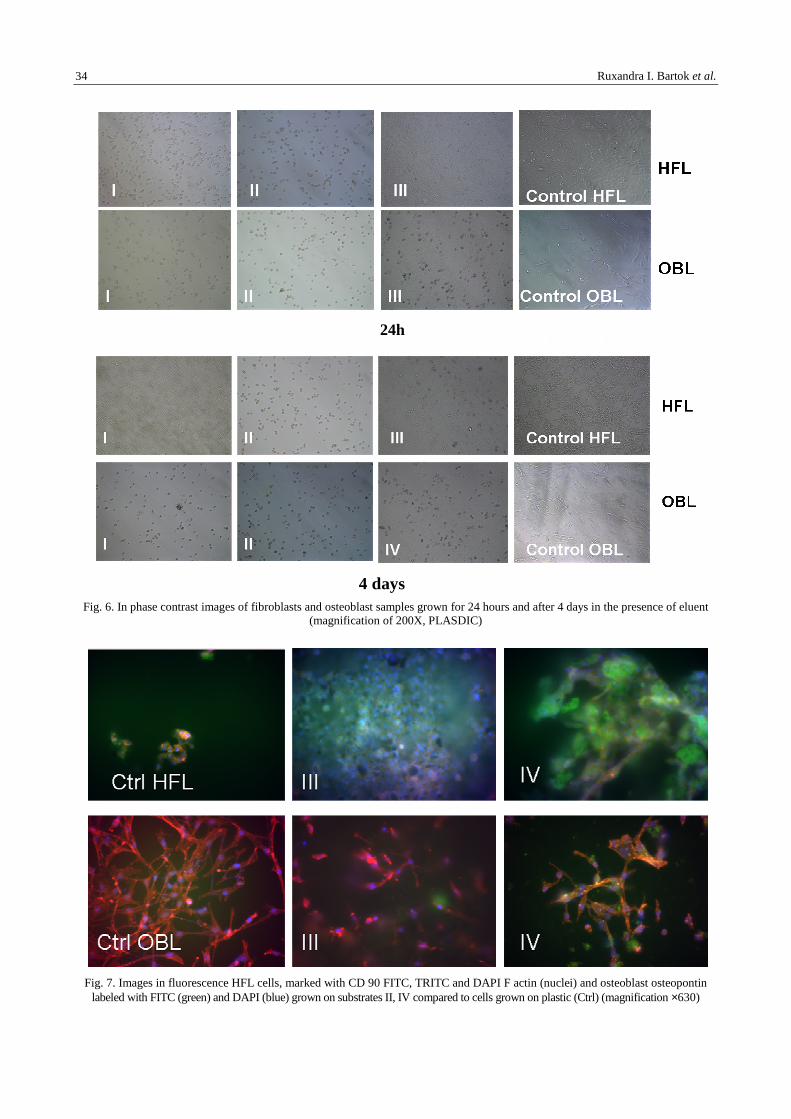
These findings are confirmed by microscopy images, taken at 24 hours and 4 days of cultivation of cells, in the presence of the eluent, (Fig. 6 A and B). Note the drastic decrease in the number of cells adhered in this eluent from the materials I and II, both after 24 hours and after 4 days of exposure, compared with the control group. Fuji Ortho LC eluent, even for lower number of cells induced at 24 hours, after 4 days had the same intensity, which confirms Alamar blue results.
Fluorescence microscopy performed at the end of 5 days of cultivation of cells on substrates confirmed the results of viability assay and microscopy (for fluorescence was used according to each fluorochrome filter: 488 nm for FITC, 546 nm for TRITC and 346 for DAPI. Taking pictures was performed with a CCD camera AxioCam MRC and analyzed with image analysis software Axiovision Rel 4.6). As shown in Fig. 7, in the upper panel corresponding HFL fibroblast cells, substrate III induced cell proliferation intense even than the control sample. Substrat IV induced a proliferation of cells with a much better adhesion,
4 days
Biocompatibility testing of four cementing materials 33
on the surface area of the cells. This is most evident in case of Transbond where the presence of osteoblastic cells substrate reveals a more pronounced display of osteoblasts with the appearance of stress fibers, characteristic
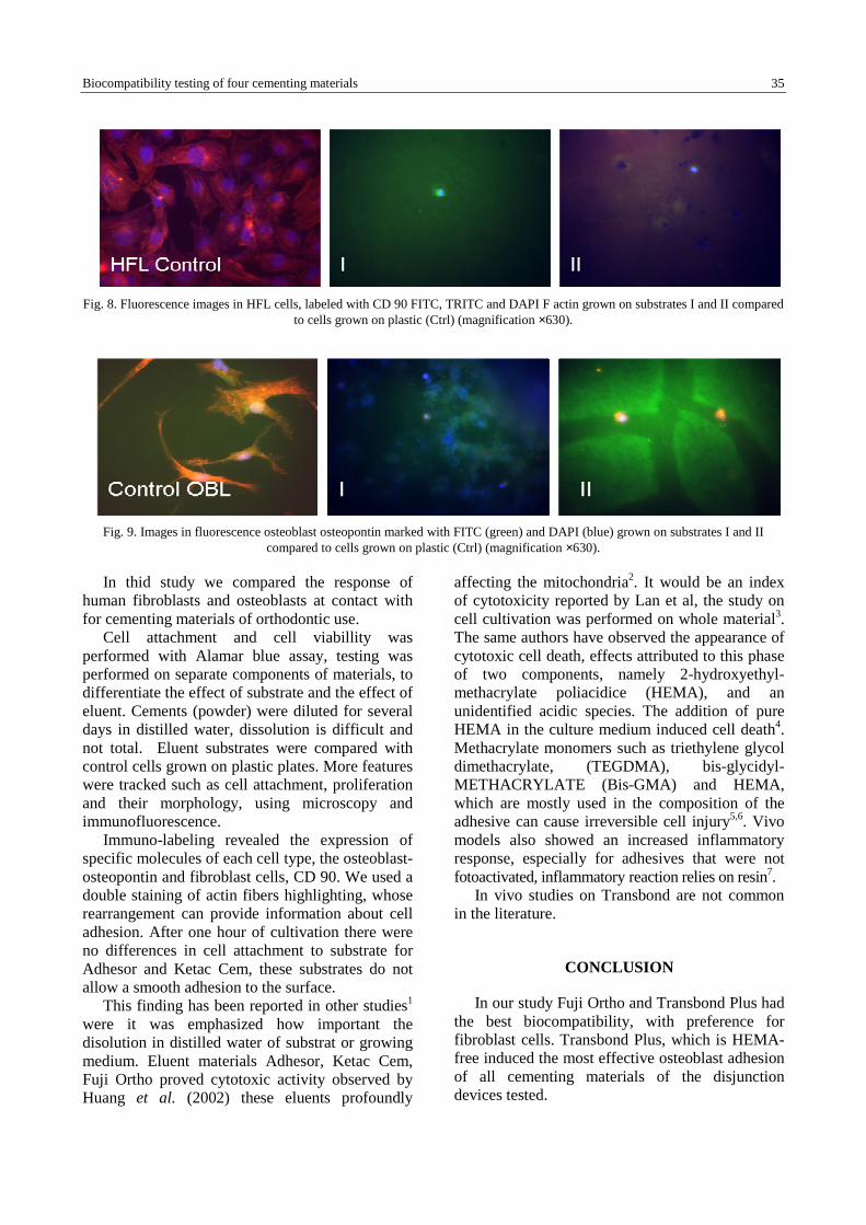
phenomenon of cell adhesion (Fig. 7). But cells grown on substrates I and II showed a very low proliferation rate compared to controls, for both fibroblasts and osteoblasts (Figs. 8 and 9).
Fig. 5. Alamar blue test for assessing adhesion and proliferation to 1 hour to 24 hours and 5 days of HFL fibroblast cells
and osteoblstilor: (A) substrate and (B) eluent.
Ruxandra I. Bartok et al. 34
Fig. 6. In phase contrast images of fibroblasts and osteoblast samples grown for 24 hours and after 4 days in the presence of eluent
(magnification of 200X, PLASDIC)
Fig. 7. Images in fluorescence HFL cells, marked with CD 90 FITC, TRITC and DAPI F actin (nuclei) and osteoblast osteopontin
labeled with FITC (green) and DAPI (blue) grown on substrates II, IV compared to cells grown on plastic (Ctrl) (magnification ×630)
4 days
24h
Biocompatibility testing of four cementing materials 35
Fig. 8. Fluorescence images in HFL cells, labeled with CD 90 FITC, TRITC and DAPI F actin grown on substrates I and II compared
to cells grown on plastic (Ctrl) (magnification ×630).
Fig. 9. Images in fluorescence osteoblast osteopontin marked with FITC (green) and DAPI (blue) grown on substrates I and II
compared to cells grown on plastic (Ctrl) (magnification ×630).
In thid study we compared the response of human fibroblasts and osteoblasts at contact with for cementing materials of orthodontic use.
Cell attachment and cell viabillity was performed with Alamar blue assay, testing was performed on separate components of materials, to differentiate the effect of substrate and the effect of eluent. Cements (powder) were diluted for several days in distilled water, dissolution is difficult and not total. Eluent substrates were compared with control cells grown on plastic plates. More features were tracked such as cell attachment, proliferation and their morphology, using microscopy and immunofluorescence.
Immuno-labeling revealed the expression of specific molecules of each cell type, the osteoblast-osteopontin and fibroblast cells, CD 90. We used a double staining of actin fibers highlighting, whose rearrangement can provide information about cell adhesion. After one hour of cultivation there were no differences in cell attachment to substrate for Adhesor and Ketac Cem, these substrates do not allow a smooth adhesion to the surface.
This finding has been reported in other studies1 were it was emphasized how important the disolution in distilled water of substrat or growing medium. Eluent materials Adhesor, Ketac Cem, Fuji Ortho proved cytotoxic activity observed by Huang et al. (2002) these eluents profoundly
affecting the mitochondria2. It would be an index of cytotoxicity reported by Lan et al, the study on cell cultivation was performed on whole material3. The same authors have observed the appearance of cytotoxic cell death, effects attributed to this phase of two components, namely 2-hydroxyethyl-methacrylate poliacidice (HEMA), and an unidentified acidic species. The addition of pure HEMA in the culture medium induced cell death4. Methacrylate monomers such as triethylene glycol dimethacrylate, (TEGDMA), bis-glycidyl-METHACRYLATE (Bis-GMA) and HEMA, which are mostly used in the composition of the adhesive can cause irreversible cell injury5,6. Vivo models also showed an increased inflammatory response, especially for adhesives that were not fotoactivated, inflammatory reaction relies on resin7.
In vivo studies on Transbond are not common in the literature.
CONCLUSION
In our study Fuji Ortho and Transbond Plus had the best biocompatibility, with preference for fibroblast cells. Transbond Plus, which is HEMA-free induced the most effective osteoblast adhesion of all cementing materials of the disjunction devices tested.
Ruxandra I. Bartok et al. 36
REFERENCES
1. Yan F, Xiao Y, Li H, Haase H, Bartold PM. A comparison of the effects of two kinds of glass-ionomer cement on human gingival fibroblast attachment, proliferation and morphology in vitro. J Int Acad Periodontol. 2000 Jan;2(1):14-8.
2. Huang FM, Chang YC. Cytotoxicity of resin-based restorative materials on human pulp cell cultures. Oral Surg Oral Med Oral Pathol Oral Radiol Endod. 2002 Sep;94(3):361-5.
3. Lan WH, Lan WC, Wang TM, Lee YL, Tseng WY, Lin CP, Jeng JH, Chang MC. Cytotoxicity of conventional and modified glass ionomer cements. Oper Dent. 2003 May-Jun;28(3):251-9.
4. Oliva A, Della Ragione F, Salerno A, Riccio V, Tartaro G, Cozzolino A, D'Amato S, Pontoni G, Zappia V. Biocompatibility studies on glass ionomer cements by primary cultures of human osteoblasts. Biomaterials. 1996 Jul;17(13):1351-6.
5. Fujisawa S, Kadoma Y, Komoda Y. 1H and 13C NMR studies of the interaction of eugenol, phenol, and triethyleneglycol dimethacrylate with phospholipid liposomes as a model system for odontoblast membranes. J Dent Res. 1988;67(11):1438-41.
6. Geurtsen W, Spahl W, Muller K, Leyhausen G. Aqueous extracts from dentin adhesives contain cytotoxic chemicals. J Biomed Mater Res. 1999;48(16):772-7.
7. Machado NP, Moysés MR, Pereira AAC, Pereira LJ, Ribeiro JCR, Dias SC. Study of dentinal adhesives compatibility using histological analysis. Braz J Oral Sci. 2007;6(20):1289-94.