Embed Size (px)
Citation preview
Heather D. Maynard Department of Chemistry and Biochemistry & California NanoSystems Institute
University of California, Los Angeles, USA
Bioconjugate Materials: Nanopatterns of Biomolecules on
Surfaces
Topic of Today’s Lecture
This lecture will focus on nanomaterials research, specifically
combining NANO and BIO on surfaces

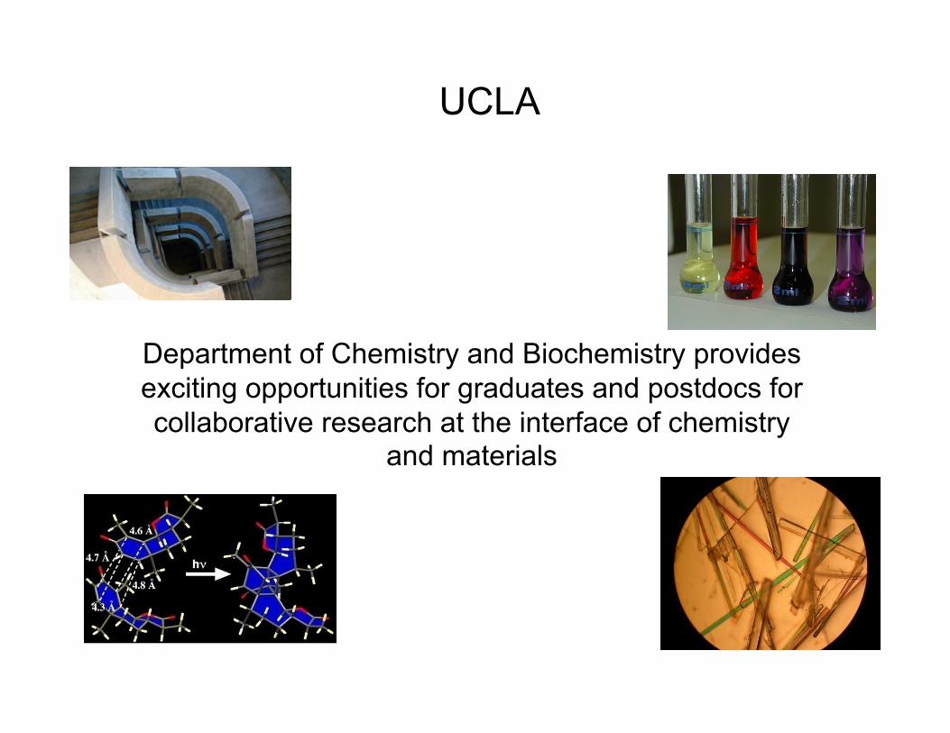
UCLA

UCLA
UCLA
Department of Chemistry and Biochemistry provides exciting opportunities for graduates and postdocs for collaborative research at the interface of chemistry
and materials
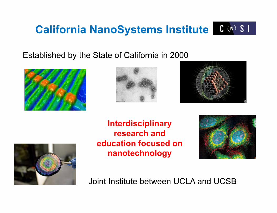
California NanoSystems Institute
Established by the State of California in 2000
Joint Institute between UCLA and UCSB
Interdisciplinary research and
education focused on nanotechnology
Topic of Today’s Lecture
This lecture will focus on nanomaterials research, specifically
combining NANO and BIO on surfaces
What is Nano?
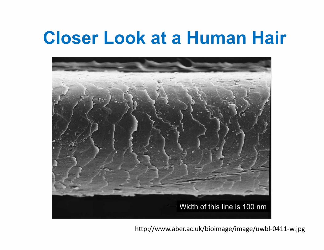
• Nanoscience is the study of objects measured in nanometers – 1-billionth of a meter – ~80,000 times smaller than the diameter of a
single human hair
h"p://www.aber.ac.uk/bioimage/image/uwbl-‐0411-‐w.jpg
Closer Look at a Human Hair
Width of this line is 100 nm
What is Nano?
• Nanoscience is the study of objects measured in nanometers – 1-billionth of a meter – ~80,000 times smaller than the diameter of a
single human hair – New properties emerge at the nanoscale
• Size and shape matter
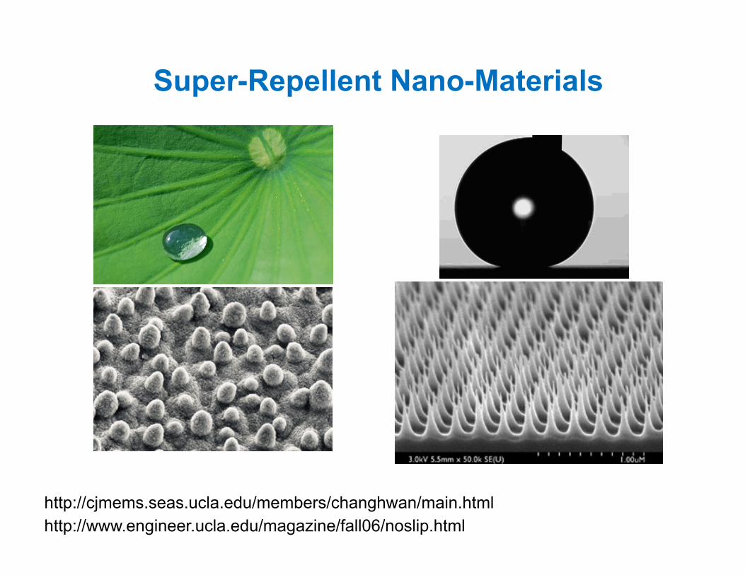
Super-Repellent Nano-Materials
http://www.engineer.ucla.edu/magazine/fall06/noslip.html http://cjmems.seas.ucla.edu/members/changhwan/main.html
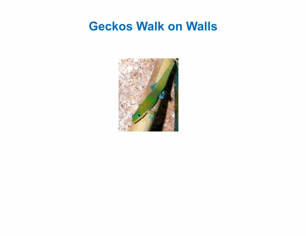
Geckos Walk on Walls
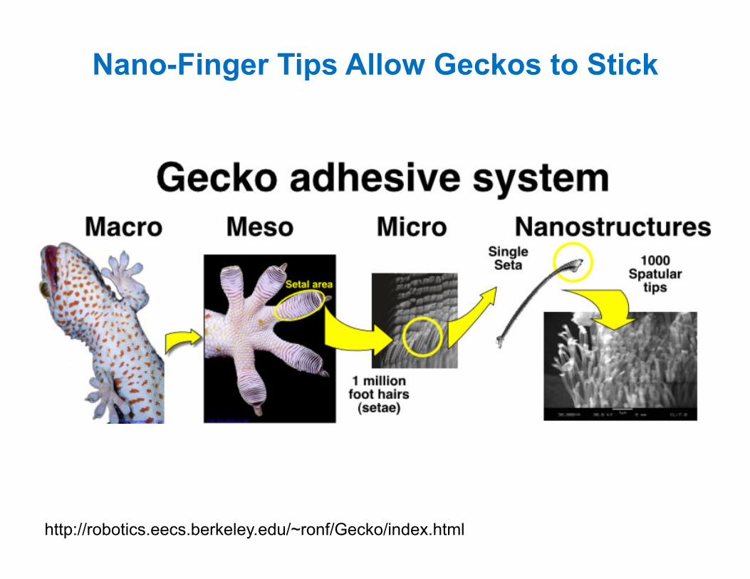
Nano-Finger Tips Allow Geckos to Stick
http://robotics.eecs.berkeley.edu/~ronf/Gecko/index.html

Man-Made Geckos
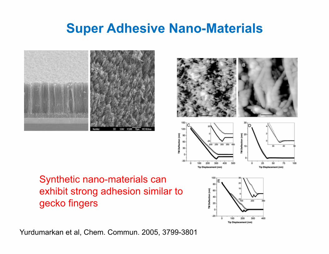
Super Adhesive Nano-Materials
Yurdumarkan et al, Chem. Commun. 2005, 3799-3801
Synthetic nano-materials can exhibit strong adhesion similar to gecko fingers
Topic of Today’s Lecture
This lecture will focus on nanomaterials research, specifically
combining NANO and BIO on surfaces
Protein
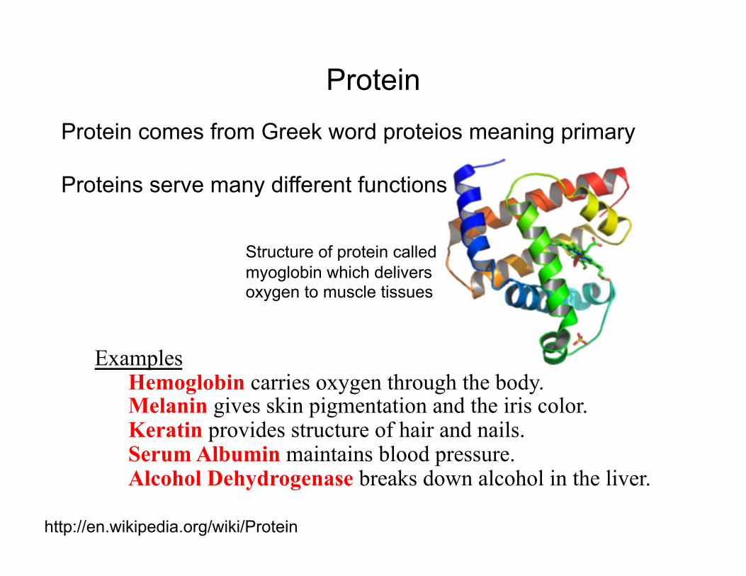
Protein comes from Greek word proteios meaning primary
Proteins serve many different functions
Examples Hemoglobin carries oxygen through the body. Melanin gives skin pigmentation and the iris color. Keratin provides structure of hair and nails. Serum Albumin maintains blood pressure. Alcohol Dehydrogenase breaks down alcohol in the liver.
Protein
http://en.wikipedia.org/wiki/Protein
Structure of protein called myoglobin which delivers oxygen to muscle tissues
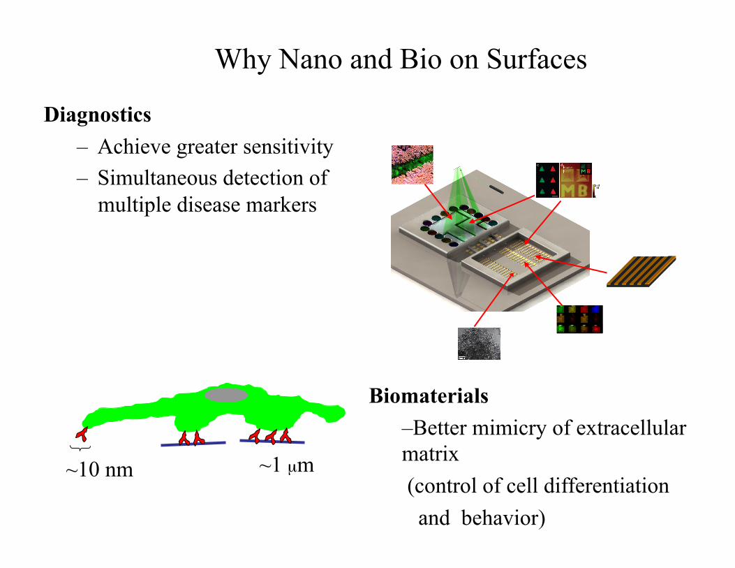
Diagnostics – Achieve greater sensitivity – Simultaneous detection of
multiple disease markers
Why Nano and Bio on Surfaces
~10 nm ~1 µm
Biomaterials – Better mimicry of extracellular matrix (control of cell differentiation and behavior)
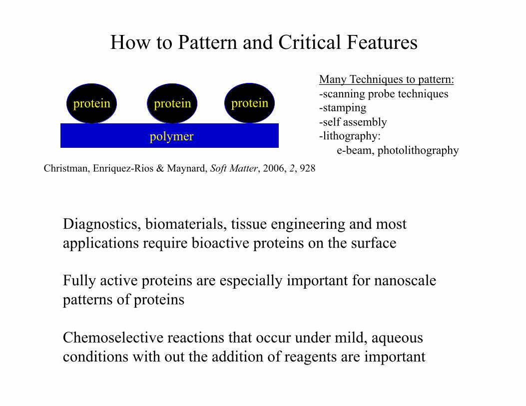
How to Pattern and Critical Features
protein protein protein
polymer
Many Techniques to pattern: -scanning probe techniques -stamping -self assembly -lithography: e-beam, photolithography
Christman, Enriquez-Rios & Maynard, Soft Matter, 2006, 2, 928
Diagnostics, biomaterials, tissue engineering and most applications require bioactive proteins on the surface
Fully active proteins are especially important for nanoscale patterns of proteins
Chemoselective reactions that occur under mild, aqueous conditions with out the addition of reagents are important
Outline
• Overview of techniques to pattern biomolecules at the nanoscale
• Example 1: Multiprotein patterns by e-beam lithography
• Example 2: Cell adhesive materials
Outline
• Overview of techniques to pattern biomolecules at the nanoscale
• Example 1: Multiprotein patterns by e-beam lithography
• Example 2: Cell adhesive materials
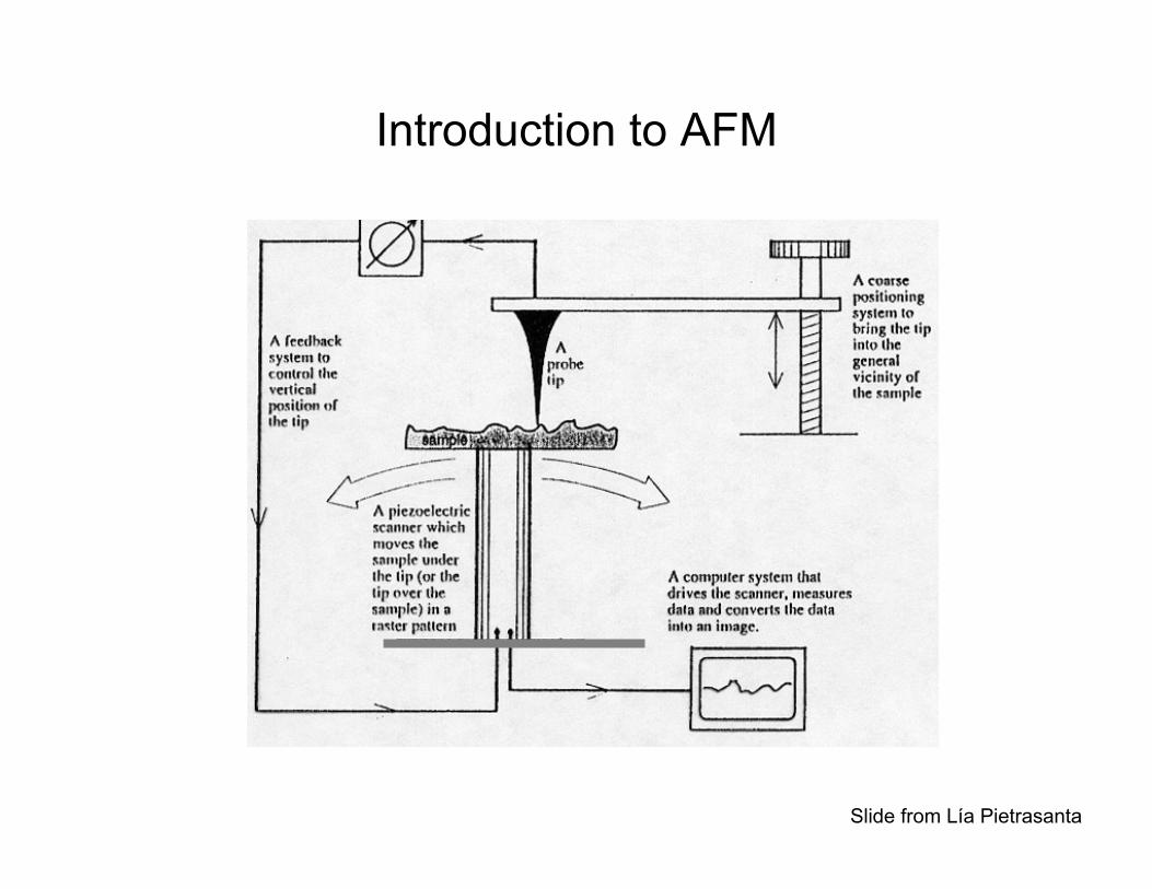
Introduction to AFM
Slide from Lía Pietrasanta
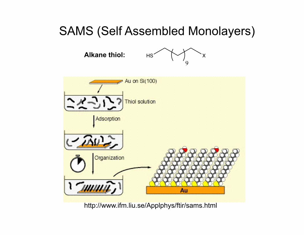
http://www.ifm.liu.se/Applphys/ftir/sams.html
SAMS (Self Assembled Monolayers)
HS X9
Alkane thiol:
AFM, Nanografting
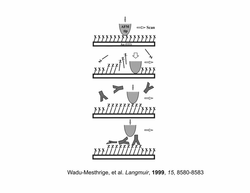
Wadu-Mesthrige, et al. Langmuir, 1999, 15, 8580-8583
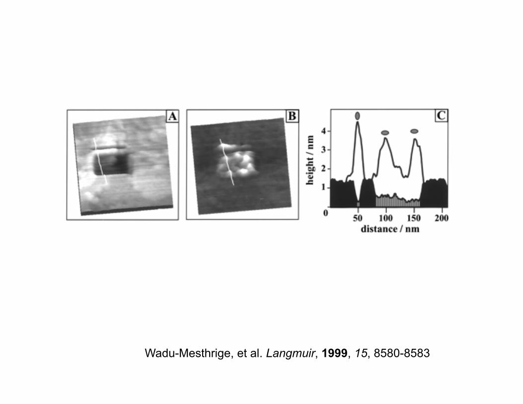
Wadu-Mesthrige, et al. Langmuir, 1999, 15, 8580-8583
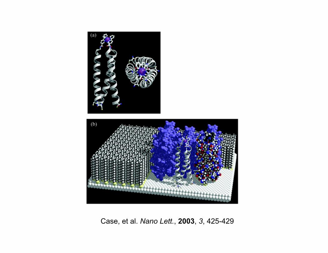
Case, et al. Nano Lett., 2003, 3, 425-429
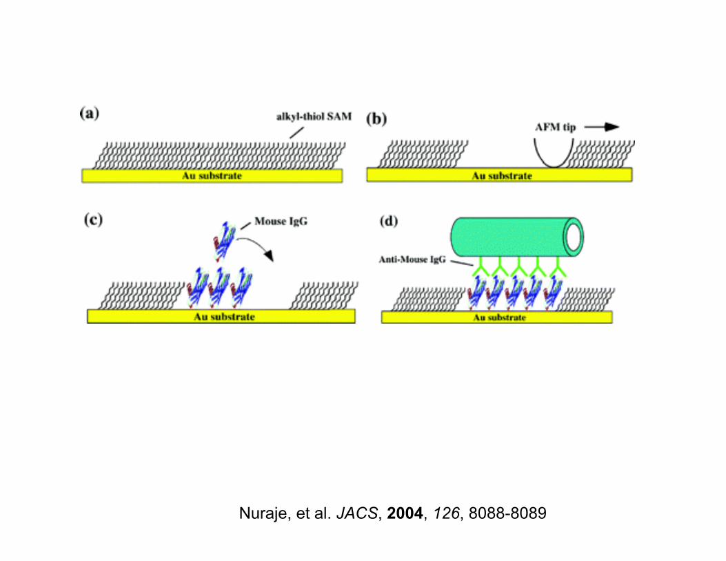
Nuraje, et al. JACS, 2004, 126, 8088-8089
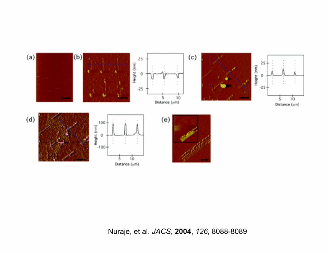
Nuraje, et al. JACS, 2004, 126, 8088-8089
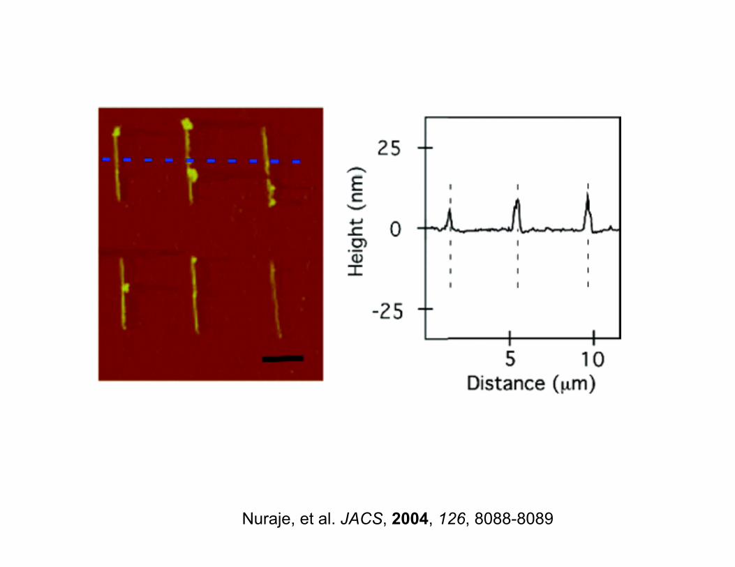
Nuraje, et al. JACS, 2004, 126, 8088-8089

AFM, Dip Pen Lithography
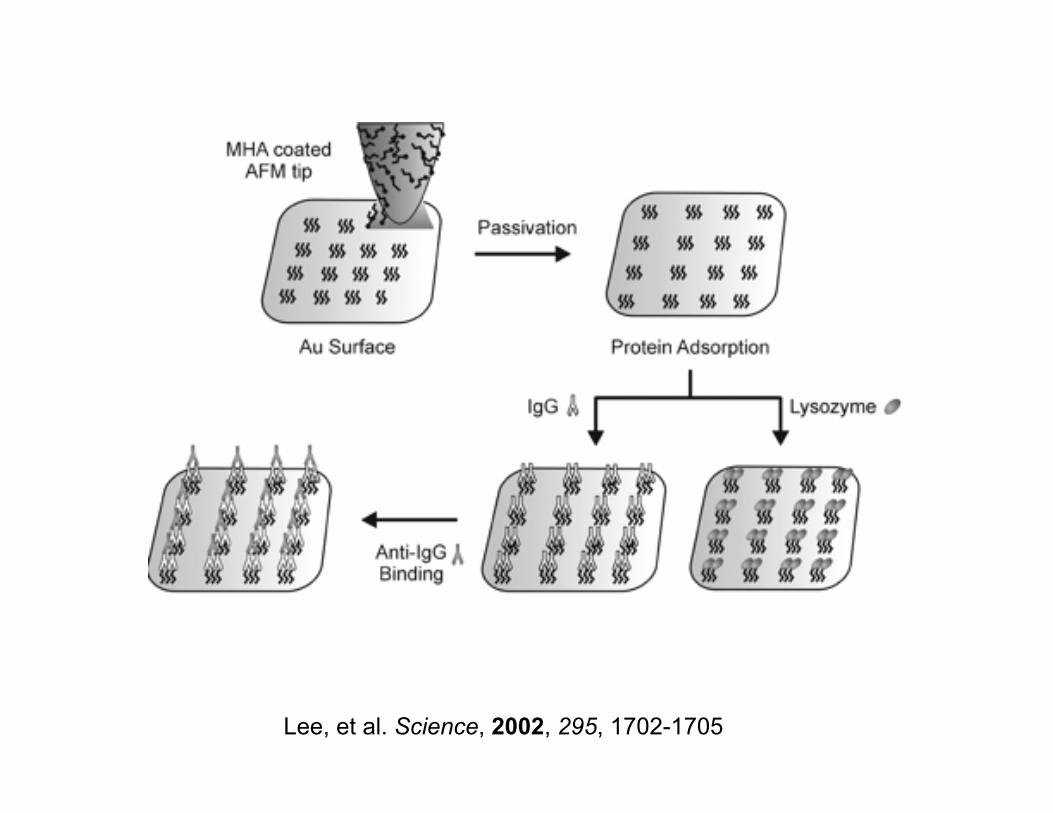
Lee, et al. Science, 2002, 295, 1702-1705
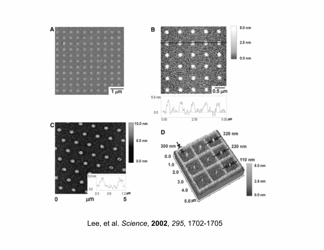
Lee, et al. Science, 2002, 295, 1702-1705
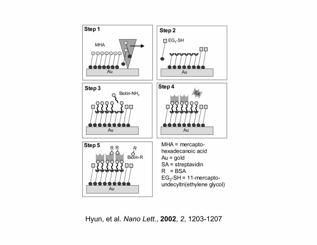
Hyun, et al. Nano Lett., 2002, 2, 1203-1207
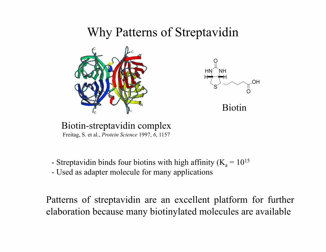
- Streptavidin binds four biotins with high affinity (Ka = 1015 - Used as adapter molecule for many applications
Biotin-streptavidin complex Freitag, S. et al., Protein Science 1997, 6, 1157
Biotin
Why Patterns of Streptavidin
Patterns of streptavidin are an excellent platform for further elaboration because many biotinylated molecules are available
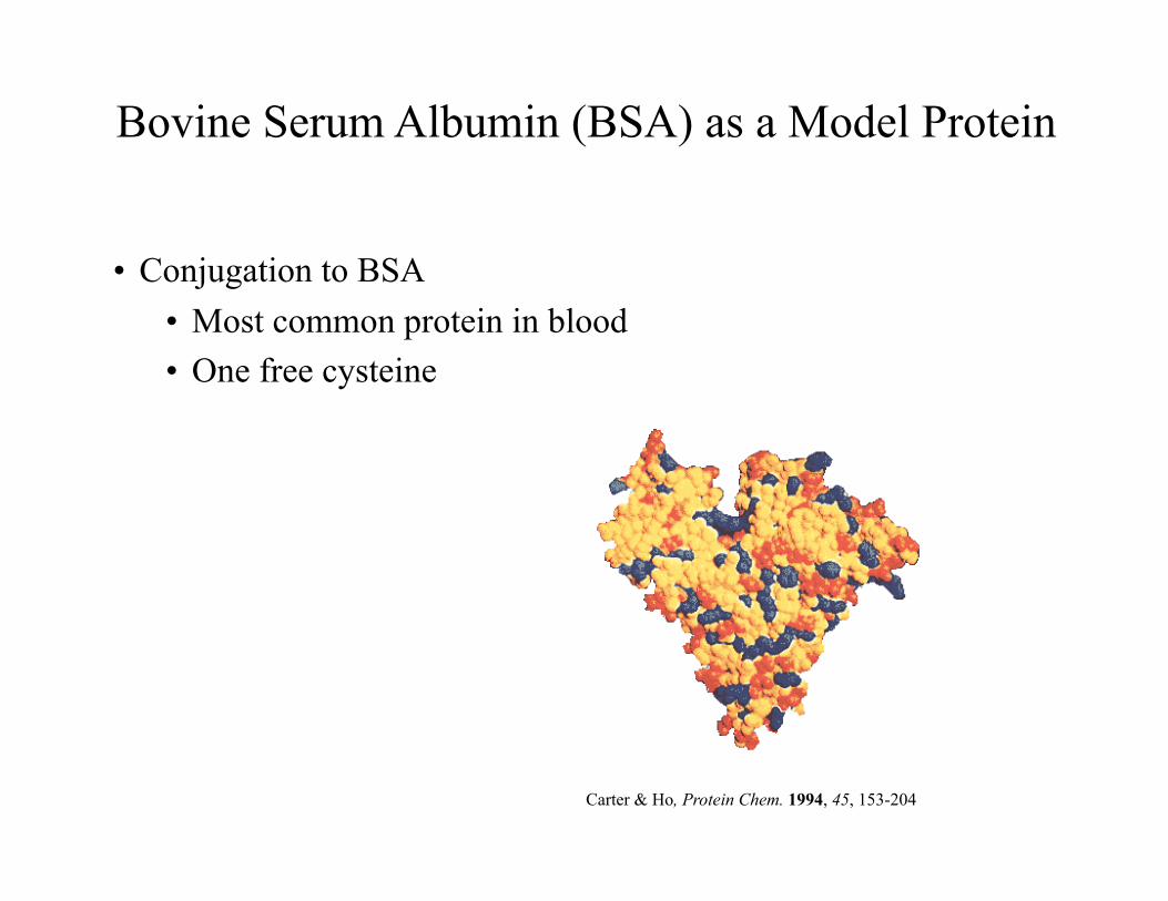
Bovine Serum Albumin (BSA) as a Model Protein
• Conjugation to BSA • Most common protein in blood • One free cysteine
Carter & Ho, Protein Chem. 1994, 45, 153-204
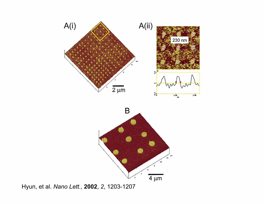
Hyun, et al. Nano Lett., 2002, 2, 1203-1207
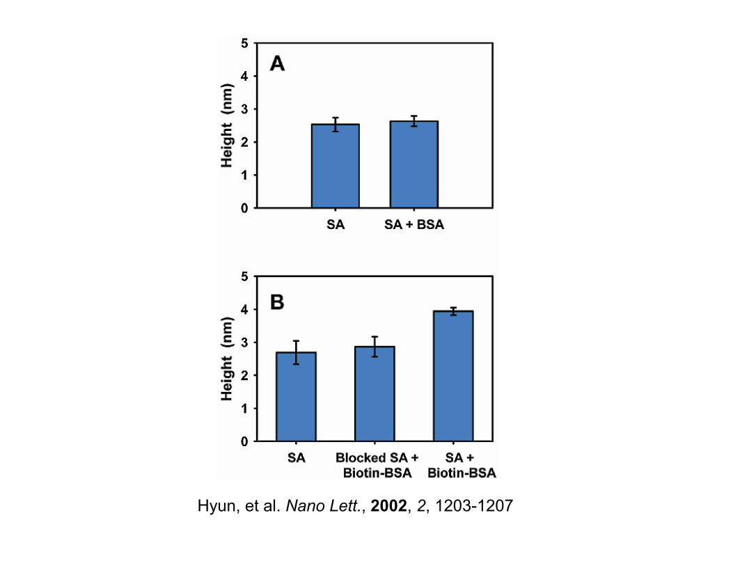
Hyun, et al. Nano Lett., 2002, 2, 1203-1207
Electron Beam (E-beam) Lithography
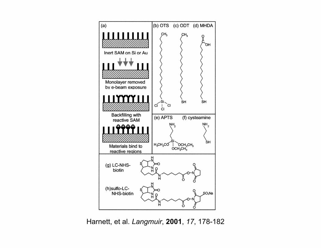
Harnett, et al. Langmuir, 2001, 17, 178-182
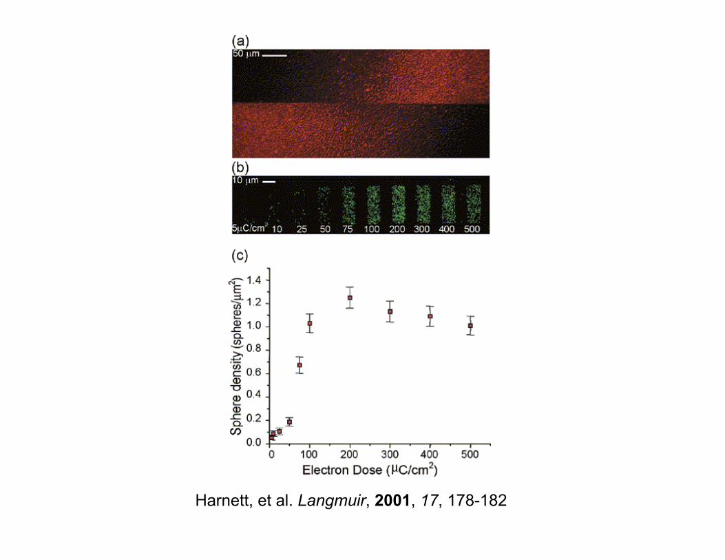
Harnett, et al. Langmuir, 2001, 17, 178-182
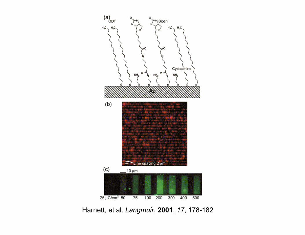
Harnett, et al. Langmuir, 2001, 17, 178-182
NanoStamping
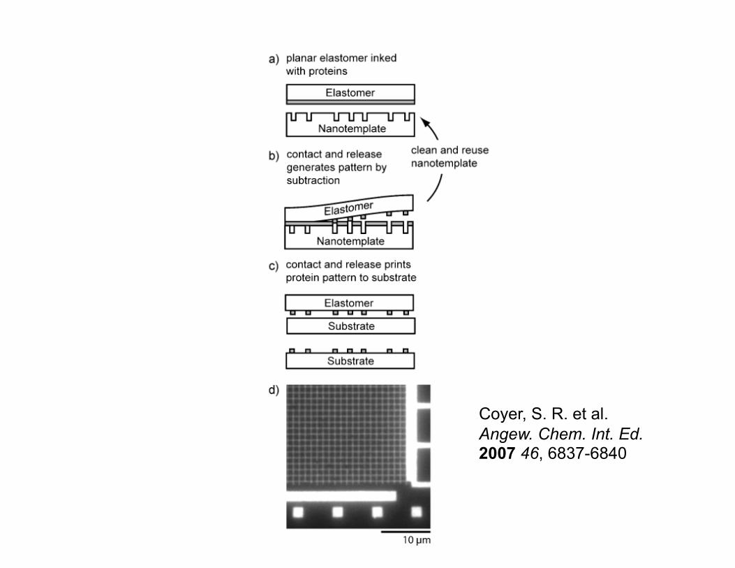
Coyer, S. R. et al. Angew. Chem. Int. Ed. 2007 46, 6837-6840
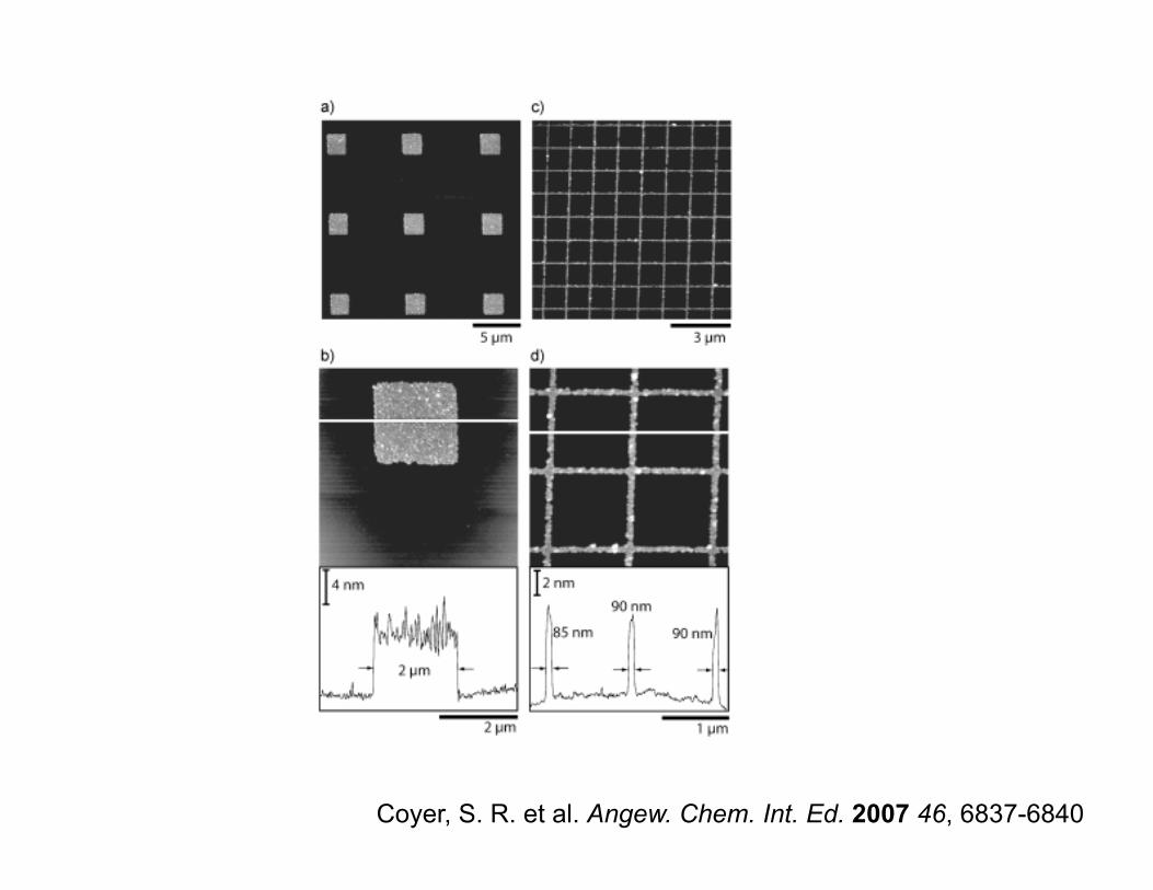
Coyer, S. R. et al. Angew. Chem. Int. Ed. 2007 46, 6837-6840

Self Assembly - DNA
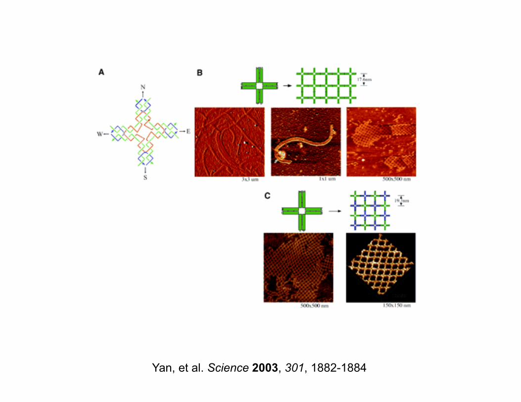
Yan, et al. Science 2003, 301, 1882-1884
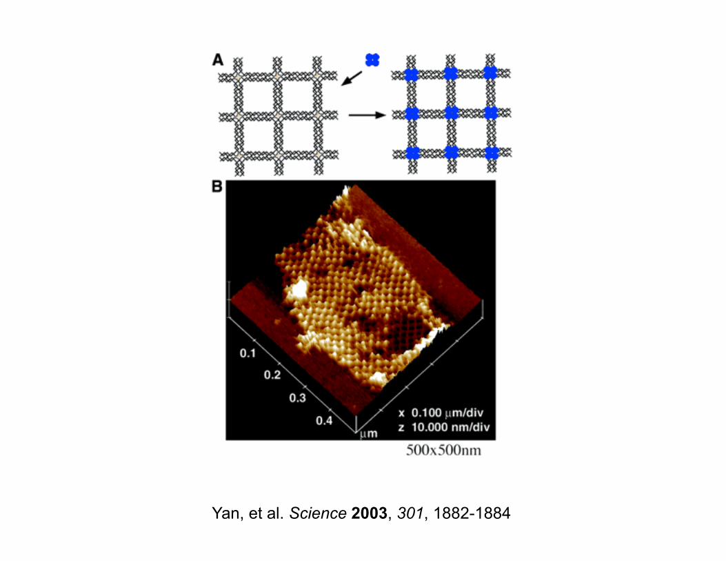
Yan, et al. Science 2003, 301, 1882-1884
Self Assembly - Proteins
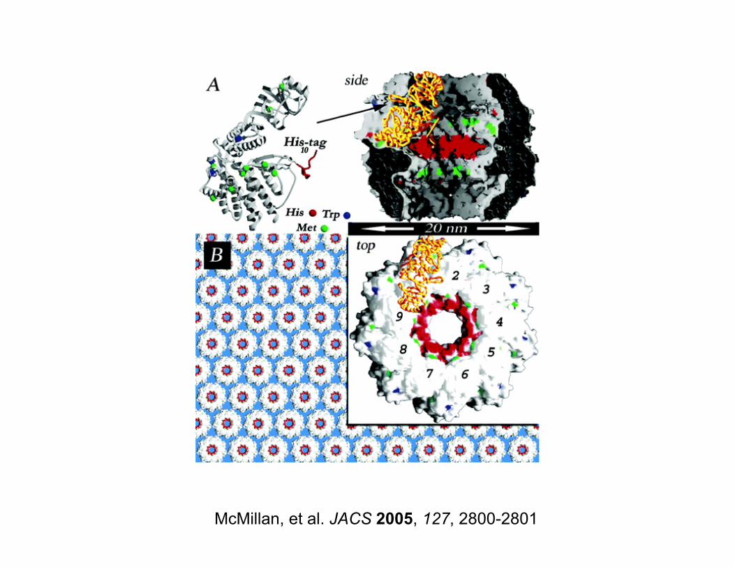
McMillan, et al. JACS 2005, 127, 2800-2801
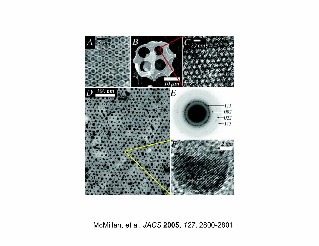
McMillan, et al. JACS 2005, 127, 2800-2801
Outline
• Overview of techniques to pattern biomolecules at the nanoscale
• Example 1: Multiprotein patterns by e-beam lithography
• Example 2: Cell adhesive materials
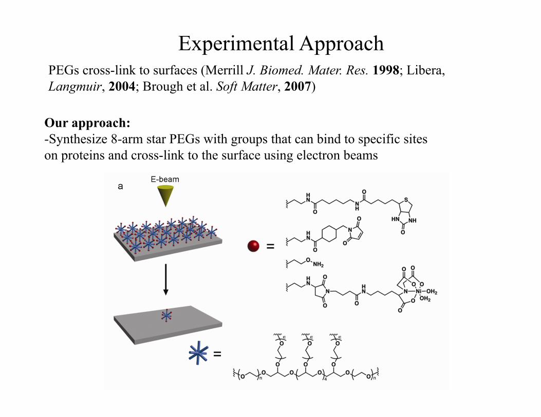
Experimental Approach
Our approach: -Synthesize 8-arm star PEGs with groups that can bind to specific sites on proteins and cross-link to the surface using electron beams
PEGs cross-link to surfaces (Merrill J. Biomed. Mater. Res. 1998; Libera, Langmuir, 2004; Brough et al. Soft Matter, 2007)
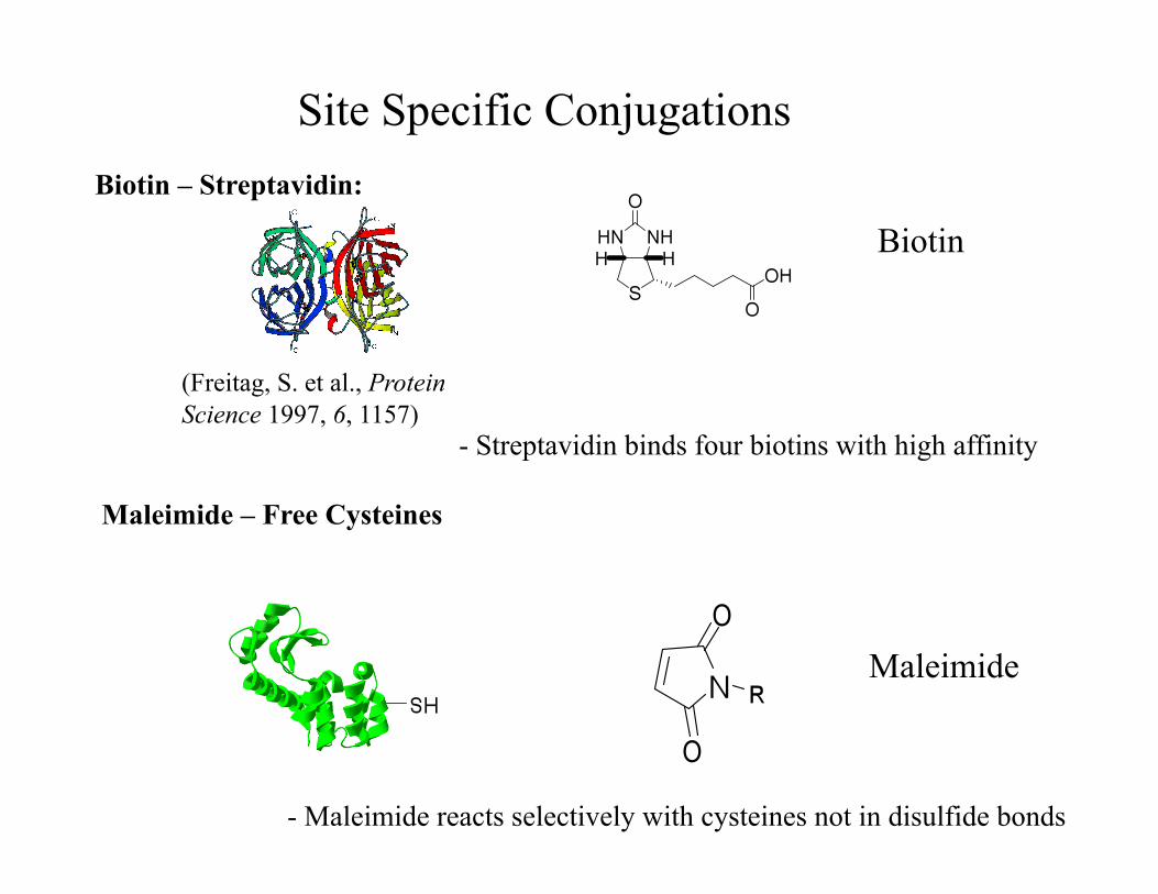
Site Specific Conjugations Biotin – Streptavidin:
Maleimide – Free Cysteines
- Streptavidin binds four biotins with high affinity
Biotin
(Freitag, S. et al., Protein Science 1997, 6, 1157)
- Maleimide reacts selectively with cysteines not in disulfide bonds
Maleimide
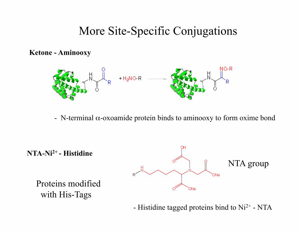
More Site-Specific Conjugations
NTA-Ni2+ - Histidine NTA group
Proteins modified with His-Tags
- Histidine tagged proteins bind to Ni2+ - NTA
Ketone - Aminooxy
- N-terminal α-oxoamide protein binds to aminooxy to form oxime bond
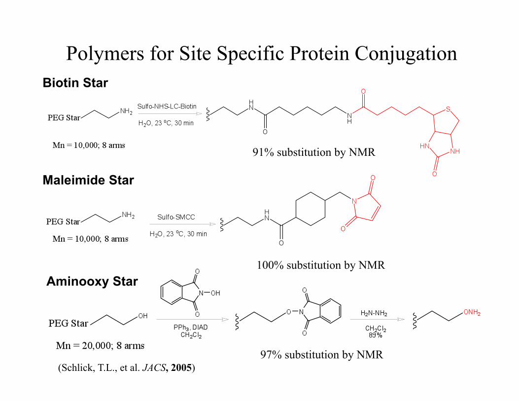
Polymers for Site Specific Protein Conjugation Biotin Star
Maleimide Star
Aminooxy Star
91% substitution by NMR
100% substitution by NMR
97% substitution by NMR (Schlick, T.L., et al. JACS, 2005)
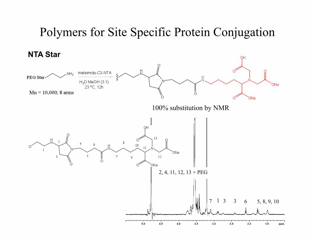
Polymers for Site Specific Protein Conjugation
NTA Star
100% substitution by NMR
2, 4, 11, 12, 13 + PEG
7 1 3 3 6 5, 8, 9, 10
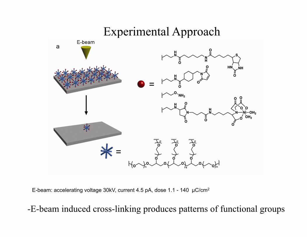
Experimental Approach
-E-beam induced cross-linking produces patterns of functional groups
E-beam: accelerating voltage 30kV, current 4.5 pA, dose 1.1 - 140 µC/cm2
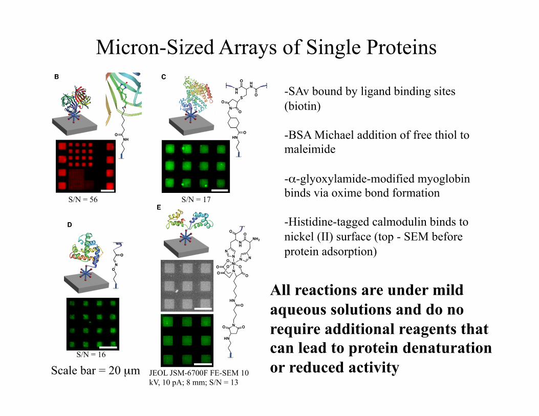
Micron-Sized Arrays of Single Proteins
-SAv bound by ligand binding sites (biotin)
-BSA Michael addition of free thiol to maleimide
-α-glyoxylamide-modified myoglobin binds via oxime bond formation
-Histidine-tagged calmodulin binds to nickel (II) surface (top - SEM before protein adsorption)
All reactions are under mild aqueous solutions and do no require additional reagents that can lead to protein denaturation or reduced activity Scale bar = 20 µm JEOL JSM-6700F FE-SEM 10
kV, 10 pA; 8 mm; S/N = 13
S/N = 56 S/N = 17
S/N = 16
Multicomponent Nanopatterns
For many desired applications, multiple proteins are required
Yet this is difficult to achieve See examples by: Mirkin and coworkers, JACS 2003; Angew. Chem. Int. Ed. 2003; Zhao, Banerjee, Matsui, JACS 2005; Coyer, S. R. et al. Angew. Chem. Int. Ed. 2007 46, 6837-6840; Tinazli, et al. Nature Nanotech. 2, 220-225 (2007).
Can we utilize e-beam lithography to achieve this? With e-beams, nanoscale spacings are possible.
Pattern PEGs with orthogonal reactivity side-by-side
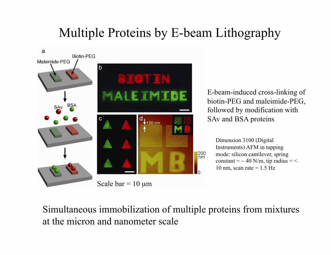
Multiple Proteins by E-beam Lithography
E-beam-induced cross-linking of biotin-PEG and maleimide-PEG, followed by modification with SAv and BSA proteins
Simultaneous immobilization of multiple proteins from mixtures at the micron and nanometer scale
Scale bar = 10 µm
Dimension 3100 (Digital Instruments) AFM in tapping mode: silicon cantilever, spring constant = ~ 40 N/m, tip radius = < 10 nm, scan rate = 1.5 Hz
Multilayer Three-Dimensional Patterning
PEG can be cross-linked to itself
Can we use this strategy to prepare 3D multilayer patterns of multiple proteins?
This would be interesting to produce multiplexed biomolecules in three-dimensional multilayer formats for a wide variety of applications such as site-isolation enzyme cascades, “nanoscale factories,” mimic natural complex structures such as protein-signaling assembles and viral capsids, present chemical and topographical cues to study and control cell adhesion
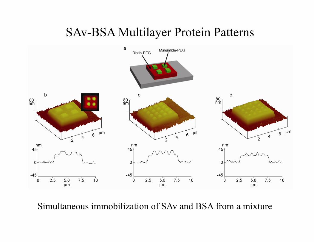
SAv-BSA Multilayer Protein Patterns
Simultaneous immobilization of SAv and BSA from a mixture
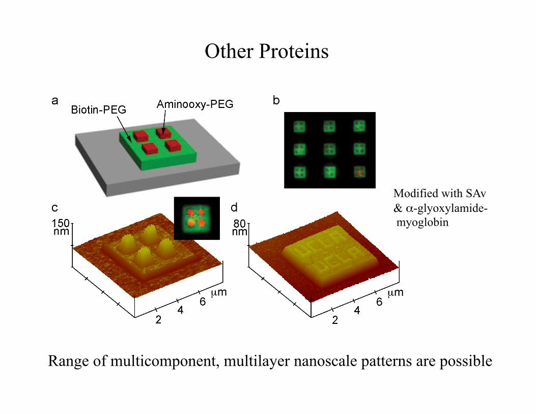
Other Proteins
Range of multicomponent, multilayer nanoscale patterns are possible
Modified with SAv & α-glyoxylamide- myoglobin
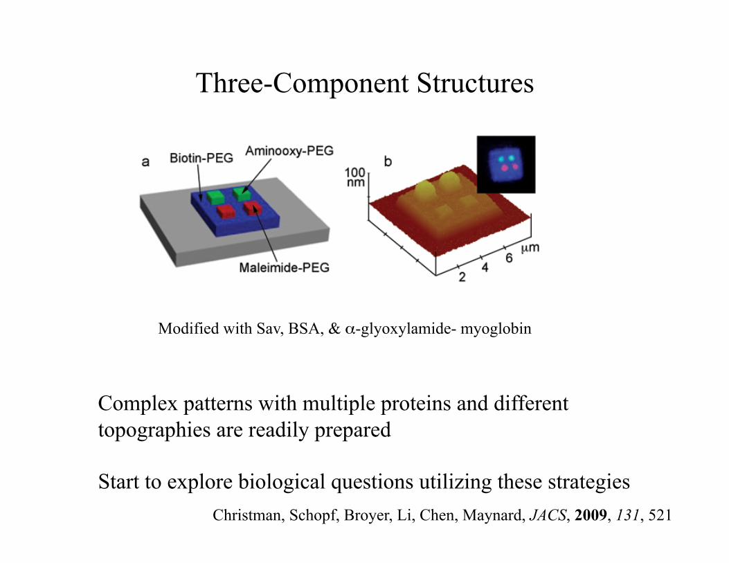
Three-Component Structures
Complex patterns with multiple proteins and different topographies are readily prepared
Start to explore biological questions utilizing these strategies
Modified with Sav, BSA, & α-glyoxylamide- myoglobin
Christman, Schopf, Broyer, Li, Chen, Maynard, JACS, 2009, 131, 521
Outline
• Overview of techniques to pattern biomolecules at the nanoscale
• Example 1: Multiprotein patterns by e-beam lithography
• Example 2: Cell adhesive materials
• Results in bioactive surfaces that mediate cell attachment
• Causes attachment of cells – Sometimes advantageous: osteoblasts in a bone
implant – Sometimes disadvantageous: platelets on the lining of
an vascular graft
• How and why do cells attach to these surfaces?
Protein adsorption
• Adsorbed adhesion proteins such as fibronectin, fibrinogen, and vitronectin
• Cells attach to adsorbed proteins as they do to native extracellular matrix (ECM) proteins
Cell Attachment
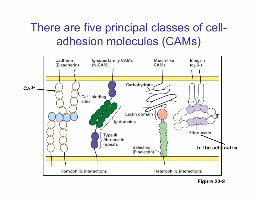
There are five principal classes of cell-adhesion molecules (CAMs)
Figure 22-2!
Ca 2+
In the cell matrix
Integrins
A family of membrane glycoproteins that bind to collagen, laminin, fibronectin and other ECM components.
Cell Surface Receptors for ECM Constituents
Involved in cell adhesion, migration, survival, growth, differentiation, and gene expression
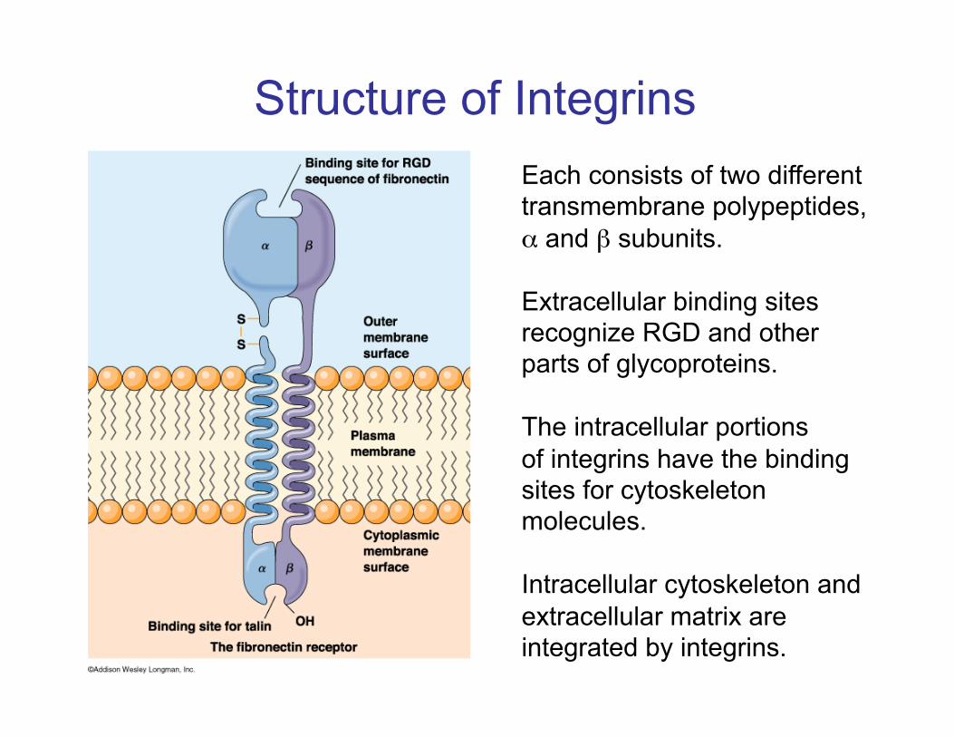
Structure of Integrins Each consists of two different transmembrane polypeptides, α and β subunits.
Extracellular binding sites recognize RGD and other parts of glycoproteins.
The intracellular portions of integrins have the binding sites for cytoskeleton molecules.
Intracellular cytoskeleton and extracellular matrix are integrated by integrins.
Receptors
• Specific amino acid sequences in ECM molecules bind to cell surface receptors (integrins)
– arginine-glycine-aspartic acid (RGD) tripeptide: 1st discovered in fibronectin (Pierschbacher and Ruoslahti, 1984)
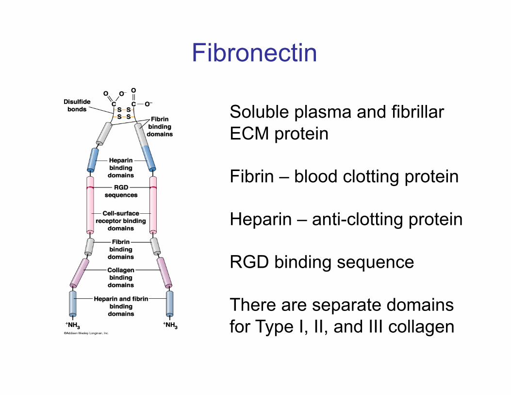
Soluble plasma and fibrillar ECM protein
Fibrin – blood clotting protein
Heparin – anti-clotting protein
RGD binding sequence
There are separate domains for Type I, II, and III collagen
Fibronectin
The influence between cytoskeleton and ECM is mutual.
By binding to integrin, fibronectin can trigger the reorganization of cytoskeleton inside the cell, which affects cell shape and motility.
Intracellular cytoskeleton can also influence the attachment and orientation of ECM.
Cytoskeleton-ECM
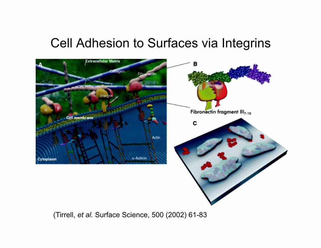
Cell Adhesion to Surfaces via Integrins
(Tirrell, et al. Surface Science, 500 (2002) 61-83
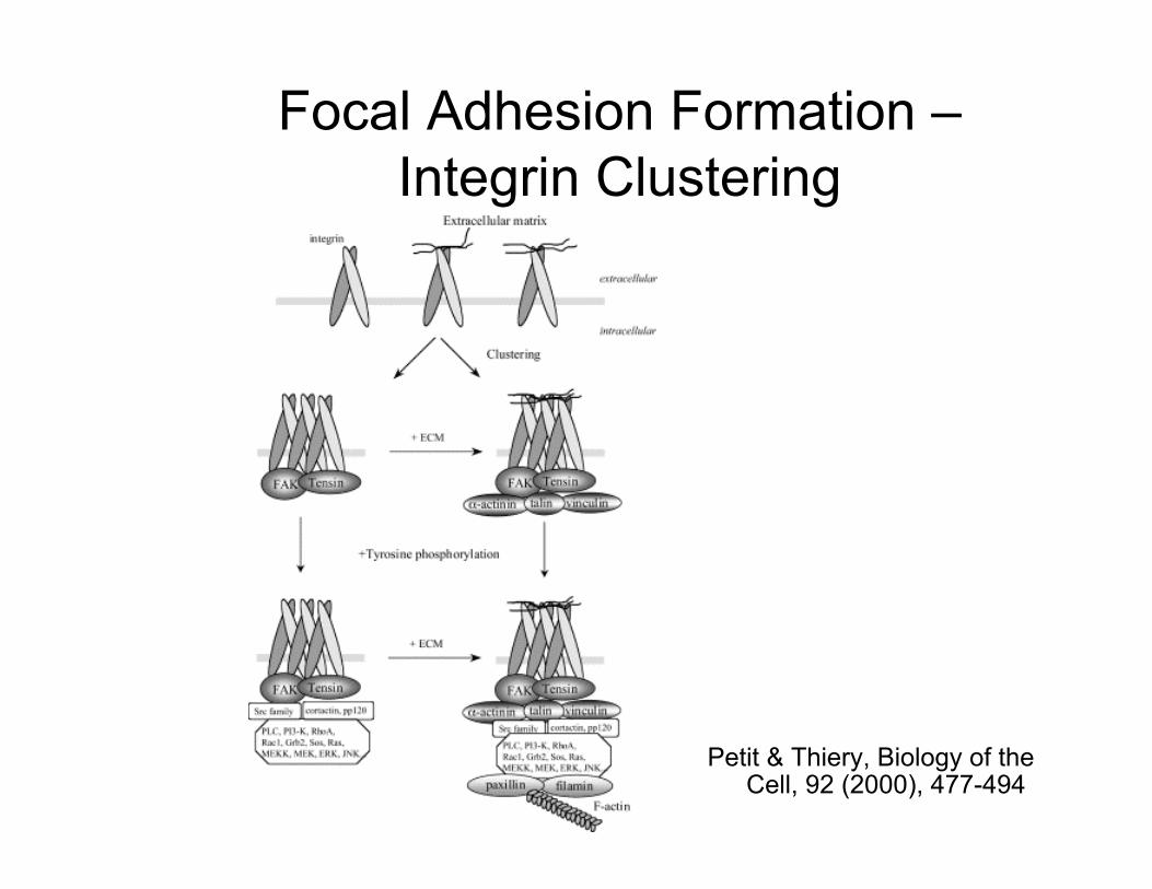
Focal Adhesion Formation – Integrin Clustering
Petit & Thiery, Biology of the Cell, 92 (2000), 477-494
Leahy, et al. Cell, 1996
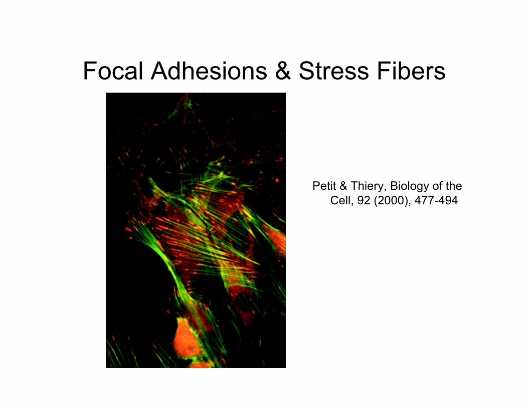
Focal Adhesions & Stress Fibers
Petit & Thiery, Biology of the Cell, 92 (2000), 477-494
Leahy, et al. Cell, 1996
Bioengineering Surfaces
• Coat surfaces with ECM molecules (for example, fibronectin)
• Design ligands and ligand-bearing surfaces to optimize attachment (and/or cell function) by mimicking the ECM
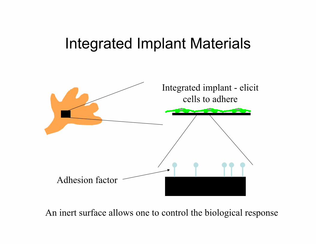
Integrated Implant Materials
Integrated implant - elicit cells to adhere
Inert material
An inert surface allows one to control the biological response
Adhesion factor
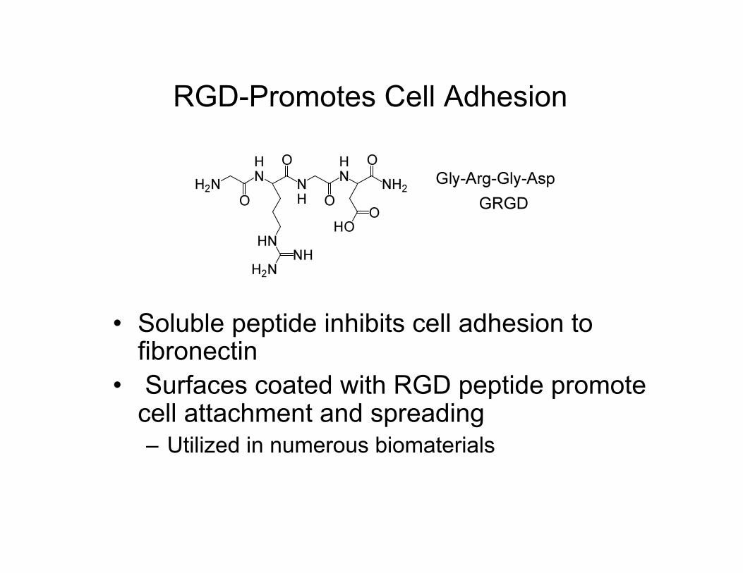
RGD-Promotes Cell Adhesion
• Soluble peptide inhibits cell adhesion to fibronectin
• Surfaces coated with RGD peptide promote cell attachment and spreading – Utilized in numerous biomaterials
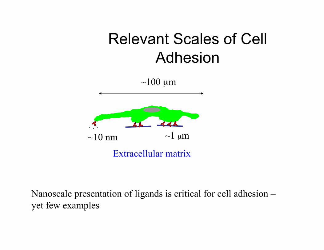
Relevant Scales of Cell Adhesion
Extracellular matrix
~10 nm
Nanoscale presentation of ligands is critical for cell adhesion – yet few examples
~1 µm
~100 µm
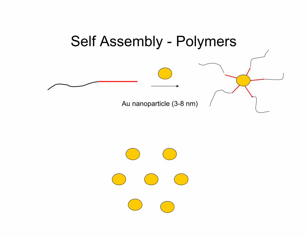
Self Assembly - Polymers
Au nanoparticle (3-8 nm)
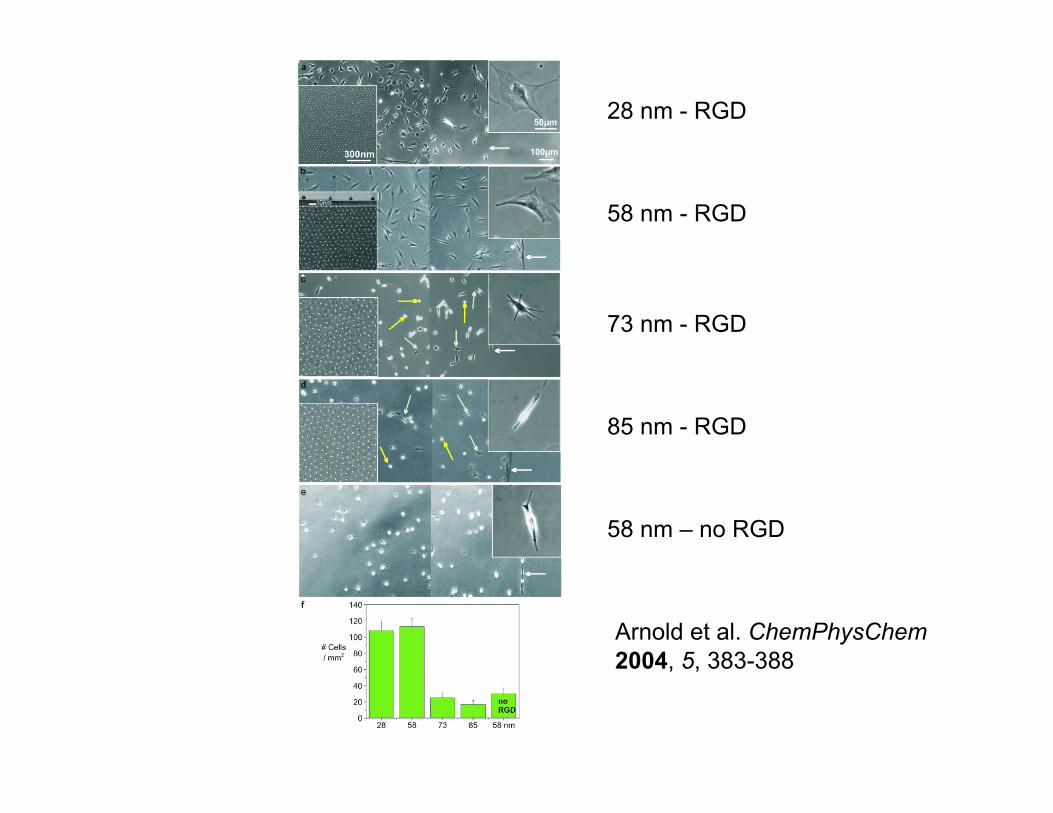
Arnold et al. ChemPhysChem 2004, 5, 383-388
28 nm - RGD
58 nm - RGD
85 nm - RGD
58 nm – no RGD
73 nm - RGD
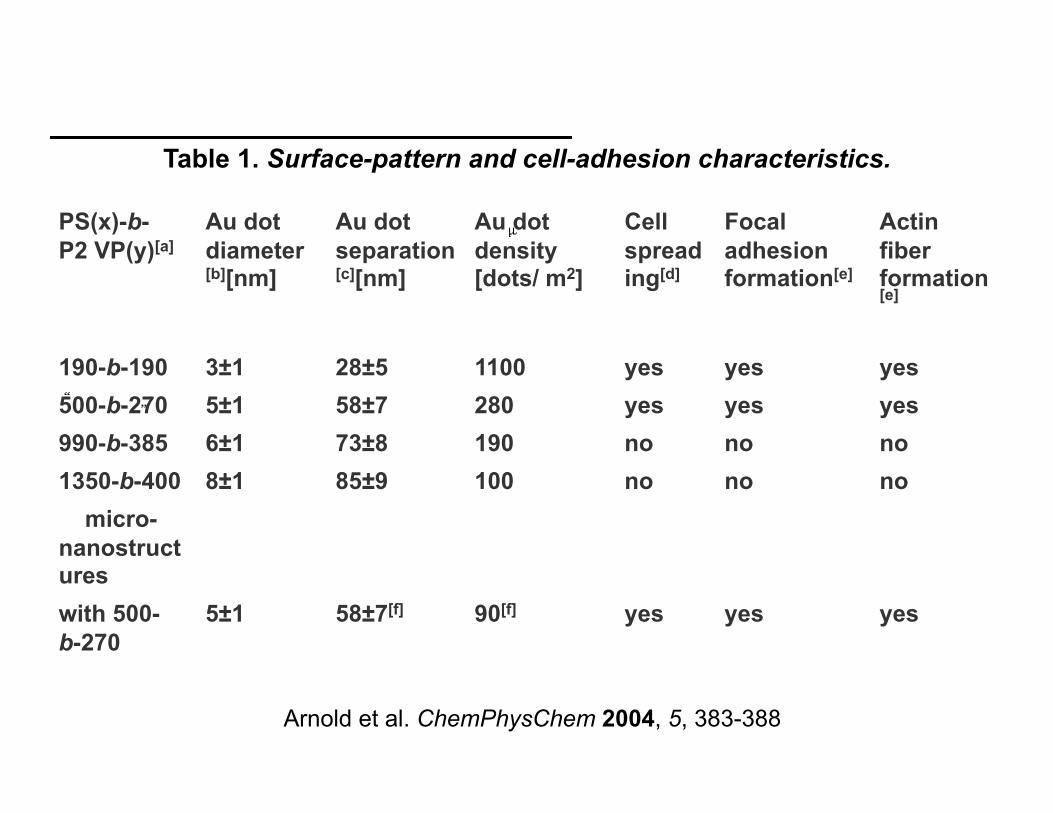
Arnold et al. ChemPhysChem 2004, 5, 383-388
Table 1. Surface-pattern and cell-adhesion characteristics.
PS(x)-b-P2 VP(y)[a]
Au dot diameter[b][nm]
Au dot separation[c][nm]
Au dot density [dots/ m2]
Cell spreading[d]
Focal adhesion formation[e]
Actin fiber formation[e]
190-b-190 3±1 28±5 1100 yes yes yes 500-b-270 5±1 58±7 280 yes yes yes 990-b-385 6±1 73±8 190 no no no 1350-b-400 8±1 85±9 100 no no no micro-nanostructures with 500-b-270
5±1 58±7[f] 90[f] yes yes yes
Self Assembly or E-beam lithography – Polymers
to probe cell adhesion at the nanoscale
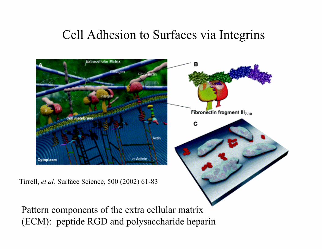
Cell Adhesion to Surfaces via Integrins
Tirrell, et al. Surface Science, 500 (2002) 61-83
Pattern components of the extra cellular matrix (ECM): peptide RGD and polysaccharide heparin
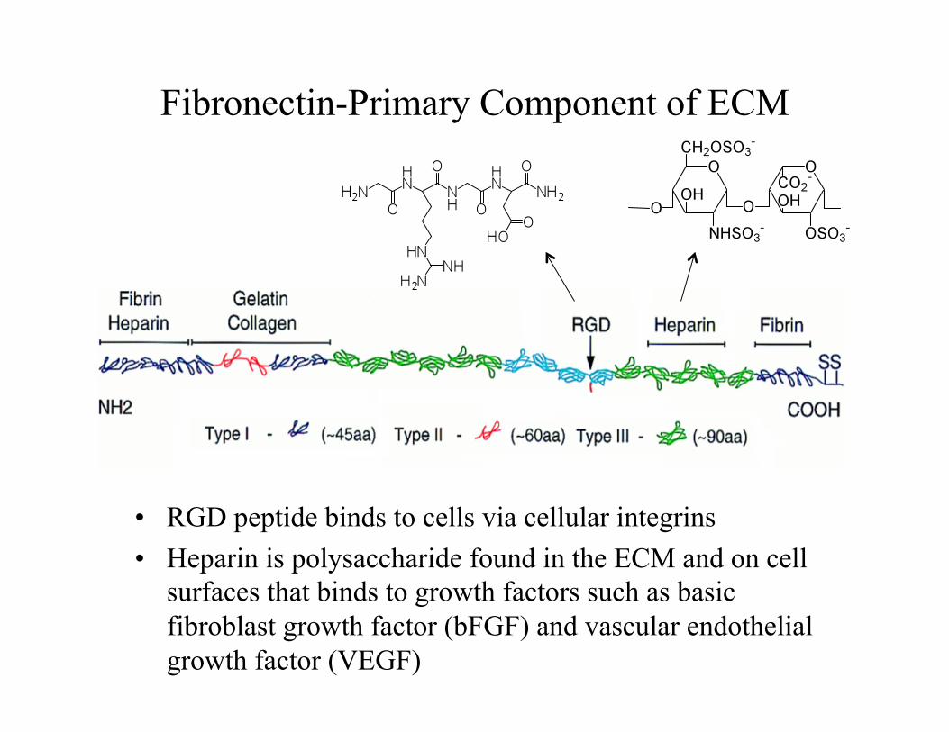
Fibronectin-Primary Component of ECM
• RGD peptide binds to cells via cellular integrins • Heparin is polysaccharide found in the ECM and on cell
surfaces that binds to growth factors such as basic fibroblast growth factor (bFGF) and vascular endothelial growth factor (VEGF)
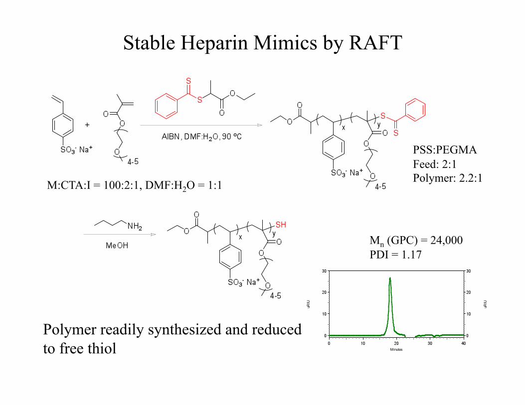
Stable Heparin Mimics by RAFT
Polymer readily synthesized and reduced to free thiol
M:CTA:I = 100:2:1, DMF:H2O = 1:1
PSS:PEGMA Feed: 2:1 Polymer: 2.2:1
Mn (GPC) = 24,000 PDI = 1.17
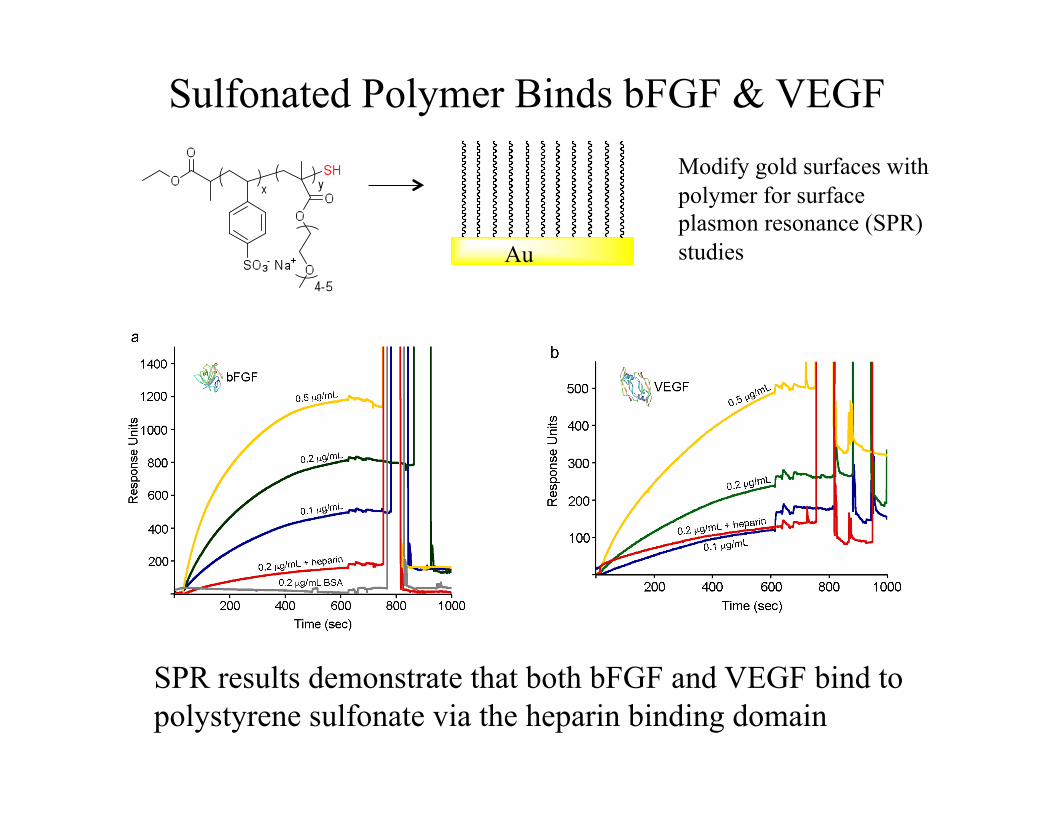
Sulfonated Polymer Binds bFGF & VEGF Modify gold surfaces with polymer for surface plasmon resonance (SPR) studies Au
SPR results demonstrate that both bFGF and VEGF bind to polystyrene sulfonate via the heparin binding domain
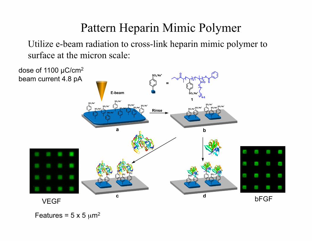
Pattern Heparin Mimic Polymer Utilize e-beam radiation to cross-link heparin mimic polymer to surface at the micron scale:
VEGF
Features = 5 x 5 µm2
bFGF
dose of 1100 µC/cm2 beam current 4.8 pA
VEGF
bFGF
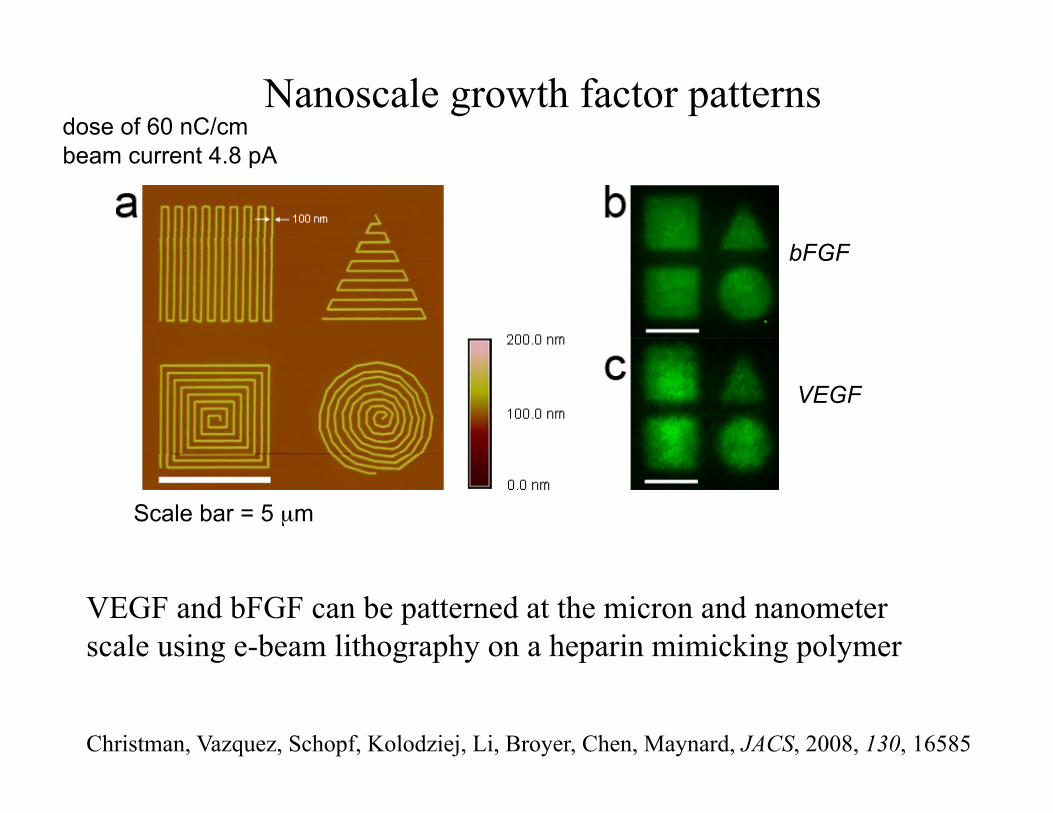
Nanoscale growth factor patterns
VEGF and bFGF can be patterned at the micron and nanometer scale using e-beam lithography on a heparin mimicking polymer
Scale bar = 5 µm
dose of 60 nC/cm beam current 4.8 pA
Christman, Vazquez, Schopf, Kolodziej, Li, Broyer, Chen, Maynard, JACS, 2008, 130, 16585
Topic of Today’s Lecture
Combining NANO and BIO on surfaces provides exciting
opportunities for engineering development, as well as application
Acknowledgements
Students and Postdocs
Dr. Karen Christman (UCSD) Rebecca Broyer Chris Kolodziej Dr. Ronald Li (PPG Aerospace) Vimary Vázquez-Dorbatt
Collaborators
Prof. Yong Chen, UCLA Eric Scopf
Funding
NSF CAREER SINAM NIH Amgen (New Faculty Award) Alfred P. Sloan Fellowship
Thank you!