Embed Size (px)
Citation preview
JOURNAL OF CLINICAL MICROBIOLOGY, Apr. 1990, p. 756-7630095-1137/90/040756-08$02.00/0Copyright C 1990, American Society for Microbiology
Purification and Characterization of a Pilin Specific for BrazilianPurpuric Fever-Associated Haemophilus influenza
Biogroup Aegyptius (H. aegyptius) StrainsROBBIN S. WEYANT,lt* WILLIAM F. BIBB,2 DAVID S. STEPHENS,3 BRIAN P. HOLLOWAY,4
WINSTON F. MOO-PENN,5 KRISTIN A. BIRKNESS,2 LETA O. HELSEL,2 AND LEONARD W. MAYER2
Department of Pathology and Laboratory Medicine' and Veterans Administration Medical Center and Departments ofMedicine, Microbiology, and Immunology,3 Emory University, Atlanta, Georgia 30322, and Meningitis and
Special Pathogens Branch, Division of Bacterial Diseases,2 Division of Viral Diseases,4 andDivision ofHost Factors,5 Centers for Disease Control, Atlanta, Georgia 30333
Received 30 June 1989/Accepted 28 November 1989
Brazilian purpuric fever (BPF) is a recently described fatal pediatric disease caused by systemic infectionwith Haemophilus influenza biogroup aegyptius. Previous studies have shown that all H. influenza biogroupaegyptius strains isolated from BPF cases and case contacts share several unique phenotypic and genotypiccharacteristics that differentiate them from other H. influenza biogroup aegyptius strains isolated fromconjunctivitis cases in Brazil. One key characteristic of this BPF clone is reactivity in a BPF-specific monoclonalantibody enzyme-linked immunosorbent assay. We have purified and partially characterized a pilin, referredto as the 25-kilodalton (kDa) protein. Aggregates of this protein contain a heat-labile epitope which isrecognized by a monoclonal antibody used in the BPF-specific enzyme-linked immunosorbent assay. Theprotein has a molecular weight of approximately 25,000, is insoluble in most detergents, and fractionates withouter membrane vesicles after LiCl extraction. Biochemical analysis of the 25-kDa protein shows it to have an
amino acid composition similar but not identical to that of the H. influenza type b pilin. The sequence of 20N-terminal amino acids of the 25-kDa protein shows almost complete homology with the N terminus of the H.influenzae type b pilin and the types 1 and P pilins of Escherichia coli. Transmission electron microscopicanalysis of the purified protein shows the presence of filamentous structures similar in morphology to those ofH. influenza pili. Reactivity between the 25-kDa protein and the BPF-specific monoclonal antibody isdemonstrated by Western blotting (immunoblotting) and colloidal gold-enhanced immunoelectron microscopy.Hemadsorption analysis shows that expression of this protein is associated with increases in piliated cells andenhanced binding of these cells to human erythrocytes. These studies indicate that expression of the 25-kDaprotein is a characteristic unique to the BPF clone and suggest that this protein plays a role in the pathogenesisof BPF.
Brazilian purpuric fever (BPF) is a newly described fatalpediatric disease caused by systemic infection with Hae-mophilus influenza biogroup aegyptius (4, 5, 8, 9). Exceptfor two cases identified in Australia (27, 29), all known cases
of BPF have occurred in the Sao Paulo and Parana states ofBrazil. Recently, Brenner et al. (6) reported the results of a
comprehensive investigation of H. influenza biogroup ae-
gyptius isolates obtained from children with conjunctivitisand children with BPF in Sao Paulo State during outbreaksin 1985 and 1986. This study concluded that all cases of BPFof Brazilian origin were caused by a unique H. influenzabiogroup aegyptius clone, termed the BPF clone, which hasthe following features: (i) a 24-megadalton plasmid with an
identical restriction endonuclease profile, (ii) a characteristicwhole-cell sodium dodecyl sulfate-polyacrylamide gel elec-trophoresis (SDS-PAGE) profile, (iii) a characteristic isoen-zyme electrophoretic profile, (iv) the ability to agglutinate inrabbit antisera made by immunization with a BPF casestrain, (v) a positive enzyme-linked immunosorbent assay
(ELISA) reaction in an assay that uses monoclonal antibod-
* Corresponding author.t Present address: National Institutes of Health, Building 28A,
9000 Rockville Pike, Bethesda, MD 20892.
ies made by immunization with a BPF case strain, (vi) one oftwo characteristic rRNA hybridization patterns, and usually(vii) resistance to sulfamethoxazole-trimethoprim.An incidental finding of this study was the identification of
a protein with an apparent molecular weight of 25,000 thatdisplayed an unusual charcoal gray color in silver-stainedwhole-cell SDS-PAGE profiles of BPF clone strains. Thisprotein was initially detected in 59% (27 of 46) of BPF cloneisolates versus 8% (3 of 36) of non-BPF clone H. influenzabiogroup aegyptius strains of Brazilian origin. Interestingly,the three non-BPF clone strains in which the 25-kilodalton(kDa) protein was detected exhibited some of the BPF clonecharacteristics and were epidemiologically associated withBPF. They contained a 24-megadalton plasmid with a re-
striction pattern similar but not identical to that of theBPF-associated plasmid, and all three gave positive resultsin the BPF-specific agglutination assays and ELISAs. Noother non-BPF clone strains exhibited these characteristics.These observations led us to investigate the 25-kDa proteinas a potential virulence factor for BPF or as a diagnosticmarker for the BPF clone.
In this study we describe the purification of the 25-kDaprotein along with biochemical characterization and trans-mission electron microscopic analysis, which indicate it tobe a pilin similar in composition and morphology to other
756
Vol. 28, No. 4
Dow
nloa
ded
from
http
s://j
ourn
als.
asm
.org
/jour
nal/j
cm o
n 15
Feb
ruar
y 20
22 b
y 17
5.11
6.22
0.83
.
A BRAZILIAN PURPURIC FEVER-LINKED H. INFLUENZAE PILIN 757
Haemophilus pilins. We also show that aggregates of thisprotein contain a heat-labile epitope recognized by themonoclonal antibody B5E8 that is the basis for BPF-specificELISAs and latex agglutination assays (3, 6).
MATERIALS AND METHODS
Bacterial strains. Strain F3031 is a BPF clone strainisolated from the blood of a BPF patient. Strain F3049 is aBPF clone strain isolated from the conjunctiva of a patientwith conjunctivitis who lived in a town with concurrent BPF.These strains were obtained from the Special PathogensReference Laboratory, Division of Bacterial Diseases, Cen-ters for Disease Control, Atlanta, Ga. Strain 34b is ahemagglutination-positive variant of strain F3049. Perma-nent stock cultures were maintained in brain heart infusionbroth supplemented with 50% glycerol at -70°C. Platecultures were obtained by inoculating chocolate agar plates(BBL Diagnostic Systems, Cockeysville, Md.) and incubat-ing them at 37°C for 18 to 24 h in a candle-extinction jar. Forbroth culture, bacteria were removed from the plate cultureswith sterile Dacron swabs and suspended in 5 ml of sterilephosphate-buffered saline (pH 7.2). These suspensions wereinoculated into 2-liter flasks containing 1 liter of brain heartinfusion broth supplemented with 1.5 ,ug of nicotine adeninedinucleotide per ml and 5.0 ,tg of hemin per ml. The brothculture flasks were incubated overnight at 37°C with gentleshaking.
Purification of 25-kDa protein. The 25-kDa protein waspurified from strain F3031 outer membrane vesicles ex-tracted from 20-liter broth cultures by the LiCl method ofMcDade and Johnston (28) as modified by Gulig et al. (18).The purification protocol is a modification of a methodreported by Munson and Granoff (31) for the purification ofthe H. influenza outer membrane protein P5. Unless other-wise indicated, all chemicals used in the purification proce-dure were obtained from the Sigma Chemical Co., St. Louis,Mo. The outer membrane extraction was conducted in 0.015M MES-0.3 M LiCl (pH 6.0), and membrane vesicles wereisolated by differential centrifugation [MES is 2-(N-mor-pholino)ethanesulfonic acid; Research Organics Inc., Cleve-land, Ohio]. Outer membrane vesicles suspended in phos-phate-buffered saline were added to 4 volumes of 4% sodiumN-lauroyl sarcosine (sarcosyl) and incubated for 2 h at roomtemperature. Sarcosyl-insoluble outer membrane vesiclescontaining the 25-kDa protein were removed by centrifuga-tion at 100,000 x g for 4 h (type 35 rotor; BeckmanInstruments,Inc., Palo Alto, Calif.). The resulting pellet wassuspended in 1% octylglucoside (Calbiochem-BehringCorp., La Jolla, Calif.)-20 mM HEPES (N-2-hydroxyeth-ylpiperazine-N'-2-ethanesulfonic acid)-5 mM EDTA (pH8.0) (buffer A) at a detergent-to-protein ratio of >20:1,homogenized in a Potter-Elvehjem homogenizer, and incu-bated for 1 h at room temperature. The octylglucoside-insoluble material that contained the 25-kDa protein wasremoved by centrifugation at 100,000 x g for 2 h (Beckman75-Ti rotor), homogenized, and resolubilized in buffer Aunder the same conditions. The resulting octylglucoside-insoluble pellet was suspended in 1% SDS-10 mM HEPES-0.5 M NaCl-0.1% 2-mercaptoethanol (pH 8.0) (buffer B) at adetergent-to-protein ratio of >20:1 and incubated for 1 h at80°C. The SDS-insoluble material was pelleted at 100,000 xg for 2h at 25°C, homogenized in buffer B, and resolubilizedunder the same conditions. The resulting pellet, referred toas purified 25-kDa protein, produced a single 25,000-Da bandon silver-stained SDS-PAGE gels. This material was used as
an immunogen for producing rabbit anti-25-kDa-proteinantiserum and analyzed for amino acid content andtransmission electron microscopic morphology. Samples fortransmission electron microscopy were washed twice inphosphate-buffered saline. Samples for biochemical analysiswere solubilized by incubation in buffer B for 5 min at 100°C.Amino acid sequencing was performed on samples dialyzedin 0.5% SDS-0.1 mM dithiothreitol. Amino acid compositionanalysis was performed on samples dialyzed in 0.01% SDS-50% acetic acid.Immunologic reagents and Western blots (immunoblots).
Polyclonal antiserum directed against the 25-kDa proteinwas made by immunizing a rabbit subcutaneously with 250,ug of purified 25-kDa protein in phosphate-buffered salinemixed with an equal volume of Freund complete adjuvant.The animal was boosted twice intramuscularly at 21-dayintervals with 150 ,ug of purified protein in Freund incom-plete adjuvant. Sera were collected immediately beforeimmunization and 7 days after each booster dose. TheBPF-specific monoclonal antibody B5E8 (immunoglobulinG2b) is produced by a murine-hybridoma line derived byfusing X63-Ag 8.653 myeloma cells with spleen cells fromBALB/c mice immunized with Formalin-killed cells of strainF3031. Mice were immunized intraperitoneally with 108 cells32 days before fusion. Booster doses of 108 cells were givenintraperitoneally at 18 days and intravascularly at 1, 2, 3, and4 days before fusion. The fusion was performed by themethod of Claflin and Williams (10) as modified by Helsel etal. (19). Hybridoma supernatants were initially screened forspecificity by ELISA against whole-cell lysates of strainF3031 and two noncase H. influenza biogroup aegyptiusstrains. Specificity for the BPF clone was further establishedby extensive testing of H. influenza biogroup aegyptiusstrains obtained during outbreaks of the disease in Brazil (6).Antibody subclass was determined by ELISA, using sub-class-specific peroxidase-labeled antibodies (Southern Bio-technology, Birmingham, Ala.). Mouse immunoglobulin Gantibodies were purified by affinity chromatography, usingprotein A-Sepharose (Pharmacia, Inc., Piscataway, N.J.) asdescribed by the manufacturer. The monoclonal antibodyPEC-1 (41) was obtained from the Immunodiagnostics Lab-oratory, Meningitis and Special Pathogens Branch, Divisionof Bacterial Diseases, Center for Infectious Diseases, Cen-ters for Disease Control. This antibody, which is directedagainst a staphylococcal protein, was used as a negativecontrol for experiments involving B5E8.Western blotting was performed by the method of Towbin
et al. (38) as modified by Cohen et al. (11). Nonspecificbinding was blocked by the casein buffer described byKenna et al. (22), and antibody binding was detected withhorseradish peroxidase-conjugated protein A (Bio-Rad Lab-oratories, Richmond, Calif.) as described by the manufac-turer. Prelabeled molecular weight standards (AmershamCorp., Arlington Heights, Ill.) were used for reference onblots.SDS-PAGE. Separating gels (12.5%) with a 5% stacking
gel were used for SDS-PAGE as described by Laemmli (24)with the modifications of Swanson (35, 36). The solubiliza-tion buffer consisted of 4% SDS (BDH Chemicals Ltd.,Poole, United Kingdom), 8% 2-mercaptoethanol, and 20%glycerol in 1.25 M Tris hydrochloride (pH 6.8). Sampleswere electrophoresed at 30 mA of constant current for 1.5 hand then fixed overnight in 50% methanol-10% acetic acid.The method of Hitchcock was used for two-dimensionalSDS-PAGE experiments (20). Outer membrane vesicles,
VOL. 28, 1990
Dow
nloa
ded
from
http
s://j
ourn
als.
asm
.org
/jour
nal/j
cm o
n 15
Feb
ruar
y 20
22 b
y 17
5.11
6.22
0.83
.
758 WEYANT ET AL.
extracted from strain F3031 as described above, were incu-bated in solubilization buffer at 40°C for 1 h and electro-phoresed in an 8.5% gel at 30 mA of constant current for 1 h.A slice of the gel containing the separated proteins was thenplaced in solubilization buffer and boiled for 5 min. Afterbeing boiled, the piece was placed at the top of a 12.5% geland oriented perpendicularly to the direction of the current.Samples consisting of boiled F3031 outer membrane vesiclesand molecular weight standards (Bio-Rad) were run on theside of the second-dimension gel for reference. This gel wasthen electrophoresed at 30 mA of constant current for 1.5 h.All gels were silver stained by the method of Morrissey (30).
Enrichment for hemadsorption. Hemadsorbing (HA') col-onies were detected by the colony filter hemadsorptionmethod of Connor and Loeb (12), a procedure used to detectcolonies of H. influenza type b expressing pili. Briefly,cultures were plated for isolation on chocolate agar, incu-bated overnight at 37°C in a candle-extinction jar, and thentransferred to nitrocellulose. The membranes were blockedwith 1% bovine serum albumin and incubated in a 1%solution of freshly obtained human erythrocytes. HA' col-onies, which appeared red, were selected for further enrich-ment until a >99% HA' variant was obtained.
Electron microscopy. Analysis of the purified 25-kDa pro-tein and whole bacteria was performed by the methods ofStephens et al. (33). Briefly, purified protein or organismswere examined by transmission electron microscopy afterbeing transferred to copper electron microscopic grids andnegatively stained with 1% phosphotungstic acid.For colloidal gold-enhanced immunoelectron microscopy,
specimens were glutaraldehyde fixed to copper electronmicroscopic grids and then incubated at 37°C with B5E8. Asa negative control, specimens were reacted with PEC-1.Bovine serum albumin was used to block nonspecific anti-body binding. The grids were then stained with gold-proteinA conjugates (Auroprobe EM Protein A G15; Janssen LifeScience Products, Olen, Belgium).
Analytical methods. Protein concentration was determinedby the method of Markwell et al. (26), using a commerciallyavailable kit (Pierce Chemical Co., Rockford, 111.). A bovineserum albumin reference was used to produce a standardcurve. Amino acid composition was determined by high-performance liquid chromatography, using the WatersPICOTAG system. The protein was hydrolyzed for 24 h at110°C in 6 N HCl before composition analysis. N-terminalamino acid sequencing was performed, using the AppliedBiosystems GP-407 automated protein sequencer.
RESULTS
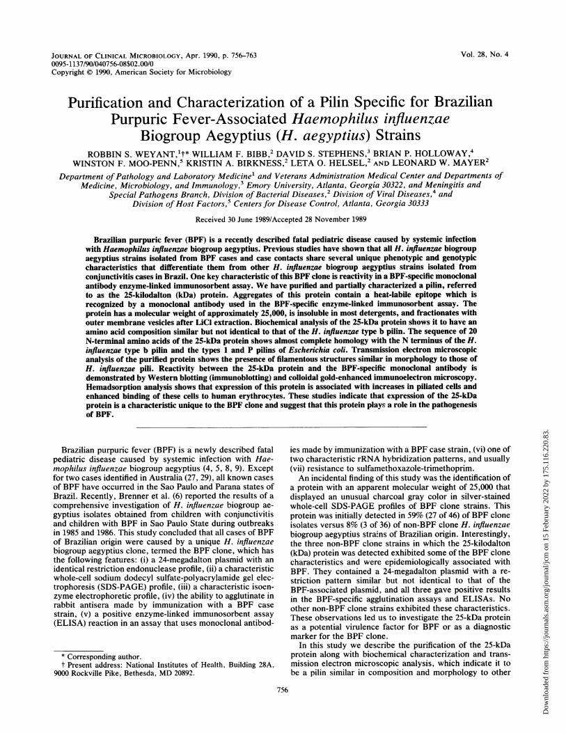
25-kDa protein purification. By using the purification pro-tocol outlined above, a yield of 150 to 200 p.g of purifiedprotein could be obtained from 20 g (wet weight) of station-ary-phase F3031 cells. The protein exhibits a characteristiccharcoal gray color on silver-stained SDS-PAGE gels andcould be readily detected in both whole-cell and outermembrane profiles. Figure 1A illustrates the effect of solu-bilization temperature on detection of the 25-kDa protein bySDS-PAGE. Purified protein was solubilized at 100 and 30°Cbefore electrophoresis. The 25-kDa protein is visualized onlywhen solubilized at 100°C. To determine whether this findingresulted from changes in silver-staining characteristics or inelectrophoretic mobility, we performed two-dimensionalSDS-PAGE (Fig. 1B). This experiment showed a proteinthat migrated with a high apparent molecular weight aftersolubilization at 30°C in the first dimension and a molecular
A O O* aE m
93
6
45 n
3 -
22 a
14 _
Looo 0E
3 0E 1
B
09366 te45 *
3 1 -LI
om 222 *
14 -
FIG. 1. Effect of heat on electrophoretic mobility of the 25-kDaprotein. (A) Silver-stained 12.5% SDS-PAGE gel of purified proteinsolubilized at 30'C (lane 30C) and 100°C (lane 100C). (B) Two-dimensional silver-stained SDS-PAGE gel of BPF strain F3031 outermembrane vesicles. Vesicles solubilized at 100°C (lane 100C) areshown. The remainder of the gel represents outer membrane vesi-cles solubilized at 30'C, separated by 8% SDS-PAGE, and thensolubilized at 100°C (see text for details). Molecular sizes (mws) arepresented in kilodaltons.
weight of approximately 25,000 after high-temperature solu-bilization in the second dimension. This protein has silver-staining and electrophoretic characteristics identical to thoseof the 25-kDa protein included in the F3031 outer membranesample solubilized at 100°C (Fig. 1B, lane 2). These resultssuggest that the native 25-kDa protein exists in a heat-labilecomplex with a molecular weight of >200,000.A transmission electron micrograph of the purified protein
is shown (see Fig. 6A). Filamentous structures approxi-mately 2 nm in diameter are observed. These structures aremorphologically similar to those of purified pili of H. influ-enzae type b (17, 34, 39).
Biochemical analysis. The amino acid composition of the25-kDa protein is listed in Table 1. Asparagine and glutamineare converted to the corresponding acids during acid hydrol-ysis ofthe protein, so an estimate of amide content could notbe made. In addition, cysteine and tryptophan residues werenot quantitated. Hydrophobic amino acids, including gly-cine, alanine, proline, valine, methionine, isoleucine, leu-cine, and phenylalanine, make up approximately 56% of theprotein, which may explain its resistance to solubilization inaqueous solutions. Charged amino acids such as aspartateand glutamate make up at most 25% of the protein. Alsoincluded in this table is the amino acid composition of theH. influenzae type b pilin as reported by Guerina et al. (17).Although these proteins are similar in overall composition,differences exist in aspartate/asparagine, glutamate/glu-tamine, serine, and glycine content. N-terminal amino acidsequence data for the 25-kDa protein are compared with thepublished N-terminal sequence of the H. influenza type bpilin (17) in Fig. 2. From N-terminal positions 3 through 20,these sequences are identical, except for position 4 which isan alanine in the 25-kDa protein and a threonine in the H.influenzae type b pilin. Positions 1 and 2 of the 25-kDaprotein could not be determined, presumably because of thepresence of residual Tris buffer in the sample. This buffercontains primary amine groups which may react with theamino acid labeling reagent.
J. CLIN. MICROBIOL.
Dow
nloa
ded
from
http
s://j
ourn
als.
asm
.org
/jour
nal/j
cm o
n 15
Feb
ruar
y 20
22 b
y 17
5.11
6.22
0.83
.
A BRAZILIAN PURPURIC FEVER-LINKED H. INFLUENZAE PILIN 759
TABLE 1. Amino acid composition of the H. influenza biogroupaegyptius 25-kDa protein and the H. influenza type b pilina
Approx no. of residues/molecule in:Amino acid H.influenzae
25-kDa protein type b pilin
Aspartate/asparagine 13 41Glutamate/glutamine 8 25Serine 25 6Glycine 34 17Histidine 3 6Arginine 2 2Threonine 22 20Alanine 28 20Proline 7 8Tyrosine 9 8Valine 24 21Methionine 7 3Cysteine NDb 4Isoleucine 18 9Leucine 16 15Phenylalanine 6 10Lysine 26 21Tryptophan ND ND
a As reported by Guerina et al. (17).b ND, Not determined.
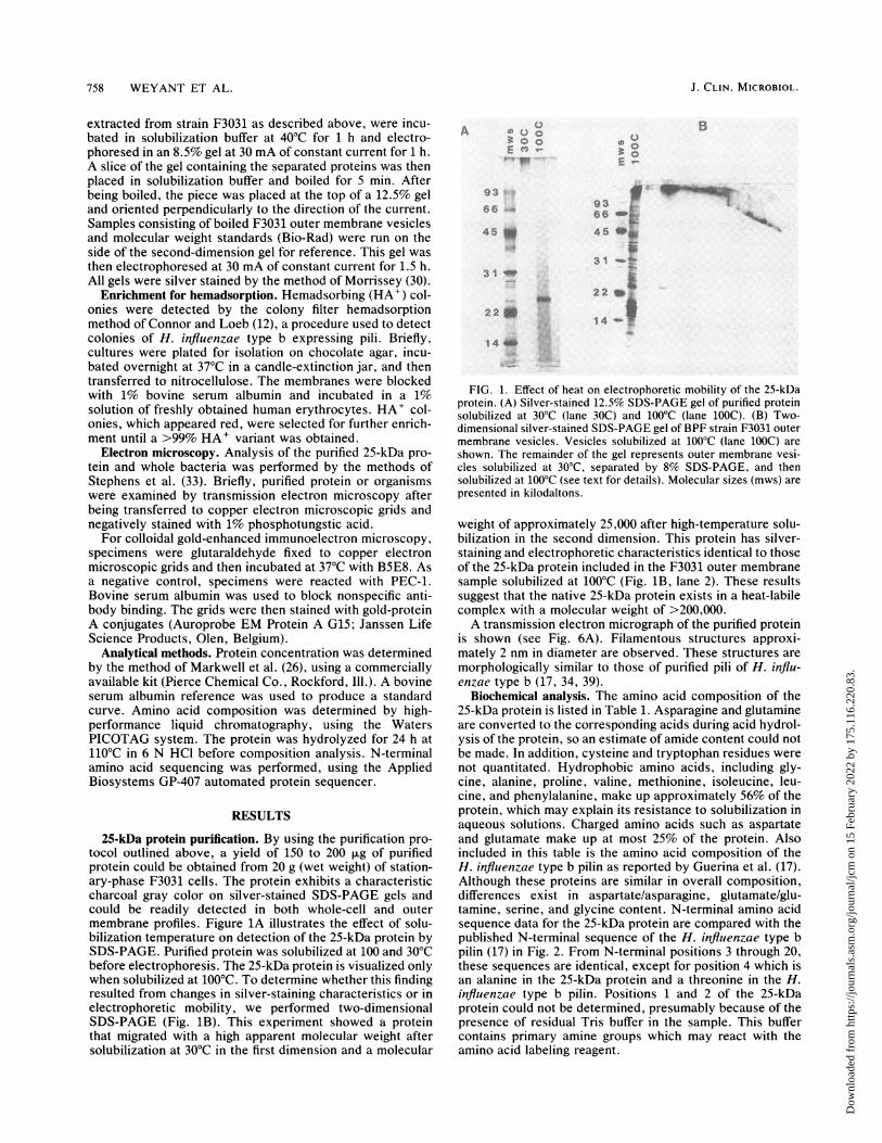
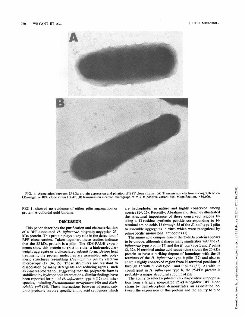
Hemadsorption activity. Strain F3049 is a BPF clone strainwhich is negative for the 25-kDa protein when analyzed bywhole-cell SDS-PAGE. Using the colony filter hemadsorp-tion assay for detecting HA' colonies, we found approxi-mately 0.1% of F3049 colonies to be HA'. By subculturingthe HA' colonies and reselecting HA' variants, we couldisolate a variant, designated 34b, which gives rise to >99%HA' colonies. Western blotting (Fig. 3) of strains F3049 and34b with polyclonal antiserum raised against purified pilinshowed expression of the 25-kDa protein in the HA' variantbut not in the HA- variant. Strain F3031 (Fig. 3, lane 1) wasincluded as a 25-kDa-positive reference. The bands observedat 40, 70, and 80 kDa represent antibodies also detected inthe preimmune serum of the animal. An equivalent silver-stained SDS-PAGE gel of these variants showed the pres-ence of the 25-kDa protein in 34b but not in F3049 (data notshown). This represented the only observable difference inSDS-PAGE profiles between the two variants. We haveattempted to apply the colony filter hemadsorption assay tonon-BPF clone strains to determine whether these strainsalso contain 25-kDa-positive subpopulations. However,these strains produced virtually 100% HA' colonies in theabsence of 25-kDa-protein expression, suggesting that theyhave other mechanisms of erythrocyte binding.Transmission electron microscopy was used to determine
whether morphologic differences exist between the 25-kDa-negative strain F3049 and its 25-kDa-positive variant 34b.Representative views are presented in Fig. 4. Strain F3049
Position 1 2 3 4 5 6 7 8 9 10 11 12 13Hae 25Kd XXX XXX ASN ALA GLU THR SER GLY LYS VAL THR PHE PHEHfl pilin SER ILE ASN TER GLU THR SER GLY LYS VAL THR PHE PHE
Position 14 15 16 17 18 19 20Hae 25Kd GLY LYS VAL VAL GLU ASN THRHfI pilin GLY LYS VAL VAL GLU ASN THR
FIG. 2. N-terminal amino acid sequence of the 25-kDa protein(Hae 25Kd) and H. influenza type b (Hfl) pilin (17). XXX, Notdetermined.
_CD
O O DEU. uLco>
200
92.5
69 lu --BMt _tIM
46
30
25 Kd-4 -
21.5
14.3
FIG. 3. Association between 25-kDa-protein expression anderythrocyte binding. Western blots of BPF clone strains F3031(25-kDa positive), F3049 (25-kDa negative), and 34b, a 25-kDa-positive hemagglutinating variant of F3049. Blots were developedwith rabbit anti-25-kDa-protein antiserum. Molecular sizes (mws)are expressed in kilodaltons.
(Fig. 4A) is essentially unpiliated, although rare (<1%)piliated forms were observed. Strain 34b (Fig. 4B) is piliated.The piliated cells contained between 5 and 15 observable piliper cell. The pili were morphologically similar to H. influ-enzae type b pili as previously described (17, 34, 39), withpilus diameters ranging from 2 to 5 nm and lengths rangingfrom 50 to 250 nm.
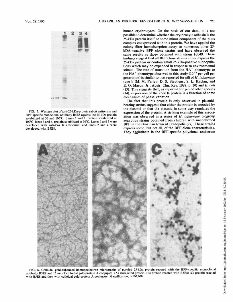
Reactivity of the 25-kDa protein with B5E8. When purified25-kDa protein and strain F3031 were reacted with mono-clonal antibody B5E8 in Western blotting experiments, noreactivity was observed when the antigens were solubilizedat 100°C (Fig. 5). When the antigens were solubilized at30°C, however, high-molecular-weight complexes were ob-served, suggesting that the antibody recognizes a heat-labileepitope present in aggregates of the 25-kDa protein.The recognition ofthe 25-kDa protein by B5E8 was further
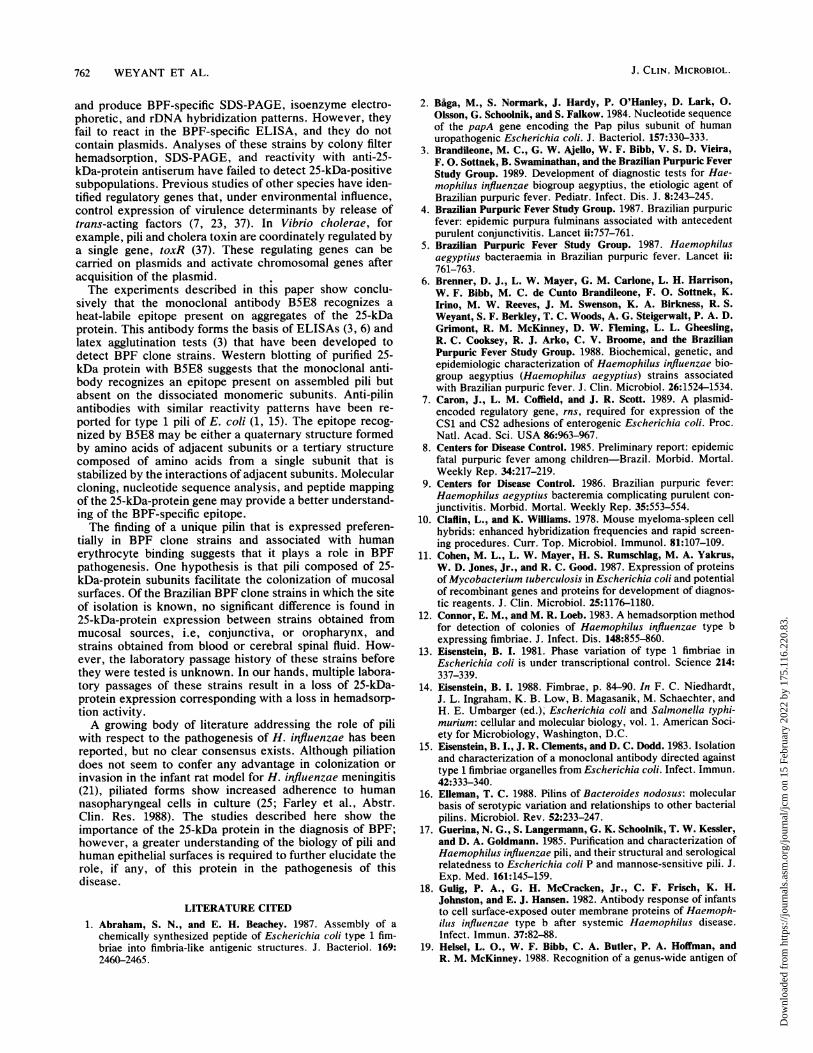
investigated, using colloidal gold-enhanced immunoelectronmicroscopy (Fig. 6). Native 25-kDa protein self assembledinto filamentous structures 2 to 5 nm in diameter andapproximately 150 to 200 nm in length (Fig. 6A). Whenexposed to B5E8, these structures formed aggregates 10 to20 nm in diameter (Fig. 6B). Specific antibodies binding tothese aggregates were detected with colloidal gold-protein Aconjugate (Fig. 6C). Similar results were obtained whenwhole-cell samples of strains F3031 and 34b were examined(data not shown). Control grids, which were developed witheither no antibody or the unrelated monoclonal antibody
VOL. 28, 1990
Dow
nloa
ded
from
http
s://j
ourn
als.
asm
.org
/jour
nal/j
cm o
n 15
Feb
ruar
y 20
22 b
y 17
5.11
6.22
0.83
.
760 WEYANT ET AL.
A
B
FIG. 4. Association between 25-kDa protein expression and piliation of BPF clone strains. (A) Transmission electron micrograph of 25-kDa-negative BPF clone strain F3049; (B) transmission electron micrograph of 25-kDa-positive variant 34b. Magnification, x80,000.
PEC-1, showed no evidence of either pilin aggregation or
protein A-colloidal gold binding.
DISCUSSION
This paper describes the purification and characterizationof a BPF-associated H. influenza biogroup aegyptius 25-kDa protein. This protein plays a key role in the detection ofBPF clone strains. Taken together, these studies indicatethat the 25-kDa protein is a pilin. The SDS-PAGE experi-ments show this protein to exist in either a high-molecular-weight aggregate or a dissociated subunit form. Before heattreatment, the protein molecules are assembled into poly-meric structures resembling Haemophilus pili by electronmicroscopy (17, 34, 39). These structures are resistant todissociation by many detergents and reducing agents, suchas 2-mercaptoethanol, suggesting that the polymeric form isstabilized by hydrophobic interactions. Similar findings havebeen reported for pili of H. influenza type b (17) and otherspecies, including Pseudomonas aeruginosa (40) and Esch-erichia coli (14). These interactions between adjacent sub-units probably involve specific amino acid sequences which
are hydrophobic in nature and highly conserved amongspecies (14, 16). Recently, Abraham and Beachey illustratedthe structural importance of these conserved regions byusing a 13-residue synthetic peptide corresponding to N-terminal amino acids 13 through 35 of the E. coli type 1 pilinto assemble aggregates in vitro which were recognized bypilin-specific monoclonal antibodies (1).The amino acid composition of the 25-kDa protein appears
to be unique, although it shares many similarities with the H.influenza type b pilin (17) and the E. coli type 1 and P pilins(2, 32). N-terminal amino acid sequencing shows the 25-kDaprotein to have a striking degree of homology with the Nterminus of the H. influenza type b pilin (17) and also toshare a highly conserved region from N-terminal positions 8through 17 with E. coli type 1 and P pilins (32). As with itscounterpart in H. influenza type b, the 25-kDa protein isprobably a major structural subunit of pili.The ability to select a piliated 25-kDa-positive subpopula-
tion from a largely nonpiliated 25-kDa-negative BPF clonestrain by hemadsorption demonstrates an association be-tween the expression of this protein and the ability to bind
J. CLIN. MICROBIOL.
Dow
nloa
ded
from
http
s://j
ourn
als.
asm
.org
/jour
nal/j
cm o
n 15
Feb
ruar
y 20
22 b
y 17
5.11
6.22
0.83
.
A BRAZILIAN PURPURIC FEVER-LINKED H. INFLUENZAE PILIN 761
1 2 3 4
25 Kd _
FIG. 5. Western blot of anti-25-kDa-protein rabbit antiserum andBPF-specific monoclonal antibody B5E8 against the 25-kDa proteinsolubilized at 30 and 100°C. Lanes 1 and 2, protein solubilized at100°C; lanes 3 and 4, protein solubilized at 30°C. Lanes 1 and 3 weredeveloped with anti-25-kDa antiserum, and lanes 2 and 4 weredeveloped with B5E8... s ...... ^ 8.ws . ' '`,..
Sh .,* * ,,
.. #. .. ..
e si F i >>^ eS 4_ ».. ,= . .< + .t `: : .e` t 3+ b
v " w ::
/
9,_. tr * ;,e 5f :
t- ,ep eR wr*S > <
7 r.
i sv » f i.
4 e «.:<X: b Xh w t ," S*
t *
b
t,
human erythrocytes. On the basis of our data, it is notpossible to determine whether the erythrocyte adhesin is the25-kDa protein itself or some minor component of the piluscomplex coexpressed with this protein. We have applied thecolony filter hemadsorption assay to numerous other 25-kDA-negative BPF clone strains and have observed thesame results as those obtained with strain F3049. Thesefindings suggest that all BPF clone strains either express the25-kDa protein or contain small 25-kDa-positive subpopula-tions which may be expanded in response to environmentalstimuli. The rate of transition from the HA- phenotype tothe HA' phenotype observed in this study (10-3 per cell pergeneration) is similar to that reported for pili of H. influenzatype b (M. M. Farley, D. S. Stephens, S. L. Kaplan, andE. O. Mason, Jr., Abstr. Clin. Res. 1988, p. 20) and E. coli(13). This suggests that, as reported for pili of other species(14), expression of the 25-kDa protein is a function of somemechanism of phase variation.The fact that this protein is only observed in plasmid-
bearing strains suggests that either the protein is encoded bythe plasmid or that the plasmid in some way regulates theexpression of the protein. A striking example of this associ-ation was observed in a series of H. influenza biogroupaegyptius strains obtained from children with unconfirmedBPF in the Brazilian town of Pradopolis (27). These strainsexpress some, but not all, of the BPF clone characteristics.They agglutinate in the BPF-specific polyclonal antiserum
sca} J
. a_60
a
*o.0
I`r4
FIG. 6. Colloidal gold-enhanced immunoelectron micrographs of purified 25-kDa protein reacted with the BPF-specific monoclonalantibody B5E8 and 15 nm of colloidal gold-protein A conjugate. (A) Unreacted protein; (B) protein reacted with B5E8; (C) protein reactedwith B5E8 and then with colloidal gold-protein A conjugate. Magnification, x 100,000.
*_
4t...
VOL. 28, 1990
'L:,14 W .1t.-
i-
'r, ..* -:,k, lk,
4 Âe l
Dow
nloa
ded
from
http
s://j
ourn
als.
asm
.org
/jour
nal/j
cm o
n 15
Feb
ruar
y 20
22 b
y 17
5.11
6.22
0.83
.
762 WEYANT ET AL.
and produce BPF-specific SDS-PAGE, isoenzyme electro-phoretic, and rDNA hybridization patterns. However, theyfail to react in the BPF-specific ELISA, and they do notcontain plasmids. Analyses of these strains by colony filterhemadsorption, SDS-PAGE, and reactivity with anti-25-kDa-protein antiserum have failed to detect 25-kDa-positivesubpopulations. Previous studies of other species have iden-tified regulatory genes that, under environmental influence,control expression of virulence determinants by release oftrans-acting factors (7, 23, 37). In Vibrio cholerae, forexample, pili and cholera toxin are coordinately regulated bya single gene, toxR (37). These regulating genes can becarried on plasmids and activate chromosomal genes afteracquisition of the plasmid.The experiments described in this paper show conclu-
sively that the monoclonal antibody B5E8 recognizes a
heat-labile epitope present on aggregates of the 25-kDaprotein. This antibody forms the basis of ELISAs (3, 6) andlatex agglutination tests (3) that have been developed todetect BPF clone strains. Western blotting of purified 25-kDa protein with B5E8 suggests that the monoclonal anti-body recognizes an epitope present on assembled pili butabsent on the dissociated monomeric subunits. Anti-pilinantibodies with similar reactivity patterns have been re-
ported for type 1 pili of E. coli (1, 15). The epitope recog-
nized by B5E8 may be either a quaternary structure formedby amino acids of adjacent subunits or a tertiary structurecomposed of amino acids from a single subunit that isstabilized by the interactions of adjacent subunits. Molecularcloning, nucleotide sequence analysis, and peptide mappingof the 25-kDa-protein gene may provide a better understand-ing of the BPF-specific epitope.The finding of a unique pilin that is expressed preferen-
tially in BPF clone strains and associated with humanerythrocyte binding suggests that it plays a role in BPFpathogenesis. One hypothesis is that pili composed of 25-kDa-protein subunits facilitate the colonization of mucosalsurfaces. Of the Brazilian BPF clone strains in which the siteof isolation is known, no significant difference is found in25-kDa-protein expression between strains obtained frommucosal sources, i.e, conjunctiva, or oropharynx, andstrains obtained from blood or cerebral spinal fluid. How-ever, the laboratory passage history of these strains beforethey were tested is unknown. In our hands, multiple labora-tory passages of these strains result in a loss of 25-kDa-protein expression corresponding with a loss in hemadsorp-tion activity.A growing body of literature addressing the role of pili
with respect to the pathogenesis of H. influenza has beenreported, but no clear consensus exists. Although piliationdoes not seem to confer any advantage in colonization or
invasion in the infant rat model for H. influenza meningitis(21), piliated forms show increased adherence to humannasopharyngeal cells in culture (25; Farley et al., Abstr.Clin. Res. 1988). The studies described here show theimportance of the 25-kDa protein in the diagnosis of BPF;however, a greater understanding of the biology of pili andhuman epithelial surfaces is required to further elucidate therole, if any, of this protein in the pathogenesis of thisdisease.
LITERATURE CITED1. Abraham, S. N., and E. H. Beachey. 1987. Assembly of a
chemically synthesized peptide of Escherichia coli type 1 fim-briae into fimbria-like antigenic structures. J. Bacteriol. 169:2460-2465.
2. Baga, M., S. Normark, J. Hardy, P. O'Hanley, D. Lark, O.Olsson, G. Schoolnik, and S. Falkow. 1984. Nucleotide sequenceof the papA gene encoding the Pap pilus subunit of humanuropathogenic Escherichia coli. J. Bacteriol. 157:330-333.
3. Brandileone, M. C., G. W. Ajello, W. F. Bibb, V. S. D. Vieira,F. O. Sottnek, B. Swaminathan, and the Brazilian Purpuric FeverStudy Group. 1989. Development of diagnostic tests for Hae-mophilus influenza biogroup aegyptius, the etiologic agent ofBrazilian purpuric fever. Pediatr. Infect. Dis. J. 8:243-245.
4. Brazilian Purpuric Fever Study Group. 1987. Brazilian purpuricfever: epidemic purpura fulminans associated with antecedentpurulent conjunctivitis. Lancet ii:757-761.
5. Brazilian Purpuric Fever Study Group. 1987. Haemophilusaegyptius bacteraemia in Brazilian purpuric fever. Lancet ii:761-763.
6. Brenner, D. J., L. W. Mayer, G. M. Carlone, L. H. Harrison,W. F. Bibb, M. C. de Cunto Brandileone, F. O. Sottnek, K.Irino, M. W. Reeves, J. M. Swenson, K. A. Birkness, R. S.Weyant, S. F. Berkley, T. C. Woods, A. G. Steigerwalt, P. A. D.Grimont, R. M. McKinney, D. W. Fleming, L. L. Gheesling,R. C. Cooksey, R. J. Arko, C. V. Broome, and the BrazilianPurpuric Fever Study Group. 1988. Biochemical, genetic, andepidemiologic characterization of Haemophilus influenza bio-group aegyptius (Haemophilus aegyptius) strains associatedwith Brazilian purpuric fever. J. Clin. Microbiol. 26:1524-1534.
7. Caron, J., L. M. Coffield, and J. R. Scott. 1989. A plasmid-encoded regulatory gene, rns, required for expression of theCS1 and CS2 adhesions of enterogenic Escherichia coli. Proc.Natl. Acad. Sci. USA 86:963-967.
8. Centers for Disease Control. 1985. Preliminary report: epidemicfatal purpuric fever among children-Brazil. Morbid. Mortal.Weekly Rep. 34:217-219.
9. Centers for Disease Control. 1986. Brazilian purpuric fever:Haemophilus aegyptius bacteremia complicating purulent con-junctivitis. Morbid. Mortal. Weekly Rep. 35:553-554.
10. Claflin, L., and K. Williams. 1978. Mouse myeloma-spleen cellhybrids: enhanced hybridization frequencies and rapid screen-ing procedures. Curr. Top. Microbiol. Immunol. 81:107-109.
11. Cohen, M. L., L. W. Mayer, H. S. Rumschlag, M. A. Yakrus,W. D. Jones, Jr., and R. C. Good. 1987. Expression of proteinsof Mycobacterium tuberculosis in Escherichia coli and potentialof recombinant genes and proteins for development of diagnos-tic reagents. J. Clin. Microbiol. 25:1176-1180.
12. Connor, E. M., and M. R. Loeb. 1983. A hemadsorption methodfor detection of colonies of Haemophilus influenza type bexpressing fimbriae. J. Infect. Dis. 148:855-860.
13. Eisenstein, B. I. 1981. Phase variation of type 1 fimbriae inEscherichia coli is under transcriptional control. Science 214:337-339.
14. Eisenstein, B. I. 1988. Fimbrae, p. 84-90. In F. C. Niedhardt,J. L. Ingraham, K. B. Low, B. Magasanik, M. Schaechter, andH. E. Umbarger (ed.), Escherichia coli and Salmonella typhi-murium: cellular and molecular biology, vol. 1. American Soci-ety for Microbiology, Washington, D.C.
15. Eisenstein, B. I., J. R. Clements, and D. C. Dodd. 1983. Isolationand characterization of a monoclonal antibody directed againsttype 1 fimbriae organelles from Escherichia coli. Infect. Immun.42:333-340.
16. Elleman, T. C. 1988. Pilins of Bacteroides nodosus: molecularbasis of serotypic variation and relationships to other bacterialpilins. Microbiol. Rev. 52:233-247.
17. Guerina, N. G., S. Langermann, G. K. Schoolnik, T. W. Kessler,and D. A. Goldmann. 1985. Purification and characterization ofHaemophilus influenza pili, and their structural and serologicalrelatedness to Escherichia coli P and mannose-sensitive pili. J.Exp. Med. 161:145-159.
18. Gulig, P. A., G. H. McCracken, Jr., C. F. Frisch, K. H.Johnston, and E. J. Hansen. 1982. Antibody response of infantsto cell surface-exposed outer membrane proteins of Haemoph-ilus influenza type b after systemic Haemophilus disease.Infect. Immun. 37:82-88.
19. Helsel, L. O., W. F. Bibb, C. A. Butler, P. A. Hoffman, andR. M. McKinney. 1988. Recognition of a genus-wide antigen of
J. CLIN. MICROBIOL.
Dow
nloa
ded
from
http
s://j
ourn
als.
asm
.org
/jour
nal/j
cm o
n 15
Feb
ruar
y 20
22 b
y 17
5.11
6.22
0.83
.
A BRAZILIAN PURPURIC FEVER-LlNKED H. INFLUENZAE PILIN 763
Legionella by a monoclonal antibody. Curr. Microbiol. 16:201-208.
20. Hitchcock, P. J. 1983. Aberrant migration of lipopolysaccharidein sodium dodecyl sulfate polyacrylamide gel electrophoresis.Eur. J. Biochem. 133:685-688.
21. Kaplan, S. L., E. O. Mason, Jr., and B. L. Weidermann. 1983.Role of adherence in the pathogenesis of Haemophiluis influ-enzae type b infection in infant rats. Infect. Immun. 42:612-617.
22. Kenna, J. G., G. N. Major, and R. S. Williams. 1985. Methodsfor reducing non-specific antibody binding in enzyme-linkedimmunosorbent assays. J. Immunol. Methods 85:409-419.
23. Klemm, P. 1986. Two regulatory genes,fimnB andfimE, controlthe phase variation of type 1 fimbrae in Escherichia coli. EMBOJ. 5:1389-1393.
24. Laemmli, U. K. 1970. Cleavage of structural proteins during theassembly of the head of bacteriophage T4. Nature (London)227:680-685.
25. Loeb, M. R., E. Connor, and D. Penney. 1988. A comparison ofthe adherence of fimbriated and nonfimbriated Hauemophilfsinfluenzae type b to human adenoids in organ culture. Infect.Immun. 56:484--489.
26. Markwell, M. A. K., S. M. Haas, L. L. Beiber, and N. E.Tolbert. 1978. A modification of the Lowery procedure tosimplify protein determination in membrane and lipoproteinsamples. Anal. Biochem. 87:206-210.
27. Mayer, L. W., W. F. Bibb, K. A. Birkness, K. Irino, R. S.Weyant, M. W. Reeves, J. M. Swenson, and the BrazilianPurpuric Fever Study Group. 1989. Distinguishing clonal char-acteristics of the Brazilian purpuric fever-producing strain.Pediatr. Infect. Dis. J. 8:241-242.
28. McDade, R. L., Jr., and K. H. Johnston. 1980. Characterizationof serologically dominant outer membrane proteins of Neisseriagonorrhoeae. J. Bacteriol. 141:1183-1191.
29. McIntyre, P., G. Wheaton, J. Erlich, and D. Hansman. 1987.Brazilian purpuric fever in Central Australia. Lancet i:112.
30. Morrissey, J. H. 1981. Silver stain for proteins in polyacryl-amide gels: a modified procedure with enhanced sensitivity.Anal. Biochem. 117:307-310.
31. Munson, R. S., Jr., and D. M. Granoif. 1985. Purification andpartial characterization of outer membrane proteins P5 and P6
from Haemnophilhs infliuenzae type b. Infect. Immun. 49:544-549.
32. O'Hanley, P., D. Lark, S. Normark, S. Falkow, and G. K.Schoolnik. 1983. Mannose-sensitive and Gal-Gal binding E. colipili from recombinant strains: chemical, functional, and sero-logic properties. J. Exp. Med. 158:1713.
33. Stephens, D. S., A. M. Whitney, G. K. Schoolnik, and W. D.Zollinger. 1988. Common epitopes of pilin of Neisseria menin-gitidis. J. Infect. Dis. 158:332-342.
34. Stull, T. C., P. M. Mendelman, J. E. Haas, M. A. Schoenborn,K. D. Mack, and A. L. Smith. 1984. Characterization of Hae-mophilus influenzae type b fimbriae. Infect. Immun. 46:787-796.
35. Swanson, J. 1979. Studies on gonococcus infection. XVIII.'251-labeled peptide mapping of the major protein of the gono-coccal cell wall outer membrane. Infect. Immun. 23:799-810.
36. Swanson, J. 1982. Colony opacity and protein Il compositions ofgonococci. Infect. Immun. 37:359-368.
37. Taylor, R. K., V. L. Miller, D. B. Furlong, and J. J. Mekalanos.1987. Use of phoA gene fusions to identify a pilus colonizationfactor coordinately regulated with cholera toxin. Proc. Natl.Acad. Sci. USA 84:2833-2837.
38. Towbin, H., T. Staehelin, and J. Gordon. 1979. Electrophoretictransfer of proteins from polyacrylamide gels to nitrocellulosesheets: procedure and some applications. Proc. Natl. Acad. Sci.USA 76:4350-4354.
39. van Alphen, L., N. van den Berghe, and L. Galen van den Broek.1988. Interaction of Haemnophilus influenzae type b with humanerythrocytes and oropharyngeal cells is mediated by a commonfimbral epitope. Infect. Immun. 56:1800-1806.
40. Watts, T. H., D. G. Scraba, and W. Paranchych. 1982. Forma-tion of 9-nm filaments from pilin monomers obtained by octyl-glucoside dissociation of Pseudomonas aeruginosa pili. J. Bac-teriol. 151:1508-1573.
41. Wells, D. E., M. W. Reeves, R. M. McKinney, L. M. Graves, O.Olsvik, T. Bergan, and J. C. Feeley. 1987. Production andcharacterization of monoclonal antibodies to toxic shock syn-drome toxin 1 and use of a monoclonal antibody in a rapid,one-step enzyme-linked immunosorbent assay for detection ofpicogram quantities of toxic shock syndrome toxin 1. J. Clin.Microbiol. 25:516-521.
VOL. 28, 1990
Dow
nloa
ded
from
http
s://j
ourn
als.
asm
.org
/jour
nal/j
cm o
n 15
Feb
ruar
y 20
22 b
y 17
5.11
6.22
0.83
.


![Biogroup propuesta alcaldia 3[1]](https://img.pdfslide.net/doc/110x75/55ac0a591a28ab26388b4637/biogroup-propuesta-alcaldia-31.jpg)